1. Introduction
Chemical communication plays a key role in mediating intra- and interspecific interactions [
1]. In the aquatic environment—a complex chemical system—the detection of chemical cues and signals is crucial for several aspects of animal biology, such as food detection, predator avoidance and finding conspecifics for mating [
2]. In animals with external fertilisation, such as bivalves, spawning synchrony is fundamental to maximise the success of fertilisation: eggs and sperm must be released simultaneously under favourable conditions to ensure fertilisation and early developmental stages critical for the maintenance of the species [
3]. Spawning synchrony is usually mediated by pheromones—chemical signals released by an individual that trigger an innate, specific and adaptive response in conspecifics [
4,
5]. Pheromones have been identified in many species, with their chemical nature and biochemical composition characterised for a wide range of taxa, including invertebrates such as insects [
6], polychaetes [
7,
8,
9], echinoderms [
10], molluscs [
11,
12,
13,
14], and crustaceans [
15,
16], as well as vertebrates, such as fishes [
17,
18,
19,
20] and amphibians [
21]. In the aquatic environment, these compounds encompass a wide range of chemical classes, including steroids, prostaglandins, bile acids, amino acids, proteins and peptides, among others [
22]. Although spawning-inducing pheromones have been implicated in bivalve reproduction and play a vital role in spawning synchrony, their chemical nature and mechanisms of action remain to be elucidated [
23].
In addition to pheromonal regulation, spawning in bivalves is also triggered by environmental (exogenous) and endogenous factors [
24]. In the natural environment, male oysters rapidly respond to increases in temperature, spawning first and subsequently stimulating females and other males to spawn [
25]. In bivalve hatcheries, several methods—biological, physical and chemical—are commonly used to induce spawning in mature individuals [
26]. To achieve more effective spawning, hatcheries often combine two methods, for example, physical (e.g., thermal shock) with biological (e.g., sperm emulsion) or chemical stimulation (e.g., synthetic compounds) [
26]. However, these are time-consuming and not always successful. Another common practice is to strip the gametes—a technique known as scarification—and fertilise the eggs directly. Nevertheless, this approach requires the sacrifice of the broodstock and results in the release of all eggs, whether mature or not, consequently compromising the success of larval culture. These limitations represent major bottlenecks in bivalve aquaculture. Bivalve hatcheries could therefore benefit from the chemical identification and synthesis of spawning-inducing pheromones (SIPs). The application of SIPs would allow the controlled spawning induction without sacrificing any broodstock, improve egg quality by promoting the release of mature gametes only, and provide greater control over, as well as help simplify and standardise, hatchery procedures by increasing reproducibility and predictability of breeding events [
26].
The presence of an SIP in oyster sperm was first suggested by Galtsoff [
27]. Spawning in
Crassostrea virginica occurred within seconds to one hour after adding conspecific sperm to the water, suggesting that sperm carried a chemical cue responsible for inducing the release of gametes in females and other males [
27]. In this species, the SIP has been characterised as a proteinaceous and heat-sensitive compound associated with the spermatozoa cell membrane [
28]. Taylor et al. [
23] also reported the presence of an SIP in the sperm of the silver-lip pearl oyster (
Pinctada maxima). Similarly to
C. virginica, the SIP was proteinaceous, as evidenced by loss of biological activity after proteinase K treatment, and was also associated with the spermatozoa membrane, since milt components alone failed to induce spawning [
23]. The authors further suggested that the SIP in
P. maxima is a highly stable, resistant and multi-component proteinaceous compound associated with both intrinsic and extrinsic sperm membranes.
The Pacific oyster,
Magallana gigas, is one of the most widely farmed bivalves worldwide. Its fast growth, high adaptability to diverse environmental conditions and resilience to disease, combined with a strong consumer preference, have contributed to its global production in aquaculture and high market demand [
29]. It is a protandric hermaphrodite, meaning that it is first a functional male, and then it may transition to a functional female. Like most bivalves, it is a broadcast spawner, releasing the gametes (eggs or sperm) into the water, where fertilisation occurs. The characterisation of SIPs in this species would improve fertilisation success, contributing to the development and standardisation of hatchery protocols. Electrophysiological techniques, particularly the electro-olfactogram (EOG), are powerful tools for pheromone identification, as most of the studied fish species exhibit high olfactory sensitivity to such compounds [
18] and allow the isolation and identification of individual components in a mixture that—alone—do not evoke the full biological response.
Therefore, the current study aimed to identify the chemical nature of SIP in the Pacific oyster (M. gigas) using an electrophysiological approach—the electro-osphradiogram, a technique adapted from the EOG, widely used in vertebrates.
2. Materials and Methods
2.1. Animal Preparation and Electro-Osphradiogram (EOsG)
Adult Pacific oysters (
Magallana gigas) (86.6 ± 20.5 g total weight; 9.4 ± 1.0 cm total length; mean ± SD), collected from Ria de Alvor, southern Portugal 37° 07’ 50” N 8° 37’ 38” W) were prepared following the protocol described by Rato et al. [
30]. Briefly, oysters were anaesthetised in aerated seawater containing 50 g L
−1 magnesium chloride (MgCl
2, hexahydrate, ≥99.0%) (Sigma-Aldrich, Darmstadt, Germany). Once the adductor muscle was relaxed, the right valve was carefully removed by cutting the adductor muscle, and a small incision was made in the gonads for sex identification. Oysters were then left overnight in natural aerated seawater to recover before being used in EOsG recordings on the following day. Ethical approval is not required in Portugal for work on non-cephalopod molluscs.
The chemosensory response of the osphradium was recorded by the electro-osphradiogram (EOsG), as previously described by Rato et al. [
30]. In brief, oysters were kept in an experimental chamber continuously irrigated, under gravity, with clean charcoal-filtered seawater delivered via a glass tube at a rate of 10 mL min
−1. Stimuli were introduced into this flow through a remotely operated solenoid valve. The recording electrode was positioned near the ventral area of the adductor muscle, close to the osphradium, while the reference electrode was placed nearby on the mantle. Electrode and stimulus tube positions were optimised using 10
−3 M L-cysteine (Merck KGaA, Darmstadt, Germany) as a standard stimulus, placing the recording electrode where the response was largest. Borosilicate glass electrodes were filled with 3 M NaCl (Merck KGaA, Darmstadt, Germany) in 4% agar (Merck KGaA, Darmstadt, Germany) and connected to the DC amplifier via Ag/AgCl pellets in 3 M KCl (Bioanalytical Systems, Inc., West Lafayette, IN, USA). Oysters were connected to earth via a silver/silver chloride pellet placed in the mantle cavity.
The D.C. signal was amplified (×5000–×20,000) using a Neurolog head-stage NL100 and NL109 amplifier, with the low-pass filter (NL 125) set at 30 Hz (
https://www.digitimer.com/ (accessed on 18 June 2025)). The signal was then digitised (Digidata 1440 A, Molecular Devices, Sunnyvale, CA, USA;
https://www.moleculardevices.com/ (accessed on 18 June 2025)) and recorded on a computer running AxoScope TM software (version 12.1, Molecular Devices). A minimum of 1 min was allowed between successive stimuli. The order of the odorants was varied. Blank and standard solution (10
−3 M L-cysteine) responses were recorded at regular intervals throughout the recording period. As no differences in the responses were seen between the sexes, data were pooled for males, females and undetermined.
2.2. Experiment 1: Fresh vs. Frozen Sperm Milt in Natural-Induced Spawning and Sperm Stripping Trials
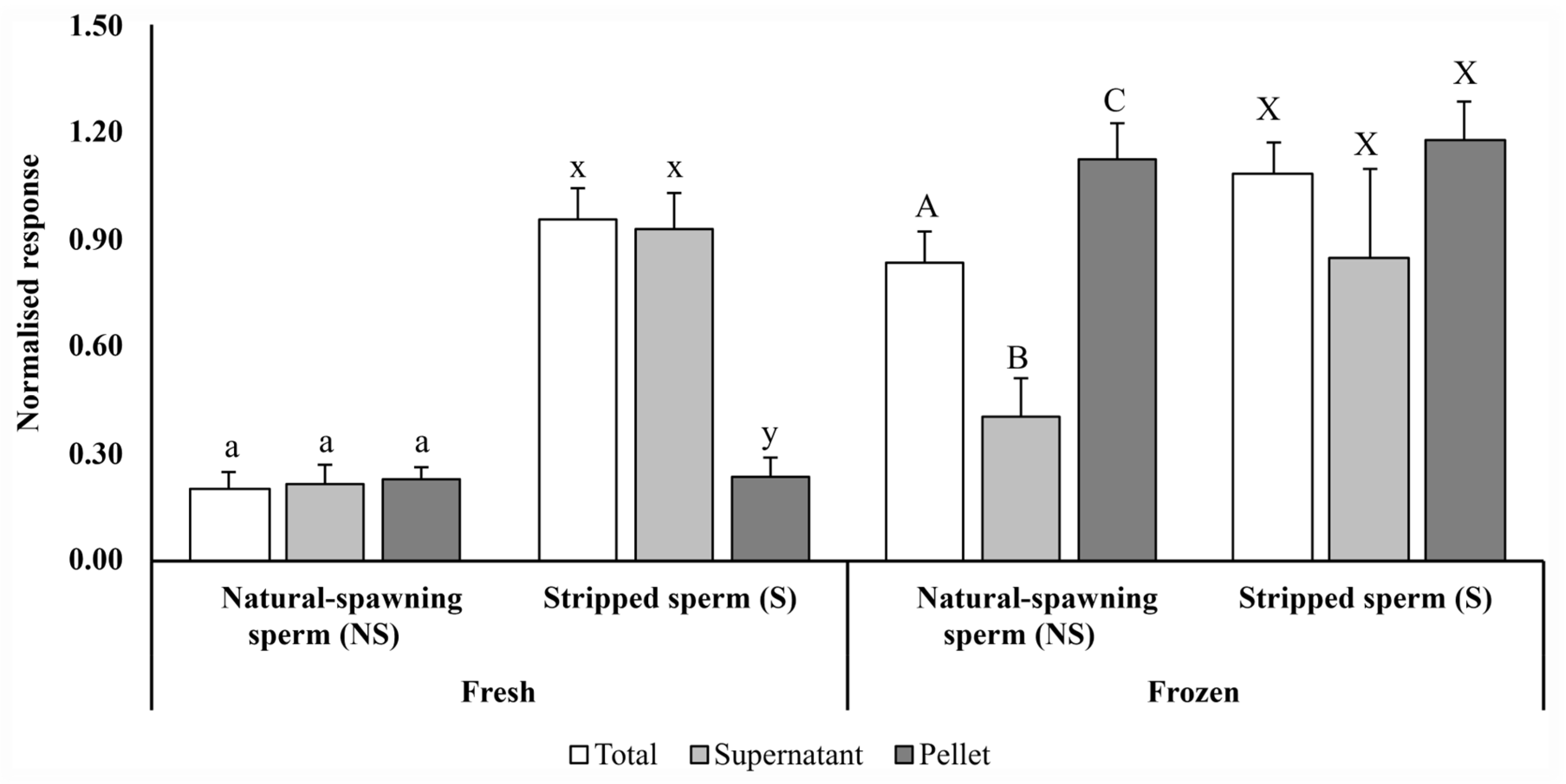
In a first approach to identify the chemical nature of the male pheromone, two different sperm milt collection methods—natural-induced spawning and sperm stripping—were tested to evaluate any differences in chemosensory responses.
Oysters (n = 32; 9.55 ± 0.96 cm total length and 78.91 ± 8.98 g total weight; mean ± SD) were induced to spawn by thermal shock (15–29 °C, at intervals of 1 h). Once males started to spawn, they were separated into individual beakers with 200 mL of charcoal-filtered seawater. Males were allowed to spawn in the beakers for 15 to 20 min, thus obtaining natural spawning sperm milt (NS) (n = 4). Afterwards, these males were sacrificed by cutting the adductor muscle and opening the valves, and sperm was collected through small incisions in the gonad (scarification), thereby obtaining stripped sperm milt (S) (n = 3). Potential differences between fresh (immediately after collection) and frozen (stored at −20 °C) sperm were also evaluated.
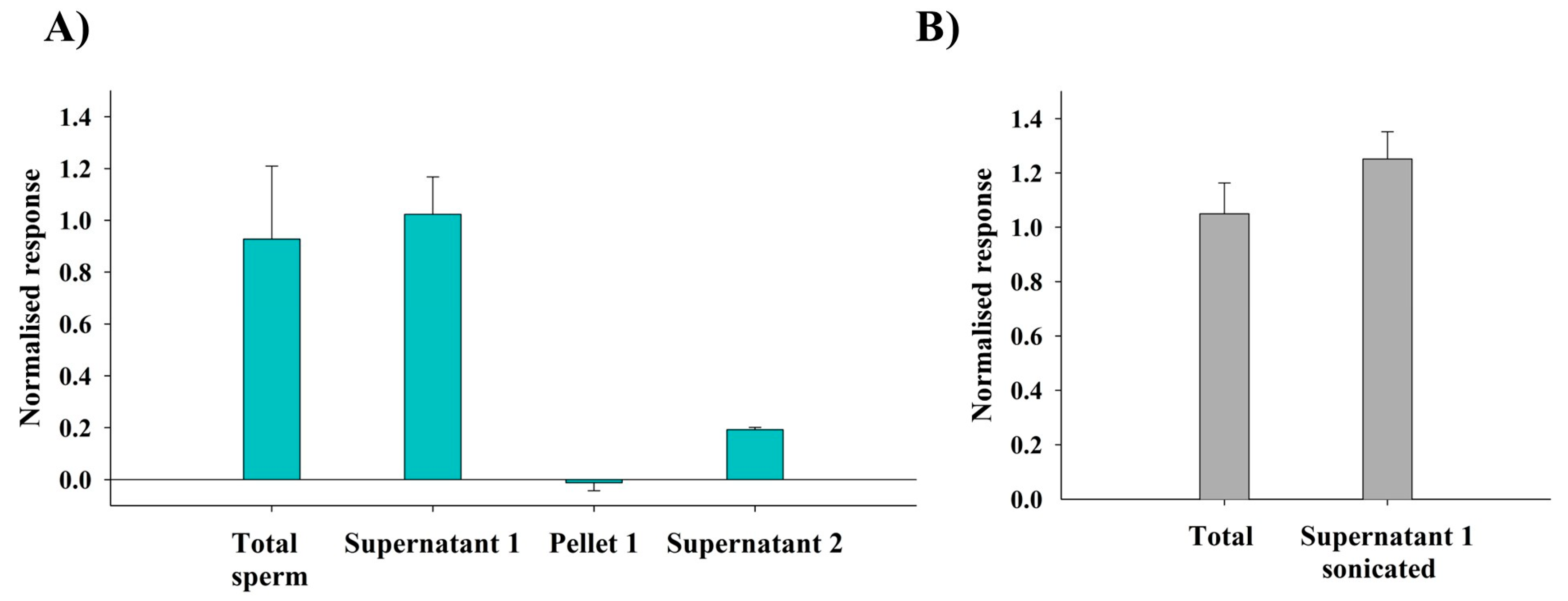
NS sperm milt and S sperm milt from each male were centrifuged (10 min, 1900× g) to obtain supernatant and pellet fractions. The chemosensitivity of the osphradium to total sperm milt (unfractionated) and respective fractions (supernatant and pellet) of both NS and S sperm milt (fresh and frozen) was assessed through electro-osphradiogram (EOsG).
2.3. Experiment 2: Solid-Phase Extraction (SPE)
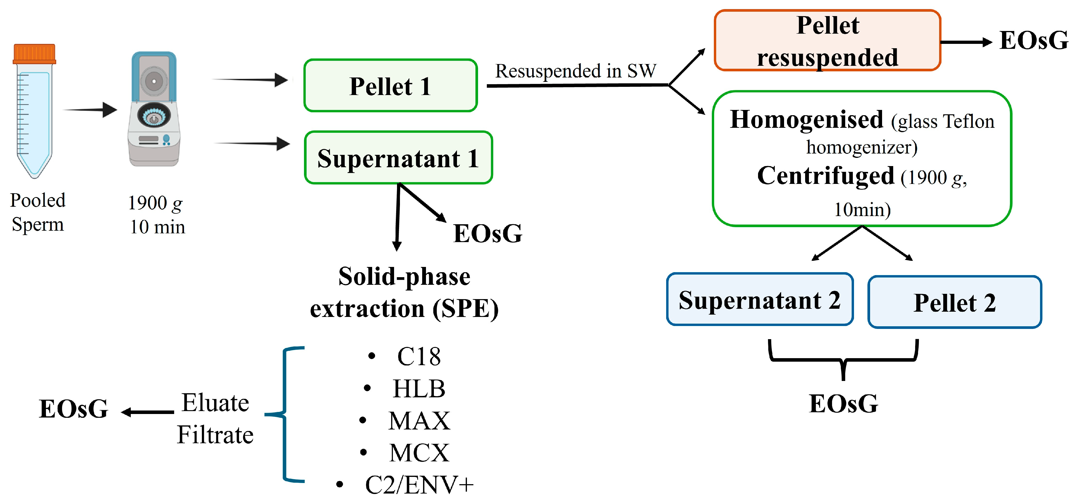
Thawed stripped sperm milt (total) was centrifuged (1900×
g, 10 min) to separate supernatant (supernatant 1) and pellet fractions (
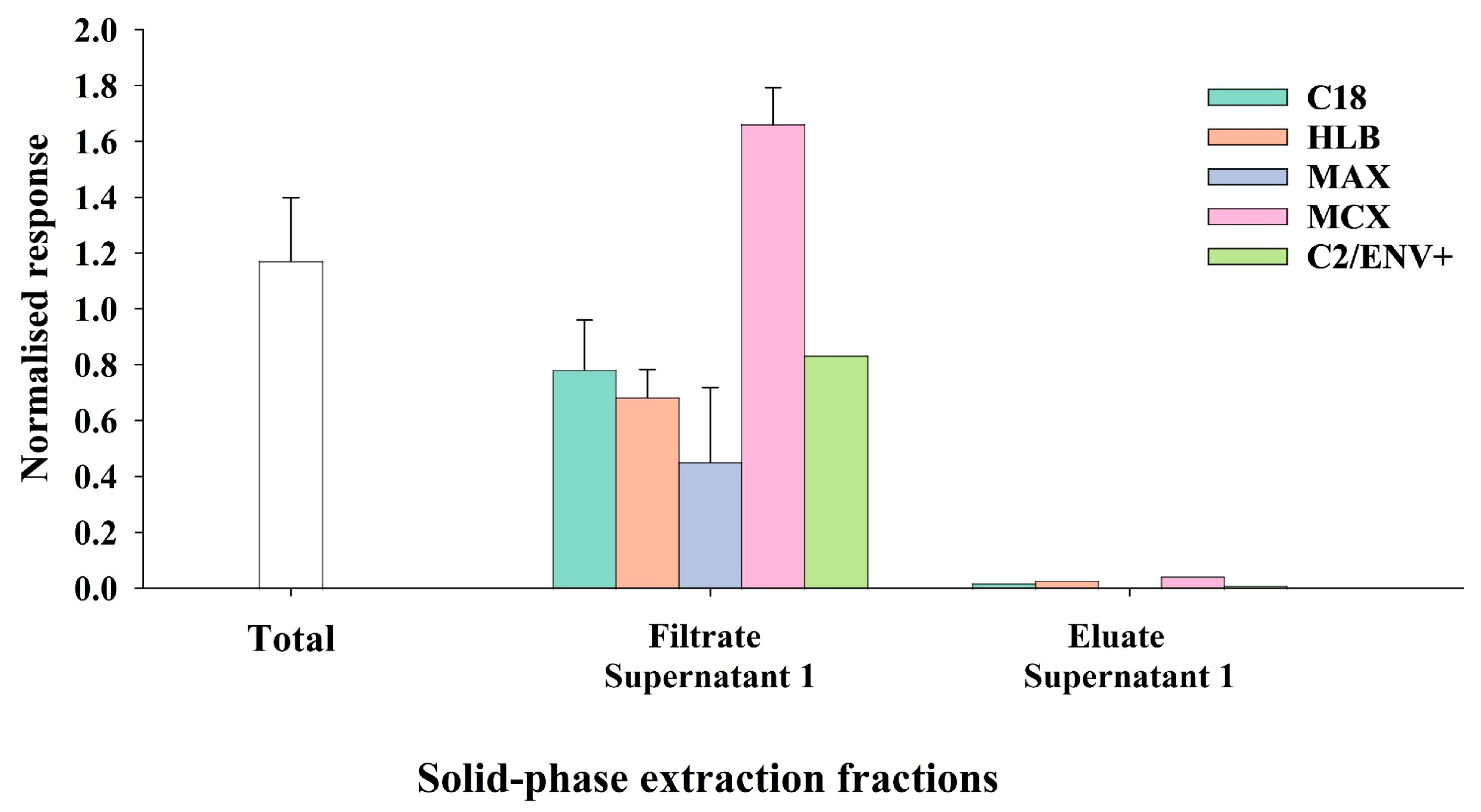
Figure 1). Supernatant 1 was divided into two aliquots: one was directly tested by the EOsG, and the other was subjected to solid-phase extraction (SPE), following the manufacturers’ instructions, using methanol as the eluant. Based on the polarity and charge characteristics of the target compounds, several SPE cartridges were tested: C18, MAX (Mixed-Mode Anion Exchange), MCX (Mixed-Mode Cation Exchange), HLB (Hydrophilic-Lipophilic Balanced) (Waters Sep-Pak™, Milford, MA, USA) and C2/ENV+ (Environmental Plus). Following SPE, both the filtrate and eluate from each cartridge were collected and assessed for olfactory activity via EOsG.
2.4. Experiment 3: Proteinaceous Nature of the SIP
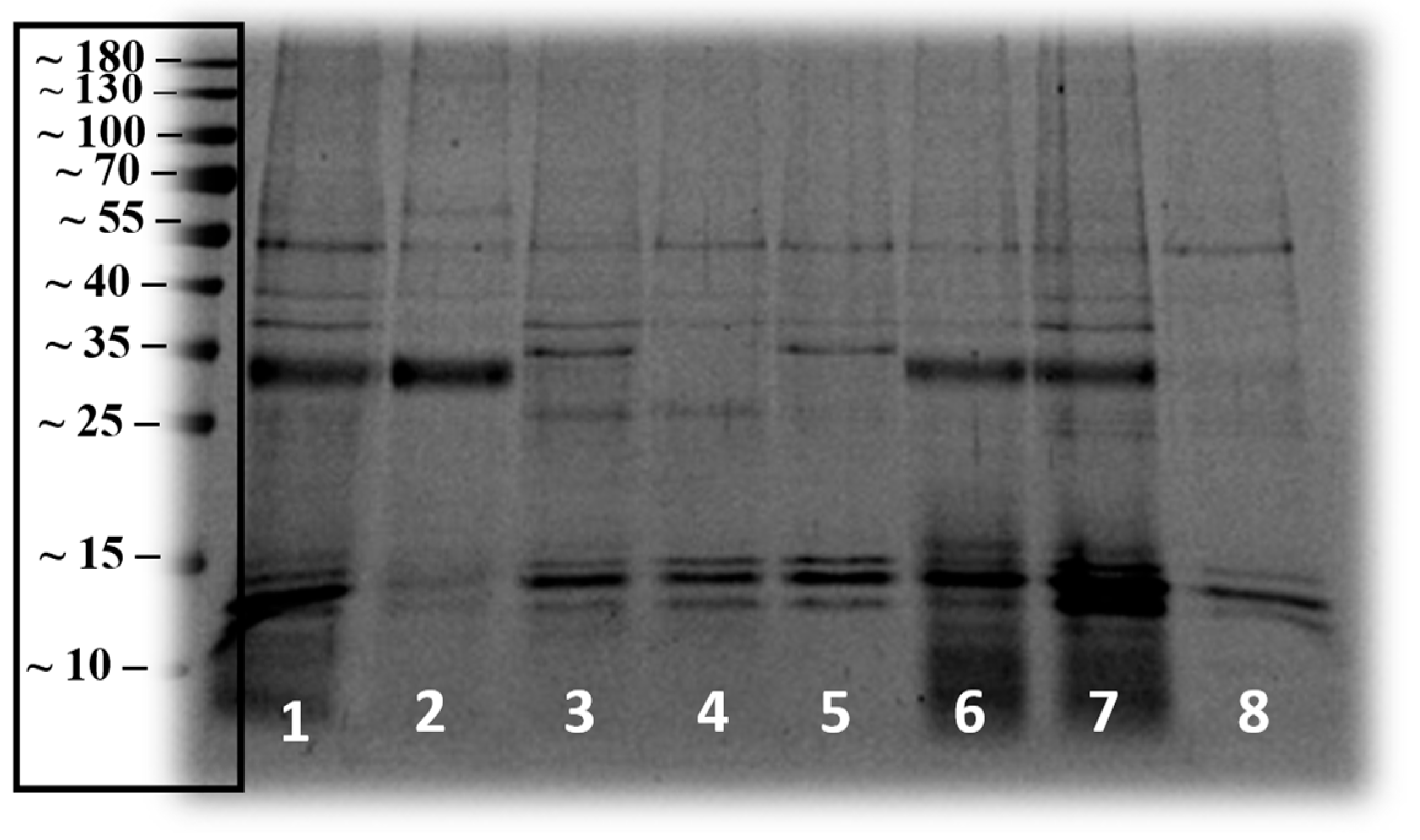
To investigate the possible proteinaceous nature of SIP, the protein profile of each sperm fraction (fresh sperm milt and the respective supernatant 1, resuspended pellet 1, supernatant 2 and resuspended pellet 2; frozen sperm milt and the respective supernatant and resuspended pellet) was analysed by one-dimensional (1D) sodium dodecyl sulphate-polyacrylamide gel electrophoresis (SDS-PAGE). Samples, prepared as mentioned previously (
Figure 1), were run on a vertical gel electrophoresis system (Mini-PROTEAN Tetra Cell, Bio-Rad, Hercules, CA, USA) using standard SDS-PAGE gels (1.5 mm thick) composed of a 5% polyacrylamide stacking gel and a 12% polyacrylamide resolving gel.
SDS loading buffer (6 µL) was added to each 30 µL sample. After centrifugation (10 s), samples were denatured at 95 °C for 3–5 min, followed by a second centrifugation. Samples were then loaded onto the gel and run with a PageRulerTM Prestained Protein Ladder (10–180 kDa) (Thermo Fisher Scientific Inc., Waltham, MA, USA) to allow molecular weight estimation. Then, the gel was stained for 1 h with Coomassie Brilliant Blue R-250 (0.1% w/v in 40% methanol and 10% acetic acid) (Bio-Rad, Hercules, CA, USA) and destained overnight with gentle agitation in a solution of 40% methanol (Merck KGaA, Darmstadt, Germany) and 10% acetic acid (Merck KGaA, Darmstadt, Germany).
Protein quantification was performed with a colorimetric assay based on the Bradford method [
31], following the manufacturer’s instructions for the Quick Start™ Bradford Protein Assay Kit (Bio-Rad). Bovine serum albumin (BSA) was used for the standard curve (Quick Start BSA standard, Bio-Rad) with a range of 0.125–2 mg/mL. The absorbance was read at 595 nm using a microplate reader.
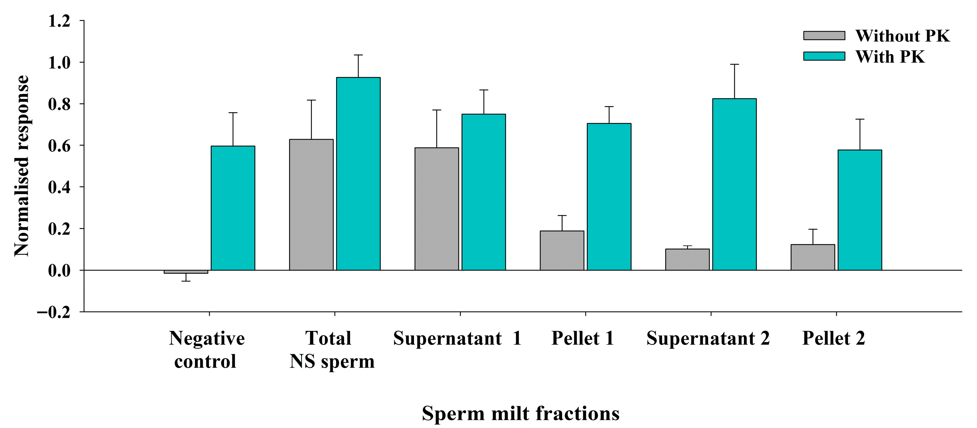
Additionally, to explore the proteinaceous nature of the target compound, the effect of the proteinase K (PK) (Fisher Bioreagents, Fair Lawn, NJ, USA) treatment on various fractions of sperm milt samples was evaluated. The fractions tested included unfractionated sperm milt (total), supernatant 1, pellet 1, supernatant of the pellet homogenate (supernatant 2), and pellet homogenate (pellet 2). Proteinase K was added to each sample at a final concentration of 0.5 mg/mL. Samples were incubated at 40 °C for 24 h, after which the enzyme was inactivated by boiling the samples for 10 min. Afterwards, all samples were tested by the EOsG.
2.5. Experiment 4: Assessment of Compound Stability
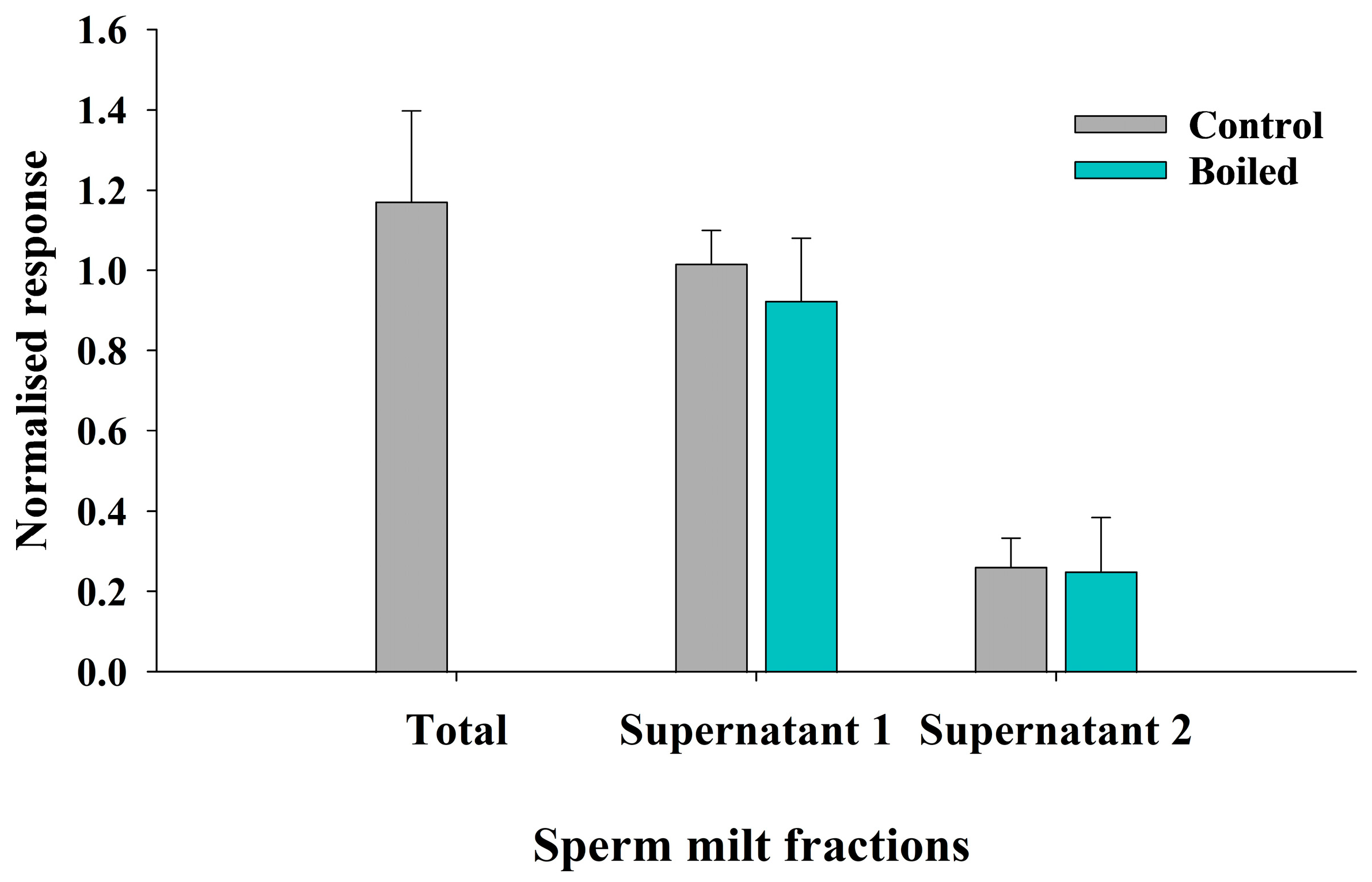
To assess the stability of the compound, supernatant 1 and supernatant 2 of pooled sperm milt frozen samples were boiled for a period of 5–10 min at 100 °C. The electrophysiological responses to these samples were compared to those of the control (non-boiled) samples.
2.6. Experiment 5: Sonication of Sperm Milt
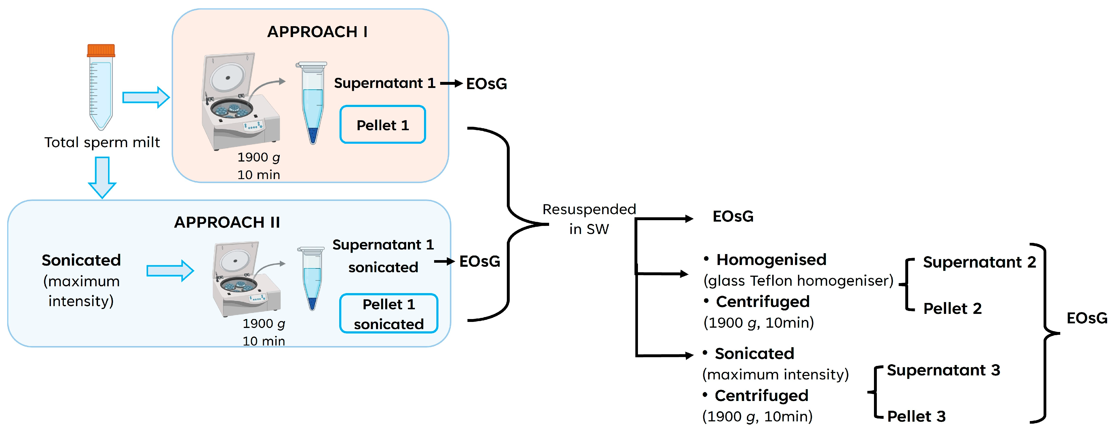
Sonication of total sperm milt samples was applied in order to try to release the cues from the sperm membrane. Two different approaches (
Figure 2) were tested. In the first approach, total sperm milt was centrifuged (1900×
g, 10 min) to obtain supernatant 1 and pellet 1. After resuspension in charcoal-filtered seawater, the pellet 1 fraction was then divided into subsamples: one subsample remained untreated, another subsample was centrifuged (1900×
g, 10 min) after being homogenised with a Teflon-glass homogeniser, and a third subsample of pellet 1 was sonicated at maximum intensity for 5 min (Ultrasons-H; J.P. Selecta,
https://grupo-selecta.com/en/the-company/ (accessed on 9 April 2024)) and centrifuged again. The second approach involved the sonication, at the highest setting (5 min), of the unfractionated (total sperm milt) sperm (
Figure 2). After sonication, total sperm milt was centrifuged (1900×
g, 10 min) to obtain the sonicated supernatant and pellet fractions. The sonicated pellet fraction was further resuspended in charcoal-filtered seawater and, similarly to the first approach, divided into three subsamples: one remained untreated; a second subsample was homogenised with a glass-Teflon homogeniser and centrifuged to obtain supernatant 2 sonicated and pellet 2 sonicated; the third subsample was sonicated and centrifuged again to obtain supernatant 3 sonicated and pellet 3 sonicated. Afterwards, all resulting fractions were then tested for olfactory activity using EOsG.
2.7. Experiment 6: Ultrafiltration
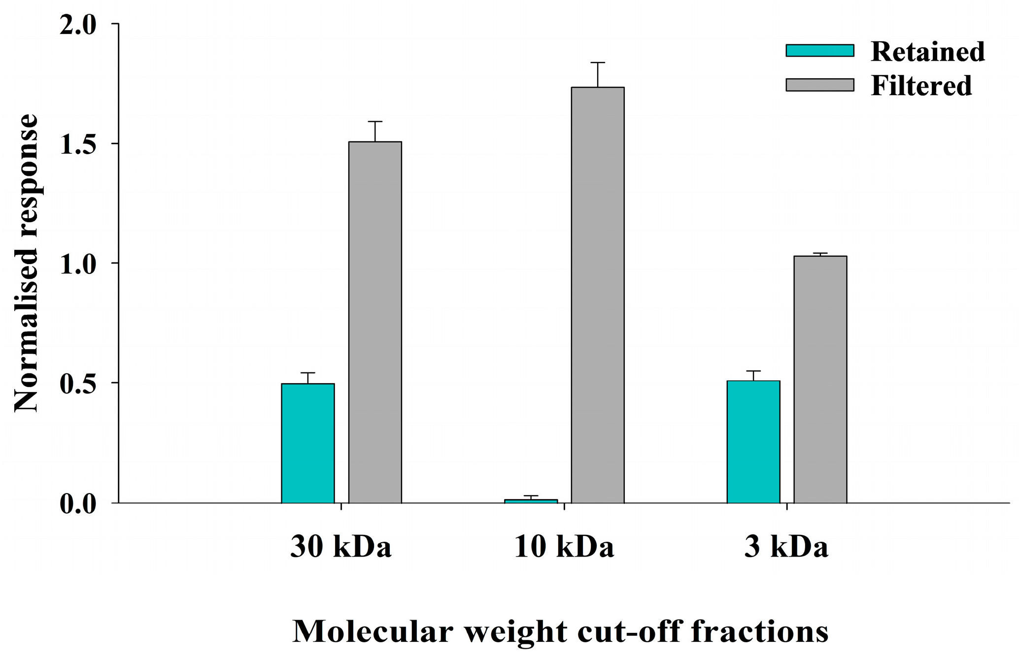
Frozen sperm milt was centrifuged after sonication as described above. The resulting supernatant was subjected to sequential ultrafiltration using centrifugal filter units with molecular weight cut-offs (MWCO) of 30 kDa, 10 kDa (Vivaspin 15RC, regenerated cellulose membrane, Sartorius;
https://www.sartorius.com/en (accessed on 12 February 2025)) and 3 kDa (Amicon
® Ultra 0.5 mL;
https://www.sigmaaldrich.com/PT/en (accessed on 12 February 2025)), following the manufacturer’s instructions. The supernatant was first loaded into a 30 kDa MWCO filter and centrifuged at 5000×
g for 10 min. The retained fraction (molecules > 30 kDa) and filtrate (molecules < 30 kDa) were collected separately. The resulting filtrate was then transferred to 10 kDa and 3 kDa MWCO filters and centrifuged (10 kDa: 5000×
g, 10 min; 3 kDa: 14,000×
g, 10 min). These steps resulted in a retained fraction (>10 kDa; >3 kDa, respectively) and new filtrates (molecules < 10 kDa; <3 kDa, respectively). All fractions were then assessed for olfactory activity by the EOsG.
2.8. Experiment 7: Bioassay
That a given compound can evoke an olfactory response does not necessarily mean that it has full biological activity, i.e., can induce spawning in conspecifics. To address this, a bioassay was conducted in which oysters were exposed to sperm milt fractions, and the results were compared with their respective ‘olfactory’ activity.
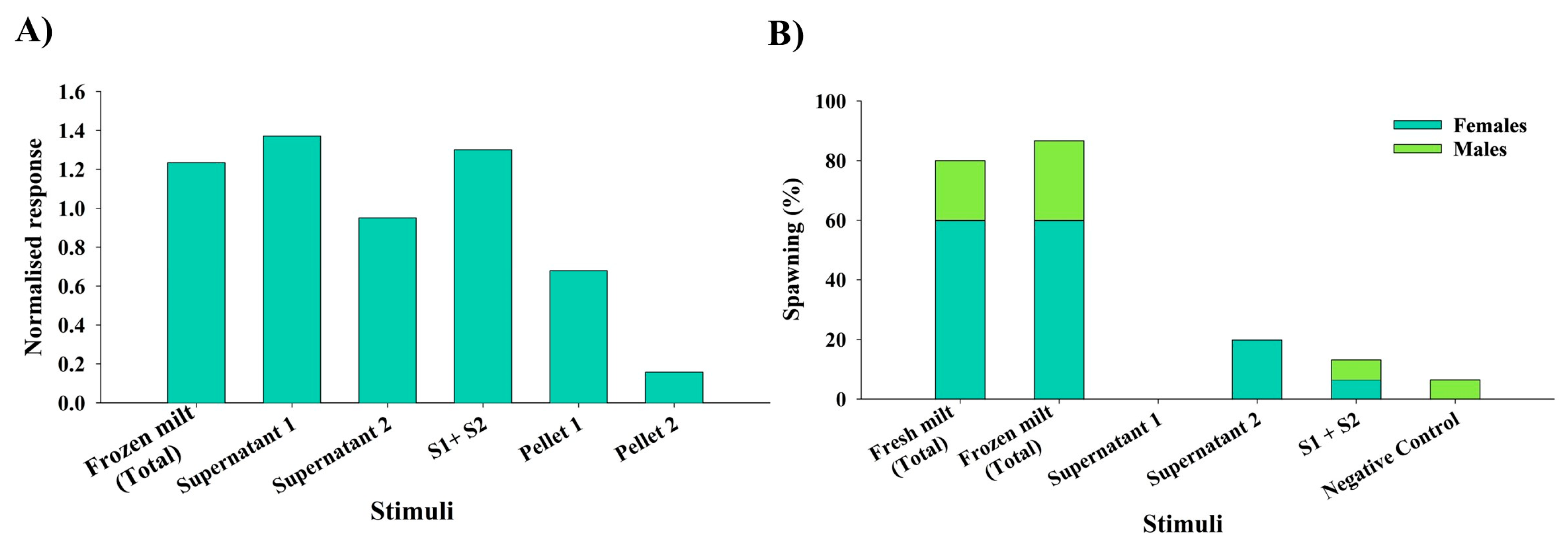
Initially, oysters (n = 15 per treatment) were induced to spawn using only chemical stimulation, with freshly stripped sperm milt, frozen stripped sperm milt, supernatant 1 (S1), supernatant 2 (S2), and a combination of S1 and S2 (S1 + S2) used as stimuli. Seawater was used as a negative control. Oysters were placed in spawning tanks with approximately 5 L of natural and filtered seawater at 22 ± 1 °C. However, chemical stimulation alone failed to induce spawning. Consequently, spawning induction was attempted by combining chemical stimulation with thermal shock (15–30 °C; at intervals of 1 h). Every 30 min, a volume of 4 mL of the corresponding stimulus was added to each experimental treatment at a final dilution of approximately 1:1000. Spawning success was determined as the release or not of gametes and expressed as a percentage.
2.9. Statistical Analysis
The amplitude of all recorded EOsG responses was measured in millivolts, blank-subtracted, and normalised to the blank-subtracted amplitude evoked by the standard stimulus, 10−3 M L-cysteine. Two-way ANOVA followed by the Holm–Sidak post hoc test was applied to assess statistical differences between results from Experiment 1. Student’s t-test was applied to compare boiled and control (non-boiled) samples from Experiment 4 and to compare fractions from non-sonicated and sonicated fractions of samples from Experiment 6. Since the aim of this study was to provide an overall perspective rather than individual comparisons, the standard error of the mean (SEM) was selected as the measure of data dispersion. Results are therefore expressed as mean ± SEM, unless otherwise stated. Statistical significance was set as p ≤ 0.05. Statistical analysis was performed using software Sigmaplot (version 15.0) (Systat Software, Inc., San Jose, CA, USA).
4. Discussion
To our knowledge, this is the first work providing evidence on the chemical nature of a putative spawning-inducing pheromone (SIP) in
Magallana gigas, a species of major economic importance for bivalve aquaculture worldwide. In this species, the SIP—at least those compounds detected by the osphradium—appears to be a heat-stable, polar multi-component cue, with molecular weights of about 35 kDa, between 3 and 10 kDa and at least one of less than 3 kDa, with one or more components associated with the spermatozoa membrane. Using a different experimental approach, however, these results largely agree with Taylor et al. [
23] in the silver-lip pearl oyster (
Pinctada maxima).
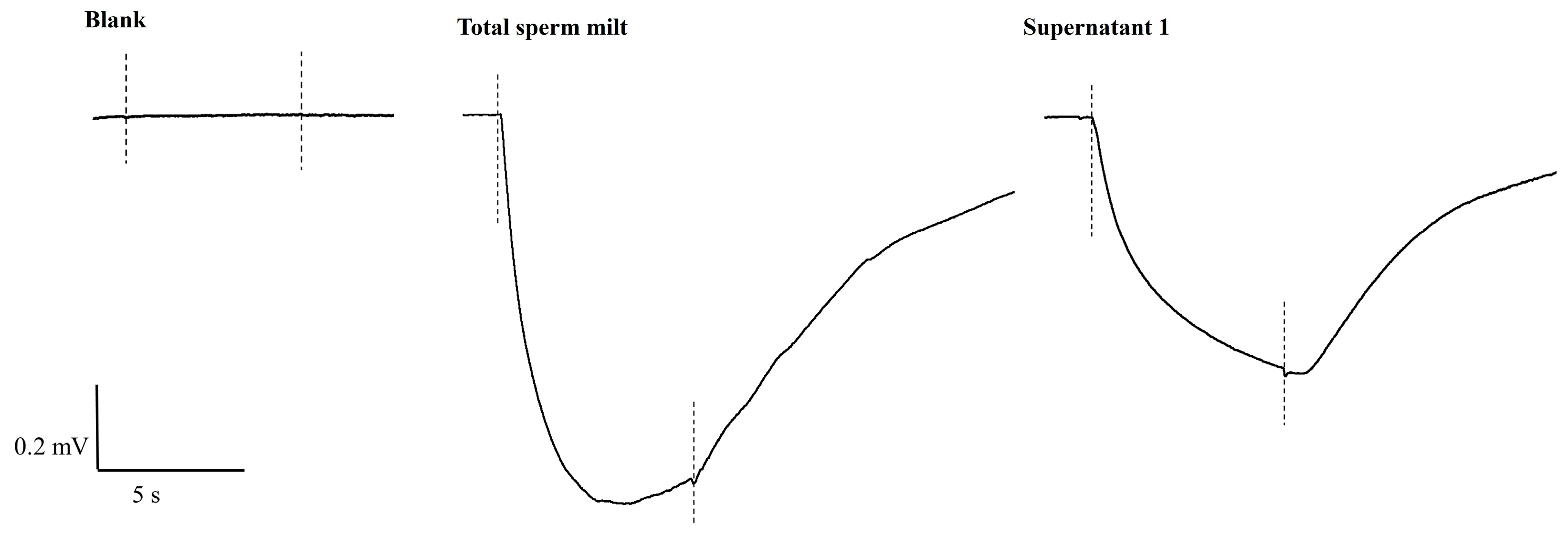
The EOsG recordings revealed significant differences between naturally spawned (NS) and stripped sperm milt (S). Indeed, sperm milt collected by stripping the gonads evoked a higher EOsG response than the naturally spawned sperm milt. However, this may be simply due to sperm collected at higher density; there was no easy way to control for this. Also, stripped sperm milt may contain some remains of gonadal tissue, and this may contribute to the response. Therefore, to minimise interference from other components, we used natural-spawning sperm milt for the rest of the study.
Frozen sperm induced a higher electrophysiological response than fresh sperm milt. This was probably a consequence of the freezing process releasing additional odorants from the spermatozoa, similar to sonication (see below). These findings support the hypothesis that the active cue is multicomponent, occurring both in free solution in the milt and associated with the spermatozoa membrane. Similarly, some olfactory activity was contained in supernatant 2 (after the first approach of sonication and subsequent centrifugation of resuspended pellet 1), therefore indicating the multicomponent nature—supernatant and supernatant of the pellet homogenate—of the cue. Nevertheless, after sonication and centrifugation of the total sperm milt (second sonication approach), the majority of the olfactory activity was retained in the supernatant, providing further evidence for the multicomponent nature of the putative SIP. This is, therefore, a method for obtaining all active components in free solution for subsequent fractionation.
Olfactory activity was mostly contained in the supernatant, whereas the pellet appeared to be pheromonally inactive. This is consistent with findings in other invertebrates, where pheromonal cues are predominantly water-borne, and retained in the supernatant fractions, e.g., in the coelomic fluid of polychaetes [
7,
8,
9,
32] and in the homogenised albumen glands of different species of the genus
Aplysia [
11,
12,
33]. Moreover, after solid-phase extraction (SPE), the eluate of the supernatant failed to evoke an electrophysiological response, whereas the aqueous fraction (filtrate) elicited responses similar to those of the unfractionated sample (total sperm milt) and supernatant, regardless of the type of cartridges used. Together, this suggests that the pheromone consists largely of polar compound(s). That the filtrate of cation-exchange cartridges (MCX) elicited approximately double the response supports the hypothesis that the active molecule is not positively charged under the assay conditions but rather neutral or negatively charged and highly polar. However, this needs further clarification.
Proteins and peptides have been suggested to act as pheromones in several invertebrates, particularly molluscs [
34]. In the gastropod
Aplysia, a family of water-borne peptide pheromones involved in egg-laying and mate attraction (attractin, enticin, temptin and seductin) have been identified and suggested to act as a mixture with synergistic effects [
34,
35,
36]. In the cuttlefish,
Sepia officinalis, a tetrapeptide (Ile-Leu-Met-Glu; ILME) eluted from egg masses [
37] and a sperm-attracting peptide (SepSAP) [
38] were also characterised as pheromones in this species. In a marine polychaete,
Nereis succinea, males and females use different compounds to synchronise spawning; females release a tetrapeptide pheromone (cysteinyl-glutathione) [
32] that attracts males, which, in turn, release sperm and the male pheromone (inosine and glutamic acid) [
7,
34,
39]. Similarly, in a tropical oyster species,
Pinctada maxima, Taylor and co-authors [
23] suggested that the spawning-inducing pheromone (SIP) was a proteinaceous multi-component associated with sperm membrane, since it had lost activity after treatment with proteinase K. For the current study, this remains an open question, as the osphradium responded to proteinase K even after the enzyme had been deactivated by boiling, indicating that its denatured remains were still detected and thus masking any potential loss of activity. This hypothesis is further supported by the results from boiled samples, where olfactory activity remained.
Similarly to
Crassostrea virginica [
28] and
P. maxima [
23], the putative SIP in
M. gigas is proteinaceous, and at least one component is associated with the spermatozoa cell membrane. However, they are different in enzymatic and heat sensitivity;
M. gigas SIP evoked olfactory activity even after enzymatic treatment, freezing and boiling. This suggests that it may not be a large globular protein, since such molecules typically lose their biological activity upon freezing–thawing and/or boiling due to denaturation [
40]. The persistence of olfactory activity, even after freezing and boiling, suggests instead that active compounds—or at least those regions that bind to the receptors—are heat-stable and may include high molecular weight proteins, as well as lower molecular weight peptides and other compounds below 3 kDa. It is possible that boiling may reduce biological activity (this was not tested) while the molecule is still able to bind to its receptor. Peptides, unlike proteins, which are large three-dimensional macromolecules, are short linear molecules composed of amino acids, and even dipeptides—composed of only two amino acids—can act as biologically active molecules [
34]. The thermal stability of the putative SIP may be related to the type of habitat, where oysters spawn in response to thermal shock caused by air exposure at low tide, followed by the incoming tide. Therefore, it makes evolutionary sense for the active compound(s) to be more heat stable. In addition, the vast distribution of the species (worldwide, except in Antarctica), with areas with high thermal variability, reinforces the adaptive value of such heat-stable molecules.
The molecular weight cut-offs (MWCO) for ultrafiltration were initially selected based on the SDS-PAGE electrophoresis results, which revealed two distinct bands of ~35 kDa and ~15 kDa. Ultrafiltration suggested that the SIP of
M. gigas is most likely a multicomponent signal, composed of molecules larger than 30 kDa, possibly the band at 35 kDa, as well as fractions between 3 and 10 kDa and below 3 kDa. Small peptides have been suggested to act as pheromones in several animals. For example, Forward et al. [
41] suggested that specific small peptides—di- and tripeptides with a neutral amino acid at the N-terminus and a basic amino acid at the C-terminus—function as crustacean pheromones in the crab
Rhithropanopeus harrisii. Also, sex pheromones in the shore crab (
Carcinus maenas) [
42] and in the helmet crab (
Telmessus cheiragonus) [
43] were characterised as small, polar, water-soluble molecules with molecular weights below 1000 Da. Similarly, Zhang and co-authors [
44] suggested that the sex pheromone of the shrimp
Lysmata wurdemanni was most likely a molecule between 500 and 1000 Da.
As the population of a species diverges into distinct species, their pheromones typically evolve, contributing to reproductive isolation [
34]. Animals that use multicomponent pheromones composed of small molecules typically diverge by adding or losing molecules from the mixture, whereas those relying on peptide or protein pheromones may diverge by changes in the amino acid sequence [
34]. Although several peptide pheromones act as single molecules, some are in fact multicomponent, involving several molecules acting together to evoke a response, as is the case of the combination of protein pheromones in
Aplysia [
34,
36]. Furthermore, pheromonal specificity may be determined by the nature of components as well as by their relative concentrations [
45]. For instance, for the mussel
Mytilopsis sallei, He et al. [
46] identified that the synergistic and exact ratio (1:1.125:3.25) of three purines (adenosine, inosine and hypoxanthine) acts as an aggregation pheromone, inducing conspecific larvae to settle and metamorphose. These findings suggest that such metabolites may function as species-specific pheromones when present in exact combinations [
46]. The same may also apply to the SIP in
M. gigas, which is likely a combination of proteins and peptides that, in a specific ratio, act as a spawning-inducing pheromone.
The bioassay revealed that the frozen sperm milt had a similar biological activity to fresh sperm milt. This not only further supports the stability of the active compound but also represents a useful and profitable opportunity for bivalve hatcheries, as sperm milt can be stored (−20 °C) for later use. Despite the strong olfactory response evoked by a given compound, this effect was not reflected in a corresponding biological activity, as sperm milt fractions failed to induce spawning in conspecifics to the same extent as untreated sperm milt. Therefore, even if a given fraction evokes a strong olfactory response, the other components must also be present to trigger spawning.
These results represent the first evidence of the role of the osphradium in the detection of spawning cues and consequent synchronisation, further supporting the hypothesis of Haszprunar [
47] and Beninger et al. [
48]. However, further work is necessary to identify each active component and which, if not all, are necessary for full pheromonal activity.