Article

Effects of Astragalus–Ginseng Dietary Supplementation on the Growth and Stress Resistance of Yellow Catfish (Pelteobagrus fulvidraco)

1
Key Laboratory of Exploration and Utilization of Aquatic Genetic Resources by the Ministry of Education, Shanghai Ocean University, Shanghai 201306, China
2
Key Laboratory of Freshwater Aquatic Genetic Resources by the Ministry of Agriculture, Shanghai Ocean University, Shanghai 201306, China
3
Key Laboratory of Aquatic Genetic Resources and Aquaculture Ecosystem Certified by the Ministry of Agriculture, Shanghai Ocean University, Shanghai 201306, China
4
College of Marine and Biology Engineering, Yancheng Institute of Technology, Yancheng 224051, China
*
Authors to whom correspondence should be addressed.
Fishes 2025, 10(5), 208; https://doi.org/10.3390/fishes10050208
Submission received: 11 March 2025 / Revised: 17 April 2025 / Accepted: 28 April 2025 / Published: 2 May 2025
(This article belongs to the Section Nutrition and Feeding)

Abstract

Astragalus and ginseng, esteemed as traditional Chinese herbal medicines, have demonstrated the ability to bolster physical health and enhance the immune function of organisms. In this study, the effects of a dietary astragalus–ginseng mixture on the growth performance, intestinal health, and nonspecific immunity of yellow catfish (Pelteobagrus fulvidraco) were evaluated, by measuring growth performance indices, intestinal villus morphology, enzyme activities, and expression levels of immune-related genes. Yellow catfish (n = 120, initial weight: 5.07 ± 0.18 g) were randomly assigned to four dietary groups: a control group (CT, 0 mg/kg) and three astragalus–ginseng treatment groups (AG1, 500 mg/kg; AG2, 1000 mg/kg; AG3, 2000 mg/kg). Each group had three replicates and was fed for six weeks. The results demonstrate that the treatment significantly enhanced the growth performance, as evidenced by increases in FBW, WG, WGR, SGR, and HSI. These improvements may be related to an increase in intestinal villi length and increased LPS activity, both of which are associated with enhanced digestive function. Meanwhile, the activity of antioxidant enzymes in the liver, including CAT, SOD, and GSH, was increased, whereas the level of MDA was decreased. In the serum, GSH was up-regulated, while SOD activity was decreased. Immune-related enzyme activities, such as ALT and LZM, were up-regulated, while AST showed no significant difference. Moreover, the treatment also promoted the expression of the anti-inflammatory factor IL-10. The pro-inflammatory factors, such as IL-1β, IL-6, and TNF-α, were decreased with the addition of low concentrations but increased with high concentrations. In conclusion, supplementation with an astragalus–ginseng mixture could promote growth performance by increasing digestive enzyme activity and intestinal villi length, and improve disease and stress resistance traits by modulating immune genes and antioxidant enzyme activity. A dosage of 1000 mg/kg was found to be optimal.
Key Contribution: (1) Dietary astragalus–ginseng mixture enhanced the growth performance and improved intestinal health in Pelteobagrus fulvidraco. (2) Dietary astragalus–ginseng mixture boosted antioxidant and immune functions in Pelteobagrus fulvidraco.

1. Introduction

As the largest aquaculture country, China’s aquaculture industry has gradually become a vital growth driver for the rural economy [1]. Constrained by nutritional limitations, environmental challenges, and pathogen infections, intensive aquaculture faces the issues of disease outbreaks and high mortality rates, which significantly impede the development of the aquaculture industry [2,3]. Therefore, improving disease and stress resistance traits of cultured animals is crucial. Previously, antibiotics have been widely used in aquaculture for disease prevention and treatment. Recently, the accumulation of active antibiotic residues from aquaculture in the environment has raised public concerns [4,5]. Since the prohibition of antibiotics, research has increasingly focused on investigating alternatives, such as plant extracts and polysaccharides derived from medicinal plants. These products have been shown to enhance appetite and weight gain, with demonstrated antibacterial and anti-parasitic properties [6,7,8]. Hence, plant extracts can significantly enhance fish immunity and effectively control infectious diseases in aquaculture.
Astragalus membranaceus and Panax ginseng are two of the most widely utilized traditional herbal medicines worldwide, recognized for their diverse pharmacological properties. Astragalus polysaccharide (APS) is the main bioactive component of A. membranaceus and is primarily composed of glucose, along with varying amounts of rhamnose, galactose, arabinose, mannose, and other monosaccharides [9,10,11]. As a pleiotropic factor, APS has been reported to exhibit anti-tumor, antioxidant, anti-viral, and antimicrobial activities as well as beneficial impacts on intestinal health through the modulation of villus morphology and gut microbiota composition [8,12,13,14,15]. Ginsenosides are the major active components of ginseng and trigger an array of pharmacological responses, related to anti-hyperglycemia, anti-cancer, immunomodulatory, and antioxidant activities [16,17,18,19,20,21,22]. In recent years, ginseng polysaccharides have also attracted increasing attention as an important class of bioactive compounds. Polysaccharides isolated from P. ginseng are mainly composed of starch-like glucans and pectin [23,24]. The ginseng starch-like polysaccharide consists of α-D-(1,4)-glucans, 6-branched α-D-(1,4)-glucans, 3-branched α-D-(1,6)-glucans, and α-D-(1,6)-glucans without side chains, while ginseng pectin is mainly composed of homogalacturonan, rhamnogalacturonan I, and arabinogalactans [25,26,27]. Ginseng polysaccharides exhibit a range of biological activities, including immuno-modulatory, anti-tumor, antioxidant, and antidiabetic effects, primarily through the regulation of immune responses and oxidative stress pathways [28,29,30].
Building on their established pharmacological properties, both astragalus polysaccharides (APSs) and ginseng polysaccharides have increasingly been investigated for their potential to enhance antioxidant capacity, modulate immune function, and pro-mote disease resistance in aquatic species. Dietary supplementation with APSs has been shown to improve weight gain, growth rate, meat quality, immunity, and disease resistance in Carassius auratus, Scophthalmus maximu, and Channa argus [31,32,33]. In addition, APS has demonstrated adjuvant potential in aquaculture vaccines by enhancing antigen-specific immunity and protective efficacy in species such as Epinephelus fuscoguttatus and Oncorhynchus mykiss [34,35]. In contrast, ginseng polysaccharides are primarily used as immunostimulants rather than growth promoters in aquaculture, with studies in Paralichthys olivaceus and Oncorhynchus mykiss demonstrating enhanced immune responses following dietary supplementation [36,37]. Due to their complementary immunomodulatory and physiological effects, the combination of APSs and ginseng polysaccharides may provide additive or synergistic benefits in aquaculture. However, supporting evidence in fish species remains limited. A relevant research is the combination of APS and Eucommia ulmoides leaf extract in Larimichthys crocea, which showed improved growth, antioxidant capacity, and immune regulation through MAPK and mTOR signaling pathways [38]. Given these promising findings, further studies are warranted to explore the potential synergistic effects of combining APSs and ginseng polysaccharides in fish, which could provide valuable insights for improving aquaculture practices.
Yellow catfish (Pelteobagrus fulvidraco) is an important economic species that is extensively distributed across China. With the intensification of aquaculture practices, infectious diseases caused by bacterial pathogens such as Edwardsiella ictaluri and Aeromonas veronii present a serious threat to the sustainable development of yellow catfish farming [39,40,41]. As feed additives, polysaccharides and plant extracts have been shown in the relevant literature to positively influence body composition and boost innate immunity in yellow catfish [42,43,44]. However, limited research has examined whether the mixture of APS and ginseng can enhance the stress resistance and immunity of yellow catfish to support its aquaculture. This study investigated the effects of APS and ginseng on the growth, intestinal health, enzyme activity, and non-specific immunity of yellow catfish, thereby providing a theoretical foundation for the development of feed additives, specifically for this species.

2. Materials and Methods

2.1. Experimental Design and Diets

The astragalus–ginseng mixture (crude polysaccharide content ≥ 40%) used in the study was provided by Shanghai Shangshang Biotechnology Co., Ltd. (Shanghai, China). Four experimental groups (CT, AG1, AG2, AG3) were fed diets supplemented with an astragalus–ginseng mixture at the following concentrations: 0 mg/kg (CT), 500 mg/kg (AG1), 1000 mg/kg (AG2), and 2000 mg/kg (AG3). The basal diet used in this study was purchased from Tongwei culture Co., Ltd. (Chengdu, China), with a proximate composition consisting of 41% crude protein, 10% crude lipid, 6% fibers, 16% crude ash, 1–4% calcium, and 2.6% lysine. The specific components are presented in Table 1. The basal diet was supplemented with an astragalus–ginseng mixture at four inclusion levels (0, 500, 1000, and 2000 mg/kg) to formulate the experimental diets. The astragalus–ginseng mixture was first dissolved in 500 mL of deionized water (25 ± 1 °C, pH 6.5) to prepare stock solutions of the respective concentrations. Subsequently, 50 g of the basal diet was thoroughly mixed with the corresponding solution to ensure uniform distribution. The diets were then oven-dried at 65 °C for 30 min and stored at 4 °C. To ensure freshness, new batches were prepared every two weeks.

2.2. Experimental Animals

All animal procedures were strictly conducted in accordance with the Statute of the Experimental Animal Ethics Committee of Shanghai Ocean University. All operations were taken to minimize negative impacts on animals. The yellow catfish originated from wild yellow catfish that were induced for breeding through a commercial breeding program. After two weeks of acclimatization, 120 yellow catfish (5 ± 0.5 g) were randomly assigned to 12 glass tanks, with 10 fish per tank. A basal diet was provided during acclimatization period. Tanks were maintained at 25 ± 1 °C, with a pH range from 6.5 to 7.0, and continuous aeration was supplied to ensure adequate dissolved oxygen levels, which were maintained above 6.2 mg/L, with a circulating water volume of 8 L. Before the treatment, all fish were starved for 48 h. The fish were fed at a rate of 5% of their body weight on a daily basis. Every two weeks, the fish in each group were weighed to adjust the feed quantity as needed and were fed to satiation twice a day (9:00 and 18:00) for 45 days.

2.3. Growth Performance Assay

All test fish were fasted for 48 h prior sampling, and the final body weight (FBW) values were recorded for each group. At least three fish from each tank were sampled, anesthetized immediately with 100 mg/L MS-222 (Shanghai Reagent Corp., Shanghai, China) solution. The liver and other organs were removed and weighed accurately to evaluate the visceral somatic index (VSI) and hepato-somatic index (HSI). Growth performance index was calculated using the formula:
  • Weight gain (WG) = (FBW − IBW) × 100%
  • Weight gain rate (WGR) = (FBW − IBW)/IBW × 100%
  • Specific growth rate (SGR) = (lnFBW − lnIBW)/Days of test × 100%
  • Viscera-somatic index (VSI) = (Visceral Weight/FBW − Visceral Weight) × 100%
  • Hepato-somatic index (HSI) = (Liver Weight/FBW − Liver Weight) × 100%

2.4. Sample Collection

Blood samples were collected from the caudal vein into 1.5 mL tubes and were allowed to clot at room temperature for 2 h. Afterward, serum was obtained by centrifugation at 4000 rpm for 10 min, the supernatant was transferred to a new tube, and was stored at −80 °C.
The intestines were harvested for histological examination and analysis of digestive enzyme activity. One portion was immersed in paraformaldehyde (PFA), with the solution replaced six times at 10-min intervals, and was stored at 4 °C. The other portion was washed with pre-cooled phosphate-buffered saline (PBS) and homogenized (1:10 w/v) in freshly prepared ice-cold PBS, centrifuged at 4000 rpm for 10 min at 4 °C, and the resulting supernatant was collected for analysis.
The liver was separated into two parts: One part was washed with ice-cold phosphate-buffered saline and homogenized (1:10 w/v) in freshly prepared PBS, centrifuged at 4000 rpm for 10 min at 4 °C, and the resulting supernatant was collected for measurement. The other part was stored at −80 °C for subsequent immune-related index analysis.
The spleen and head kidney were collected for immune-related gene analysis, dissected on ice, and then stored at −80 °C.

2.5. Intestinal Microvillus Morphology

The intestine fixed in PFA was embedded in paraffin, using Automated Rotary Microtome (Leica, Berlin, Germany), and sectioned into 10 μm slices; the slices were stained by hematoxylin and eosin (H&E, Baton Rouge, LA, USA). A Ni-E microscope with a Nikon Ds-Ri2 camera (Nikon, Tokyo, Japan) was used to observe and photograph the slices.

2.6. Enzyme Activity Assay

Amylase (AMS) and lipase (LPS) were measured to evaluate intestinal absorptive capacity. Catalase (CAT), superoxide dismutase (SOD), glutathione peroxidase (GSH), and malondialdehyde (MDA) were measured to assess the liver’s antioxidant enzyme activity, while alanine aminotransferase (ALT), aspartate aminotransferase (AST), and lysozyme (LZM) were analyzed to determine the liver immune function. Serum was used to measure SOD and GSH levels. Enzyme activity was assessed using commercial kits (Nanjing Jiancheng Biotechnic Institute, Nanjing, China.

2.7. Quantitative Real-Time PCR Analysis

Total RNA was obtained using Trizol reagent (Sigma, Burlington, VT, USA), and the Evo M-MLV RT with gDNA Clean for qPCR II (Accurate biotechnology, Changsha, China) was used for cDNA synthesis and gDNA removal. The mRNA expression levels of immune factors IL-1β, IL-6, IL-10, and TNF-α were measured; qPCR primers (Table A1) were designed based on the sequences available in GenBank and synthesized by Sangon Biotechnology, Inc (Shanghai, China). β-actin was used as the housekeeping gene for the normalization of the data. Real-time PCR (qPCR) was performed on the Roche LightCycler®480 II Real-Time System (Roche, Switzerland) using the SYBR Green PCR Master Mix Kit (TaKaRa, Kusatsu, Japan). β-actin was used as the reference gene. The mRNA expression levels of the target genes were analyzed using the formula R = 2−ΔΔCt, and each sample was replicated three times.

2.8. Statistical Analysis

A one-way ANOVA was performed using SPSS 26, after confirming data normality and homogeneity of variance with the Shapiro–Wilk and Levene’s tests, respectively. Tukey’s HSD test was used to compare differences between means. Data are presented as mean ± standard deviation (SD), with statistical significance set at p < 0.05.

3. Results

3.1. Growth Performance

After six weeks of feeding with experimental diets, no significant differences were observed in the VSI. However, FBW, WG, WGR, SGR, and HSI were significantly higher in both additive groups compared to the CT group (p < 0.05), with the group receiving the highest concentration exhibiting the highest values (Table 2).

3.2. Intestinal Morphology

The H&E-stained intestinal sections showed that the length of intestinal villi was increased in all treatment groups compared to the control group, with the 1000 mg/kg group exhibiting the greatest length (Figure 1C), whereas the microvillus morphology of the 2000 mg/kg group showed signs of shrinkage (Figure 1D). To enhance clarity, intestinal muscular thickness (a) and intestinal villus length (b) were marked in the figures, highlighting the structural differences among groups. This result highlights the adverse effects of excessive intake of experimental diets and suggests that a moderate concentration is a more suitable choice for dietary purposes.

3.3. Digestive Enzyme Activity

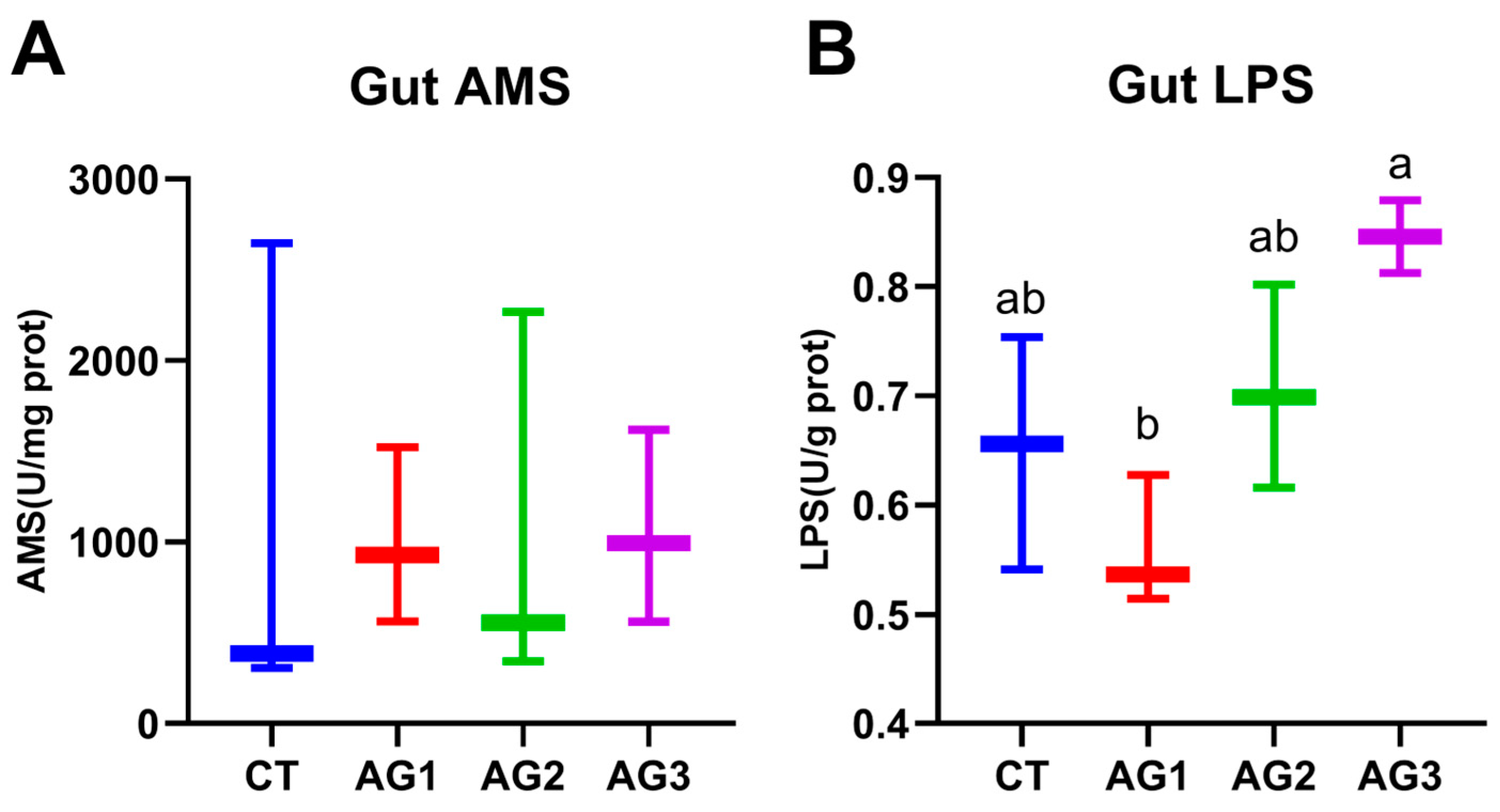
The intestinal digestive enzyme activity results show that LPS activity was significantly increased following the administration of the astragalus–ginseng mixture (Figure 2B). At the same time, there were no significant differences in AMS activity among all groups (Figure 2A). Based on these results, we infer that the increase in LPS promotes food absorption, leading to improved growth performance. Meanwhile, the enhanced lipid absorption may contribute to the increase in the hepato-somatic index.

3.4. Antioxidant Enzyme Activity

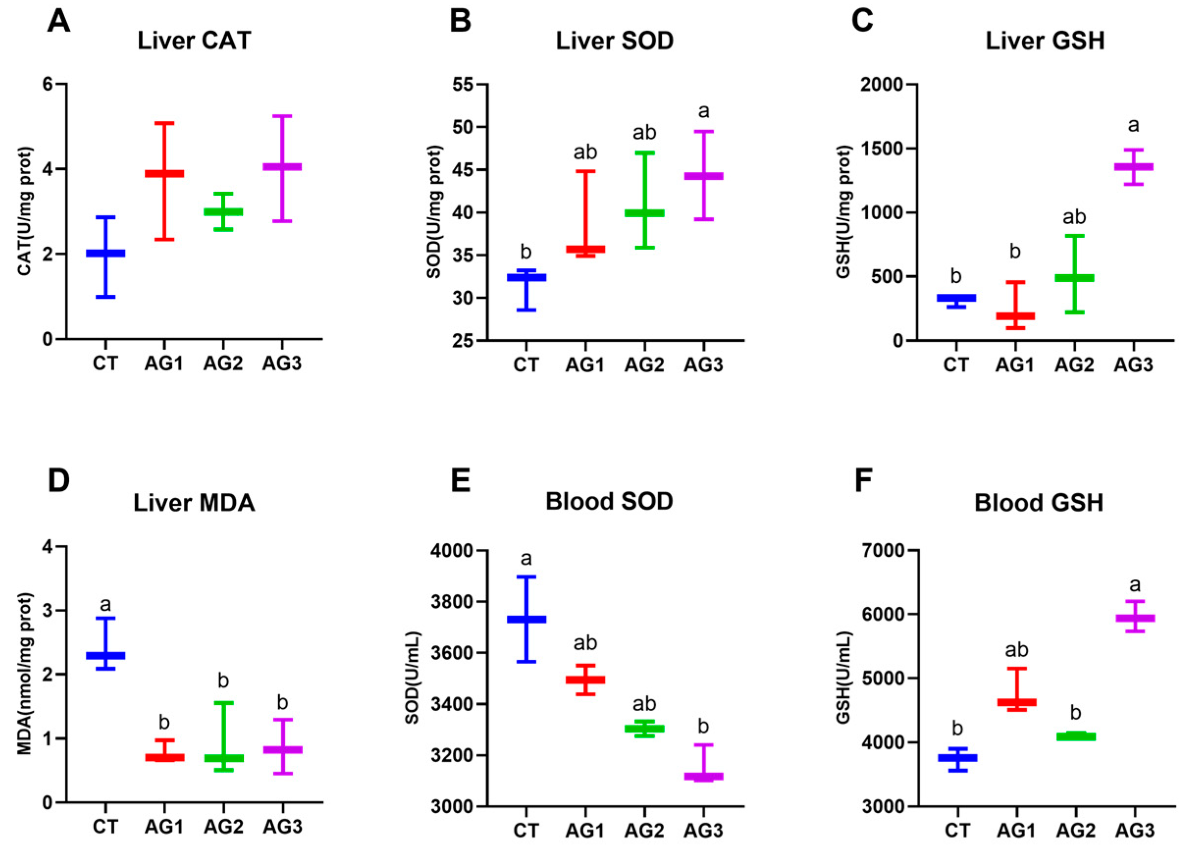
An antioxidant analysis was performed by measuring the MDA content and the activity of antioxidant enzymes in the liver and blood of yellow catfish. Liver SOD and GSH activities showed an increasing trend after feeding (Figure 3B,C), while MDA was significantly decreased (Figure 3D). Blood GSH showed the same trend as in the liver (Figure 3E). However, blood SOD activity decreased after feeding (Figure 3F). The liver CAT activity showed no significant differences among all groups (Figure 3A). In conclusion, feeding with experimental diets can upregulate the antioxidant capacity in the liver.

3.5. Immune-Related Enzyme Activity

The results of immune-related enzyme activity show that ALT and LZM activities were significantly elevated following the administration of the astragalus–ginseng mixture (Figure 4A,C). The activity of AST in all groups showed no significant differences (Figure 4B). These results indicate that experimental diets can enhance the production of the LZM. However, the increase in high concentration intake may burden liver function, contributing to the increase in AST activity.

3.6. Immune-Related Gene Expression

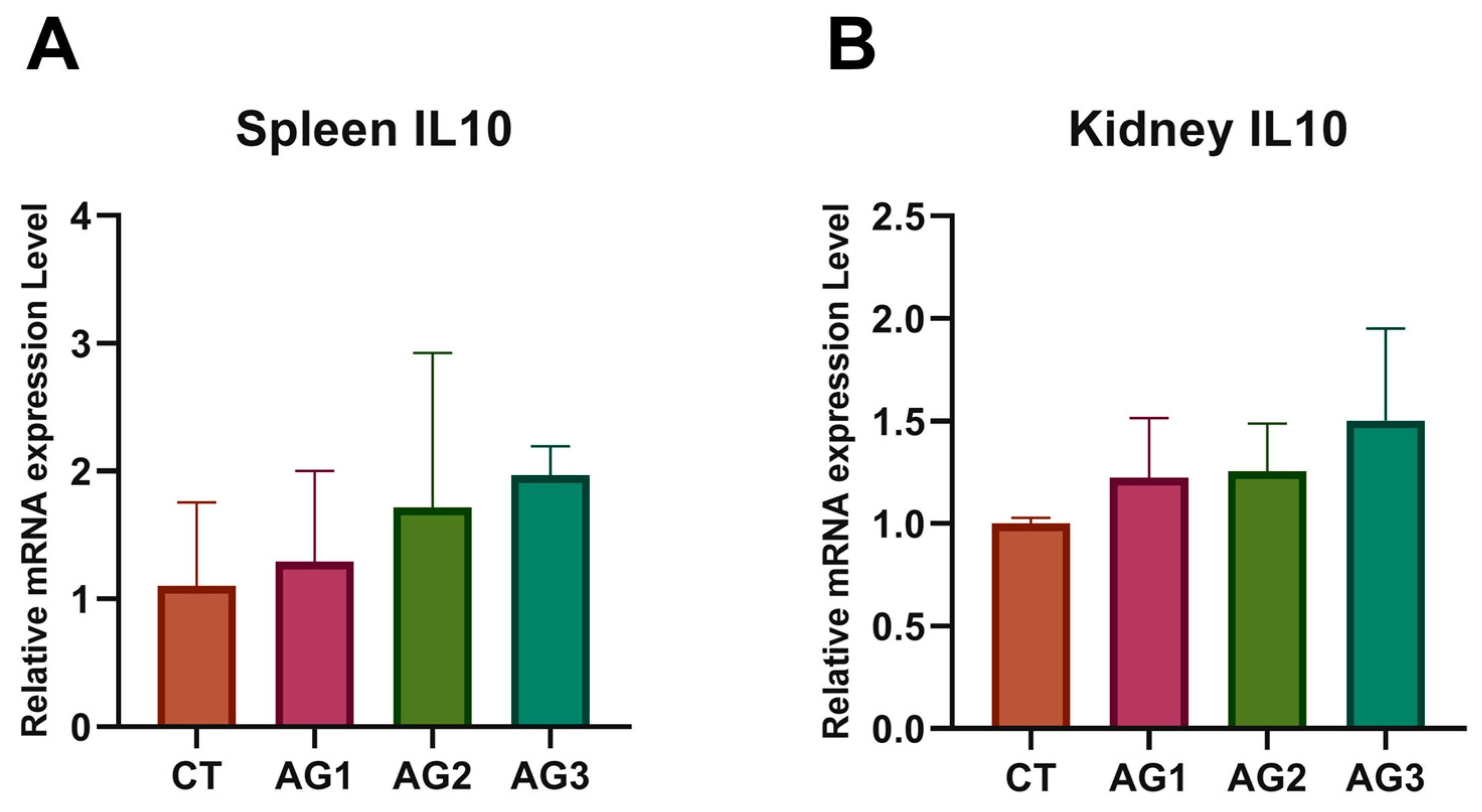
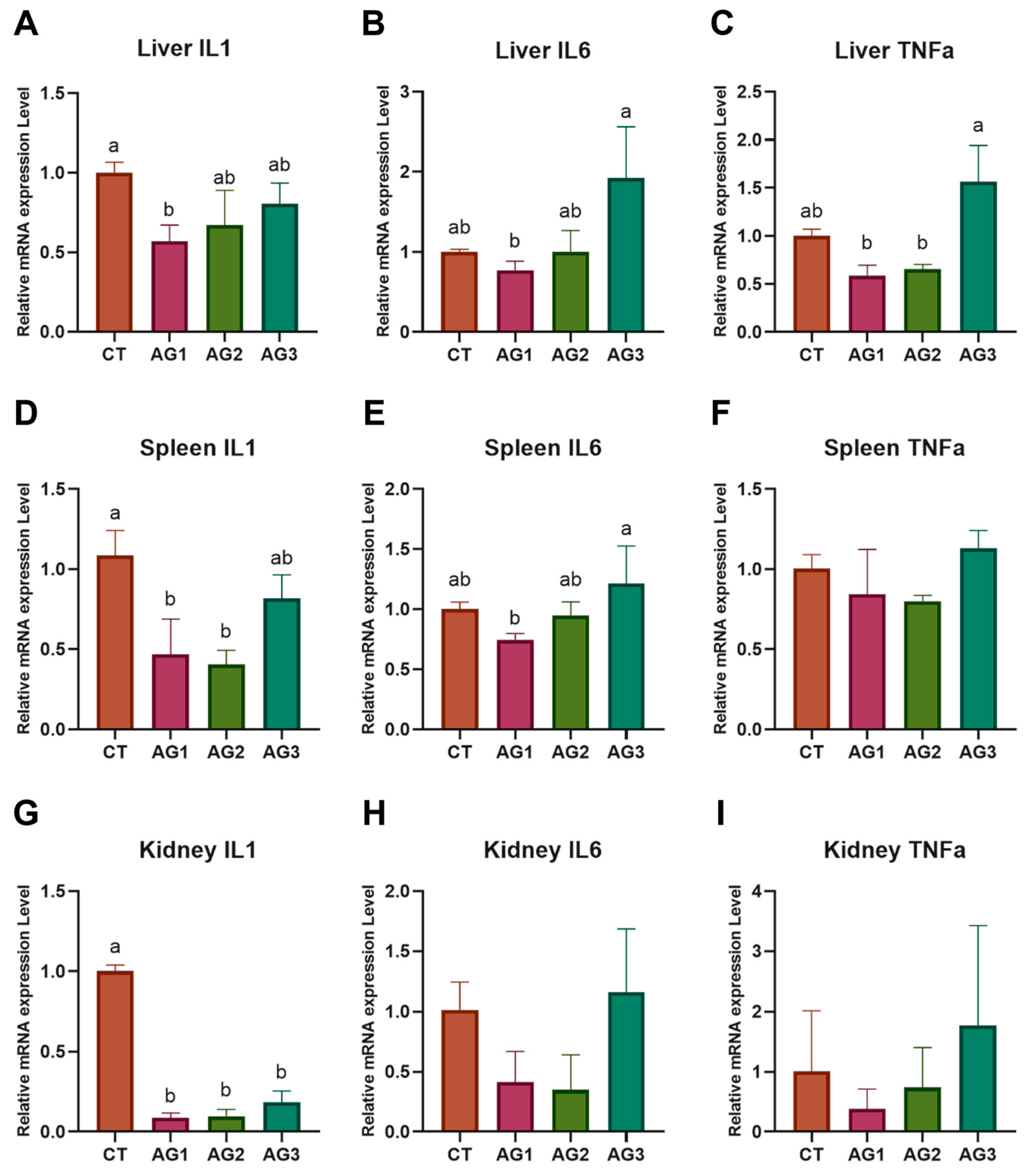
The expression levels of inflammatory genes in the liver of yellow catfish showed that IL-1β was significantly reduced in the fed group compared to the control group (Figure 5A). The expression levels of IL-6 and TNF-α genes decreased at low concentrations but increased at high concentrations (Figure 5B,C). The relative expression of IL-1β was significantly reduced, while IL-6 decreased initially and then increased in the spleen (Figure 5D,E). There were no significant differences in the relative expression level of TNF-α gene in the spleen among all groups (Figure 5F). In the kidney, the expression level of the IL-1β gene was significantly decreased with supplementation of the astragalus–ginseng mixture (Figure 5G), while the expression levels of IL-6 and TNF-α genes showed no significant differences (Figure 5H,I). The relative expression of IL-10 in all organs had no significant differences (Figure A1). In general, we found that all pro-inflammatory factors initially decreased first and then increased, suggesting that a low concentration of experimental diets can reduce the inflammatory response, while a high concentration may induce it.

4. Discussion

Recently, astragalus and ginseng have attracted considerable attention as food additives to enhance resilience and immunity, primarily due to their ability to improve growth performance and immune function. It has been reported that 1000 mg/kg of astragalus is optimal for the growth of snakehead (Channa argus). A supplementation dosage of 1500 mg of astragalus is optimal for Nile tilapia (Oreochromis niloticus), while 688 mg/kg of astragalus is optimal for spotted sea bass (Lateolabrax maculatus) [15,32,45,46]. Ginseng has positive effects on growth; however, an optimal dosage has not yet been established [47]. Although the dietary supplementation of astragalus and ginseng has been studied in farmed fish, current supplementation strategies are still limited to single-component additions. Whether these two components exhibit synergistic effects that could further enhance the economic benefits of aquaculture remains unexplored. Addressing this gap, the present study investigates the physiological and immunological effects of varying concentrations of an astragalus–ginseng mixture in yellow catfish (Pelteobagrus fulvidraco).
In the present study, the effects of varying concentrations of an astragalus–ginseng mixture were evaluated in yellow catfish. The results indicate that 1000 mg/kg is the most effective concentration for enhancing growth performance, as reflected by improvements in FBW, WG, WGR, SGR, and HSI. These enhancements are likely associated with increased digestive enzyme activity and the elongation of intestinal villi. However, further increases in concentration resulted in adverse physiological responses. Notably, at 2000 mg/kg, signs of villus shrinkage were observed, along with changes in immune-related indicators, suggesting that excessive AG supplementation may disrupt intestinal health and trigger immune responses. To determine the optimal addition level of AG supplementation, we first evaluated the effects of different dosages on growth performance and digestive functions in yellow catfish.
Growth performance is typically influenced by two key factors: digestive enzyme activity and mucosal folds length [48]. The results show that LPS activity was lowest at 500 mg/kg, and with increasing concentrations, LPS activity gradually increased, reaching the highest level at 2000 mg/kg. No significant differences were observed in AMS activity across the treatment groups. Therefore, it can be hypothesized that the improvement in growth performance, particularly in HSI, may be attributed to the enhanced activity of LPS, which leads to an increase in liver fat mass induced by the astragalus–ginseng mixture. Similar results are observed in the present study, which investigates the effects of the APS diet on Nile tilapia (Oreochromis niloticus) and zebrafish [15,49]. On the other hand, the length of intestinal villi directly reflects nutrient absorption capacity [50]. The intestine is a vital digestive organ where digestion and absorption primarily occur, with absorption occurring mainly in the intestinal villi. Longer villi increase the surface area, enhancing nutrient absorption and ultimately improving growth performance. Studies have confirmed that APS can preserve gut structure by reducing immunological stress while enhancing intestinal barrier function [51,52]. Concurrently, multiple studies have demonstrated that APS can improve gut morphology and stimulate intestinal development [53,54]. Research on ginseng primarily emphasizes its role in modulating the intestinal microbiota by promoting beneficial probiotics and inhibiting harmful bacteria [55,56]. In this study, the dietary astragalus–ginseng mixture significantly increased intestinal villus length, particularly at a concentration of 1000 mg/kg. However, based on the histological results of intestine, as the concentration increase to 2000 mg/kg, gut villi showed signs of adhesion and even shrinkage, suggesting that higher concentrations may negatively impact intestinal health. However, this study did not investigate the effects of the dietary astragalus–ginseng mixture on the intestinal microbiota. Future studies should investigate how different concentrations of the mixture impact gut microbiota, as it plays a key role in digestive health and immune function.
Given the significance of antioxidant capacity, the effects of the astragalus–ginseng mixture at different concentrations on antioxidant function were further explored. In many studies, the antioxidant capacity determines an animal’s ability to withstand environmental stress. Under conditions of excessive stress, cells generate reactive oxygen species (ROS), which damage nucleic acids, oxidize proteins, and trigger lipid peroxidation, disrupting various cellular functions [57]. As ubiquitous antioxidant enzymes, SOD, CAT, and GSH collaboratively transform reactive oxygen species into harmless substances, protecting cells from oxidative damage [58,59]. Meanwhile, MDA level is commonly used as an indicator of lipid peroxidation and antioxidant capacity [60]. As effective immunopotentiators, the dietary intake of APS and ginseng has been shown to enhance antioxidant function [61,62]. CAT and SOD activity in the liver shows an increasing trend, which is consistent with findings observed in yellow perch (Perca flavescens). In the serum, SOD activity exhibits a significant decrease, similar to the trend in catla (Catla catla) [63,64]. GSH levels exhibited a significant increase in both the liver and serum, whereas the MDA level showed a significant decrease in the liver. These results indicate that the exogenous addition of astragalus–ginseng mixture in Pelteobagrus fulvidraco enhances the conversion of convert reactive oxygen species by increasing the level of antioxidant enzyme activity, thereby improving the antioxidant defense system. Notably, as the concentration of the mixture increased, antioxidant enzyme activities showed a corresponding increase, suggesting a potential dose-dependent effect, where higher concentrations may lead to more pronounced antioxidant effects.
Lysozyme is an immune and hydrolytic enzyme that plays a crucial role in non-specific immunity responses. Lysozyme accomplishes the bactericidal process by hydrolyzing the peptidoglycan component of the bacterial cell wall [65]. In addition to its bactericidal function, lysozyme exhibits anti-inflammatory and antiviral properties and modulates the secretion of other immune factors [66]. It has already been demonstrated that dietary APS can increase the activity of LZM [61,67]. ALT and AST are commonly recognized as reliable indicators of liver pathology [68]. Recent studies have demonstrated that following hepatocytic injury, both ALT and AST levels exhibit a significant upward trend [69]. Studies on piglets indicate that dietary APSs and ginseng polysaccharides can decrease ALT and AST levels, thereby alleviating liver dysfunction and reduced immunological stress [52]. In this research, the dietary astragalus–ginseng mixture significantly increases the level of LZM. However, following dietary intervention, no significant difference was observed in liver AST, whereas ALT levels increased significantly with higher concentrations of the astragalus–ginseng mixture, suggesting that a higher dose may impose greater stress on liver function. The effects of dietary APS on zebrafish yield consistent results [49].
The innate immune system serves as the first line of defense against invading pathogens and function through the pattern recognition receptors. In teleosts, the innate immunity system plays a more crucial role than in mammals due to the less diversified adaptive immune system of teleosts [70,71]. In vertebrates, cytokines serve as key regulators of the immune response and play a crucial role in linking the innate and adaptive immune systems [72]. IL-1β, IL-6, and TNF-α are key pro-inflammatory cytokines that function as pleiotropic molecules, exerting a wide range of biological effects in immune regulation [73,74,75]. The primary function of IL-10 is to limit and eventually terminate inflammatory responses, as well as regulate the proliferation of B cells and NK cells [76]. Recent research has confirmed that APSs and ginseng polysaccharides can regulate the levels of IL-1β, IL-10, and TNF-a during immune responses [52]. In this study, the relative mRNA expression of IL-1β, IL-6, and TNF-α in all three organs exhibited a trend of initial decrease followed by an increase with higher concentrations of the astragalus–ginseng mixture; nevertheless, significant differences were observed only in certain cases. Meanwhile, the relative expression of IL-10 showed an increasing trend in all groups. However, no significant differences were observed. This phenomenon suggests that a low concentration of the astragalus–ginseng mixture in the diet can alleviate the immune response and enhance the immune function. Additionally, a high concentration of the astragalus–ginseng mixture may elicit some immune responses, which could have adverse effects with long-term use.
Based on the preceding findings, the astragalus–ginseng mixture at 1000 mg/kg was the most effective in enhancing the growth and physiological performance of Pelteobagrus fulvidraco, while higher concentrations, particularly 2000 mg/kg, adversely affected intestinal health and triggered immune responses. Therefore, aside from determining the optimal dosage, the duration of administration is also an important factor that warrants further consideration. For instance, a study on Nile tilapia (Oreochromis niloticus) demonstrated that the inclusion of astragalus–ginseng mixture in their diet resulted in a time-dependent lysozyme activity and phagocytic capacity, which initially rose and then declined, with the peak activity being observed in the third week of a 1–4 week observation period [77]. Subsequently, a study on crucian carp (Carassius auratus) showed that pro-inflammatory factors such as IL-1 and IFN-α increased progressively over the 4-week feeding period. Meanwhile, the anti-inflammatory factor IL-10 exhibited a trend of initially decreasing and then increasing, with the lowest expression observed in the third week, suggesting that the third week might be the most optimal duration for supplementation [78]. Most studies currently focus on a single time point, with limited attention to changes over time. Future research should address this gap by exploring the effects of supplementation across different periods.
In recent years, the use of herbal mixtures in aquaculture, particularly for improving growth performance, antioxidant capacity, and immune function, has been widely studied, and these mixtures have played a significant role in replacing antibiotics. Additionally, novel findings have emerged regarding the use of herbal mixtures as vaccine adjuvants and their ability to enhance resistance against pathogens in fish. Recent research demonstrated the dietary astragalus enhancement in immune responses in carp (Cyprinus carpio), which also provides protection against Aeromonas hydrophila. Furthermore, ginseng has been shown to positively impact growth performance, immune response, and resistance to Yersinia ruckeri in rainbow trout (Oncorhynchus mykiss), thereby further underscoring the potential of herbal extracts in aquaculture [36,79]. As a leading producer of traditional Chinese herbs, China’s aquaculture industry faces challenges from diseases in intensive farming. The use of low-cost herbal mixtures as feed additives can reduce losses by providing antibacterial properties, offering significantly economic benefits. Building on the findings of this study, future research on herbal mixtures for yellow catfish could include pathogen challenge experiments, particularly with Edwardsiella ictaluri or Aeromonas veronii, to further assess and confirm the immune-enhancing effects of the astragalus–ginseng mixture.

5. Conclusions

In summary, the dietary astragalus–ginseng mixture in yellow catfish enhanced the activities of liver antioxidant enzymes, thereby further increasing the body’s antioxidant capacity. Moreover, by promoting the activity of intestinal digestive enzymes and increasing the length of intestinal villi, the dietary astragalus–ginseng mixture improved intestinal health and enhance digestive capacity. Furthermore, the dietary astragalus–ginseng mixture also downregulated the expression level of pro-inflammatory genes, such as IL-1β, IL-6, and TNF-α, while upregulating the expression levels of IL-10, an anti-inflammatory gene. Based on these data, the excessive supplementation of the astragalus–ginseng mixture may lead to immune responses and even toxicological effects, and the optimal concentration for the dietary supplementation of the astragalus–ginseng mixture is approximately 1000 mg/kg. While this study provides valuable insights, there are a few areas that could benefit from further exploration in future research. While this study provides a comprehensive characterization of the astragalus–ginseng mixture, it does not explore the optimal ratio of astragalus to ginseng, which warrants further investigation. Additionally, although the study focuses on a 6-week feeding trial, it lacks insights into the weekly progression, suggesting that future research should include a time-dependent analysis. Furthermore, although the effects on the immune system were assessed, the underlying mechanisms remain unexplored. Future studies could incorporate toxicity challenge experiments to further validate immune enhancement. These avenues will deepen our understanding and optimize the practical application of the astragalus–ginseng mixture in aquaculture.

Author Contributions

Conceptualization, methodology, software, formal analysis, data curation, writing—original draft preparation: W.L. Conceptualization, methodology, formal analysis, data curation, writing—review and editing: H.X. Conceptualization, investigation, validation: X.M., Z.Y., and A.W. Writing—review and editing, supervision, funding: J.Q. and M.L. All authors have read and agreed to the published version of the manuscript.

Funding

This research was supported by the National Key R&D Program of China (2022YFD2400901).

Institutional Review Board Statement

The animal procedures were strictly compliance with the regulations outlined in the Statute of Experimental Animal Ethics Committee of Shanghai Ocean University Approval Code: SHOU-2023-031 Approval Date: 1 September 2023.

Data Availability Statement

The original contributions presented in the study are included in the article; further inquiries can be directed to the corresponding authors.

Conflicts of Interest

The authors declare no conflicts of interest.

Appendix A

Table A1. Real-time PCR primers.
GenesForward PrimerReverse PrimerAccession No.
IL-1β5-CCTGTGTGTTTGGGGATTG-35-CCTTGATGGTCTTTAGGCTCT-3MF770571.1
IL-65-CCATCGGAGGAACACAGA-35-GTAGATAAGGCGCAGACATT-3XM_027176013.2
IL-105-TCTGTAGGTTCCTCCTGCTT-35-AGGTCATCCTTGGATTCGT-3XM_027144360.2
TNF-α5-CAGGCAAACACACAAAGGC-35-GAGAAAGCTCCGAAAAACG-3XM_027133763.2
β-actin5-CCTAAAGCCAACAGGGAAAAG-35-GTCACGGCCAGCCAAATC-3XM_027148463.2

Appendix B

Figure A1. IL-10 gene expression levels of yellow catfish fed diets with graded levels of astragalus–ginseng mixture for 6 weeks. Spleen IL-10 (A), kidney IL-10 (B).
Fishes 10 00208 g0a1

References

  1. Li, X.P.; Li, J.R.; Wang, Y.B.; Fu, L.L.; Fu, Y.Y.; Li, B.Q.; Jiao, B.H. Aquaculture Industry in China: Current State, Challenges, and Outlook. Rev. Fish. Sci. 2011, 19, 187–200. [Google Scholar] [CrossRef]
  2. Bondad-Reantaso, M.G.; Subasinghe, R.P.; Arthur, J.R.; Ogawa, K.; Chinabut, S.; Adlard, R.; Tan, Z.; Shariff, M. Disease and health management in Asian aquaculture. Vet. Parasitol. 2005, 132, 249–272. [Google Scholar] [CrossRef] [PubMed]
  3. Lafferty, K.D.; Harvell, C.D.; Conrad, J.M.; Friedman, C.S.; Kent, M.L.; Kuris, A.M.; Powell, E.N.; Rondeau, D.; Saksida, S.M. Infectious Diseases Affect Marine Fisheries and Aquaculture Economics. Annu. Rev. Mar. Sci. 2015, 7, 471–496. [Google Scholar] [CrossRef] [PubMed]
  4. Cabello, F.C.; Godfrey, H.P.; Tomova, A.; Ivanova, L.; Dolz, H.; Millanao, A.; Buschmann, A.H. Antimicrobial use in aquaculture re-examined: Its relevance to antimicrobial resistance and to animal and human health. Environ. Microbiol. 2013, 15, 1917–1942. [Google Scholar] [CrossRef]
  5. Liu, X.; Steele, J.C.; Meng, X.Z. Usage, residue, and human health risk of antibiotics in Chinese aquaculture: A review. Environ. Pollut. 2017, 223, 161–169. [Google Scholar] [CrossRef] [PubMed]
  6. Reverter, M.; Bontemps, N.; Lecchini, D.; Banaigs, B.; Sasal, P. Use of plant extracts in fish aquaculture as an alternative to chemotherapy: Current status and future perspectives. Aquaculture 2014, 433, 50–61. [Google Scholar] [CrossRef]
  7. Tan, B.K.H.; Vanitha, J. Immunomodulatory and antimicrobial effects of some traditional Chinese medicinal herbs: A review. Curr. Med. Chem. 2004, 11, 1423–1430. [Google Scholar] [CrossRef]
  8. Xie, J.H.; Jin, M.L.; Morris, G.A.; Zha, X.Q.; Chen, H.Q.; Yi, Y.; Li, J.E.; Wang, Z.J.; Gao, J.; Nie, S.P.; et al. Advances on Bioactive Polysaccharides from Medicinal Plants. Crit. Rev. Food Sci. Nutr. 2016, 56 (Suppl. 1), S60–S84. [Google Scholar] [CrossRef]
  9. Fu, J.; Wang, Z.; Huang, L.; Zheng, S.; Wang, D.; Chen, S.; Zhang, H.; Yang, S. Review of the botanical characteristics, phytochemistry, and pharmacology of Astragalus membranaceus (Huangqi). Phytother. Res. 2014, 28, 1275–1283. [Google Scholar] [CrossRef]
  10. Liu, D.; Tang, W.; Yin, J.-Y.; Nie, S.-P.; Xie, M.-Y. Monosaccharide composition analysis of polysaccharides from natural sources: Hydrolysis condition and detection method development. Food Hydrocoll. 2021, 116, 106641. [Google Scholar] [CrossRef]
  11. Xu, D.-J.; Xia, Q.; Wang, J.-J.; Wang, P.-P. Molecular Weight and Monosaccharide Composition of Astragalus Polysaccharides. Molecules 2008, 13, 2408–2415. [Google Scholar] [CrossRef] [PubMed]
  12. Li, S.-G.; Chen, Y.; Zhang, Y.-Q. Effects of Astragalus polysaccharide on nephritis induced by cationic bovine serum albumin in rats. J. Chin. Med. Mater. 2010, 33, 1913–1916. [Google Scholar]
  13. Shao, B.M.; Xu, W.; Dai, H.; Tu, P.; Li, Z.; Gao, X.M. A study on the immune receptors for polysaccharides from the roots of Astragalus membranaceus, a Chinese medicinal herb. Biochem. Biophys. Res. Commun. 2004, 320, 1103–1111. [Google Scholar] [CrossRef] [PubMed]
  14. Li, S.P.; Zhao, X.J.; Wang, J.Y. Synergy of Astragalus polysaccharides and probiotics (Lactobacillus and Bacillus cereus) on immunity and intestinal microbiota in chicks. Poult. Sci. 2009, 88, 519–525. [Google Scholar] [CrossRef]
  15. Zahran, E.; Risha, E.; AbdelHamid, F.; Mahgoub, H.A.; Ibrahim, T. Effects of dietary Astragalus polysaccharides (APS) on growth performance, immunological parameters, digestive enzymes, and intestinal morphology of Nile tilapia (Oreochromis niloticus). Fish Shellfish Immunol. 2014, 38, 149–157. [Google Scholar] [CrossRef]
  16. Attele, A.S.; Wu, J.A.; Yuan, C.S. Ginseng pharmacology—Multiple constituents and multiple actions. Biochem. Pharmacol. 1999, 58, 1685–1693. [Google Scholar] [CrossRef]
  17. Attele, A.S.; Zhou, Y.P.; Xie, J.T.; Wu, J.A.; Zhang, L.; Dey, L.; Pugh, W.; Rue, P.A.; Polonsky, K.S.; Yuan, C.S. Antidiabetic effects of Panax ginseng berry extract and the identification of an effective component. Diabetes 2002, 51, 1851–1858. [Google Scholar] [CrossRef]
  18. Mochizuki, M.; Yoo, Y.C.; Matsuzawa, K.; Sato, K.; Saiki, I.; Tonooka, S.; Samukawa, K.; Azuma, I. Inhibitory Effect of Tumor-Metastasis in Mice by Saponins, Ginsenoside-Rb2, 20(R)-Ginsenoside-Rg3 and 20(S)-Ginsenoside-Rg3, of Red-ginseng. Biol. Pharm. Bull. 1995, 18, 1197–1202. [Google Scholar] [CrossRef]
  19. Shibata, S. Chemistry and cancer preventing activities of ginseng saponins and some related triterpenoid compounds. J. Korean Med. Sci. 2001, 16, S28–S37. [Google Scholar] [CrossRef]
  20. Fu, Y.; Ji, L.L. Chronic ginseng consumption attenuates age-associated oxidative stress in rats. J. Nutr. 2003, 133, 3603–3609. [Google Scholar] [CrossRef]
  21. Jung, S.; Lee, M.-S.; Shin, Y.; Kim, C.-T.; Kim, I.-H.; Kim, Y. High Hydrostatic Pressure Extract of Red Ginseng Attenuates Inflammation in Rats with High-fat Diet Induced Obesity. Prev. Nutr. Food Sci. 2015, 20, 253–259. [Google Scholar] [CrossRef]
  22. Uluisik, D.; Keskin, E. The Effects of Ginseng and Echinacea on Some Plasma Cytokine Levels in Rats. Kafkas Univ. Vet. Fak. Derg. 2012, 18, 65–68. [Google Scholar]
  23. Ovodov, Y.S.; Solov’eva, T.F. Polysaccharides ofPanax ginseng. Chem. Nat. Compd. 1966, 2, 243–245. [Google Scholar] [CrossRef]
  24. Sun, L.; Wu, D.; Ning, X.; Yang, G.; Lin, Z.; Tian, M.; Zhou, Y. α-Amylase-assisted extraction of polysaccharides from Panax ginseng. Int. J. Biol. Macromol. 2015, 75, 152–157. [Google Scholar] [CrossRef] [PubMed]
  25. Zhang, X.; Yu, L.; Bi, H.; Li, X.; Ni, W.; Han, H.; Li, N.; Wang, B.; Zhou, Y.; Tai, G. Total fractionation and characterization of the water-soluble polysaccharides isolated from Panax ginseng C. A. Meyer. Carbohydr. Polym. 2009, 77, 544–552. [Google Scholar] [CrossRef]
  26. Jiang, X.L.; Ma, G.F.; Zhao, B.B.; Meng, Y.; Chen, L.L. Structural characterization and immunomodulatory activity of a novel polysaccharide from Panax notoginseng. Front. Pharmacol. 2023, 14, 1190233. [Google Scholar] [CrossRef] [PubMed]
  27. Tomoda, M.; Shimada, K.; Konno, C.; Sugiyama, K.; Hikino, H.J.P.M. Partial structure of panaxan A, a hypoglycaemic glycan of Panax ginseng roots. Planta Medica 1984, 50, 436–438. [Google Scholar] [CrossRef]
  28. Cheng, H.; Li, S.; Fan, Y.; Gao, X.; Hao, M.; Wang, J.; Zhang, X.; Tai, G.; Zhou, Y. Comparative studies of the antiproliferative effects of ginseng polysaccharides on HT-29 human colon cancer cells. Med. Oncol. 2011, 28, 175–181. [Google Scholar] [CrossRef]
  29. Na, H.S.; Lim, Y.-J.; Yun, Y.S.; Choi, Y.H.; Oh, J.S.; Rhee, J.H.; Lee, H.C. Protective Effect of Ginsan Against Vibrio vulnificus Infection. J. Bacteriol. Virol. 2009, 39, 113–118. [Google Scholar] [CrossRef]
  30. He, Z.; Wang, X.; Li, G.; Zhao, Y.; Zhang, J.; Niu, C.; Zhang, L.; Zhang, X.; Ying, D.; Li, S. Antioxidant activity of prebiotic ginseng polysaccharides combined with potential probiotic Lactobacillus plantarum C88. Int. J. Food Sci. Technol. 2015, 50, 1673–1682. [Google Scholar] [CrossRef]
  31. Li, Y.G.; Dong, X.H.; Zhang, Y.L.; Xiao, T.Y.; Zhao, Y.R.; Wang, H.Q. Astragalus polysaccharide improves the growth, meat quality, antioxidant capacity and bacterial resistance of Furong crucian carp (Furong carp♀ × red crucian carp♂). Int. J. Biol. Macromol. 2023, 244, 124999. [Google Scholar] [CrossRef] [PubMed]
  32. Zhu, X.M.; Liu, X.Y.; Xia, C.G.; Li, M.Y.; Niu, X.T.; Wang, G.Q.; Zhang, D.M. Effects of dietary Astragalus propinquus Schischkin polysaccharides on growth performance, immunological parameters, antioxidants responses and inflammation-related gene expression in Channa argus. Comp. Biochem. Physiol. C-Toxicol. Pharmacol. 2021, 249, 109121. [Google Scholar] [CrossRef]
  33. Sun, Y.K.; Wang, X.; Zhou, H.H.; Mai, K.S.; He, G. Dietary Astragalus polysaccharides ameliorates the growth performance, antioxidant capacity and immune responses in turbot (Scophthalmus maximus L.). Fish Shellfish Immunol. 2020, 99, 603–608. [Google Scholar] [CrossRef]
  34. Pan, Y.C.; Liu, Z.; Quan, J.Q.; Gu, W.; Wang, J.W.; Zhao, G.Y.; Lu, J.H.; Wang, J.F. Purified Astragalus Polysaccharide Combined with Inactivated Vaccine Markedly Prevents Infectious Haematopoietic Necrosis Virus Infection in Rainbow Trout (Oncorhynchus mykiss). ACS Biomater. Sci. Eng. 2024, 10, 6938–6953. [Google Scholar] [CrossRef]
  35. Lin, G.X.; Da, F.; Wan, X.J.; Huang, Y.C.; Yang, S.P.; Jian, J.C.; Cai, S.H. Immune-enhancing effects of Astragalus polysaccharides and Ganoderma lucidum polysaccharides on Vibrio harveyi flgJ DNA vaccine in grouper. J. Fish Dis. 2023, 46, 147–156. [Google Scholar] [CrossRef]
  36. Bulfon, C.; Bongiorno, T.; Messina, M.; Volpatti, D.; Tibaldi, E.; Tulli, F. Effects of Panax ginseng extract in practical diets for rainbow trout (Oncorhynchus mykiss) on growth performance, immune response and resistance to Yersinia ruckeri. Aquac. Res. 2017, 48, 2369–2379. [Google Scholar] [CrossRef]
  37. In-Chul, B.; Kwon, M.G.; Cho, S.H. Effects of Dietary Inclusion of Red Ginseng Byproduct on Growth, Body Composition, Serum Chemistry, and Lysozyme Activity in Juvenile Olive Flounder (Paralichthys olivaceus). Fish. Aquat. Sci. 2010, 13, 300–307. [Google Scholar]
  38. Shao, J.C.; Wang, X.X.; Liu, Q.Q.; Lv, H.Y.; Qi, Q.; Li, C.H.; Zhang, J.N.; Chen, X.J.; Chen, X.H. Eucommia ulmoides leaf extracts combined with Astragalus polysaccharides: Effects on growth, antioxidant capacity, and intestinal inflammation in juvenile large yellow croaker (Larimichthys crocea). Fish Shellfish. Immunol. 2025, 161, 110229. [Google Scholar] [CrossRef]
  39. Jin, R.M.; Huang, H.Z.; Zhou, Y.; Wang, Y.Y.; Fu, H.C.; Li, Z.; Fu, X.Z.; Li, N.Q. Characterization of mandarin fish (Siniperca chuatsi) IL-6 and IL-6 signal transducer and the association between their SNPs and resistance to ISKNV disease. Fish Shellfish Immunol. 2021, 113, 139–147. [Google Scholar] [CrossRef]
  40. Xiong, Y.; Zheng, X.; Ke, W.; Gong, G.; Wang, Y.; Dan, C.; Huang, P.; Wu, J.; Guo, W.; Mei, J. Function and association analysis of Cyclophilin A gene with resistance to Edwardsiella ictaluri in yellow catfish. Dev. Comp. Immunol. 2020, 113, 103783. [Google Scholar] [CrossRef]
  41. Ye, S.; Li, H.; Qiao, G.; Li, Z. First case of Edwardsiella ictaluri infection in China farmed yellow catfish Pelteobagrus fulvidraco. Aquaculture 2009, 292, 6–10. [Google Scholar] [CrossRef]
  42. Bai, D.Q.; Xu, H.L.; Wu, X.; Zhai, S.L.; Yang, G.; Qiao, X.T.; Guo, Y.J. Effect of Dietary Ganoderma lucidum Polysaccharides (GLP) on Cellular Immune Responses and Disease Resistance of Yellow Catfish (Pelteobagrus fulvidraco). Isr. J. Aquac.-Bamidgeh 2015, 67, 9. [Google Scholar] [CrossRef]
  43. Li, S.; Li, C.; Wu, S. Dietary chitosan modulates the growth performance, body composition and nonspecific immunity of juvenile yellow catfish (Pelteobagrus fulvidraco). Int. J. Biol. Macromol. 2022, 217, 188–192. [Google Scholar] [CrossRef]
  44. Song, Z.X.; Jiao, C.R.; Chen, B.Y.; Xu, W.Y.; Wang, M.Y.; Zou, J.H.; Xu, W.Q.; Xu, Z.; Wang, Q.C. Dietary Acanthopanax senticosus extracts modulated the inflammatory and apoptotic responses of yellow catfish to protect against Edwardsiella ictaluriinfection. Aquac. Res. 2021, 52, 5078–5092. [Google Scholar] [CrossRef]
  45. Wei, F.; Abulahaiti, D.; Tian, C.C.; Chen, Y.; Jiang, S.S.; Lu, J.X.; Zhang, G.H. Effects of dietary Astragalus mongholicus, Astragalus polysaccharides and Lactobacillus on growth performance, immunity and antioxidant status in Qingjiaoma finishing broilers. Czech J. Anim. Sci. 2022, 67, 275–285. [Google Scholar] [CrossRef]
  46. Huang, Z.F.; Ye, Y.L.; Xu, A.L.; Li, Z.B. Effects of Astragalus membranaceus Polysaccharides on Growth Performance, Physiological and Biochemical Parameters, and Expression of Genes Related to Lipid Metabolism of Spotted Sea Bass, Lateolabrax maculatus. Aquac. Nutr. 2023, 2023, 6191330. [Google Scholar] [CrossRef] [PubMed]
  47. Hua, M.; Liu, Z.; Sha, J.; Li, S.; Dong, L.; Sun, Y. Effects of ginseng soluble dietary fiber on serum antioxidant status, immune factor levels and cecal health in healthy rats. Food Chem. 2021, 365, 130641. [Google Scholar] [CrossRef]
  48. Heidarieh, M.; Mirvaghefi, A.R.; Akbari, M.; Farahmand, H.; Sheikhzadeh, N.; Shahbazfar, A.A.; Behgar, M. Effect of dietary Ergosan on growth performance, digestive enzymes, intestinal histology, hematological parameters and body composition of rainbow trout (Oncorhynchus mykiss). Fish Physiol. Biochem. 2012, 38, 1169–1174. [Google Scholar] [CrossRef]
  49. Li, Y.; Ran, C.; Wei, K.; Xie, Y.; Xie, M.; Zhou, W.; Yang, Y.; Zhang, Z.; Lv, H.; Ma, X.; et al. The effect of Astragalus polysaccharide on growth, gut and liver health, and anti-viral immunity of zebrafish. Aquaculture 2021, 540, 736677. [Google Scholar] [CrossRef]
  50. Liu, L.; Ling, H.Y.; Zhang, W.; Zhou, Y.; Li, Y.G.; Peng, N.; Zhao, S.M. Functional Comparison of Clostridium butyricum and Sodium Butyrate Supplementation on Growth, Intestinal Health, and the Anti-inflammatory Response of Broilers. Front. Microbiol. 2022, 13, 914212. [Google Scholar] [CrossRef]
  51. Liang, H.; Tao, S.M.; Wang, Y.Y.; Zhao, J.; Yan, C.; Wu, Y.J.; Liu, N.; Qin, Y.H. Astragalus polysaccharide: Implication for intestinal barrier, anti-inflammation, and animal production. Front. Nutr. 2024, 11, 1364739. [Google Scholar] [CrossRef] [PubMed]
  52. Wang, K.L.; Zhang, H.R.; Han, Q.J.; Lan, J.H.; Chen, G.Y.; Cao, G.T.; Yang, C.M. Effects of astragalus and ginseng polysaccharides on growth performance, immune function and intestinal barrier in weaned piglets challenged with lipopolysaccharide. J. Anim. Physiol. Anim. Nutr. 2020, 104, 1096–1105. [Google Scholar] [CrossRef]
  53. Liu, Y.T.; Miao, Y.Q.; Xu, N.; Ding, T.; Cui, K.; Chen, Q.C.; Zhang, J.Z.; Fang, W.; Mai, K.S.; Ai, Q.H. Effects of dietary Astragalus polysaccharides (APS) on survival, growth performance, activities of digestive enzyme, antioxidant responses and intestinal development of large yellow croaker (Larimichthys crocea) larvae. Aquaculture 2020, 517, 734752. [Google Scholar] [CrossRef]
  54. Qiao, Y.Y.; Liu, C.Z.; Guo, Y.P.; Zhang, W.; Guo, W.B.; Oleksandr, K.; Wang, Z.X. Polysaccharides derived from Astragalus membranaceus and Glycyrrhiza uralensis improve growth performance of broilers by enhancing intestinal health and modulating gut microbiota. Poult. Sci. 2022, 101, 101905. [Google Scholar] [CrossRef] [PubMed]
  55. Geng, S.; Zheng, W.; Zhao, Y.; Xu, T. METTL3-Mediated m6A Modification of TRIF and MyD88 mRNAs Suppresses Innate Immunity in Teleost Fish, Miichthys miiuy. J. Immunol. 2023, 211, 130–139. [Google Scholar] [CrossRef]
  56. Sun, Y.-F.; Zhang, X.; Wang, X.-Y.; Jia, W. Effect of long-term intake of ginseng extracts on gut microbiota in rats. China J. Chin. Mater. Medica 2018, 43, 3927–3932. [Google Scholar] [CrossRef]
  57. Scandalios, J.G. Oxidative stress: Molecular perception and transduction of signals triggering antioxidant gene defenses. Braz. J. Med. Biol. Res. 2005, 38, 995–1014. [Google Scholar] [CrossRef]
  58. Gill, S.S.; Tuteja, N. Reactive oxygen species and antioxidant machinery in abiotic stress tolerance in crop plants. Plant Physiol. Biochem. 2010, 48, 909–930. [Google Scholar] [CrossRef]
  59. He, L.; He, T.; Farrar, S.; Ji, L.B.; Liu, T.Y.; Ma, X. Antioxidants Maintain Cellular Redox Homeostasis by Elimination of Reactive Oxygen Species. Cell. Physiol. Biochem. 2017, 44, 532–553. [Google Scholar] [CrossRef]
  60. Ayala, A.; Muñoz, M.F.; Argüelles, S. Lipid Peroxidation: Production, Metabolism, and Signaling Mechanisms of Malondialdehyde and 4-Hydroxy-2-Nonenal. Oxidative Med. Cell. Longev. 2014, 2014, 360438. [Google Scholar] [CrossRef]
  61. Chang, Z.Q.; Ge, Q.Q.; Sun, M.; Wang, Q.; Lv, H.Y.; Li, J. Immune responses by dietary supplement with Astragalus polysaccharides in the Pacific white shrimp, Litopenaeus vannamei. Aquac. Nutr. 2018, 24, 702–711. [Google Scholar] [CrossRef]
  62. Weini, Z.; Yongyang, W.; Anyi, C.; Ruoyu, L.; Fuyu, K.; Jinpeng, Z.; Jianchun, S.; Xiaohong, H.; Xinhua, C. Protective effects of dietary Astragalus polysaccharides on large yellow croaker (Larimichthys crocea) against Vibrio alginolyticus infection. Aquaculture 2024, 581, 740398. [Google Scholar] [CrossRef]
  63. Elabd, H.; Wang, H.P.; Shaheen, A.; Yao, H.; Abbass, A. Feeding Glycyrrhiza glabra (liquorice) and Astragalus membranaceus (AM) alters innate immune and physiological responses in yellow perch (Perca flavescens). Fish Shellfish Immunol. 2016, 54, 374–384. [Google Scholar] [CrossRef] [PubMed]
  64. Harikrishnan, R.; Devi, G.; Doan, H.V.; Tapingkae, W.; Balasundaram, C.; Arockiaraj, J.; Ringo, E. Changes in immune genes expression, immune response, digestive enzymes-antioxidant status, and growth of catla (Catla catla) fed with Astragalus polysaccharides against edwardsiellosis disease. Fish Shellfish Immunol. 2022, 121, 418–436. [Google Scholar] [CrossRef] [PubMed]
  65. Ragland, S.A.; Criss, A.K. From bacterial killing to immune modulation: Recent insights into the functions of lysozyme. PLoS Pathog. 2017, 13, e1006512. [Google Scholar] [CrossRef]
  66. Song, Q.; Xiao, Y.; Xiao, Z.H.; Liu, T.; Li, J.C.; Li, P.; Han, F. Lysozymes in Fish. J. Agric. Food Chem. 2021, 69, 15039–15051. [Google Scholar] [CrossRef]
  67. Liu, J.; Zhang, P.J.; Wang, B.; Lu, Y.T.; Li, L.; Li, Y.H.; Liu, S.J. Evaluation of the effects of Astragalus polysaccharides as immunostimulants on the immune response of crucian carp and against SVCV in vitro and in vivo. Comp. Biochem. Physiol. C-Toxicol. Pharmacol. 2022, 253, 109249. [Google Scholar] [CrossRef]
  68. Anderson, F.H.; Zeng, L.C.; Rock, N.R.; Yoshida, E.M. An assessment of the clinical utility of serum ALT and AST in chronic hepatitis C. Hepatol. Res. 2000, 18, 63–71. [Google Scholar] [CrossRef]
  69. Huynh, T.; Zhang, J.; Hu, K.Q. Hepatitis C Virus Clearance by Direct-acting Antiviral Results in Rapid Resolution of Hepatocytic Injury as Indicated by Both Alanine Aminotransferase and Aspartate Aminotransferase Normalization. J. Clin. Transl. Hepatol. 2018, 6, 258–263. [Google Scholar] [CrossRef]
  70. Hoffmann, J.A.; Kafatos, F.C.; Janeway, C.A.; Ezekowitz, R.A.B. Phylogenetic perspectives in innate immunity. Science 1999, 284, 1313–1318. [Google Scholar] [CrossRef]
  71. Zhu, L.Y.; Nie, L.; Zhu, G.; Xiang, L.X.; Shao, J.Z. Advances in research of fish immune-relevant genes: A comparative overview of innate and adaptive immunity in teleosts. Dev. Comp. Immunol. 2013, 39, 39–62. [Google Scholar] [CrossRef] [PubMed]
  72. Akira, S.; Uematsu, S.; Takeuchi, O. Pathogen recognition and innate immunity. Cell 2006, 124, 783–801. [Google Scholar] [CrossRef] [PubMed]
  73. Dinarello, C.A. Biologic basis for interleukin-1 in disease. Blood 1996, 87, 2095–2147. [Google Scholar] [CrossRef] [PubMed]
  74. Kishimoto, T. IL-6: From its discovery to clinical applications. Int. Immunol. 2010, 22, 347–352. [Google Scholar] [CrossRef]
  75. Tracey, K.J.; Cerami, A. Tumor necrosis factor: A pleiotropic cytokine and therapeutic target. Annu. Rev. Med. 1994, 45, 491–503. [Google Scholar] [CrossRef]
  76. Moore, K.W.; Malefyt, R.D.; Coffman, R.L.; O’Garra, A. Interleukin-10 and the interleukin-10 receptor. Annu. Rev. Immunol. 2001, 19, 683–765. [Google Scholar] [CrossRef]
  77. Yin, G.J.; Jeney, G.; Racz, T.; Xu, P.; Jun, M.; Jeney, Z. Effect of two Chinese herbs (Astragalus radix and Scutellaria radix) on non-specific immune response of tilapia, Oreochromis niloticus. Aquaculture 2006, 253, 39–47. [Google Scholar] [CrossRef]
  78. Shi, L.S.; Xue, M.Y.; Xing, Y.Y.; Xu, C.; Jiang, N.; Fan, Y.D.; Chen, J.W.; Liu, W.; Wu, Y.Y.; Wu, M.L.; et al. Dietary supplementation of Astragalus fermentation products improves the growth performance, immunological characteristics, and disease resistance of crucian carp (Carassius auratus). Isr. J. Aquac.-Bamidgeh 2024, 76, 190–199. [Google Scholar] [CrossRef]
  79. Yin, G.J.; Ardó, L.; Thompson, K.D.; Adams, A.; Jeney, Z.; Jeney, G. Chinese herbs (Astragalus radix and Ganoderma lucidum) enhance immune response of carp, Cyprinus carpio, and protection against Aeromonas hydrophila. Fish Shellfish Immunol. 2009, 26, 140–145. [Google Scholar] [CrossRef]
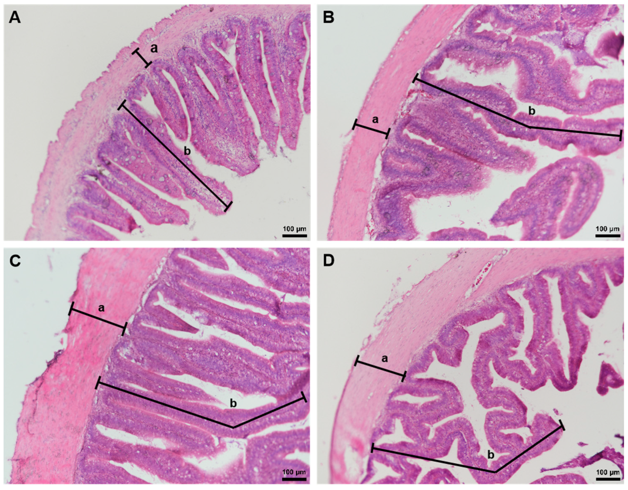
Figure 1. The intestinal morphology of yellow catfish fed diets with graded level of astragalus–ginseng mixture for 6 weeks (×100): 0 mg/kg (control) (A), 500 mg/kg (B), 1000 mg/kg (C), 2000 mg/kg (D); (a) intestinal muscular thickness, (b) intestinal villus length.
Fishes 10 00208 g001
Figure 2. AMS (A) and LPS (B) activities in the gut of yellow catfish. Different letters indicate a significant difference among astragalus–ginseng mixture concentrations (p < 0.05).
Fishes 10 00208 g002
Figure 3. Antioxidant enzyme activity in yellow catfish. Liver CAT (A), liver SOD (B), liver GSH (C), liver MDA (D), blood SOD (E), and blood GSH (F). Different letters indicate a significant difference among astragalus–ginseng mixture concentrations (p < 0.05).
Fishes 10 00208 g003
Figure 4. ALT (A), AST (B), and LZM (C) activities in the liver of yellow catfish. Different letters indicate a significant difference among astragalus–ginseng mixture concentrations. (p < 0.05).
Fishes 10 00208 g004
Figure 5. Immune-related genes expression levels of yellow catfish fed diets with graded levels of astragalus–ginseng mixture for 6 weeks. Liver IL-1β (A), liver IL-6 (B), liver TNF-α (C), spleen IL-1β (D), spleen IL-6 (E), spleen TNF-α (F), kidney IL-1β (G), kidney IL-6 (H), kidney TNF-α (I). Different letters indicate a significant difference among astragalus–ginseng mixture concentrations (p < 0.05).
Fishes 10 00208 g005
Table 1. Ingredient composition of the experimental diets.
IngredientsExperimental Feeds (Dry Matter %)
CTAG1AG2AG3
Fish meal44444444
Flour19.5519.5519.5519.55
Cottonseed meal4444
Soybean meal15151515
Corn gluten meal5555
Fish oil4.54.54.54.5
Brewer’s yeast6666
Minerals1111
Choline chloride0.60.60.60.6
Multivitamin0.20.20.20.2
Mold inhibitor0.150.150.150.15
Total100100100100
Astragalus–ginseng mixture (mg/kg)050010002000
Table 2. Effects of dietary astragalus–ginseng mixture levels on the growth performance of Pelteobagrus fulvidraco.
Parameters0 mg/kg500 mg/kg1000 mg/kg2000 mg/kg
IBW (g)5.13 ± 0.125.04 ± 0.235.05 ± 0.225.06 ± 0.26
FBW (g)9.75 ± 0.55 b10.81 ± 0.12 ab10.97 ± 0.27 ab11.61 ± 0.76 a
WG (%)46.35 ± 3.88 b56.9 ± 1.41 ab59.16 ± 0.58 a65.53 ± 6.33 a
WGR (%)90.54 ± 4.72 b111.23 ± 8.43 a117.20 ± 4.08 a129.60 ± 12.36 a
SGR (%)1.43 ± 0.05 b1.66 ± 0.08 ab1.72 ± 0.04 a1.84 ± 0.12 a
VSI (%)12.06 ± 1.8013.18 ± 1.1112.76 ± 1.1513.24 ± 0.91
HSI (%)1.01 ± 0.01 c1.29 ± 0.06 b1.63 ± 0.03 a1.53 ± 0.02 a
Note: Data are express as mean ± SE. Data marked with different letters differ significantly (p < 0.05) among groups based on a one-way ANOVA. IBW: Initial body weight; FBW: Final body weight; WG: Weight gain; WGR: Weight gain rate; SGR: Specific growth rate; VSI: Visceral somatic index; HIS: Hepato-somatic index.
Disclaimer/Publisher’s Note: The statements, opinions and data contained in all publications are solely those of the individual author(s) and contributor(s) and not of MDPI and/or the editor(s). MDPI and/or the editor(s) disclaim responsibility for any injury to people or property resulting from any ideas, methods, instructions or products referred to in the content.