Article

Development and Application of Infectious Hematopoietic Necrosis Virus Antigen-Specific DAS-ELISA Detection Method

1
Department of Aquatic Animal Diseases and Control, Heilongjiang River Fisheries Research Institute, Chinese Academy of Fishery Sciences, Harbin 150070, China
2
Key Laboratory of Aquatic Animal Diseases and Immune Technology of Heilongjiang Province, Harbin 150070, China
*
Author to whom correspondence should be addressed.
Fishes 2025, 10(10), 533; https://doi.org/10.3390/fishes10100533
Submission received: 20 August 2025 / Revised: 28 September 2025 / Accepted: 10 October 2025 / Published: 20 October 2025
(This article belongs to the Special Issue Advances in Rainbow Trout: 2nd Edition)

Abstract

Infectious hematopoietic necrosis virus (IHNV), a salmonid rhabdovirus, causes severe mortality exceeding 90% in both wild and farmed salmon and trout. Frequent outbreaks of IHNV highlight the urgent need for rapid detection methods to support effective prevention and control. This study developed a double-antibody sandwich ELISA (DAS-ELISA) targeting the nucleocapsid (N) protein of IHNV. Two peptides derived from the N protein—selected for their strong antigenicity, high level of conservation, and surface accessibility—were used as immunogens to generate two specific monoclonal antibodies. Following optimization, the DAS-ELISA was established using monoclonal antibody N-15 as the capture antibody and horseradish peroxidase (HRP)-conjugated antibody N-106 as the detection antibody. The results of this study demonstrated that DAS-ELISA exhibited high specificity for multiple IHNV strains and showed no cross-reactivity with IPNV, SVCV, or VHSV. The detection sensitivity of DAS-ELISA for IHNV was determined to be 103 TCID50/mL. Parallel analysis of 293 clinical samples using DAS-ELISA and WOAH reference method demonstrated a concordance rate of 92.83% (κ = 0.856). These results confirm that the established DAS-ELISA exhibits high sensitivity, specificity, broad-spectrum applicability, and repeatability. In conclusion, this DAS-ELISA provides a reliable and efficient tool for high-throughput early detection of IHNV infection in clinical settings.
Key Contribution: The DAS-ELISA established in this study represents a viable alternative to a laboratory diagnosis of IHNV and provides a robust foundation for developing commercial detection kits.

1. Introduction

Infectious hematopoietic necrosis (IHN) is a viral disease that has led to substantial economic losses in salmon and trout aquaculture worldwide [1]. IHN was first identified in the 1950s at sockeye salmon farms in Oregon and Washington State, USA [2]. Until the early 1980s, IHN spread to many countries through the trade of fry and adult fish [3]. Depending on host species and viral strains, outbreaks of IHN can cause mortality rates ranging from 90% to 100% [4,5]. In China, the first reported outbreak of IHN occurred in 1985 at a rainbow trout farm located in Liaoning Province [6]. IHN has now become a common disease found in fish in China and its occurrence has been reported in nine Chinese provinces [6].
The pathogen responsible for IHN is the infectious hematopoietic necrosis virus (IHNV), a member of the genus Salmonid Novirhabdovirus within the family Rhabdoviridae [7]. The genome of IHNV encodes a total of six proteins in the following order, from 3′ to 5′: nucleocapsid protein (N), phosphoprotein (P), matrix protein (M), glycoprotein (G), non-virion protein (NV), and large polymerase protein (L) [8]. Phylogenetic analysis of IHNV showed that this virus can be divided into five genogroups: J, E, L, M, and U [6]. In China, only J and U genogroups have been reported, with the J genogroup being dominant and widely prevalent [9]. To date, no commercial vaccine against IHNV has been approved in China. Therefore, the most effective way of controlling the spread of IHNV is early detection and timely intervention to block transmission.
At present, there are several methods used for IHNV detection, such as virus isolation, enzyme-linked immunosorbent assay (ELISA), reverse-transcription PCR (RT-PCR), isothermal amplification, and indirect immunofluorescence (IFA) [10,11,12,13,14]. Among these, virus isolation is a time-consuming method, while RT-PCR requires high-quality RNA, specialized handling procedures, and expensive equipment [15]. Although ELISA and IFA are the most widely used serological methods for IHNV detection, IFA is not suitable for high-throughput testing of clinical samples [16]. In contrast, ELISA offers high sensitivity and specificity and has low requirements for experimental conditions [17]. ELISA is a suitable method for the rapid diagnosis of large numbers of clinical samples.
In this study, we developed a double-antibody sandwich enzyme-linked immunosorbent assay (DAS-ELISA) for IHNV detection. The assay uses two monoclonal antibodies (mAbs) targeting distinct epitopes of the IHNV nucleocapsid (N) protein—one as the capture antibody and the other as the detection antibody. The DAS-ELISA method showed high specificity, sensitivity, reproducibility, and broad-spectrum detection capability for both J and U genogroups of IHNV. When applied to clinical samples, the diagnostic results of the DAS-ELISA showed a high level of agreement with the method recommended by the World Organization for Animal Health (WOAH).

2. Materials and Methods

2.1. Ethics Statements

All animal procedures in this study were conducted according to the guidelines for the care and use of laboratory animals of Heilongjiang River Fisheries Research Institute, CAFS. The animal studies were reviewed and approved by the Committee for the Welfare and Ethics of Laboratory Animals of Heilongjiang River Fisheries Research Institute, CAFS.

2.2. Cells and Viruses

The epithelioma papulosum cyprini (EPC) cell line and mouse myeloma cells (SP2/0) were stored in our laboratory. The J genogroup IHNV strains, Sn1203 (GenBank No: KC660147.1), XJ-13 (GenBank No: KF871191.1), BJ15 (GenBank No: MH170329.1), LN12 (GenBank No: MH170322.1), YN-13 (GenBank No: KF871192.1), QH16 (GenBank No: MH170342.1), GS15 (GenBank No: MH170319.1), and HLJ13 (GenBank No: MH170313.1) were isolated from epidemiological investigations and stored in our laboratory [6]. The U genogroup IHNV strain Blk94 (GenBank No: DQ164100) was kindly provided by Dr. Gael Kurath [18]. The infectious pancreatic necrosis virus (IPNV) strain ChRtm213 (GenBank No: KX234591.1), and spring viremia of carp virus (SVCV) strain shlj4 (GenBank No: MT675953.1) were also isolated from epidemiological investigations and stored in our laboratory [19]. The viral hemorrhagic septicemia virus (VHSV) strain was obtained from the China center for type culture collection (CCTCC).

2.3. Preparation of IHNV N Protein Antigen

The N gene sequences from the five IHNV genogroups were downloaded from NCBI. The conservation, antigenicity, and surface accessibility of amino acids in N protein were analyzed using IEDB (https://www.iedb.org/), DNAMAN version 6.0, AlphaFold version 3.0 (https://golgi.sandbox.google.com/), and PyMOL version 2.2.0 software. The optimal peptides were conjugated to keyhole limpet hemocyanin (KLH) or bovine serum albumin (BSA) (SS2163, Biofount, Beijing, China) and synthesized by Genescript Biotechnology Co., Ltd. (Nanjing, China).

2.4. Generation of mAbs

BALB/c mice were immunized with 50 μg of KLH-conjugated peptides in 50 μL of QuickAntibody-Mouse 3W adjuvant (KX0210042, Biodragon, Suzhou, China). After 14 days of immunization, booster immunization was performed using the same method as that used for the first immunization. On day 21 after the initial immunization, the mice were euthanized and spleen cells were fused with SP2/0 cells using a standard polyethylene glycol (PEG) fusion protocol. The resulting hybridoma cell suspension was seeded into 96-well plates and cultured in RPMI-1640 (C11875500BT, Gibco, Shanghai, China) medium supplemented with 2% HAT (H0262, Sigma, Beijing, China) and 20% FBS (A5669701, Gibco, Melbourne, Australia) at 37 °C under 5% CO2. The supernatants of hybridoma cells were screened by indirect ELISA using BSA-conjugated peptides as coating antigens. Positive hybridoma cells were subcloned three times by limiting dilution to obtain monoclonal cells. To produce ascites, BALB/c mice were pre-treated with 500 μL Freund’s incomplete adjuvant (F5506, Sigma, St. Louis, MO, USA). On day 7 post-injection, the positive hybridoma cells (1 × 10⁶ cells) were injected intraperitoneally into pristine-primed BALB/c mice. Ascitic fluid was collected after 7–10 days, and mAbs were purified using Protein G Sepharose 4 FF affinity chromatography resin (17061801, Cytiva, Shanghai, China). The purity of the antibodies was verified by SDS-PAGE, and mAbs isotypes were determined using a commercial mouse mAb isotyping kit (PK20003, Proteintech, Wuhan, China). The purified antibodies were coupled to horseradish peroxidase (HRP) using an HRP conjugation kit (ab102890, Abcam, Cambridge, UK).

2.5. Specificity Identification of the Antibodies

EPC cells seeded in 6-well plates were infected with IHNV, IPNV, SVCV, or VHSV for 1 h, respectively. After being washed with PBS (BL302A, Biosharp, Beijing, China), the infected cells were cultured at 15 °C under 1% CO2 for 48 h. For Western blot analysis, the cells were lysed with IP lysis buffer (87787, Thermo Scientific, Shanghai, China) and then analyzed with purified mAbs or an anti-β-tubulin mouse mAb (AC021, ABclonal, Wuhan, China) as the primary antibody, and HRP-labeled goat anti-mouse IgG antibody (C31430100, Invitrogen, Shanghai, China) as the second antibody. For IFA, virus-infected EPC cells were initially fixed with 4% paraformaldehyde (P0099, Beyotime, Shanghai, China) and then permeabilized with 0.5% Triton X-100 (P0096, Beyotime, Shanghai, China). After undergoing incubation at room temperature for 20 min, respectively, the purified antibodies were used as primary antibody, and Alexa Fluor 488 goat anti-mouse IgG antibody (A11001, Invitrogen, Shanghai, China) was used as the second antibody. Photos were acquired using a fluorescence microscope (DMi8, Leica, Wetzlar, Germany).

2.6. Optimization of the DAS-ELISA

The DAS-ELISA was performed as follows: (a) Coating: capture antibody diluted in carbonate buffer (C1055, Solarbio, Beijing, China)was added to 96-well ELISA plates (100 µL/well) and incubated at 4 °C for 12 h. (b) Blocking: plates were blocked with 200 µL/well of blocking solution. (c) Antigen incubation: test samples (100 µL/well) were added and incubated at 37 °C. (d) Detection: HRP-labeled detection antibody (100 µL/well) was added and incubated at 37 °C. (e) Signal development: TMB substrate solution (TMB-S-004, Biopanda, Huzhou, China) (100 µL/well) was added and the reaction was stopped with 2 M HCl (Beijing Chemical Works, Beijing, China) (50 µL/well). (f) Measurement: absorbance was measured at 450 nm using a microplate reader (SpectraMax i3x, Molecular Devices, Linz, Austria). Following each step from (a) to (d), the plate was washed three times with PBS containing 0.1% (v/v) tween-20 (ST825, Beyotime, Shanghai, China) (5 min per wash). To establish the optimal assay conditions, we evaluated which mAb (N-15 or N-106) was more suitable as a capture or detection antibody; the capture antibody concentration (from 0.01 μg to 1 μg per well); detection antibody dilution (from 1:1000 to 1:8000); the blocking solution type (skimmed milk (3191345, Difco, NJ, USA), FBS, BSA (SS2163, Biofount, Beijing, China)), concentration (3% and 5%), and duration (from 0.5 h to 3.0 h); antigen incubation time (from 0.5 h to 3.0 h); detection antibody incubation time (from 0.5 h to 3.0 h); and TMB substrate solution incubation time (5 min to 20 min).

2.7. Preparation of Tissue Samples

Virus-negative or -positive fish tissue samples (kidney, liver, and spleen) were collected and homogenized in PBS containing 1% Triton X-100 (1:1, w/v) using a tissue grinder (N9548R, Hoder, Beijing, China) for 5 min at 4 °C. Homogenates were centrifuged at 10,000× g for 10 min at 4 °C, and the supernatant was collected for DAS-ELISA detection.

2.8. Determination of the Cut-Off Value

A total of 80 negative and 157 IHNV-positive tissue samples were tested by the established DAS-ELISA. The obtained OD450 values were calculated by receiver operating characteristic (ROC) curve analysis and the cut-off value was determined using MedCalc software (v20.0.10).

2.9. Determination of the Specificity, Broad-Spectrum Sensitivity, and Repeatability of DAS-ELISA

The specificity of the established DAS-ELISA was assessed by testing tissue samples positive for IHNV, IPNV, SVCV, and VHSV. Broad-spectrum detection capability was evaluated using the J and U genogroup IHNV-positive tissue samples. The sensitivity was evaluated by testing the serial dilutions of an IHNV-positive tissue sample. The repeatability was assessed by testing five positive and three negative tissue samples. To assess intra-batch repeatability, triplicate replicates of each sample were performed using the same batch of precoated plates. For inter-batch repeatability evaluation, each sample was analyzed across three different batches of precoated plates. Intra-batch and inter-batch repeatability was determined by calculating the coefficient of variation (CV).

2.10. Clinical Validation of DAS-ELISA

To evaluate the clinical performance of DAS-ELISA, 293 tissue samples were collected from 23 different rainbow trout farms located across China. The results were compared with the WOAH recommended gold standard method (virus isolation coupled with RT-PCR) [14,20].

2.11. Statistical Analysis

The ROC analysis and the cut-off value for DAS-ELISA were determined using MedCalc software (v20.0.10). All statistical analyses were performed using GraphPad Prism software (version 6.0).

3. Results

3.1. Antigen Epitope Analysis of IHNV N Protein

To generate specific antibodies targeting N protein across all five IHNV genogroups, we initially conducted a conservation analysis of the N protein sequence. The DNAMAN analysis identified six highly conserved regions: 1–28 aa, 63–78 aa, 106–146 aa, 182–202 aa, 206–268 aa, and 291–311 aa (Figure 1A). Potential immunogens were further evaluated based on antigenicity and hydrophilicity. The IEDB analysis revealed five strongly antigenic peptides longer than 14 aa: 6–28 aa, 95–121 aa, 151–173 aa, 308–325 aa, and 360–388 aa (Table 1). Based on the combined analysis of high conservation, strong antigenicity, and favorable hydrophilicity, the 6–28 aa and 106–121 aa peptides were selected as promising immune antigens for antibody production. Structural modeling using AlphaFold and PyMOL confirmed that these two peptides are all located in surface-accessible regions of the N protein (Figure 1B). These two peptides were synthesized and conjugated to KLH or BSA for subsequent immunization and mAbs screening.

3.2. Production and Characterization of mAbs Against the N Protein

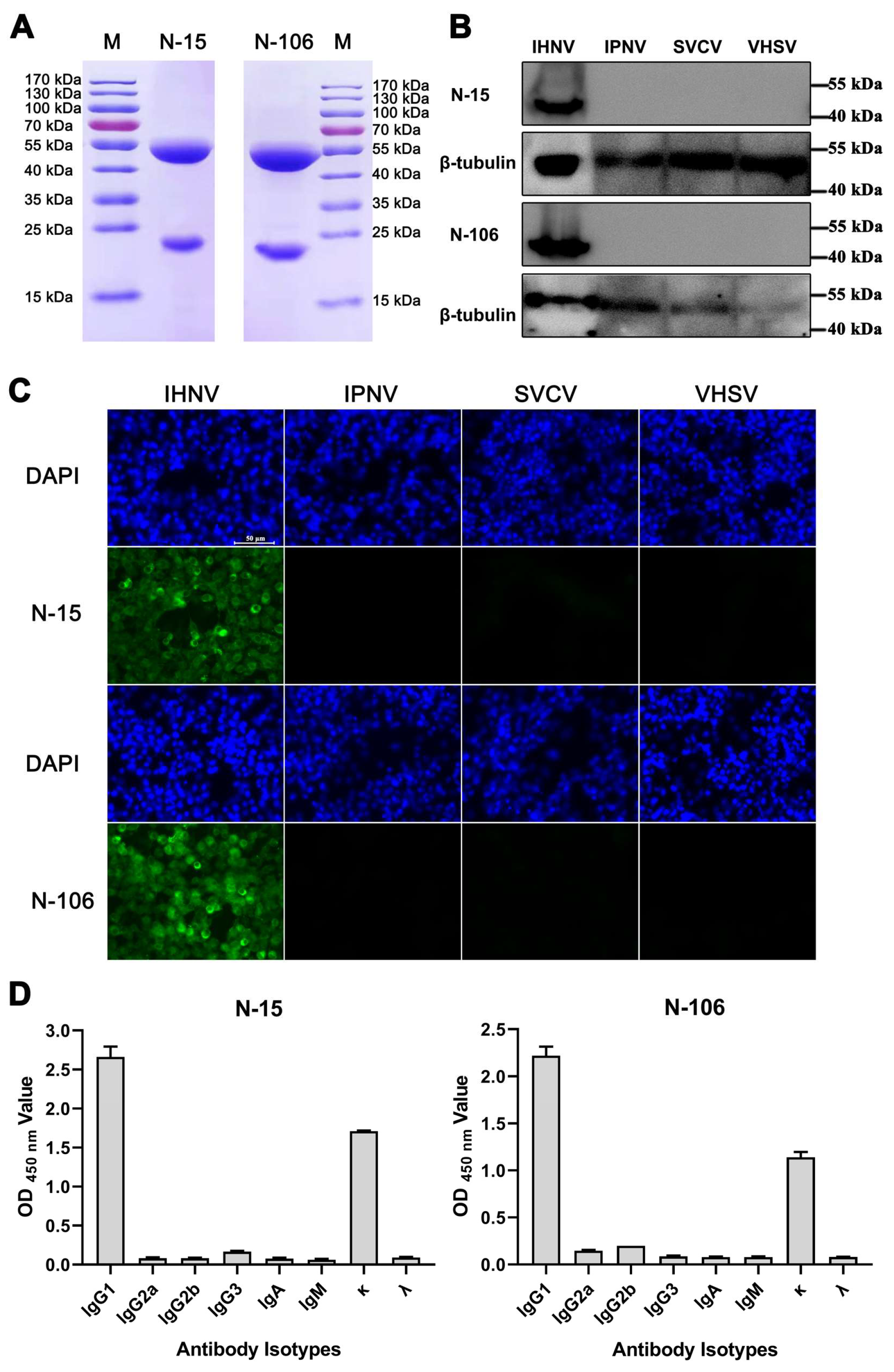
Following hybridoma cell generation and three rounds of limited dilution, two hybridoma cells that stably secreted the specific antibody against N protein were obtained, designated N-15-5H3 (specific to peptide 6–28 aa) and N-106-3A9 (specific to peptide 106–121 aa). The corresponding mAbs were named N-15 and N-106, respectively. mAbs were purified from mouse ascites, and SDS-PAGE analysis under reducing conditions confirmed the presence of light and heavy chains, indicating successful purification (Figure 2A). Concentrations of the purified mAb, N-15, and N-106 were quantified and adjusted to 1 mg/mL. Western blotting analysis showed that both mAbs specifically recognized a protein of the expected size in lysates from cells infected with IHNV, but not in lysates from cells infected with IPNV, SVCV, and VHSV (Figure 2B). Similarly, IFA results demonstrated specific staining only in IHNV-infected cells, with no cross-reactivity observed with IPNV, SVCV, and VHSV (Figure 2C). Both Western blot and IFA results demonstrated that the mAbs specifically bind to the N protein in its linear (Western blotting) and native (IFA) conformations (Figure 2B,C). Antibody isotyping analysis revealed that both mAbs are of the IgG1 isotype with kappa light chains (Figure 2D). The purified mAbs were conjugated with HRP for use in subsequent assay development.

3.3. Development and Optimization of the DAS-ELISA

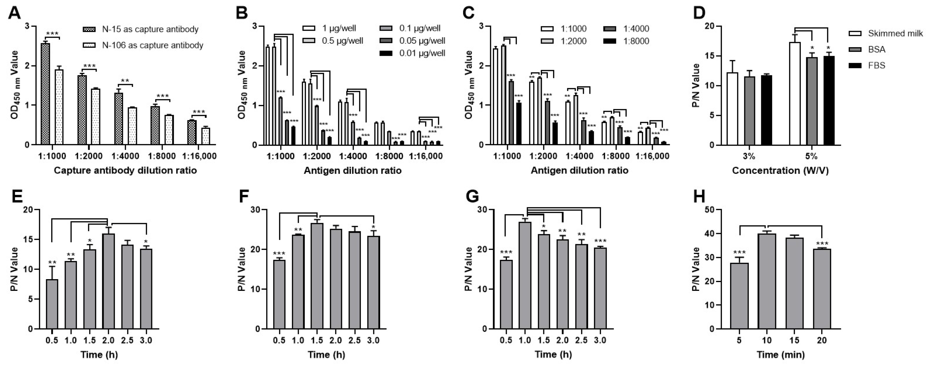
We systematically optimized the DAS-ELISA parameters to establish a robust assay for IHNV detection. An evaluation of the two mAbs indicated that using N-15 mAb as the capture antibody (at an optimal concentration of 0.5 μg/well) and HRP-conjugated N-106 mAb as the detection antibody (at an optimal dilution of 1:2000) yielded the best performance (Figure 3A,B). The optimal blocking condition was determined to be 5% skimmed milk for 2 h (Figure 3D,E). Further optimization indicated ideal incubation times of 1.5 h for the antigen and 1 h for the detection antibody (Figure 3F,G). Finally, the optimal incubation period for the TMB substrate solution was determined to be 10 min (Figure 3H).
Based on the above optimization results, the specific operation procedures for the established DAS-ELISA are as follows: (a) Coating: capture antibody (N-15 mAb) diluted in carbonate buffer was added to a 96-well ELISA plates (0.5 μg/100 µL/well) and incubated at 4 °C for 12 h. (b) Blocking: plates were blocked with 5% skimmed milk (200 µL/well) at 37 °C for 2 h. (c) Antigen incubation: test samples (100 µL/well) were added and incubated at 37 °C for 1.5 h. (d) Detection: HRP-labeled detection antibody (N-15 mAb, dilution of 1:2000, 100 µL/well) was added and incubated at 37 °C for 1 h. (e) Signal development: TMB substrate solution (100 µL/well) was added and incubated at 37 °C for 10 min. The reaction was stopped by adding 2 M HCl (50 µL/well). (f) Measurement: absorbance was measured at 450 nm using a microplate reader.

3.4. Determination of the DAS-ELISA Cut-Off Value

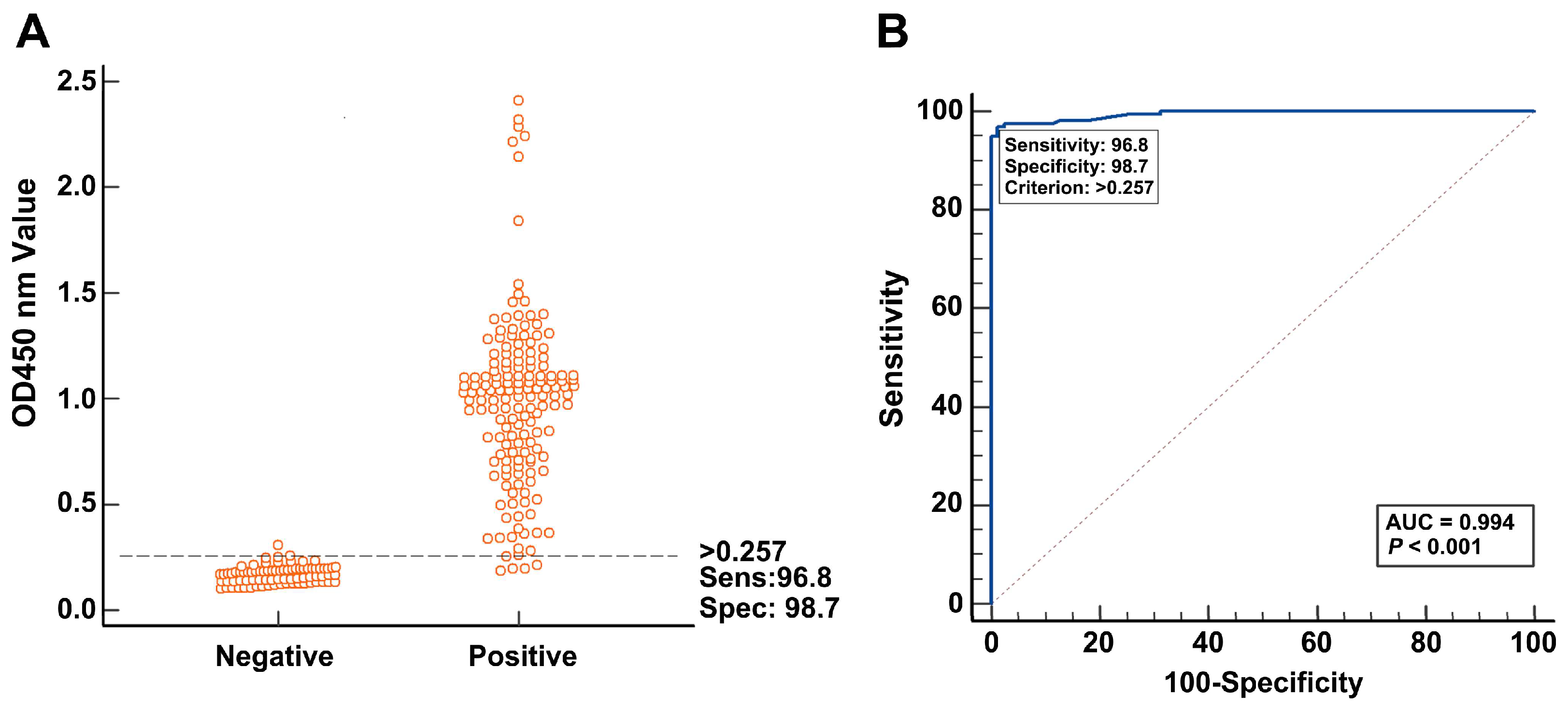
Using the optimized protocol, we analyzed 80 negative and 157 IHNV-positive tissue samples to determine the diagnostic cut-off value, sensitivity, and specificity. The OD450 values of different samples were analyzed using MedCalc software and displayed with an interactive dot plot diagram (Figure 4A). When the cut-off value of DAS-ELISA was set at 0.257, the diagnostic sensitivity and specificity were 96.8% (with a 95% confidence interval between 0.925 and 0.988) and 98.7% (with a 95% confidence interval between 0.923 and 0.999), respectively (Figure 4A). The area under the ROC curve (AUC) was 0.994 with a 95% confidence interval between 0.974 and 1.000, indicating excellent diagnostic accuracy (Figure 4B). Therefore, samples with an OD450 value ≥ 0.257 were regarded as positive, while those with an OD450 value < 0.257 were regarded as negative.

3.5. Specificity and Broad-Spectrum Detection Capability of DAS-ELISA

The specificity of DAS-ELISA was confirmed by testing IHNV, IPNV, SVCV, and VHSV positive samples. The results showed that only the IHNV-positive samples yielded OD450 values above the cut-off value, while all other virus (IPNV, SVCV, and VHSV) positive samples and negative controls yielded OD450 values below the cut-off value (Figure 5A). These results indicated that the DAS-ELISA specifically recognized IHNV and exhibited no cross-reactivity with other tested fish viruses.
To evaluate the broad-spectrum detection capability, we tested eight J genogroup strains (Sn1203, XJ-13, BJ15, LN12, YN-13, QH16, GS15, and HLJ13) and one U genogroup strain (Blk94). The results showed that all IHNV strains yielded OD450 values above the cut-off value (Figure 5B). These findings indicated that the DAS-ELISA was capable of broad-spectrum detection of IHNV strains.

3.6. Sensitivity and Repeatability of the DAS-ELISA

The sensitivity of the DAS-ELISA was determined by testing ten-fold serial dilutions of an IHNV-positive sample with a virus titer of 107 TCID50/mL. The results indicated that the detection limit of the DAS-ELISA was 103 TCID50/mL (Figure 5C).
The repeatability of the DAS-ELISA was assessed using five positive and three negative samples. The intra-batch CV values ranged from 0.906% to 6.028%, and the inter-batch CV values ranged from 0.787% to 3.653% (Table 2). All the CV values were consistently below 7%, indicating the superior repeatability performance of this newly established DAS-ELISA.

3.7. Validation of the DAS-ELISA

The clinical performance of the DAS-ELISA was evaluated using 293 field tissue samples and compared against the WOAH-recommended method (virus isolation coupled with RT-PCR). The results showed that the positive consistency rate between the DAS-ELISA and WOAH-recommended method was 94.04% (142/151), the negative consistency rate was 91.55% (130/142), and the overall consistency rate was 92.83% (272/293). The kappa value of 0.856 indicated a high level of concordance between these two methods (Table 3). These findings suggest that the DAS-ELISA possesses significant potential for clinical diagnostic applications.

4. Discussion

Over the past two decades, IHNV has caused persistent outbreaks in Chinese salmon and trout farms, leading to substantial economic losses [21]. The dominance of the J genogroup, coupled with the confirmed presence of the U genogroup, underscores a dynamic viral ecology that demands robust diagnostic tools [9]. In the absence of commercially available vaccines in China [7], strategies for disease control are fundamentally reliant on early, accurate detection to facilitate timely intervention and prevent outbreaks.
At present, there are some methods that are used for IHNV detection, but some issues hamper their clinical application. The current gold standard for IHNV detection, virus isolation coupled with RT-PCR, is constrained by its time-consuming, costly, and technically demanding nature [20,22]. Moreover, the sensitivity of RT-PCR can be compromised by sequence variations in primer-binding regions [15,23,24]. Although serological assays are valuable for detecting immune responses, their utility in early diagnosis is limited by the delay in antibody production. Antigen-detection methods such as lateral flow devices (LFDs) and ELISA offer attractive alternatives [25]. While LFDs are advantageous for field use, ELISA typically provides superior sensitivity and is more suitable for high-throughput screening, striking an optimal balance between accuracy, cost, and operational feasibility.
For the development of an effective antigen-detection ELISA, the choice of target antigen is the first key factor. During the development of IHNV detection methods, the glycoprotein (G protein) has been a primary target in previous studies due to its role as a surface antigen [26]. However, the accuracy of G protein-based detection methods may be affected by the frequent mutations in the G gene [27]. In contrast, the nucleocapsid (N) protein, essential for viral replication, is highly conserved and abundantly expressed during early infection, making it an ideal candidate for developing a sensitive, reliable, and broad-spectrum diagnostic assay [22]. Therefore, the N protein was selected as the target diagnostic antigen. We employed a rational design strategy for epitope selection, integrating analyses of sequence conservation, antigenicity, hydrophilicity, and structural surface accessibility across all five genogroups IHNV. This comprehensive bioinformatic approach enabled the identification of two optimal peptide regions (6–28 aa and 106–121 aa) for antibody development, aiming to maximize the potential for broad reactivity from the outset.
In addition to the target antigen, the selection of specific antibodies is a fundamental factor determining the success of an antigen-detection ELISA. Monoclonal antibodies (mAbs) are ideal candidates and offer significant advantages over polyclonal antisera, including superior specificity, accuracy, and efficiency in ELISA [28]. Furthermore, they provide an unlimited supply of reagents with consistent quality. To this end, we generated two mAbs using a strategy involving immunization with KLH-conjugated peptides and subsequent screening with BSA-conjugated versions. In theory, using different carrier proteins for immunization (KLH) and screening (BSA) may introduce a risk of selecting clones against common carrier epitopes or experiencing epitope masking. Considering that the BSA carrier provides a completely different protein background from the immunization carrier (KLH), this method positively selects only for hybridomas that secrete antibodies binding specifically to the peptide antigen. This method also ensures that any antibodies that recognize epitopes common to both KLH and BSA (a rare occurrence given their structural differences) are not detected during the screening process. To avoid the concern regarding epitope masking, the peptides were conjugated to both KLH and BSA using the same cross-linking chemistry (e.g., via cysteine residue). This ensures that the orientation and chemical environment of the peptide epitope are comparable between the immunogen and the screening antigen. Our peptides were designed based on predicted linear B-cell epitopes. For such short, linear sequences, the presentation on a carrier protein is less likely to alter the core antigenic structure compared to a conformational epitope. Finally, the resulting antibodies (N-15 and N-106) were assessed by Western blot and IFA, and both antibodies exhibited no cross-reactivity with other common fish viruses.
Beyond the selection of a target antigen and specific antibodies, the establishment of a reliable antigen-detection ELISA requires the meticulous optimization of several operational steps [29,30]. We systematically optimized key parameters, including antibody pairing, coating, blocking, antigen incubation, detection incubation, and the TMB reaction. This process resulted in a robust DAS-ELISA method that showed high specificity for IHNV, no cross-reactivity with other fish viruses (IPNV, VHSV, and SVCV), a sensitivity of 103 TCID50/mL, and excellent reproducibility (CV below 7%). Most importantly, validation with 293 clinical samples showed strong agreement (92.83%; κ = 0.856) with the WOAH-recommended method, confirming its potential for clinical diagnosis. It is important to note that while the assay exhibited a clear dose–response under controlled conditions, a perfect linear correlation between OD values and viral titers was not observed in complex clinical samples. Therefore, we position the DAS-ELISA primarily as a highly reliable qualitative screening tool rather than a precise quantitative assay. Its key value lies in enabling rapid, cost-effective, and high-throughput detection of IHNV, which is essential for large-scale surveillance and early outbreak response.

5. Conclusions

In conclusion, we have developed and validated a novel DAS-ELISA for IHNV detection based on mAbs against conserved epitopes of the N protein. This assay showed excellent sensitivity, specificity, broad-spectrum applicability, repeatability, and cost-effective solution for the large-scale screening of IHNV, particularly in settings where RT-PCR is not feasible. Furthermore, this method provides a solid foundation for the further development of commercial diagnostic kits in China.

Author Contributions

Conceptualization, J.-Z.Z. and T.-Y.L.; methodology, Y.-Z.S.; software, M.W.; validation, M.W., W.-T.L. and J.-Z.Z.; formal analysis, M.W.; investigation, M.W. and W.-T.L.; writing—original draft preparation, J.-Z.Z.; writing—review and editing, L.-M.X.; funding acquisition, J.-Z.Z. and T.-Y.L. All authors have read and agreed to the published version of the manuscript.

Funding

This study was supported by the Central Public-interest Scientific Institution Basal Research Fund, Chinese Academy of Fishery Sciences [HSY202204M and 2023TD45], the National Natural Science Foundation of China [32202988], the Key Research and Development Program of Heilongjiang Province [JD24A013].

Institutional Review Board Statement

All animal procedures in this study were conducted according to the guidelines for the care and use of laboratory animals of Heilongjiang River Fisheries Research Institute, CAFS. The studies in animals were reviewed and approved by the Committee for the Welfare and Ethics of Laboratory Animals of Heilongjiang River Fisheries Research Institute, CAFS (Approval code: 20220616-001 and approval date: 16 June 2022).

Data Availability Statement

All datasets generated for this study are included in the article and further inquiries can be directed to the corresponding author.

Acknowledgments

The authors express their gratitude to the reviewers for their critical feedback and to the colleagues at the Heilongjiang River Fisheries Research Institute of the Chinese Academy of Fishery Sciences (CAFS) for their valuable assistance during this research.

Conflicts of Interest

The authors declare no conflicts of interest.

References

  1. Ballesteros, N.A.; Alonso, M.; Saint-Jean, S.R.; Perez-Prieto, S.I. An oral DNA vaccine against infectious haematopoietic necrosis virus (IHNV) encapsulated in alginate microspheres Induces dose-dependent immune responses and significant protection in rainbow trout (Oncorhynchus Mykiss). Fish Shellfish Immunol. 2015, 45, 877–888. [Google Scholar] [CrossRef]
  2. Hoffmann, B.; Beer, M.; Schütze, H.; Mettenleiter, T.C. Fish rhabdoviruses: Molecular epidemiology and evolution. Curr. Top. Microbiol. Immunol. 2005, 292, 81–117. [Google Scholar] [CrossRef]
  3. Jia, P.; Zheng, X.-C.; Shi, X.-J.; Kan, S.-F.; Wang, J.-J.; He, J.-Q.; Zheng, W.; Yu, L.; Lan, W.-S.; Hua, Q.-Y.; et al. Determination of the complete genome sequence of infectious hematopoietic necrosis virus (IHNV) Ch20101008 and viral molecular evolution in China. Infect. Genet. Evol. 2014, 27, 418–431. [Google Scholar] [CrossRef] [PubMed]
  4. Liu, L.; Shan, L.-P.; Xue, M.-Y.; Lu, J.-F.; Hu, Y.; Liu, G.-L.; Chen, J. Potential application of antiviral coumarin in aquaculture against IHNV infection by reducing viral adhesion to the epithelial cell surface. Antivir. Res. 2021, 195, 105192. [Google Scholar] [CrossRef] [PubMed]
  5. LaPatra, S.E.; Corbeil, S.; Jones, G.R.; Shewmaker, W.D.; Lorenzen, N.; Anderson, E.D.; Kurath, G. Protection of rainbow trout against infectious hematopoietic necrosis virus four days after specific or semi-specific DNA vaccination. Vaccine 2001, 19, 4011–4019. [Google Scholar] [CrossRef]
  6. Xu, L.; Zhao, J.; Liu, M.; Kurath, G.; Breyta, R.B.; Ren, G.; Yin, J.; Liu, H.; Lu, T. Phylogeography and evolution of infectious hematopoietic necrosis virus in China. Mol. Phylogenet. Evol. 2019, 131, 19–28. [Google Scholar] [CrossRef]
  7. Li, J.; Xia, D.; Zhang, M.; Zhang, Y.; Liu, X.; Sun, J.; Xu, B.; Yang, J.; Wang, N.; Shi, W.; et al. Infectious hematopoietic necrosis virus (IHNV) nucleoprotein amino acid residues affect viral virulence and immunogenicity in rainbow trout (Oncorhynchus mykiss). Fish Shellfish Immunol. 2022, 130, 572–581. [Google Scholar] [CrossRef]
  8. Abbadi, M.; Fusaro, A.; Ceolin, C.; Casarotto, C.; Quartesan, R.; Pozza, M.D.; Cattoli, G.; Toffan, A.; Holmes, E.C.; Panzarin, V. Molecular evolution and phylogeography of co-circulating IHNV and VHSV in Italy. Front. Microbiol. 2016, 7, 1306. [Google Scholar] [CrossRef]
  9. Huo, C.; Ma, Z.; Li, F.; Xu, F.; Li, T.; Zhang, Y.; Jiang, N.; Xing, W.; Xu, G.; Luo, L.; et al. First isolation and pathogenicity analysis of a genogroup U strain of infectious hematopoietic necrosis virus from rainbow trout in China. Transbound. Emerg. Dis. 2022, 69, 337–348. [Google Scholar] [CrossRef] [PubMed]
  10. Jia, P.; Purcell, M.K.; Pan, G.; Wang, J.; Kan, S.; Liu, Y.; Zheng, X.; Shi, X.; He, J.; Yu, L.; et al. Analytical validation of a reverse transcriptase droplet digital PCR (RT-ddPCR) for quantitative detection of infectious hematopoietic necrosis virus. J. Virol. Methods 2017, 245, 73–80. [Google Scholar] [CrossRef] [PubMed]
  11. Lapatra, S.E.; Roberti, K.A.; Rohovec, J.S.; Fryer, J.L. Fluorescent antibody test for the rapid diagnosis of infectious hematopoietic necrosis. J. Aquat. Anim. Health 1989, 1, 29–36. [Google Scholar] [CrossRef]
  12. Choi, H.D.; Baek, E.J.; Hong, S.; Kim, Y.C.; Jeong, J.M.; Kwon, M.G.; Il Kim, K. Development and validation of reverse-transcription cross-priming amplification-based lateral flow assay for the detection of infectious hematopoietic necrosis virus. J. Virol. Methods 2024, 329, 115008. [Google Scholar] [CrossRef] [PubMed]
  13. Rong, F.; Wang, H.; Tang, X.; Xing, J.; Sheng, X.; Chi, H.; Zhan, W. The development of RT-RPA and CRISPR-Cas12a based assay for sensitive detection of infectious hematopoietic necrosis virus (IHNV). J. Virol. Methods 2024, 326, 114892. [Google Scholar] [CrossRef] [PubMed]
  14. Dixon, P.; Paley, R.; Alegria-Moran, R.; Oidtmann, B. Epidemiological characteristics of infectious hematopoietic necrosis virus (IHNV): A Review. Vet. Res. 2016, 47, 63. [Google Scholar] [CrossRef] [PubMed]
  15. Bustin, S.A.; Benes, V.; Garson, J.; Hellemans, J.; Huggett, J.; Kubista, M.; Mueller, R.; Nolan, T.; Pfaffl, M.W.; Shipley, G.; et al. The need for transparency and good practices in the qPCR literature. Nat. Methods 2013, 10, 1063–1067. [Google Scholar] [CrossRef]
  16. Meroni, P.L.; Bizzaro, N.; Cavazzana, I.; Borghi, M.O.; Tincani, A. Automated tests of ANA immunofluorescence as throughput autoantibody detection technology: Strengths and limitations. BMC Med. 2014, 12, 38. [Google Scholar] [CrossRef]
  17. Hayrapetyan, H.; Tran, T.; Tellez-Corrales, E.; Madiraju, C. Enzyme-linked immunosorbent assay: Types and applications. Methods Mol. Biol. 2023, 2612, 1–17. [Google Scholar] [CrossRef]
  18. Zhao, J.-Z.; Liu, M.; Xu, L.-M.; Zhang, Z.-Y.; Cao, Y.-S.; Shao, Y.-Z.; Yin, J.-S.; Liu, H.-B.; Lu, T.-Y. A chimeric recombinant infectious hematopoietic necrosis virus induces protective immune responses against infectious hematopoietic necrosis and infectious pancreatic necrosis in rainbow trout. Mol. Immunol. 2019, 116, 180–190. [Google Scholar] [CrossRef] [PubMed]
  19. Zhao, J.-Z.; Li, L.-F.; Xu, L.-M.; Shao, Y.-Z.; Ren, G.-M.; Liu, Q.; Lu, T.-Y. Traditional Chinese medicine bufalin inhibits infectious hematopoietic necrosis virus genogroups I and V infection in vitro and in vivo. Aquaculture 2023, 576, 739789. [Google Scholar] [CrossRef]
  20. World Organisation for Animal Health OIE. Chapter 2.3.4.—Infectious Haematopoietic Necrosis. In Manual of Diagnostic Tests for Aquatic Animals; OIE: Paris, France, 2015. [Google Scholar]
  21. He, M.; Ding, N.Z.; He, C.Q. Novirhabdoviruses versus fish innate immunity: A review. Virus Res. 2021, 304, 198525. [Google Scholar] [CrossRef]
  22. Purcell, M.; Thompson, R.; Garver, K.; Hawley, L.; Batts, W.; Sprague, L.; Sampson, C.; Winton, J. Universal reverse-transcriptase real-time PCR for infectious hematopoietic necrosis virus (IHNV). Dis. Aquat. Org. 2013, 106, 103–115. [Google Scholar] [CrossRef] [PubMed]
  23. Dhar, A.K.; Bowers, R.M.; Licon, K.S.; LaPatra, S.E. Detection and quantification of infectious hematopoietic necrosis virus in rainbow trout (Oncorhynchus Mykiss) by SYBR green real-time reverse transcriptase-polymerase chain reaction. J. Virol. Methods 2008, 147, 157–166. [Google Scholar] [CrossRef] [PubMed]
  24. Purcell, M.K.; Hart, S.A.; Kurath, G.; Winton, J.R. Strand-specific, real-time RT-PCR assays for quantification of genomic and positive-sense RNAs of the fish rhabdovirus, infectious hematopoietic necrosis virus. J. Virol. Methods 2006, 132, 18–24. [Google Scholar] [CrossRef] [PubMed]
  25. Yong, C.Y.; Yeap, S.K.; Omar, A.R.; Tan, W.S. Advances in the study of nodavirus. PeerJ 2017, 5, e3841. [Google Scholar] [CrossRef]
  26. Xu, L.; Zhao, J.; Liu, M.; Kurath, G.; Ren, G.; Lapatra, S.E.; Yin, J.; Liu, H.; Feng, J.; Lu, T. A effective DNA vaccine against diverse genotype J infectious hematopoietic necrosis virus strains prevalent in China. Vaccine 2017, 35, 2420–2426. [Google Scholar] [CrossRef]
  27. Einer-Jensen, K.; Harmache, A.; Biacchesi, S.; Bremont, M.; Stegmann, A.; Lorenzen, N. High virulence differences among phylogenetically distinct isolates of the fish rhabdovirus viral hemorrhagic septicaemia virus are not explained by variability of the surface glycoprotein G or the non-virion protein Nv. J. Gen. Virol. 2014, 95, 307–316. [Google Scholar] [CrossRef]
  28. Gao, Y.; Huang, X.; Zhu, Y.; Lv, Z. A brief review of monoclonal antibody technology and its representative applications in immunoassays. J. Immunoass. Immunochem. 2018, 39, 351–364. [Google Scholar] [CrossRef]
  29. Han, F.; Shan, F.; Hou, J.; Guo, D.; Xiang, Y.; Yuan, J.; Wei, Z. Establishment and application of PDCoV antigen-specific DAS-ELISA detection method. BMC Vet. Res. 2024, 20, 342. [Google Scholar] [CrossRef] [PubMed]
  30. Wang, J.; Yin, J.; Wang, S.-H.; Ding, C.-Z.; Wang, J.-F. Development and application of an indirect ELISA for the serological detection of bovine viral diarrhea virus infection based on the protein E2 antigen. Mol. Biol. Rep. 2023, 50, 4707–4713. [Google Scholar] [CrossRef]
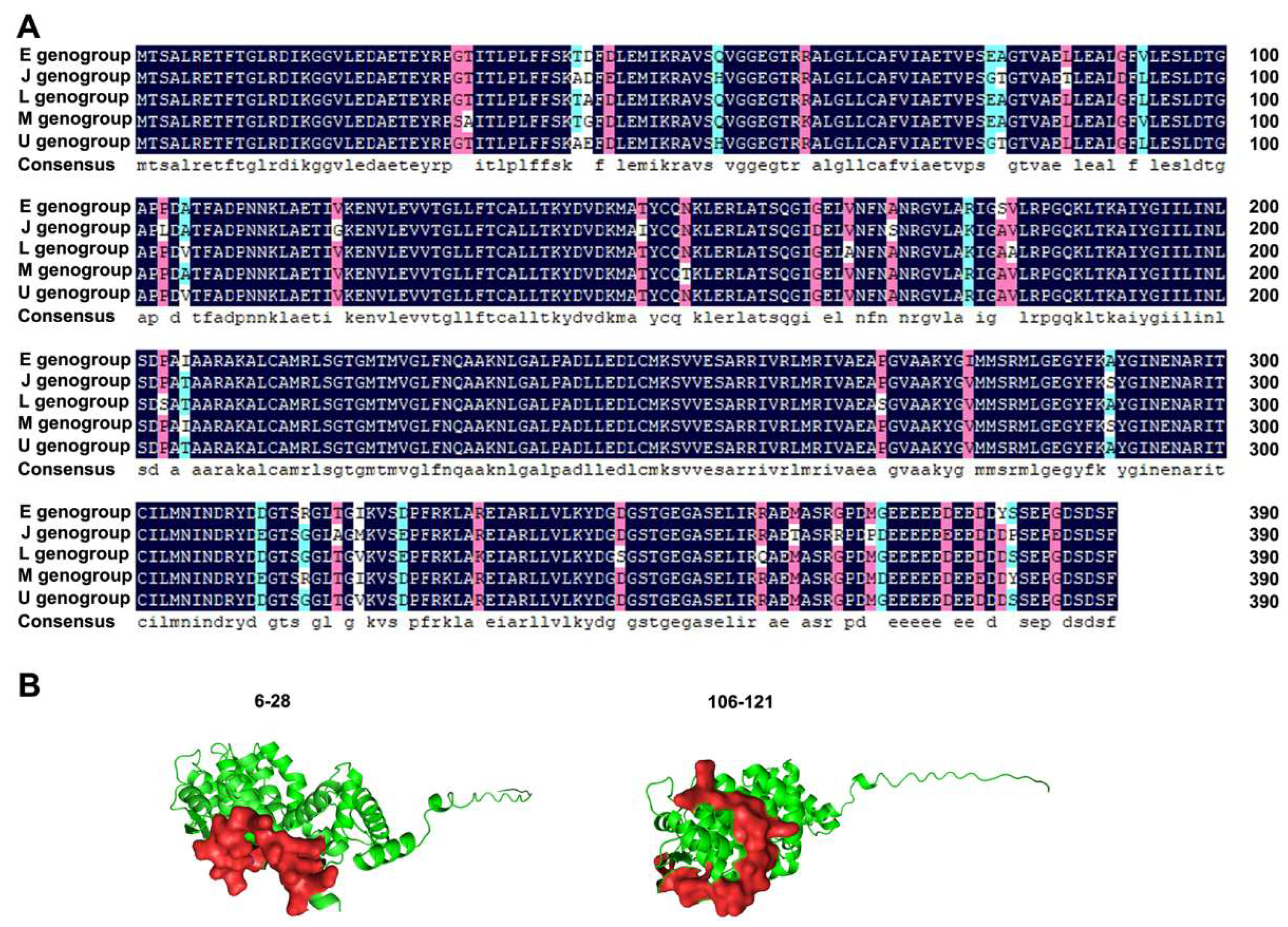
Figure 1. Conservation and surface accessibility analysis of IHNV N protein. (A) Conservation analysis of N protein across the five IHNV genogroups. Black: indicates full identity at that position across all sequences. Blue or red: indicates partial identity at that position. White: indicates a mismatch with other sequence. (B) Surface accessibility analysis of the selected epitopes on the N protein. The target peptides were highlighted in red and the rest amino acids were in green.
Fishes 10 00533 g001
Figure 2. Characterization of mAbs against N protein. (A) SDS-PAGE analysis of purified N-15 and N-106 antibodies. (B) Western blotting analysis of N-15 and N-106 specificity. (C) IFA analysis of N-15 and N-106 specificity. (D) Antibody isotype identification of N-15 and N-106.
Fishes 10 00533 g002
Figure 3. Optimization of the DAS-ELISA. (A) Determining the optimal capture and detection antibodies. (B) Titrating the capture antibody concentration. (C) Titrating the detection antibody concentration. (D) Selecting the optimal blocking solution. (E) Optimizing the blocking duration. (F) Determining the optimal antigen incubation time. (G) Determining the optimal detection antibody incubation time. (H) Determining the optimal TMB reaction time. Significant differences are indicated as * p < 0.05, ** p < 0.01, and *** p < 0.001.
Fishes 10 00533 g003
Figure 4. Determination of the DAS-ELISA cut-off value. (A) Distribution of OD450 values for negative and positive samples. The dashed line indicates the determined cut-off value. (B) Receiver operating characteristic (ROC) curve analysis evaluating the diagnostic performance of DAS-ELISA. The solid curve represents the ROC of the proposed model. The dashed diagonal line indicates the performance of a random classifier, for reference.
Fishes 10 00533 g004
Figure 5. Assessing the performance of the DAS-ELISA. (A) Analyzing assay specificity. (B) Evaluating broad-spectrum detection capability. (C) Determining assay sensitivity.
Fishes 10 00533 g005
Table 1. Antigenicity analysis of N protein.
NumberAmino Acid SiteSequenceLength
16–28RETFTGLRDIKGGVLEDAETEYR23
241–45ADFEL5
354–60HVGGEGT7
475–82TVPSGTGT8
595–121ESLDTGAPLDATFADPNNKLAETIGKE27
6143–144DK5
7151–173NKLERLATSQGIDELVNFNSNRG23
8186–187QK2
9203–207PATAA5
10234–238NLGAL5
11285–295EGYFKSYGINE11
12308–325DRYDEGTSGGLAGMKVSE18
13343–351DGDGSTGEG9
14360–388ETASRRPDPDEEEEEEEEDDDPSEPEDSD29
Table 2. Repeatability analysis of the DAS-ELISA.
SamplesIntra-BatchInter-Batch
X ± SDCV (%)X ± SDCV (%)
Positive 12.191 ± 0.0542.4452.133 ± 0.0170.787
Positive 22.178 ± 0.0401.8192.115 ± 0.0432.010
Positive 31.520 ± 0.0523.4021.487 ± 0.0312.052
Positive 41.045 ± 0.0090.9061.043 ± 0.0222.131
Positive 52.101 ± 0.0512.4362.137 ± 0.0482.261
Negative 10.102 ± 0.0066.0280.090 ± 0.0033.653
Negative 20.110 ± 0.0054.1330.109 ± 0.0222.247
Negative 30.112 ± 0.0043.3410.109 ± 0.0191.891
Table 3. Comparison of DAS-ELISA and the WOAH-recommended method.
DAS-ELISAWOAH-Recommended MethodConcordanceKappa Value
PositiveNegativeTotal
Positive142915194.04%
Negative1213014291.55%
Total15413929392.83%0.856
Disclaimer/Publisher’s Note: The statements, opinions and data contained in all publications are solely those of the individual author(s) and contributor(s) and not of MDPI and/or the editor(s). MDPI and/or the editor(s) disclaim responsibility for any injury to people or property resulting from any ideas, methods, instructions or products referred to in the content.