Article

Dietary Inclusion of Micro-Algal Astaxanthin on Gut Health of Rainbow Trout Oncorhynchus mykiss: Insights from Gut Morphology, Physiological Indices and Microbiota Diversity

by
Min Zhang
1,
Xiaowen Long
2,
Yaopeng Li
3,
Yong Zhang
4,
Weihong Sun
5,* and
Xugan Wu
1,*
1
Centre for Research on Environmental Ecology and Fish Nutrition of the Ministry of Agriculture and Rural Affairs of China, Shanghai Ocean University, Shanghai 201306, China
2
College of Agriculture and Biological Sciences, Dali University, Dali 671003, China
3
R&D Center of Qinghai Minze Longyangxia Ecological Aquaculture Co., Ltd., Hainan Tibetan Autonomous Prefecture 811899, China
4
Yunnan Alphy Biotech Co., Ltd., Chuxiong 675000, China
5
Yellow Sea Fisheries Research Institute, Chinese Academy of Fishery Sciences, Qingdao 266000, China
*
Authors to whom correspondence should be addressed.
Fishes 2025, 10(10), 505; https://doi.org/10.3390/fishes10100505
Submission received: 1 September 2025 / Revised: 1 October 2025 / Accepted: 3 October 2025 / Published: 8 October 2025
(This article belongs to the Section Nutrition and Feeding)

Abstract

The green alga Haematococcus pluvialis, rich in natural astaxanthin, is a key feed additive for salmonid pigmentation. This study evaluated dietary micro-algal astaxanthin effects on structure, antioxidative and immune response, as well as microbiota in different gut segments of rainbow trout Oncorhynchus mykiss (initial average weight: 0.67 ± 0.02 kg). Three diets contained 0 (Diet 1, control), 18.57 (Diet 2) and 31.25 mg/kg (Diet 3) micro-algal astaxanthin. After a 4-month feeding trial, dietary astaxanthin promoted the goblet cell proliferation of pyloric caeca and increased hindgut tunica muscularis thickness (p < 0.05). It also improved antioxidant capacity, characterized by the upregulation of gpx and cat expression in the midgut, accompanied by a significant decrease in MDA content (p < 0.05). Furthermore, dietary astaxanthin could upregulate tgf-β, tor1 and pcna levels in midgut and igm in hindgut, while il1β, il6, il8 and tnfα in hindgut were significantly downregulated in Diet 2 (p < 0.05). Additionally, dietary astaxanthin also enhanced the α-diversity of hindgut and altered the core microbiota (reduced Proteobacteria, increased Actinobacteria). Diet 2 increased microbic abundance associated with reducing gut inflammation and promoting nutrient absorption while decreasing that of pathogenic bacteria. Overall, dietary 18.57 mg/kg astaxanthin supplementation could promote gut structure, antioxidant and immune capacity, reduce inflammation and modulate microbiota. These findings indicate that natural astaxanthin from H. pluvialis has potential as an immunostimulant to promote gut health in salmonids.
Key Contribution: This work highlights the segment-specific effects of dietary micro-algal astaxanthin on the gut health of rainbow trout, as well as the potential of an optimal astaxanthin concentration (18.57 mg/kg dry diet) to improve gut structural integrity, enhance antioxidant and immune capacities, mitigate inflammation and modulate gut microbiota—providing insights for astaxanthin application as a functional feed additive in salmonid aquaculture.

1. Introduction

As intensive aquaculture has developed, the production of aquatic economic animals has been significantly improved. This intensification, however, simultaneously exerts negative impacts on fish health, as limited rearing space increases competition and accelerates water quality deterioration [1,2]. Environmental changes, such as dissolved oxygen, temperature, pH and crowded conditions, can cause stress responses in fish, damage their health and eventually cause death [3]. As a result, farmed aquatic animals tend to be vulnerable. Modern aquaculture fisheries mainly rely on the conversion and utilization of feed substances to meet basic nutritional needs. In addition, the supplementation of beneficial substances (such as carotenoids, vitamins, etc.) at various recommended levels in feeds has become a hotly debated topic [4,5,6].
Carotenoid, a kind of essential nutrient for aquatic animals, is used as a feed additive to improve the wellness of aquatic animals [7,8,9,10]. Astaxanthin, a natural ketone carotenoid, is regarded as an ecologically friendly functional feed additive in aquaculture [11]. Haematococcus pluvialis, a green alga and high-quality natural astaxanthin source, serves as an astaxanthin supplement for farmed fish—either via its dried powder in feeds or astaxanthin extracted from its cells. It has been applied to the reddish or pinkish color of fish [12,13,14,15]. In recent years, numerous biological functions have reported on its antioxidant and immune activities, since it possesses the highest oxygen radical absorbance capacity [12,14,16]. Previous studies have shown that diets with optimized astaxanthin sources and concentrations could significantly enhance antioxidant and immune capabilities [17,18,19], even improving resistance against various stresses [20,21,22]. Of note, the relevant research is focused on the serum [23,24,25] and liver [26,27] of fish; less information has been available about its effects on fish gut. The homeostasis of the gut microbial community is another important component of gut health, and dietary components play important roles in shaping gut microbial community [28]. Previous findings highlight the potential of astaxanthin to regulate host–microbe symbiosis in the gut, and its inclusion into the diet may help improve disease conditions associated with gut health [29]. To our knowledge, there are some reports on the effects of astaxanthin on the gut microbiota of fish [25,30,31]; however, there has been no study investigating the effects of different dietary astaxanthin levels on the gut content microbiota across different gut segments of fish.
Rainbow trout Oncorhynchus mykiss, a globally critical cold-water species, is a major component of salmonid aquaculture—with annual global production exceeding 400,000 tons [32] and 47,822 tons trout in China [33]. As an important cold-water economic fish, it depends on dietary supply for carotenoids, and it is common practice in farming to supplement diets with astaxanthin. H. pluvialis is increasingly integrated into trout feed as a high-quality natural astaxanthin source. However, to our knowledge, it has been rarely reported that astaxanthin, as an immunopotentiator, has an impact on gut immunity, inflammatory responses and microbiota of fish. Therefore, the present study aimed to investigate for the first time the influence of different levels of dietary astaxanthin on morphology, physiological metabolic response and microbiota in different gut segments of rainbow trout. This study will provide a basis for healthy culture and micro-ecological regulation of rainbow trout.

2. Materials and Methods

2.1. Experimental Diets

The experimental diets were based on the commercial feed of rainbow trout (Aller Aqua (Qingdao) Co., Ltd., Qingdao, China) with the addition of astaxanthin. Haematococcus pluvialis powder (Yunnan Alphy Biotech Co., Ltd., Chuxiong, China) was used as a source of natural astaxanthin for experimental diets. Three experimental diets were formulated to target different astaxanthin levels: Diet 1 (control, 0 mg/kg), Diet 2 (18.57 mg/kg) and Diet 3 (31.25 mg/kg). Each diet was tested in 3 replicate cages, with 150 fish per cage (n = 450 fish per diet). The proximate composition and actual astaxanthin content of these three diets are presented in Table 1 [12]. The experimental diets were separately kept in sealed plastic bags at 8–10 °C to be used in the feeding trial.

2.2. Fish Breeding and Treatment

Apparently healthy rainbow trout (initial average weight: 0.67 ± 0.02 kg) with no external lesions, normal swimming behavior and consistent body size were obtained from Qinghai Minze Longyangxia Ecological Hydroponics Co., Ltd., Hainan, China. The experiment was conducted in the Longyangxia Reservoir, Hainan, China. Fish were randomly distributed in 9 net cages (4 m × 4 m × 5 m) at a density of 150 fish in each cage. The water quality parameters were monitored daily using an automatic monitoring system (LDO HQ1130, Hach, Loveland, CO, USA), with water temperature ranging from 12 to 19 °C and dissolved oxygen ranging from 6 to 9 mg/L. Additionally, water transparency was measured daily using a Secchi disk (SD30, Shanghai Precision Instrument & Meter Co., Ltd., Shanghai, China), with values ranging from 1.0 to 2.5 m. In this study, there were a total of 3 diet treatments, and each treatment was set up with 3 replicates. Fish were fed the diets (Table 1) at a feeding rate of 3–5% of body weight twice a day (8:00 and 15:00), which was appropriately adjusted according to temperature and fish growth to keep a similar feeding level for each cage.

2.3. Sample Collection

After four months rearing, the fish fasted for 24 h. For growth index calculation, 9 fish were randomly selected and weighed from each cage (9 fish/diet treatment) (Table 2). Subsequently, these fish were anesthetized using tricaine methane sulfonate (MS-222, 300 mg/L) to collect gut tissues. The whole gut was quickly collected from the fish; the gut contents were carefully collected following the aseptic operation and preserved in a 2 mL sterile centrifuge tube filled with ethanol for 24 h, and then they were stored at −80 °C until DNA extraction. Also, gut samples were taken, emptied, washed in ice-cold 0.85% saline solution two times and patted dry on the filter paper. A small portion (approximately 50 mg) was transferred to 2 mL RNase-free tubes, immediately put into liquid nitrogen and then stored at −80 °C for gene expression profiling; another portion (approximately 500 mg) was transferred to 5 mL sterile tubes and then stored at −40 °C for biochemical parameters analysis. Finally, small pieces of gut (pyloric caeca, midgut and hindgut) collected from each fish were taken and fixed in 4% paraformaldehyde for histological evaluation. In addition, the delineation of the midgut and hindgut segments was referred to in a previous study [34]. All rainbow trout were treated in strict accordance with the guidelines for the care and use of experimental animals established by the Committee on Experimental Animal Management at Shanghai Ocean University (ethics code: SHOU-DW-2020-152, date: 22 March 2020).

2.4. Gut Histology

After being fixed in 4% paraformaldehyde for 24 h, the guts were routinely dehydrated through an alcohol gradient, equilibrated in xylene and embedded in paraffin. The 7 μm paraffin sections were cut and then H.E. staining was carried out. The slides were examined under a light microscope (Nikon Eclipse 80i, Tokyo, Japan) equipped with a Nikon DS-Ri1 12.7-megapixel camera. Data were processed by ImageJ software (version 1.x).

2.5. Analysis of Antioxidant and Immune Parameters

The frozen gut samples were thawed, weighted and homogenized for enzymatic assays using a micro-homogenizer (HR-6, Shanghai Huxi Industrial Co., Ltd., Shanghai, China), and three replicates of around 0.1 g per example were added to ice-cold physiological saline solution in a proportion of 1:5 (w/v). The homogenate was centrifuged at 13,000× g for 10 min at 4 °C. Then, the aqueous supernatant was collected for further analysis.
The total antioxidant capacity (T-AOC, A015-1), total superoxide dismutase (T-SOD, A001-1), catalase (CAT, A007-1), malondialdehyde (MDA, A003-1), alkaline phosphatase (AKP, A059-1), acid phosphatase (ACP, A060-1) and lysozyme (LZM, A050-1) in the gut were spectrophotometrically (TECAN infinite M200 NanoQuant, Tecan Trading AG, Männedorf, Switzerland) analyzed with commercial assay kits (Nanjing Jiancheng Bioengineering Institute, Nanjing, China) according to the manufacturer’s instructions.

2.6. Gene Expression Analysis

The total RNA was extracted from midguts and hindguts (three samples per cage and three cages per treatment) using RNAiso Plus (TaKaRa Biotechnology (Dalian) Co., Ltd., Dalian, China). The integrity of total RNA samples was tested by electrophoresing on a 1.0% denaturing agarose gel. Meanwhile, the purity and concentration were determined by a spectrophotometer (NanoDrop® ND-2000,Thermo Fisher Scientific, Wilmington, DE, USA). RNA from three fish in one cage was mixed in equal amounts to form one sample, with 3 replicates per treatment group. Subsequently, total RNA was used to synthesize complementary DNA (cDNA) using the Prime Script™ RT reagent kit (Takara, Dalian, China).
Gene expression levels were determined by qRT-PCR using the FAST-7500 system (ABI-7500, ThermoFisher, Singapore). Analyses were performed on 1 μL of cDNA using 2× SYBR Green Master Mix (Takara, Dalian, China) in a total PCR reaction volume of 10 μL, containing 5 μL of TB Green, 0.2 μL of ROX Reference Dye II, 0.2 μL of each primer, and 3.4 μL of nuclease-free water. All samples were run in triplicate. Relative constant expression of target genes was done using β-actin as the reference gene within all groups. All nucleotide sequences of primers used in this study are presented in Table 3. The 2−ΔΔCt method was used for calculation of the relative quantification of qRT-PCR data [35].

2.7. DNA Extraction and Sequencing of Intestinal Microbiota

The E.Z.N.A.® Soil DNA Kit (Omega Bio-tek, Norcross, GA, USA) was used for microbial DNA extraction from the intestinal contents according to the manufacturer’s instructions. The V1-V9 region of the 16S ribosomal RNA (rRNA) gene of gut bacteria was amplified using the 27F/1492R primer set to analyze the diversity and composition of gut bacterial community used by ABI GeneAmp® 9700 (Applied Biosystems, Temecula, CA, USA). The reactions were conducted in a 20 μL sample volume using 4 μL of 5× FastPfu Buffer, 2 μL of 2.5 mM dNTPs, 0.4 μL of FastPfu Polymerase, 5 μM primers, 10 ng template DNA and nuclease-free water. The PCR program consisted of 95 °C for 5 min, 30 cycles of “denaturation at 95 °C for 30 s, annealing at 55 °C for 30 s, extension at 72 °C for 45 s” and a final extension at 72 °C for 10 min. Amplicons were extracted from 2% agarose gels and purified using the AxyPrep DNA Gel Extraction Kit (Axygen Biosciences, Union City, CA, USA) according to the manufacturer’s instructions. SMRTbell libraries were prepared from the amplified DNA by blunt ligation according to the manufacturer’s instructions (Pacific Biosciences of California, Inc., Menlo Park, CA, USA). Purified SMRTbell libraries from the Zymo and HMP mock communities were sequenced on dedicated PacBio Sequel II 8M cells using Sequencing Kit 2.0 chemistry. Purified SMRTbell libraries from the pooled and barcoded samples were sequenced on a single PacBio Sequel II cell. All amplicon sequencing was performed by Shanghai Biozeron Biotechnology Co., Ltd. (Shanghai, China).

2.8. Data Statistical Analysis

Statistical analysis was conducted using SPSS 26.0 software. Data normality was assessed via the Shapiro-Wilk test (p > 0.05 indicating normality) and variance homogeneity via Levene’s test. When necessary, data were arcsine-square root or logarithmically transformed prior to analysis. One-way ANOVA followed by Tukey’s test was used for comparisons. Differences were considered statistically significant at p < 0.05. Each result was expressed as the mean of triplicate cages ± SD. All graphs were produced using GraphPad Prism version 7.03.

3. Results

3.1. Dietary Astaxanthin Improves Intestinal Morphology of Rainbow Trout

There was no statistical difference in villus height and villus width among the three diets in these three gut sections (p > 0.05) (Table 4). Compared to the control, Diet 2 increased the tunica muscularis thickness, villus height and villus width in the pyloric caeca (though not significantly, p > 0.05) and also resulted in slightly higher villus height and villus width in the midguts compared to the other two treatments (p > 0.05). In the hindguts, the tunica muscularis thickness of Diet 2 and 3 treatments was significantly higher than the control (Diet 1) (p < 0.05). Notably, goblet cells (GCs) in the pyloric caeca of astaxanthin-supplemented groups (Diet 2 and 3) showed obvious proliferation, which was confirmed by histological observation (Figure 1).

3.2. Antioxidant Responses of Guts Were Induced by Dietary Astaxanthin

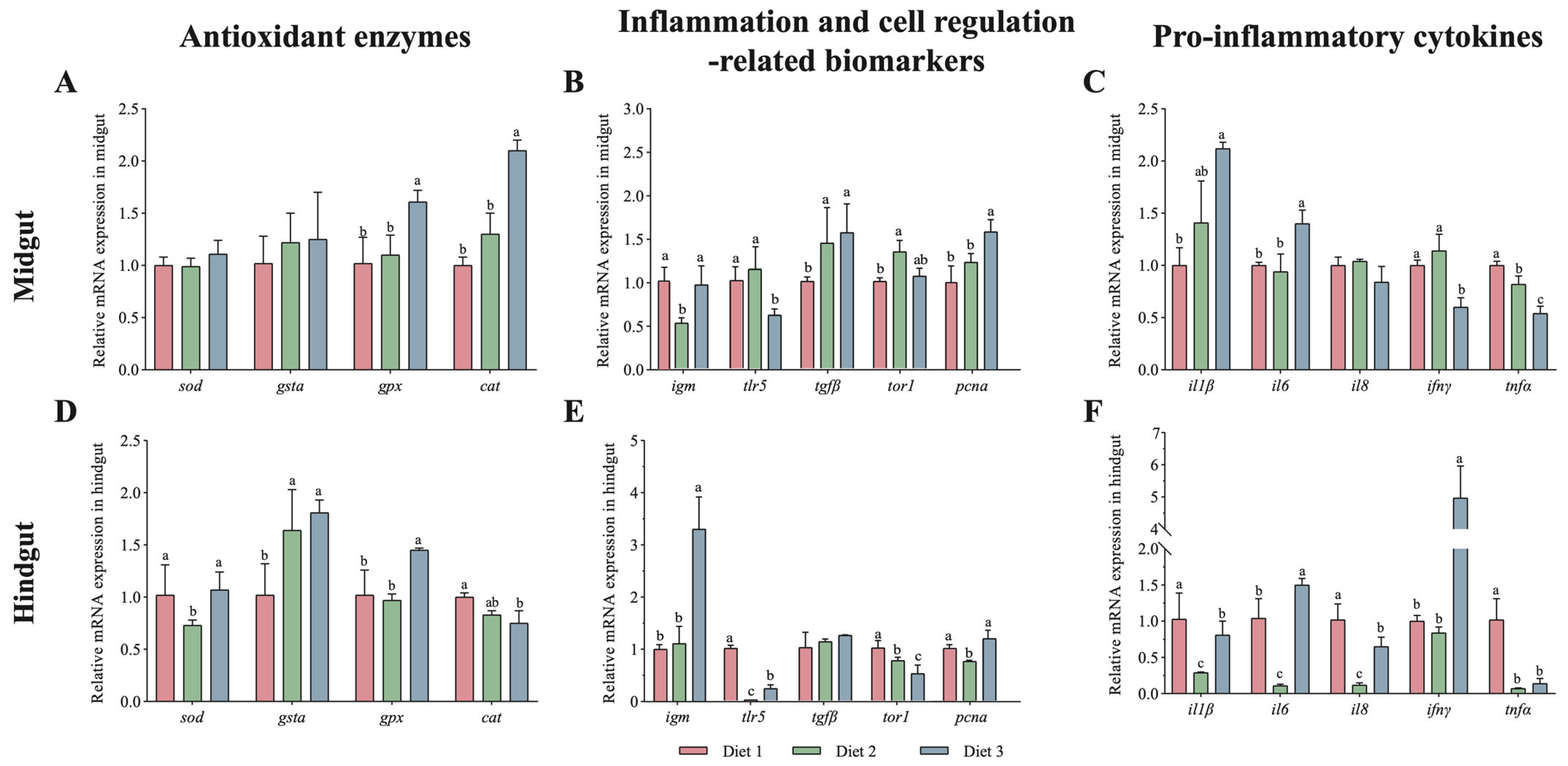
In the midgut, compared with the control (Diet1), Diet 2 and 3 treatments significantly decreased T-AOC activity and MDA content (p < 0.05, Table 5) while significantly increasing T-SOD activity (p < 0.05); nevertheless, no significant differences in these indices were observed in the hindgut (p > 0.05). Additionally, antioxidant enzyme gene expression showed that in the midgut, the expression levels of gpx and cat increased significantly with dietary astaxanthin (p < 0.05, Figure 2A). In the hindgut, dietary astaxanthin also upregulated gsta and gpx but downregulated cat (p < 0.05, Figure 2D).

3.3. Immune Responses of Guts Were Induced by Dietary Astaxanthin

Regarding immune parameters (Table 5), LZM activity in both the midgut and hindgut significantly increased as dietary astaxanthin levels reached 31.25 mg/kg (Diet 3) (p < 0.05). In the midgut, compared with the control (Diet1), significantly lower AKP activity was observed in Diet 2 and 3 treatments (p < 0.05). The gene expression levels of pcna and tgfβ were significantly increased (p < 0.05) with dietary astaxanthin content, while Diet 2 treatment had the highest gene expression levels of tor1 and tlr5 in the midguts compared to the other treatments (p < 0.05, Figure 2B); for the pro-inflammatory cytokines, the gene expression levels of ifn-γ and tnf-α genes were significantly decreased, while the gene expression levels of il1β and il6 were significantly increased in the midgut with dietary astaxanthin content (p < 0.05, Figure 2C). In the hindgut, the dietary natural astaxanthin also improved the gene expression levels of gsta and gpx but significantly decreased cat expression level (p < 0.05, Figure 2D); the gene expression levels of igm were significantly increased, but tor1 and tlr5 decreased significantly in the midguts with dietary astaxanthin content (p < 0.05, Figure 2E). Diet 2 treatment had the lowest gene expression levels of all pro-inflammatory cytokines, and Diet 3 had the highest gene expression levels of ifn-γ and il6 among all treatments (p < 0.05, Figure 2F).

3.4. Dietary Astaxanthin Alters the Gut Microbiota Diversity and Community

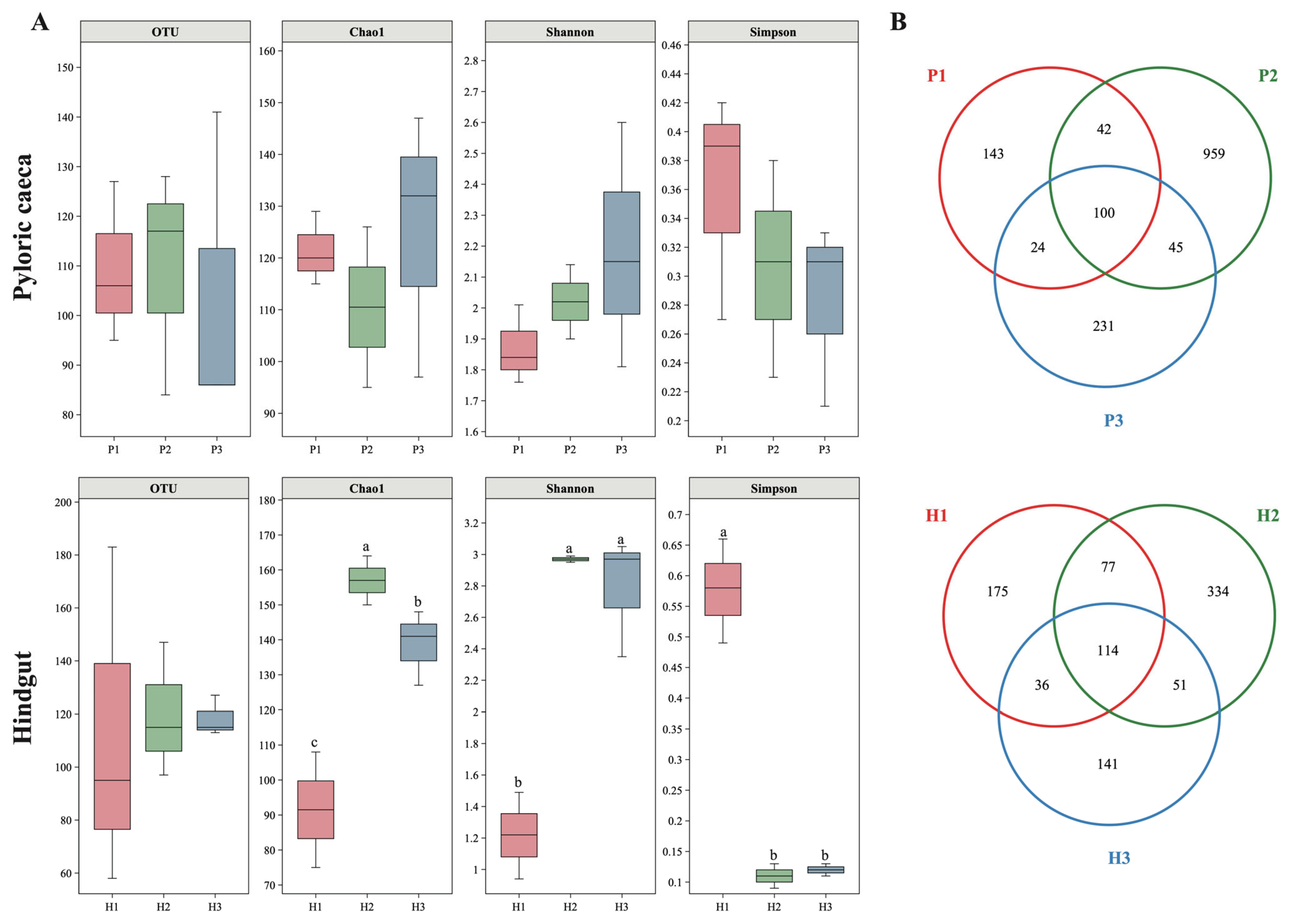
The α-diversity indices (OTUs, Chao, Shannon and Simpson) in the pyloric caeca were unaffected by diet (p > 0.05), but in the hindgut, Chao and Shannon indices increased significantly (p < 0.05) and Simpson index decreased significantly (p < 0.05) in astaxanthin-supplemented groups compared to the control (Figure 3A). The Venn diagram shows that a total of 100 OTUs were commonly identified in pyloric caeca among the three treatments, while Diet 1, 2 and 3 treatments had 143, 959 and 231 uniquely identified OTUs, respectively (Figure 3B). Moreover, 175 (Diet 1), 334 (Diet 2) and 141 OTUs (Diet 3) were unique in the hindgut, accompanied by the overlap of 114 OTUs (Figure 3B). It is noticeable that Diet 2 treatment had significantly higher OTUs in both the pyloric caeca and hindgut than the other two treatments (p < 0.05).
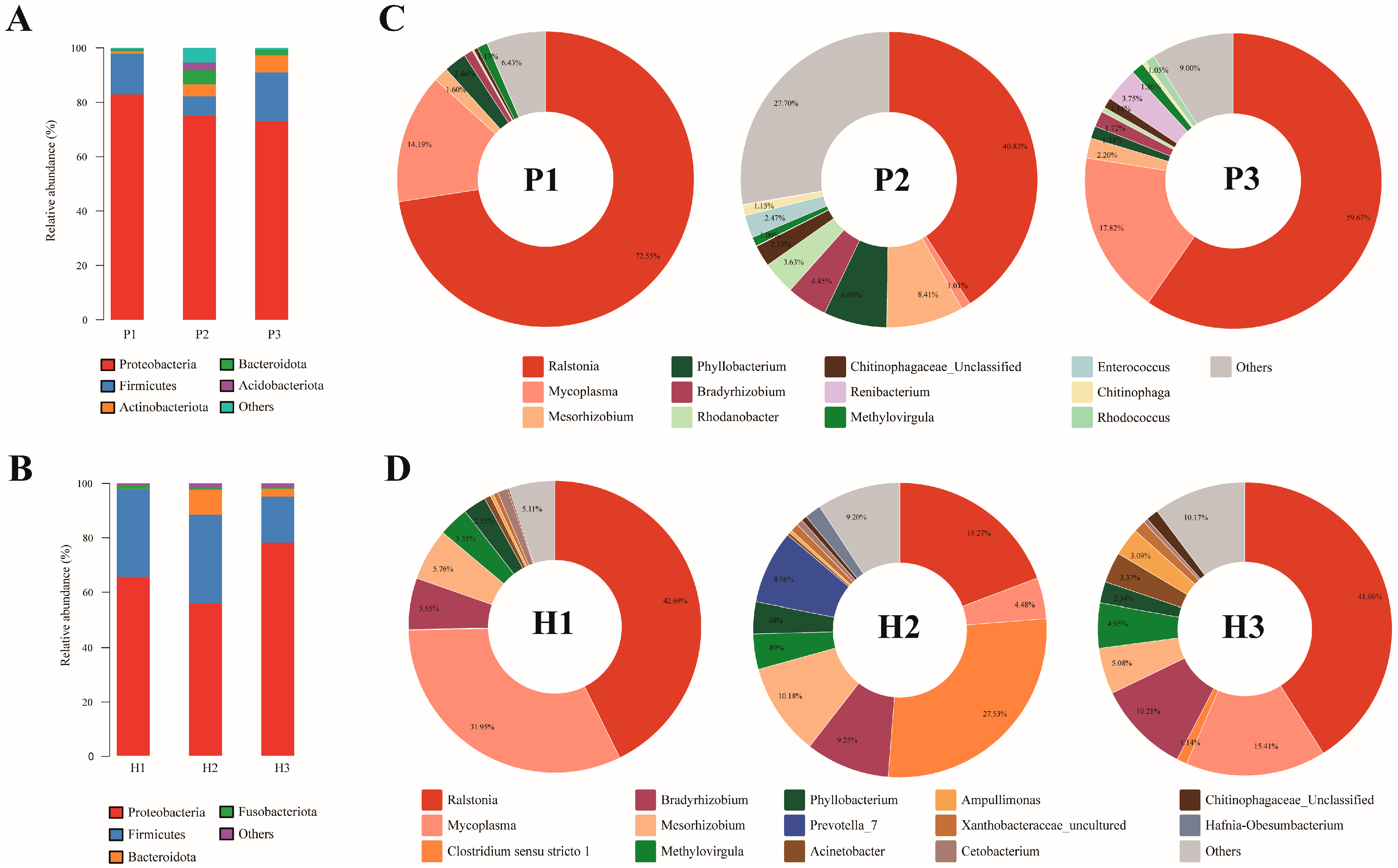
The microbial community composition and abundance of all gut samples at phylum level and genus level are presented in Figure 4. At the phylum level, the first two dominant species in both pyloric cecum and hindgut were Proteobacteria and Firmicutes, of which Proteobacteria accounted for more than 50% (Figure 4A,B). Moreover, Actinobacteriota, Bacteroidota and Acidobacteriota had higher abundance values in pyloric cecum (Figure 4A), and Bacteroidota had higher values in the hindgut (Figure 4B). Among them, the proportions of Actinobacteriota and Bacteroidota were higher in the diet supplemented with astaxanthin in pyloric cecum, and similarly, Bacteroidota in the hindgut. Firmicutes abundance was lower in P2 than in P1 and P3, while Bacteroidota in H2 showed the opposite results. At the genus level (Figure 4C,D), the most dominant microbial community member was Ralstonia, accounting for 40–73% in pyloric caecum and 19–43% in hindgut, respectively. Mycoplasma, as the second top genus in gut contents, had the lowest significant abundance values in P2 and H2. However, Mesorhizobium, Phyllobacterium and Bradyrhizobium were lowest in P2; meanwhile, Clostridium sensu stricto 1, Mesorhizobium and Prevotella_7 in H2 were higher than those in other groups.

4. Discussion

During the 4-month feeding trial, all astaxanthin-supplemented groups maintained stable survival rates and no growth inhibition [12], confirming that the observed gut health changes were not confounded by adverse physiological stress. The present findings instead highlight that dietary astaxanthin derived from H. pluvialis contributes to improved gut health in rainbow trout, with distinct responses across gut segments. Morphological adaptations represent a foundational response to astaxanthin. The significant increase in hindgut tunica muscularis thickness strengthens gut structural integrity, potentially enhancing motility and reducing permeability to pathogens [36]. Concurrently, the proliferation of goblet cells in the pyloric caeca—consistent with observations in astaxanthin-supplemented pompano Trachinotus ovatus—augments mucin secretion, reinforcing the mucosal barrier against pathogenic adhesion [37]. These structural changes provide a physical framework for improved digestive efficiency and immune defense.
Astaxanthin modulates antioxidant and immune pathways in a segment-specific manner. In the midgut, reduced MDA levels and elevated T-SOD activity, coupled with upregulated gpx and cat expression, indicate enhanced clearance of reactive oxygen species (ROS) [38,39], likely driven by astaxanthin’s direct radical-scavenging capacity. Similarly, astaxanthin supplementation was found to enhance gut antioxidant capacity in other fishes [30,40]. In the hindgut, the mRNA expression of sod in the hindgut for Diet 2 was shown to be significantly lower than for other diets; simultaneously, cat expression showed a decreasing trend with astaxanthin supplementation. This result indicates that it is possibly linked to localized metabolic demands; there was a lower oxidative stress level in fish feed Diet 2. Studies on E. sinensis [22], oriental river prawn Macrobrachium nipponense [41] and black tiger shrimp Penaeus monodon [19] showed a significant reduction in SOD in the astaxanthin-supplemented diets compared with the control, which is consistent with the present study. Notably, SOD is also closely related to the robustness of the immune system of the organism [42]; this may be the reason for the lower expression of pro-inflammatory cytokines in the hindguts for Diet 2.
Immune enhancements are highlighted by increased lysozyme activity in hindguts (peaking in Diet 3) and downregulated tlr5, tor1 and pcna in Diet 2, reflecting strengthened bactericidal capacity and tissue repair. Previous studies have found that dietary astaxanthin supplementation improved AKP and LZM in the gut of loach Paramisgurnus dabryanus [40]. A previous study on dietary astaxanthin in Plectropomus leopardus showed that the relative expression of gpx, lz-c and igm tended to increase as the amount of astaxanthin added to the feed increased to 0.1 g/kg [43], which is consistent with the result for hindguts in the present study. Interestingly, fish fed Diet 2 had significantly lower IgM expression than other diets for midguts. The reasons for this difference may be that the midgut, along with the hindgut, has local immunological significance. Critically, the downregulation of pro-inflammatory cytokines (il1β, il6, il8, tnfα) in the hindgut—consistent with suppressed tlr5 expression—directly attenuates excessive inflammation. This is because reduced levels of these cytokines inhibit the activation of pro-inflammatory transcription factors, leading to a significant decrease in inflammatory cytokines. Excessive inflammation, in turn, is a key hallmark of gut dysfunction [44,45]. Similarly, astaxanthin supplementation prevented the lipopolysaccharide-induced upregulation of pro-inflammatory cytokines tnfα, il6 and il1β in Chinese snakehead Channa argus [46]. Similarly, in mice with dextran sulfate sodium-induced colitis, astaxanthin supplementation was found to suppress expression of colitis inflammatory cytokines, including tnfα and il1β [47]. Downregulated interleukin (a cytokine) may be associated with the fatty acid transporter CD36 [48,49]. CD36, a membrane protein, is involved in facilitating carotenoid uptake [50]. Moreover, a study found a steep decreasing gradient in CD36 levels of mice from proximal to distal gut segments [51]. Thus, it is reasonable to hypothesize that the differential capacity of various gut segments to absorb dietary astaxanthin may influence interleukin mRNA levels. Therefore, there was a positive effect of appropriate astaxanthin on reducing the inflammatory response in the gut (especially the hindgut), and it may be attributed to the inhibition of pro-inflammatory cytokines. Diet is a key environmental factor shaping the gut microbial community [52,53], and dietary astaxanthin supplementation significantly influenced gut microbial composition in the present study. Consistent with previous reports [54,55], Proteobacteria and Firmicutes were the dominant phyla, with Ralstonia and Mycoplasma as the principal genera, though discrepancies with other studies of rainbow trout [56] may arise from differences in fish source, rearing conditions or microbiota analysis methods.
Diet is a key environmental factor shaping gut microbial community [28], and dietary astaxanthin supplementation significantly influenced gut microbial composition in the present study. Consistent with previous reports [54,55], Proteobacteria and Firmicutes dominated at the phylum level, with Ralstonia and Mycoplasma as the principal genera, though there are discrepancies with some previous studies on gut bacteria of rainbow trout [56]. It may reflect differences in fish source, rearing environment or microbiota analysis methods. Proteobacteria is susceptible to diet, and its abundance correlated with microbial dysbiosis or disease state [57]. It decreased progressively in the pyloric cecum with astaxanthin supplementation (from 82.95% to 73.11% as astaxanthin increased to 29.76 mg/kg), suggesting alleviated microbiota dysbiosis. Conversely, Diet 3 induced overgrowth of Proteobacteria in the hindgut, potentially disrupting microbial homeostasis—consistent with findings in tiger puffer, where high astaxanthin (500 mg/kg) selectively promoted Proteobacteria [30]. In addition, dietary astaxanthin supplementation also increased Actinobacteriota (0.09% to 0.63% in pyloric cecum), a phylum linked to beneficial metabolites and reduced pathogenicity [58], further supporting its role in optimizing microbial balance. Moreover, the propagation of Proteobacteria can further promote inflammation or invasion by exogenic pathogens [57]. This may be the reason why the expression of pro-inflammatory cytokines (il1β, il6, il8 and ifnγ) in the hindguts for Diet 3 was significantly higher than for Diet 2, and some were even higher than for Diet 1 (Control) (Figure 2F). Of the Proteobacteria, Ralstonia was well represented, dominating the sequence libraries in the pyloric cecum and hindgut of the three diets, accompanied by the lowest relative abundance value of Diet 2. Ralstonia is increasingly recognized as an opportunistic pathogen for infections leading to bacteremia, especially in immunosuppressed patients [59,60].
Firmicutes were involved in energy resorption and have demonstrated probiotic properties in fish. Mycoplasma decreased in the relative abundance of the hindgut with astaxanthin supplementation, with Diet 2 exhibiting the lowest levels in both the pyloric cecum and hindgut. This aligns with the improved gut immune profile in Diet 2, supporting the early view of Mycoplasma as a potential pathogen disrupting host immunity [61]. However, a recent study demonstrated that Mycoplasma was the dominant taxon in the gut microbiota of both resistant and susceptible lines of rainbow trout [62]; it is also possible that the high occurrence of these prokaryotes in other diets (Diet 1 and 3) was due to their active struggle against pathogenic microorganisms. Conversely, some studies have found the role of Mycoplasma may be to maintain the normal physiological state of the host [62,63,64]. Astaxanthin significantly increased Clostridium sensu stricto 1 in the hindgut (notably in H2), a genus critical for amino acid utilization in protein diets [65,66], suggesting it may enhance amino acid fermentation and absorption in trout. The Firmicutes/Bacteroidota ratio of the gut directly affects host fat accumulation, with Firmicutes abundance associated with fat accumulation and Bacteroidota dominance characteristic of healthy gut ecosystems [67]. Astaxanthin alleviated gut microbiota dysbiosis by optimizing the Firmicutes/Bacteroidota ratio and inhibiting the abundance of obesity-related pathogenic microbiota [68]. In the present study, P2 had a lower relative abundance of Firmicutes, while the Bacteroidota of P2 and H2 were reversed, indicating that P2 and H2 had healthier gut status. Moreover, H2 exhibited an 8% relative abundance of Prevotella_7, a Bacteroidota genus implicated in gut inflammation regulation [69]. The level of Prevotella is known to decline in enteritis and can be elevated by probiotics [42], consistent with its enrichment of Diet 2 linked to reduced gut inflammation. The absence of Prevotella_7 in Diet 3 indicates that the impact of astaxanthin on gut microbiota is not merely a dose-dependent effect or a compensatory response to environmental stress but rather involves selective modulation at an optimal level.
Hence, allowing for the changes in gut structure and immune cytokines in the present study, we have reason to believe that dietary astaxanthin supplementation may decrease the incidence of gut inflammation responses by decreasing the abundance of pathogens and increasing the abundance of probiotics. Combined with the changes in gut immunity, antioxidant capacity and gut content microbiota mentioned above, we inferred that the effects of astaxanthin-supplemented diets on growth performance were closely related to gut health.

5. Conclusions

The present study revealed dietary supplementation with 18.57 mg/kg micro-algal astaxanthin can promote the proliferation of goblet cells in gut mucosal epithelium, increase the tunica muscularis thickness, improve gut antioxidant and immune capacities and modulate gut content microbiota. All astaxanthin-supplemented groups maintained stable growth performance with no adverse effects, confirming that the observed gut health improvements were not confounded by growth inhibition. These findings not only clarify the segment-specific regulatory role of micro-algal astaxanthin in rainbow trout gut health but also provide practical guidance for using natural astaxanthin from Haematococcus pluvialis as a functional feed additive to support gut health in salmonid aquaculture, independent of growth optimization goals.

Author Contributions

M.Z.: conducting of experiments, data curation and writing of the original draft. X.L.: conducting of experiments, data curation and writing of the original draft. Y.L.: conducting of experiments. Y.Z.: design of the experiment. W.S.: methodology, paper revision. X.W.: idea for the project, experiment design, paper revision. All authors have read and agreed to the published version of the manuscript.

Funding

The study was supported by the Yunnan Basic Research Program from Science and Technology Department of Yunnan province (No. 202101AT070028), the technology transformation project of Yunnan Alphy Biotech Co., Ltd. (No. 2019310031000118) and the Innovation Team Project of High-level Local Universities from the Shanghai education committee (No. HJWK-2021-21).

Institutional Review Board Statement

The study was conducted in accordance with the Animal Ethics Committee (AEC) of Shanghai Ocean University, approval code: SHOU-DW-2020-152, 22 March 2020.

Data Availability Statement

The original contributions presented in this study are included in the article. Further inquiries can be directed to the corresponding authors.

Acknowledgments

The authors would like to thank the Aller Aqua (Qingdao) Co., Ltd. for their help in the preparation of the experimental feeds, as well as Peng Zhao and Wenyan Fan from Qinghai Minze Longangxia Ecological Aquaculture Co., Ltd. for their assistance with fish culture and sampling. Most sincere thanks are also given to Lei Wang, Haoxiang Ding and Meijun Tang from Shanghai Ocean University for their contribution to the sample collection of this study.

Conflicts of Interest

Authors Yaopeng Li and Yong Zhang were employed by the R&D center, Qinghai Minze Longangxia Ecological Hydroponics Co., Ltd. and Yunnan Alphy Biotech Co., Ltd., respectively. The remaining authors declare that the research was conducted in the absence of any commercial or financial relationships that could be construed as a potential conflict of interest.

References

  1. Dan, X.M.; Zhang, T.W.; Li, Y.W.; Li, A.X. Immune responses and immune-related gene expression profile in orange-spotted grouper after immunization with Cryptocaryon irritans vaccine. Fish Shellfish Immunol. 2013, 34, 885–891. [Google Scholar] [CrossRef]
  2. Ali, M.; Nicholson, H.L. Increasing zebrafish (Danio rerio) numbers in a limited tank space reduces night-time fish sleep-like state and induces aggressive behaviour. Depress. Anxiety 2018, 1, 1003. [Google Scholar]
  3. Portz, D.E.; Woodley, C.M.; Cech, J.J. Stress-associated impacts of short-term holding on fishes. Rev. Fish Biol. Fish. 2006, 16, 125–170. [Google Scholar] [CrossRef]
  4. Lock, E.J.; Waagbo, R.; Bonga, S.W.; Flik, G. The significance of vitamin D for fish: A review. Aquac. Nutr. 2010, 16, 100–116. [Google Scholar] [CrossRef]
  5. Hernandez, L.H.; Hardy, R.W. Vitamin A functions and requirements in fish. Aquac. Res. 2020, 51, 3061–3071. [Google Scholar] [CrossRef]
  6. Wade, N.M.; Gabaudan, J.; Glencross, B.D. A review of carotenoid utilisation and function in crustacean aquaculture. Rev. Aquac. 2017, 9, 141–156. [Google Scholar] [CrossRef]
  7. Besen, K.P.; Melim, E.W.H.; da Cunha, L.; Favaretto, E.D.; Moreira, M.; Fabregat, T.E.P. Lutein as a natural carotenoid source: Effect on growth, survival and skin pigmentation of goldfish juveniles (Carassius auratus). Aquac. Res. 2019, 50, 2200–2206. [Google Scholar] [CrossRef]
  8. Abd El-Gawad, E.A.; Wang, H.P.; Yao, H. Diet supplemented with synthetic carotenoids: Effects on growth performance and biochemical and immunological parameters of yellow perch (Perca flavescens). Front. Physiol. 2019, 10, 1056. [Google Scholar] [CrossRef]
  9. Mansour, A.T.; El-Feky, M.M.M.; El-Beltagi, H.S.; Sallam, A.E. Synergism of dietary co-supplementation with lutein and bile salts improved the growth performance, carotenoid content, antioxidant capacity, lipid metabolism, and lipase activity of the marbled spinefoot rabbitfish, Siganus rivulatus. Animals 2020, 10, 1643. [Google Scholar] [CrossRef]
  10. Xie, W.; Deng, H.G.; Li, K.; Ma, Y.C.; Gao, M.R.; Duan, H.; Sui, L.Y. Dietary supplementation of archaeal carotenoids improved antioxidative capacity and regulated immune-related gene expression of golden trout Oncorhynchus mykiss against challenge. Aquac. Res. 2022, 53, 5053–5062. [Google Scholar] [CrossRef]
  11. Elbahnaswy, S.; Elshopakey, G.E. Recent progress in practical applications of a potential carotenoid astaxanthin in aquaculture industry: A review. Fish Physiol. Biochem. 2023, 14, 100685. [Google Scholar] [CrossRef]
  12. Long, X.W.; Wang, L.; Li, Y.P.; Sun, W.H.; Wu, X.G. Effects of long-term Haematococcus pluvialis astaxanthin feeding on the growth, coloration, and antioxidant capacity of commercial-sized Oncorhynchus mykiss. Aquac. Rep. 2023, 30, 101603. [Google Scholar] [CrossRef]
  13. Boe, M.R.; Vo, T.T.M.; Hansen, A.K.G.; Lerfall, J. Effect of natural carotenoids obtained from Haematococcus pluvialis, Paracoccus carotinifaciens, and Phaffia rhodozyma on flesh pigmentation and related biochemical mechanisms in Atlantic salmon (Salmo salar L.). Aquaculture 2025, 596, 741743. [Google Scholar] [CrossRef]
  14. Sha, Y.P.; Chen, Y.H.; Dong, J.; Gao, X.F.; Yuan, H.T.; Zhang, J.X.; Gao, Y.N.; Li, X.J. Effects of astaxanthin microencapsulated from Haematococcus pluvialis on the growth, muscle quality, antioxidant system, and related gene expression of Cyprinus carpio. Aquac. Int. 2025, 33, 110. [Google Scholar] [CrossRef]
  15. Wang, L.; Long, X.W.; Li, Y.P.; Zhang, Y.; Sun, W.H.; Wu, X.G. Effects of three sources of astaxanthin on the growth, coloration, and antioxidant capacity of rainbow trout (Oncorhynchus mykiss) during long-term feeding. Fishes 2024, 9, 174. [Google Scholar] [CrossRef]
  16. Zhang, C.Q.; Chen, X.X.; Too, H.P. Microbial astaxanthin biosynthesis: Recent achievements, challenges, and commercialization outlook. Appl. Microbiol. Biotechnol. 2020, 104, 5725–5737. [Google Scholar] [CrossRef]
  17. Sheikhzadeh, N.; Tayefi-Nasrabadi, H.; Oushani, A.K.; Enferadi, M.H. Effects of Haematococcus pluvialis supplementation on antioxidant system and metabolism in rainbow trout (Oncorhynchus mykiss). Fish Physiol. Biochem. 2012, 38, 413–419. [Google Scholar] [CrossRef]
  18. Long, X.W.; Wu, X.G.; Zhao, L.; Liu, J.G.; Cheng, Y.X. Effects of dietary supplementation with Haematococcus pluvialis cell powder on coloration, ovarian development and antioxidation capacity of adult female Chinese mitten crab, Eriocheir sinensis. Aquaculture 2017, 473, 545–553. [Google Scholar] [CrossRef]
  19. Wang, W.L.; Liu, M.T.; Fawzy, S.; Xue, Y.C.; Wu, M.Q.; Huang, X.X.; Yi, G.F.; Lin, Q. Effects of dietary Phaffia rhodozyma astaxanthin on growth performance, carotenoid analysis, biochemical and immune-physiological parameters, intestinal microbiota, and disease resistance in Penaeus monodon. Front. Microbiol. 2021, 12, 762689. [Google Scholar] [CrossRef]
  20. Kalinowski, C.T.; Larroquet, L.; Veron, V.; Robaina, L.; Izquierdo, M.S.; Panserat, S.; Kaushik, S.; Fontagne-Dicharry, S. Influence of dietary astaxanthin on the hepatic oxidative stress response caused by Episodic Hyperoxia in rainbow trout. Antioxidants 2019, 8, 626. [Google Scholar] [CrossRef]
  21. Wang, Z.; Cai, C.F.; Cao, X.M.; Zhu, J.M.; He, J.; Wu, P.; Ye, Y.T. Supplementation of dietary astaxanthin alleviated oxidative damage induced by chronic high pH stress, and enhanced carapace astaxanthin concentration of Chinese mitten crab Eriocheir sinensis. Aquaculture 2018, 483, 230–237. [Google Scholar] [CrossRef]
  22. Jiang, X.D.; Zu, L.; Wang, Z.Y.; Cheng, Y.X.; Yang, Y.H.; Wu, X.G. Micro-algal astaxanthin could improve the antioxidant capability, immunity and ammonia resistance of juvenile Chinese mitten crab, Eriocheir sinensis. Fish Shellfish Immun. 2020, 102, 499–510. [Google Scholar] [CrossRef]
  23. Wang, Y.J.; Chien, Y.H.; Pan, C.H. Effects of dietary supplementation of carotenoids on survival, growth, pigmentation, and antioxidant capacity of characins, Hyphessobrycon callistus. Aquaculture 2006, 261, 641–648. [Google Scholar] [CrossRef]
  24. Zhang, J.J.; Li, X.Q.; Leng, X.J.; Zhang, C.L.; Han, Z.Y.; Zhang, F.G. Effects of dietary astaxanthins on pigmentation of flesh and tissue antioxidation of rainbow trout (Oncorhynchus mykiss). Aquac. Int. 2013, 21, 579–589. [Google Scholar] [CrossRef]
  25. Wang, C.; Qin, S.; Sun, F.J.; Shao, Y.P.; Du, R.; Gao, Z.Q.; Cui, Y.L. Effects of feeding Haematococcus pluvialis powder on astaxanthin accumulation, biochemical compositions, antioxidant activity, and gut microbial community in juvenile crucian carp (Carassius auratus L.). Aquac. Rep. 2024, 36, 102109. [Google Scholar] [CrossRef]
  26. Song, X.L.; Wang, L.; Li, X.Q.; Chen, Z.Z.; Liang, G. Dietary astaxanthin improved the body pigmentation and antioxidant function, but not the growth of discus fish (Symphysodon spp.). Aquac. Res. 2017, 48, 1359–1367. [Google Scholar] [CrossRef]
  27. Carmen Tatiana, K.; Monica, B.B.; Silvia, T.; Matthew, S.; Laurence, L.; Vincent, V.; Stephane, P.; Maria Soledad, I.; Sadasivam, J.K.; Stephanie, F.-D. More than an antioxidant: Role of dietary astaxanthin on lipid and glucose metabolism in the liver of rainbow trout (Oncorhynchus mykiss). Antioxidants 2023, 12, 136. [Google Scholar] [CrossRef]
  28. Ringø, E.; Zhou, Z.; Vecino, J.L.G.; Wadsworth, S.; Romero, J. Effect of dietary components on the gut microbiota of aquatic animals. A never-ending story? Aquac. Nutr. 2015, 22, 219–282. [Google Scholar] [CrossRef]
  29. Pratap, K.; Majzoub, M.E.; Taki, A.C.; Hernandez, S.M.; Magnusson, M.; Glasson, C.R.K.; de Nys, R.; Thomas, T.; Lopata, A.L.; Kamath, S.D. The algal polysaccharideulvan and carotenoid astaxanthin both positively modulate gut microbiota in mice. Foods 2022, 11, 565. [Google Scholar] [CrossRef]
  30. Ou, W.H.; Liao, Z.B.; Yu, G.J.; Xu, H.G.; Liang, M.Q.; Mai, K.S.; Zhang, Y.J. The effects of dietary astaxanthin on intestinal health of juvenile tiger puffer Takifugu rubripes in terms of antioxidative status, inflammatory response and microbiota. Aquac. Nutr. 2019, 25, 466–476. [Google Scholar] [CrossRef]
  31. Ma, F.; Ma, R.L.; Zou, Y.L.; Zhao, L. Effect of astaxanthin on the antioxidant capacity and intestinal microbiota of tsinling lenok trout (Brachymystax lenok tsinlingensis). Mar. Biotechnol. 2022, 24, 1125–1137. [Google Scholar] [CrossRef] [PubMed]
  32. Food and Agriculture Organization of the United Nations. The State of World Fisheries and Aquaculture 2024—Blue Transformation in Action; FAO: Rome, Italy, 2024. [Google Scholar]
  33. Ministry of Agriculture Fisheries and Fishery Administration Bureau. China Fishery Statistical Yearbook 2024; Agriculture Press: Beijing, China, 2024.
  34. Mansfield, G.S.; Desai, A.R.; Nilson, S.A.; Van Kessel, A.G.; Drew, M.D.; Hill, J.E. Characterization of rainbow trout (Oncorhynchus mykiss) intestinal microbiota and inflammatory marker gene expression in a recirculating aquaculture system. Aquaculture 2010, 307, 95–104. [Google Scholar] [CrossRef]
  35. Livak, K.J.; Schmittgen, T.D. Analysis of relative gene expression data using real-time quantitative PCR and the 2-ΔΔCT method. Methods 2001, 25, 402–408. [Google Scholar] [CrossRef] [PubMed]
  36. Wang, J.Y.; Hao, Y.T.; Zhang, L.H.; Gao, X.W.; Xu, Y.H.; Wang, J.J.; Hanafiah, F.; Khor, W.; Sun, Y.F.; Wu, C.B. Profiling the gut structure and microbiota, and identifying two dominant bacteria belonging to the Weissella genus in mandarin fish (Siniperca chuatsi) fed an artificial diet. Front. Microbiol. 2024, 15, 1486501. [Google Scholar] [CrossRef]
  37. Fang, H.H.; Xie, J.J.; Zhao, W.; Liu, Z.L.; Liu, Y.J.; Tian, L.X.; Niu, J. Study supplementation of astaxanthin in high-fat diet on growth performance, antioxidant ability, anti-inflammation, non-specific immunity and intestinal structure of juvenile Trachinotus ovatus. Aquac. Nutr. 2021, 27, 2575–2586. [Google Scholar] [CrossRef]
  38. Poljšak, B.; Jamnik, P.; Raspor, P.; Pesti, M. Oxidation-antioxidation-reduction processes in the cell: Impacts of environmental pollution. Encycl. Environ. Health 2011, 300–306. [Google Scholar] [CrossRef]
  39. Duan, Y.F.; Zhang, J.S.; Dong, H.B.; Wang, Y.; Liu, Q.S.; Li, H. Oxidative stress response of the black tiger shrimp Penaeus monodon to Vibrio parahaemolyticus challenge. Fish Shellfish Immun. 2015, 46, 354–365. [Google Scholar] [CrossRef]
  40. Chen, X.M.; Gao, C.S.; Du, X.Y.; Yao, J.M.; He, F.F.; Niu, X.T.; Wang, G.Q.; Zhang, D.M. Effects of dietary astaxanthin on the growth, innate immunity and antioxidant defence system of Paramisgurnus dabryanus. Aquac. Nutr. 2020, 26, 1453–1462. [Google Scholar] [CrossRef]
  41. Ettefaghdoost, M.; Haghighi, H. Impact of different dietary lutein levels on growth performance, biochemical and immuno-physiological parameters of oriental river prawn (Macrobrachium nipponense). Fish Shellfish Immunol. 2021, 115, 86–94. [Google Scholar] [CrossRef]
  42. Wang, L.; Wu, Z.Q.; Wang, X.L.; Ren, Q.; Zhang, G.S.; Liang, F.F.; Yin, S.W. Immune responses of two superoxide dismutases (SODs) after lipopolysaccharide or Aeromonas hydrophila challenge in pufferfish, Takifugu obscurus. Aquaculture 2016, 459, 1–7. [Google Scholar] [CrossRef]
  43. Zhu, X.W.; Hao, R.J.; Zhang, J.P.; Tian, C.X.; Hong, Y.C.; Zhu, C.H.; Li, G.L. Dietary astaxanthin improves the antioxidant capacity, immunity and disease resistance of coral trout (Plectropomus leopardus). Fish Shellfish Immunol. 2022, 122, 38–47. [Google Scholar] [CrossRef]
  44. Takeda, K.; Kaisho, T.; Akira, S. Toll-like receptors. Annu. Rev. Immunol. 2003, 21, 335–376. [Google Scholar] [CrossRef]
  45. Lim, K.H.; Staudt, L.M. Toll-like receptor signaling. Cold Spring Harb. Perspect. Biol. 2013, 5, a011247. [Google Scholar] [CrossRef]
  46. Li, M.Y.; Gao, C.S.; Du, X.Y.; Zhao, L.; Zhang, D.M. Effect of sub-chronic exposure to selenium and astaxanthin on Channa argus: Bioaccumulation, oxidative stress and inflammatory response. Chemosphere 2019, 244, 125546. [Google Scholar] [CrossRef]
  47. Yasui, Y.; Hosokawa, M.; Mikami, N.; Miyashita, K.; Tanaka, T. Dietary astaxanthin inhibits colitis and colitis-associated colon carcinogenesis in mice via modulation of the inflammatory cytokines. Chem. Biol. Interact. 2011, 193, 79–87. [Google Scholar] [CrossRef] [PubMed]
  48. Murakami, A.; Ikeda, Y.; Ohigashi, H. Aggregated ursolic acid, a natural triterpenoid, binds to CD36 for inducing interleukin-1β release from murine peritoneal macrophages. Cancer Res. 2007, 67, 3383. [Google Scholar]
  49. Wei, L.; Wu, J.; Zhao, L.; Chen, Y.X.; Yang, P. CD36 knockdown alleviates lipid accumulation induced by tumor necrosis factor-α and interleukin-6 in HepG2 cell. J. Army Med. Univ. 2018, 40, 1833–1838. [Google Scholar]
  50. Sakudoh, T.; Kuwazaki, S.; Iizuka, T.; Narukawa, J.; Yamamoto, K.; Uchino, K.; Sezutsu, H.; Banno, Y.; Tsuchida, K. CD36 homolog divergence is responsible for the selectivity of carotenoid species migration to the silk gland of the silkworm Bombyx mori. J. Lipid Res. 2013, 54, 482–495. [Google Scholar] [CrossRef]
  51. Nassir, F.; Wilson, B.; Han, X.L.; Gross, R.W.; Abumrad, N.A. CD36 is important for fatty acid and cholesterol uptake by the proximal but not distal intestine. J. Biol. Chem. 2007, 282, 19493–19501. [Google Scholar] [CrossRef]
  52. De Filippo, C.; Cavalieri, D.; Di Paola, M.; Ramazzotti, M.; Poullet, J.B.; Massart, S.; Collini, S.; Pieraccini, G.; Lionetti, P. Impact of diet in shaping gut microbiota revealed by a comparative study in children from Europe and rural Africa. Proc. Natl. Acad. Sci. USA 2010, 107, 14691–14696. [Google Scholar] [CrossRef]
  53. Carmody, R.N.; Gerber, G.K.; Luevano, J.M., Jr.; Gatti, D.M.; Somes, L.; Svenson, K.L.; Turnbaugh, P.J. Diet dominates host genotype in shaping the murine gut microbiota. Cell Host Microbe 2015, 17, 72–84. [Google Scholar] [CrossRef]
  54. Desai, A.R.; Links, M.G.; Hill, J.E. Effects of plant-based diets on the distal gut microbiome of rainbow trout (Oncorhynchus mykiss). Aquaculture 2012, 350, 134–142. [Google Scholar] [CrossRef]
  55. Rimoldi, S.; Antonini, M.; Gasco, L.; Moroni, F.; Terova, G. Intestinal microbial communities of rainbow trout (Oncorhynchus mykiss) may be improved by feeding a Hermetia illucens meal/low-fishmeal diet. Fish Physiol. Biochem. 2021, 47, 365–380. [Google Scholar] [CrossRef] [PubMed]
  56. Lyons, P.P.; Turnbull, J.F.; Dawson, K.A.; Crumlish, M. Effects of low-level dietary microalgae supplementation on the distal intestinal microbiome of farmed rainbow trout Oncorhynchus mykiss (Walbaum). Aquac. Res. 2016, 48, 2438–2452. [Google Scholar] [CrossRef]
  57. Shin, N.R.; Whon, T.W.; Bae, J.W. Proteobacteria: Microbial signature of dysbiosis in gut microbiota. Trends Biotechnol. 2015, 33, 496–503. [Google Scholar] [CrossRef] [PubMed]
  58. Barka, E.A.; Vatsa, P.; Sanchez, L.; Gaveau-Vaillant, N.; Jacquard, C.; Meier-Kolthoff, J.P.; Klenk, H.P.; Clement, C.; Ouhdouch, Y.; van Wezel, G.P. Taxonomy, physiology, and natural products of Actinobacteria. Microbiol. Mol. Biol. Rev. 2016, 80, 1–43. [Google Scholar] [CrossRef]
  59. Tejera, D.; Limongi, G.; Bertullo, M.; Cancela, M. Ralstonia pickettii bacteremia in hemodialysis patients: A report of two cases. Rev. Bras. Ter. Intensiv. 2016, 28, 195–198. [Google Scholar]
  60. Chi, C.Y.; Fung, C.P.; Wong, W.W.; Liu, C.Y. Brevundimonas bacteremia: Two case reports and literature review. Scand. J. Infect. Dis. 2004, 36, 59–61. [Google Scholar] [CrossRef]
  61. Holben, W.E.; Williams, P.; Gilbert, M.A.; Saarinen, M.; Sarkilahti, L.K.; Apajalahti, J.H.A. Phylogenetic analysis of intestinal microflora indicates a novel mycoplasma phylotype in farmed and wild salmon. Microbiol. Ecol. 2002, 44, 175–185. [Google Scholar] [CrossRef]
  62. Brown, R.M.; Wiens, G.D.; Salinas, I. Analysis of the gut and gill microbiome of resistant and susceptible lines of rainbow trout (Oncorhynchus mykiss). Fish Shellfish Immun. 2019, 86, 497–506. [Google Scholar] [CrossRef]
  63. Ciric, M.; Waite, D.; Draper, J.; Jones, J.B. Characterization of mid-intestinal microbiota of farmed Chinook salmon using 16S rRNA gene metabarcoding. Arch. Biol. Sci. 2019, 71, 577–587. [Google Scholar] [CrossRef]
  64. Mora-Sanchez, B.; Balcazar, J.L.; Perez-Sanchez, T. Effect of a novel postbiotic containing lactic acid bacteria on the intestinal microbiota and disease resistance of rainbow trout (Oncorhynchus mykiss). Biotechnol. Lett. 2020, 42, 1957–1962. [Google Scholar] [CrossRef]
  65. Heinritz, S.N.; Mosenthin, R.; Weiss, E. Use of pigs as a potential model for research into dietary modulation of the human gut microbiota. Nutr. Res. Rev. 2013, 26, 191–209. [Google Scholar] [CrossRef]
  66. Fan, P.; Liu, P.; Song, P.; Chen, X.; Ma, X. Moderate dietary protein restriction alters the composition of gut microbiota and improves ileal barrier function in adult pig model. Sci. Rep. 2017, 7, 43412. [Google Scholar] [CrossRef] [PubMed]
  67. Ley, R.E.; Turnbaugh, P.J.; Klein, S.; Gordon, J.I. Microbial ecology: Human gut microbes associated with obesity. Nature 2006, 444, 1022–1023. [Google Scholar] [CrossRef]
  68. Wang, M.; Ma, H.T.; Guan, S.Y.; Luo, T.; Zhao, C.C.; Cai, G.P.; Zheng, Y.B.; Jia, X.Y.; Di, J.B.; Li, R.Z.; et al. Astaxanthin from Haematococcus pluvialis alleviates obesity by modulating lipid metabolism and gut microbiota in mice fed a high-fat diet. Food Funct. 2021, 12, 9719–9738. [Google Scholar] [CrossRef]
  69. Iljazovic, A.; Amend, L.; Galvez, E.J.C.; Oliveira, R.D.; Strowig, T. Modulation of inflammatory responses by gastrointestinal Prevotella spp.—From associations to functional studies. Int. J. Med. Microbiol. 2021, 311, 151472. [Google Scholar] [CrossRef]
Figure 1. Histological characteristics of the different intestine regions (pyloric caeca, midgut and hindgut) rainbow trout fed three experimental diets with different micro-algal astaxanthin contents. (a,b): pyloric caeca, (c): midgut, (d): hindgut, TMT: tunica muscularis thickness, VH: villus height, VW: villus width, GCs: goblet cells.
Fishes 10 00505 g001
Figure 2. Expression profiles of antioxidant enzymes, inflammation and cell regulation-related biomarkers and pro-inflammatory cytokines in the midguts (AC) and hindguts (DF) of rainbow trout fed three experimental diets with different micro-algal astaxanthin contents. Values are mean ± standard deviation (SD) (n = 3). Means of same gene expression with different superscripts are significantly different (p < 0.05).
Fishes 10 00505 g002
Figure 3. Alpha diversity indexes (A) and Venn diagram analysis (B) of intestinal microbiota in the pyloric caeca and hindgut of the rainbow trout fed three experimental diets with different micro-algal astaxanthin contents. P1, P2 and P3 represented pyloric caeca of Diet 1, Diet 2 and Diet 3 treatments, while H1, H2 and H3 represented hindgut of Diet 1, Diet 2 and Diet 3 treatments, respectively. OTU: operational taxonomic units. Numbers in the circles refer to the OTUs that belong to each treatment. Means of column with different superscripts are significantly different (p < 0.05).
Fishes 10 00505 g003
Figure 4. Abundance distribution of intestinal microbiota at phylum level (A,B) and genus level (C,D) of rainbow trout fed three experimental diets with different micro-algal astaxanthin contents. (A): Phyla abundance in pyloric caeca; (B): phyla abundance in hindgut; (C): genera abundance in pyloric caeca; (D): genera abundance in hindgut. Only top 4–5 bacterial phyla or 12–13 genera (based on relative abundance) are shown in the figures, while the other phyla or genera are assigned as “Others”. P1, P2 and P3 represent pyloric caeca of Diet 1, Diet 2 and Diet 3 treatments, while H1, H2 and H3 represent hindgut of Diet 1, Diet 2 and Diet 3 treatments, respectively. Numbers in the circles refer to the OTUs that belong to each treatment.
Fishes 10 00505 g004
Table 1. Proximate composition and actual astaxanthin content of experimental diets.
ItemsExperimental Diets
Diet 1Diet 2Diet 3
Moisture (% dry diet)10.1010.2110.13
Crud protein (% dry diet)42.4541.5642.02
Crud lipid (% dry diet)23.6523.8223.63
Ash (% dry diet)6.916.936.88
Astaxanthin (mg/kg dry diet)0.0018.5731.25
Note: Moisture content was determined by oven drying at 105 °C for 24 h, crude protein by the Kjeldahl method, crude lipid by Soxhlet extraction and ash by muffle furnace incineration at 550 °C for 6 h. Astaxanthin content was measured using high-performance liquid chromatography (HPLC, Agilent 1260, Los Angeles, CA, USA). Detailed procedures for all analyses followed the method described by Long et al. (2023) [12].
Table 2. Effects of dietary supplementation with micro-algal astaxanthin on growth performance of rainbow trout.
IndicesExperimental Diets
Diet 1Diet 2Diet 3
IW (kg)0.67 ± 0.040.67 ± 0.020.65 ± 0.02
FW (kg)2.19 ± 0.162.18 ± 0.072.30 ± 0.12
WG (kg)1.52 ± 0.131.51 ± 0.051.65 ± 0.10
FCR0.85 ± 0.080.84 ± 0.010.87 ± 0.03
SGR (%/d)0.99 ± 0.030.97 ± 0.021.03 ± 0.02
SR (%)94.00 ± 2.6791.56 ± 4.7389.78 ± 4.02
Note: Values are mean ± standard deviation (SD) (n = 9). IW: initial weight, FW: final weight, WG: weight gain, FCR: feed conversion ratio, SGR: specific growth rate, SR: survival rate. WG = FW − IW; SGR (%/d) = 100 × (lnFW − lnIW)/trial days; FCR = 100 × (total feed casting-total food residue)/(total final weight − total initial weight + total mortality weight) dry feed intake/wet weight gain; SR (%) = 100 × (final number of fish/initial number of fish).
Table 3. Primers used for intestinal gene expression analysis of rainbow trout in this study.
Target GenePrimer Sequence (5′→3′)Accession Number
sod1F:GTAGTCGTGGCTCAATGGTAAGXM_021590204.2
R:GCTTTATATTCTGCGGGTCATT
catF:TGATGTCACACAGGTGCGTATC99600
R:GTGGGCTCAGTGTTGTTGAG
gpx1F:AAATTGCCATTCCCCTCCGAXM_021569971.2
R:TCCATCAGGACTGACCAGGA
gstaF:CAGAGGACAGCTCCCTGCTTNM_001160559.1
R:CTGAACCGGCTCTCCAGGTA
igmF:CACTTCATCAGATGGTCCAGTCCX83372.1
R:ACAGTCCCATTGCTCCAGTCC
pcnaF:TGTGACCGCAACCTCGCAATGGXM_036936092.1
R:CACGGCAGATACGGGCAAACTCC
tgfβF:AGATAAATCGGAGAGTTGCTGTGX99303.1
R:CCTGCTCCACCTTGTGTTGT
tor1F:ATGGTTCGATCACTGGTCATCAEU179853
R:TCCACTCTTGCCACAGAGAC
tlr5F:CTTACAGGAAACTCTATTCGCNM_001124208
R:CTGTTAGCAAAGCCCAAGAGG
il1βF:ACATTGCCAACCTCATCATCGAJ278242
R:TTGAGCAGGTCCTTGTCCTTG
il6F:ACTCCCCTCTGTCACACACCDQ866150
R:GGCAGACAGGTCCTCCACTA
il8F:GCTGCATTGAGACGGAGAGCHG917307
R:CCAGACAAATCTCCTGACCG
ifnγF:CTGTTCAACGGAAACCCTGTNM_001160503
R:AACACCCTCCGATCACTGTC
tnfαF:AAGCAGCCATCCATTTAGAGGAJ277604
R:GTGTGTGGGATGAGGATTTGG
β-actinF:ATCCTGACAGAGCGCGGTTACAGTAJ43815
R:TGCCCATCTCCTGCTCAAAGTCAA
Accession numbers are from http://www.ncbi.nlm.nih.gov/ (accessed on 13 January 2021).
Table 4. Histological parameters of the different gut sections of rainbow trout fed three experimental diets with different micro-algal astaxanthin contents.
Histological ParametersExperimental Diets
Diet 1Diet 2Diet 3
Pyloric caeca
Tunica muscularis thickness (μm)164.45 ± 43.37201.42 ± 45.54196.76 ± 23.82
Villus height (μm)970.41 ± 133.38980.82 ± 68.47974.33 ± 53.46
Villus width (μm)135.04 ± 18.05137.42 ± 11.03136.91 ± 8.77
Midgut
Tunica muscularis thickness (μm)399.87 ± 84.53359.25 ± 66.60366.82 ± 87.72
Villus height (μm)841.24 ± 149.48956.50 ± 278.26838.84 ± 215.50
Villus width (μm)146.67 ± 19.35162.81 ± 16.03154.71 ± 6.88
Hindgut
Tunica muscularis thickness (μm)208.37 ± 17.90 b314.14 ± 20.34 a307.83 ± 26.89 a
Villus height (μm)1421.73 ± 220.551330.62 ± 249.591381.74 ± 110.28
Villus width (μm)186.04 ± 41.87163.20 ± 13.27170.42 ± 26.86
Values are mean ± standard deviation (SD) (n = 3). Means in the same row with different superscripts are significantly different (p < 0.05).
Table 5. The antioxidant and immunological parameters of midgut and hindgut of rainbow trout fed three experimental diets with different micro-algal astaxanthin contents.
ItemsMidgutHindgut
Diet 1Diet 2Diet 3Diet 1Diet 2Diet 3
Antioxidant parameters
T-AOC (U/mgprot)1.65 ± 0.47 a1.16 ± 0.27 b1.11 ± 0.14 b 3.38 ± 0.853.46 ± 0.623.14 ± 0.54
T-SOD (U/mgprot)540.46 ± 65.62 b596.94 ± 11.29 a617.20 ± 57.73 b266.66 ± 37.25284.24 ± 31.08283.07 ± 27.67
CAT (U/mgprot)4.06 ± 0.923.86 ± 0.783.46 ± 0.870.77 ± 0.150.67 ± 0.160.50 ± 0.36
MDA (nmol/mgprot)24.29 ± 5.71 a15.18 ± 2.83 b17.62 ± 3.51 b18.01 ± 4.0015.98 ± 2.2917.89 ± 3.92
Immunological parameters
ACP (U/gprot)440.94 ± 116.25482.88 ± 87.03490.73 ± 42.66258.08 ± 23.55310.05 ± 29.06288.61 ± 43.88
AKP (U/gprot)3594.70 ± 381.41 a1966.01 ± 563.35 b2699.26 ± 521.64 b2005.84 ± 262.772222.77 ± 507.791701.58 ± 431.36
LZM (U/mgprot)6.71 ± 1.95 b4.76 ± 1.09 b9.57 ± 2.43 a47.99 ± 7.27 b39.82 ± 9.49 b64.30 ± 13.91 a
Note: Values are mean ± standard deviation (SD) (n = 3). Values for the same row of the same tissue with different superscripts are significantly different (p < 0.05). T-AOC: total antioxidant capacity, T-SOD: total superoxide dismutase, CAT: catalase, MDA: malonaldehyde, ACP: acid phosphatase, AKP: alkaline phosphatase, LZM: lysozyme.
Disclaimer/Publisher’s Note: The statements, opinions and data contained in all publications are solely those of the individual author(s) and contributor(s) and not of MDPI and/or the editor(s). MDPI and/or the editor(s) disclaim responsibility for any injury to people or property resulting from any ideas, methods, instructions or products referred to in the content.