Quantitative Assessment of Airway Changes in Fibrotic Interstitial Lung Abnormality Patients by Chest CT According to Cumulative Cigarette Smoking
Abstract
1. Introduction
2. Materials and Methods
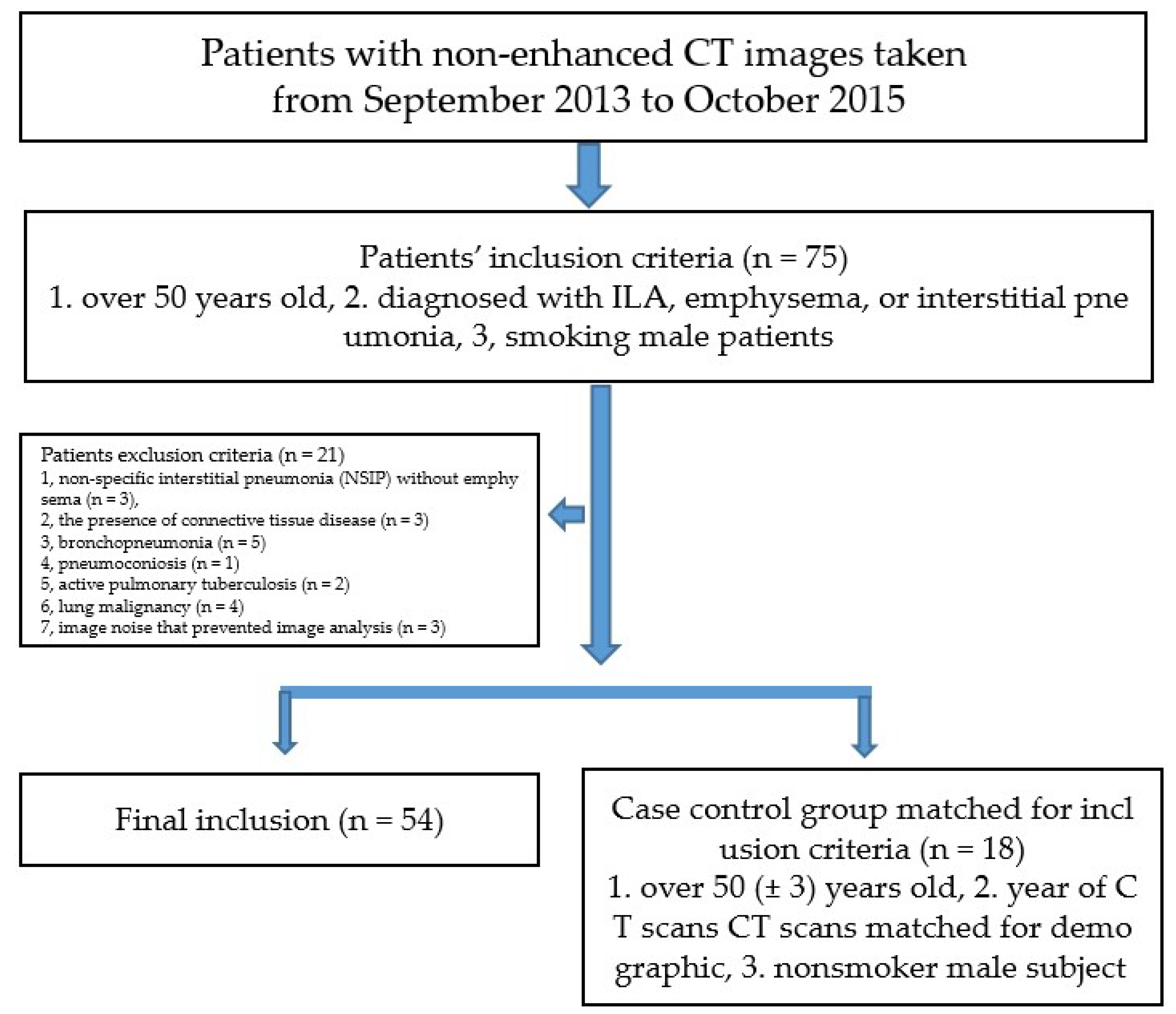
2.1. Study Subjects
2.2. Pulmonary Function Tests
2.3. Smoking
2.4. CT Acquisition
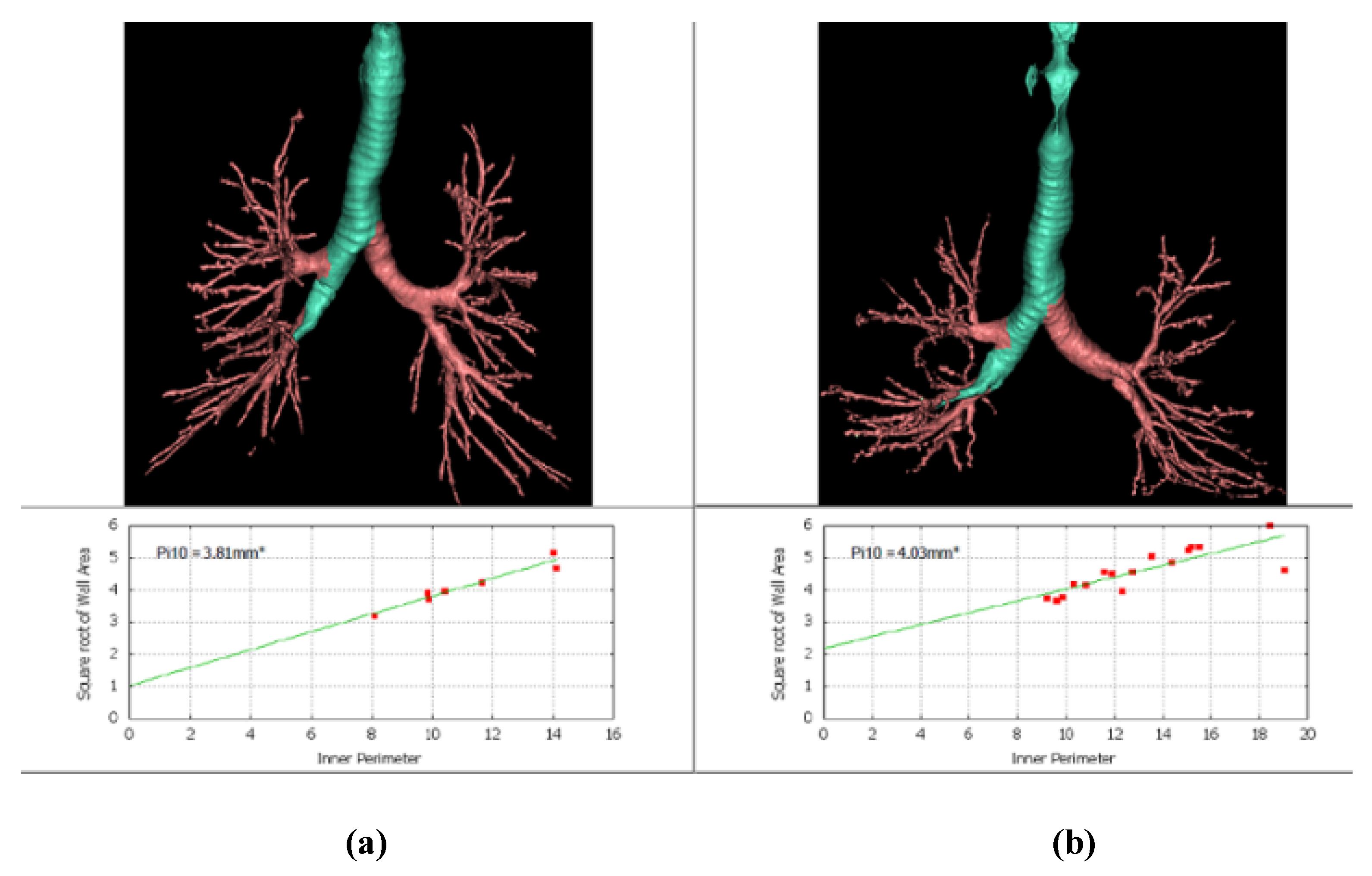
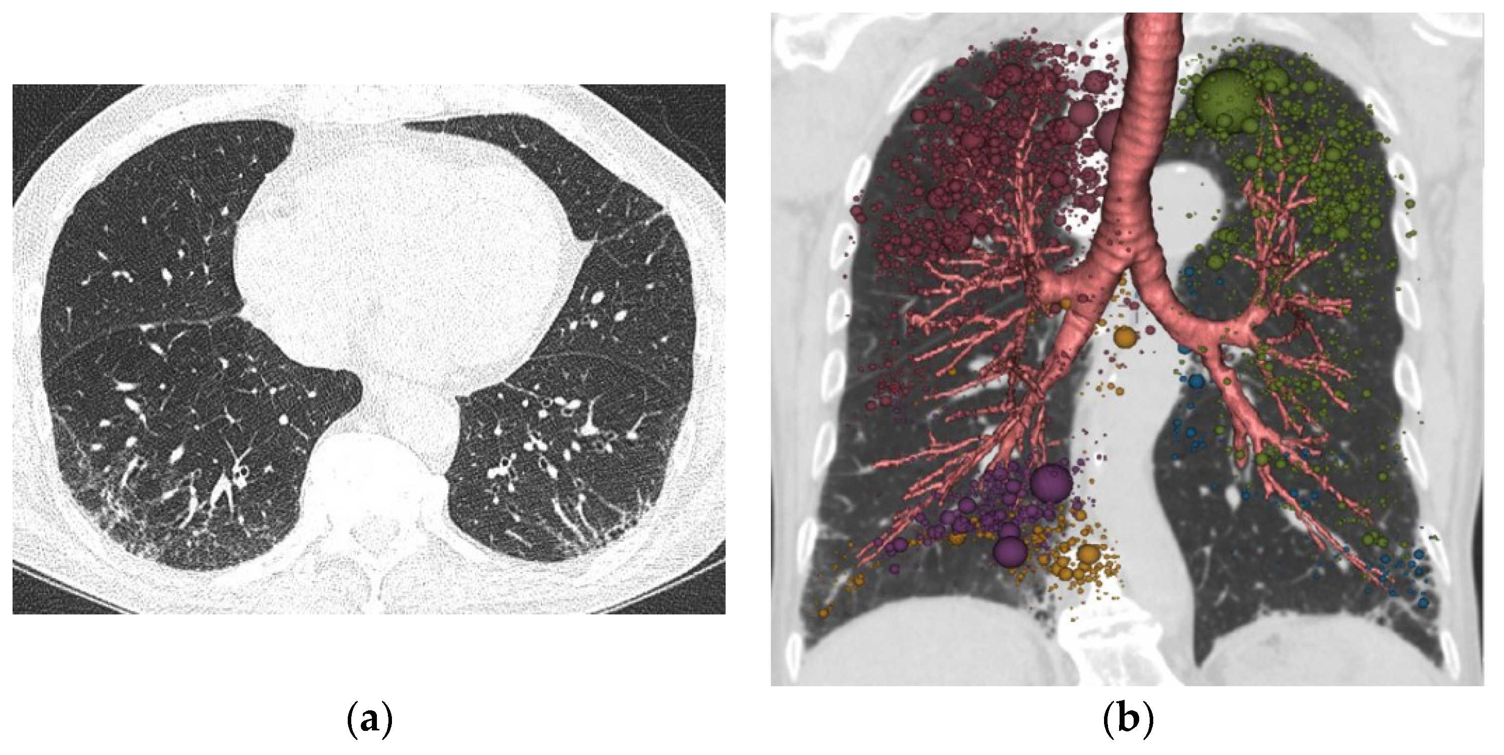
2.5. Quantitative CT Evaluation
2.6. Statistical Analysis
3. Results
4. Discussion
5. Conclusions
Author Contributions
Funding
Institutional Review Board Statement
Informed Consent Statement
Data Availability Statement
Conflicts of Interest
References
- Chae, K.J.; Chung, M.J.; Jin, G.Y.; Song, Y.J.; An, A.R.; Choi, H.; Goo, J.M. Radiologic-pathologic correlation of interstitial lung abnormalities and predictors for progression and survival. Eur. Radiol. 2022, 32, 2713–2723. [Google Scholar] [CrossRef]
- Chae, K.J.; Jin, G.Y.; Goo, J.M.; Chung, M.J. Interstitial Lung Abnormalities: What Radiologists Should Know. Korean J. Radiol. 2021, 22, 454–463. [Google Scholar] [CrossRef] [PubMed]
- Jin, G.Y.; Lynch, D.; Chawla, A.; Garg, K.; Tammemagi, M.C.; Sahin, H.; Misumi, S.; Kwon, K.S. Interstitial lung abnormalities in a CT lung cancer screening population: Prevalence and progression rate. Radiology 2013, 268, 563–571. [Google Scholar] [CrossRef] [PubMed]
- Hino, T.; Hida, T.; Nishino, M.; Lu, J.; Putman, R.K.; Gudmundsson, E.F.; Hata, A.; Araki, T.; Valtchinov, V.I.; Honda, O.; et al. Progression of traction bronchiectasis/bronchiolectasis in interstitial lung abnormalities is associated with increased all-cause mortality: Age Gene/Environment Susceptibility-Reykjavik Study. Eur. J. Radiol. Open 2021, 8, 100334. [Google Scholar] [CrossRef]
- Hata, A.; Schiebler, M.L.; Lynch, D.A.; Hatabu, H. Interstitial Lung Abnormalities: State of the Art. Radiology 2021, 301, 19–34. [Google Scholar] [CrossRef] [PubMed]
- Hunninghake, G.M.; Hatabu, H.; Okajima, Y.; Gao, W.; Dupuis, J.; Latourelle, J.C.; Nishino, M.; Araki, T.; Zazueta, O.E.; Kurugol, S.; et al. MUC5B promoter polymorphism and interstitial lung abnormalities. N. Engl. J. Med. 2013, 368, 2192–2200. [Google Scholar] [CrossRef] [PubMed]
- Tsushima, K.; Sone, S.; Yoshikawa, S.; Yokoyama, T.; Suzuki, T.; Kubo, K. The radiological patterns of interstitial change at an early phase: Over a 4-year follow-up. Respir. Med. 2010, 104, 1712–1721. [Google Scholar] [CrossRef]
- Menon, A.A.; Putman, R.K.; Sanders, J.L.; Hino, T.; Hata, A.; Nishino, M.; Ghosh, A.J.; Ash, S.Y.; Rosas, I.O.; Cho, M.H.; et al. Interstitial Lung Abnormalities, Emphysema, and Spirometry in Smokers. Chest 2021. [Google Scholar] [CrossRef]
- Hackx, M.; Bankier, A.A.; Gevenois, P.A. Chronic obstructive pulmonary disease: CT quantification of airways disease. Radiology 2012, 265, 34–48. [Google Scholar] [CrossRef]
- Hersh, C.P.; Washko, G.R.; Estépar, R.S.J.; Lutz, S.; Friedman, P.J.; Han, M.K.; Hokanson, J.E.; Judy, P.F.; Lynch, D.A.; Make, B.J.; et al. Paired inspiratory-expiratory chest CT scans to assess for small airways disease in COPD. Respir. Res. 2013, 14, 42. [Google Scholar] [CrossRef]
- Kim, S.S.; Jin, G.Y.; Li, Y.Z.; Lee, J.E.; Shin, H.S. CT Quantification of Lungs and Airways in Normal Korean Subjects. Korean J. Radiol. 2017, 18, 739–748. [Google Scholar] [CrossRef] [PubMed][Green Version]
- Lynch, D.A.; Al-Qaisi, M.A. Quantitative computed tomography in chronic obstructive pulmonary disease. J. Thorac. Imaging 2013, 28, 284–290. [Google Scholar] [CrossRef] [PubMed]
- Miller, E.R.; Putman, R.K.; Diaz, A.A.; Xu, H.; San José Estépar, R.; Araki, T.; Nishino, M.; Poli de Frías, S.; Hida, T.; Ross, J.; et al. Increased Airway Wall Thickness in Interstitial Lung Abnormalities and Idiopathic Pulmonary Fibrosis. Ann. Am. Thorac. Soc. 2019, 16, 447–454. [Google Scholar] [CrossRef] [PubMed]
- Hatabu, H.; Hunninghake, G.M.; Richeldi, L.; Brown, K.K.; Wells, A.U.; Remy-Jardin, M.; Verschakelen, J.; Nicholson, A.G.; Beasley, M.B.; Christiani, D.C.; et al. Interstitial lung abnormalities detected incidentally on CT: A Position Paper from the Fleischner Society. Lancet. Respir. Med. 2020, 8, 726–737. [Google Scholar] [CrossRef]
- American Thoracic Society. Standardization of Spirometry, 1994 Update. Am. J. Respir. Crit. Care Med. 1995, 152, 1107–1136. [Google Scholar] [CrossRef]
- Yun, W.-J.; Shin, M.-H.; Kweon, S.-S.; Ryu, S.-Y.; Rhee, J.-A. Association of smoking status, cumulative smoking, duration of smoking cessation, age of starting smoking, and depression in Korean adults. BMC Public Health 2012, 12, 724. [Google Scholar] [CrossRef]
- Kim, V.; Desai, P.; Newell, J.D.; Make, B.J.; Washko, G.R.; Silverman, E.K.; Crapo, J.D.; Bhatt, S.P.; Criner, G.J. Airway wall thickness is increased in COPD patients with bronchodilator responsiveness. Respir. Res. 2014, 15, 84. [Google Scholar] [CrossRef]
- Grydeland, T.B.; Thorsen, E.; Dirksen, A.; Jensen, R.; Coxson, H.O.; Pillai, S.G.; Sharma, S.; Eide, G.E.; Gulsvik, A.; Bakke, P.S. Quantitative CT measures of emphysema and airway wall thickness are related to D(L)CO. Respir. Med. 2011, 105, 343–351. [Google Scholar] [CrossRef]
- Oelsner, E.C.; Smith, B.M.; Hoffman, E.A.; Kalhan, R.; Donohue, K.M.; Kaufman, J.D.; Nguyen, J.N.; Manichaikul, A.W.; Rotter, J.I.; Michos, E.D.; et al. Prognostic Significance of Large Airway Dimensions on Computed Tomography in the General Population. The Multi-Ethnic Study of Atherosclerosis (MESA) Lung Study. Ann. Am. Thorac. Soc. 2018, 15, 718–727. [Google Scholar] [CrossRef]
- Grydeland, T.B.; Dirksen, A.; Coxson, H.O.; Pillai, S.G.; Sharma, S.; Eide, G.E.; Gulsvik, A.; Bakke, P.S. Quantitative computed tomography: Emphysema and airway wall thickness by sex, age and smoking. Eur. Respir. J. 2009, 34, 858–865. [Google Scholar] [CrossRef]
- Nambu, A.; Zach, J.; Schroeder, J.; Jin, G.; Kim, S.S.; Kim, Y.-I.; Schnell, C.; Bowler, R.; Lynch, D.A. Quantitative computed tomography measurements to evaluate airway disease in chronic obstructive pulmonary disease: Relationship to physiological measurements, clinical index and visual assessment of airway disease. Eur. J. Radiol. 2016, 85, 2144–2151. [Google Scholar] [CrossRef] [PubMed]
- Kim, V.; Davey, A.; Comellas, A.P.; Han, M.K.; Washko, G.; Martinez, C.H.; Lynch, D.; Lee, J.H.; Silverman, E.K.; Crapo, J.D.; et al. Clinical and computed tomographic predictors of chronic bronchitis in COPD: A cross sectional analysis of the COPDGene study. Respir. Res. 2014, 15, 52. [Google Scholar] [CrossRef] [PubMed]
- Grydeland, T.B.; Dirksen, A.; Coxson, H.O.; Eagan, T.M.L.; Thorsen, E.; Pillai, S.G.; Sharma, S.; Eide, G.E.; Gulsvik, A.; Bakke, P.S. Quantitative computed tomography measures of emphysema and airway wall thickness are related to respiratory symptoms. Am. J. Respir. Crit. Care Med. 2010, 181, 353–359. [Google Scholar] [CrossRef]
- Mohamed Hoesein, F.A.A.; de Jong, P.A.; Lammers, J.-W.J.; Mali, W.P.T.M.; Schmidt, M.; de Koning, H.J.; van der Aalst, C.; Oudkerk, M.; Vliegenthart, R.; Groen, H.J.M.; et al. Airway wall thickness associated with forced expiratory volume in 1 second decline and development of airflow limitation. Eur. Respir. J. 2015, 45, 644–651. [Google Scholar] [CrossRef] [PubMed]
- Camiciottoli, G.; Orlandi, I.; Bartolucci, M.; Meoni, E.; Nacci, F.; Diciotti, S.; Barcaroli, C.; Conforti, M.L.; Pistolesi, M.; Matucci-Cerinic, M.; et al. Lung CT densitometry in systemic sclerosis: Correlation with lung function, exercise testing, and quality of life. Chest 2007, 131, 672–681. [Google Scholar] [CrossRef]
- Orlandi, I.; Moroni, C.; Camiciottoli, G.; Bartolucci, M.; Pistolesi, M.; Villari, N.; Mascalchi, M. Chronic obstructive pulmonary disease: Thin-section CT measurement of airway wall thickness and lung attenuation. Radiology 2005, 234, 604–610. [Google Scholar] [CrossRef] [PubMed]
- Boschetto, P.; Miniati, M.; Miotto, D.; Braccioni, F.; De Rosa, E.; Bononi, I.; Papi, A.; Saetta, M.; Fabbri, L.M.; Mapp, C.E. Predominant emphysema phenotype in chronic obstructive pulmonary. Eur. Respir. J. 2003, 21, 450–454. [Google Scholar] [CrossRef]


| Parameters | Control (n = 18) | Fibrotic ILA (n = 54) | p Value |
|---|---|---|---|
| Age(year) * | 68.56 ± 6.03 | 70.51 ± 1.51 | 0.123 |
| BMI(kg/m2) * | 24.32 ± 2.78 | 23.40 ± 3.22 | 0.329 |
| FVC (%) | 95.89 ± 11.94 | 95.97 ± 15.32 | 0.983 |
| FEV1 (%) * | 105.89 ± 16.61 | 100.19 ± 18.79 | 0.439 |
| FEV1/FVC * | 77.00 ± 5.29 | 72.51 ± 9.97 | 0.070 |
| FEF25–75% (%) * | 91.00 ± 26.87 | 80.94 ± 35.45 | 0.161 |
| DLCO (%) | 110.00 ± 9.56 | 72.90 ± 21.48 | <0.001 |
| TLCCT(L) | 5146.77 ± 824.47 | 4909.71 ± 875.84 | 0.317 |
| AIP (mm) | 29.23 ± 2.62 | 29.96 ± 3.31 | 0.528 |
| AWT (mm) | 1.77 ± 0.11 | 1.79 ± 0.14 | 0.296 |
| WAF (%) | 0.49 ± 0.02 | 0.49 ± 0.02 | 0.439 |
| Pi10(mm) | 3.97 ± 0.05 | 3.98 ± 0.06 | 0.192 |
| MLA(HU) | −839.86 ± 15.95 | −823.64 ± 28.23 | 0.004 |
| LAAI-950(HU) * | 0.64 ± 0.58 | 5.39 ± 6.40 | <0.001 |
| Skewness * | 2.58 ± 0.36 | 1.89 ± 0.37 | <0.001 |
| Kurtosis * | 7.64 ± 2.36 | 3.62 ± 1.70 | <0.001 |
| Parameters | Control (n = 18) | Fibrotic ILA Patients (n = 54) | ||||
|---|---|---|---|---|---|---|
| Light (n = 12) | Moderate (n = 24) | Heavy (n = 18) | p-Value | Post hoc | ||
| FVC (%) | 95.89 ± 11.94 | 93.67 ± 19.21 | 96.76 ± 12.81 | 96.46 ± 16.32 | 0.933 | |
| FEV1 (%) | 105.89 ± 16.61 | 101.00 ± 25.89 | 97.74 ± 17.82 | 102.92 ± 14.87 | 0.406 | |
| FVE1/FVC | 77.00 ± 5.29 | 73.33 ± 12.09 | 70.06 ± 10.86 | 75.24 ± 6.21 | 0.080 | |
| FEF25–75% (%) | 91.00 ± 26.87 | 87.92 ± 47.85 | 70.87 ± 26.75 | 90.19 ± 34.26 | 0.139 | |
| DLCO (%) | 110.00 ± 9.56 | 80.45 ± 24.45 | 77.41 ± 20.69 | 58.36 ± 11.83 | <0.001 | I, II III, IV |
| TLCCT(L) | 5146.77 ± 824.47 | 4972.09 ± 1076.39 | 5031.03 ± 864.51 | 4706.35 ± 750.56 | 0.342 | |
| MLA (HU) | −839.86 ± 15.95 | −837.54 ± 25.31 | −826.00 ± 28.80 | −811.22 ± 25.30 | 0.004 | I II III, IV |
| LAAI-950(HU) * | 0.64 ± 0.58 | 6.10 ± 5.20 | 6.55 ± 8.19 | 3.38 ± 3.60 | 0.006 | I IV, II III |
| AIP (mm) | 29.23 ± 2.62 | 30.18 ± 2.32 | 29.42 ± 2.80 | 29.99 ± 3.04 | 0.867 | |
| AWT (mm) | 1.77 ± 0.11 | 1.71 ± 0.09 | 1.72 ± 0.08 | 1.83 ± 0.14 | 0.012 | II III, IV |
| WAF (%) | 0.49 ± 0.02 | 0.48 ± 0.02 | 0.49 ± 0.03 | 0.50 ± 0.02 | 0.146 | |
| Skewness * | 2.58 ± 0.36 | 1.94 ± 0.54 | 1.90 ± 0.33 | 1.82 ± 0.28 | 0.057 | I IV, II III |
| Kurtosis * | 7.64 ± 2.3 | 4.38 ± 2.03 | 3.64 ± 1.78 | 3.09 ± 1.17 | 0.019 | I IV, II III |
| Pi10(mm)* | 3.97 ± 0.05 | 3.96 ± 0.07 | 3.96 ± 0.05 | 4.01 ± 0.05 | 0.026 | I II III, IV |
Publisher’s Note: MDPI stays neutral with regard to jurisdictional claims in published maps and institutional affiliations. |
© 2022 by the authors. Licensee MDPI, Basel, Switzerland. This article is an open access article distributed under the terms and conditions of the Creative Commons Attribution (CC BY) license (https://creativecommons.org/licenses/by/4.0/).
Share and Cite
Li, Y.Z.; Jin, G.Y.; Chae, K.J.; Han, Y.M. Quantitative Assessment of Airway Changes in Fibrotic Interstitial Lung Abnormality Patients by Chest CT According to Cumulative Cigarette Smoking. Tomography 2022, 8, 1024-1032. https://doi.org/10.3390/tomography8020082
Li YZ, Jin GY, Chae KJ, Han YM. Quantitative Assessment of Airway Changes in Fibrotic Interstitial Lung Abnormality Patients by Chest CT According to Cumulative Cigarette Smoking. Tomography. 2022; 8(2):1024-1032. https://doi.org/10.3390/tomography8020082
Chicago/Turabian StyleLi, Yuan Zhe, Gong Yong Jin, Kum Ju Chae, and Young Min Han. 2022. "Quantitative Assessment of Airway Changes in Fibrotic Interstitial Lung Abnormality Patients by Chest CT According to Cumulative Cigarette Smoking" Tomography 8, no. 2: 1024-1032. https://doi.org/10.3390/tomography8020082
APA StyleLi, Y. Z., Jin, G. Y., Chae, K. J., & Han, Y. M. (2022). Quantitative Assessment of Airway Changes in Fibrotic Interstitial Lung Abnormality Patients by Chest CT According to Cumulative Cigarette Smoking. Tomography, 8(2), 1024-1032. https://doi.org/10.3390/tomography8020082


