Basal Ganglia Iron Content Increases with Glioma Severity Using Quantitative Susceptibility Mapping: A Potential Biomarker of Tumor Severity
 and
and
Abstract
1. Introduction
2. Materials and Methods
3. Results
4. Discussion
5. Conclusions
Author Contributions
Funding
Institutional Review Board Statement
Informed Consent Statement
Data Availability Statement
Acknowledgments
Conflicts of Interest
References
- Youssef, L.A.; Spitalnik, S.L. Iron: A double-edged sword. Transfusion 2017, 57, 2293–2297. [Google Scholar] [CrossRef] [Scilit] [PubMed]
- Crichton, R. (Ed.) The Importance of Iron for Biological Systems. In Iron Metabolism: From Molecular Mechanisms to Clinical Consequences, 3rd ed.; Wiley Online Books: Hoboken, NJ, USA, 2009; pp. 17–58. [Google Scholar] [CrossRef] [Scilit]
- Dizdaroglu, M.; Jaruga, P. Mechanisms of free radical-induced damage to DNA. Free Radic. Res. 2012, 46, 382–419. [Google Scholar] [CrossRef] [Scilit] [PubMed]
- Stevens, R.G.; Beasley, R.P.; Blumberg, B.S. Iron-Binding Proteins and Risk of Cancer in Taiwan. JNCI J. Natl. Cancer Inst. 1986, 76, 605–610. [Google Scholar] [CrossRef] [Scilit] [PubMed]
- Stevens, R.G.; Jones, D.Y.; Micozzi, M.S.; Taylor, P.R. Body Iron Stores and the Risk of Cancer. N. Engl. J. Med. 1988, 319, 1047–1052. [Google Scholar] [CrossRef] [Scilit] [PubMed]
- Selby, J.V.; Friedman, G.D. Epidemiologic evidence of an association between body iron stores and risk of cancer. Int. J. Cancer 1988, 41, 677–682. [Google Scholar] [CrossRef] [Scilit] [PubMed]
- Okada, S. Iron-induced tissue damage and cancer: The role of reactive oxygen species-free radicals. Pathol. Int. 2018, 46, 311–332. [Google Scholar] [CrossRef] [Scilit] [PubMed]
- Bystrom, L.M.; Rivella, S. Cancer cells with irons in the fire. Free Radic. Biol. Med. 2015, 79, 337–342. [Google Scholar] [CrossRef] [Scilit] [PubMed]
- Torti, S.V.; Torti, F.M. Iron and cancer: More ore to be mined. Nat. Rev. Cancer 2013, 13, 342–355. [Google Scholar] [CrossRef] [Scilit] [PubMed]
- Jung, M.; Mertens, C.; Tomat, E.; Brüne, B. Iron as a Central Player and Promising Target in Cancer Progression. Int. J. Mol. Sci. 2019, 20, 273. [Google Scholar] [CrossRef] [Scilit]
- Voth, B.; Nagasawa, D.T.; Pelargos, P.E.; Chung, L.K.; Ung, N.; Gopen, Q.; Tenn, S.; Kamei, D.T.; Yang, I. Transferrin receptors and glioblastoma multiforme: Current findings and potential for treatment. J. Clin. Neurosci. 2015, 22, 1071–1076. [Google Scholar] [CrossRef] [Scilit]
- Calzolari, A.; Larocca, L.M.; Deaglio, S.; Finisguerra, V.; Boe, A.; Raggi, C.; Ricci-Vitani, L.; Pierconti, F.; Malavasi, F.; De Maria, R.; et al. Transferrin Receptor 2 Is Frequently and Highly Expressed in Glioblastomas. Transl. Oncol. 2010, 3, 123–134. [Google Scholar] [CrossRef] [Scilit] [PubMed]
- Schonberg, D.L.; Miller, T.E.; Wu, Q.; Flavahan, W.A.; Das, N.K.; Hale, J.S.; Hubert, C.G.; Mack, S.C.; Jarrar, A.M.; Karl, R.T.; et al. Preferential Iron Trafficking Characterizes Glioblastoma Stem-like Cells. Cancer Cell 2015, 28, 441–455. [Google Scholar] [CrossRef] [Scilit] [PubMed]
- Legendre, C.; Garcion, E. Iron metabolism: A double-edged sword in the resistance of glioblastoma to therapies. Trends Endocrinol. Metab. 2015, 26, 322–331. [Google Scholar] [CrossRef] [Scilit] [PubMed]
- Chitambar, C.R.; Al-Gizawiy, M.M.; Alhajala, H.S.; Pechman, K.R.; Wereley, J.P.; Wujek, R.; Clark, P.A.; Kuo, J.S.; Antholine, W.E.; Schmainda, K.M. Gallium Maltolate Disrupts Tumor Iron Metabolism and Retards the Growth of Glioblastoma by Inhibiting Mitochondrial Function and Ribonucleotide Reductase. Mol. Cancer Ther. 2018, 17, 1240–1250. [Google Scholar] [CrossRef] [Scilit] [PubMed]
- Hallgren, B.; Sourander, P. The effect of age on the non-haemin iron in the human brain. J. Neurochem. 1958, 3, 41–51. [Google Scholar] [CrossRef] [Scilit] [PubMed]
- Dexter, D.T.; Wells, F.R.; Agid, F.; Agid, Y.; Lees, A.J.; Jenner, P.; Marsden, C.D. Increased nigral iron content in postmortem parkinsonian brain. Lancet 1987, 330, 1219–1220. [Google Scholar] [CrossRef] [Scilit]
- Haacke, E.M.; Cheng, N.Y.C.; House, M.J.; Liu, Q.; Neelavalli, J.; Ogg, R.J.; Khan, A.; Ayaz, M.; Kirsch, W.; Obenaus, A. Imaging iron stores in the brain using magnetic resonance imaging. Magn. Reson. Imaging 2005, 23, 1–25. [Google Scholar] [CrossRef] [Scilit]
- Schenck, J.F.; Dumoulin, C.L.; Redington, R.W.; Kressel, H.Y.; Elliott, R.T.; McDougall, I.L. Human exposure to 4.0-Tesla magnetic fields in a whole-body scanner. Med. Phys. 1992, 19, 1089–1098. [Google Scholar] [CrossRef] [Scilit]
- Schenck, J.F. The role of magnetic susceptibility in magnetic resonance imaging: MRI magnetic compatibility of the first and second kinds. Med. Phys. 1996, 23, 815–850. [Google Scholar] [CrossRef] [Scilit]
- Reichenbach, J.R. The future of susceptibility contrast for assessment of anatomy and function. NeuroImage 2012, 62, 1311–1315. [Google Scholar] [CrossRef] [Scilit]
- Zheng, W.; Nichol, H.; Liu, S.; Cheng, Y.-C.; Haacke, E.M. Measuring iron in the brain using quantitative susceptibility mapping and X-ray fluorescence imaging. NeuroImage 2013, 78, 68–74. [Google Scholar] [CrossRef] [Scilit] [PubMed]
- Langkammer, C.; Schweser, F.; Krebs, N.; Deistung, A.; Goessler, W.; Scheurer, E.; Sommer, K.; Reishofer, G.; Yen, K.; Fazekas, F.; et al. Quantitative susceptibility mapping (QSM) as a means to measure brain iron? A post mortem validation study. NeuroImage 2012, 62, 1593–1599. [Google Scholar] [CrossRef] [Scilit] [PubMed]
- Bilgic, B.; Pfefferbaum, A.; Rohlfing, T.; Sullivan, E.V.; Adalsteinsson, E. MRI estimates of brain iron concentration in normal aging using quantitative susceptibility mapping. NeuroImage 2012, 59, 2625–2635. [Google Scholar] [CrossRef] [Scilit]
- Yoon, J.; Gong, E.; Chatnuntawech, I.; Bilgic, B.; Lee, J.; Jung, W.; Ko, J.; Jung, H.; Setsompop, K.; Zaharchuk, G.; et al. Quantitative susceptibility mapping using deep neural network: QSMnet. NeuroImage 2018, 179, 199–206. [Google Scholar] [CrossRef] [Scilit] [PubMed]
- Jung, W.; Yoon, J.; Ji, S.; Choi, J.Y.; Kim, J.M.; Nam, Y.; Kim, E.Y.; Lee, J. Exploring linearity of deep neural network trained QSM: QSMnet+. NeuroImage 2020, 211, 116619. [Google Scholar] [CrossRef] [Scilit] [PubMed]
- Louis, D.N.; Perry, A.; Reifenberger, G.; von Deimling, A.; Figarella-Branger, D.; Cavenee, W.K.; Ohgaki, H.; Wiestler, O.D.; Kleihues, P.; Ellison, D.W. The 2016 World Health Organization Classification of Tumors of the Central Nervous System: A summary. Acta Neuropathol. 2016, 131, 803–820. [Google Scholar] [CrossRef] [Scilit] [PubMed]
- Li, W.; Avram, A.V.; Wu, B.; Xiao, X.; Liu, C. Integrated Laplacian-based phase unwrapping and background phase removal for quantitative susceptibility mapping. NMR Biomed. 2014, 27, 219–227. [Google Scholar] [CrossRef] [Scilit] [PubMed]
- Jenkinson, M.; Bannister, P.; Brady, M.; Smith, S. Improved Optimization for the Robust and Accurate Linear Registration and Motion Correction of Brain Images. NeuroImage 2002, 17, 825–841. [Google Scholar] [CrossRef] [PubMed]
- Jenkinson, M.; Smith, S. A global optimisation method for robust affine registration of brain images. Med. Image Anal. 2001, 5, 143–156. [Google Scholar] [CrossRef] [Scilit]
- Greve, D.N.; Fischl, B. Accurate and robust brain image alignment using boundary-based registration. NeuroImage 2009, 48, 63–72. [Google Scholar] [CrossRef] [Scilit]
- Bedekar, D.; Jensen, T.; Rand, S.; Malkin, M.G.; Connelly, J.; Schmainda, K.M. Delta T1 method: An automatic post-contrast ROI selection technique for brain tumors. In Proceedings of the 18th Annual Meeting and Exhibition of the International Society for Magnetic Resonance in Medicine, Stockholm, Sweden, 1–7 May 2010. [Google Scholar]
- Reith, T.; Wujek, R.; Karr, R.; Koch, K.; Al-Gizawiy, M.; Schmainda, K. Basal ganglia iron deposition as a biomarker of brain tumor severity. In Proceedings of the 27th Annual Meeting and Exhibition of the International Society for Magnetic Resonance in Medicine, Montreal, QC, Canada, 11–16 May 2019. [Google Scholar]
- Herynek, V.; Wagnerová, D.; Malucelli, A.; Vymazal, J.; Sameš, M.; Hájek, M. Alterations in the basal ganglia in patients with brain tumours may be due to excessive iron deposition. Oncol. Lett. 2015, 9, 43–46. [Google Scholar] [CrossRef] [Scilit]
- Aquino, D.; Bizzi, A.; Grisoli, M.; Garavaglia, B.; Bruzzone, M.G.; Nardocci, N.; Savoiardo, M.; Chiapparini, L. Age-related Iron Deposition in the Basal Ganglia: Quantitative Analysis in Healthy Subjects. Radiology 2009, 252, 165–172. [Google Scholar] [CrossRef] [Scilit]
- Xu, M.; Guo, Y.; Cheng, J.; Xue, K.; Yang, M.; Song, X.; Feng, Y.; Cheng, J. Brain iron assessment in patients with First-episode schizophrenia using quantitative susceptibility mapping. NeuroImage Clin. 2021, 31, 102736. [Google Scholar] [CrossRef] [Scilit]
- Blazejewska, A.I.; Al-Radaideh, A.; Wharton, S.; Lim, S.Y.; Bowtell, R.; Constantinescu, C.; Gowland, P.A. Increase in the iron content of the substantia nigra and red nucleus in multiple sclerosis and clinically isolated syndrome: A 7 Tesla MRI study. J. Magn. Reson. Imaging 2015, 41, 1065–1070. [Google Scholar] [CrossRef] [Scilit]
- Langkammer, C.; Liu, T.; Khalil, M.; Enzinger, C.; Jehna, M.; Fuchs, S.; Fazekas, F.; Wang, Y.; Ropele, S. Quantitative Susceptibility Mapping in Multiple Sclerosis. Radiology 2013, 267, 551–559. [Google Scholar] [CrossRef] [Scilit]
- He, N.; Ling, H.; Ding, B.; Huang, J.; Zhang, Y.; Zhang, Z.; Liu, C.; Chen, K.; Yan, F. Region-specific disturbed iron distribution in early idiopathic Parkinson’s disease measured by quantitative susceptibility mapping. Hum. Brain Mapp. 2015, 36, 4407–4420. [Google Scholar] [CrossRef] [Scilit]
- Langkammer, C.; Pirpamer, L.; Seiler, S.; Deistung, A.; Schweser, F.; Franthal, S.; Homayoon, N.; Katschnig-Winter, P.; Koegl-Wallner, M.; Pendl, T.; et al. Quantitative Susceptibility Mapping in Parkinson’s Disease. PLoS ONE 2016, 11, e0162460. [Google Scholar] [CrossRef] [Scilit]
- Wen, C.P.; Lee, J.H.; Tai, Y.-P.; Wen, C.; Wu, S.B.; Tsai, M.K.; Hsieh, D.P.; Chiang, H.-C.; Hsiung, C.A.; Hsu, C.Y.; et al. High Serum Iron Is Associated with Increased Cancer Risk. Cancer Res. 2014, 74, 6589–6597. [Google Scholar] [CrossRef] [Scilit]
- Fonseca-Nunes, A.; Jakszyn, P.; Agudo, A. Iron and Cancer Risk—A Systematic Review and Meta-analysis of the Epidemiological Evidence. Cancer Epidemiol. Biomark. Prev. 2014, 23, 12–31. [Google Scholar] [CrossRef] [Scilit]
- Sato, Y.; Honda, Y.; Asoh, T.; Oizumi, K.; Ohshima, Y.; Honda, E. Cerebrospinal fluid ferritin in glioblastoma: Evidence for tumor synthesis. J. Neuro-Oncol. 1998, 40, 47–50. [Google Scholar] [CrossRef] [Scilit]
- Dubrow, R.; Darefsky, A.S. Demographic variation in incidence of adult glioma by subtype, United States, 1992–2007. BMC Cancer 2011, 11, 325. [Google Scholar] [CrossRef] [Scilit] [PubMed]
- Persson, N.; Wu, J.; Zhang, Q.; Liu, T.; Shen, J.; Bao, R.; Ni, M.; Liu, T.; Wang, Y.; Spincemaille, P. Age and sex related differences in subcortical brain iron concentrations among healthy adults. NeuroImage 2015, 122, 385–398. [Google Scholar] [CrossRef] [Scilit] [PubMed]
- Bartzokis, G.; Tishler, T.A.; Lu, P.H.; Villablanca, P.; Altshuler, L.L.; Carter, M.; Huang, D.; Edwards, N.; Mintz, J. Brain ferritin iron may influence age- and gender-related risks of neurodegeneration. Neurobiol. Aging 2007, 28, 414–423. [Google Scholar] [CrossRef] [Scilit] [PubMed]
- Li, J.; Zhang, Q.; Zhang, N.; Guo, L. Increased Brain Iron Deposition in the Putamen in Patients with Type 2 Diabetes Mellitus Detected by Quantitative Susceptibility Mapping. J. Diabetes Res. 2020, 2020, 7242530. [Google Scholar] [CrossRef] [Scilit] [PubMed]

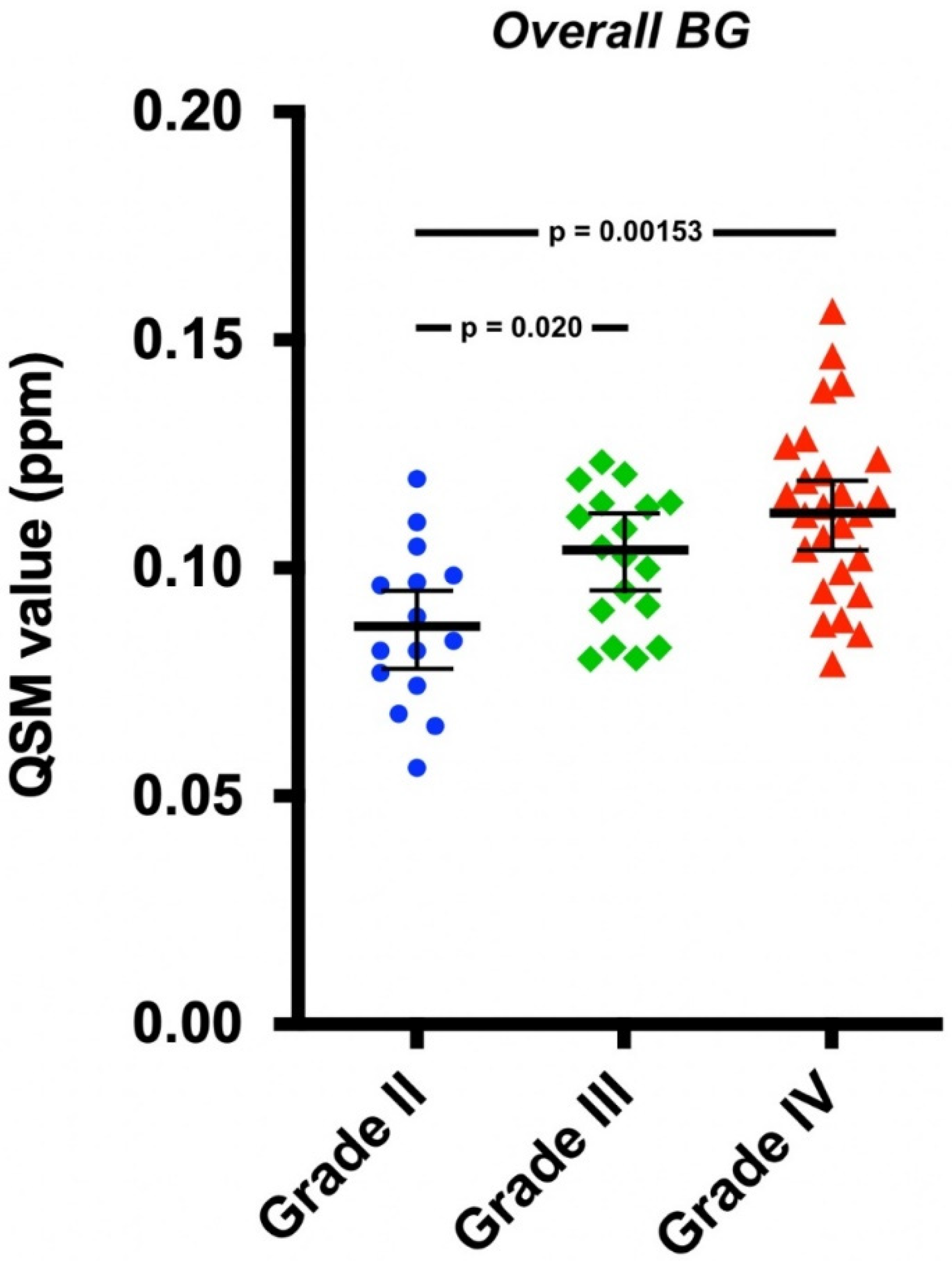
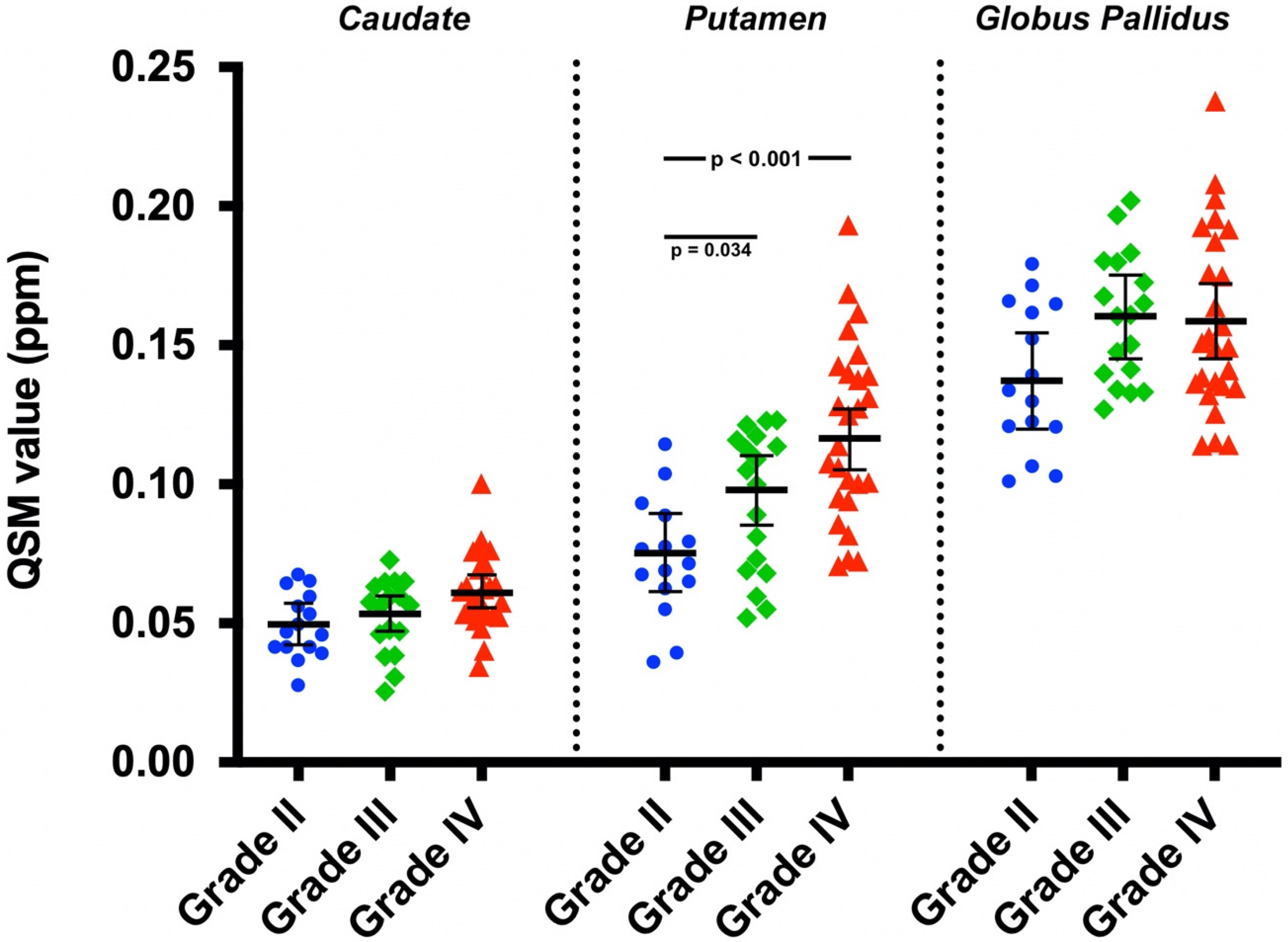

| Covariate Adjusted Mean QSM Value (95% CI) (ppm) | |||
|---|---|---|---|
| Grade II | Grade III | Grade IV | |
| Caudate | 0.050 (0.042, 0.057) | 0.053 (0.047, 0.060) | 0.061 (0.055, 0.067) |
| Putamen | 0.075 (0.061, 0.089) | 0.098 (0.085, 0.110) | 0.116 (0.105, 0.127) |
| Globus pallidus | 0.137 (0.120, 0.154) | 0.160 (0.145, 0.175) | 0.159 (0.145, 0.172) |
| Overall BG | 0.087 (0.078, 0.096) | 0.104 (0.095, 0.112) | 0.112 (0.104, 0.119) |
| Covariate Adjusted Mean QSM Value (95% CI) (ppm) | |
|---|---|
| Astrocytoma | |
| Caudate | 0.055 (0.051, 0.059) |
| Putamen | 0.096 (0.089, 0.104) |
| Globus pallidus | 0.151 (0.142, 0.160) |
| Overall BG | 0.101 (0.096, 0.106) |
| Oligodendroglioma | |
| Caudate | 0.059 (0.051, 0.066) |
| Putamen | 0.112 (0.097, 0.126) |
| Globus pallidus | 0.162 (0.144, 0.180) |
| Overall BG | 0.111 (0.101, 0.121) |
| Mean QSM Value ± SD (ppm) | |||
|---|---|---|---|
| Grade II | Grade III | Grade IV | |
| All Patients/Tumor Types | |||
| Caudate | 0.049 ± 0.012 | 0.052 ± 0.013 | 0.062 ± 0.014 |
| Putamen | 0.073 ± 0.021 | 0.094 ± 0.025 | 0.119 ± 0.032 |
| Globus pallidus | 0.138 ± 0.026 | 0.160 ± 0.023 | 0.158 ± 0.032 |
| Overall BG | 0.087 ± 0.018 | 0.102 ± 0.015 | 0.113 ± 0.019 |
| Male | |||
| Caudate | 0.051 ± 0.011 | 0.051 ± 0.013 | 0.0611 ± 0.0080 |
| Putamen | 0.073 ± 0.024 | 0.094 ± 0.026 | 0.116 ± 0.025 |
| Globus pallidus | 0.137 ± 0.029 | 0.157 ± 0.021 | 0.164 ± 0.038 |
| Overall BG | 0.087 ± 0.018 | 0.101 ± 0.016 | 0.114 ± 0.017 |
| Female | |||
| Caudate | 0.048 ± 0.013 | 0.057 ± 0.012 | 0.062 ± 0.016 |
| Putamen | 0.074 ± 0.020 | 0.094 ± 0.029 | 0.121 ± 0.036 |
| Globus pallidus | 0.140 ± 0.025 | 0.169 ± 0.029 | 0.155 ± 0.030 |
| Overall BG | 0.087 ± 0.018 | 0.1066 ± 0.0088 | 0.113 ± 0.021 |
| Astrocytoma | |||
| Caudate | 0.046 ± 0.013 | 0.050 ± 0.015 | 0.062 ± 0.014 |
| Putamen | 0.067 ± 0.024 | 0.084 ± 0.025 | 0.119 ± 0.032 |
| Globus pallidus | 0.131 ± 0.026 | 0.157 ± 0.023 | 0.158 ± 0.032 |
| Overall BG | 0.081 ± 0.020 | 0.097 ± 0.015 | 0.113 ± 0.019 |
| Oligodendroglioma | |||
| Caudate | 0.052 ± 0.010 | 0.0562 ± 0.0074 | – |
| Putamen | 0.079 ± 0.018 | 0.1139 ± 0.0086 | – |
| Globus pallidus | 0.144 ± 0.027 | 0.166 ± 0.023 | – |
| Overall BG | 0.092 ± 0.015 | 0.1120 ± 0.0094 | – |
Publisher’s Note: MDPI stays neutral with regard to jurisdictional claims in published maps and institutional affiliations. |
© 2022 by the authors. Licensee MDPI, Basel, Switzerland. This article is an open access article distributed under the terms and conditions of the Creative Commons Attribution (CC BY) license (https://creativecommons.org/licenses/by/4.0/).
Share and Cite
Reith, T.P.; Prah, M.A.; Choi, E.-J.; Lee, J.; Wujek, R.; Al-Gizawiy, M.; Chitambar, C.R.; Connelly, J.M.; Schmainda, K.M. Basal Ganglia Iron Content Increases with Glioma Severity Using Quantitative Susceptibility Mapping: A Potential Biomarker of Tumor Severity. Tomography 2022, 8, 789-797. https://doi.org/10.3390/tomography8020065
Reith TP, Prah MA, Choi E-J, Lee J, Wujek R, Al-Gizawiy M, Chitambar CR, Connelly JM, Schmainda KM. Basal Ganglia Iron Content Increases with Glioma Severity Using Quantitative Susceptibility Mapping: A Potential Biomarker of Tumor Severity. Tomography. 2022; 8(2):789-797. https://doi.org/10.3390/tomography8020065
Chicago/Turabian StyleReith, Thomas P., Melissa A. Prah, Eun-Jung Choi, Jongho Lee, Robert Wujek, Mona Al-Gizawiy, Christopher R. Chitambar, Jennifer M. Connelly, and Kathleen M. Schmainda. 2022. "Basal Ganglia Iron Content Increases with Glioma Severity Using Quantitative Susceptibility Mapping: A Potential Biomarker of Tumor Severity" Tomography 8, no. 2: 789-797. https://doi.org/10.3390/tomography8020065
APA StyleReith, T. P., Prah, M. A., Choi, E.-J., Lee, J., Wujek, R., Al-Gizawiy, M., Chitambar, C. R., Connelly, J. M., & Schmainda, K. M. (2022). Basal Ganglia Iron Content Increases with Glioma Severity Using Quantitative Susceptibility Mapping: A Potential Biomarker of Tumor Severity. Tomography, 8(2), 789-797. https://doi.org/10.3390/tomography8020065