Article

The Effect of Iron Oxide Insertion on the In Vitro Bioactivity, and Antibacterial Properties of the 45S5 Bioactive Glass

by
Imen Hammami
1,
Suresh Kumar Jakka
1,
Isabel Sá-Nogueira
2,3,
João Paulo Borges
4 and
Manuel Pedro Fernandes Graça
1,*
1
I3N and Physics Department, Aveiro University, 3810-193 Aveiro, Portugal
2
Associate Laboratory i4HB—Institute for Health and Bioeconomy, NOVA School of Science and Technology, NOVA University Lisbon, 2819-516 Caparica, Portugal
3
UCIBIO—Applied Molecular Biosciences Unit, Department of Life Sciences, NOVA School of Science and Technology, NOVA University Lisbon, 2819-516 Caparica, Portugal
4
I3N-CENIMAT and Materials Science Department, NOVA School of Science and Technology, Campus de Caparica, Nova University Lisbon, 2829-516 Caparica, Portugal
*
Author to whom correspondence should be addressed.
Biomimetics 2024, 9(6), 325; https://doi.org/10.3390/biomimetics9060325
Submission received: 30 April 2024 / Revised: 24 May 2024 / Accepted: 27 May 2024 / Published: 29 May 2024
(This article belongs to the Special Issue Advances in Bioceramics for Bone Regeneration)

Abstract

:
The aging population and increasing incidence of trauma among younger age groups have heightened the increasing demand for reliable implant materials. Effective implant materials must demonstrate rapid osseointegration and strong antibacterial properties to ensure optimal patient outcomes and decrease the chance of implant rejection. This study aims to enhance the bone–implant interface by utilizing 45S5 bioglass modified with various concentrations of Fe3O4 as a coating material. The effect of the insertion of Fe3O4 into the bioglass structure was studied using Raman spectroscopy which shows that with the increase in Fe3O4 concentration, new vibration bands associated with Fe-related structural units appeared within the sample. The bioactivity of the prepared glasses was evaluated using immersion tests in simulated body fluid, revealing the formation of a calcium phosphate-rich layer within 24 h on the samples, indicating their potential for enhanced tissue integration. However, the sample modified with 8 mol% of Fe3O4 showed low reactivity, developing a calcium phosphate-rich layer within 96 h. All the bioglasses showed antibacterial activity against the Gram-positive and Gram-negative bacteria. The modified bioglass did not present significant antibacterial properties compared to the bioglass base.

1. Introduction

The field of biomedical implants has evolved from having a main emphasis on mechanical characteristics to a more sophisticated understanding that incorporates critical biological qualities. In the past, the mechanical aspects of implants remained crucial for restoring normal tissue functions, with titanium being the predominant material due to its outstanding mechanical properties [1,2]. However, recent findings emphasize the need to integrate biocompatibility, osseointegration, tissue regeneration, and other biological factors as fundamental components of successful implant design [3,4]. The realization that clinical outcomes are intricately linked to the interaction of the implant with the biological fluid has prompted advancements in materials and surface modifications [5,6,7,8]. In recent years, there has been a growing interest in coating implants with bioactive materials to enhance biological performance including biocompatibility, osseointegration, and tissue regeneration, therefore ensuring enhanced longevity, stability, and overall success in clinical applications [7,9,10].
One of these bioactive materials is the 45S5 bioglass® (45% SiO2, 24.5% Na2O, 24.5% CaO, and 6% P2O5 (wt)), invented by L.L. Hench, which has emerged as a compelling class of ceramic materials, notably for bone regeneration [11,12,13]. Their innate bioactivity, characterized by the capacity to form a robust and direct bond with tissues through subsequent chemical reactions, makes them promising candidates for various medical applications [14,15,16]. Beyond their efficacy in stimulating bone growth, bioglasses exhibit intrinsic antibacterial properties, a feature that has attracted significant attention in recent research [17,18]. Notably, this antibacterial activity is not dependent on the inclusion of ions to their composition but arises from factors such as localized pH elevation caused by the exchange of glass network-modifier ions with H+ ions from surrounding fluids and osmotic pressure increases [19].
Researchers have explored the incorporation of therapeutic metal ions into bioglass to confer additional biological properties, including improved antibacterial performance, osteogenesis, and anti-inflammatory properties [7,20,21,22,23,24,25,26]. Iron (Fe) has demonstrated exceptional performance in numerous biomedical applications and is considered a promising candidate due to its chemical stability, biocompatibility, and resistance to corrosion [27]. Additionally, it is involved in bone metabolism, particularly in the processes of osteoblastic differentiation, proliferation, and calcification [28]. Furthermore, recent research investigates the use of iron nanoparticles in cancer treatment, exploiting their magnetic properties for targeted drug delivery and hyperthermia treatment [29]. Moreover, studies have demonstrated the antibacterial effects of iron oxide against several types of bacteria, thereby inhibiting biofilm formation [29,30].
The electrical characteristics of bioglass, particularly its capacity to store electrical charges, have been investigated for their potential influence on osseointegration [31,32,33]. The charge storage capacity of bioglass is attributed to the high mobility of the sodium ion chain. Because of its distinctive chemical composition, bioglass can effectively store and release electrical charges, with potential therapeutic application. In their research, Obata et al. [32,33] showed that the bioglass was able to store larger electrical charges compared to hydroxyapatite (HA). Specifically, the calculated stored electrical charges of the bioglass were found to be 1000 times greater than those of HA. In another study, Obata et al. [31] demonstrated that the presence of surface charges on bioglass improves its osteoconductivity by controlling the growth of calcium phosphate crystals. Moreover, Singh et al. [34] observed in their work an increased growth rate of osteoblast cells on negatively polarized 1393B3 bioglass (56% B2O3, 5.5% Na2O, 18.5% CaO, 11.1% K2O, 3.7% P2O5, 4.6% MgO (wt.%)). The enhanced cellular response suggests that surface polarization has a positive effect on the biological performance of the bioglass.
In this study, 45S5 bioglasses modified with the insertion of various percentages of iron oxide (Fe3O4) were prepared using the melt-quenching method. While the incorporation of hematite Fe2O3 into the bioglass system has been thoroughly explored, the insertion and effect of magnetite Fe3O4 in the preparation of the 45S5 bioglass is not widely documented in the literature. The influence of Fe3O4 on the bioglass structure was evaluated using Raman spectroscopy. Impedance spectroscopy (IS) was employed to validate alterations in the electrical properties of the prepared glasses. To assess the bioactivity of the prepared samples, an immersion test in simulated body fluid (SBF) was used. The antibacterial properties of the bioglasses were analyzed by examining their ability to hinder the growth of S. aureus bacteria, a common source of implant infections, as well as other bacterial strains known to contribute to the formation of detrimental biofilms.

2. Materials and Methods

2.1. Materials Synthesis

The melt-quenching method was employed to fabricate the bioglass, following the composition of 45S5 Bioglass® (46.1SiO2-24.4Na2O-26.9CaO-2.6P2O5, mol%) as outlined by Hench et al. [11,12]. Various concentrations of iron oxide (Fe3O4) (0, 1, 2, 4, and 8 mol%) were introduced to the bioglass network (denoted as Fe0, Fe1, Fe2, Fe4, and Fe8 respectively). The planetary ball-milling technique was employed for 1 h at 300 rpm to mix and homogenize the chemical precursors, namely SiO2, P2O5, CaCO3, Na2CO3, and Fe3O4. These precursors were supplied by Sigma-Aldrich in Darmstadt, Germany, with a high purity grade of ≥99%. Then, the mixed powder underwent a calcination process at 800 °C for 1h followed by a melting process at 1300 °C for 1 h. To ensure greater sample homogeneity, the bioactive glass was re-melted using the same conditions. The resulting bulk samples from the melt-quenching process were manually ground and subsequently milled in a planetary ball mill system for 1 h at 300 rpm. A series of bioglass compositions (mol%): (100 − x) 45S5 bioglass + x Fe3O4 is obtained. The nominal composition of the prepared bioglass samples is described in Table 1.

2.2. Structural Characterization

Raman spectroscopy was performed on the bulk materials using a Jobin Yvon HR800 spectrometer, equipped with an Ar+ laser (λ = 532 nm). The spectra were obtained in back-scattering geometry, encompassing the spectral range of 200 to 1400 cm−1.
The XRD diffractograms were obtained at room temperature on an Aeris-Panalytical diffractometer using CuKα radiation (λ = 1.54056 Å). The measurement parameters were a 2θ range of 10°–70° and a scan step of 0.002°.

2.3. Electrical Characterization

Electrical measurements were carried out on glass bulk samples having a consistent thickness of 1 mm. Utilizing ImageJ software 1.8.0, the surface area of the samples was determined. The samples were then coated with silver conducting paste on their opposite parallel sides. A nitrogen bath cryostat was employed for alternating current (AC) investigations, enabling temperature measurements ranging from 100 to 400 K. The temperature of the sample was regulated using an Oxford Research IT-C4 system equipped with a platinum sensor for temperature monitoring. To assess dielectric properties in AC experiments, an impedance analyzer (Agilent 4294A, Santa Clara, CA, USA) was utilized. Measurements were performed using the Cp−Rp configuration and a 0.5 V AC signal across a frequency range spanning from 100 Hz to 1 MHz. Using Equations (1) and (2), the complex electric permittivity ε* and the complex dielectric modulus M* were calculated [35,36,37,38,39].
ε* = ε′ − j ε″ = Cp (d/ε0 A) − j d (ω Rp ε0 A),
M* = 1/ε* = M′ + j M″ = ε′/(ε′2 + ε″2) + j ε″/(ε′2 + ε″2),
where Cp and Rp are the measured capacitance and resistance, d is the sample thickness, A the electrode area, ω is the angular frequency, and ε0 the permittivity of the space (8.8542 × 10⁻¹² F/m).
To calculate the activation energy, the Arrhenius model was employed to adjust the temperature dependence of the relaxation frequency [35,37,40,41,42]:
fmax = f0 exp (−Ea/(kB T)),
where fmax is the frequency where the M″ reaches a maximum for a specific temperature, f0 is a preexponential factor, Ea is the activation energy, KB is the Boltzmann constant, and T is the temperature.

2.4. Bioactivity

Bioactivity test was carried out in accordance with ISO23317:2017 Standards: “Implants for surgery—In vitro evaluation for apatite-forming ability of implant materials” [43]. For the experiment, pellets made from bioactive glass powder with a diameter of 7 mm were immersed in simulated body fluid (SBF) and placed in an incubator set at 37 °C, equipped with a continuous oscillating support. It is crucial to note that the SBF solution was renewed every 48 h to replicate the biological environment accurately. At intervals of 12, 24, 48, 96 h, 14, and 28 days of SBF immersion, the samples were removed from the medium, rinsed with deionized water, and left to dry at room temperature. The surface of the samples was examined using SEM (Scanning Electron Microscopy), a microscope from TESCAN model Vega 3 (TESCAN ORSAY HOLDING, Brno-Kohoutovice, Czech Republic), equipped with Bruker EDS (energy dispersive spectroscopy) system. Additionally, the pH of the SBF medium was evaluated in both the group of samples that underwent medium changes and the group that remained unchanged. The test was conducted in duplicate.

2.5. Antibacterial Activity

Agar diffusion assay plates were utilized to observe the antimicrobial behavior of all the samples. Reference strains used were Escherichia coli K12 DSM498 (DSMZ, Braunschweig, Germany), Staphylococcus aureus COL MRSA (a methicillin-resistant strain provided by Rockefeller University), and Streptococcus mutans DSM20523 (DSMZ, Braunschweig, Germany). The bacterial strains were cultivated in tryptic soy broth (TSB) at 37 °C overnight. Prior to testing, the disks with a diameter of 6 mm were sterilized at 180 °C for 2 h. The bioassay consisted of two layers: a base layer solidified with agar 1.5% w/v, and a top layer at 0.8% w/v. Plates were prepared with a base layer of 18–20 mL and an overlay of 4 mL of molten seeded bacteria containing around 108 CFU/mL of the relevant indicator bacteria. The disks of material to be tested were placed in the center of the plates, and then placed in an incubator at 37 °C for 24 h. For S. mutans, an incubator with 5% CO2 was utilized [14].
Photographs of the pellets were captured, and the diameter of the resulting inhibition zone was assessed using ImageJ software. To ensure precision, each pellet underwent thorough measurement from various angles, with a total of 30 measurements per pellet. Statistical analysis of the data was conducted using GraphPad Prism 8.0 software, utilizing an unpaired t-test, to compare the antibacterial effects of the bioactive glass base composition with each of the different samples.

3. Results and Discussion

3.1. Structural Characterization

The structural spectroscopic characteristics of the bioglass samples are analyzed by Raman spectroscopy, as depicted in Figure 1. One can notice that the insertion of iron oxide alters the glass structure. In the low-frequency region (>800 cm−1), the bioglass base, Fe0, exhibits a band at around 630 cm−1 attributed to the rocking motion of bridging oxygen in structural units containing non-bridging oxygen ions (NBOs) [44,45,46]. However, with an increase in the concentration of Fe3O4, the intensity of this band decreases, and additional bands are observed. The appearance of the band at around 730 cm−1 in the spectra of the iron-containing glasses is likely attributed to the introduction of Fe3O4, which results in the formation of Fe-related structural units within the sample. A similar feature has been observed in the Raman spectra of nano-Fe3O4 samples and silicate glasses doped with Fe3O4 [47,48]. Moreover, the presence of two bands at 560 cm−1, and 480 cm−1, with intensities increasing as the concentration of Fe3O4 inserted in the bioglass increases, matches well with the vibrational modes of hematite [49,50].
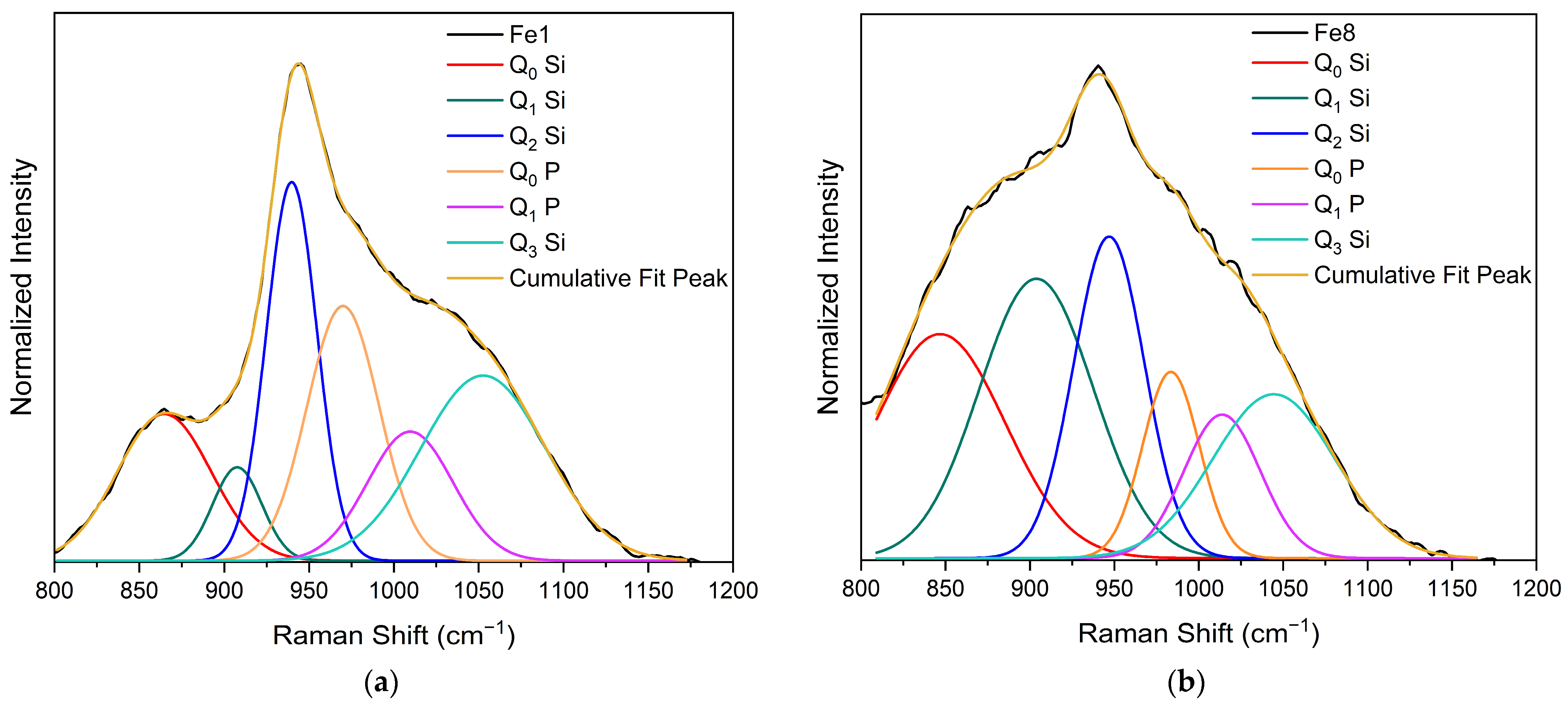
In the high-frequency region (<800 cm−1), three vibrational bands can be observed at around 850 cm−1, 940 cm−1, and 1050 cm−1. This region is considered significant as it contains the most intriguing vibrational modes for silicate glasses, characterized by symmetric and asymmetric stretching. Consequently, a deconvolution of the Raman spectra within this region was conducted. The spectral deconvolutions for the bioglass modified with 1, and 8% mol of Fe3O4 are illustrated in Figure 2a and Figure 2b, respectively. The results show the presence of six vibrational modes located at around 845–860 cm−1, 905–908 cm−1, 940–906 cm−1, 970–985 cm−1, 1009–1014 cm−1, and 144–1053 cm−1, associated with Q0 Si units, Q1 Si units, Q2 Si units, Q0 P units, Q1 P units and Q3 Si units, respectively [44,51,52,53,54,55].

3.2. Electrical Characterization

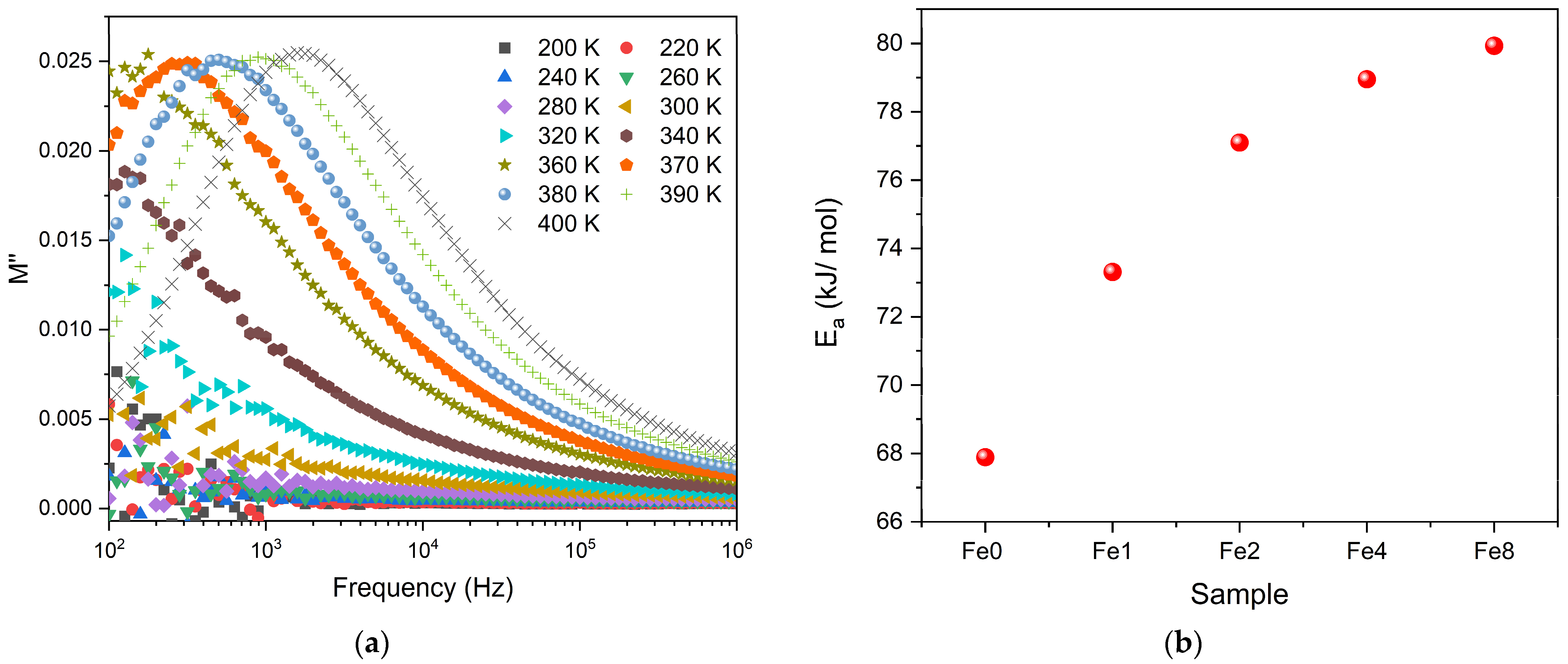
The dielectric properties of the bioglasses were represented in the form of modulus formalism, M*, expressed as 1/ε*. This method offers the advantage of reducing the impact of low capacitance factors, such as electrode polarization and low-frequency conductivity, on the dielectric data. The results, shown in Figure 3a, indicate a single dielectric relaxation process that shifts to higher frequencies with increasing temperature which indicates the presence of a thermally activated process. Notably, other formalisms, such as permittivity, impedance, or admittance, do not clearly show this dielectric relaxation behavior. Therefore, the observed relaxation phenomenon is due to an intrinsic characteristic related to the formation of dipoles between the network modifier ions and the nearby non-bridging oxygen ions. The variation of the relaxation frequency versus temperature determined from M″, was adjusted using the Arrhenius model to calculate the activation energy of this relaxation process. Figure 3b shows that the activation energy Ea increases with the increase in Fe3O4 concentration in the bioglass network. This suggests that the glass structure becomes more rigid, creating higher barriers for ion movement and thereby increasing the activation energy [51].
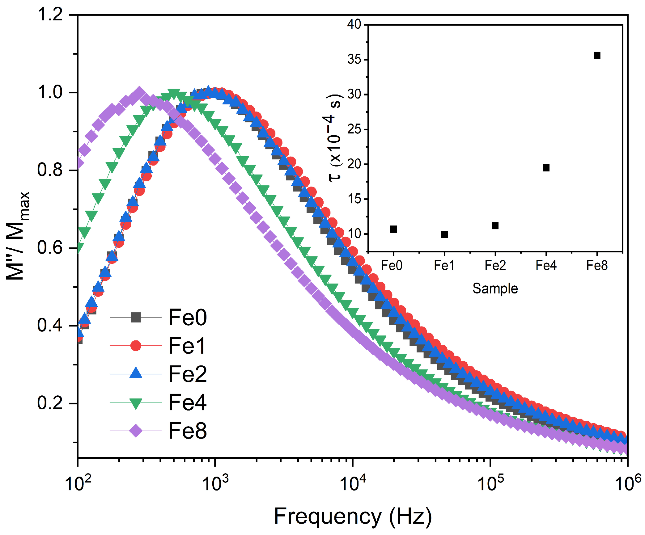
Figure 4 displays a comparison of the normalized imaginary parts of the electric modulus M″/M″max versus frequency for different concentrations of Fe3O4 incorporated into the Bioglass network. A shift in the electrical modulus to a lower frequency was observed with the insertion of a high concentration of Fe3O4 suggesting an increase in relaxation time (inset of Figure 4). This suggests that the dipoles in the bioglass system have less freedom to align with the direction of the applied electric field. Consequently, the network of bioglasses modified with more than 2 mol% Fe3O4 is more polymerized.

3.3. Bioactivity

To assess the kinetics of apatite precipitation on bioactive glass surfaces within a physiological environment, bioglass pellets underwent immersion in simulated body fluid (SBF) for varying durations: 12, 24, 48, 96 h, 14, and 28 days. SEM-EDS analysis was utilized to examine the atomic percentage of the elements presented on the surface. The Figure 5a,b reveals a gradual decline in silicon and sodium ions on the glass surface over time, reaching stabilization after 96 h. Indeed, the exposure of the bioglass to the SBF medium initiates ionic exchange between them. The pH rises as a consequence of the exchange of H+ and H3O+ ions in the medium with the alkali and alkaline earth ions (Na+ and Ca2+) on the bioglass surface. The breakage of Si-O-Si bonds in the glass network is facilitated by this rise in pH, which accelerates the glass’s dissolution and promotes the creation of silanol units (Si(OH)4). These silanol units condense onto the glass surface to create a layer of hydrated silica. This layer serves as a nucleation site for the subsequent crystallization of a carbonated hydroxyapatite layer [56,57].
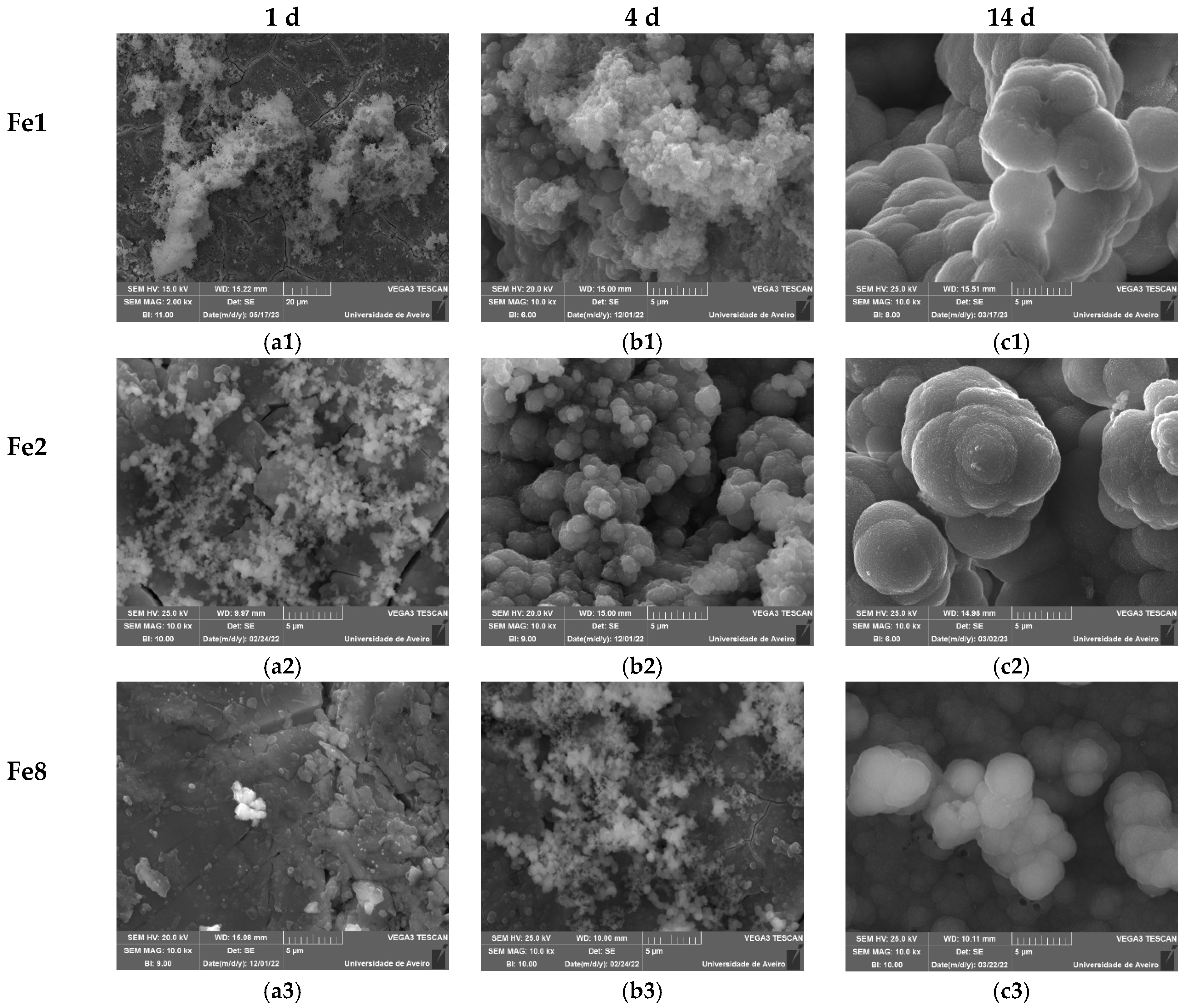
Figure 5c illustrates the calcium-to-phosphorus (Ca/P) ratio, showing that for the samples with a low content of Fe3O4, this ratio decreases with increasing immersion time in SBF and stabilizes to reach a value close to that of hydroxyapatite in natural bone (Ca/P = 1.67) within the first 24 h [58]. However, for the samples with 8 mol% of Fe3O4, this value is considered high, reaching approximately 4 after 28 days of SBF immersion. SEM micrographs (Figure 6) validate the bioactivity of the glasses by revealing the presence of spherical (cauliflower-like) particles on the surfaces of the pellets. This observation provides evidence of the development of an apatitic layer. As immersion time in SBF increased, the apatite particles aggregated and became denser. After 14 days of immersion, this process resulted in the complete coating of the surface with an apatite layer. In the case of the bioglass modified with 8 mol% of Fe3O4, small amounts of apatite particles are observed during the initial days of immersion in SBF. After 14 days, the size of these particles is notably smaller compared to those found in other bioglass samples, indicating reduced bioactivity.
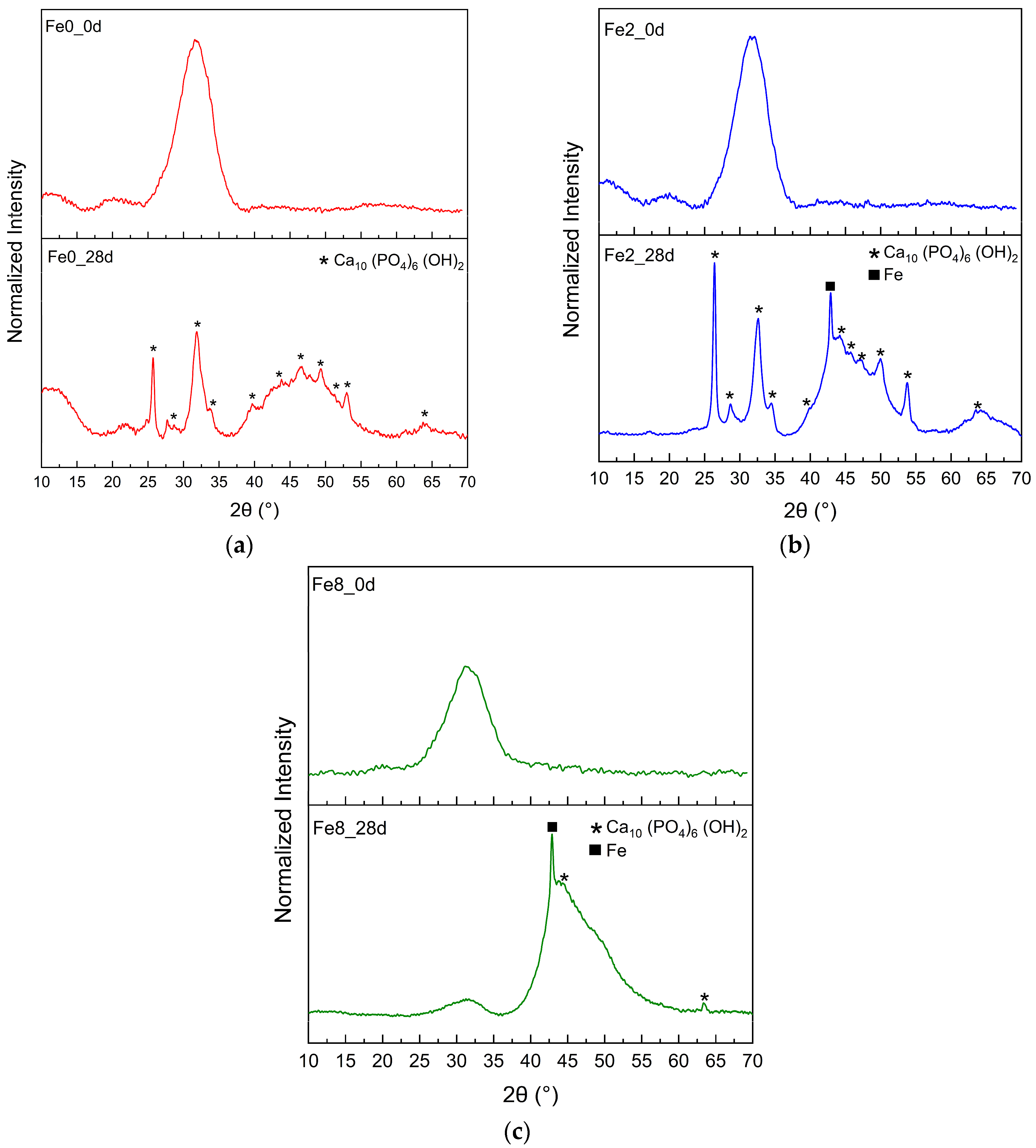
Figure 7 shows the XRD pattern of the Fe0, Fe2, and Fe3 samples before and after immersion in SBF for 28 d. Before immersion in SBF, the XRD patterns of the samples did not exhibit any distinct diffraction peaks but rather an amorphous hump, indicating a lack of long-range atomic order in their molecular arrangement. However, the immersion of the samples in SBF for 28 d led to the formation of a crystalline phase, with the main diffraction peak corresponding to hydroxyapatite Ca10 (PO4)6 (OH)2 (ICCD. No 00-001-1008) [59]. Moreover, the XRD results indicate that the apatite formed on the surface of bioglass with a high concentration of Fe3O4 exhibits a lower crystallinity than those formed on the samples with a low concentration of Fe3O4, suggesting that a high content of iron delays the crystallization of the apatite layer. These results are in agreement with the SEM analysis, which shows the formation of an apatite layer on the bioglass surface immersed in SBF. Additionally, the SEM analysis reveals that the Fe8 sample exhibits the smallest apatite particles on its surface compared to the other bioglass samples.
Figure 8 represents Raman spectra of bioglass samples before and after immersion of 28 d in SBF solution. The samples with low content of Fe3O4, after immersion in SBF for 28 d, show changes in their Raman spectra compared to their initial spectra, becoming very similar to that of commercial hydroxyapatite [60,61]. With SBF immersion, the bands attributed to the vibration of Si structural units at 800–1100 cm−1 become less intense, and the band associated with PO4 vibration becomes more pronounced and shifts to 960 cm−1. Moreover, additional bands related to PO4 vibration appeared at 440 and 580 cm−1. The band around 1080 cm−1 is associated with a carbonate group, indicating that the in vitro-grown hydroxyapatite is carbonated [61,62]. However, the Fe8 sample with a high Fe3O4 concentration did not reveal an alteration in Raman spectra. This result is consistent with the SEM and XRD findings, revealing that at high concentrations, Fe3O4 decreases the formation of the hydroxyapatite layer.
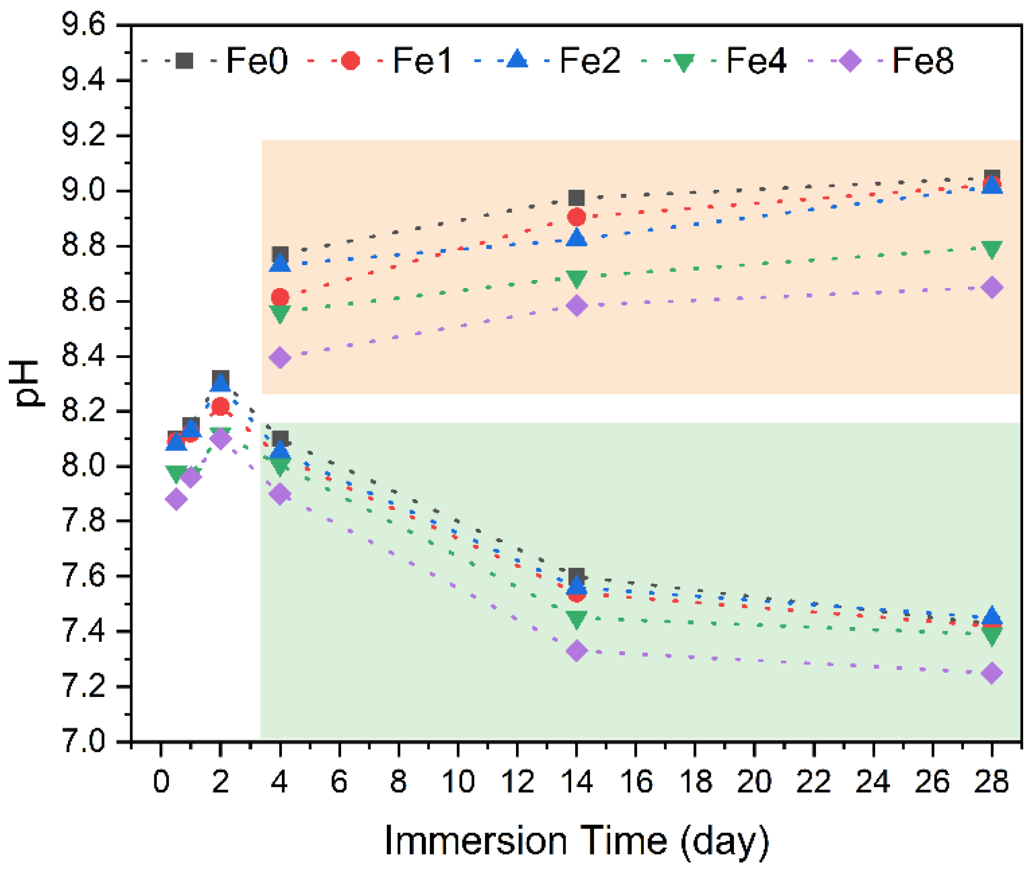
Figure 9 depicts the pH variations in SBF over different immersion times for pellets under two conditions: with regular medium changes every two days to mimic the behavior of the material in the physiological environment, and continuous exposure without medium replacement. As previously mentioned, the pH levels of the surrounding SBF solution in contact with the bioglass gradually increased over time due to the dissolution of alkaline metal ions (Na+ and Ca2+) from the bioglass pellets. Notably, during the initial two days, this increase was particularly prominent, with a more gradual rise observed during the subsequent period, as evidenced by the values enclosed within the orange rectangle (representing samples where the medium remained unchanged). However, when the immersion conditions were altered to mimic those of the in vivo environment, with medium changes every two days, a decrease in pH was observed (the values enclosed within the green rectangle). The decrease in pH suggests the formation of a hydroxyapatite layer on the bioglass surface [56,63].

3.4. Antibacterial Activity

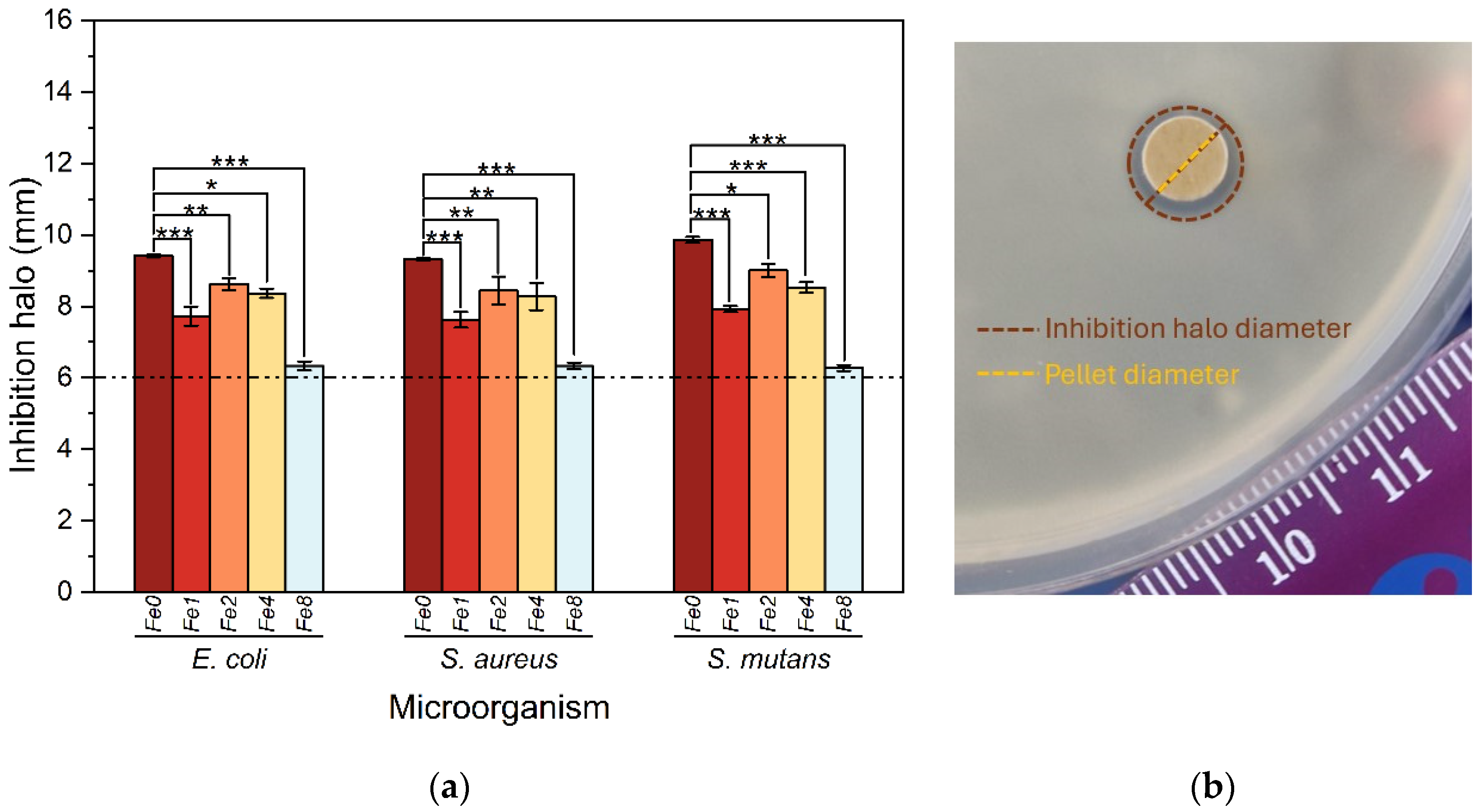
The antibacterial activity of the prepared bioglasses was assessed using the agar diffusion method against E. coli, S. aureus, and S. mutans. The inhibition halo diameters of the pellets against various bacteria are presented in Figure 10.
The results obtained demonstrate the antibacterial efficacy of all samples, as evidenced by the presence of an inhibition zone surrounding the Bioglass pellets. The observed mean diameter of these zones, exceeding 6 mm, surpasses the diameter of the pellets, indicating substantial antibacterial activity against all tested bacterial strains. Two main processes elucidate the antibacterial properties of 45S5 bioglass. These processes involve the release of bioglass ions, particularly Na+ and Ca2+, which induce alkalinity in the surrounding medium, thereby increasing osmotic pressure [18,19]. This alkaline pH inhibits bacterial growth and metabolic processes by disrupting proteins and enzymes. Furthermore, the discharge of ions and subsequent fluctuations in their concentrations affect the integrity of bacterial cell membranes and intramembrane pressure, leading to changes in cellular size, shape, and membrane tension levels, ultimately resulting in bacterial death. A notable decrease in antibacterial efficacy was observed in samples modified with the insertion of Fe3O4 compared to the base material. This decline in antibacterial effect can be attributed to the presence of iron. This finding is supported by the analysis conducted to evaluate the pH levels of the SBF medium after immersion of the samples. Remarkably, the pH measurements show that samples containing Fe3O4 exhibited lower pH values compared to the base material, with higher concentrations of iron displaying minimal pH values. Therefore, the presence of iron in the modified samples contributes to the alteration of pH levels, which in turn could potentially influence their antibacterial properties. The bioglass modified with 2 mol% Fe3O4 showed a higher antibacterial activity compared to all modified bioglasses with Fe3O4. It exhibits a mean inhibition of halo diameter of 8.61 mm, 8.44 mm and 9.01 mm against E. coli, S. aureus and S. mutans bacteria, respectively.

4. Conclusions

Several bioglass samples modified with the addition of Fe3O4 were successfully produced using the melt-quenching technique. Structural analysis of the samples using Raman spectroscopy revealed that the insertion of Fe3O4 alters the bioglass structure, leading to the appearance of new vibrations related to the formation of Fe-related structural units. The bioactivity test showed that within 24 h, most samples formed a calcium phosphate-rich layer, except for the sample modified with 8 mol% of Fe3O4, which exhibited slower reactivity, developing this layer within 96 h. The antibacterial tests showed that the prepared glasses exhibited antibacterial activity against E. coli, S. aureus, and S. mutans bacteria. However, the modified bioglass with Fe3O4 did not exhibit notable antibacterial properties compared to the 45S5 bioglass. Among all modified bioglasses with Fe3O4 addition, the bioglass with 2 mol% Fe3O4 showed higher antibacterial properties.

Author Contributions

Conceptualization, I.H., M.P.F.G. and I.S.-N.; methodology, I.H., M.P.F.G., S.K.J. and I.S.-N.; software, I.H.; validation, I.H., M.P.F.G., I.S.-N. and J.P.B.; formal analysis, I.H.; investigation, I.H., S.K.J., I.S.-N. and M.P.F.G.; resources, J.P.B., I.S.-N. and M.P.F.G.; data curation, I.H.; writing—original draft preparation, I.H.; writing—review and editing, M.P.F.G., I.S.-N. and J.P.B.; visualization, I.H.; supervision, M.P.F.G. and J.P.B. All authors have read and agreed to the published version of the manuscript.

Funding

This research was funded by FEDER funds through the COMPETE 2020 Program and National Funds through the FCT—Portuguese Foundation for Science and Technology under the projects LISBOA-01-0247-FEDER-039985/POCI-01-0247-FEDER-039985, LA/P/0037/2020, UIDP/50025/2020, and UIDB/50025/2020 of the Associate Laboratory Institute of Nanostructures, Nanomodelling and Nanofabrication—i3N, UIDP/04378/2020 and UIDB/04378/2020 of the Research Unit on Applied Molecular Biosciences—UCIBIO, and LA/P/0140/2020 of the Associate Laboratory Institute for Health and Bioeconomy—i4HB. S.K. Jakka acknowledges FCT—Fundaçao para a Ciência e a Tecnologia, Portugal, I.P., in the scope of the framework contract foreseen in the numbers 4, 5, and 6 of article 23 of the Decree Law 57/2016 of 29 August, changed by Law 57/2017 of 19 July.

Institutional Review Board Statement

Not applicable.

Informed Consent Statement

Not applicable.

Data Availability Statement

The data presented in this study are available from the corresponding author upon request.

Conflicts of Interest

The authors declare no conflicts of interest.

References

  1. Wu, H.; Chen, X.; Kong, L.; Liu, P. Mechanical and Biological Properties of Titanium and Its Alloys for Oral Implant with Preparation Techniques: A Review. Materials 2023, 16, 6860. [Google Scholar] [CrossRef] [PubMed]
  2. Hoque, M.E.; Showva, N.-N.; Ahmed, M.; Rashid, A.B.; Sadique, S.E.; El-Bialy, T.; Xu, H. Titanium and Titanium Alloys in Dentistry: Current Trends, Recent Developments, and Future Prospects. Heliyon 2022, 8, e11300. [Google Scholar] [CrossRef]
  3. Cruz, M.B.; Silva, N.; Marques, J.F.; Mata, A.; Silva, F.S.; Caramês, J. Biomimetic Implant Surfaces and Their Role in Biological Integration—A Concise Review. Biomimetics 2022, 7, 74. [Google Scholar] [CrossRef] [PubMed]
  4. Hammami, I.; Gavinho, S.R.; Pádua, A.S.; Lança, M.D.C.; Borges, J.P.; Silva, J.C.; Sá-Nogueira, I.; Jakka, S.K.; Graça, M.P.F. Extensive Investigation on the Effect of Niobium Insertion on the Physical and Biological Properties of 45S5 Bioactive Glass for Dental Implant. Int. J. Mol. Sci. 2023, 24, 5244. [Google Scholar] [CrossRef] [PubMed]
  5. Civantos, A.; Martínez-Campos, E.; Ramos, V.; Elvira, C.; Gallardo, A.; Abarrategi, A. Titanium Coatings and Surface Modifications: Toward Clinically Useful Bioactive Implants. ACS Biomater. Sci. Eng. 2017, 3, 1245–1261. [Google Scholar] [CrossRef]
  6. Pádua, A.S.; Gavinho, S.R.; Vieira, T.; Hammami, I.; Silva, J.C.; Borges, J.P.; Graça, M.P.F. In Vitro Characterization of Doped Bioglass 45S5/HAp Coatings Obtained by CoBlastTM Deposition. Coatings 2023, 13, 1775. [Google Scholar] [CrossRef]
  7. Kulkarni Aranya, A.; Pushalkar, S.; Zhao, M.; LeGeros, R.Z.; Zhang, Y.; Saxena, D. Antibacterial and Bioactive Coatings on Titanium Implant Surfaces. J Biomed. Mater. Res 2017, 105, 2218–2227. [Google Scholar] [CrossRef] [PubMed]
  8. Wang, Q.; Zhou, P.; Liu, S.; Attarilar, S.; Ma, R.L.-W.; Zhong, Y.; Wang, L. Multi-Scale Surface Treatments of Titanium Implants for Rapid Osseointegration: A Review. Nanomaterials 2020, 10, 1244. [Google Scholar] [CrossRef] [PubMed]
  9. Joy-anne, N.O.; Su, Y.; Lu, X.; Kuo, P.-H.; Du, J.; Zhu, D. Bioactive Glass Coatings on Metallic Implants for Biomedical Applications. Bioact. Mater. 2019, 4, 261–270. [Google Scholar]
  10. Bargavi, P.; Chitra, S.; Durgalakshmi, D.; Radha, G.; Balakumar, S. Zirconia Reinforced Bio-Active Glass Coating by Spray Pyrolysis: Structure, Surface Topography, in-Vitro Biological Evaluation and Antibacterial Activities. Mater. Today Commun. 2020, 25, 101253. [Google Scholar] [CrossRef]
  11. Hench, L.L. The Story of Bioglass®. J. Mater. Sci. Mater. Med. 2006, 17, 967–978. [Google Scholar] [CrossRef]
  12. Hench, L.L. An Introduction to Bioceramics, 2nd ed.; World Scientific Publishing: Singapore, 2013; pp. 1–600. [Google Scholar] [CrossRef]
  13. Hench, L.L.; Greenspan, D. Interactions between Bioactive Glass and Collagen: A Review and New Perspectives. J. Aust. Ceram. Soc. 2013, 49, 1–40. [Google Scholar]
  14. Mačković, M.; Hoppe, A.; Detsch, R.; Mohn, D.; Stark, W.J.; Spiecker, E.; Boccaccini, A.R. Bioactive Glass (Type 45S5) Nanoparticles: In Vitro Reactivity on Nanoscale and Biocompatibility. J. Nanopart. Res. 2012, 14, 966. [Google Scholar] [CrossRef]
  15. Kumar, A.; Murugavel, S.; Aditya, A.; Boccaccini, A.R. Mesoporous 45S5 Bioactive Glass: Synthesis, in Vitro Dissolution and Biomineralization Behavior. J. Mater. Chem. B 2017, 5, 8786–8798. [Google Scholar] [CrossRef] [PubMed]
  16. Silver, I.A.; Deas, J.; Erecińska, M. Interactions of Bioactive Glasses with Osteoblasts in Vitro: Effects of 45S5 Bioglass®, and 58S and 77S Bioactive Glasses on Metabolism, Intracellular Ion Concentrations and Cell Viability. Biomaterials 2001, 22, 175–185. [Google Scholar] [CrossRef] [PubMed]
  17. Allan, I.; Newman, H.; Wilson, M. Antibacterial Activity of Particulate Bioglass® against Supra-and Subgingival Bacteria. Biomaterials 2001, 22, 1683–1687. [Google Scholar] [CrossRef] [PubMed]
  18. Hu, S.; Chang, J.; Liu, M.; Ning, C. Study on Antibacterial Effect of 45S5 Bioglass®. J. Mater. Sci. Mater. Med. 2009, 20, 281–286. [Google Scholar] [CrossRef] [PubMed]
  19. Drago, L.; Toscano, M.; Bottagisio, M. Recent Evidence on Bioactive Glass Antimicrobial and Antibiofilm Activity: A Mini-Review. Materials 2018, 11, 326. [Google Scholar] [CrossRef] [PubMed]
  20. Hammami, I.; Gavinho, S.R.; Jakka, S.K.; Valente, M.A.; Graça, M.P.F.; Pádua, A.S.; Silva, J.C.; Sá-Nogueira, I.; Borges, J.P. Antibacterial Biomaterial Based on Bioglass Modified with Copper for Implants Coating. J. Funct. Biomater. 2023, 14, 369. [Google Scholar] [CrossRef] [PubMed]
  21. Baino, F. Copper-Doped Ordered Mesoporous Bioactive Glass: A Promising Multifunctional Platform for Bone Tissue Engineering. Bioengineering 2020, 7, 45. [Google Scholar] [CrossRef] [PubMed]
  22. Balamurugan, A.; Balossier, G.; Laurent-Maquin, D.; Pina, S.; Rebelo, A.H.S.; Faure, J.; Ferreira, J.M.F. An in Vitro Biological and Anti-Bacterial Study on a Sol-Gel Derived Silver-Incorporated Bioglass System. Dent. Mater. 2008, 24, 1343–1351. [Google Scholar] [CrossRef]
  23. Hammami, I.; Gavinho, S.R.; Pádua, A.S.; Sá-Nogueira, I.; Silva, J.C.; Borges, J.P.; Valente, M.A.; Graça, M.P.F. Bioactive Glass Modified with Zirconium Incorporation for Dental Implant Applications: Fabrication, Structural, Electrical, and Biological Analysis. Int. J. Mol. Sci. 2023, 24, 10571. [Google Scholar] [CrossRef]
  24. Cacciotti, I. Bivalent Cationic Ions Doped Bioactive Glasses: The Influence of Magnesium, Zinc, Strontium and Copper on the Physical and Biological Properties. J. Mater. Sci. 2017, 52, 8812–8831. [Google Scholar] [CrossRef]
  25. Tabia, Z.; Mabrouk, K.E.; Bricha, M.; Nouneh, K. Mesoporous Bioactive Glass Nanoparticles Doped with Magnesium: Drug Delivery and Acellular in Vitro Bioactivity. RSC Adv. 2019, 9, 12232–12246. [Google Scholar] [CrossRef]
  26. Teerakanok, S.; Zhao, M.; Giordano, R.; Fan, Y. Interaction of Doped Magnesium, Zinc and Fluoride Ions on Hydroxyapatite Crystals Grown on Etched Human Enamel. J. Cryst. Growth 2021, 571, 126262. [Google Scholar] [CrossRef]
  27. Arias, L.S.; Pessan, J.P.; Vieira, A.P.M.; de Lima, T.M.T.; Delbem, A.C.B.; Monteiro, D.R. Iron Oxide Nanoparticles for Biomedical Applications: A Perspective on Synthesis, Drugs, Antimicrobial Activity, and Toxicity. Antibiotics 2018, 7, 46. [Google Scholar] [CrossRef] [PubMed]
  28. Zhu, R.; Cai, M.; Fu, T.; Yin, D.; Peng, H.; Liao, S.; Du, Y.; Kong, J.; Ni, J.; Yin, X. Fe-Based Metal Organic Frameworks (Fe-MOFs) for Bio-Related Applications. Pharmaceutics 2023, 15, 1599. [Google Scholar] [CrossRef] [PubMed]
  29. Ansari, K.; Ahmad, R.; Tanweer, M.S.; Azam, I. Magnetic Iron Oxide Nanoparticles as a Tool for the Advancement of Biomedical and Environmental Application: A Review. Biomed. Mater. Devices 2023, 2, 139–157. [Google Scholar] [CrossRef]
  30. Nehra, P.; Chauhan, R.P.; Garg, N.; Verma, K. Antibacterial and Antifungal Activity of Chitosan Coated Iron Oxide Nanoparticles. Br. J. Biomed. Sci. 2017, 75, 13–18. [Google Scholar] [CrossRef] [PubMed]
  31. Obata, A.; Nakamura, S.; Moriyoshi, Y.; Yamashita, K. Electrical Polarization of Bioactive Glass and Assessment of Their in Vitro Apatite Deposition. J. Biomed. Mater. Res. Part A Biomater. 2003, 67, 413–420. [Google Scholar] [CrossRef] [PubMed]
  32. Obata, A.; Nakamura, S.; Yamashita, K. Interpretation of Electrical Polarization and Depolarization Mechanisms of Bioactive Glasses in Relation to Ionic Migration. Biomaterials 2004, 25, 5163–5169. [Google Scholar] [CrossRef] [PubMed]
  33. Obata, A.; Nakamura, S.; Sekijima, Y.; Yamashita, K. Control of Surface Reactions of Bioactive Glass by Electrical Polarization. J. Ceram. Soc. Jpn. Suppl. 2004, 112, S822–S825. [Google Scholar] [CrossRef]
  34. Singh, A.; Singh, P.; Dubey, A.K. Effect of Incorporation of Piezoelectric Phases on Antibacterial and Cellular Response of Borate Bioactive Glass. Open Ceram. 2022, 9, 100234. [Google Scholar] [CrossRef]
  35. Graça, M.P.F.; da Silva, M.F.; Sombra, A.S.B.; Valente, M.A. Electric and Dielectric Properties of a SiO2–Na2O–Nb2O5 Glass Subject to a Controlled Heat-Treatment Process. Phys. B Condens. Matter 2007, 396, 62–69. [Google Scholar] [CrossRef]
  36. Hammami, I.; Sales, A.M.J.; Benhamou, K.; Arous, M.; Costa, L.C.; da Cruz, J.A.; Kaddami, H. Dielectric Response and Molecular Dynamics of Nanocomposites Based on TEMPO-Oxidized Cellulose Nanofibrils and Polyvinyl Acetate. Therm. Sci. Eng. Prog. 2022, 34, 101428. [Google Scholar] [CrossRef]
  37. Barsoukov, E.; Macdonald, J.R. Impedance Spectroscopy: Theory, Experiment, and Applications; John Wiley & Sons: Hoboken, NJ, USA, 2018. [Google Scholar]
  38. El-Mallawany, R.A. Tellurite Glasses Handbook: Physical Properties and Data; CRC Press: Boca Raton, FL, USA, 2014. [Google Scholar]
  39. Ashok, J.; Purnachand, N.; Suresh Kumar, J.; Srinivasa Reddy, M.; Suresh, B.; Graça, M.P.F.; Veeraiah, N. Studies on Dielectric Dispersion, Relaxation Kinetics and a.c. Conductivity of Na2OCuOSiO2 Glasses Mixed with Bi2O3-Influence of Redox Behavior of Copper Ions. J. Alloys Compd. 2017, 696, 1260–1268. [Google Scholar] [CrossRef]
  40. Hammami, I.; Gavinho, S.R.; Pádua, A.S.; Graça, M.P.F.; Silva, J.C. Synthesis and Characterization of Iron Containing Bioactive Glass for Implants. In Proceedings of the 2022 E-Health and Bioengineering Conference (EHB), Iasi, Romania, 17–18 November 2022; pp. 1–4. [Google Scholar]
  41. Macdonald, J.R. Emphasizing Solid Materials and Systems. In Impedance Spectroscopy; John Wiley Sons Inc.: New York, NY, USA, 1987. [Google Scholar]
  42. Kumari, K.; Prasad, A.; Kumari, K.; Prasad, K. Dielectric, Impedance/Modulus and Conductivity Studies on [Bi0.5(Na1−xKx)0.5]0.94Ba0.06TiO3, (0.16 ≤ x ≤ 0.20) Lead-Free Ceramics. Am. J. Mater. Sci. 2016, 6, 1–18. [Google Scholar] [CrossRef]
  43. ISO 23317:2012; Implants for Surgery: In Vitro Evaluation for Apatite-Forming Ability of Implant Material. ISO Copyright Office: Geneva, Switzerland, 2012.
  44. Aguiar, H.; Serra, J.; González, P.; León, B. Structural Study of Sol–Gel Silicate Glasses by IR and Raman Spectroscopies. J. Non-Cryst. Solids 2009, 355, 475–480. [Google Scholar] [CrossRef]
  45. Aguiar, H.; Solla, E.L.; Serra, J.; González, P.; León, B.; Almeida, N.; Cachinho, S.; Davim, E.J.C.; Correia, R.; Oliveira, J.M. Orthophosphate Nanostructures in SiO2–P2O5–CaO–Na2O–MgO Bioactive Glasses. J. Non-Cryst. Solids 2008, 354, 4075–4080. [Google Scholar] [CrossRef]
  46. Lopes, J.H.; Magalhães, A.; Mazali, I.O.; Bertran, C.A. Effect of Niobium Oxide on the Structure and Properties of Melt-Derived Bioactive Glasses. J. Am. Ceram. Soc. 2014, 97, 3843–3852. [Google Scholar] [CrossRef]
  47. Li, Y.-S.; Church, J.S.; Woodhead, A.L. Infrared and Raman Spectroscopic Studies on Iron Oxide Magnetic Nano-Particles and Their Surface Modifications. J. Magn. Magn. Mater. 2012, 324, 1543–1550. [Google Scholar] [CrossRef]
  48. Nayak, M.T.; Desa, J.A.E. Roles of Iron and Lithium in Silicate Glasses by Raman Spectroscopy. J Raman Spectrosc. 2018, 49, 1507–1513. [Google Scholar] [CrossRef]
  49. Zhang, Z.; Zhang, N.; Li, X.; Li, G.; Zhang, K.; Jing, A.; Li, J.; Tang, H. Porous Magnetic Fe3O4/Bioactive Glass–Ceramic (CaO-SiO2-P2O5-MgO) Scaffold with Enhanced Self-Heating Ability for Hyperthermia Treatment of Bone Tumor—An in Vitro Study. J. Aust. Ceram. Soc. 2022, 58, 1729–1745. [Google Scholar] [CrossRef]
  50. Chamritski, I.; Burns, G. Infrared- and Raman-Active Phonons of Magnetite, Maghemite, and Hematite: A Computer Simulation and Spectroscopic Study. J. Phys. Chem. B 2005, 109, 4965–4968. [Google Scholar] [CrossRef] [PubMed]
  51. Hammami, I.; Graça, M.P.F.; Gavinho, S.R.; Jakka, S.K.; Borges, J.P.; Silva, J.C.; Costa, L.C. Exploring the Impact of Copper Oxide Substitution on Structure, Morphology, Bioactivity, and Electrical Properties of 45S5 Bioglass®. Biomimetics 2024, 9, 213. [Google Scholar] [CrossRef] [PubMed]
  52. Dziadek, M.; Zagrajczuk, B.; Jelen, P.; Olejniczak, Z.; Cholewa-Kowalska, K. Structural Variations of Bioactive Glasses Obtained by Different Synthesis Routes. Ceram. Int. 2016, 42, 14700–14709. [Google Scholar] [CrossRef]
  53. Berezicka, A.; Szumera, M.; Sułowska, J.; Jeleń, P.; Olejniczak, Z.; Stępień, J.; Zając, M.; Pollastri, S.; Olivi, L. Unraveling the Nature of Sulfur-Bearing Silicate-Phosphate Glasses: Insights from Multi-Spectroscopic (Raman, MIR, 29Si, 31P MAS-NMR, XAS, XANES) Investigation. Ceram. Int. 2022, 48, 4238–4254. [Google Scholar] [CrossRef]
  54. Sun, Y.; Zhang, Z.; Liu, L.; Wang, X. FTIR, Raman and NMR Investigation of CaO–SiO2–P2O5 and CaO–SiO2–TiO2–P2O5 Glasses. J. Non-Cryst. Solids 2015, 420, 26–33. [Google Scholar] [CrossRef]
  55. Gavinho, S.R.; Hammami, I.; Jakka, S.K.; Teixeira, S.S.; Silva, J.C.; Borges, J.P.; Graça, M.P.F. Influence of the Addition of Zinc, Strontium, or Magnesium Oxides to the Bioglass 45S5 Network on Electrical Behavior. Materials 2024, 17, 499. [Google Scholar] [CrossRef] [PubMed]
  56. Plewinski, M.; Schickle, K.; Lindner, M.; Kirsten, A.; Weber, M.; Fischer, H. The Effect of Crystallization of Bioactive Bioglass 45S5 on Apatite Formation and Degradation. Dent. Mater. 2013, 29, 1256–1264. [Google Scholar] [CrossRef]
  57. Maximov, M.; Maximov, O.-C.; Craciun, L.; Ficai, D.; Ficai, A.; Andronescu, E. Bioactive Glass—An Extensive Study of the Preparation and Coating Methods. Coatings 2021, 11, 1386. [Google Scholar] [CrossRef]
  58. Boukha, Z.; Yeste, M.P.; Cauqui, M.Á.; González-Velasco, J.R. Influence of Ca/P Ratio on the Catalytic Performance of Ni/Hydroxyapatite Samples in Dry Reforming of Methane. Appl. Catal. A Gen. 2019, 580, 34–45. [Google Scholar] [CrossRef]
  59. Miola, M.; Verné, E.; Ciraldo, F.E.; Cordero-Arias, L.; Boccaccini, A.R. Electrophoretic Deposition of Chitosan/45S5 Bioactive Glass Composite Coatings Doped with Zn and Sr. Front. Bioeng. Biotechnol. 2015, 3, 159. [Google Scholar] [CrossRef] [PubMed]
  60. Głąb, M.; Kudłacik-Kramarczyk, S.; Drabczyk, A.; Kordyka, A.; Godzierz, M.; Wróbel, P.S.; Tomala, A.; Tyliszczak, B.; Sobczak-Kupiec, A. Evaluation of the Impact of pH of the Reaction Mixture, Type of the Stirring, and the Reagents’ Concentration in the Wet Precipitation Method on Physicochemical Properties of Hydroxyapatite so as to Enhance Its Biomedical Application Potential. J. Biomed. Mater. Res. 2022, 110, 2649–2666. [Google Scholar] [CrossRef] [PubMed]
  61. Bellucci, D.; Bolelli, G.; Cannillo, V.; Cattini, A.; Sola, A. In Situ Raman Spectroscopy Investigation of Bioactive Glass Reactivity: Simulated Body Fluid Solution vs TRIS-Buffered Solution. Mater. Charact. 2011, 62, 1021–1028. [Google Scholar] [CrossRef]
  62. Awonusi, A.; Morris, M.D.; Tecklenburg, M.M.J. Carbonate Assignment and Calibration in the Raman Spectrum of Apatite. Calcif. Tissue Int. 2007, 81, 46–52. [Google Scholar] [CrossRef] [PubMed]
  63. Gavinho, S.R.; Pádua, A.S.; Holz, L.I.V.; Sá-Nogueira, I.; Silva, J.C.; Borges, J.P.; Valente, M.A.; Graça, M.P.F. Bioactive Glasses Containing Strontium or Magnesium Ions to Enhance the Biological Response in Bone Regeneration. Nanomaterials 2023, 13, 2717. [Google Scholar] [CrossRef] [PubMed]
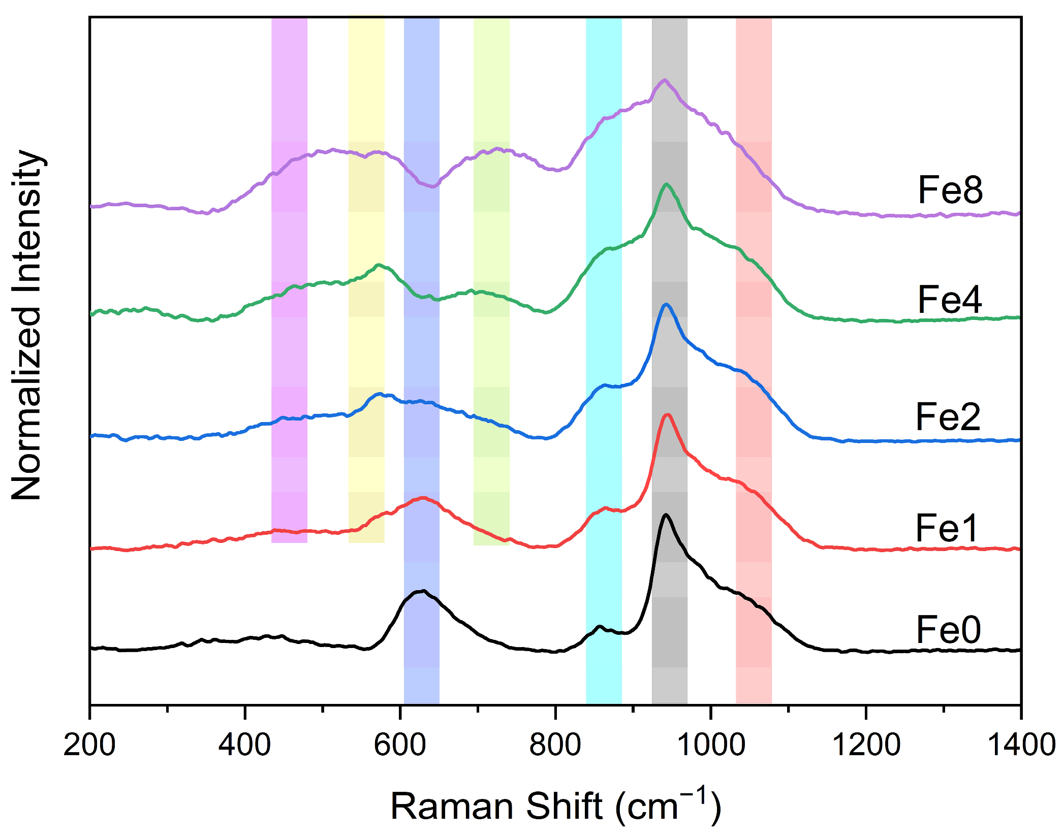
Figure 1. Raman spectra of the various bioglasses.
Biomimetics 09 00325 g001
Figure 2. Deconvoluted Raman spectra of (a) Fe1 and (b) Fe8 samples.
Biomimetics 09 00325 g002
Figure 3. (a) The imaginary part of the electrical modulus M″ versus frequency for Fe2 sample and (b) the variation activation energy Ea with increasing the Fe3O4 content.
Biomimetics 09 00325 g003
Figure 4. The normalized imaginary part of the modulus M″/M″max versus the frequency at 390 K for all prepared bioglasses (the inset depicts the relaxation time for all samples at 390 K).
Biomimetics 09 00325 g004
Figure 5. The variation in the atomic percentage of (a) Si and (b) Na elements and (c) the Ca/P ratio presented on the surface of the bioglass pellets after immersion in SBF.
Biomimetics 09 00325 g005
Figure 6. SEM micrographs of the surface of the bioglasses after immersion in SBF for (a1a3) 1 d; (b1b3) 4 d; (c1c3) 14 d. (The magnification of SEM images is 10 kX).
Biomimetics 09 00325 g006
Figure 7. XRD patterns of (a) Fe0 (b) Fe2 and (c) Fe8 samples before (_0d) and after immersion in SBF for 28 d.
Biomimetics 09 00325 g007
Figure 8. Raman spectra of (a) Fe0 (b) Fe2 and (c) Fe8 samples before (_0d) and after immersion in SBF for 28 d.
Biomimetics 09 00325 g008
Figure 9. Variation in pH values of the SBF solution resulting from pellets immersion for different times, with the medium changed every two days (green rectangle) and without medium change (orange rectangle).
Biomimetics 09 00325 g009
Figure 10. (a) Values of inhibition halo diameters of the bioglass samples modified with the insertion of various Fe3O4 concentrations against E. coli, S. aureus, and S. mutans bacteria after incubation for 24 h. Results are reported as mean ± SD. The asterisks indicate significance in an unpaired t-test; * p ≤ 0.05; ** p ≤ 0.01; *** p ≤ 0.001. (b) An example of an essay plate illustrating the inhibition halo of a Fe2 pellet on E. coli.
Biomimetics 09 00325 g010
Table 1. The composition of different bioglass samples.
Composition (mol%)
SampleSiO2Na2OCaOP2O5Fe3O4
Fe046.1024.4026.902.60-
Fe145.6424.1626.632.571
Fe245.1823.9126.362.552
Fe444.2623.4225.822.504
Fe842.4122.4524.752.398
Disclaimer/Publisher’s Note: The statements, opinions and data contained in all publications are solely those of the individual author(s) and contributor(s) and not of MDPI and/or the editor(s). MDPI and/or the editor(s) disclaim responsibility for any injury to people or property resulting from any ideas, methods, instructions or products referred to in the content.