Review

Recognizing the Clinical and Pathophysiologic Overlap Between Allergy and Lupus

Department of Medicine, Division of Rheumatology, Dell Medical School at The University of Texas at Austin, Austin, TX 78712, USA
Allergies 2026, 6(3), 24; https://doi.org/10.3390/allergies6030024
Submission received: 23 April 2026 / Revised: 29 May 2026 / Accepted: 30 June 2026 / Published: 6 July 2026
(This article belongs to the Section Physiopathology)

Abstract

Systemic lupus erythematosus (SLE) is a heterogeneous autoimmune disease characterized by diverse and evolving clinical manifestations, often resulting in delayed diagnosis and increased morbidity. Although traditionally viewed as distinct, allergic and autoimmune diseases share overlapping immunologic pathways and clinical features. Symptoms commonly encountered in allergy practice, including urticaria, angioedema, and pruritus, may represent early or coexisting manifestations of underlying lupus rather than primary allergic disease. This narrative review examines the clinical and pathophysiologic overlap between allergy and lupus, with a focus on presentations that may bring patients with undiagnosed SLE to the allergy setting. Shared mechanisms, including complement dysregulation, autoreactive IgE, and mast cell activation, are discussed as contributors to overlapping symptomatology. Key clinical features that distinguish lupus-associated presentations from primary allergic conditions are outlined, along with associated systemic findings that should prompt further evaluation. A practical approach to assessment is emphasized, including targeted history, judicious use of serologic testing, and indications for dermatologic and rheumatologic referral. Improved recognition of these patterns in allergy practice may facilitate earlier diagnosis, reduce unnecessary testing, and improve patient outcomes.

1. Introduction

Autoimmune and allergic diseases are both driven by immune dysregulation and can present with overlapping clinical manifestations. Systemic lupus erythematosus [SLE] is a chronic autoimmune disease of considerable diagnostic complexity, characterized by heterogeneous clinical manifestations that evolve gradually and often mimic more common conditions. Symptoms frequently encountered in the allergy setting, such as pruritus, urticaria, and angioedema, can also occur in SLE, creating a diagnostic challenge.
Delayed recognition of SLE is common, and its consequences are significant. More than three-quarters of patients wait over six months for a diagnosis, and longer delays are independently associated with worse outcomes [1]. Diagnostic delay also carries a significant quality of life burden, with patients reporting reduced ability to work and socialize as a consequence of prolonged misdiagnosis [2]. Irreversible organ damage is present in 40% of patients within the first year of diagnosis and accrues linearly over time [3]. SLE carries a mortality rate two to three times that of the general population, a gap that has not narrowed since the mid-1990s [4].
Recognizing the severity of delayed diagnosis does not mean testing everyone. Serologic tests lack specificity, and their value depends heavily on pretest probability. A focused, clinically guided approach is needed to identify which patients warrant further rheumatologic evaluation.
This narrative review examines the clinical presentations most likely to bring patients with undiagnosed lupus to the allergy setting, with a focus on practical guidance for recognition, targeted evaluation, and timely referral. Earlier recognition in the allergy setting may shorten diagnostic delay and reduce its associated consequences.
This narrative review was conducted through a comprehensive search of PubMed/MEDLINE through May 2025. Search terms included combinations of “systemic lupus erythematosus,” “cutaneous lupus,” “allergy,” “urticaria,” “chronic spontaneous urticaria,” “angioedema,” “urticarial vasculitis,” “hypocomplementemic urticarial vasculitis,” “pruritus,” “drug hypersensitivity,” “drug-induced lupus,” “complement deficiency,” “IgE autoantibodies,” and “mast cell activation.” Reference lists of identified articles were reviewed to capture additional relevant publications. Priority was given to systematic reviews, meta-analyses, large cohort studies, and clinical practice guidelines, with case series and case reports included when higher-level evidence was limited. No formal systematic review protocol was applied, consistent with the narrative scope of this review.

2. Shared Pathophysiology of Allergy and Lupus

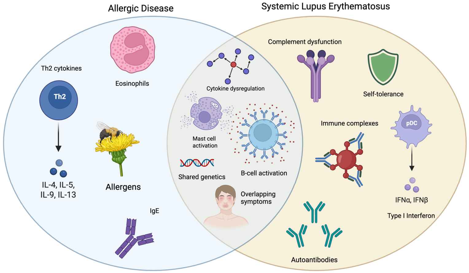
Autoimmune and allergic disorders both arise from immune dysregulation but have been traditionally viewed as opposite ends of the immunologic spectrum. Autoimmune diseases have been traditionally associated with Th1/Th17-driven cell-mediated immune responses [5], whereas allergy and asthma encompass Th2-mediated responses directed against environmental antigens [6]. This traditional dichotomy, however, is increasingly challenged by growing evidence of substantial mechanistic overlap [7] (Figure 1). Genome-wide association studies have identified significant enrichment of shared susceptibility loci between allergic and autoimmune diseases, with approximately 48% of common loci showing the same direction of effect for both conditions, suggesting shared pathogenic mechanisms rather than opposing processes [8].
The complement system illustrates how the same immune pathway can function differently in allergy and autoimmunity. In allergic diseases, complement activation enhances mast cell degranulation and amplifies inflammatory responses. In contrast, complement plays a paradoxical dual role in SLE: while complement activation via the classical pathway contributes to immune complex-mediated tissue damage, deficiency of early complement components (C1q, C4, C2) is strongly associated with lupus development. C1q deficiency is associated with a 93% risk of developing SLE, and C4 deficiency with a 75% risk [9]. This paradox reflects complement’s essential role in clearing apoptotic debris and immune complexes; when this clearance fails, autoantigens accumulate and drive autoimmunity [10].
Mast cells and IgE, traditionally considered hallmarks of allergy, are now recognized as contributors to autoimmune pathogenesis [7]. Autoreactive IgE antibodies targeting self-antigens have been identified in SLE [11], bullous pemphigoid, and chronic spontaneous urticaria, where they activate mast cells and basophils via FcεRI receptors [12]. In SLE specifically, elevated total IgE levels are observed independent of atopy, and IgE autoantibodies can induce type I interferon responses from plasmacytoid dendritic cells, a key pathogenic pathway in lupus [13]. Mast cells further contribute to autoimmunity through cytokine secretion, antigen presentation, and recruitment of inflammatory cells to target tissues. This mechanistic convergence between allergic and autoimmune pathways helps explain why conditions such as urticaria, angioedema, and pruritus may represent manifestations of underlying systemic autoimmune disease when occurring outside a typical allergic pattern.

3. Lupus Background for the Clinician

Lupus erythematosus exists on a spectrum. Cutaneous lupus erythematosus (CLE) refers to a disease confined to the skin. Systemic lupus erythematosus (SLE) involves multiple organ systems and can subsequently cause life-threatening organ damage. Skin involvement is common, affecting up to 70% of patients with systemic disease, and may be the presenting feature before additional systemic manifestations become apparent.

3.1. Cutaneous Lupus Erythematosus

CLE includes three major subtypes. Acute cutaneous lupus presents as the classic malar or butterfly rash, a photosensitive erythematous eruption across the cheeks and nasal bridge that characteristically spares the nasolabial folds. It typically occurs during periods of active SLE and resolves without scarring. Subacute CLE is a photosensitive annular or papulosquamous rash affecting 10–15% of SLE patients [14]. Chronic cutaneous lupus includes discoid lupus, the most common subtype, which presents as indurated, scaly plaques that heal with scarring and dyspigmentation. Less common variants include chilblain lupus, which presents as painful erythematous lesions on acral surfaces triggered by cold and damp conditions, and lupus tumidus, characterized by smooth urticarial plaques without surface change.
Up to 30% of patients with discoid lupus will progress to SLE [14]. Risk factors for progression include younger age, darker skin phototype, and high-titer ANA (anti-nuclear antibody) [15]. The probability of developing SLE within 3 years of a CLE diagnosis has been estimated at 16.7%, with the highest risk in the first year [16].
Lupus rashes typically occur in sun-exposed distributions, including the face, neck, upper chest, and extensor surfaces of the arms, and are triggered or worsened by ultraviolet light exposure. They do not respond to antihistamines and may persist or recur over weeks to months rather than resolving within hours as urticarial lesions do. Depending on the subtype, lesions may heal with permanent scarring and dyspigmentation. These features should prompt consideration of an underlying autoimmune process.

3.2. Delays in Lupus Diagnosis

SLE is a chronic autoimmune disease characterized by immune dysregulation, autoantibody production, and inflammation affecting multiple organ systems. It disproportionately affects Black, Hispanic, and Asian populations, who often develop disease at a younger age and experience more severe manifestations, including lupus nephritis. Clinical manifestations are highly variable and often evolve gradually over time. Diagnosis is therefore often delayed.
Median delays of 18–24 months from symptom onset have been reported, with some patients experiencing delays of up to 6 years [1,17,18]. During this interval, patients may seek care from other specialists, including allergists, before the full clinical picture becomes apparent. Fatigue, joint pain, oral ulcers, and hair loss are common but may not be part of the chief complaint, particularly when intermittent or overshadowed by more acute symptoms.
SLE nonetheless carries significant risk of internal organ involvement, with lupus nephritis developing in approximately 40% of patients [14], the majority within the first five years of diagnosis [19,20]. Early recognition remains critical.

3.3. ANA: Clinical Utility and Limitations

The diagnosis of lupus is based on a constellation of clinical findings combined with specific autoantibody positivity, guided by the 2019 ACR/EULAR classification criteria [21]. These criteria were developed primarily for research enrollment and are best viewed as a clinical guide rather than an absolute diagnostic standard. A positive ANA at a titer ≥ 1:80 is required as an entry criterion, followed by weighted clinical and immunologic domains; a score of ≥10 classifies a patient as SLE with 96.1% sensitivity and 93.4% specificity [21].
The ANA warrants particular attention given its limitations. Most laboratories report a positive result at a titer of ≥1:40. At this value, up to 20–30% of healthy individuals test positive, making the result clinically unreliable for diagnosing autoimmune disease [22], which is why entry criterion uses a higher titer of ≥1:80. In populations with low pretest probability for lupus, the positive predictive value of a positive ANA for diagnosing SLE is as low as 2–3% [23,24]. This likely reflects frequent ANA testing in settings where clinical suspicion is low.
Compounding this issue, many laboratories now use solid-phase assays such as multiplex bead-based or chemiluminescent immunoassays rather than traditional Hep-2 indirect immunofluorescence [IIF], and the two methods do not always agree. Substantial discordance between multiplex ANA assays and Hep-2 IIF has been reported, including 55% of multiplex-positive results being negative on IIF, and 47% of multiplex-negative results being positive on IIF in one cohort [25].
ANA prevalence in the US population has increased significantly over the past three decades, rising from 11% in 1988–1991 to 16.1% in 2011–2012, with the most dramatic increase observed in adolescents, where prevalence nearly tripled [26]. A positive ANA requires careful clinical interpretation and does not independently establish a diagnosis of lupus. Despite these limitations, the ANA remains the initial screening test when evaluating for lupus. Clinicians in allergy practice will frequently encounter patients with a positive ANA; understanding its limitations is essential to avoid overdiagnosis and unnecessary evaluation.

3.4. Initial Evaluation of Suspected Lupus

When lupus is suspected and the ANA is positive, more specific antibody testing should follow. Anti-dsDNA and anti-Smith are the most lupus-specific autoantibodies, and complement levels (C3/C4) should be obtained as they can reflect disease activity and complement consumption. Anti-Ro/SSA antibodies are present in 72.1% of SCLE and 47.4% of ACLE patients, making them a useful serologic marker when cutaneous lupus is suspected [14]. Urinalysis with spot urine protein-to-creatinine ratio should also be obtained, as lupus nephritis occurs in approximately 30% of patients and proteinuria is often its earliest sign. A negative ANA does not fully exclude autoimmune disease, particularly when assay variability exists or another connective tissue disease is possible. If the ANA is negative but clinical suspicion remains high for autoimmune disease, rheumatology or dermatology referral is appropriate.
Urticaria, angioedema, and pruritus are common allergy symptoms that can also be seen in SLE yet are absent from lupus classification criteria and classic presentations, making them easy to overlook. Awareness of these overlapping presentations and a low threshold for further evaluation can help prevent diagnostic delay.

4. Urticaria and Lupus

Chronic spontaneous urticaria (CSU) is defined as recurrent pruritic wheals, angioedema, or both lasting more than six weeks without an identifiable trigger. It affects approximately 1% of the general population, predominantly females aged 30–50 years, and is typically responsive to antihistamines [27]. CSU is not simply an allergic condition; it is autoimmune in a substantial subset of patients. Known autoimmune endotypes, present in more than 50% of CSU cases, include two distinct subtypes: type I autoimmune (autoallergic) CSU, which is mediated by IgE antibodies directed against self-antigens such as thyroid peroxidase and IL-24, and type IIb autoimmune CSU, characterized by IgG autoantibodies that activate mast cells through IgE and FcεRI pathways [28]. Type IIb is associated with more severe disease, lower total IgE, and reduced response to antihistamines and omalizumab [29].
Approximately 10–28% of CSU patients have at least one additional autoimmune disease, with Hashimoto thyroiditis being the most common, affecting approximately 20% of CSU patients. CSU is also seen in association with rheumatoid arthritis, Sjogren’s disease, celiac disease, type 1 diabetes mellitus, and SLE. CSU is associated with an increased risk of multiple autoimmune diseases, with autoimmune diagnoses most commonly occurring in the 10 years following a CSU diagnosis, suggesting that urticaria may precede or signal underlying systemic autoimmune disease [28,30,31]. Emerging genetic data suggest that CSU and SLE may share a common autoimmune trajectory rather than representing coincidental comorbidity [28,29]. The relationship between chronic urticaria and SLE appears to be bidirectional. Patients with chronic urticaria have approximately twice the risk of developing SLE compared to controls, with a dose-response relationship observed with increasing urticaria episodes [32,33]. Mendelian randomization data further support a genetic causal association from urticaria to SLE, though not in the reverse direction, suggesting urticaria-related immune dysregulation may predispose to lupus development rather than the converse [29].
A patient with CSU may have concurrent lupus not because urticaria itself is a classic lupus manifestation, but because both conditions may arise from shared immune dysregulation. Maintaining a broad differential and asking directly about systemic symptoms is therefore essential. Persistent wheals, residual dyspigmentation, or systemic features should shift suspicion away from CSU and toward autoimmune pathology.

Urticarial Vasculitis

Urticarial vasculitis is a distinct autoimmune entity characterized by lesions that persist longer than 24 h and leave a bruise-like post-inflammatory hyperpigmentation when they resolve, unlike urticarial wheals, which resolve completely without residual skin change. Two forms of urticarial vasculitis are recognized. Normocomplementemic urticarial vasculitis (NUV) is more common and generally skin-limited. Hypocomplementemic urticarial vasculitis (HUV) is more severe and can have extracutaneous manifestations including arthralgias, glomerulonephritis, uveitis, episcleritis, abdominal pain, and obstructive lung disease [34].
HUV is strongly associated with SLE. In one cohort, SLE was present or subsequently occurred in 54% of HUV cases versus 3% of NUV [35], and more recent studies support this continued association [36]. HUV is driven by immune complex-mediated vascular injury in the presence of anti-C1q antibodies. Angioedema occurs in over 50% of HUV cases, compared to fewer than 5% of SLE patients [34].
When urticarial vasculitis is suspected, skin biopsy is needed for diagnosis and should ideally be performed within 24–48 h of lesion onset for optimal yield. Biopsy will demonstrate leukocytoclastic vasculitis with fibrinoid necrosis. Specific findings, including neutrophil alignment along the dermal-epidermal junction and dermal C4d deposition, are strongly associated with SLE [36].
Urticarial vasculitis can occasionally present with lesions lasting fewer than 24 h, mimicking CSU. Guidelines therefore recommend that any urticaria with residual purpura, burning pain, or systemic symptoms should prompt biopsy regardless of lesion duration [37].
In patients with SLE, urticarial eruptions may represent true CSU (a comorbid autoimmune condition), urticarial vasculitis (immune complex-mediated, often hypocomplementemic), or non-specific urticaria-like lesions associated with active disease. The boundaries between these categories are blurred, and biopsy is essential for classification.

5. Angioedema

SLE patients who develop angioedema are more often younger, female, and African American, and are more likely to experience severe episodes requiring hospitalization [38]. Not all angioedema presenting in the allergy clinic is histamine-mediated. While mast cell-mediated angioedema can occur and is typically associated with urticaria and triggers, lupus-associated angioedema is often bradykinin-mediated due to complement dysregulation [39]. Angioedema may occasionally precede a formal lupus diagnosis or be an early clue to underlying autoimmune disease.
Clinically, this form of angioedema presents as nonpruritic, non-erythematous swelling without urticaria, most commonly involving the face, lips, eyelids, and extremities, and may also involve the gastrointestinal tract or airway. The edema is usually soft, non-pitting, and painless, and develops over hours [40].
Lupus-associated angioedema occurs in an estimated 15–30% of SLE patients [41]. Not all angioedema in this setting is the same, and distinguishing between acquired C1-INH deficiency, hereditary angioedema (HAE), and ACE inhibitor-associated angioedema has direct therapeutic implications. Acquired C1-INH deficiency typically presents after age 40 without a family history and with low C1q levels. By contrast, C1q is normal in hereditary angioedema [42,43]. In a Spanish cohort of patients with acquired C1-INH angioedema, SLE was identified as an associated condition in 10.9% of cases [42]. Conversely, in a large Italian cohort of HAE patients, 2.1% had concomitant connective tissue disease, with SLE being the most common [44]. ACE inhibitor-associated angioedema is also bradykinin-mediated and should always be excluded by medication review in any patient presenting with recurrent angioedema [43].
Low complement levels help distinguish lupus-associated angioedema from histaminergic forms. Histaminergic angioedema typically responds to antihistamines, whereas bradykinin-mediated angioedema does not. Symptoms often improve with treatment of the underlying disease [40].
When lupus-associated angioedema is suspected, initial workup should include a C4 level as a useful screening test. If abnormal, C1q should be checked next, followed by C1-INH level and function to guide further evaluation [39,43]. In patients with additional systemic symptoms or unexplained complement abnormalities, evaluation for underlying autoimmune disease is reasonable.

6. Drug Hypersensitivity in Lupus

Drug hypersensitivity reactions, both immediate and delayed, occur more frequently in patients with SLE than in the general population. Because lupus often requires long-term treatment with multiple medications, cumulative drug exposure is high, increasing the risk of sensitization over time. The underlying immune dysregulation in SLE, including abnormal T-cell activation, autoantibody production, and cytokine imbalance, is thought to lower the threshold for immune responses to medications [45]. Repeated medication exposure during flares, infections, and disease complications may further amplify this risk.
Diagnosing true drug allergy in this population is challenging. Lupus patients frequently report multiple drug allergies, but this likely reflects a mix of true immune-mediated hypersensitivity, medication tolerance, and disease-related symptoms misattributed to drugs. Rash, oral ulcers, and fatigue are common lupus manifestations that can easily be confused with drug reactions, leading to unnecessary drug discontinuation and incorrect drug allergy labels that can limit future treatment options [45]. In patients requiring first-line therapy, incorrect allergy labels may create avoidable barriers to disease control. Careful history taking, including timing of symptoms and reproducibility with exposure, is therefore especially important.
Drug-induced lupus erythematosus (DILE) is a distinct condition in which specific medications trigger lupus symptoms. Patients typically present with arthralgia, serositis, and rashes. Severe manifestations such as renal and central nervous system involvement are uncommon. Classic culprits include hydralazine, procainamide, isoniazid, minocycline, and anti-TNF agents [14,46,47]. Procainamide has the highest rate at 15–20%, followed by hydralazine at 7–13% [14]. The mechanism differs from SLE and involves enhanced immune responses to reactive drug metabolites that modify self-proteins, triggering autoimmunity [46,48,49].
Serologically, DILE is characterized by positive ANA and positive anti-histone antibodies with negative dsDNA. Once patients stop the offending agent, the symptoms improve within a few weeks of drug discontinuation, though resolution may take several months. DILE does not generally progress to idiopathic SLE, making it a reversible condition when recognized early.

7. Pruritus

Pruritus is a common and underrecognized symptom in lupus, affecting approximately 77% of patients with CLE [50]. It may be the initial presenting symptom in patients who have not yet received a lupus diagnosis. However, pruritus in lupus is driven by cytokine dysregulation, mast cell degranulation, and small fiber neuropathy rather than histamine release [51], so antihistamines provide limited benefit, and persistent or refractory pruritus in this population should prompt consideration of active underlying disease rather than escalation of allergy-directed therapy.
Pruritus in lupus is associated with increased disease activity and correlates with validated measures of lupus activity and cutaneous involvement [50]. It often precedes or accompanies an active rash rather than occurring in isolation. Importantly, while pruritus correlates with disease activity, it is not associated with increased organ damage, suggesting it reflects inflammatory burden rather than irreversible injury [50].
When pruritus fails to respond to antihistamines, particularly in the setting of active rash or systemic symptoms, addressing the underlying inflammatory disease is more likely to provide relief than further allergy-directed therapy.

8. Pediatric Considerations

Childhood-onset SLE is more aggressive than adult-onset disease, with higher rates of renal involvement, neuropsychiatric manifestations, and hematologic abnormalities [52,53]. Several features are particularly relevant in the allergy setting. Urticaria may precede a lupus diagnosis in children, and a history of urticaria is associated with a 2.2-fold increased risk of developing SLE, with the strongest association seen in female adolescents [33]. Drug-induced lupus in children differs from adults as minocycline and ethosuximide are more commonly implicated than procainamide and hydralazine, and anti-TNF agents are increasingly recognized as culprits in pediatric patients on biologics [54]. Co-existing atopy in juvenile SLE is associated with higher disease activity, more frequent flares, and longer time to stable disease [55]. These distinctions underscore the importance of considering lupus in children and adolescents presenting with allergic symptoms that do not follow an expected course.

9. Practical Approach to Evaluation and Management

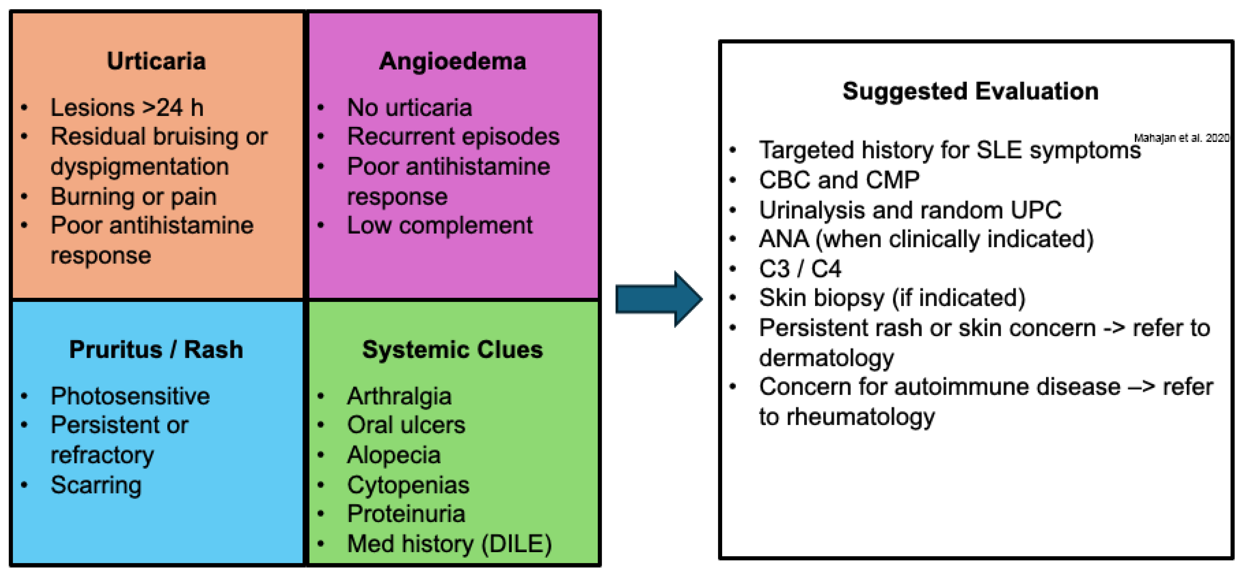
When lupus is suspected, the goal of evaluation is to avoid both underdiagnosis and unnecessary testing, a balance that requires a focused and stepwise approach (Figure 2). The practical question is rarely whether to evaluate, but how much evaluation is justified by the presentation. A thorough history should include direct inquiry about systemic symptoms, including joint pain and swelling, rashes, oral ulcers, hair loss, and photosensitivity, as these are easy to miss if not explicitly asked. When systemic features are present alongside allergic complaints, basic lab evaluation is a reasonable first step: complete blood count with differential, comprehensive metabolic panel, urinalysis with microscopy, and random urine protein-to-creatinine ratio, which can reveal cytopenias, renal dysfunction, or proteinuria that significantly raises diagnostic suspicion.
Serologic testing should be sequential. Testing should begin with an ANA. If the result is negative in a low-suspicion patient, lupus is unlikely, and further autoimmune testing is low yield. If positive, proceed to more specific antibody testing, including anti-dsDNA, anti-Smith, and complement levels (C3/C4) [56]. The ANA is most useful when ordered to answer a specific clinical question rather than as a screening test for nonspecific symptoms. Broad multiplex autoantibody panels should be avoided in low-pretest-probability patients, as this is a reliable way to generate anxiety-provoking, clinically meaningless results that complicate rather than clarify the evaluation.
Before ordering an ANA, patients should be counseled that a positive result requires clinical interpretation and does not independently establish a diagnosis of lupus. This is particularly important given the high background positivity rate in the general population and the anxiety a positive result can generate, especially when patients research their results independently.
If a persistent or unusual rash is present, or if urticarial vasculitis is on the differential, referring to dermatology for skin biopsy is recommended. Tissue diagnosis may be more informative than expanded serologic testing when the primary manifestation is cutaneous. If the ANA is negative, but clinical suspicion remains high given recurrent symptoms or failure to respond to antihistamines, both rheumatologic and dermatologic evaluation are reasonable next steps.
Rheumatology referral is appropriate when lupus is suspected, though patients should be informed that wait times are often prolonged. In the interim, chronic corticosteroids should be avoided, as they carry significant long-term side effects and can complicate the diagnostic picture. Short courses may be reasonable for acute inflammatory flares, but longitudinal steroid management should be deferred to the rheumatologist overseeing care.
Hydroxychloroquine is the cornerstone of lupus treatment and is recommended for virtually all patients with SLE given its established benefits in decreasing disease activity, organ protection, and long-term survival. While initiation is appropriately deferred to rheumatology, earlier referral may expand the opportunity to benefit from treatment.

10. Conclusions

Lupus does not always present with classic multisystem features. Cutaneous and mucosal symptoms commonly encountered in the allergy setting, including pruritus, urticaria, and angioedema, can be early or coexisting manifestations of underlying systemic autoimmune disease. The challenge is recognizing when a familiar presentation is no longer behaving like a routine allergy. The mechanistic overlap between allergic and autoimmune disease provides a biological basis for the clinical intersection and helps explain why these conditions can be so difficult to distinguish.
When lupus is suspected, a targeted history and basic laboratory evaluation will identify most patients who warrant further workup. The ANA is a useful but imperfect tool, and its value depends heavily on the clinical context in which it is ordered. In populations with low pretest probability, a positive result is far more likely to represent background noise than true autoimmune disease, and ordering it indiscriminately can lead to patient anxiety and unnecessary downstream testing.
As the science connecting allergic and autoimmune diseases continues to evolve, so too does the case for greater cross-specialty awareness. Many patients with lupus will encounter an allergist, sometimes before a rheumatologist. For some, recognition in that setting may shorten a diagnostic journey that otherwise may span years.

Funding

This research received no external funding.

Institutional Review Board Statement

Not applicable.

Informed Consent Statement

Not applicable.

Data Availability Statement

No new data were created or analyzed in this study.

Acknowledgments

During the preparation of this manuscript/study, the author(s) used [ChatGPT based on OpenAI’s GPT-5.3 model] for the purposes of literature review and language editing for grammar and syntax. The author has reviewed and edited the output and take full responsibility for the content of this publication.

Conflicts of Interest

The author declares no conflicts of interest.

References

  1. Kernder, A.; Richter, J.G.; Fischer-Betz, R.; Winkler-Rohlfing, B.; Brinks, R.; Aringer, M.; Schneider, M.; Chehab, G. Delayed diagnosis adversely affects outcome in systemic lupus erythematosus: Cross sectional analysis of the LuLa cohort. Lupus 2021, 30, 431–438. [Google Scholar] [CrossRef] [PubMed]
  2. Kent, T.; Davidson, A.; Newman, D.; Buck, G.; D’Cruz, D. Burden of illness in systemic lupus erythematosus: Results from a UK patient and carer online survey. Lupus 2017, 26, 1095–1100. [Google Scholar] [CrossRef] [PubMed]
  3. Taraborelli, M.; Cavazzana, I.; Martinazzi, N.; Lazzaroni, M.G.; Fredi, M.; Andreoli, L.; Franceschini, F.; Tincani, A. Organ damage accrual and distribution in systemic lupus erythematosus patients followed-up for more than 10 years. Lupus 2017, 26, 1197–1204. [Google Scholar] [CrossRef] [PubMed]
  4. Hoi, A.; Igel, T.; Mok, C.C.; Arnaud, L. Systemic lupus erythematosus. Lancet 2024, 403, 2326–2338. [Google Scholar] [CrossRef] [PubMed]
  5. Steinman, L. A brief history of TH17, the first major revision in the TH1/TH2 hypothesis of T cell–mediated tissue damage. Nat. Med. 2007, 13, 139–145. [Google Scholar] [CrossRef] [PubMed]
  6. Lloyd, C.M.; Hessel, E.M. Functions of T cells in asthma: More than just TH2 cells. Nat. Rev. Immunol. 2010, 10, 838–848. [Google Scholar] [CrossRef] [PubMed]
  7. Ramirez, G.A.; Cardamone, C.; Lettieri, S.; Fredi, M.; Mormile, I. Clinical and Pathophysiological Tangles Between Allergy and Autoimmunity: Deconstructing an Old Dichotomic Paradigm. Clin. Rev. Allergy Immunol. 2025, 68, 13. [Google Scholar] [CrossRef] [PubMed]
  8. Kreiner, E.; Waage, J.; Standl, M.; Brix, S.; Pers, T.H.; Couto Alves, A.; Warrington, N.M.; Tiesler, C.M.T.; Fuertes, E.; Franke, L.; et al. Shared genetic variants suggest common pathways in allergy and autoimmune diseases. J. Allergy Clin. Immunol. 2017, 140, 771–781. [Google Scholar] [CrossRef] [PubMed]
  9. Macedo, A.C.L.; Isaac, L. Systemic Lupus Erythematosus and Deficiencies of Early Components of the Complement Classical Pathway. Front. Immunol. 2016, 7, 55. [Google Scholar] [CrossRef] [PubMed]
  10. Ling, G.S.; Crawford, G.; Buang, N.; Bartok, I.; Tian, K.; Thielens, N.M.; Bally, I.; Harker, J.A.; Ashton-Rickardt, P.G.; Rutschmann, S.; et al. C1q restrains autoimmunity and viral infection by regulating CD8+ T cell metabolism. Science 2018, 360, 558–563. [Google Scholar] [CrossRef] [PubMed]
  11. Dema, B.; Pellefigues, C.; Hasni, S.; Gault, N.; Jiang, C.; Ricks, T.K.; Bonelli, M.M.; Scheffel, J.; Sacré, K.; Jablonski, M.; et al. Autoreactive IgE Is Prevalent in Systemic Lupus Erythematosus and Is Associated with Increased Disease Activity and Nephritis. PLoS ONE 2014, 9, e90424. [Google Scholar] [CrossRef] [PubMed]
  12. Sanjuan, M.A.; Sagar, D.; Kolbeck, R. Role of IgE in autoimmunity. J. Allergy Clin. Immunol. 2016, 137, 1651–1661. [Google Scholar] [CrossRef] [PubMed]
  13. Henault, J.; Riggs, J.M.; Karnell, J.L.; Liarski, V.M.; Li, J.; Shirinian, L.; Xu, L.; Casey, K.A.; Smith, M.A.; Khatry, D.B.; et al. Self-reactive IgE exacerbates interferon responses associated with autoimmunity. Nat. Immunol. 2016, 17, 196–203. [Google Scholar] [CrossRef] [PubMed]
  14. Siegel, C.H.; Sammaritano, L.R. Systemic Lupus Erythematosus: A Review. JAMA 2024, 331, 1480. [Google Scholar] [CrossRef] [PubMed]
  15. Fredeau, L.; Courvoisier, D.S.; Ait Mehdi, R.; Ingen-Housz-Oro, S.; Mahe, E.; Costedoat-Chalumeau, N.; Arnaud, L.; Francès, C.; Mathian, A.; Jachiet, M.; et al. Risk factors of progression from discoid lupus to severe systemic lupus erythematosus: A registry-based cohort study of 164 patients. J. Am. Acad. Dermatol. 2023, 88, 551–559. [Google Scholar] [CrossRef] [PubMed]
  16. Grönhagen, C.M.; Fored, C.M.; Linder, M.; Granath, F.; Nyberg, F. Subacute cutaneous lupus erythematosus and its association with drugs: A population-based matched case-control study of 234 patients in Sweden: SCLE and its association with drugs. Br. J. Dermatol. 2012, 167, 296–305. [Google Scholar] [CrossRef] [PubMed]
  17. Kapsala, N.N.; Nikolopoulos, D.S.; Flouda, S.P.; Chavatza, A.P.; Tseronis, D.D.; Aggelakos, M.D.; Katsimbri, P.P.; Bertsias, G.K.; Fanouriakis, A.C.; Boumpas, D.T. From first symptoms to diagnosis of systemic lupus erythematosus: Mapping the journey of patients in an observational study. Clin. Exp. Rheumatol. 2021, 41, 74–81. [Google Scholar] [CrossRef] [PubMed]
  18. Ramírez-Flores, M.F.; Hernandez-Garduno, A.; Quintana, R.; Fuentes-Silva, Y.; Nieto, R.; Cano-Gámez, T.; Ferreyra, L.; Ceballos, M.F.; Gastelum-Strozzi, A.; Pons-Estel, B.A.; et al. Factors associated with delay in the diagnosis and treatment of systemic lupus erythematosus in adult patients: A systematic review. Rheumatology 2025, 64, 5597–5610. [Google Scholar] [CrossRef] [PubMed]
  19. Parodis, I.; Rovin, B.H.; Tektonidou, M.G.; Anders, H.-J.; Malvar, A.; Mok, C.C.; Mohan, C. Lupus nephritis. Nat. Rev. Dis. Primer 2025, 11, 69. [Google Scholar] [CrossRef] [PubMed]
  20. Mahajan, A.; Amelio, J.; Gairy, K.; Kaur, G.; Levy, R.A.; Roth, D.; Bass, D. Systemic lupus erythematosus, lupus nephritis and end-stage renal disease: A pragmatic review mapping disease severity and progression. Lupus 2020, 29, 1011–1020. [Google Scholar] [CrossRef] [PubMed]
  21. Aringer, M.; Costenbader, K.; Daikh, D.; Brinks, R.; Mosca, M.; Ramsey-Goldman, R.; Smolen, J.S.; Wofsy, D.; Boumpas, D.T.; Kamen, D.L.; et al. 2019 European League Against Rheumatism/American College of Rheumatology Classification Criteria for Systemic Lupus Erythematosus. Arthritis Rheumatol. 2019, 71, 1400–1412. [Google Scholar] [CrossRef] [PubMed]
  22. Pisetsky, D.S. Antinuclear antibody testing—Misunderstood or misbegotten? Nat. Rev. Rheumatol. 2017, 13, 495–502. [Google Scholar] [CrossRef] [PubMed]
  23. Kang, S.-H.; Seo, Y.-I.; Lee, M.H.; Kim, H.A. Diagnostic Value of Anti-Nuclear Antibodies: Results From Korean University-Affiliated Hospitals. J. Korean Med. Sci. 2022, 37, e159. [Google Scholar] [CrossRef] [PubMed]
  24. Abeles, A.M.; Abeles, M. The Clinical Utility of a Positive Antinuclear Antibody Test Result. Am. J. Med. 2013, 126, 342–348. [Google Scholar] [CrossRef] [PubMed]
  25. Ali, M.; Baban, Y.; Kumar, G. Diagnostic discrepancies: A retrospective analysis of two antinuclear antibody platforms. J. Investig. Med. 2026, 74, 134–139. [Google Scholar] [CrossRef] [PubMed]
  26. Dinse, G.E.; Parks, C.G.; Weinberg, C.R.; Co, C.A.; Wilkerson, J.; Zeldin, D.C.; Chan, E.K.L.; Miller, F.W. Increasing Prevalence of Antinuclear Antibodies in the United States. Arthritis Rheumatol. 2022, 74, 2032–2041. [Google Scholar] [CrossRef] [PubMed]
  27. Lang, D.M. Chronic Urticaria. N. Engl. J. Med. 2022, 387, 824–831. [Google Scholar] [CrossRef] [PubMed]
  28. Kolkhir, P.; Bonnekoh, H.; Metz, M.; Maurer, M. Chronic Spontaneous Urticaria: A Review. JAMA 2024, 332, 1464. [Google Scholar] [CrossRef] [PubMed]
  29. Yang, M.; Su, Y.; Xu, K.; Wen, P.; Zhang, B.; Guo, J.; Nan, K.; Yang, P.; Shao, X.; Liu, L.; et al. Common autoimmune diseases and urticaria: The causal relationship from a bidirectional two-sample mendelian randomization study. Front. Immunol. 2023, 14, 1280135. [Google Scholar] [CrossRef] [PubMed]
  30. Confino-Cohen, R.; Chodick, G.; Shalev, V.; Leshno, M.; Kimhi, O.; Goldberg, A. Chronic urticaria and autoimmunity: Associations found in a large population study. J. Allergy Clin. Immunol. 2012, 129, 1307–1313. [Google Scholar] [CrossRef] [PubMed]
  31. Kolkhir, P.; Pogorelov, D.; Olisova, O.; Maurer, M. Comorbidity and pathogenic links of chronic spontaneous urticaria and systemic lupus erythematosus—A systematic review. Clin. Exp. Allergy 2016, 46, 275–287. [Google Scholar] [CrossRef] [PubMed]
  32. Yong, S.; Su, K.; Chen, H.; Huang, J.; Wu, H.; Wei, J.C. Impact of chronic urticaria on systemic lupus erythematosus: A nationwide population-based study in Taiwan. J. Dermatol. 2019, 46, 26–32. [Google Scholar] [CrossRef] [PubMed]
  33. Lin, C.; Hung, P.; Hu, H.; Chung, C.; Chen, T.; Hung, K. Clinically diagnosed urticaria and risk of systemic lupus erythematosus in children: A nationwide population-based case-control study. Pediatr. Allergy Immunol. 2018, 29, 732–739. [Google Scholar] [CrossRef] [PubMed]
  34. Ouerdani, Y.; Ben Achour, T.; Ben Hmid, A.; Said, F.; Ayadi, I.; Zehani Kassar, A.; Jridi, M.; Ben Ghorbel, I.; Naceur, I.; Samoud, S.; et al. Urticarial hypocomplementemic vasculitis syndrome and systemic lupus erythematosus: A case report and review of the literature. Front. Immunol. 2025, 16, 1649699. [Google Scholar] [CrossRef] [PubMed]
  35. Davis, M.D.; Daoud, M.S.; Kirby, B.; Gibson, L.E.; Rogers, R.S. Clinicopathologic correlation of hypocomplementemic and normocomplementemic urticarial vasculitis. J. Am. Acad. Dermatol. 1998, 38, 899–905. [Google Scholar] [CrossRef] [PubMed]
  36. Damman, J.; Mooyaart, A.L.; Seelen, M.A.J.; Van Doorn, M.B.A. Dermal C4d Deposition and Neutrophil Alignment Along the Dermal–Epidermal Junction as a Diagnostic Adjunct for Hypocomplementemic Urticarial Vasculitis (Anti-C1q Vasculitis) and Underlying Systemic Disease. Am. J. Dermatopathol. 2020, 42, 399–406. [Google Scholar] [CrossRef] [PubMed]
  37. Bernstein, J.A.; Lang, D.M.; Khan, D.A.; Craig, T.; Dreyfus, D.; Hsieh, F.; Sheikh, J.; Weldon, D.; Zuraw, B.; Bernstein, D.I.; et al. The diagnosis and management of acute and chronic urticaria: 2014 update. J. Allergy Clin. Immunol. 2014, 133, 1270–1277.e66. [Google Scholar] [CrossRef] [PubMed]
  38. Luo, Y.; Fan, X.; Jiang, C.; Ramos-Rodriguez, A.; Wen, Y.; Zhang, J.; Huang, F.; Guan, X.; Xu, J. Systemic Lupus Erythematosus and Angioedema: A Cross-Sectional Study From the National Inpatient Sample. Arch. Rheumatol. 2019, 34, 301–307. [Google Scholar] [CrossRef] [PubMed]
  39. Lahiri, M.; Lim, A.Y. Angioedema and Systemic Lupus Erythematosus—A Complementary Association? Ann. Acad. Med. Singap. 2007, 36, 142–145. [Google Scholar] [CrossRef]
  40. Bienstock, D.; Mandel, L. Facial Angioedema and Systemic Lupus Erythematosus: Case Report. J. Oral Maxillofac. Surg. 2015, 73, 928–932. [Google Scholar] [CrossRef] [PubMed]
  41. Lisiecka, M.Z. Symptoms Causing Urticaria and Angioedema in Systemic Connective Tissue Diseases: Investigation and Analysis of Early Manifestations. Adv. Skin Wound Care 2026, 39, E239–E247. [Google Scholar] [CrossRef] [PubMed]
  42. Baeza, M.L.; González-Quevedo, T.; Caballero, T.; Guilarte, M.; Lleonart, R.; Varela, S.; Castro, M.; Díaz, C.; Escudero, E.; García, M.G.; et al. Angioedema Due to Acquired Deficiency of C1-Inhibitor: A Cohort Study in Spain and a Comparison With Other Series. J. Allergy Clin. Immunol. Pract. 2022, 10, 1020–1028. [Google Scholar] [CrossRef] [PubMed]
  43. Zuraw, B.L.; Bernstein, J.A.; Lang, D.M.; Craig, T.; Dreyfus, D.; Hsieh, F.; Khan, D.; Sheikh, J.; Weldon, D.; Bernstein, D.I.; et al. A focused parameter update: Hereditary angioedema, acquired C1 inhibitor deficiency, and angiotensin-converting enzyme inhibitor–associated angioedema. J. Allergy Clin. Immunol. 2013, 131, 1491–1493.e25. [Google Scholar] [CrossRef] [PubMed]
  44. Triggianese, P.; Senter, R.; Perego, F.; Gidaro, A.; Petraroli, A.; Arcoleo, F.; Brussino, L.; Giardino, F.; Rossi, O.; Bignardi, D.; et al. Rare connective tissue diseases in patients with C1-inhibitor deficiency hereditary angioedema: First evidence on prevalence and distribution from a large Italian cohort study. Front. Immunol. 2024, 15, 1461407. [Google Scholar] [CrossRef] [PubMed]
  45. Watanabe, Y.; Yamaguchi, Y. Drug allergy and autoimmune diseases. Allergol. Int. 2022, 71, 179–184. [Google Scholar] [CrossRef] [PubMed]
  46. Vaglio, A.; Grayson, P.C.; Fenaroli, P.; Gianfreda, D.; Boccaletti, V.; Ghiggeri, G.M.; Moroni, G. Drug-induced lupus: Traditional and new concepts. Autoimmun. Rev. 2018, 17, 912–918. [Google Scholar] [CrossRef] [PubMed]
  47. Arnaud, L.; Mertz, P.; Gavand, P.-E.; Martin, T.; Chasset, F.; Tebacher-Alt, M.; Lambert, A.; Muller, C.; Sibilia, J.; Lebrun-Vignes, B.; et al. Drug-induced systemic lupus: Revisiting the ever-changing spectrum of the disease using the WHO pharmacovigilance database. Ann. Rheum. Dis. 2019, 78, 504–508. [Google Scholar] [CrossRef] [PubMed]
  48. He, Y.; Sawalha, A.H. Drug-induced lupus erythematosus: An update on drugs and mechanisms. Curr. Opin. Rheumatol. 2018, 30, 490–497. [Google Scholar] [CrossRef] [PubMed]
  49. Rubin, R.L.; Kretz-Rommel, A. Initiation of autoimmunity by a reactive metabolite of a lupus-inducing drug in the thymus. Environ. Health Perspect. 1999, 107, 803–806. [Google Scholar] [CrossRef]
  50. Samotij, D.; Szczęch, J.; Antiga, E.; Bonciani, D.; Caproni, M.; Chasset, F.; Dańczak-Pazdrowska, A.; Furukawa, F.; Hasegawa, M.; Hashizume, H.; et al. Clinical characteristics of itch in cutaneous lupus erythematosus: A prospective, multicenter, multinational, cross-sectional study. Lupus 2021, 30, 1385–1393. [Google Scholar] [CrossRef] [PubMed]
  51. Pathania, Y.S. An overview of pruritus in autoimmune connective tissue diseases. Int. J. Dermatol. 2022, 61, 515–518. [Google Scholar] [CrossRef] [PubMed]
  52. Bundhun, P.K.; Kumari, A.; Huang, F. Differences in clinical features observed between childhood-onset versus adult-onset systemic lupus erythematosus: A systematic review and meta-analysis. Medicine 2017, 96, e8086. [Google Scholar] [CrossRef] [PubMed]
  53. Huang, X.; Jia, N.; Xiao, F.; Sun, C.; Zhu, J.; Lai, J.; Cui, X. Differences in the Clinical Manifestations and Mortality of Systemic Lupus Erythematosus Onset in Children and Adults: A Systematic Review and Meta-Analysis. Int. Arch. Allergy Immunol. 2022, 183, 116–126. [Google Scholar] [CrossRef] [PubMed]
  54. Kaya Akca, U.; Sener, S.; Batu, E.D.; Balik, Z.; Basaran, O.; Bilginer, Y.; Ozen, S. Drug-induced lupus erythematosus in childhood: Case-based review. Lupus 2024, 33, 737–748. [Google Scholar] [CrossRef] [PubMed]
  55. Guo, R.; Zhou, Y.; Lu, L.; Cao, L.; Cao, J. Atopy in children with juvenile systemic lupus erythematosus is associated with severe disease. PLoS ONE 2017, 12, e0177774. [Google Scholar] [CrossRef] [PubMed]
  56. Yazdany, J.; Schmajuk, G.; Robbins, M.; Daikh, D.; Beall, A.; Yelin, E.; Barton, J.; Carlson, A.; Margaretten, M.; Zell, J.; et al. Choosing wisely: The American College of Rheumatology’s top 5 list of things physicians and patients should question. Arthritis Care Res. 2013, 65, 329–339. [Google Scholar] [CrossRef] [PubMed]
Figure 1. Shared and distinct immunopathogenic mechanisms in allergic disease and lupus. Allergic disease is classically characterized by IgE-mediated sensitization, mast cell activation, eosinophilic inflammation, and type 2 cytokine signaling. SLE is characterized by autoantibody formation, immune complex deposition, complement dysregulation, impaired self-tolerance, and type I interferon pathway activation. Overlapping pathways, including shared genetic susceptibility, B-cell activation, cytokine dysregulation, and common clinical manifestations, may contribute to diagnostic overlap in clinical practice. Of note, complement activation contributes to allergic pathogenesis, while complement dysfunction is specific to SLE.
Allergies 06 00024 g001
Figure 2. Recognizing Lupus in Patients with Allergy-Like Presentations [20]. Common symptoms encountered in allergy practice, including urticaria, angioedema, and pruritus, are paired with distinguishing clinical features that should prompt consideration of underlying lupus. Associated systemic findings and a stepwise approach to initial evaluation, including targeted history, laboratory testing, and referral, are outlined to support timely diagnosis.
Allergies 06 00024 g002
Disclaimer/Publisher’s Note: The statements, opinions and data contained in all publications are solely those of the individual author(s) and contributor(s) and not of MDPI and/or the editor(s). MDPI and/or the editor(s) disclaim responsibility for any injury to people or property resulting from any ideas, methods, instructions or products referred to in the content.