Review

An Overview of Fruit Allergens: Structural, Functional, Phylogenetical, and Clinical Aspects

UMR 152 PharmaDev, Faculté de Pharmacie, Institut de Recherche et Développement, Université Paul, Sabatier, 35 Chemin des Maraîchers, 31062 Toulouse, France
*
Author to whom correspondence should be addressed.
Allergies 2023, 3(3), 134-176; https://doi.org/10.3390/allergies3030010
Submission received: 2 June 2023 / Revised: 5 July 2023 / Accepted: 10 July 2023 / Published: 13 July 2023
(This article belongs to the Section Food Allergy)

Abstract

:
Most of the allergenic proteins from fruits identified so far belong to different families of pathogenesis-related (PR) proteins. These PR proteins have been classified in different families of structurally and functionally unrelated proteins, but the majority of all fruit allergens belong to three groups, in particular PR-5 thaumatin-like proteins (TLP), PR-10 Bet v 1-like proteins, and PR-14 non-specific lipid transfer proteins (nsTLP). Some allergenic proteins from fruits can also be found among PR-protein families of PR-2 β1,3-glucanase proteins, PR-3 chitinases I, II, IV–VII, and PR-8 chitinases III. In addition, other important fruit allergens occur in protein families unrelated to the PR-protein families, such as the profilins and the newly emerging group of gibberellin-regulated proteins (GBRP). Finally, proteins that belong to seed storage proteins from higher plants, including 2S albumins, 7S globulins (vicilin), and 11S globulins (legumin), must be retained as possible potential fruit allergens resulting from the unintended consumption of the seeds. Here, we present an overview of the structural organization, functional properties, and phylogenetical relationships among these different groups of fruit allergens, supporting the occurrence of cross-reactivity and cross-allergenicity often described between fruit allergens, and the corresponding allergens from vegetables and pollens.

1. Introduction

Allergies to edible fruits have grown dramatically during the last years, due to increased fruit consumption and the introduction of many exotic fruits into our eating habits. Edible fruits, especially fleshy fruits, contain allergenic proteins that are responsible for various allergic manifestations, ranging from a simple oral syndrome (OAS) to a more severe anaphylactic shock. In this respect, fruits from the Rosaceae family, and kiwi fruits from the Actinidiaceae family, have become a worrying source of food allergies largely distributed in many countries.
Botanically, fruits are derived from the transformation and development of the ovary from flower parts after pollination. Depending on the fruits, the transport of the pollen to the pistil is either self-pollinated, or carried out by insects (entomophilous plants) or wind (anemophilous plants). In fleshy fruits from the Rosaceae family, the edible part of the fruit consists of the mesocarp and the exocarp, which develop from the ripened ovary or carpels and the floral envelopes, respectively. The exocarp is limited by the skin or epicarp. The seeds, derived from the pollinated ovules, are located in the endocarp (Figure 1). In many fruits from the Rosaceae family, such as peach (Prunus persica), apricot (Prunus armeniaca), or plum (Prunus dulcis), the mesocarp and endocarp are sclerified and form a kernel. A similar organization occur in other fruits from the Solanaceae (tomato), Cucurbitaceae (pumpkin), Musaceae (banana) or Lythraceae (pomegranate) families.
Besides the commonly consumed fleshy fruits, e.g., fruits from the Rosaceae family, a large diversity of edible fruits has been described, including the caryopses from Poaceae (maize, wheat, rice) which are, however, essentially made up of the seeds and are therefore considered more like seeds, or false fruits like strawberries (Fragaria vesca), resulting from the development of the calyx accrescent from the flowers covered with an aggregate of achenes containing tiny seeds. Accordingly, defining edible fruits is not an easy task because, in common language, some genuine fruits (tomato, bell pepper, cucumber) are often considered as vegetables and, vice versa, some so-called fruits (rhubarb stalks) readily correspond to genuine vegetables [1].
Most of the allergenic proteins from fruits identified so far belong to different families of PR-protein [2]. PR proteins correspond to a disparate set of proteins, which differ in their structures and properties, and are all involved in various processes of plant defense against phytopathogens and, more generally, in all the stress situations that plants have to face. PR proteins have been classified in seventeen distinct families of structurally and functionally unrelated proteins [3], but the majority of all fruit allergens belong to three families, in particular PR-5 thaumatin-like proteins (TLP), PR-10 Bet v 1-like proteins, and PR-14 non-specific lipid transfer proteins (nsTLP) (Figure 2). Other PR-protein families, such as PR-2 β1,3-glucanase proteins, PR-3 chitinases I, II, IV-VII, and PR-8 chitinases III, also contain some allergenic proteins from fruits. However, other important fruit allergens are not related to the PR-protein families, such as the profilins and the newly emerging group of gibberellin-regulated proteins (GBR-protein). Although not restricted to fruits, the small knottin-folded defensins occur as food allergens susceptible to cross-reaction with their well-identified pollen allergen counterparts. In addition, a few allergens, including Cuc m 3 of the PR-1 family, bromelain (Ana c 2) and kiwelin (Act c 5), belong to a single fruit or a restricted number of fruits. Finally, proteins that belong to the three classes of seed storage proteins from higher plants, 2S albumins, 7S globulins (vicilin), and 11S globulins (legumin) [4], should also be retained as possible potential fruit allergens resulting from the unintended consumption of the seeds of kernels [5]. Many of these fruit allergens represent widely distributed pan-allergens, which are responsible for IgE-binding cross-reactivity often associated with some cross-allergenicity.
Here we present a review on the molecular structures and properties of the main categories of fruit allergen families, with some insights into the clinical aspects of the corresponding food allergies.

2. Repertoire of the Main Fruit Allergen Families

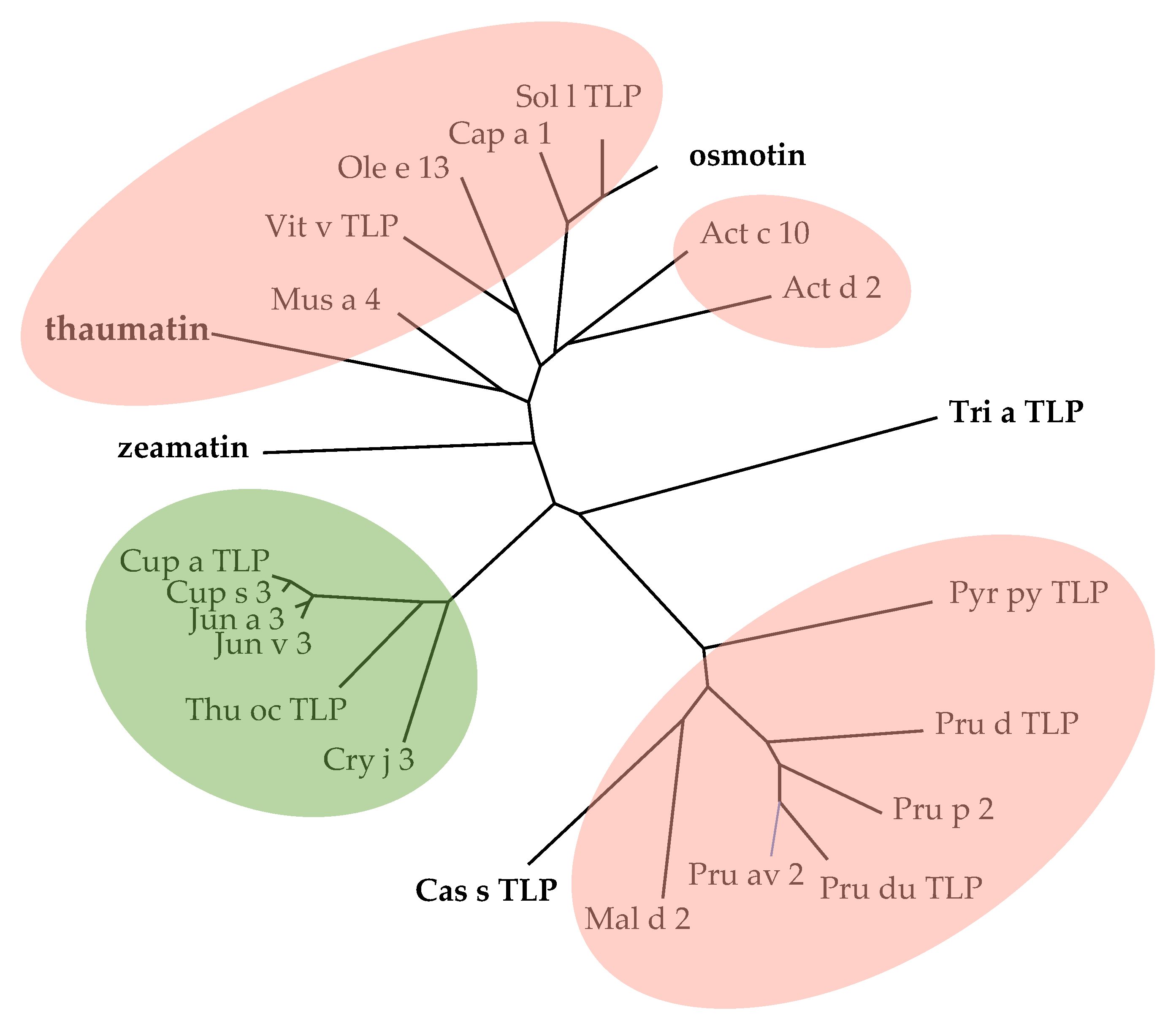
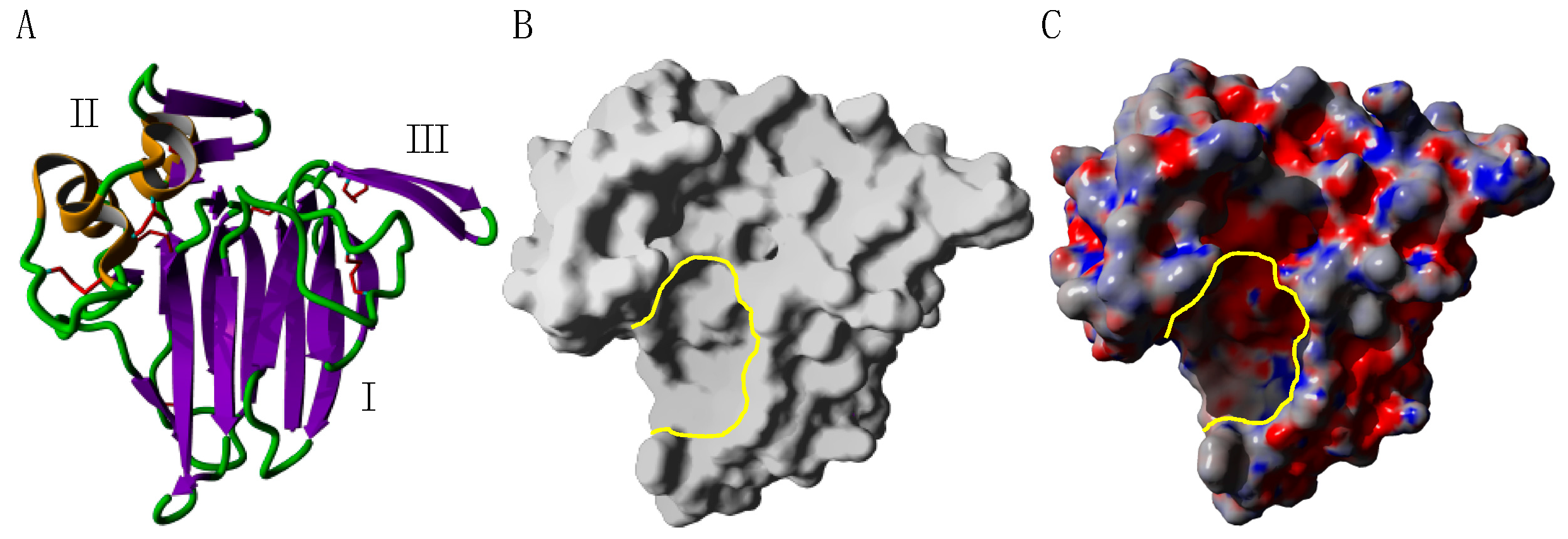
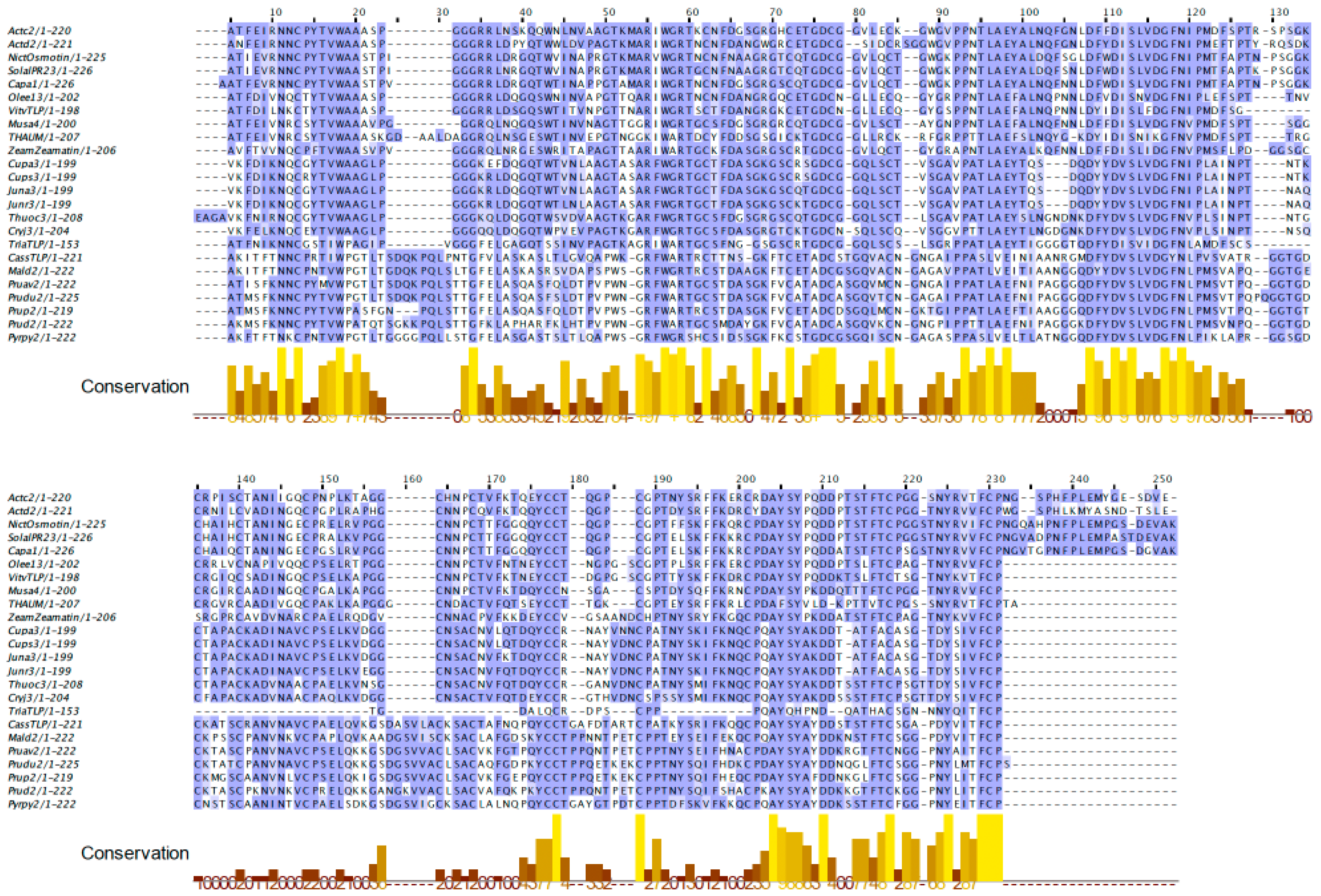
2.1. PR-5 Thaumatin-like Proteins (TLP)

The group of PR-5 thaumatin-like proteins (TLP) is characterized by an extreme homogeneity of both the molecular structure and the endo-β1,4-glucanase properties associated with their structural fold. All TLPs possess a structure similar to that found in thaumatin, the sweet-tasting protein isolated from the arils of Thaumatococcus daniellii [6], made of three covalently associated domains: a central domain I with a β-sandwich structure, flanked on both sides by a predominantly α-helical domain II, and a small domain III which consists of a short β-hairpin (Figure 3A) [7]. Up to 16 extremely conserved cysteine residues form 8 disulfide bridges that confer to this tightly structured fold a good resistance to digestive proteases and heat denaturation, both properties susceptible to enhance the allergenicity of protein allergens [8]. A highly electronegative cleft with an endo-β1,4-glucanase activity, occurs between domains I and II (Figure 3B,C).
The allergenicity of fruit TLPs has been scarcely investigated and, thus, only a few insights are available on the IgE-binding epitopic regions of fruit TLPs. An in silico approach allowed the prediction of four IgE-binding epitopic areas corresponding to the amino acid sequence stretches 36–48, 51–63, 58–70, and 73–85 on the surface of the sapodilla (Manilkara zapota) fruit, that are essentially located around the electronegative cleft on the front face of the allergen (Figure 4a) [9]. Previously, some overlap had been detected between the predicted IgE-binding epitopes from Mus a 4, the banana (Musa acuminata) TLP, and the IgE-binding epitopes identified in the mountain cedar pollen TLP Jun a 3 [7]. These B-cell epitopes occur in the α-helical domain II of the TLP. More recently, three IgE-binding epitopes from Cup s 3, the green cypress (Cupressus sempervirens) TLP allergen, and TLPs from other phylogenetically related Cupressaceae species, strongly interacted with all the tested allergic patient’s sera, using the Pepscan technique (Figure 4b) [10]. The presence of common epitopes offers a molecular basis for the cross-reactivity and crossed allergenicity observed between fruit and pollen TLP allergens [11].
The multiple amino acid sequence alignments of TLP allergens show a high degree of conservation that corresponds to a very conserved structural organization (Figure 5). However, TLPs from the Rosaceae family slightly differ from TLPs of other families by two additional insertions in regions corresponding to domain I (1st insertion) and III (2nd insertion), respectively.
The TLPs of Rosaceae form a very homogeneous group of closely related proteins, whereas TLPs from other families are clustered in a rather distant group within the phylogenetic tree of TLPs built from the amino acid sequence comparison. TLPs from the Cupressaceae pollen form an intermediate cluster equidistant from the other two fruit clusters (Figure 6). Depending on these phylogenetic affinities, the IgE-binding cross-reactivities observed among the fruit TLPs, e.g., between Mal d 2, Pru av 2 and Mus a 4, are so frequent than those observed between fruit and pollen TLPs, e.g., between Mal d 2, Pru av 2 and Cup a 3 [12].

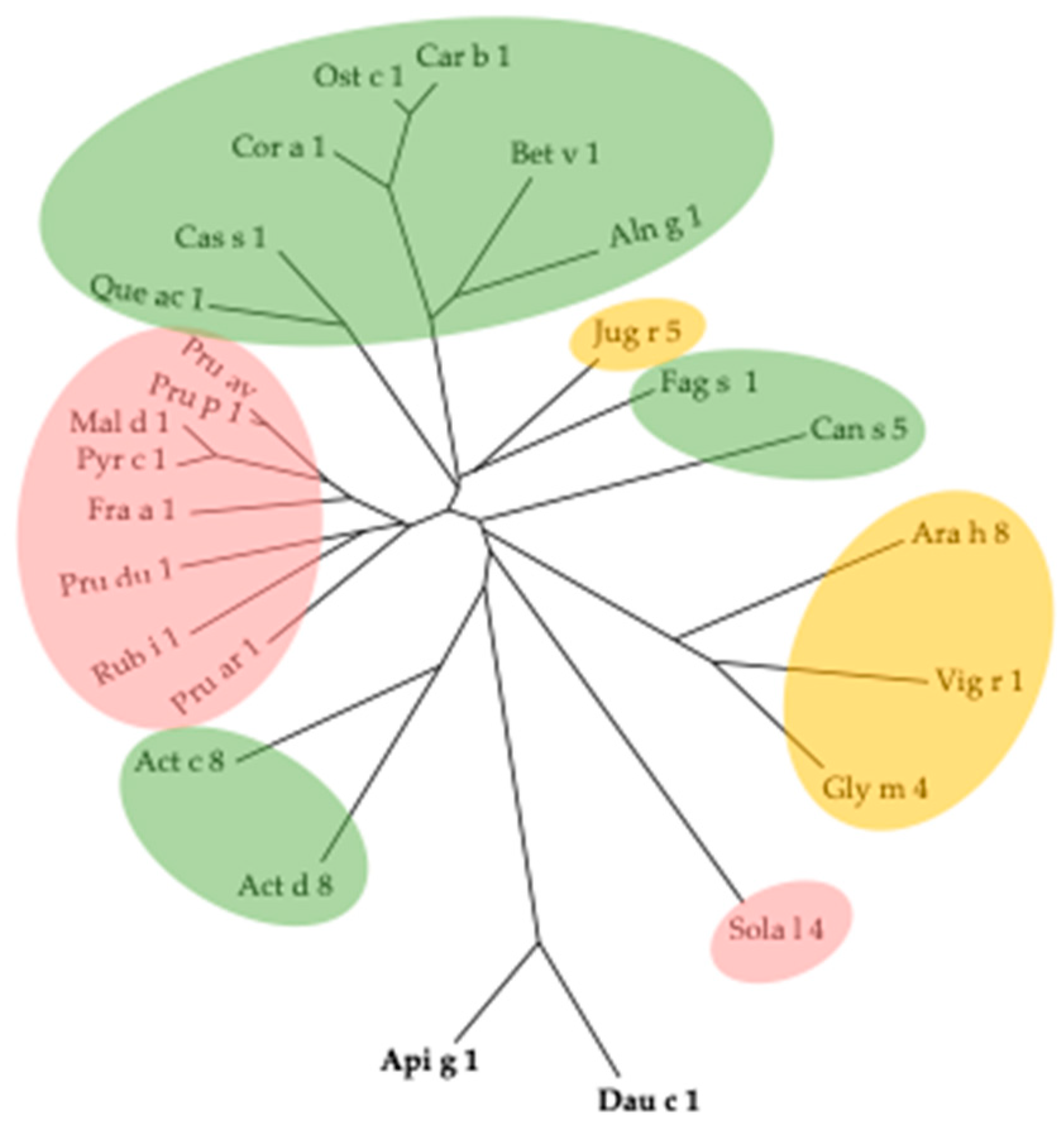
2.2. PR-10 Bet v 1-like Proteins

Bet v 1, the major pollen allergen of birch (Betula verrucosa), has been identified for a long time as the essential component of the birch pollen allergy [13]. Bet v 1 and other closely related PR-10 Bet v 1-like allergens consist of a homogeneous group of polypeptide chains of about 150 amino-acid residues, typically folded as an αβ protein made of a large curved β-sheet associated with a long α-helix (Figure 7A), exhibiting an overall cylindrical shape (Figure 7B,C) [14]. These loosely structured proteins exhibit a weak resistance to digestive proteases and heat denaturation, so they are quickly inactivated upon the cooking of PR-10 Bet v 1 like protein-containing foods and food products [15].
Structural and mutational analyses of Pru av 1 showed that the amino acid sequence stretch 44LEGDGGPGT52, forming the so-called P-loop of Pru av 1 and other PR10 Bet v 1-like protein allergens, played a key role in the IgE-binding capacity of Pru av 1, together with two other residues N28 and P108, located in other parts of the protein (Figure 8) [16,17]. In addition, the residue stretch 142–156 of Bet v 1 was identified as the dominant T-cell epitope of the major birch pollen allergen [18]. All these key residues participating in the discontinuous IgE-binding epitopes and T-cell epitopes should be responsible for the cross-reactivity between food and pollen Bet v 1-like proteins [19].
The multiple amino acid sequence alignment of PR10-Bet v 1-like allergens show a high degree of conservation (Figure 9), which results in a very conserved structural organization for all the members of the PR10 family (Figure 8).
Bet v 1 allergens from the Fagaceae pollen form a homogeneous group of proteins sharing conserved amino acid sequences and closely-related three-dimensional structures (Figure 10) [20]. However, Bet v 1-like allergens exhibiting very close amino acid sequences and structural scaffolds also occur in various fruits from the Rosaceae family (Fra a 1, Mal d 1, Pru p 1, Pru ar 1, Pru av 1), seeds (Gly m 4, Jug r 5), and vegetables (Api g 1, Dau c 1), that account for possible cross-reactivities and cross-allergies with these food products.

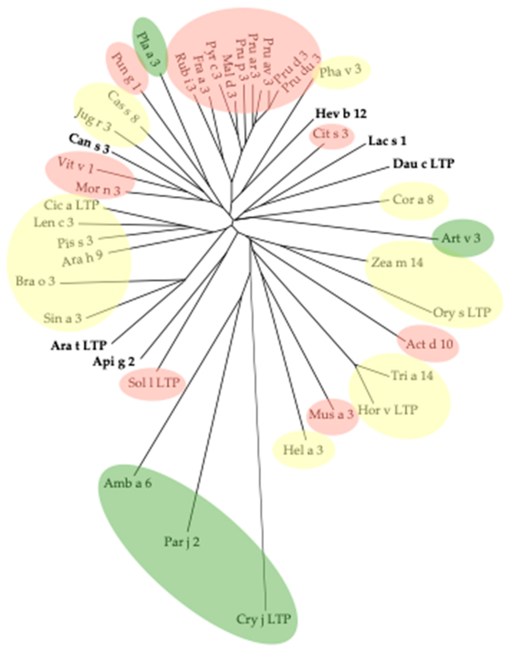
2.3. PR14 Non-Specific Lipid Transfer Proteins (nsLTP)

Non-specific lipid transfer proteins (nsLTPs) are small polypeptides of about 90 amino acids, which consist of a tightly packed core of four α-helices (α1–α4) extended by a C-terminal tail, which adopt the so-called “saxophone-like” conformation (Figure 11) [21]. Four disulfide bridges occurring between 8 conserved cysteine residues contribute to creating a compact structure extremely resistant to digestive proteases and heat denaturation that probably accounts for their high allergenic potential [22,23]. In fleshy fruits like peach, apple or apricot, once synthesized in the pulp cells, nsLTPs migrate into the epidermic cells to accumulate in the fuzzy covering the peach or the outer cuticular cells of the apple [24]. As a practical consequence of this surface localization, peeling the skin off the peach and apple allows the removal of most of the allergens contained in these fruits.
Three-major IgE-binding epitopic regions have been identified on the surface of the Rosaceae nsLTPs, Pru p 3 [25] and Mal d 3 [26], probing synthetic peptides spanning the nsLTP sequences with specific IgE-containing sera from allergic patients. Although few in number, these IgE-binding epitopic regions cover an extended area of the molecular surface, which could explain the enhanced allergenicity of both allergens (Figure 12). In addition, most of these IgE-binding regions are well conserved in nsLTPs from other fruits and other sources, like seeds and pollen [27].
A high degree of conservation of their amino acid sequences characterizes the nsLTP allergens, as shown in the amino acid sequence alignment (Figure 13).
In this respect, the phylogenetic tree built for nsLTPs of different origins shows some rather close relationships of fruit nsLTPs with other seeds and pollen nsLTPs, irrespective of their phylogenetic relationships (Figure 14). In this regard, nsLTP allergens from the Rosaceae family form a very homogeneous group with a high degree of cross-reactivity among the members, e.g., strong cross-reactivity of Pru p 3 with Mal d 3, Pru av 3, and Pru ar 3, and lower cross-reactivity with nsLTP allergens from other families, e.g., low cross-reactivity of Pru p 3 with Jug r 3 (Juglandaceae), Ara h 9 (Fabaceae), Cor a 8 (Betulaceae), Zea m 14 (Poaceae), Cit s 3 (Rutaceae), and Art v 3 (Asteraceae) [27]. These IgE-binding cross-reactivities depend on very similar three-dimensional conformations and exposed molecular surfaces.
The nsLTP allergens and PR10 Bet v 1-like protein allergens possess internal cavities, which usually accommodate a set of small molecules ligands including unsaturated fatty acids and other lipidic compounds (nsLTPs), polyphenols, and alkaloids (PR10 Bet v 1-like proteins) (Figure 15) [28]. In addition to enhancing the stability and eventually modifying the allergenicity of nsLTPs and PR10 Bet v 1-like protein allergens [29,30,31], these small molecule ligands have an adjuvant effect, and participate in the activation and regulation of the allergic and inflammatory responses via the activation of signaling pathways common to innate immunity and allergic responses [32,33,34].

2.4. Profilins

Profilins are typical pollen allergens that also occur in the vegetative organs of plants and fruits. They correspond to small polypeptides of ~130 amino-acid residues exhibiting a canonical structural organization made of a central β-sheet flanked on both sides by a few α-helices (Figure 16A) that gives a rather flattened shape to these molecules (Figure 16B,C) [35].
This rather loose structural organization, not sustained by disulfide bridges, accounts for a low resistance to thermal denaturation, that explains why profilins lose their allergenic potency after cooking [36]. However, despite the cooking of profilin-containing foods destroying their IgE-binding epitopes, the remaining continuous epitopes keep some cross-reactive T cell activity in vitro [37].
Using synthetic peptides spanning the full Cuc m 2 amino acid sequence, the IgE-binding epitopes of the muskmelon profilin, Cuc m 2, were identified to stretches 66–75 + 81–93 (E1) and 95–99 + 121–131 (E2) of the amino acid sequence, which collapses in two patches (E1 and E2) well exposed on the molecular surface of the profilin (Figure 17). An additional stretch corresponding to residues 2–10 + 35–45 forms another discontinuous epitope E3 on the molecular surface of Cuc m 2 [38]. A discontinuous mimotope was further identified on the Cuc m 2 surface, corresponding to the sequence stretch 2SW + 5AY + 9DH + 111TPGQ + 116NM + 121RL, which plays a role in the cross-reactivity of Cuc m 2 with pollen profilins [39].
Similarly, the IgE-binding activity of a recombinant soybean profilin, rGly m 3, is mediated by extended conformational epitopes, since three overlapping profilin fragments comprising amino acid residues 1–65, 38–88, and 50–131 displayed no significant IgE-binding activity towards the sera from soybean allergic patients [40]. In addition, plant food profilins and pollen profilins exhibited an equivalent IgE-binding reactivity, which suggests that food and pollen profilins share most of their IgE-binding epitopes [41].
Recently, the distribution of negative charges on the molecular surface of various profilins, including Amb a 4 and Amb a 8 from Ambrosia artemisiifolia, Art v 4 from Artemisia vulgaris, Bet v 2 from Betula verrucosa, Fra e 2 from Fraxinus excelsior, Hev b 8 from Hevea brasiliensis, Phl p 2 from Phleum pratense, and Zea m 12 from Zea mays, was relevant for the recognition of profilins by monoclonal antibodies, and should participate in their IgE-binding cross-reactivity [42].
The alignment of their amino acid sequences shows a high degree of conservation for the profilin allergens, independent of their botanical origin (Figure 18). Accordingly, all these allergens exhibit a very conserved three-dimensional structure. As widely distributed among fruits, seeds and pollens, profilins constitute a very homogenous family of phylogenetically related pan-allergens.
The close phylogenetic relationships clearly appear in the phylogenetic tree built from the amino acid sequences of profilins (Figure 19). Consequently, IgE-binding cross-reactivity commonly occurs between apparently distantly related plant sources, e.g., between melon (Cuc m 2) and mugwort (Art v 4), between celery (Api g 4) and chestnut (Cor a 2), between peach (Pru p 4) and birch (Bet v 2), etc. [43]. However, due to their weak resistance to heat denaturation (cooking), profilins usually behave as mild allergens.

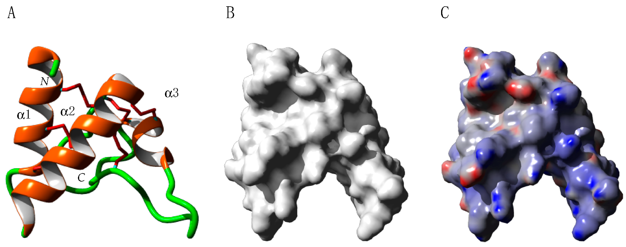
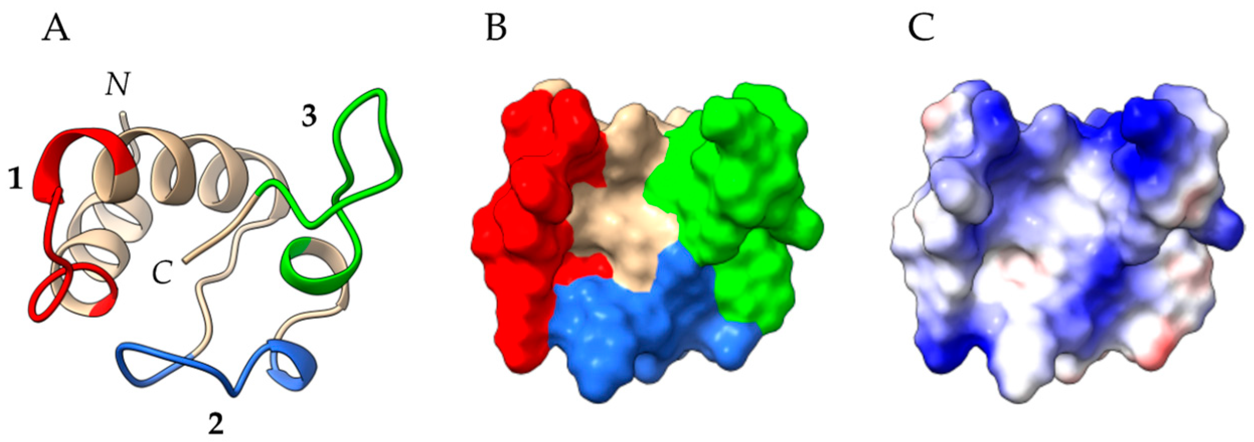
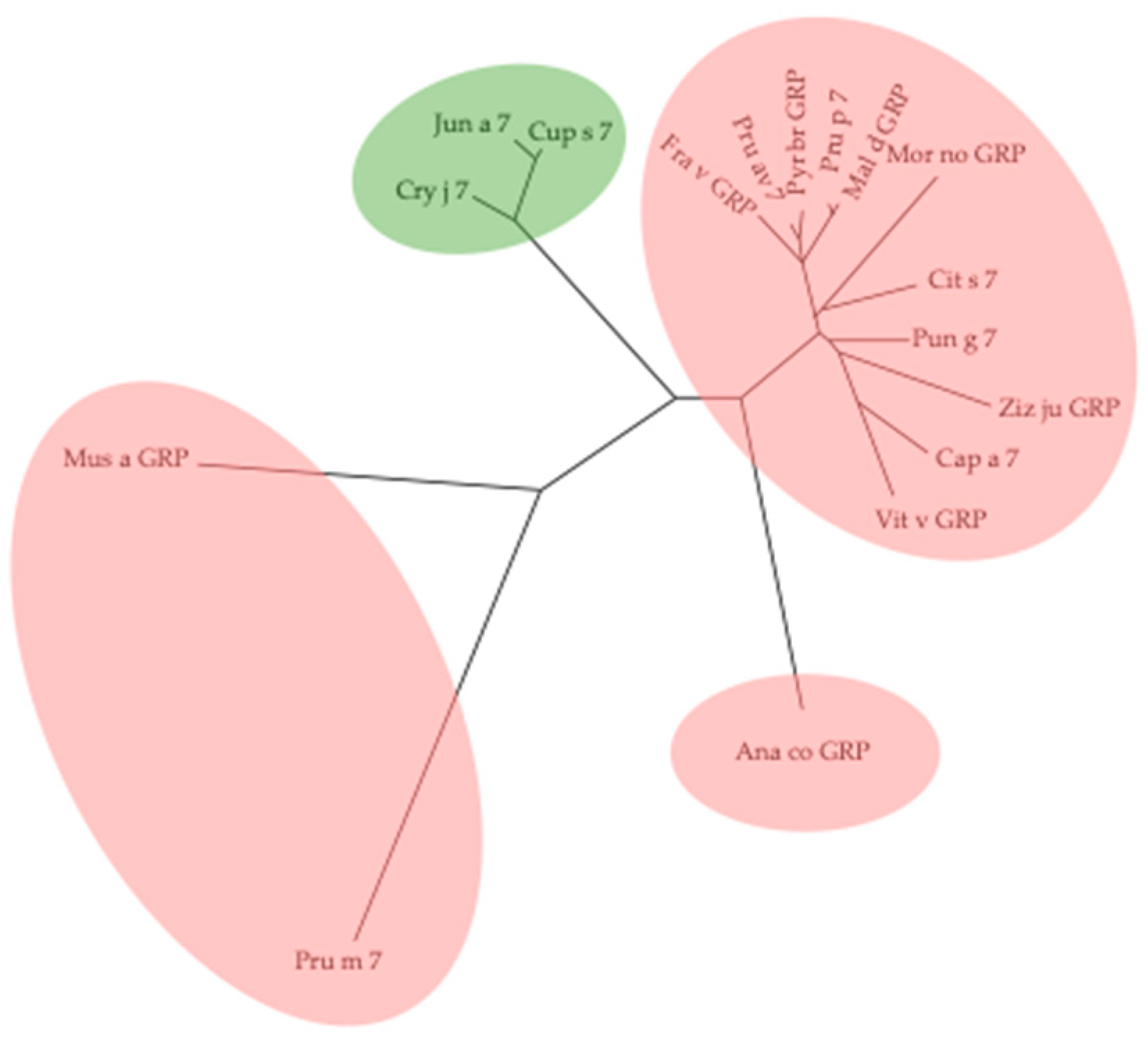
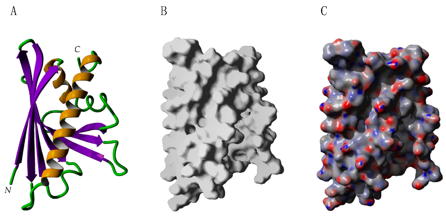
2.5. Gibberellin-Regulated Proteins (GRPs)
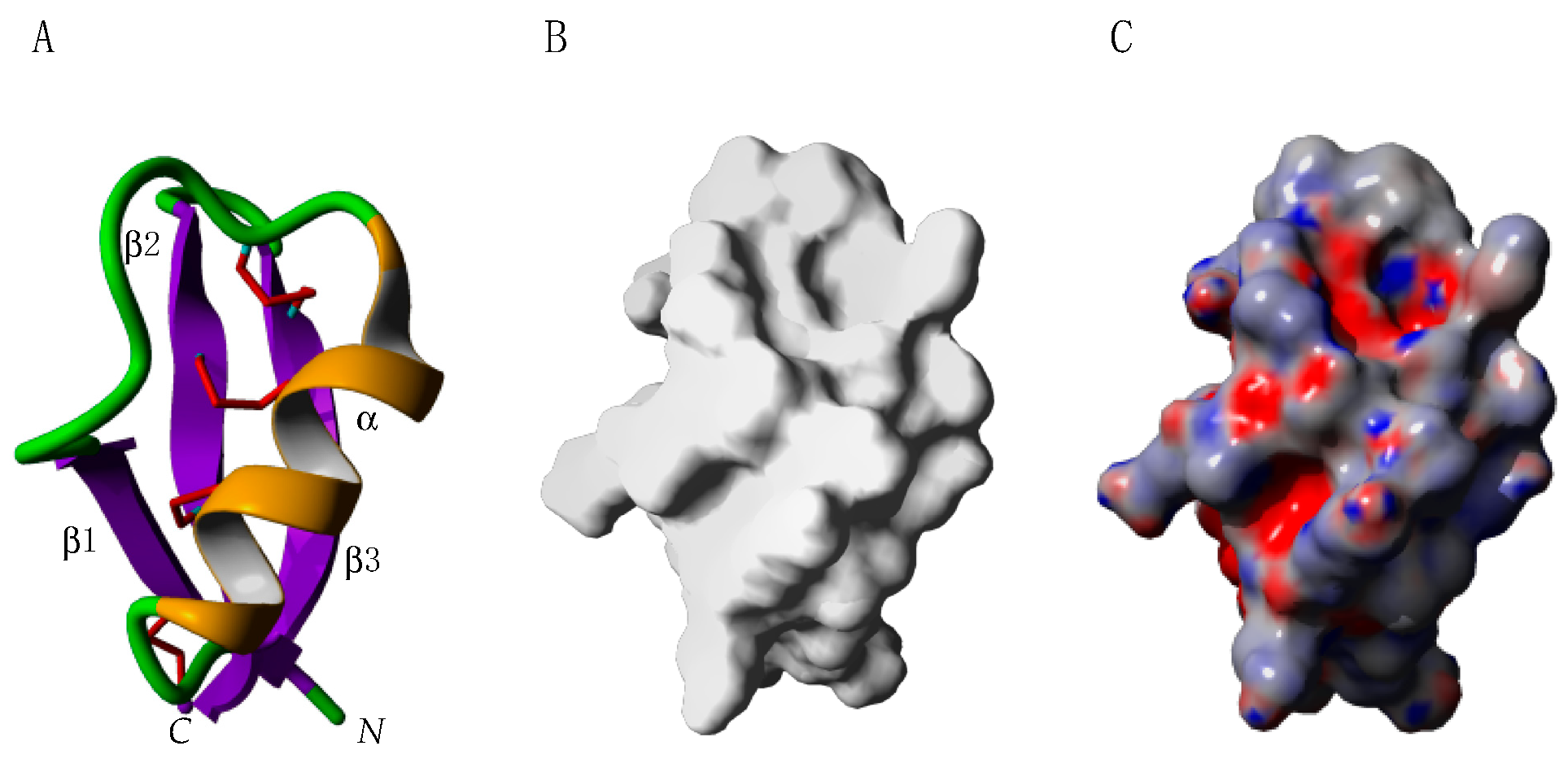
The GRPs (also known as snakins) sequenced so far share a superposable canonical structural organization made of three short α-helices α1, α2, and α3, tightly linked by two series of three disulfide bonds interconnecting helices α1–α2 and helices α2–α3, respectively (Figure 20A). The resulting three-dimensional structure consists of a dome-shaped molecule, crossed by a deep cleft located at its bottom. Depending on the GRPs, the cleft is enlarged, e.g., in the peach peamaclein Pru p 7 (Figure 20B), or narrower, e.g., in the potato snakin-1. The distribution of electronegatively charged residues Asp and Glu are primarily located at the top of the dome, while other parts of the GRPs, especially the cleft, exhibit an electropositive character, due to the high concentration of electropositive Arg and Lys residues on the molecular surface (Figure 20C). Accordingly, GRPs behave as predominantly basic proteins, displaying a high pI value (calculated pI of 8.52 for Pru p 7).
By virtue of their extremely tightly packed structure, sustained by a high number of conserved disulfide bridges, peamaclein (Pru p 7) and other GRPs from fruit or pollen consist of small proteins resistant to digestive proteases and heat denaturation. In this regard, Pru p 7 was shown to become sensitive to the digestive proteolytic degradation only after heat denaturation [44].
Although no data on the allergenicity of GRPs are available, a hydrophobic cluster analysis (HCA) of GRPs, together with a surface analysis of the modeled proteins, allows one to predict the presence of exposed areas rich in hydrophilic and electropositive residues [45,46] that are susceptible to participate in the IgE-binding activity of these allergens. The three predicted potential IgE-binding epitopic regions are located on the bottom of the dome-shaped GRPs, essentially around the cleft, and roughly coincide with electropositively charged areas (Figure 21). In contrast, no potential IgE-binding areas were predicted to occur on the dome face opposite to the cleft. In addition, all of the GRP allergens share a very similar distribution of these predicted potential IgE-binding areas, which could account for the cross-reactivity observed between GRP allergens from fruits and pollens.
The alignment of amino acid sequences of GRP allergens shows a high degree of conservation of GRPs from fruits and pollens (Figure 22).
Fruit GRPs constitute a homogeneous group of phylogenetically related proteins, except for GRPs from ananas (Ana co GRP), banana (Mus a GRP) and Japanese plum (Pru m 7), which are less closely related to the fruit clade (Figure 23). In addition, GRP from the Cupressaceae pollen, Cry j 7, Cup s 7 and Jun a 7, are phylogenetically close to the fruit GRPs. Accordingly, it is not surprising that GRPs are highly immunochemically cross-reactive allergens from fruit (Pru p 7, Pun g 7) and pollen (Cup s 7, Cry j 7) sources [47,48].

2.6. Defensins

The plant defensin PR-12 family comprises small acidic molecules of about 110 amino acid residues, exhibiting the canonical knottin fold, a structural αβ motif made of an α-helix linked to an antiparallel three-standed β-sheet, stabilized by 3 or 4 disulfide bridges which adopt a disposition known as the so-called “disulfide through disulfide knot” (Figure 24) [49]. Genuine defensins, e.g., brazzein from the Pentadiplandra brazzeana, are restricted to the knottin domain, whereas the closely related defensin-like proteins, e.g., Art v 1 from mugwort (Artemisia vulgaris) and Amb a 4 from ragweed (Ambrosia artemisiifolia), possess a knottin domain extended at the C-terminus by a long prolin-rich tail (Figure 25). With the exception of brazzein, a sweet-tasting and taste-modifying protein of fruits from the African shrub Pentadiplandra brazzaeana [50,51], whose the sweetening power is 500 to 2000 times greater than that of sucrose [52], other defensins essentially occur in the pollen (Art v 1, Amb a 4), vegetative parts (defensins from pea (Pisum sativum), and fenugreek (Trigonella foenum-graecum) of the plants.
The IgE-binding activity of the pollen defensin allergens Art v 1 (Artemisia vulgaris), Amb a 4 (Ambrosia artemisiifolia), and Par h 1 (Parthenium hysterophorus), which all consist of extended defensin-like proteins comprising a N-terminal defensin domain of about 50 amino acid residues associated with a C-terminal proline-rich domain of >60 amino acid residues, was investigated using sera from Austrian, Canadian and Korean allergic patients [53]. Both domains participated in the defensin allergenicity (defensin domain) and IgE-binding cross-reactivity (proline-rich domain). In fact, structurally altered defensin-like proteins are still capable of IgE-binding capacity, suggesting that continuous epitopes in the defensin domain should be responsible for the IgE-binding activity (Figure 25). Interaction of Art v 1 with the corresponding IgE allowed identification of some surface exposed amino acid residues (S3, K4, K8, S14, R40, E41, E45, S46, K55, A63) involved in the binding of antibodies (Figure 26) [49]. Cross-inhibition experiments showed that the proline-rich domain is also involved in the IgE-binding cross-reactivity, but different degrees of cross-reactivity depend on the origin of sera with a high, medium, and low cross-reactivity for sera from Austrian, Canadian and Korean allergic patients, respectively.
The alignment of defensin allergens shows a dichotomy between short and long amino acid sequences (Figure 27). In addition, the highly conserved defensins from pollens differ from the rather less conserved defensins from fruits.
The phylogenetic tree built up from the amino acid sequences of plant defensins suggests that possible cross-reactivities and cross-allergies could occur between defensins of pollens, fruits, seeds, and vegetables (Figure 28) [54].

2.7. PR3 Chitinases

Plant chitinases essentially consist of endo chitinases that are classically divided into five classes, including class-I, class-II, class-III, class-IV, and class-V [55]:
-
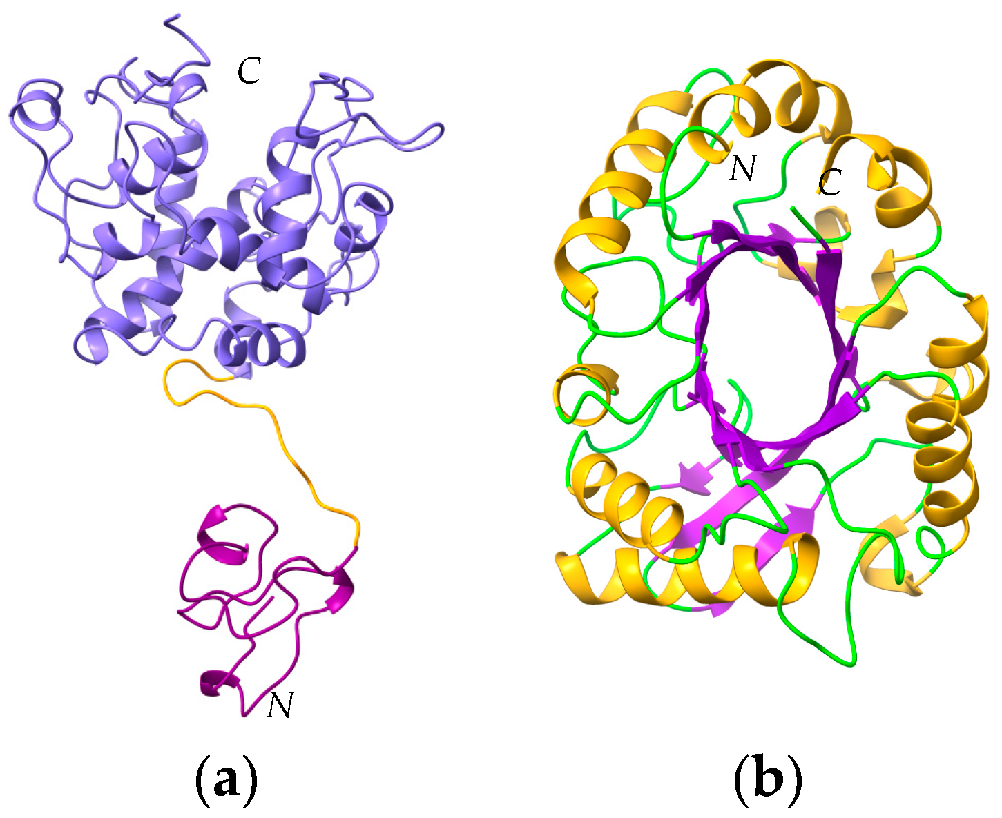
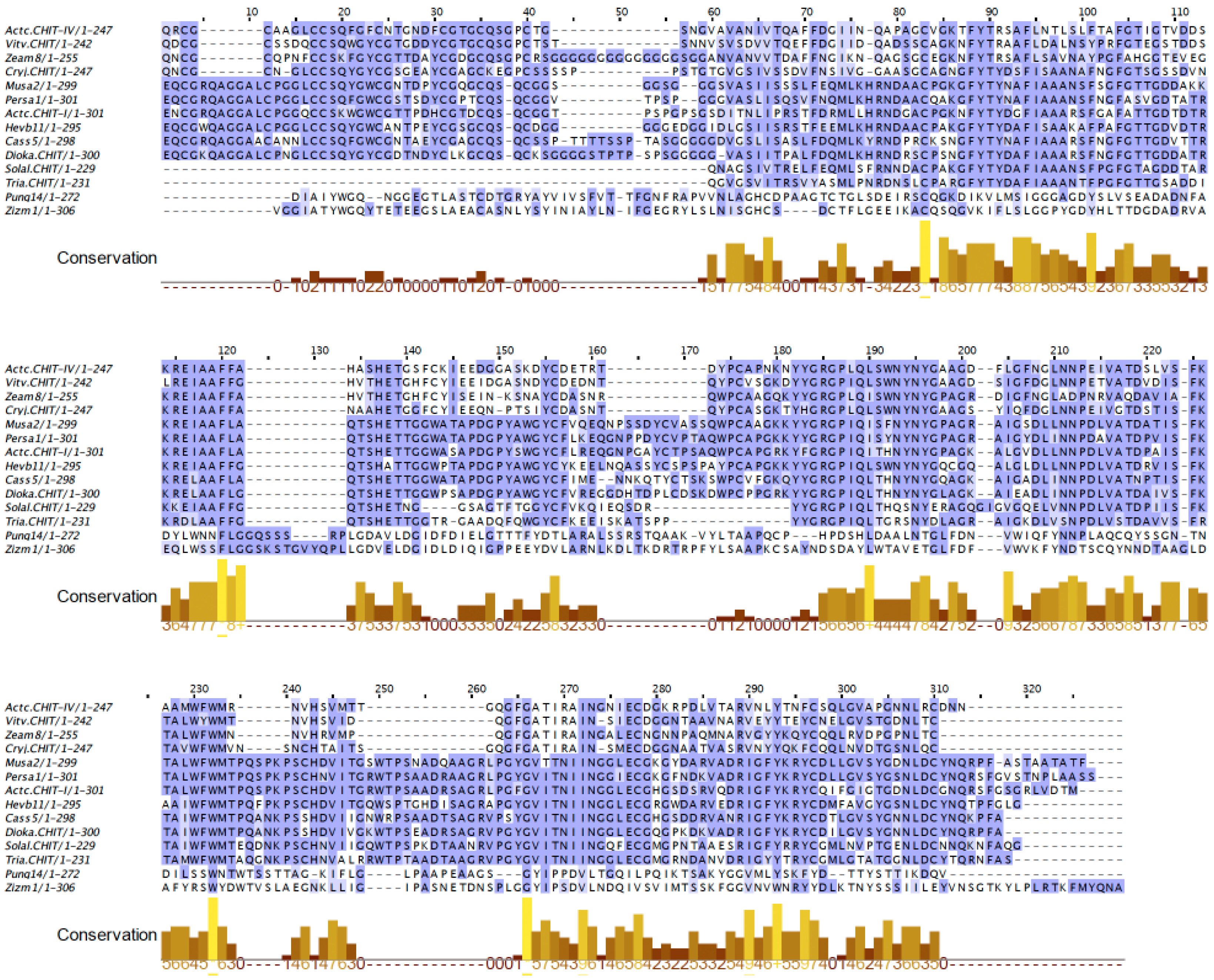
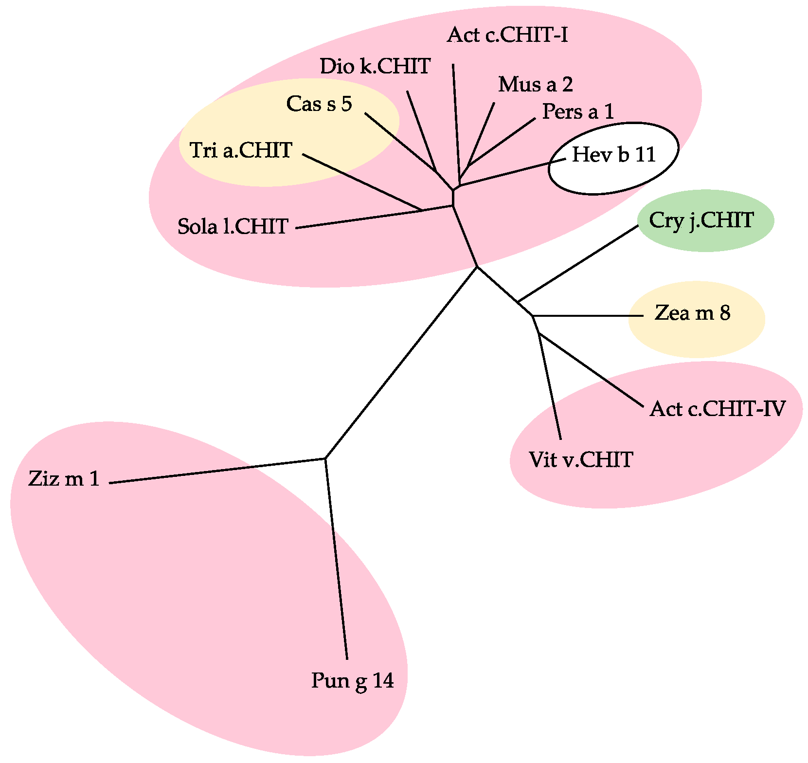
Class-I and class-IV chitinases exhibit a structural organization in two domains, comprising a N-terminal chitin-binding domain made of a cysteine-rich polypeptide, similarly folded as the hevein domain of the latex from rubber tree Hevea brasiliensis (Hev b 6), linked by an extended linker loop to a C-terminal α-helical folded domain with a chitin-cleavage activity (Figure 29a). Class-I and class-IV chitinases use an inverting mechanism for cleaving the β1,4-GlcNAc linkage of chitin to generate shorter fragments of chitobiose (GlcNAc)2, chitotriose (GlcNAc)3 and chitotetraose (GlcNAc)4. The chitinase allergens from banana (Mus a 2), avocado (Pers a 1), and Hev b 11 from the latex of rubber tree, belong to this group of chitinases.
-
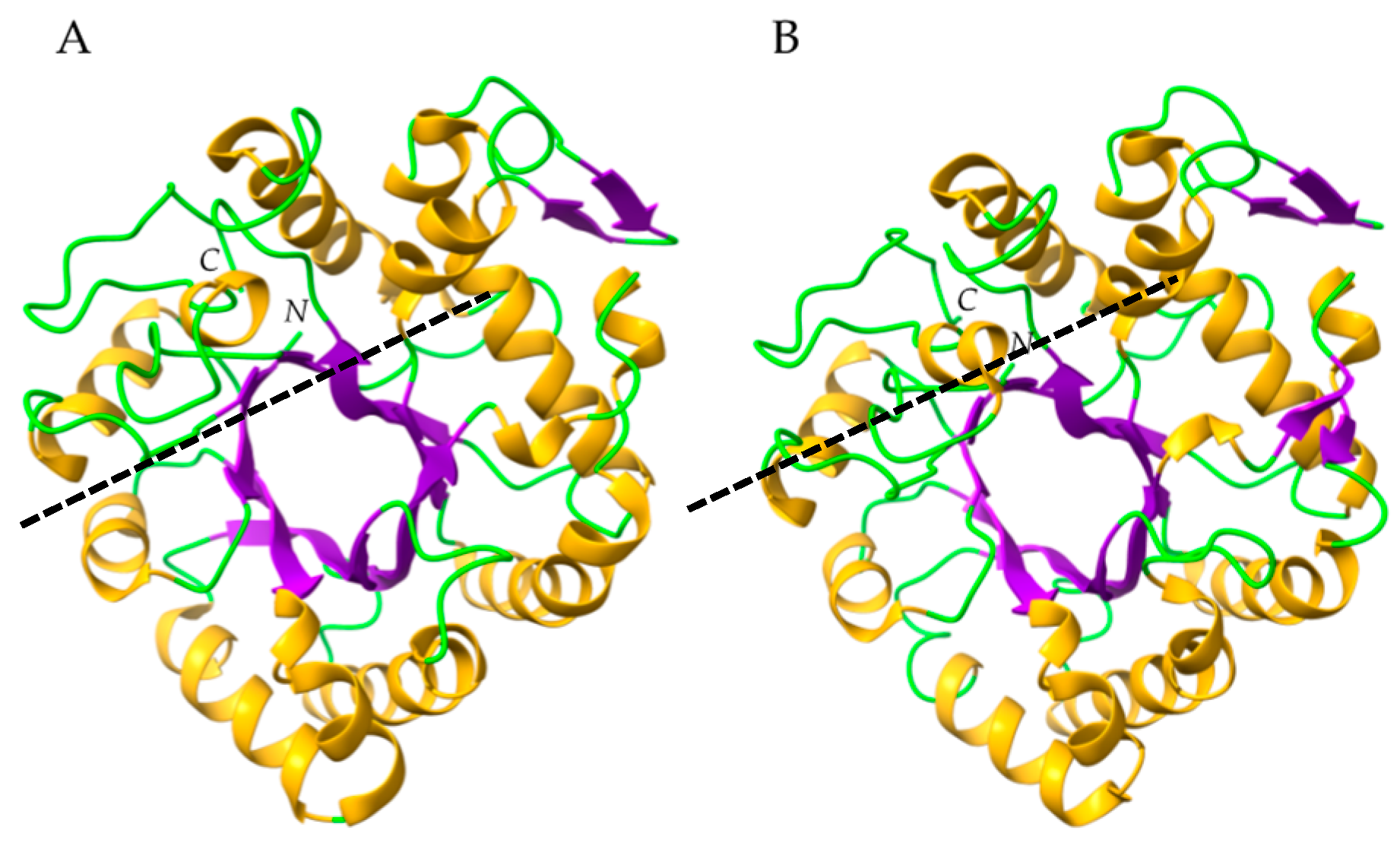
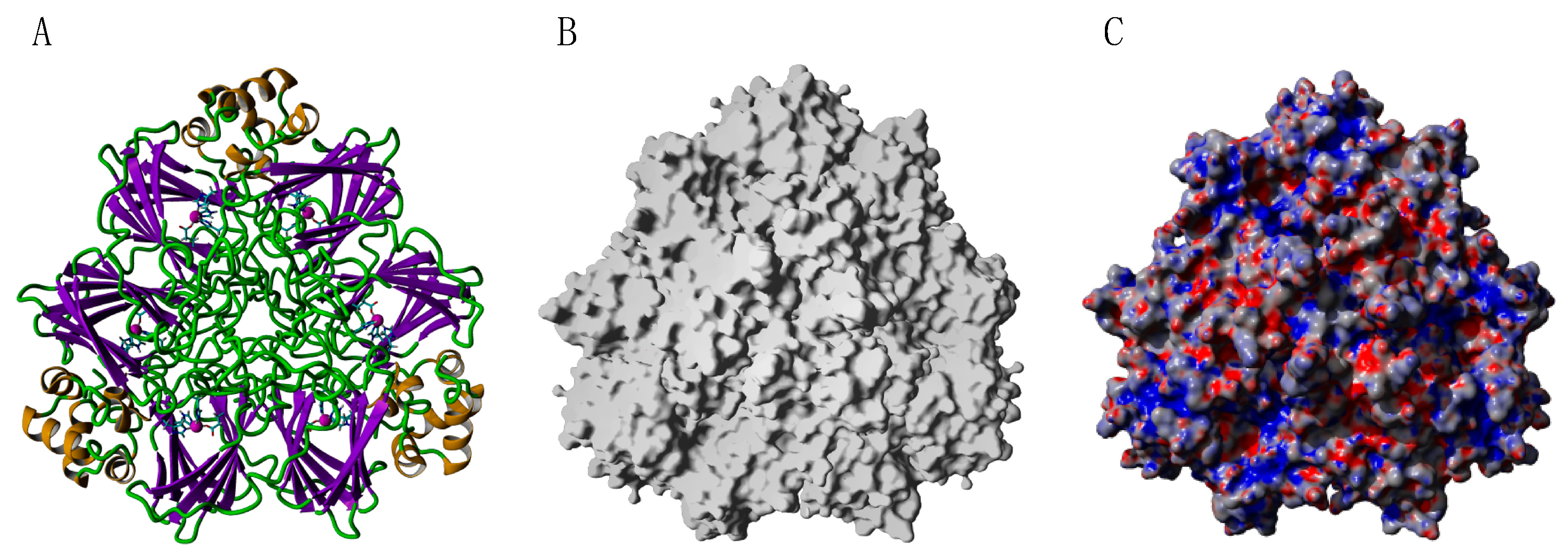
Class-III chitinases readily differ from class-I chitinases by a different α8β8 β-barrel organization made of a central crown of eight β-strands, linked to a peripheral crown of eight α-helix by interconnecting loops (Figure 29b) [56]. In addition, class-III chitinases use a different retaining mechanism for the cleavage of chitin chains in shorter fragments. The chitinase allergens from pomegranate (Pun g 14) and jujuba fruit (Ziz m 1), belong to the class-III chitinase group.
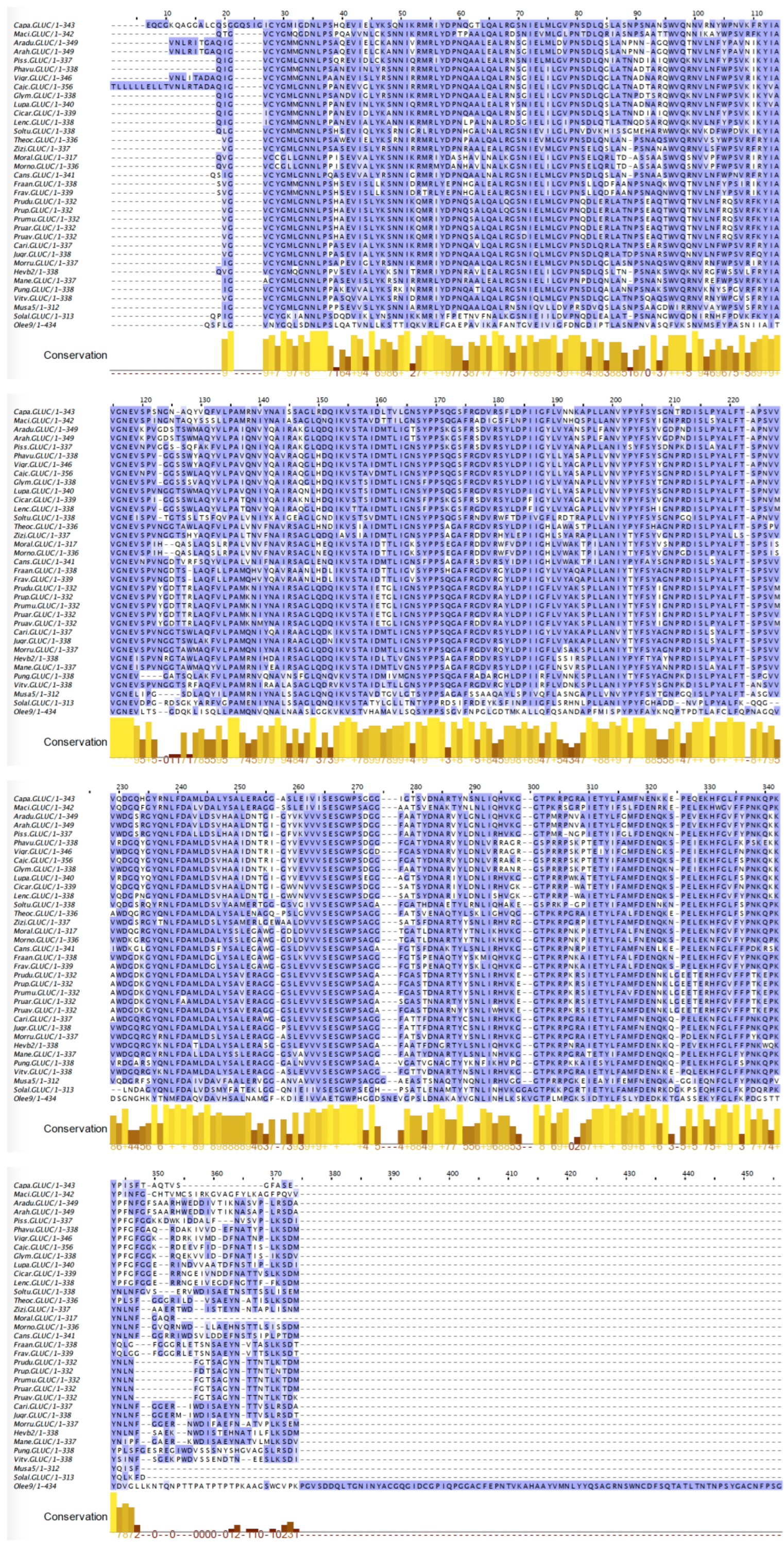
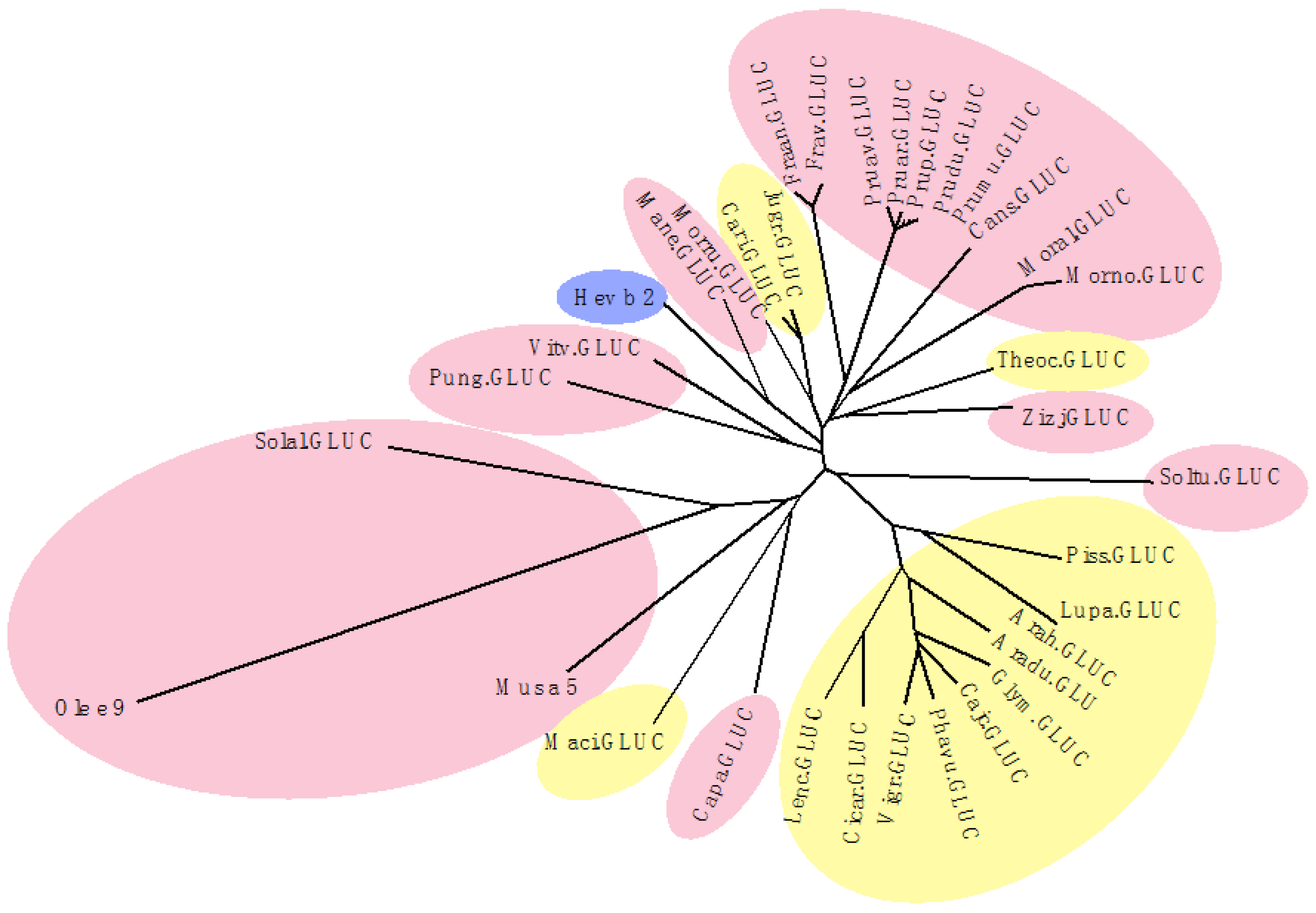
Hevein (Hev b 6), consists of a short polypeptide chain of 43 amino acids folded made of two short β-strands associated with two α-helical stretches, folded via four disulfide bridges into a tightly packed three-dimensional structure (Figure 30A) [57]. Both B- and T-epitopes have been identified on the molecular surface of Hev b 6, and show some overlap (Figure 30B,C). A N-terminal hevein-like domain exhibiting a similar fold occurs in class-I and class-IV chitinase allergens, including Hev b 11, Mus a 2, and Per a 1.
The alignments of PR3 chitinase allergens from higher plants clearly discriminate between class-I/class-IV and class-III chitinase (Pun g 14, Ziz m 1) groups (Figure 31). However, class-I and class-IV chitinases exhibit rather different amino acid sequences, particularly in their N-terminal part.
According to the discrepancies observed in the amino acid sequences of the different classes of chitinases, class-I, -III, and -IV chitinases cluster in three distinct groups in the phylogenetic tree built from the amino acid sequence alignment (Figure 32). Depending on these phylogenetic relationships, some cross-reactivity should be predicted among the chitinases from class-I group, whereas no cross-reactivity should occur between chitinases from the class-I and class-III groups. In this respect, class-I chitinases, which are closely related to Hev b 11, should participate in the latex-fruit syndrome [58].

2.8. PR2 Glucanases

The β1,4-endoglucanases from higher plants all exhibit the α8β8 β-barrel organization, made of an internal crown of eight β-strands connected by loops to a peripheral crown of eight α-helices. A short additional β-hairpin completes the three-dimensional structure (Figure 33). A long catalytic groove centered on the β-strand crown contains the active site responsible for the cleavage of the β1,4-linked glucan chains by plant glucanases. This three-dimensional organization pattern is conserved in all the β1,3-endoglucanases from higher plants, especially in the glucanase allergens identified so far, e.g., Mus a 5 from banana [7], Pru av 2 cherry [59], VVTL-1 from grape [60], and Hev b 2 from the latex of rubber tree [61].
Probing synthetic peptides spanning the amino acid sequence of Hev b 2 with specific IgE-containing sera from allergic patients allowed identification of up to eight continuous IgE-binding epitopes. They correspond to different stretches of amino acid sequences arrayed on the molecular surface, essentially located on the back face opposite to the catalytic groove [62] (Figure 34A,B). Compared to Hev b 2, the continuous IgE-binding epitopic regions identified in the banana glucanase Mus a 5, are similarly located on the back face of the allergen, and exhibit a very similar conformation on the molecular surface [7] (Figure 34C,D). The similar distribution and conformation of the epitopic regions in both allergens suggests the occurrence of a rather high degree of cross-allergenicity between these fruit and latex allergens that should participate in the latex-fruit syndrome [58].
The glucanase allergens exhibit a high degree of identity in the multiple alignments of their amino acid sequences (Figure 35). With the exception of Ole e 9 [63], the olive tree allergen, which differs from other fruit and seed glucanases by a quite different amino acid sequence and an additional long C-terminal expansion, other fruit allergens display closely related amino acid sequences. Accordingly, they are distributed in two closely related clusters; the first cluster groups Hev b 2, Rosaceae and other fruit allergens, and the second cluster contains most of the seed glucanase allergens. Another heterogeneous cluster contains Ole e 9, Mus 5, and other Solanaceae glucanases (Figure 36). The close vicinity of these different clusters points out the phylogenetic relationships that should occur among the fruit and seed allergens, and between Hev b 2 and fruit glucanase allergens (latex-fruit syndrome).

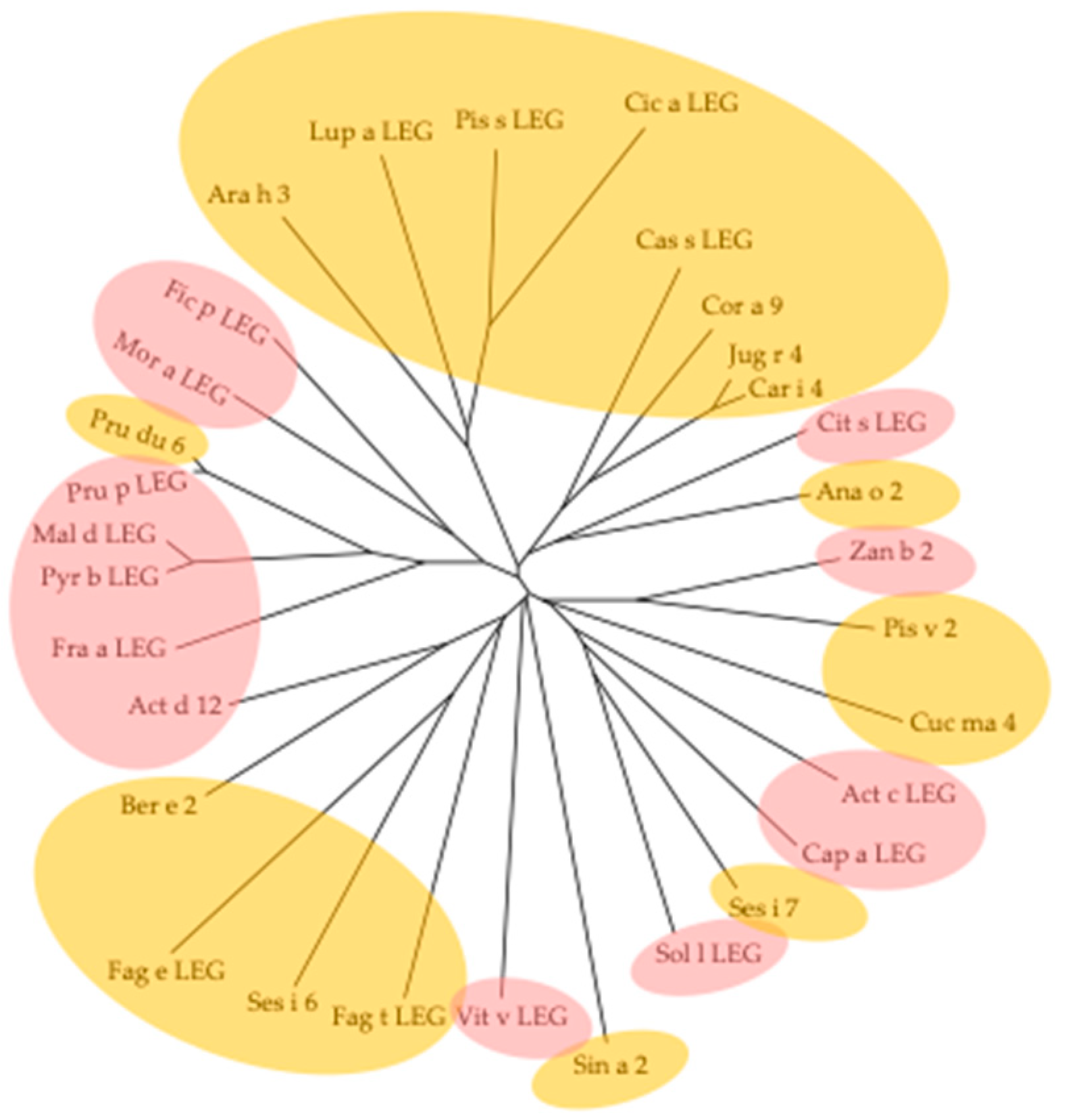
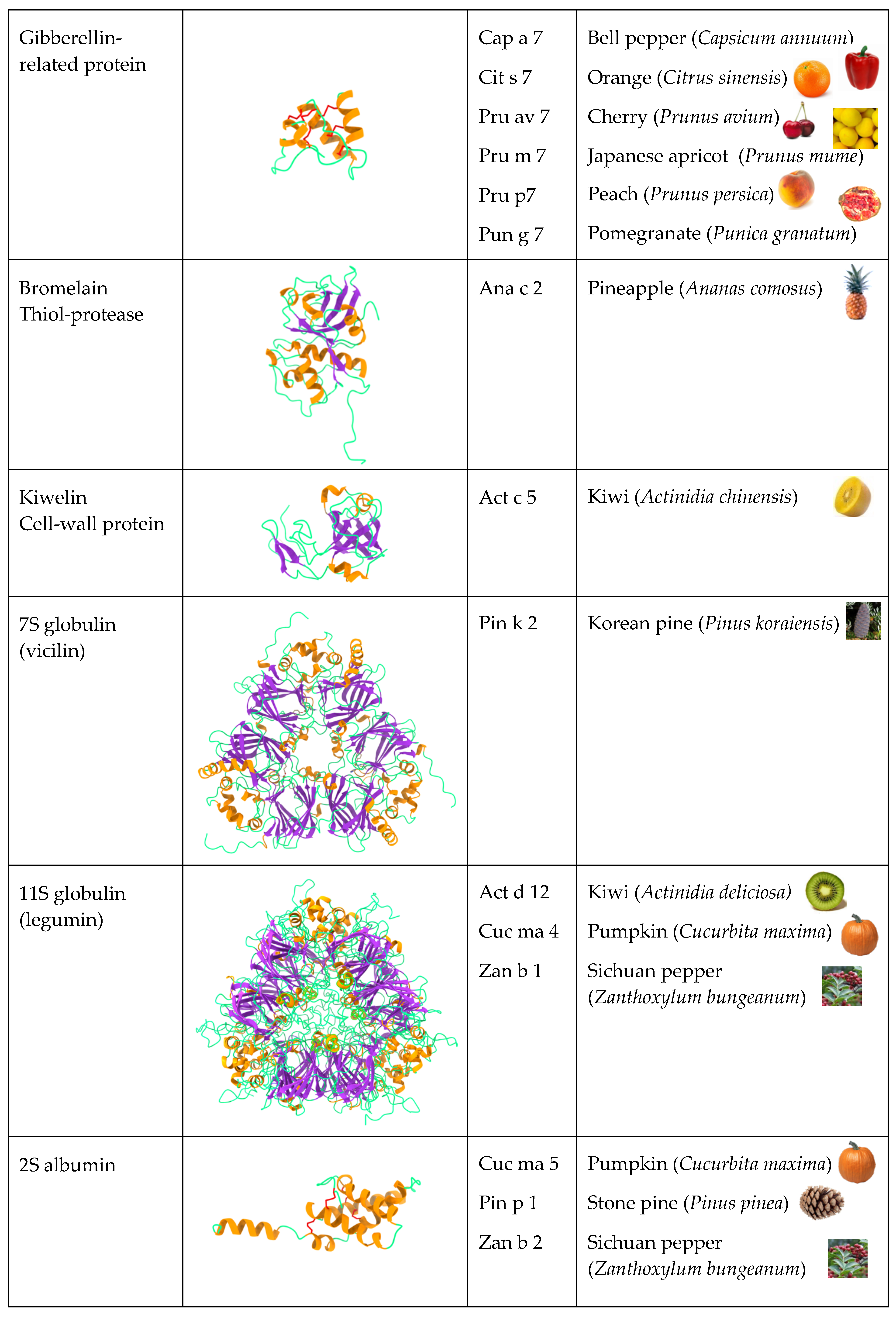
2.9. Seed Storage Proteins from Fruit Kernels

Botanically, fruits from higher plants consist of seed-bearing structures derived from the gynecium of the flowers. Accordingly, mature edible fruits contain seeds that correspond to the modified ovules of the flowers. Depending on the size of the seeds distributed in the flesh, additional allergic manifestations could result from seed allergens different from the pericarp allergens, when seeds are consumed together with the pericarp, e.g., in the case of small sized seed-containing fruits like kiwi, tomato, grape, or the Japanese lantern cherry (Physalis alkekengi) [64]. In addition, seeds or seed fragments of fruits from the Rosaceae (apple, pear) or the Rutaceae (lemon, orange) families can be accidentally consumed, and cause unexpected allergenic manifestations [5].
Seed allergens from seeds of higher plants belong to the so-called seed storages proteins, which comprise the cupin allergens 7S globulins (vicilins) and 11S globulins (legumins), and 2S albumins [65]. In addition, germins and germin-like proteins, recently recognized as relevant allergens, also belong to the cupin family of proteins [66]. Some nsLTPs proteins, e.g., Sola l 6 and Sola l 7, are located in tomato seeds [67].
Seed cupin allergens possess the canonical homotrimeric organization of their monomers, containing two cupin motifs in a flat-shaped structure (Figure 37A,B). A single homotrimer builds the 7S globulins, whereas two homotrimers associate face to face to build the 11S globulins or legumin allergens. The non-covalent association of two homotrimers mainly results from electrostatic interactions occurring between the opposite electropositive and electronegative charges of both faces of each homotrimers (Figure 37C).
Due to the conservation of both the amino acid sequences and three-dimensional structures of the 11S globulin allergens, a few continuous IgE-binding epitopic regions have been identified as common IgE-binding epitopes shared by the seed 11S globulin allergens [68]. These well-exposed, common IgE-binding epitopic regions also occur in the 11S globulin allergens Act d 12 from kiwi fruit and Fra v-Leg from strawberry (Fragaria vesca) (Figure 38), and could thus account for some allergic response in sensitized people [69,70].
The alignment of amino acid sequences of 11S globulin/legumin allergens exhibits a high degree of conservation with, however, some discrepancies, due to insertions/deletion events that have occurred in the medium part of the sequences (Figure 39). These sequence homologies are in agreement with the very conserved three-dimensional structure of these seed storage proteins. The phylogenetic tree built from the amino acid sequence alignment of 11S globulins shows that the clustering of fruit and seed 11S globulins roughly coincides with their distribution in the phylogenetically related families of higher plants (Figure 40).
The germin and germin-like allergens possess a single homotrimeric organization similar to that of vicilins, but differ from typical vicilins by their monomers, which contain a single cupin motif. Accordingly, the homotrimeric organization of germins consists of two superposerd dimers of homotrimers (Figure 41A,B). Like in other cupin proteins, both faces of the homotrimeric germins and germin-like allergens exhibit a complex distribution of electrostatic charges (Figure 41C) [71].
To date, with the exception of Cis s 1, the allergenic potential of germin-like proteins has not been clearly demonstrated. In addition, the localization of IgE-binding epitopes of germin-like protein allergens has not been investigated but IgE-binding experiments, ELISA inhibition assays and in vivo skin prick tests (SPT) performed with sera from allergic patients showed that previous deglycosylation of Cis s 1 resulted in an important loss of IgE-binding reactivity [66,72]. These results suggest that N-glycans play a prominent role in the IgE-binding capacity of Cit s 1. In fact, the Cit s 1 allergen corresponds to a heavily N-glycosylated allergen, which contains several putative N-glycosylation sites. Assuming these N-glycans are occupied by N-glycans of the complex type allowed us to build a heavily glycosylated Cit s 1 allergen, using the GlyProt server facilities (Figure 42). These predictions agree with the IgE-binding results observed with Cit s 1 [72].
The comparison of amino acid sequences of germin-like proteins shows a rather moderate degree of conservation (Figure 43), in spite of a very conserved three-dimensional conformation. According to this amino acid sequence heterogeneity, the phylogenetic tree of germins and germin-like proteins built from the amino acid sequence alignment, shows a rather marked dispersion of the allergens, even though fruit and seed germin-like allergens exhibit a tendency to clustering in well-individualized phylogenetically related groups (Figure 44).
The extreme diversity of allergens in some fruits like kiwi and tomato (Figure 45) illustrates another aspect often observed in fruit allergies. A great number of fruit allergens consist of pan-allergens widely distributed in different plant organs, including fruits, seeds, pollen and other vegetative parts of plants. According to such a distribution, a high degree of cross-reactivity often occurs between fruits, seeds, and pollen from apparently distantly related plant species.
In addition to proteins belonging to the different PR-protein families, such as the PR-10 Bet v 1 like proteins or the PR-14 LTPs, which have become prominent fruit allergens in recent years, other fruit allergens, less known because they are less investigated, such as the PR-5 TLPs [11,73], defensins and defensin-like proteins with a knottin fold [74], and GRPs [45], are now considered as important emerging fruit allergens.

3. Cross-Reactivity of Fruit Allergens

Most of the fruit allergens consist of pan-allergens widely distributed in different plant organs, which are thus responsible for cross-reactivities associated or not to cross-allergenicities between fruits, vegetables, and pollens. All these fruit allergens exhibit closely related amino acid sequences and quasi-identical folds. Fruit and pollen profilins display a high degree of cross-reactivity (Figure 46A) [43]. Depending on the relatedness between their amino acid sequences, their overall fold, and their IgE-binding epitope community, nsLTP allergens from Rosaceae fruits, especially Pru p 3, display pronounced cross-reactivities with other nsLTPs from vegetable, seed and pollen sources (Figure 46B) [11,15,27]. Due to the widespread distribution of Bet v 1-like proteins closely related to the white birch pollen Bet v 1 allergen in various tree pollens, vegetables, and seeds, extensive IgE-binding cross-reactivity between the pollen of various trees and foods are frequently observed (Figure 46D) [19]. Although less investigated as compared to profilins and PR10 Bet v 1-like proteins, the recently identified GRP allergens fall apparently into the same category of highly cross-reactive allergens (Figure 46C).
The cross-reactivities between fruit allergens and homologous proteins from other vegetable, seed, and pollen sources account for different food-pollen syndromes. The allergens responsible for these food-pollen syndromes essentially consist of the pan-allergens nsLTPs, profilins, GRPs, β-1,3-glucanases, but also the seed storage proteins [4]. Most often these pollen-food syndromes result in a benign oral allergy syndrome (OAS) but can, less frequently, lead to a more severe systemic reaction, especially with the peach nsLTP Pru p 3 [27,75].
However, it should be noted that many IgE-binding cross-reactivities measured in vitro by IgE-binding activity tests, inhibition tests of the IgE-binding activity and, more rarely, by the direct activation of previously sensitized basophil cells (BAT), have apparently no clinical significance, and the consumption of fruit or vegetable allergens does not induce any reaction in sensitized individuals [75].

4. Brief Clinical Aspects

Most of the fruit pan-allergens, like profilins, PR10 Bet v 1-like proteins, nsLTP, and GRPs, are non-sensitizing allergens, which trigger allergic reactions if a previous contact with a sensitizing homologous allergen has occurred. As an example, people sensitized by inhalation of the pollen Bet v 1 allergen can trigger an allergic reaction after eating fruits containing a cross-reactive Bet v 1 homologous allergen, e.g., peach (Pru p 1) or apple (Mal d 1). In this case, the cross-reactivity between pollen allergens and the homologous fruit or vegetable allergens has a clinical expression, which consists of various OAS syndromes induced by fruits and vegetables containing allergens as varied as profilins, PR10 Bet v 1-like proteins, PR14 nsLTPs, GRPs, PR2 β1,3-glucanases, cysteine-proteases, and seed storage proteins [27,75,76].
However, in addition this non-sensitizing character, some fruit allergens, such as the nsLTPs from peach (Pru p 3) [27], maize (Zea m 14) [77], and seed storage proteins like 2S albumins (Cor a 14) and 11S globulin (Cor a 9) [76], can act as sensitizers and trigger more or severe systemic reactions. These discrepancies in the behavior of fruit allergens depend on various factors, including:
-
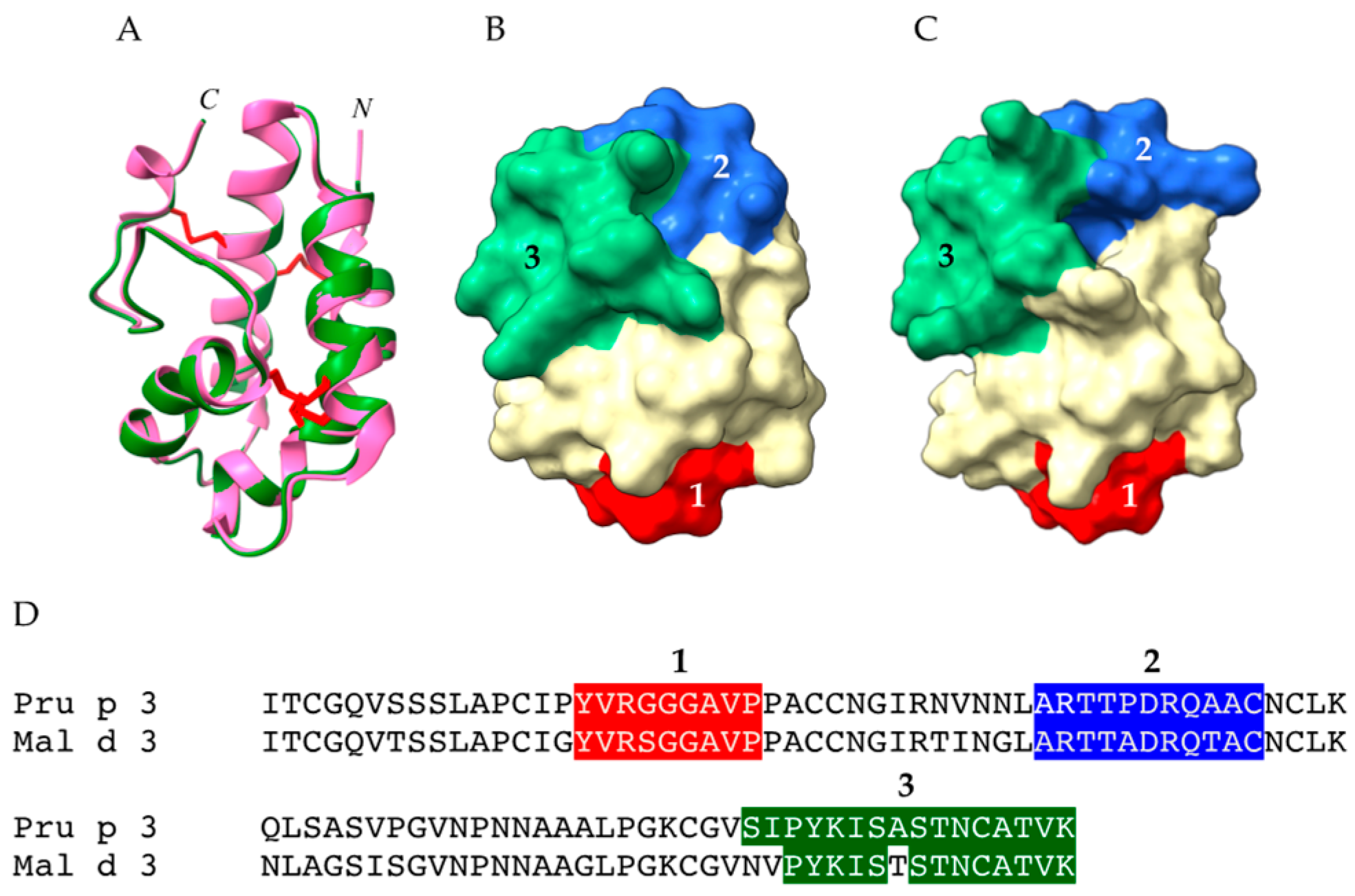
The structural characteristics of the allergens (allergens exhibiting tightly packed conformation vs. allergens loosely structured) and some structure-associated properties, like the resistance to digestive proteases and heat (cooking) denaturation [36]. In this respect, fruit nsLTPs, like Pru p 3 and Mal d 3, possess a tightly packed three-dimensional conformation stabilized by four disulfide bridges, the so-called “saxophone-like conformation”, that contributes to the extreme resistance of these protein to the degradation by digestive proteases and to heat denaturation [22]. Similarly, the tightly packed GRPs offer an enhanced resistance to protease and heat denaturation [44]. Conversely, profilins, such as Pru p 4 and Mal d 4, are more loosely structured proteins and offer a weak resistance to both enzymatic digestion and heat denaturation [36];
-
The localization and accessibility of the allergens. In this regard, the surface localization of Pru p 3 in the fuzzy covering the peel of the peach [78], and the localization of Mal d 3 in the external cell layers forming the peel of the apple [24], favors the contact of the allergens with the body. Accordingly, removing the peel from peaches and apples is sufficient to eliminate most of the allergens from the fruits;
-
The amounts of allergens present in fruits and vegetables. This is an important point to consider, because many fruit allergens consist of PR proteins which interfere with the defense of plants against phytopathogenic fungi, bacteria, and viruses [79]. In addition, their synthesis can vary considerably, depending on the response of the plant to abiotic stresses, for example water stress or heat stress [80]. Moreover, large variations in the allergen content of fruits were measured in different varieties or cultivars of peach and apple [81,82,83]. Large variations were also measured in the allergen content of fruits, depending on the cultivation conditions [84,85], the degree of ripening of the harvested fruits, and the shelf life of the postharvest fruit storage [86,87,88,89,90]. Other factors can influence the sensitizing propensity of the allergens, such as the route of exposure [76] and the processing of fruits and fruit products before consumption [91].
In addition, some geographical discrepancies have been identified concerning, for example, severe systemic reactions to Pru p 3 in people from Mediterranean countries, compared to people from northern countries [92,93]. However, the risk for a severe allergic response to Pru p 3, was also reported for people living outside the Mediterranean countries [15,94]. The high resistance of Pru p 3 to both the digestive and thermal degradation largely accounts for the dangerous character of this nsLTP [22].
In general, most of the allergic symptoms following fruit consumption consist of oropharyngeal symptoms, the so-called OAS, which usually occurs very quickly after fruit consumption. The OAS includes various symptoms, but broadly consists of itch, tearing, sneezing, lips swelling and, more rarely, difficulty swallowing, hives, and urticaria. However, severe systemic symptoms can occur after consumption of nsLTP-containing fruits, namely peach Pru p 3, which can require adrenaline injection(s) and emergency hospital care [27,75]. A more detailed description of the clinical aspects of allergy to profilins, nsLTPs, PR10 Bet v 1-like proteins, GRPs, and other allergens from fruits and vegetables, is available in the recently published second edition of the Molecular Allergy User’s Guide (MAUG2) [95]. Clinical aspects of Rosaceae fruit allergies and defensin-related food allergies are presented in some recently published reviews [12,54].

5. Comparative Prevalence and Harmful Properties of Fruit Allergens

Fruit allergens responsible for the most frequent allergic manifestations correspond to a rather limited number of structurally well-identified protein families, including profilins, GRPs, defensins, and various PR-protein families such as nsLTPs (PR14), Betv v 1-like PR10, TLPs (PR5), chitinases (PR3) and endoβ1,3-glucanases (PR2) [3]. The relationship between the structure and harmfulness of fruit allergens is a complex matter, which apparently depends on both intrinsic and extrinsic factors.
The availability of fruit allergens for the sensitization and subsequent elicitation of an allergic reaction in sensitized people requires both their solubilization and structural preservation during the digestion process. Depending on their structure and physicochemical properties, fruit allergens can readily differ in this respect [96]. This ability to resist protease denaturation is also valid in case of dermal sensitization because all tissues contain trypsin-like proteases that can degrade allergens. Furthermore, even in case of proteolytic degradation, the allergens most resistant to denaturation can release peptide fragments capable of retaining some of the allergenicity and harmfulness of native allergens [76]. Moreover, the cross-reactivity between fruit allergens and pollen homologous allergens that explains many cases of prior inhalation sensitization observed in allergies to peach and apple could also participate in their harmful character [97]. Recently, the ability of Pru p 3 to interact with its natural lipidic ligand camptothecin, associated with phytosphingosine, has been characterized as an adjuvant for promoting the sensitization to Pru p 3 [97]. Previously, the interaction of Pru p 3 with free fatty acids had been shown to enhance the Pru p 3 IgE-binding activity [29].
Among the extrinsic factors, the localization and abundancy of allergens in the fruits is another important factor for their availability. As an example, the localization and abundancy of Pru p 3 in the fuzzy covering the peach, and Mal d 3 in the cuticular cell layers of apple [97,98], favor the allergenicity and harmful properties of Rosaceae fruits [98]. Other relevant factors, such as the existence of a marked atopic terrain or ethnic predispositions, are to be considered.
Depending on all the factors that contribute to the allergenic potential and harmful potential of fruit allergens, the prevalence and severity of fruit allergies in Europe, in particular, can vary widely between countries. According to [99], the overall prevalence of fruit allergies varies between 0.1% and 4.3%. The most frequent fruit sensitizations in Europe concern peach (7.9%), apple (6.5%), and kiwi fruit (5.2%) [100,101]. In northern countries and Italy, apple allergy is frequently mild and depends on prior sensitization to birch pollen PR10 Bet v 1-like protein allergen homologous to Mal d 1, whereas in Spain, sensitization to apple allergen Mal d 3 (nsLTP) predominates in Mediterranean countries and frequently provokes severe anaphylaxis responses [92,102,103,104]. Similarly, allergen sensitization patterns to kiwi fruit differ across Europe [105]. Sensitization to kiwi fruit mostly depends on Act d 8 (PR10 Bet v 1-like protein allergen) in western, central and eastern Europe, while it depends on Act d 9 (profilin) and Act d 10 (nsLTP) in southern Europe. Symptoms range from mild OAS (Act d 8, Act d 9) to severe systemic reactions (Act d 10). In general, allergy to fruit profilins is mild, and results in OAS symptoms. Sensitization to fruit profilins is evenly distributed in Europe, but is more frequent in Mediterranean countries, particularly in Spain [106]. Sensitization to Pru p 7, the GRP from peach, was reported as the predominant cause of severe cypress pollen-associated peach allergies in southern France [107]. Recently, a high prevalence of mango allergy was reported from a self-reported food allergy survey performed in Jiangxi (China), but no indication is available on the causative allergen(s) [108]

6. Conclusions

Some general conclusions can be drawn from this review on fruit allergens:
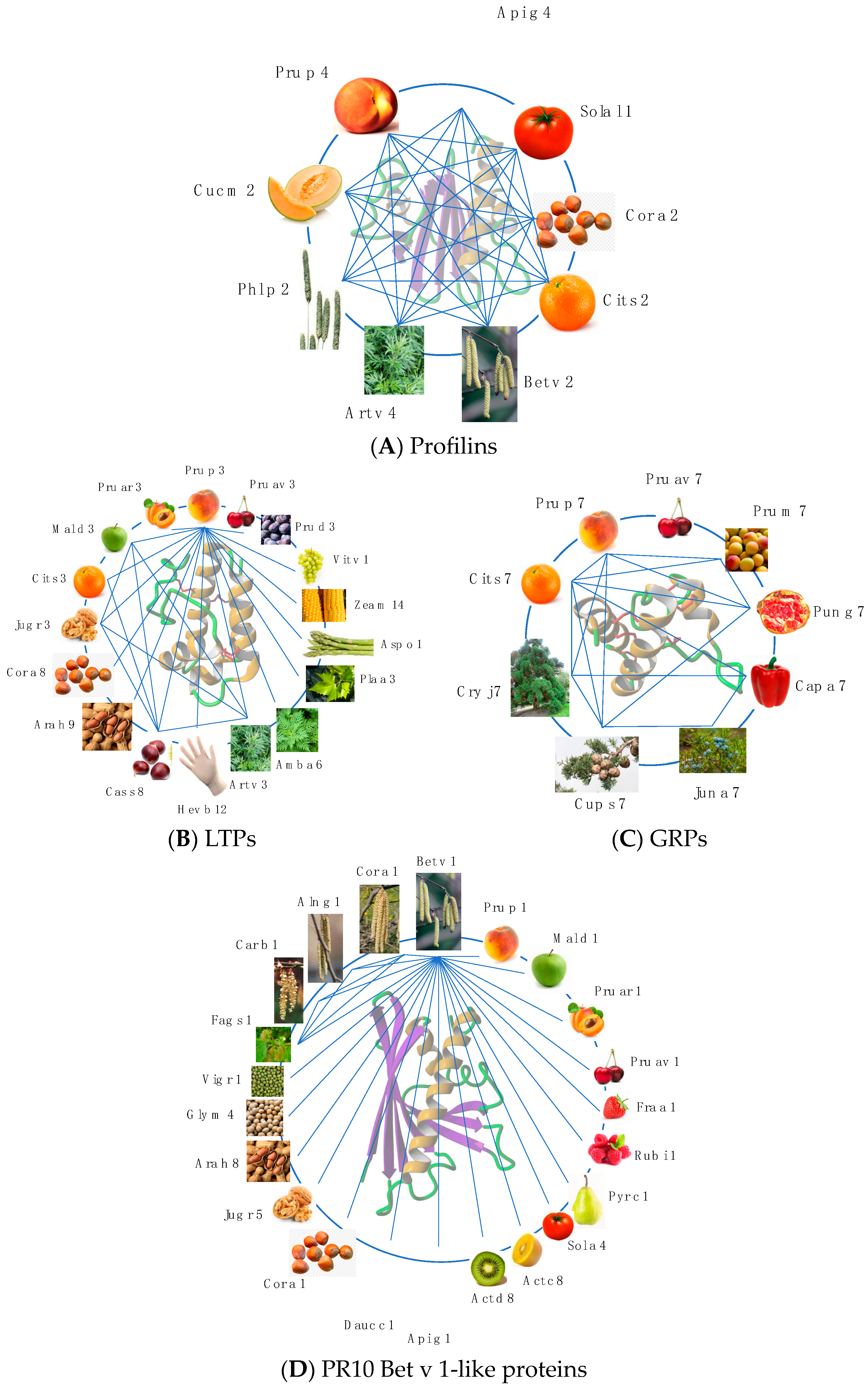
  • Allergens from edible fruits, especially fleshy fruits, correspond to pan-allergens that are widely distributed in vegetables, seeds, and pollen from other apparently unrelated plants;
  • Fruit allergens essentially belong to different PR-proteins, which play a role in the defense of plants against phytopathogenic fungi, bacteria, and viruses. Accordingly, their biosynthesis is largely influenced by the stress conditions to which the plants are subjected. The allergen content of fruits can also vary considerably, depending on the ripening stage and storage conditions of the fruits after harvest;
  • The different families of allergenic PR-proteins exhibit highly conserved amino acid sequences and three-dimensional structures and display close phylogenetic relationships;
  • Depending on their large distribution and their sequential, structural, and phylogenetical relationships, a high degree of cross-reactivity usually occurs between allergens from fruits and the counterparts from other sources like vegetables, seeds, or pollens. This cross-reactivity is at the origin of various clinical syndromes including, e.g., the apple-birch syndrome, the peach-cypress syndrome, and the peach-latex syndrome. However, many cross-sensitizations with pollen allergens are not clinically relevant;
  • The allergenicity and toxicity vary largely among fruit allergens. Although all fruit allergens are potentially dangerous, some of them, like nsLTPs and GRPs, are responsible for food allergies, and provoke severe systemic reactions, especially in Mediterranean countries;
  • In general, the consumption of allergen-containing fruits only results in mild oropharyngeal symptoms that corresponds to the so-called oral allergic syndrome, OAS. In some cases, however, more severe systemic reactions can develop, especially upon consumption of Rosaceae fruits or kiwi fruit.

7. Bioinformatics

The amino acid sequences of fruit allergens were retrieved from the WHO/IUIS database and the nr NCBI database (accessed on 18 August 2022). Multiple amino acid sequence alignments were performed with ClustalX [109]. Unrooted phylogenetic trees were built from the multiple amino acid sequence alignments using the neighbor joining method, and were represented with TreeView [110].
The atomic coordinates of the endo-β1,3-glucanase from banana (Musa acuminata) (PDB code 2CYG) [7], Pru av 2 from cherry (Prunus avium) (PDB code 2AHN) [14], Bet v 1 from birch (Betula verrucosa) (PDB code 4A88) [111], and Pru p 3 from peach (Prunus persica) (PDB code 2ALG) [21] were retrieved from the Protein Data Bank (https://www.rcsb.org) (accessed on August 2022) [112].
Homology modeling of other allergens, including the potential allergens from the tomato fruit, was performed with the YASARA Structure program [113] using various three-dimensional structures of the PDB as templates. PROCHECK [114], ANOLEA [115], and the calculated QMEAN scores [116,117] were used to assess the geometric and thermodynamic qualities of the three-dimensional models.
The surface electrostatic potentials were calculated and rendered with YASARA, using the Amber96 forcefield with dielectric constants applied to the protein and the solvent fixed at 4.0 and 80.0, respectively. Electrostatic potentials were displayed on the molecular surface as red (electro-negatively charged) and blue (electro-positively charged) patches. Neutral surfaces are white.
Continuous (sequential) and discontinuous (conformational) IgE-binding epitopes identified in various publications were mapped on the molecular surface of the corresponding X-ray-solved or modelled fruit allergens.
Molecular cartoons were drawn with YASARA, Chimera [118], and ChimeraX [119].

Author Contributions

Conceptualization, P.R.; methodology, P.R.; software, P.R.; validation, A.B. and H.B.; resources, A.B.; data curation, A.B. and H.B.; writing—original draft preparation, P.R.; writing—review and editing, P.R.; project administration, A.B. All authors have read and agreed to the published version of the manuscript.

Funding

This research received no external funding.

Institutional Review Board Statement

Not applicable.

Informed Consent Statement

Not applicable.

Data Availability Statement

Not applicable.

Conflicts of Interest

The authors declare no conflict of interest.

References

  1. Lefèvre, S.; Gregori, M.; Astier, C.; Rougé, P.; Kanny, G. Fruit allergies. Rev. Fr. Allergol. 2020, 60, 585–594. [Google Scholar] [CrossRef]
  2. Ebner, C.; Hoffmann-Sommergruber, K.; Breiteneder, H. Plant food allergens homologous to pathogenesis-related proteins. Allergy 2001, 56, 43–44. [Google Scholar] [CrossRef] [PubMed]
  3. Sinha, M.; Singh, R.P.; Singh Kushwaha, G.; Iqbal, N.; Singh, A.; Kaushik, S.; Kaur, P.; Sharma, S.; Singh, T.P. Current overview of allergens of plant pathogenesis related families. ScientificWorldJournal 2014, 2014, 543195. [Google Scholar] [CrossRef] [Green Version]
  4. Jain, A. Seed storage protein, functional diversity and association with allergy. Allergies 2023, 3, 25–38. [Google Scholar] [CrossRef]
  5. Barre, A.; Simplicien, M.; Benoist, H.; Rougé, P. Fruit allergies: Beware of the seed allergens! Rev. Fr. Allergol. 2018, 58, 308–317. [Google Scholar] [CrossRef]
  6. Mackenzie, A.; Pridham, J.B.; Saunders, N.A. Changes in the sweet proteins (thaumatins) in Thaumatoccocus daniellii fruit development. Phytochemistry 1985, 11, 2503–2506. [Google Scholar] [CrossRef]
  7. Leone, P.; Menu-Bouaouiche, L.; Peumans, W.J.; Payan, F.; Barre, A.; Roussel, A.; Van Damme, E.J.M.; Rougé, P. Resolution of the structure of the allergenic and antifungal banana fruit thaumatin-like protein at 1.7-Å. Biochimie 2006, 88, 45–52. [Google Scholar] [CrossRef]
  8. Smole, U.; Bublin, M.; Radauer, C.; Ebner, C.; Breiteneder, H. Mal d 2, the thaumatin-like allergen from apple, is highly resistant to gastrointestinal digestion and thermal processing. Int. Arch. Allergy Immunol. 2008, 147, 289–298. [Google Scholar] [CrossRef]
  9. Kumar, H.G.A.; Venkatesh, Y.P. In silico analyses of structural and allergenicity features of sapodilla (Manilkara zapota) acidic thaumatin-like protein in comparison with allergenic plant TLPs. Mol. Immunol. 2014, 57, 119–128. [Google Scholar] [CrossRef]
  10. Barre, A.; Sénéchal, H.; Nguyen, C.; Granier, C.; Poncet, P.; Rougé, P. Structural basis for the IgE-binding cross-reacting epitopic peptides of Cup s 3, a PR-5 thaumatin-like protein allergen from common cypress (Cupressus sempervirens) pollen. Allergies 2023, 3, 11–24. [Google Scholar] [CrossRef]
  11. Palacín, A.; Rivas, L.A.; Gómez-Casado, C.; Aguirre, J.; Tordesillas, L.; Bartra, J.; Blanco, C.; Carrillo, T.; Cuesta-Herranz, J.; Bonny, J.A.; et al. The involvement of thaumatin-like proteins in plant food cross-reactivity: A multicenter study using a specific protein microarray. PLoS ONE 2012, 7, e44088. [Google Scholar] [CrossRef] [PubMed] [Green Version]
  12. Costa, J.; Mafra, I. Rosaceae food allergy: A review. Crit. Rev. Food Sci. Nutr. 2022, 1–38. [Google Scholar] [CrossRef] [PubMed]
  13. Breiteneder, H.; Hassfeld, W.; Pettenburger, K.; Jarolim, E.; Breitenbach, M.; Rumpold, D.; Kraft, D.; Scheiner, O. Isolation and characterization of messenger RNA from male inflorescences and pollen of the white birch (Betula verrucosa). Int. Arch. Allergy Appl. Immunol. 1988, 87, 19–24. [Google Scholar] [CrossRef] [PubMed]
  14. Neudecker, P.; Schweimer, K.; Nerkamp, J.; Scheurer, S.; Vieths, S.; Sticht, H.; Rösch, P. Allergic cross-reactivity made visible: Solution structure of the major cherry allergen Pru av 1. J. Biol. Chem. 2001, 276, 22756–22763. [Google Scholar] [CrossRef] [Green Version]
  15. Skypala, I.J.; Asero, R.; Barber, D.; Cecchi, L.; Díaz-Perales, A.; Hoffmann-Sommergruber, K.; Pastorello, E.A.; Swoboda, I.; Bartra, J.; Ebo, D.G.; et al. Non-specific lipid-transfer proteins: Allergen structure and function, cross-reactivity, sensitization, and epidemiology. Clin. Transl. Allergy 2021, 11, e12010. [Google Scholar] [CrossRef]
  16. Neudecker, P.; Lehmann, K.; Nerkamp, J.; Haase, T.; Wangorsch, A.; Fötisch, K.; Hoffmann, S.; Rösch, P.; Vieths, S.; Scheurer, S. Mutational epitope analysis of Pru av 1 and Api g 1, the major allergens of cherry (Prunus avium) and celery (Apium graveolens): Correlating IgE reactivity with three-dimensional structure. Biochem. J. 2003, 376, 97–107. [Google Scholar] [CrossRef] [Green Version]
  17. Wiche, R.; Gubesch, M.; König, H.; Fötisch, K.; Hoffmann, A.; Wangorsch, A.; Scheurer, S.; Vieths, S. Molecular basis pf pollen-related food allergy: Identification of a second cross-reactive IgE epitope on Pru av 1, the major cherry (Prunus avium) allergen. Biochem. J. 2005, 385, 319–327. [Google Scholar] [CrossRef] [Green Version]
  18. Jahn-Schmid, B.; Radakovics, A.; Lüttkopf, D.; Scheurer, S.; Vieths, S.; Ebner, C.; Bohle, B. Bet v 1 142-156 is the dominant T-cell epitope of the major birch pollen allergen and important for cross-reactivity with Bet v 1-related food allergens. J. Allergy Clin. Immunol. 2005, 116, 213–219. [Google Scholar] [CrossRef]
  19. Breiteneder, H.; Aglas, L.; Kleine-Tebbe, J.; Hafner, C.; Dramburg, S.; Hilger, C.; Santos, A.F.; Vecillas, L.d.L.; Aalberse, R.C.; Acevedo, N.; et al. C02–PR-10-like allergens. EAACI Molecular Allergology User’s Guide 2.0. Pediatr. Allergy Immunol. 2023, 34, 272–280, e13854. [Google Scholar]
  20. Scala, E.; Maria Marra, A.; Asero, R.; Dramburg, S.; Hilger, C.; Santos, A.F.; Vecillas, L.d.L.; Aalberse, R.C.; Acevedo, N.; Aglas, L.; et al. B01—Tree pollen allergy. EAACI Molecular Allergology User’s Guide 2.0. Pediatr. Allergy Immunol. 2023, 34, 93–102, e13854. [Google Scholar]
  21. Pasquato, N.; Berni, R.; Folli, C.; Folloni, S.; Cianci, M.; Pantano, S.; Helliwell, J.R.; Zanotti, G. Crystal structure of peach Pru p 3, the prototypic member of the family of plant non-specific lipid transfer protein pan-allergens. J. Mol. Biol. 2005, 356, 684–694. [Google Scholar] [CrossRef] [PubMed]
  22. Toda, M.; Reese, G.; Gadermaier, G.; Schulten, V.; Lauer, I.; Egger, M.; Briza, P.; Randow, S.; Wolfheimer, S.; Kigongo, V.; et al. Protein unfolding strongly modulate the allergenicity and immunogenicity of Pru p 3, the major peach allergen. J. Allergy Clin. Immunol. 2011, 128, 1022–1030. [Google Scholar] [CrossRef] [PubMed]
  23. Akkerdaas, J.; Totis, M.; Barnett, B.; Bell, E.; Davis, T.; Edrington, T.; Glenn, K.; Graser, G.; Herman, R.; Knulst, A.; et al. Protease resistance of food proteins: A mixed picture for predicting alleregenicity but a useful tool for assessing exposure. Clin. Transl. Allergy 2018, 8, 30. [Google Scholar] [CrossRef] [PubMed] [Green Version]
  24. Borges, J.-P.; Jauneau, A.; Brulé, C.; Culerrier, R.; Barre, A.; Didier, A.; Rougé, P. The lipid transfer proteins (LTP) essentially concentrate in the skin of Rosaceae fruits as cell surface exposed allergens. Plant Physiol. Biochem. 2006, 44, 535–542. [Google Scholar] [CrossRef]
  25. García-Casado, G.; Pacios, L.F.; Díaz-Perales, A.; Sánchez-Monge, R.; Lombardero, M.; García-Selles, F.J.; Polo, F.; Barber, D.; Salcedo, G. Identification of IgE-binding epitopes of the major peach allergen Pru p 3. J. Allergy Clin. Immunol. 2003, 112, 599–605. [Google Scholar] [CrossRef]
  26. Borges, J.-P.; Barre, A.; Culerrier, R.; Granier, C.; Didier, A.; Rougé, P. Lipid transfer proteins from Rosaceae fruits share consensus epitopes responsible for vtheir IgE-binding cross-reactivity. Biochem. Biophys. Res. Commun. 2008, 365, 685–690. [Google Scholar] [CrossRef]
  27. Pastorello, E.A.; Gomez, F.; Barber, D.; Dramburg, S.; Hilger, C.; Santos, A.F.; Vecillas, L.d.L.; Aalberse, R.C.; Acevedo, N.; Aglas, L.; et al. C03—Nonspecific lipid transfer proteins (nsLTP). EAACI Molecular Allergology User’s Guide 2.0. Pediatr. Allergy Immunol. 2023, 34, 281–287, e13854. [Google Scholar]
  28. Scheurer, S.; Schulke, S. Interaction of non-specific lipid-transfer proteins with plant-derived lipids and its impact on allergic sensitization. Front. Immunol. 2018, 9, 1389. [Google Scholar] [CrossRef] [Green Version]
  29. Dubiela, P.; Aina, R.; Polak, D.; Geiselhart, S.; Humeniuk, P.; Bohle, B.; Alessandri, S.; Del Conte, R.; Cantini, F.; Borowski, T.; et al. Enhanced Pru p 3 IgE-binding activity by selected free fatty acid-interaction. J. Allergy Clin. Immunol. 2017, 140, 1728–1731. [Google Scholar] [CrossRef] [Green Version]
  30. Dubiela, P.; Del Conte, R.; Cantini, F.; Borowski, T.; Aina, R.; Radauer, C.; Bublin, M.; Hoffmann-Sommergruber, K.; Alessandri, S. Impact of lipid binding on the tertiary structure and allergenic potential of Jug r 3, the non-specific lipid transfer protein from walnut. Sci. Rep. 2019, 9, 2007. [Google Scholar] [CrossRef] [Green Version]
  31. Chruszcz, M.; Chew, F.T.; Hoffmann-Sommergruber, K.; Hurlburt, B.K.; Mueller, G.A.; Pomés, A.; Rouvinen, J.; Villalba, M.; Wöhrl, B.; Breiteneder, H. Allergens and their associated small molecule ligands—Their dual role in sensitization. Allergy 2021, 76, 2367–2382. [Google Scholar] [CrossRef] [PubMed]
  32. Gonzalez-Klein, Z.; Pazos-Castro, D.; Hernandez-Ramirez, G.; Garrido-Aranda, M.; Díaz-Perales, A.; Tome-Amat, J. Lipid ligands and allergenic LTPs: Redefining the paradigm of the protein-centered vision in allergy. Front. Allergy 2022, 3, 864652. [Google Scholar] [CrossRef] [PubMed]
  33. Rougé, P.; Hilger, C.; Hoffmann-Sommergruber, K.; Traidl-Hoffmann, C.; Dramburg, S.; Hilger, C.; Santos, A.F.; Vecillas, L.d.L.; Aalberse, R.C.; Acevedo, N.; et al. A11—Small molecules as immunomodulators and allergen ligands. EAACI Molecular Allergology User’s Guide 2.0. Pediatr. Allergy Immunol. 2023, 34, 80–84, e13854. [Google Scholar]
  34. Brenda Kapingidza, A.; Pye, S.E.; Hyduke, N.; Dolamore, C.; Pote, S.; Schlachter, C.; Commins, S.P.; Kowal, K.; Chruszcz, M. Comparative structural and thermal stability studies of Cuc m 2.0101, Art v 4.0101 and other allergenic profilins. Mol. Immunol. 2019, 114, 19–29. [Google Scholar] [CrossRef]
  35. Asero, R.; Mistrello, G.; Roncarolo, D.; Amato, S.; Zanoni, D.; Barocci, F.; Caldironi, G. Detection of clinical markers of sensitization to profilin in patients allergic to plant-derived foods. J. Allergy Clin. Immunol. 2003, 112, 427–432. [Google Scholar] [CrossRef] [PubMed]
  36. Offermann, L.; Schlachter, C.R.; Perdue, M.L.; Majorek, K.A.; He, J.Z.; Booth, W.T.; Kowal, K.; Chruszcz, M. Structural, functional, and immunological characterization of profilin panallergens Amb a 4, Art v 4, and Bet v 2. J. Biol. Chem. 2016, 291, 15447–15459. [Google Scholar] [CrossRef] [Green Version]
  37. Hofmann, A.; Burks, A.W. Pollen food syndrome: Update on the allergens. Curr. Allergy Asthma Rep. 2008, 8, 413–417. [Google Scholar] [CrossRef]
  38. López-Torrejón, G.; Díaz-Perales, A.; Rodríguez, J.; Sánchez-Monge, R.; Crespo, J.F.; Salcedo, G.; Pacios, L.F. An experimental and modeling-based approach to locate IgE epitopes of plant profilin allergens. J. Allergy Clin. Immunol. 2007, 119, 1481–1488. [Google Scholar] [CrossRef]
  39. Tordesillas, L.; Pacios, L.F.; Palacín, A.; Cuesta-Herranz, J.; Madero, M.; Díaz-Perales, A. Characterization of IgE epitopes of Cuc m 2, the major melon allergen, and their role in cross-reactivity with pollen profilins. Clin. Exp. Allergy 2010, 40, 174–181. [Google Scholar] [CrossRef]
  40. Rihs, H.P.; Chen, Z.; Ruëff, F.; Petersen, A.; Rozynek, P.; Heimann, H.; Baur, X.D. IgE binding of the recombinant allergen soybean profilin (rGly m 3) is mediated by conformational epitopes. J. Allergy Clin. Immunol. 1999, 104, 1293–1301. [Google Scholar] [CrossRef]
  41. Sirvent, S.; Tordesillas, L.; Villalba, M.; Díaz-Perales, A.; Cuesta-Herranz, J.; Salcedo, G.; Rodríguez, R. Pollen and plant food profilin allergens show equivalent IgE reactivity. Ann. Allergy Asthma Immunol. 2011, 106, 429–435. [Google Scholar] [CrossRef] [PubMed]
  42. Terán, M.G.; García-Ramírez, B.; Mares-Mejía, I.; Ortega, E.; O’Malley, A.; Chruszcz, M.; Rodríguez-Romero, A. Molecular basis of plant profilins’ cross-reactivity. Biomolecules 2023, 13, 608. [Google Scholar] [CrossRef] [PubMed]
  43. Asero, R.; Barber, D.; Celi, G.; Dramburg, S.; Hilger, C.; Santos, A.F.; Vecillas, L.d.L.; Aalberse, R.C.; Acevedo, N.; Aglas, L.; et al. C01—Profilins. EAACI Molecular Allergology User’s Guide 2.0. Pediatr. Allergy Immunol. 2023, 34, 268–271, e13854. [Google Scholar]
  44. Tuppo, L.; Spadaccini, R.; Alessandri, C.; Wiek, H.; Boelens, R.; Giangrieco, I.; Tamburrini, M.; Mari, A.; Picone, D.; Ciardello, M.A. Structure, stability, and IgE binding of the peach allergen peamaclein (Pru p 7). Biopolymers 2014, 102, 416–425. [Google Scholar] [CrossRef] [PubMed]
  45. Iuzuka, T.; Barre, A.; Rougé, P.; Charpin, D.; Scala, E.; Baudin, B.; Aizawa, T.; Sénéchal, H.; Poncet, P. Gibberellin-regulated proteins: Emergent allergens. Front. Allergy 2022, 3, 877553. [Google Scholar] [CrossRef]
  46. Poncet, P.; Iizuka, T.; Sénéchal, H.; Scala, E.; Dramburg, S.; Hilger, C.; Santos, A.F.; Vecillas, L.d.L.; Aalberse, R.C.; Acevedo, N.; et al. C09–Gibberellin-regulated proteins. EAACI Molecular Allergology User’s Guide 2.0. Pediatr. Allergy Immunol. 2023, 34, 322–327, e13854. [Google Scholar]
  47. Poncet, P.; Sénéchal, H.; Charpin, D. Update on pollen-food allergy syndrome. Expert Rev. Clin. Immunol. 2020, 16, 561–578. [Google Scholar] [CrossRef]
  48. Takei, M.; Nin, C.; Izuka, T.; Pawlikowski, M.; Selva, M.A.; Chantran, Y.; Nakajima, Y.; Zheng, J.; Aizawa, T.; Ebisawa, M.; et al. Capsicum allergy: Involvement of Cap a 7, a new clinically relevant gibberellin-regulated protein cross-reactive with Cry j 7, the gibberellin-regulated protein from Japanese cedar pollen. Allergy asthma Immunol. Res. 2022, 14, 328–338. [Google Scholar] [CrossRef]
  49. Razzera, G.; Gadermaier, G.; de Paula, V.; Almeida, M.S.; Egger, M.; Jahn-schmid, B.; Almeida, F.C.L.; Ferreira, F.; Valente, A.P. Mapping the interactions between a major pollen allergen and human IgE antibodies. Structure 2010, 18, 1011–1021. [Google Scholar] [CrossRef] [Green Version]
  50. Gao, G.; Dai, J.; Ding, M.; Hellekant, G.; Wang, J.; Wang, D. Studies on solution NMR structure of brazzein: Secondary structure and molecular scaffold. Sci. China C Life Sci. 1999, 42, 409–419. [Google Scholar] [CrossRef]
  51. Nagata, K.; Hongo, N.; Kameda, Y.; Yamamura, A.; Sasaki, H.; Lee, W.C.; Ishikawa, K.; Suzuki, E.; Tanokura, M. The structure of brazzein, a sweet-tasting protein from the wild African plant Pentadiplandra brazzeana. Acta Crystallogr. D Biol. Crystallogr. 2013, 69, 642–647. [Google Scholar] [CrossRef] [PubMed]
  52. Ming, D.; Hellekant, G. Brazzein, a new high-potency thermostable sweet protein from Pentadiplandra brazzeana B. FEBS Lett. 1994, 355, 106–108. [Google Scholar] [CrossRef] [PubMed] [Green Version]
  53. Pablos, I.; Eichhorn, S.; Machado, Y.; Briza, P.; Neunkirchner, A.; Jahn-Schmid, B.; Wildner, S.; Soh, W.T.; Ebner, C.; Park, J.-W.; et al. Distinct epitope structures of defensin-like proteins linked to prolin-rich regions give rise to differences in their allergenici activity. Allergy 2018, 73, 431–441. [Google Scholar] [CrossRef] [PubMed]
  54. Cosi, V.; Gadermaier, G. The role of defensins as pollen and food allergens. Curr. Allergy Asthma Rep. 2023, 23, 277–285. [Google Scholar] [CrossRef] [PubMed]
  55. Patil, R.S.; Ghormade, V.; Deshpande, M.V. Chitinolytic enzymes: An exploration. Enz. Mircrob. Technol. 2000, 26, 473–483. [Google Scholar] [CrossRef] [PubMed]
  56. Masuda, T.; Zhao, G.; Mikami, B. Crystal structure of class III chitinase from pomegranate provides the insight into its metal storage capacity. Biosci. Biotechnol. Biochem. 2015, 79, 45–50. [Google Scholar] [CrossRef] [Green Version]
  57. Reyes-López, C.; Hernández-Santoyo, A.; Pedraza-Escalona, M.; Mendoza, G.; Hernández-Arana, A.; Rodríguez-Romero, A. Insights into a conformational epitope of Hev b 6.02 (hevein). Biochem. Biophys. Res. Commun. 2004, 314, 123–130. [Google Scholar] [CrossRef]
  58. Blanco, C.; Carrillo, T.; Castillo, R.; Quiralte, J.; Cuevas, M. Latex allergy: Clinical features and cross-reactivity with fruits. Ann. Allergy 1994, 73, 309–314. [Google Scholar]
  59. Dall’Antonia, Y.; Pavlov, T.; Fuchs, H.; Breiteneder, H.; Keller, W. The high-resolution crtystal structure of an allergenic thaumatin-like protein, Pru av 2, isolated from ripe cherries. Pdb code 2AHN (to be published).
  60. Marangon, M.; Van Sluyter, S.C.; Waters, E.J.; Menz, R.I. Structure of haze forming proteins in white wines: Vitis vinifera thaumatin-like proteins. PLoS ONE 2014, 9, e113757. [Google Scholar] [CrossRef] [Green Version]
  61. Rodríguez-Romero, A.; Hernández-Santoyo, A.; Fuentes-Silva, D.; Palomares, L.A.; Muñoz-Cruz, S.; Yépez-Mulia, L.; Orozco-Martínez, S. Structural analysis of the endogenous glycoallergen Hev b 2 (endo-b-1,3-glucanase) from Hebea brasiliensis and its recognition by human basophils. Acta Crystallogr. D Biol. Crystallogr. 2014, 70, 329–341. [Google Scholar] [CrossRef] [Green Version]
  62. Barre, A.; Culerrier, R.; Granier, C.; Selman, L.; Peumans, W.J.; Van Damme, E.J.M.; Bienvenu, F.; Bienvenu, J.; Rougé, P. Mapping of IgE-binding epitopes on the major latex allergen Hev b 2 and the cross-reacting 1,3-β-glucanase fruit allergens as a molecular basis for the latex-fruit syndrome. Mol. Immunol. 2009, 46, 1595–1604. [Google Scholar] [CrossRef] [PubMed]
  63. Huecas, S.; Villalba, M.; Rodríguez, R. Ole e 9, a major olive pollen allergen is a 1,3-glucanase. Isolation, characterization, amino acid sequence, and tissue specificity. J. Biol. Chem. 2001, 276, 27959–27966. [Google Scholar] [CrossRef] [Green Version]
  64. Gallen, C.; Brunet, E.; Borges, J.P.; Rancé, F.; Barre, A.; Rougé, P. A case report of anaphylaxis to Physalis alkekengi fruit: The culprit is in the seeds! Rev. Fr. Allergol. 2018, 58, 527–529. [Google Scholar] [CrossRef]
  65. Shewry, P.R.; Napier, J.A.; Tatham, A.S. Seed storage proteins: Structures and biosynthesis. Plant Cell 1995, 7, 945–956. [Google Scholar]
  66. Jensen-Jarolim, E.; Schmid, B.; Bernier, F.; Berna, A.; Kinaciyan, T.; Focke, M.; Ebner, C.; Scheiner, O.; Boltz-Nitulescu, G. Allergologic exploration of germins and germin-like proteins, a new class of plant allergens. Allergy 2002, 57, 805–810. [Google Scholar] [CrossRef] [PubMed]
  67. Martín-Pedraza, L.; González, M.; Gómez, F.; Blanca-López, N.; Garrido-Arandia, M.; Rodríguez, R.; Torres, M.J.; Blanca, M.; Villalba, M.; Mayorga, C. Two non-specific lipid transfer proteins (nsLTPs) from tomato seeds are associated to severe symptoms of tomato-allergic patients. Mol. Nutr. Food Res. 2016, 60, 1172–1182. [Google Scholar] [CrossRef] [PubMed]
  68. Barre, A.; Nguyen, C.; Granier, C.; Benoist, H.; Rougé, P. IgE-binding epitopes of Pis v 1, Pis v 2 and Pis v 3, the pistachio (Pistacia vera) seed allergens. Allergies 2021, 1, 63–91. [Google Scholar] [CrossRef]
  69. Sirvent, S.; Cantó, B.; Cuesta-Herranz, J.; Gómez, F.; Blanca, N.; Canto, G.; Blanca, M.; Rodríguez, R.; Villalba, M.; Palomares, O. Act d 12 and Act d 13: Two novel, masked, relevant allergens in kiwifruit seeds. J. Allergy Clin. Immunol. 2014, 133, 1765–1767. [Google Scholar] [CrossRef] [Green Version]
  70. Barre, A.; Delplanque, A.; Simplicien, M.; Benoist, H.; Rougé, P. Molecular basis of cross-reactivity between Act c 12 and other seed 11S-globulin allergens. Rev. Fr. Allergol. 2017, 57, 58–66. [Google Scholar] [CrossRef]
  71. Crespo, J.F.; Retzek, M.; Foetisch, K.; Sierra-Maestro, E.; Cid-Sanchez, A.B.; Pascual, C.Y.; Conti, A.; Feliu, A.; Rodríguez, J.; Vieths, S.; et al. Germin-like protein Cit s 1 and profilin Cit s 2 are major allergens in orange (Citrus sinensis) fruits. Mol. Nutr. Food Res. 2006, 50, 282–290. [Google Scholar] [CrossRef]
  72. Ahrazem, O.; Ibañez, M.D.; López-Rorrejón, G.; Sánchez-Monge, R.; Sastre, J.; Lombardero, M.; Barber, D.; Salcedo, G. Orange germin-like glycoprotein Cit s 1: An equivocal allergen. Int. Arch. Allergy Immunol. 2006, 139, 96–103. [Google Scholar] [CrossRef] [PubMed]
  73. García, J.A.; Cuest-Herranz, J.; Perea Lam, N.; Díaz-Perales, A. Anaphylaxis mediated by thaumatin-like proteins. J. Investig. Allergol. Clin. Immunol. 2014, 24, 448–449. [Google Scholar]
  74. Bienvenu, F.; Barre, A.; Viel, S.; Garnier, L.; Guyon, C.; Favre-Metz, C.; Pauli, G.; Bienvenu, J.; Rougé, P. Are defensins important plant allergens ? Rev. Fr. Allergol. 2013, 53, 585–590. [Google Scholar] [CrossRef]
  75. Ballmer-Weber, B.; Hoffmann-Sommergruber, K.; Dramburg, S.; Hilger, C.; Santos, A.F.; Vecillas, L.d.L.; Aalberse, R.C.; Acevedo, N.; Aglas, L.; Altmann, F.; et al. B15—Fruit and vegetable allergy. EAACI Molecular Allergology User’s Guide 2.0. Pediatr. Allergy Immunol. 2023, 34, 200–208, e13854. [Google Scholar]
  76. van Ree, R.; Aalberse, R.C.; Dramburg, S.; Hilger, C.; Santos, A.F.; Vecillas, L.d.L.; Aalberse, R.C.; Acevedo, N.; Aglas, L.; Altmann, F.; et al. A02—Allergens and the allergenic composition of source materials. EAACI Molecular Allergology User’s Guide 2.0. Pediatr. Allergy Immunol. 2023, 34, 200–208, e13854. [Google Scholar]
  77. Pastorello, E.A.; Farioli, C.; Pravettoni, V.; Ispano, M.; Scibola, E.; Tranbaioli, C.; Giuffrida, M.G.; Ansaloni, R.; Godovac-Zimmermann, J.; Conti, A.; et al. The maize major allergen, which is responsible for food-induced allergic reactions, is a lipid transfer protein. J. Allergy Clin. Immunol. 2000, 106, 744–751. [Google Scholar] [CrossRef] [PubMed]
  78. Carnés, J.; Fernández-Caldas, E.; Gallego, M.T.; Ferrer, A.; Cuesta-Herranz, J. Pru p 3 (LTP) content in peach extracts. Allergy 2002, 57, 1071–1075. [Google Scholar] [CrossRef]
  79. Kaur, S.; Samota, M.K.; Choudhary, M.; Choudhary, M.; Pandey, A.K.; Sharma, A.; Thakur, J. How do plant defend themselves against pathogens—Biochemical mechanisms and genetic interventions. Physiol. Mol. Biol. Plants 2022, 28, 485–504. [Google Scholar] [CrossRef] [PubMed]
  80. Breen, S.; Williams, S.J.; Outram, M.; Kobe, B.; Solomon, P.S. Emerging insights into the functions of pathogenesis-related protein 1. Trends Plant Sci. 2017, 22, 871–879. [Google Scholar] [CrossRef]
  81. Ahrazem, O.; Jimeno, L.; López-Torrejón, G.; Herrero, M.; Espada, J.L.; Sánchez-Monge, R.; Duffort, O.; Barber, D.; Salcedo, G. Assessing allergen levels in peach and nectarine cultivars. Ann. Allergy Asthma Immunol. 2007, 99, 42–47. [Google Scholar] [CrossRef]
  82. Sancho, A.I.; van Ree, R.; van Leeuwen, A.; Meulenbroek, B.J.; van de Weg, E.W.; Gilissen, L.J.W.J.; Puehringer, H.; Laimer, M.; Martinelli, A.; Zaccharini, M.; et al. Measurement of lipid transfer protein in 88 apple cultivars. Int. Arch. Allergy Immunol. 2008, 146, 19–26. [Google Scholar] [CrossRef] [PubMed]
  83. Jin, J.; Gan, K.; Zhao, L.; Jia, H.; Zhu, Y.; Li, X.; Yang, Z.; Ye, Z.; Cao, K.; Wang, Z.; et al. Peach allergen Pru p 1 content is generally low in fruit but with large variations in different varieties. Clin. Transl. Allergy 2021, 11, e12034. [Google Scholar] [CrossRef]
  84. Matthes, A.; Schmitz-Eiberger, M. Apple (Malus domestica L. Borkh.) allergen Mad d 1: Effect of cultivar, cultivation system, and storage conditions. J. Agric. Food Chem. 2009, 57, 10548–10553. [Google Scholar] [CrossRef] [PubMed]
  85. Kurze, E.; Kock, V.; Scalzo, R.L.; Olbricht, K.; Schwab, W. Effect of strawberry genotype, cultivation and processing on the Fra a 1 allergen content. Nutrients 2018, 10, 857. [Google Scholar] [CrossRef] [PubMed] [Green Version]
  86. Sancho, A.I.; Foxall, R.; Browne, T.; Dey, R.; Zuidmeer, L.; Marzban, G.; Waldron, K.W.; van Ree, R.; Hoffmann-Sommergruber, K.; Laimer, M.; et al. Effect of postharvest storage on the expression of the apple allergen Mal d 1. J. Agric. Food Chem. 2006, 54, 5917–5923. [Google Scholar] [CrossRef] [PubMed]
  87. Schmitz-Eiberger, M.; Matthes, A. Effect of harvest maturity, duration of storage and shelf life of apples on the allergen Mal d 1, polyphenoloxidase activity and polyphenol content. Food Chem. 2011, 127, 1459–1464. [Google Scholar] [CrossRef]
  88. Kiewning, D.; Baab, G.; Schmitz-Eiberger, M. Impact of storage conditions of the apple allergen Mal d 1. Erwerbs-Obstbau 2012, 54, 177–183. [Google Scholar] [CrossRef]
  89. Hallmann, E.; Rozpara, E.; Slowianek, M.; Leszczynska, J. The effect of organic and conventional farm management on the allergenic potency and bioactive compounds status of apricots (Prunus armeniaca L.). Food Chem. 2019, 279, 171–178. [Google Scholar] [CrossRef]
  90. Song, H.; Asghari, M.; Zahedipour-Sheshglani, P.; Diao, E.; Xiang, X.; Liang, X.; Mandoulakani, B.A.; Qian, S. Investigation of pectolytic and PR genes expression, quality and phytochemical contents in organic and non-organic table grapes at harvest and during storage. Food Res. Int. 2023, 167, 112717. [Google Scholar] [CrossRef]
  91. Suriyamoorthy, P.; Madhuri, A.; Tangirala, S.; Michael, K.R.; Sivanandham, V.; Rawson, A.; Anandharaj, A. Comprehensive review of banana fruit allergy: Pathogenesis, diagnosis, management, and potential modification of allergens through food processing. Plant Foods Hum. Nutr. 2022, 77, 159–171. [Google Scholar] [CrossRef]
  92. Fernández-Rivas, M.; Bolhaar, S.; González-Mancebo, E.; Asero, R.; van Leeuwen, A.; Bohle, B.; Ma, Y.; Ebner, C.; Rigby, N.; Sancho, A.I.; et al. Apple allergy across Europe: How allergen sensitization profiles determine the clinical expression of allergies to plant foods. J. Allergy Clin. Immunol. 2006, 118, 481–488. [Google Scholar] [CrossRef] [PubMed]
  93. Gamboa, P.M.; Cáceres, O.; Antepara, I.; Sánchez-Monge, R.; Ahrazem, O.; Salcedo, G.; Barber, D.; Lombardero, M.; Sanz, M.L. Two different profiles of peach allergy in the north of Spain. Allergy 2007, 62, 408–414. [Google Scholar] [CrossRef] [PubMed]
  94. Reuter, A.; Lidholm, J.; Andersson, K.; Ostling, J.; Lundberg, M.; Scheurer, S.; Eneique, E.; Cistero-Bahima, A.; Miguel-Moncin, M.S.; Ballmer-Weber, B.K.; et al. A critical assessment of allergen component-based in vitro diagnosis in cherry allergy across Europe. Clin. Exp. Allergy 2006, 36, 815–823. [Google Scholar] [CrossRef] [PubMed]
  95. Dramburg, S.; Hilger, C.; Santos, A.F.; Vecillas, L.d.L.; Aalberse, R.C.; Acevedo, N.; Aglas, L.; Altmann, F.; Arruda, K.L.; Asero, R.; et al. EAACI Molecular Allergology User’s Guide 2.0. Pediatr. Allergy Immunol. 2023, 34, e13854. [Google Scholar]
  96. Asero, R.; Mistrello, G.; Roncarolo, D.; de Vries, S.C.; Gautier, M.F.; Ciurana, C.L.; Verbeek, E.; Mohammadi, T.; Knul-Brettlova, V.; Akkerdaas, J.H.; et al. Lipid transfer protein: A pan-allergen in plant-derived foods that is highly resistant to pepsin digestion. Int. Arch. Allergy Immunol. 2000, 122, 20–32. [Google Scholar] [CrossRef]
  97. Tordesillas, L.; Cubells-Baeza, N.; Gómez-Casado, C.; Berin, C.; Esteban, V.; Barcik, W.; O’Mahony, L.; Ramirez, C.; Pacios, L.F.; Garrodo-Arandia, M.; et al. Mechanisms underlying induction of allergic sensitization by Pru p 3. Clin. Exp. Allergy 2017, 47, 1398–1408. [Google Scholar] [CrossRef]
  98. Asero, R.; Mistrello, G.; Amato, S.; Roncarolo, D.; Martinelli, A.; Zaccarini, M. Peach fuzz contains large amounts of lipid transfer protein: Is this the cause of the high prevalence of sensitization to LTP in Mediterranean countries? Eur. Ann. Allergy Clin. Immunol. 2006, 38, 118–121. [Google Scholar]
  99. Zuidmeer, L.; Goldhahn, K.; Rona, R.J.; Gislason, D.; Madsen, C.; Summers, C.; Sodergren, E.; Dahlstrom, J.; Lindner, T.; Sigurdardottir, S.T.; et al. The prevalence of plant food allergies: A systematics review. J. Allergy Clin. Immunol. 2008, 121, 1210–1218. [Google Scholar] [CrossRef]
  100. Burney, P.G.; Summers, C.; Chinn, S.; Hooper, R.; van Ree, R.; Lidholm, J. Prevalence and distribution of sensitization to foods in the European Community Respiratory Health Suvey: A EuroPrevall Analysis. Allergy 2010, 65, 1182–1188. [Google Scholar]
  101. Burney, P.G.; Potts, J.; Kummeling, I.; Mills, E.N.; Clausen, M.; Dubakiene, R.; Barreales, L.; Fernández-Pérez, C.; Fernández-Rivas, M.; Le, T.M.; et al. The prevalence and distribution of food sensitization in European adults. Allergy 2014, 69, 365–371. [Google Scholar] [CrossRef] [Green Version]
  102. Fernández-Rivas, M.; González-Mancebo, E.; Rodríguez-Pérez, R.; Benito, C.; Sánchez-Monge, R.; Salcedo, G.; Alonso, M.D.; Rosadeo, A.; Tejedor, M.A.; Vila, C.; et al. Clinically relevant peach allergy is related to peach lipid transfer protein, Pru p 3, in the Spanish population. J. Allergy Clin. Immunol. 2003, 112, 789–795. [Google Scholar] [CrossRef] [PubMed]
  103. Boyano-Martinez, T.; Pedrosa, M.; Belver, T.; Quirce, S.; García-Ara, C. Peach allergy in Spanish children: Tolerance to the pulp and molecular sensitization profile. Pediatr. Allergy Immunol. 2013, 24, 168–172. [Google Scholar] [CrossRef] [PubMed]
  104. Pastorello, E.A.; Farioli, L.; Stafylaraki, C.; Mascheri, A.; Scibilia, J.; Pravettoni, L.; Piantanida, M.; Nichelatti, M.; Aseo, R. Anti-Pru p 3 IgE levels are inversely related to the age at oncet of peach-induced severe symptoms reported by peach-allergic adults. Int. Arch. Allergy Immunol. 2013, 162, 45–49. [Google Scholar] [CrossRef] [PubMed]
  105. Le, T.M.; Bublin, M.; Breiteneder, H.; Fernández-Rivas, M.; Asero, R.; Ballmer-Weber, B.; Barreales, L.; Bures, P.; Belohlavkova, S.; de Blay, F.; et al. Kiwifruit allergy across Europe: Clinical manifestation and IgE recognition patterns to kiwifruit allergens. J. Allergy Clin. Immunol. 2013, 131, 164–171. [Google Scholar] [CrossRef] [PubMed]
  106. González-Mancebo, E.; González-de-Olano, D.; Trujillo, M.J.; Santos, S.; Gandolfo-Cano, M.; Meléndez, A.; Juárez, R.; Morales, P.; Calso, A.; Mazuela, O.; et al. Prevalence of sensitization to lipid transfer proteins and profilins in a population of 430 patients in the south of Madrid. J. Investig. Allergol. Clin. Immunol. 2011, 21, 278–282. [Google Scholar]
  107. Klingebiel, C.; Chantran, Y.; Arif-Lusson, R.; Ehrenberg, A.; Östling, J.; Poisson, A.; Liabeuf, V.; Agabriel, C.; Birnbaum, J.; Porri, F.; et al. Pru p 7 sensitization is predominant cause of severe, cypress pollen-associated peach allergy. Clin. Exp. Allergy 2019, 49, 526–536. [Google Scholar] [CrossRef]
  108. Feng, H.; Zhou, J.; Lu, Y.; Zhao, Q.; Yang, Z.; Xiong, X.; Luo, N.; Chen, Y.; Wang, L.; Wu, Y. Prevalence of self-reported food allergy among adults in Jiangxi, China. World Allergy Organ. J. 2023, 16, 100773. [Google Scholar] [CrossRef]
  109. Thompson, J.D.; Gibson, T.J.; Plewniak, F.; Jeanmougin, F.; Higgins, D.G. The CLUSTAL-X windows interface: Flexible strategies for multiple sequence alignment aided by quality analysis tool. Nucleic Acids Res. 1997, 15, 4876–4882. [Google Scholar] [CrossRef] [Green Version]
  110. Page, R.D.M. TREEVIEW: An application to display phylogenetic trees on personal computers. Computers Appl. BioSci. 1996, 12, 357–358. [Google Scholar]
  111. Kofler, S.; Asam, C.; Eckhard, U.; Wallner, M.; Ferreira, F.; Brandstetter, H. Crystallographically mapped ligand binding differs in high and low IgE binding isoforms of birch pollen allergen Bet v 1. J. Mol. Biol. 2012, 422, 109–123. [Google Scholar] [CrossRef] [Green Version]
  112. Rose, P.W.; Prlic, A.; Bi, C.; Bluhm, W.F.; Christie, C.H.; Dutta, S.; Green, R.K.; Goodsell, D.S.; Westbrook, J.D.; Woo, J.; et al. The RCSB Protein Data Bank: Views of structural biology for basic and applied research and education. Nucleic Acids res. 2015, 43, D345–D356. [Google Scholar] [CrossRef]
  113. Krieger, E.; Koraimann, G.; Vriend, G. Increasing the precision of comparative models with YASARA NOVA—A self-parameterizing force field. Proteins 2002, 47, 393–402. [Google Scholar] [CrossRef]
  114. Laskowski, R.A.; MacArthur, M.W.; Moss, D.S.; Thornton, J.M. PROCHECK: A program to check the stereochemistry of protein structures. J. Appl. Cryst. 1993, 26, 283–291. [Google Scholar] [CrossRef]
  115. Melo, F.; Feytmans, E. Assessing protein structures with a non-local atomic interaction energy. J. Mol. Biol. 1998, 277, 1141–1152. [Google Scholar] [CrossRef] [PubMed]
  116. Benkert, P.; Biasini, M.; Schwede, T. Toward the estimation of the absolute quality of individual protein structure models. Bioinformatics 2011, 27, 343–350. [Google Scholar] [CrossRef] [PubMed] [Green Version]
  117. Arnold, K.; Bordoli, L.; Kopp, J.; Schwede, T. The SWISS-MODEL workspace: A web-based environment for protein structure homology modelling. Bioinformatics 2006, 22, 195–201. [Google Scholar] [CrossRef] [PubMed] [Green Version]
  118. Pettersen, E.F.; Goddard, T.D.; Huang, C.C.; Couch, G.S.; Greenblatt, D.M.; Meng, E.C.; Ferrin, T.E. UCSF Chimera—A visualization system for exploratory research and analysis. J. Comput. Chem. 2004, 25, 1605–1612. [Google Scholar] [CrossRef] [Green Version]
  119. Pettersen, E.F.; Goddard, T.D.; Huang, C.C.; Meng, E.C.; Couch, G.S.; Croll, T.I.; Morris, J.H.; Ferrin, T.E. UCSF ChimeraX: Structure visualization for researchers, educators, and developers. Protein Sci. 2021, 30, 70–82. [Google Scholar] [CrossRef]
Figure 1. (a) Apple flower showing the central pistil surrounded by the stamens. (b) Cut-section of the fruit showing the different parts of the apple fruit including the epicarp (Ep), the exocarp (Ex), the mesocarp (M) delineated by a black dashed line, and the sclerified endocarp (En) containing the seeds (S).
Allergies 03 00010 g001
Figure 2. Overview of the different structural scaffolds and distributions of the different families of fruit allergens.
Allergies 03 00010 g002aAllergies 03 00010 g002bAllergies 03 00010 g002c
Figure 3. (A). Ribbon diagram of Mus a 4 built from three a central β-sandwich domain (I) associated with an α-helical domain II and a β-hairpin domain III. Up to 8 disulfide bridges (colored red) contribute to the stability of the three-dimensional structure. (B). Molecular surface of Mus a 4. The yellow line delineates the cleft occurring between domains I and II. (C). Electrostatic potentials displayed on the molecular surface of Mus a 4, showing the electronegative character of the central cleft (delineated by a yellow line). Electronegative and electropositive regions are colored red and blue, respectively, and neutral regions are colored grey.
Allergies 03 00010 g003
Figure 4. IgE-binding epitopic regions predicted on the molecular surface of the TLP from sapodilla fruit (a) and Cup s 3, the green cypress pollen TLP allergen (b). The IgE-binding allergens are differently colored. The electronegative catalytic cleft is highlighted with a yellow line.
Allergies 03 00010 g004
Figure 5. Amino acid sequence comparison of TLP allergens from Actinidia chinensis (Act c 10), A. deliciosa (Act c 2), Capsicum annuum (Cap a 1), Castanea sativa (Cas s TLP), Cryptomeria japonica (Cry j TLP), Cupressus arizonica (Cup a TLP), C. sempervirens (Cup s 3), Juniperus ashei (Jun a 3), J. virginiana (Jun v 3), Malus domestica (Mal d 2), Musa acuminata (Mus a 4), Nicotiana tabacum (osmotin), Olea europaea (Ole e 13), Prunus avium (Pru av 2), P. domestica (Pru d TLP), Prunus dulcis (Pru du TLP), P. persica (Pru p 2), Pyrus pyrifolia (Pyr py LTP), Thaumatococcus daniellii (thaumatin), Thuya occidentalis (Thu oc TLP), Triticum aestivum (Tri a TLP), Vitis vinifera (Vit v TLP), and Zea mays (zeamatin). Conserved amino acids are colored blue, and the degree of conservation along the amino acid sequences is indicated (from brown to yellow).
Allergies 03 00010 g005
Figure 6. Unrooted phylogenetic tree built from the amino acid sequence alignment of PR-5 thaumatin-like (TLP) allergens from fruits (Actinidia chinensis Act c 10, A. deliciosa Act c 2, Capsicum annuum Cap a 1, Malus domestica Mal d 2, Musa acuminata Mus a 4, Olea europaea Ole e 13, Prunus avium Pru av 2, P. domestica Pru d TLP, P. persica Pru p 2, Pyrus pyrifolia Pyr py LTP, Thaumatococcus daniellii thaumatin, Vitis vinifera TLP), seeds (Castanea sativa Cas s TLP, Prunus dulcis Pru du TLP, Triticum aestivum Tri a TLP), other vegetative parts (Nicotiana tabacum osmotin, Zea mays zeamatin), and pollen (Cryptomeria japonica Cry j TLP, Cupressus arizonica Cup a TLP, C. sempervirens Cup s 3, Juniperus ashei Jun a 3, J. virginiana Jun v 3, Thuya occidentalis Thu oc TLP) of higher plants. TLP allergens of fruits and pollen are highlighted in red and green, respectively. TLP allergens located in vegetative organs of plants are in bold letters.
Allergies 03 00010 g006
Figure 7. (A) Ribbon diagram of Pru av 1 built from a bundle of β-sheet associated with α-helices. N and C correspond to the N- and C-terminal ends of the polypeptide chain, respectively. (B) Molecular surface of Pru av 1. (C) Electrostatic potentials displayed on the molecular surface of Pru av 1 (electronegative and electropositive regions are colored red and blue, respectively, and neutral regions are colored grey).
Allergies 03 00010 g007
Figure 8. (A) Ribbon diagram of Pru av 1 showing the P-loop (colored red) containing the key amino acid E45 (colored green). (B) Molecular surface of Pru av 1 showing the P-loop region (colored red) containing the key residue E45 (colored green). Other key residues N28 and P108 involved in the allergenicity of Pru av 1 are colored green.
Allergies 03 00010 g008
Figure 9. Amino acid sequence comparison of PR-10 Bet v 1-like protein allergens from Actinidia chinensis (Act c 8), A. deliciosa (Act d 8), Alnus glutinosa (Aln g 1), Apium graveolens (Api g 1), Arachis hypogaea (Ara h 8), Betula verrucosa (Bet v 1), Cannabis sativa (Can s 5), Carpinus betulus (Car b 1), Castanea sativa (Cas s 1), Corylus avellana (Cor a 1), Daucus carota (Dau c 1), Fagus sylvatica (Fag s 1), Fragaria ananassa (Fra a 1), Glycine max (Gly m 4), Juglans regia (Jug r 5), Malus domestica (Mal d 2), Ostrya capinifolia (Ost c 1), Prunus armeniaca (Pru ar 1), P. avium (Pru av 1), Prunus dulcis (Pru du PR10), P. persica (Pru p 1), Pyrus communis (Pyr c 1), Quercus acutissima (Que ac 1), Rubus idaeus (Rub i 1), Solanum lycopersicum (Sola l 4), and Vigna radiata (Vig r 1). Conserved amino acids are colored blue, and the degree of conservation along the amino acid sequences is indicated (from brown to yellow).
Allergies 03 00010 g009
Figure 10. Unrooted phylogenetic tree built from the amino acid sequence alignment of PR-10 Bet v 1-like protein allergens from fruits (Fragaria ananassa Fra a 1, Malus domestica Mal d 2, Prunus armeniaca Pru ar 1, P. avium Pru av 1, P. persica Pru p 1, Pyrus communis c 1, Rubus idaeus Rub I 1, Solanum lycopersicum Sola l 4), seeds (Arachis hypogaea Ara h 8, Glycine max Gly m 4, Juglans regiz Jug r 5, Prunus dulcis Pru du TLP, Vigna radiata Vig r 1), other vegetative parts (Apium graveolens Api g 1, Daucus carota Dau c 1), and pollen (Actinidia chinensis Act c 8, A. deliciosa Act d 8, Alnus glutinosa Aln g 1, Betula verrucosa Bet v 1, Cannabis sativa Can s 5, Carpinus betulus Car b 1, Castanea sativa Cas s 1, Corylus avellana Cor a 1, Fagus sylvatica Fag s 1, Ostrya capinifolia Ost c 1, Quercus acutissima Que ac 1) of higher plants. PR-10 allergens of fruits, seeds and pollen are highlighted in red, yellow and green, respectively. PR-10 allergens located in vegetative organs of plants are in bold letters. For clarity, PR-10 allergens Que a 1 from Quercus alba, Que I 1 from Q. ilex and Que m 1 from Q. mongolica, were not represented.
Allergies 03 00010 g010
Figure 11. (A) Ribbon diagram of Pru p 3 built from five α-helices α1–α5 (colored orange), linked by 4 disulfide bridges (colored red). N and C correspond to the N- and C-terminal ends of the polypeptide chain, respectively. (B) Molecular surface of Pru p 3. (C) Electrostatic potentials displayed on the molecular surface of Pru p 3 (electronegative and electropositive regions are colored red and blue, respectively, and neutral regions are colored grey).
Allergies 03 00010 g011
Figure 12. (A) Superposition of Pru p 3 (colored pink) and Mal d 3 (colored green), showing the very conserved overall conformation of the Prunoideae nsLTP. The conserved disulfide bonds are colored red. (B,C) Localization of the IgE-binding epitopic regions 1 (colored red), 2 (colored blue) and 3 (colored green) on the molecular surface of Pru p 3 (B) and Mal d 3 (C). (D) Comparison of the amino acid sequences of Pru p 3 and Mal d 3, showing the similar localization of the IgE-binding stretches 1 (highlighted in red), 2 (highlighted in blue), and 3 (highlighted in green) along the sequences.
Allergies 03 00010 g012
Figure 13. Multiple amino acid sequence alignment of nsLTP allergens from Actinidia chinensis (Act c 10), A. deliciosa (Act d 10), Ambrosia artemisiifolia (Amb a 6), Apium graveolens (Api g 2), Arabidopsis thaliana (Ara t LTP), Arachis hypogaea (Ara h 9), Artemisia vulgaris (Art v 3), Brassica oleracea (Bra o 3), Cannabis sativa (Can s 3), Castanea sativa (Cas s 8), Cicer arietinum (Cic a LTP), Citrus sinensis (Cit s 3), Corylus avellana (Cor a 8), Cryptomeria japonica (Cry j LTP), Daucus carota (Dau c LTP), Fragaria ananassa (Fra a 3), Helianthus annuus (Hel a 3), Hevea brasiliensis (Hev b 12), Hordeum vulgare (Hor v LTP), Juglans regia (Jug r 3), Lactuca sativa (Lac s 1), Lens culinaris (Len c 3), Malus domestica (Mal d 3), Morus nigra (Mor n 3), Musa acuminata (Mus a 3), Oryza sativa (Ory s LTP), Parietaria Judaica (Par j 2), Phaseolus vulgaris (Pha v 3), Pisum sativum (Pis s 3), Platanus acerifolia (Pla a 3), Prunus avium (Pru av 3), P. armeniaca (Pru ar 3), P. domestica (Pru d 3), P. dulcis (Pru du 3), P. persica (Pru p 3), Punica granatum (Pun g 1), Pyrus pyrifolia (Pyr c 3), Rubus idaeus (Rub i 3), Sinapis alba (Sin a 3), Solanum lycopersicum (Sol l LTP), Triticum aestivum (Tri a 14), Zea mays (Zea m 14), and Vitis vinifera (Vit v 1). Conserved amino acids are colored blue, and the degree of conservation along the amino acid sequences is indicated (from brown to yellow).
Allergies 03 00010 g013
Figure 14. Unrooted phylogenetic tree built from the amino acid sequence alignment of nsLTP allergens from fruits (Actinidia chinensis Act c 10, A. deliciosa Act d 10, Ambrosia artemisiifolia Amb a 6, Apium graveolens Api g 2, Arabidopsis thaliana Ara t LTP, Arachis hypogaea Ara h 9, Artemisia vulgaris Art v 3, Brassica oleracea Bra o 3, Cannabis sativa Can s 3, Castanea sativa Cas s 8, Cicer arietinum Cic a LTP, Citrus sinensis Cit s 3, Corylus avellana Cor a 8, Cryptomeria japonica Cry j LTP, Daucus carota Dau c LTP, Fragaria ananassa Fra a 3, Helianthus annuus Hel a 3, Hevea brasiliensis Hev b 12, Hordeum vulgare Hor v LTP, Juglans regia Jug r 3, Lactuca sativa Lac s 3, Lens culinaris Len c 3, Malus domestica Mal d 3, Morus nigra Mor n 3, Musa acuminata Mus a 3, Oryza sativa Ory s LTP, Parietaria Judaica Par j 2, Phaseolus vulgaris Pha v 3, Pisum sativum Pis s 3, Platanus acerifolia Pla a 3, Prunus avium Pru av 2, P. armeniaca Pru ar 3, P. domestica Pru d TLP, P. dulcis Pru du 3, P. persica Pru p 3, Punica granatum Pun g 1, Pyrus pyrifolia Pyr c 3, Rubus idaeus Rub i 3, Sinapis alba Sin a 3, Solanum lycopersicum (Sol l LTP), Triticum aestivum Tri a 14, Vitis vinifera (Vit v 1), and Zea mays (Zea m 14), and Vitis vinifera (Vit v 1). For clarity, many other nsLTP allergens listed in the official WHO/IUIS database have not been included in the tree (Act c 10 from Actinidia chinensis, Api g 6 from Apium graveolens, Asp o 1 from Asparagus officinalis, Ara h 16 and Ara h 17 from Arachis hypogaea; Art an 3, Art ar 3, Art c 3, Art gm 3, Art la and Art si 3 from Artemisia annua, A. argyi, A. capillaris, A. gmelinii, A. lavandulifolia and A. sieversiana, respectively; Cit l 3 from Citrus limon and Cit r 3 from C. reticulata; Jug r 8 from Juglans regia, Par j 1 from Parietaria judaica and Par o 1 from P. officinalis, Pla or 3 from Platanus orientalis).
Allergies 03 00010 g014
Figure 15. (A,B) Cut planes of Pru p 3 (A) and Zea m 14 (B) in complex with oleic acid (colored green). (C) Cut plane of Bet v 1 in complex with naringenin (colored green). Tunnel wall limits are highlighted in red. Cartoons rendered with PyMol software (W.L. DeLano, http://pymol.sourceforge.net) (accessed on 14 March 2023).
Allergies 03 00010 g015
Figure 16. (A) Ribbon diagram of Cuc m 2 built from a central bundle of β-sheet surrounded by a-helices α1–α3. N and C correspond to the N- and C-terminal ends of the polypeptide chain, respectively. (B) Molecular surface of Cuc m 2. (C) Electrostatic potentials displayed on the molecular surface of Cuc m 2 (electronegative and electropositive regions are colored red and blue, respectively, and neutral regions are colored grey).
Allergies 03 00010 g016
Figure 17. (A) Localization of IgE-binding epitopes E1 (colored red), E2 (colored green), and E3 (colored magenta) on the ribbon diagram of Cuc m 2. (B,C) Localization of IgE-binding epitopes E1 (colored red), E2 (colored green), and E3 (colored magenta) on both faces of the molecular surface of Cuc m 2.
Allergies 03 00010 g017
Figure 18. Multiple amino acid sequence alignment of the profilin allergens from Acacia farnesiana (Aca f 2), Actinidia deliciosa (Act d 9), Amaranthus retroflexus (Ama r 2), Ambrosia artemisiifolia Amb a 4, Ananas comosus (Ana c 1), Apium graveolens (Api g 4), Arachis hypogaea (Ara h 5), Artemisia vulgaris (Art v 4), Betula verrucosa (Bet v 2), Cannabis sativa (Can s 2), Capsicum annuum (Cap a 2), Chenopodium album (Che a 2), Citrullus lanatus (Citr l 2), Citrus sinensis (Cit s 2), Corylus avellana (Cor a 2), Crocus sativus (Cro s 2), Cucumis melo (Cuc m 2), Cynodon dactylum (Cyn d 12), Daucus carota (Dau c 4), Fragaria ananassa (Fra a 4), Glycine max (Gly m 3), Helianthus annuus (Hel a 2), Hevea brasiliensis (Hev b 8), Hordeum vulgare (Hor v 12), Juglans regia (Jug r 7), Kochia scoparia (Koc s 2), Ligustrum vulgare (Lig v 2), Litchi chinensis (Lit c 1), Malus domestica (Mal d 4), Mangifera indica (Man i 4), Mercurialis annua (Mer a 1), Musa acuminata (Mus a 1), Olea europaea (Ole e 2), Oryza sativa (Ory s 12), Parietaria judaica (Par j 3), Phleum pratense (Phl p 13, Phoenix dactylifera (Pho d 2), Populus nigra (Pop n 2), Proposopis juliflora (Pro j 2), Prunus avium (Pru av 4), Prunus dulcis (Pru du 4), P. persica (Pru p 4), Pyrus communis (Pyr c 4), Quercus acutissima (Que ac 2), Salsola kali (Sal k 4), Sinapis alba (Sin a 4), Solanum lycopersicum (Sola l 1), S. melongena (Sola m 1), Triticum aestivum (Tri a 12), and Zea mays (Zea m 12). Conserved amino acids are colored blue, and the degree of conservation along the amino acid sequences is indicated (from brown to yellow).
Allergies 03 00010 g018
Figure 19. Unrooted phylogenetic tree built from the amino acid sequence alignment of profilin allergens from fruits (Actinidia deliciosa Act d 9, Ananas comosus Ana c 1, Capsicum annuum Cap a 2, Citrullus lanatus Citr l 2, Citrus sinensis Cit s 2, Cucumis melo Cuc m 2, Fragaria ananassa Fra a 4, Litchi chinensis Lit c 1, Malus domestica Mand d 4, Mangifera indica Man I 4, Musa acuminata Mus a 1, Phoenix dactylifera Pho d 2, Prunus avium Pru av 4, P. persica Pru p 4, Pyrus communis Pyr c 4, Solanum lycopersicum Sola l 1, S. melongena Sola m 1), seeds (Arachis hypogaea Ara h 5, Corylus avellana Cor a 2, Glycine max Gly m 3, Hordeum vulgare Hor v 12, Juglans regia Jug r 7, Prunus dulcis Pru du 4, Sinapis alba Sin a 4, Triticum aestivum Tri a 12, Zea mays Zea m 12), other vegetative parts (Apium graveolens Api g 4, Daucus carota Dau c 4, Hevea brasiliensis Hev b 8), and pollen (Acacia farnesiana Aca f 2, Amaranthus retroflexus Ama r 2, Ambrosia artemisiifolia Amb a 4, Artemisia vulgaris Art v 4, Betula verrucosa Bet v 2, Cannabis sativa Can s 2, Chenopodium album Che a 2, Crocus sativus Cro s 2, Cynodon dactylum Cyn d 12, Helianthus annuus Hel a 2, Kochia scoparia Koc s 2, Ligustrum vulgare Lig v 2, Mercurialis annua Mer a 1, Olea europaea Ole e 2, Oryza sativa Ory s 12, Parietaria judaica Par j 3, Phleum pratense Phl p 13, Populus nigra Pop n 2, Proposopis juliflora Pro j 2, Quercus acutissima Que ac 2, Salsola kali Sal k 4) of higher plants. Profilin allergens of fruits, seeds and pollen are highlighted in red, yellow and green, respectively. TLP allergens located in vegetative organs of plants are in bold letters.
Allergies 03 00010 g019
Figure 20. (A) Ribbon diagram of the modelled Pru p 7, built from three α-helices α1–α3 (colored orange), linked by 6 disulfide bridges (colored red). N and C correspond to the N- and C-terminal ends of the polypeptide chain, respectively. (B) Molecular surface of Pru p 7. (C) Electrostatic potentials displayed on the molecular surface of Pru p 7 (electronegative and electropositive regions are colored red and blue, respectively, and neutral regions are colored grey).
Allergies 03 00010 g020
Figure 21. (A) Bottom view of the ribbon diagram of the modelled Pru p 7, showing the localization of the predicted IgE-binding epitopic regions 1 (colored red), 2 (colored blue), and 3 (colored green). N and C indicate the N- and C-termini of the polypeptide chain of Pru p 7. (B) Bottom view of the molecular surface of Pru p 7 showing the extent of the predicted IgE-binding epitopic regions 1 (red patch), 2 (blue patch), and 3 (green patch). (C). Bottom view of the molecular surface of Pru p 7 showing the distribution of the electronegative (colored red) and electropositive (colored blue) areas. Neutral areas are colored white.
Allergies 03 00010 g021
Figure 22. Multiple amino acid sequence alignment of GRP allergens from Ananas comosus (Ana co GRP), Capsicum annuum (Cap a 7), Citrus sinensis (Cit s 7), Cryptomeria japonica (Cry j 7), Cupressus sempervirens (Cup s 7), Fragaria vesca (Fra v GRP), Juniperus ashei (Jun a 7), Morus notabilis (Mor no GRP), Malus domestica (Mal d GRP), Musa acuminata (Mus a GRP), Prunus avium (Pru av 7), Prunus mume (Pru m 7), P. persica (Pru p 7), Punica granatum (Pun g 7), Pyrus bretschneideri (Pyr br GRP), Vitis vinifera (Vit v GRP), and Ziziphus jujuba (Ziz ju GRP). Conserved amino acids are colored blue and the degree of conservation along the amino acid sequences is indicated (from brown to yellow).
Allergies 03 00010 g022
Figure 23. Unrooted phylogenetic tree built from the amino acid sequence alignment of Gibberellin-regulated protein (GRP) allergens from fruits (Ananas comosus Ana co GRP, Capsicum annuum Cap a 7, Citrus sinensis Cit s 7, Fragaria vesca Fra v GRP, Morus notabilis Mor no GRP, Malus domestica Mal d GRP, Musa acuminata Mus a GRP, Prunus avium Pru av 7, Prunus mume Pru m 7, P. persica Pru p 7, Punica granatum Pun g 7, Pyrus bretschneideri Pyr br GRP, Vitis vinifera Vit v GRP, Ziziphus jujuba Ziz ju GRP), and pollen (Cryptomeria japonica Cry j 7, Cupressus sempervirens Cup s 7, Juniperus ashei Jun a 7) of higher plants. GRP allergens of fruits and pollen are highlighted in red and green, respectively.
Allergies 03 00010 g023
Figure 24. (A) Ribbon diagram of Ara h 13 built from three β-strands (β1–β3), linked to a single α-helix (α). N and C correspond to the N- and C-terminal ends of the polypeptide chain, respectively. (B) Molecular surface of Ara h 13. (C) Electrostatic potentials displayed on the molecular surface of Ara h 13 (electronegative and electropositive regions are colored red and blue, respectively, and neutral regions are colored grey).
Allergies 03 00010 g024
Figure 25. Ribbon diagram of Art v 1 (A) and modelled Amb a 4 (B) allergens. In both structures, the defensin-fold domain and the proline-rich domain are colored violet and green, respectively. The four disulfide bonds responsible for the knottin-fold of the defensin-like proteins are colored red. N and C indicate the N- and C-terminal ends of the polypeptide chains of Art v 1 and Amb a 4.
Allergies 03 00010 g025
Figure 26. Ribbon diagram (A) and molecular surface (B) of Art v 1, showing the localization of epitopic amino acid residues participating in the binding of IgE.
Allergies 03 00010 g026
Figure 27. Multiple amino acid sequence alignment of defensin allergens from Ambrosia artemisiifolia (Amb a 4), Apium graveolens (Api g 7), Arachis duranensis (Ara d DEF), Arachis hypogaea (Ara h 12 and Ara h 13), Artemisia vulgaris (Art v 1), A. annua (Art an 7), A. argyi (At ar 7), A. capillaris (Art c 7), A. gmelinii (Art gm 7), A. lavandulifolia (Art la 7), A. sieversiana (Art si 7), Avena sativa (Ave s DEF), Brassica napus (Bra na DEF), Capsicum annuum (Cap a DEF), Cicer arietinum (Cic a DEF), Citrus sinensis (Cit si DEF), Cucumis sativus (Cuc sa DEF), Cucurbita maxima (Cuc ma DEF), Helianthus annuus (Hel a DEF), Lens culinaris (Len c DEF), Malus domestica (Mal d DEF), Mangifera indica (Man i DEF), Pentadiplandra brazzeana (Brazzein), Persea americana (Per a DEF), Petunia integrifolia (Pet i DEF), Olea europaea (Ole e DEF), Pachyrhizus erosus (Pac e DEF), Parthenium hysterophorus (Par h DEF), Phaseolus vulgaris (Pha v DEF), Prunus persica (Pet h DEF), Pyrus pyrifolia (Pyr py DEF), Raphanus sativus (Raph s DEF), Ricinus communis (Ric c DEF), Saccharum officinarum (Sac o DEF), Sinapis alba (Sin a DEF), Solanum lycopersicum (Sol l DEF), Solanum tuberosum (Sol t DEF), Triticum aestivum (Tri a DEF) and Vigna radiata (Vig r DEF). Conserved amino acids are colored blue, and the degree of conservation along the amino acid sequences is indicated (from brown to yellow).
Allergies 03 00010 g027
Figure 28. Unrooted phylogenetic tree built from the amino acid sequence alignment of defensin allergens from fruits (brazzein from Pentadiplandra brazzeana, Capsicum annuum Cap a DEF, Magifera indica Man I DEF, Persea americana Per a DEF, Petunia integrifolia Pet i DEF, Prunus persica DEF, Pyrus pyrifolia Pyr py DEF, Solanum lycopersicum Sol l DEF), seeds (Arachis duranensis Ara d DEF, Arachis hypogaea Ara h 12 and Ara h 13, Brassica napus Bra na DEF, Cicer arietinum Cic a DEF, Helianthus annuus Hel a DEF, Lens culinaris Len c DEF, Phaseolus vulgaris Pha v DEF, Raphanus sativus Raph s DEF, Ricinus communis DEF, Sinapis alba Sin a DEF, Triticum aestivum Tri a DEF, Vigna radiata Vig r DEF), other vegetative parts (Apium graveolens Api g 7, Avena sativa Ave s DEF, Citrus sinensis Cit si DEF, Cucumis sativus Cuc sa DEF, Cucurbita maxima Cuc ma DEF, Malus domestica Mal d DEF, Olea europaea Ole e DEF, Pachyrhizus erosus Pac e DEF, Saccharum officinarum Sac o DEF, Solanum tuberosum Sol t DEF), and pollen (Ambrosia artemisiifolia Amb a 4, Artemisia vulgaris Art v 1, Parthenium hysterophorus DEF) of higher plants. GRP allergens of fruits, seeds and pollen are highlighted in red, yellow, and green, respectively. GRP allergens located in vegetative organs of plants are in bold letters. For clarity, many other pollen defensin allergens listed in the official WHO/IUIS database have not been included in the tree (Art an 7, Art ar 7, Art c 7, Art gm 7, Art la 7 and Art si 7 from Artemisia annua, A. argyi, A. capillaris, A. gmelinii, A. lavandulifolia and A. sieversiana, respectively).
Allergies 03 00010 g028
Figure 29. Comparison of the different structural organizations of chitinase-I from banana fruit (Mus a 2) (a) and chitinase-III from pomegranate (Pun g 14) (b). A. The different domains forming the chitinase of class-I from Mus a 2 are colored purple (N-terminal hevein-like domain), gold (linker), and slate blue (C-terminal catalytic domain). B. The central β-barrel (colored violet) linked by extended loops (colored green) to the peripheral α-helix crown (colored gold) form the α8β8 organization of the class-III chitinase Pun g 14. N and C correspond to the N- and C-termini of the polypeptide chains of Mus a 2 and Pun g 4.
Allergies 03 00010 g029
Figure 30. (A) Ribbon diagram and molecular surface shown in transparency of hevein (Hev b 6). (B) Localization of IgE-binding epitopic regions (colored cyan and violet) on the molecular surface of hevein. (C) Localization of epitopes T (colored magenta and green) on the molecular surface of hevein.
Allergies 03 00010 g030
Figure 31. Multiple amino acid sequence alignment of chitinase allergens from Actinidia chinensis (Actc.CHIT-I, Actc.CHIT-IV), Castanea sativa (Cas s 5), Cryptomeria japonica (Cry jCHIT), Diospyros kaki (Dioka.CHIT), Hevea brasiliensis (Hev b 11), Musa acuminata (Mus a 2), Persea americana (Pers a 1), Punica granatum (Pun g 14), Solanum lycopersicum (Solal.CHIT), Triticum aestivum (Tria.CHIT), Vitis vinifera (Vitv.CHIT), Zea mays (Zea m 8), and Ziziphus mauritiana Ziz m 1). Conserved amino acids are colored blue, and the degree of conservation along the amino acid sequences is indicated (from brown to yellow).
Allergies 03 00010 g031
Figure 32. Unrooted phylogenetic tree built from the amino acid sequence alignment of chitinase allergens from fruits (Act c.CHIT-I, Act c.CHIT-IV, Dio k.CHIT, Mus a 2, Per s 1, Pun g 14, Sola l.CHIT, Vit v.CHIT, Ziz m 1), seeds (Cas s 5, Tri a.CHIT, Zea m 8), pollen (Cry j.CHIT), and rubber tree latex (Hev b 11). Chitinases from fruits, seeds, pollen, and latex are highlighted in red, yellow, green, and white, respectively.
Allergies 03 00010 g032
Figure 33. (A,B) Cartoons showing the molecular organization in α8β8 β-barrel structure of Mus a 5 (PDB code 2CYG) (A), and Hev b 2 (PDB code 4HPG) (B). The catalytic groove responsible for the enzymatic activity is indicated by a black dashed line on the front face of the molecules. N and C correspond to the N- and C-termini of the polypeptide chains of Mus a 5 and Hev b 2, respectively.
Allergies 03 00010 g033
Figure 34. (A,B) Localization of continuous IgE-binding epitopes 1 (pink), 2 (green), 7 (cyan), and 8 (violet) on the ribbon diagram (A) and molecular surface (B) of Hev b 2. (C,D). Localization of continuous IgE-binding epitopes 1 (pink), 2 (green), 4 (cyan), and 5 (violet) on the ribbon diagram (C) and molecular surface (D) of Mus a 5. N and C correspond to the N- and C-termini of the polypeptide chains of Heb b 2 and Mus a 5, respectively.
Allergies 03 00010 g034
Figure 35. Multiple amino acid sequence alignment of glucanase allergens from Arachis duranensis (Ara d.GLUC), Arachis hypogaea (Ara h.GLUC), Cajanus cajan (Caj c.GLUC), Cannabis sativa (Can s.GLUC), Capsicum annuum (Cap a.GLUC), Carya illinoinensis (Car i.GLUC), Cicer arietinum (Ci car.GLUC), Fragaria x ananassa (Fra an.GLUC), Fragaria vesca (Fra v.GLUC), Glycine max (Gly m.GLUC), Hevea brasiliensis (Hev b 2), Juglans regia (Jug r.GLUC), Lens culinaris (Len c.GLUC), Lupinus albus (Lup a.GLUC), Macadamia indica (Mac i.GLUC), Manihot esculenta (Man e.GLUC), Morella rubra (Mor ru.GLUC), Morus alba (Mor al.GLUC), Morus notabilis (Mor no.GLUC), Musa acuminata (Mus a 5), Olea europaea (Ole e 9), Phaseolus vulgaris (Pha vu.GLUC), Pisum sativum (Pis s.GLUC), Prunus armeniaca (Pru ar.GLUC), Prunus avium (Pru av.GLUC), Prunus dulcis (Pru du.GLUC), Prunus mume (Pru mu.GLUC), Prunus persica (Pru p.GLUC), Punica granatum (Pun g.GLUC), Solanum lycopersicum (Sola l.GLUC), Solanum tuberosum (Sol tu.GLUC), Theobroma cacao (Theo c.GLUC), Vigna radiata (Vig r.GLUC), Vitis vinifera (Vit v.GLUC), and Zizyphus jujuba (Ziz j.GLUC). Conserved amino acids are colored blue, and the degree of conservation along the amino acid sequences is indicated (from brown to yellow).
Allergies 03 00010 g035
Figure 36. Unrooted phylogenetic tree built from the amino acid sequence alignment of glucanase allergens from Arachis duranensis (Ara d.GLUC), Arachis hypogaea (Ara h.GLUC), Cajanus cajan (Caj c.GLUC), Cannabis sativa (Can s.GLUC), Capsicum annuum (Cap a.GLUC), Carya illinoinensis (Car i.GLUC), Cicer arietinum (Ci car.GLUC), Fragaria x ananassa (Fra an.GLUC), Fragaria vesca (Fra v.GLUC), Glycine max (Gly m.GLUC), Hevea brasiliensis (Hev b 2), Juglans regia (Jug r.GLUC), Lens culinaris (Len c.GLUC), Lupinus albus (Lup a.GLUC), Macadamia indica (Mac i.GLUC), Manihot esculenta (Man e.GLUC), Morella rubra (Mor ru.GLUC), Morus alba (Mor al.GLUC), Morus notabilis (Mor no.GLUC), Musa acuminata (Mus a 5), Olea europaea (Ole e 9), Phaseolus vulgaris (Pha vu.GLUC), Pisum sativum (Pis s.GLUC), Prunus armeniaca (Pru ar.GLUC), Prunus avium (Pru av.GLUC), Prunus dulcis (Pru du.GLUC), Prunus mume (Pru mu.GLUC), Prunus persica (Pru p.GLUC), Punica granatum (Pun g.GLUC), Solanum lycopersicum (Sola l.GLUC), Solanum tuberosum (Sol tu.GLUC), Theobroma cacao (Theo c.GLUC), Vigna radiata (Vig r.GLUC), Vitis vinifera (Vit v.GLUC), and Zizyphus jujuba (Ziz j.GLUC). Glucanases from fruits, seeds and latex (Hev b 2) are highlighted in red, yellow, and violet, respectively.
Allergies 03 00010 g036
Figure 37. (A) Ribbon diagram structure of the modelled homohexameric allergen Zan b 2, the Sichuan pepper (Xanthoxylum bungeanum) 11S globulin, made of two superimposed trimers of monomers with a cupin fold. (B) Molecular surface of Zan b 2. (C) Electrostatic potentials displayed on the molecular surface of Zan b 2 (electronegatively and electropositively charged regions are colored red and blue, respectively, and neutral regions are colored grey).
Allergies 03 00010 g037
Figure 38. Distribution of four continuous IgE-binding epitopic regions 1 (colored red), 2 (colored orange), 3 (colored cyan), and 4 (colored magenta), shared in common by the 11S globulin allergens from fruits and seeds, on the molecular surface of the homodimeric structures of Act d 12 (A) and Fra v-Leg (B).
Allergies 03 00010 g038
Figure 39. Multiple amino acid sequence alignment of 11S globulin allergens from Actinidia chinensis (Act c LEG), A. deliciosa (Act d 12), Anacardium occidentalis (Ana o 2), Arachis hypogaea (Ara h 3), Bertolletia excelsa (Ber e 2), Capsicum annuum (Cap a LEG), Carya illinoinensis (Car i 4), Castanea sativa (Cas s LEG), Cicer arietinum (Cic a LEG), Citrus sinensis (Cit s LEG), Corylus avellana (Cor a 9), Cucurbita maxima (Cuc ma 4), Fagopyrum esculentum (Fag e LEG), F. tataricum (Fag t LEG), Ficus pumila (Fic p LEG), Fragaria ananassa (Fra a LEG), Juglans regia (Jug r 4), Lupinus albus (Lup a LEG), Malus domestica (Mal d LEG), Morus alba (Mor a LEG), Pistacia vera (Pis v 2), Pisum sativum (Pis s LEG), Prunus dulcis (Pru du 6, amandin), Prunus persica (Pru p LEG), Pyrus pyrifolia (Pyr pyr LEG), Pyrus x bretschneideri (Pyr b LEG), Sesamum indicum (Ses I 6 and Ses I 7), Sinapis alba (Sin a 2), Solanum lycopersicum (Sol l LEG), Vitis vinifera (Vit v LEG), and Zanthoxylum burgearum (Zan b 2). Conserved amino acids are colored blue, and the degree of conservation along the amino acid sequences is indicated (from brown to yellow).
Allergies 03 00010 g039
Figure 40. Unrooted phylogenetic tree built from the amino acid sequence alignment of cupin 11S/legumin allergens from fruits (Actinidia chinensis Act c LEG, A. deliciosa Act d 12, Capsicum annuum Cap a LEG, Citrus sinensis Cit s LEG, Ficus pumila Fic p LEG, Fragaria ananassa Fra a LEG, Malus domestica Mal d LEG, Morus alba Mor a LEG, Prunus persica Leg, Pyrus pyrifolia Pyr py DEF, Pyrus x bretschneideri Pyr b LEG, Solanum lycopersicum Sol l LEG, Vitis vinifera Vit v LEG, Zanthoxylum burgearum Zan b 2) and seeds (Anacardium occidentalis Ana o 2, Arachis hypogaea Ara h 3, Bertolletia excelsa Ber e 2, Carya illinoinensis Car I 4, Castanea sativa Cas s LEG, Cicer arietinum Cic a LEG, Corylus avellana Cor a 9, Cucurbita maxima Cuc ma 4, Fagopyrum esculentum Fag e LEG, F. tartaricum Fag t LEG, Juglans regia Jug r 4, Lupinus albus Lup a LEG, Pistacia vera Pis v 2, Pisum sativum Pis s LEG, Prunus dulcis Pru du 6 (amandin), Sesamum indicum Ses I 6 and Ses I 7, Sinapis alba Sin a 2). Legumin allergens of fruits and seeds are highlighted in red and yellow, respectively.
Allergies 03 00010 g040
Figure 41. (A) Ribbon diagram structure of the modelled germin-like protein Cit s 1 from orange fruit, made of a trimer of dimers with a cupin fold. (B) Molecular surface of Cit s 1. (C) Electrostatic potentials displayed on the molecular surface of Cit s 1 (electronegative and electropositive regions are colored red and blue, respectively, and neutral regions are colored grey).
Allergies 03 00010 g041
Figure 42. Extended N-glycan chains of the complex type (colored cyan), associated with the front face (A) and back face (B) of Cit s 1. N-glycans of the complex type were modelled with the GlyProt server.
Allergies 03 00010 g042
Figure 43. Multiple amino acid sequence alignment of germin-like allergens from Arachis hypogaea (Ara h GERM), Capsicum annuum (Cap a GERM), Cicer arietinum (Cic a GERM), Citrus sinensis (Cit s 1), Glycine max (Gly m GERM), Malus domestica (Mal d GERM), Morus alba (Mor a GERM), Musa acuminata (Mus a GERM), Phaseolus vulgaris (Pha v GERM), Pisum sativum (Pis s GERM), Prunus persica (Pru p GERM), Solanum lycopersicum (Sol l GERM), Theobroma cacao (The c GERM), Vigna radiata (Vig r GERM), and Vitis vinifera (Vit v GERM). Conserved amino acids are colored blue, and the degree of conservation along the amino acid sequences is indicated (from brown to yellow).
Allergies 03 00010 g043
Figure 44. Unrooted phylogenetic tree built from the amino acid sequence alignment of germin-like protein allergens from fruits (Capsicum annuum Cap a GERM, Citrus sinensis Cit s 1, Malus domestica Mal d GERM, Morus alba Mor a GERM, Musa acuminata Mus a GERM, Prunus persica Pru p GERM, Solanum lycopersicum Sol l GERM, Vitis vinifera Vit v GERM) and seeds (Arachis hypogaea Ara h 3 GERM, Cicer arietinum Cic a GERM, Glycine max Gly m GERM, Phaseolus vulgaris Pha v GERM, Pisum sativum Pis s GERM, Theobroma cacao The c GERM, Vigna radiata Vig r GERM). Germin allergens of fruits and seeds are highlighted in red and yellow, respectively.
Allergies 03 00010 g044
Figure 45. Cartoon showing the diversity of potential allergens of pericarp and seeds from tomato fruit (Solanum lycopersicum). Allergens listed in the official WHO/IUIS database are highlighted in red. Other tomato allergens were retrieved from Allergome (https://www.Allergome.org), accessed on 16 April 2023, and either modelled or retrieved from the PDB (https://www.rcsb.org), accessed on 16 April 2023.
Allergies 03 00010 g045
Figure 46. (A). Cross-reactivity between profilins from fruit (Cit s 2, Cuc m 2, Pru p 4, Sola l 1), vegetable (Api g 4), seed (Cor a 2), and pollen (Art v 4, Bet v 2, Phl p 2) sources. (B). Cross-reactivity between LTPs from fruit (Cit s 3, Mal d 3, Pru ar 3, Pru av 3, Pru d 4, Pru p 3, Vit v 1), vegetable (Asp o 1), seed (Ara h 9, Cas s 8, Cor a 1, Jug r 3, Zea m 14), pollen (Amb a 6, Art v 3, Pla a 3), and latex (Hev b 12) sources. (C). Cross-reactivity between GRPs from fruit (Cap a 7, Cit s 7, Pru av 7, Pru m 7, Pru p 7, Pun g 7) and pollen (Cry j 7, Cup s 7, Jun a 7) sources. (D). Cross-reactivity between PR10 bet v 1-like proteins from fruit (Act c 8, Act d 8, Fra a 1, Mal d 1, Pru ar 1, Pru av 1, Pru p 1, Pyr c 1, Rub I 1, Sol a 4), vegetable (Api g 1, Dauc c 1), seed (Ara h 8, Cor a 1, Gly m 4, Jug r 5, Vig r 1), and pollen (Aln g 1, Bet v 1, Carb 1, Cor a 1, Fag s 1) sources. IgE-binding cross-reactivities are indicated by blue lines.
Allergies 03 00010 g046
Disclaimer/Publisher’s Note: The statements, opinions and data contained in all publications are solely those of the individual author(s) and contributor(s) and not of MDPI and/or the editor(s). MDPI and/or the editor(s) disclaim responsibility for any injury to people or property resulting from any ideas, methods, instructions or products referred to in the content.

