Article

Chiral Dy(III) Fluorescent Single-Molecule Magnet Based on an Achiral Flexible Ligand

1
Department of Chemistry, Tsinghua University, Beijing 100084, China
2
Beijing National Laboratory for Molecular Sciences, Centre for Molecular Science, Institute of Chemistry, Chinese Academy of Sciences, Beijing 100190, China
*
Author to whom correspondence should be addressed.
Magnetochemistry 2022, 8(12), 166; https://doi.org/10.3390/magnetochemistry8120166
Submission received: 29 October 2022 / Revised: 17 November 2022 / Accepted: 21 November 2022 / Published: 23 November 2022
(This article belongs to the Special Issue Advances in Molecular Magnetism)

Abstract

:
A novel multi-channel barcode module was developed by using chiral co-crystals which contain field-induced SMM behavior and different emission bands. The chiral co-crystals [Zn(H2L)Dy(DBM)2]4(ClO4)4⋅9CH3OH⋅H2O (1a) and [Zn(H2L)Dy(DBM)2]4(ClO4)4⋅8CH3OH⋅0.5H2O (1b) (H4L = 2,2′-[1,2-ethanediylbis[(hydroxyethylimino)methylene]]bis[6-methoxy-4-methyl-phenol], HDBM = dibenzoylmethane) were obtained through one-pot reaction of ZnII and DyIII with the achiral ligands H4L and HDBM. X-ray single crystal diffraction and CD spectroscopy confirmed that they are enantiomers crystallized in P43 (1a) and P41 (1b), both consisting of two ∆-[Zn(H2L)Dy(DBM)2]+ cations, two Λ-[Zn(H2L)Dy(DBM)2]+ cations and four (ClO4) anions. The presence of DyIII ions endow them with the property of field-induced slow magnetic relaxation. The relatively low energy barrier of 35.0(9) K for complex 1 may be due to the poor axiality of the ligand field caused by the long Dy-Ophenoxy bond lengths and the small Ophenoxy-Dy-Ophenoxy bond angles. Moreover, when the organic ligands H4L (λex = 350 nm) and DyIIIex = 420 nm) are excited, different emission spectra are observed.

1. Introduction

Barcodes, as an important medium for information storage, play a crucial role in transportation, education and national security [1,2,3]. The ever-increasing demand for information security calls for higher requirements on the coding ability of barcodes [4,5,6,7]. Combining multiple storage technologies to construct multi-channel barcode modules is an effective strategy to improve information storage capability [8]. Circular dichroism (CD) is the differential absorption of left- and right-handed chiral species, which is related to the ground states of the chiral structures [9,10], and has important applications in optical storage. Single-molecule magnets (SMMs) are special metal-organic complexes that can switch between “0” and “1” in the direction of magnetic field at a certain temperature, which can be used for magnetic storage and have a larger storage capacity compared with conventional magnets [11,12,13,14,15,16]. Therefore, chiral species combining CD and SMM behavior form a family of potential two-channel magneto-optical storage materials. In recent years, luminescent materials with multiple emission bands have been widely used in barcodes [8,17,18,19,20,21,22]. For example, Zhao et al. designed multicolor photonic barcodes using 1D Ln-MOF multi-block heterostructures [20]. Su et al. established a dual-channel barcode module by PrIII-MOF crystals [21].
Based on the above considerations, we are interested in designing a three-channel barcode module with multiple emission bands through chiral SMMs. According to our previous report, a chiral CoIIDyIII SMM with the energy gap of 89.9 K was obtained based on the achiral ligands 2,2′-[1,2-ethanediylbis[(hydroxyethylimino)methylene]]bis[6-methoxy-4-methyl-phenol] (H4L) and dibenzoylmethane (HDBM) [23]. H4L is a luminescent ligand and the DyIII ion can emit light in the visible range. In order to generate and maintain multiple emission bands, the CoIII ion is replaced with the ZnII ion in this paper.
Herein, we propose a novel multi-channel barcode module based on a chiral SMM which contains two different emission bands upon excitation of the metal ion and organic ligand. As shown in Scheme 1, the as-prepared chiral co-crystals [Zn(H2L)Dy(DBM)2]4(ClO4)4⋅9CH3OH⋅H2O (1a) and [Zn(H2L)Dy(DBM)2]4(ClO4)4⋅8CH3OH⋅0.5H2O (1b) are enantiomers crystallized in P43 (1a) and P41 (1b) with mirror-symmetric CD spectra and allow for the definition of one channel of the barcode. Moreover, the presence of DyIII ions gives the kryptoracemate (hidden racemate) [24] complex 1 the property of field-induced SMM behavior with the relatively low energy barrier of 35.0(9) K, which can also be defined as the other one channel of the barcode. Furthermore, complex 1 emits different emission spectra upon excitation of the organic ligand H4L (λex = 350 nm) and the metal ion DyIIIex = 420 nm), further increasing the barcoding channel. This study not only provides some valuable information for the design of novel chiral complexes, but also opens up a new way to construct multi-channel barcodes.

2. Results

2.1. Crystal Structures

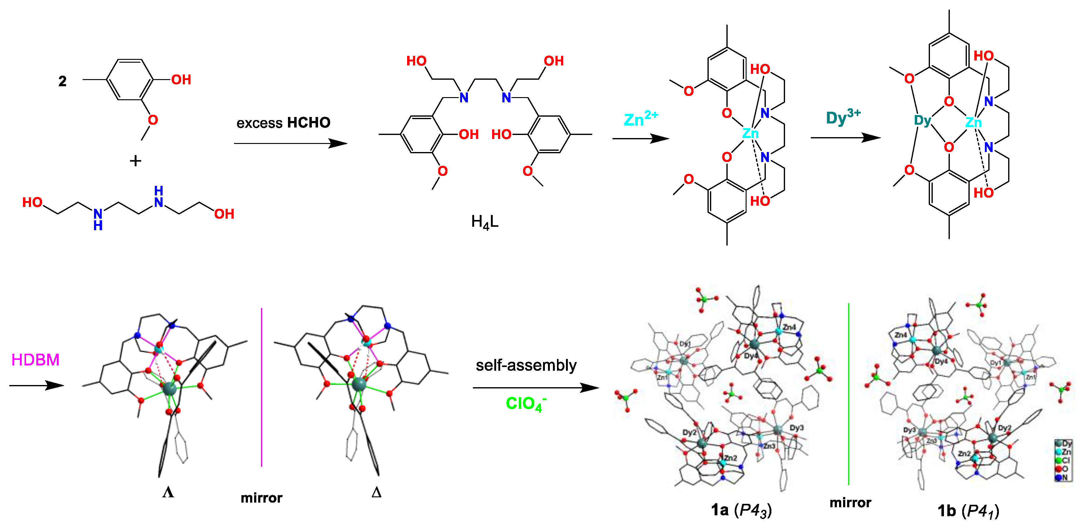
H4L, HDBM, Zn(ClO4)2⋅6H2O, Dy(ClO4)3⋅6H2O and triethylamine was dissolved in a MeOH/MeCN mixture, and the resultant solution was slowly evaporated at room temperature, giving rise to the concurrent crystallization of [Zn(H2L)Dy(DBM)2]4(ClO4)4⋅9CH3OH⋅H2O 1a or [Zn(H2L)Dy(DBM)2]4(ClO4)4⋅8CH3OH⋅0.5H2O 1b. The crystallographic data (Table 1) show that (1a) and (1b) crystallize in the chiral space groups P43 and P41 with the Flack factor of −0.0025(14) and 0.0012(16), respectively, indicating that 1a and 1b are chiral (hidden racemate). The powder X-ray diffraction (PXRD) patterns of the products are in good agreement with the simulated diffraction patterns of 1a and 1b (Figure S1), which further indicates that there is no other impurity phase. The calculated formulas of 1a or 1b were consistent with the results of CHN element analysis for the kryptoracemates. As shown in Figure 1, the achiral flexible ligand H4L was synthesized by the reaction of 2-methoxy-4-methylphenol, N,N′-Bis(2-hydroxyethyl)ethylenediamine and formaldehyde through a Mannich condensation reaction [25]. The detailed self-assembly process of complexes 1a and 1b is as follows: Firstly, H4L chelates with Zn2+ through two N atoms, two Ohydroxy atoms and two Ophenoxy atoms to form Zn(H2L); secondly, Zn(H2L) is connected to Dy3+ through two Ophenoxy atoms and two Oether atoms, and then the generated [Zn(H2L)Dy]3+ moiety is linked to two DBM to form a [Zn(H2L)Dy(DBM)2]+ cation with ∆- or Λ-configuration around Dy(III); finally, four [Zn(H2L)Dy(DBM)2]+ cations and four ClO4 anions self-assemble into mirror-symmetric 1a or 1b. The existence of ClO4 anion was confirmed by the peak at ∼1087 cm−1 in the IR spectrum.
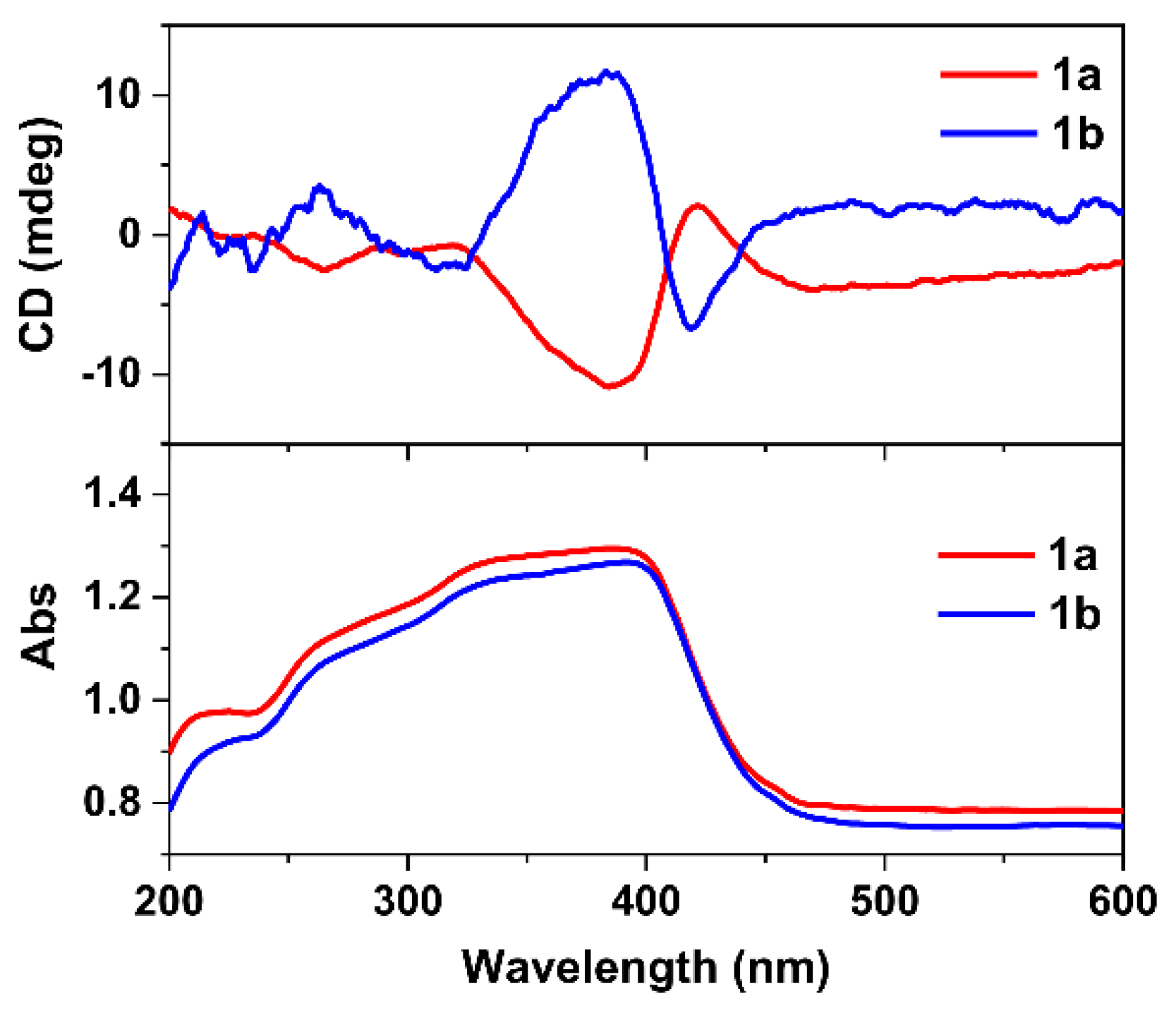
The solid-state CD spectra of 1a and 1b were measured with two individual single crystals by several attempts because the crystal shapes of 1a and 1b are indistinguishable. Two opposite CD signals are successfully obtained, as shown in Figure 2. For 1a, negative CD signal is detected at 265 and 385 nm, and positive CD signal is detected at 420 nm. In contrast, the CD signal for 1b is positive at 265 and 385 nm, and negative at 420 nm. The near-perfect mirror-image CD spectra in the range of 200–600 nm confirm the optical purity of 1a and 1b. The UV-vis spectra of complexes 1a and 1b in the solid state have obvious broad absorption in the range of 200–470 nm, which is basically consistent with the CD peaks.
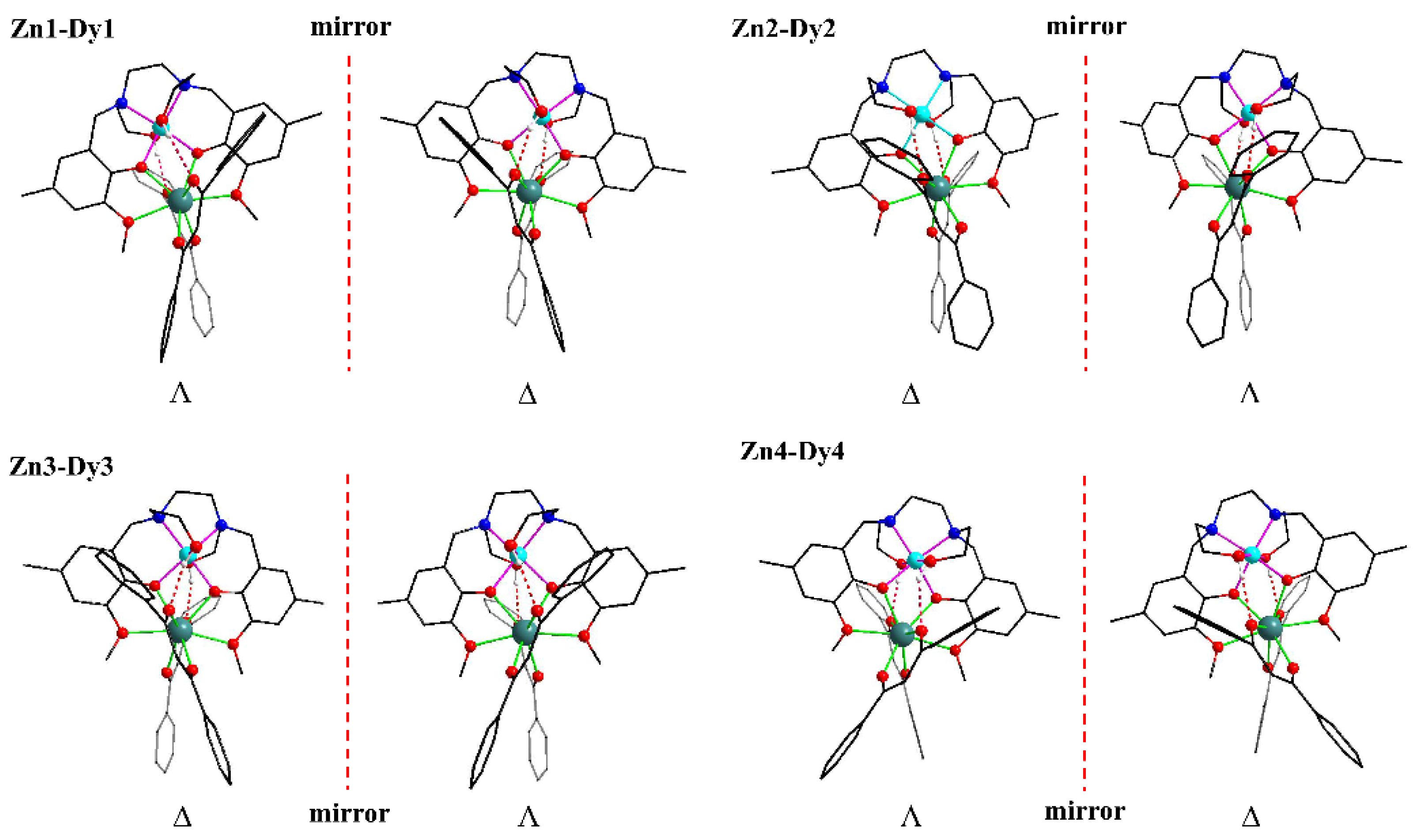
Chiral co-crystals formed from the same number of ∆-configurations and Λ-configurations are rare. Complexes 1a and 1b are the first reported enantiomers which consist of two ∆-configurations and two Λ-configurations. As shown in Figure 3, for 1a, Zn1-Dy1 and Zn4-Dy4 are of Λ-configurations, Zn2-Dy2 and Zn3-Dy3 are of ∆-configurations and the ∆- and Λ-ZnDy moieties are diastereomers. In contrast, for 1b, Zn1-Dy1 and Zn4-Dy4 are ∆-configurations, and Zn2-Dy2 and Zn3-Dy3 are Λ-configurations, and similarly the ∆- and Λ-ZnDy moieties are diastereomers. The ∆- and Λ-ZnDy moieties in 1a and 1b are enantiomers (Figure 3). Here, only the crystal structure of Zn1-Dy1 in 1a is selected as the example to be described. Specifically, two Ophenoxy atoms and two N atoms of H2L2− occupy the equatorial position of octahedral Zn atom with the Zn-Ophenoxy bond distances of 2.005(4) Å and the Zn-N bond distances of 2.088(5) and 2.096(5) Å. Two Ohydroxy atoms of H2L2− are situated at the axial position with the Ohydroxy-Zn-Ohydroxy angle of 172.55° and Zn-Ohydroxy bond lengths of 2.239(4) and 2.290(5) Å. The Dy atom is eight-coordinate with two Ophenoxy atoms and two Oether atoms from H2L2−, and four Ocarbonyl atoms from two BDM. The Dy-Oether bond distances are 2.468(4) and 2.497(4) Å, and the Dy-Ocarbonyl bond distances of 2.266(4)–2.394(4) Å are within the normal range [26,27]. The Dy-Ophenoxy bond distances are 2.307(4) and 2.268(4) Å, and the Ophenoxy-Dy-Ophenoxy bond angle is 71.48(13)°. The coordination geometry of Dy1 in complex 1a calculated by SHAPE software is close to D4d and D2d, and the deviation parameters are 1.674 and 1.615 (Table S1), respectively. Zn and Dy atoms are connected by two Ophenoxy atoms with a Zn1-Dy1 distance of 3.350 Å. H2L2− and BDM are connected through strong intramolecular hydrogen bond with Ohydroxy-H-Ocarbonyl angles of 125.7 and 160.2°, and Ohydroxy-Ocarbonyl distances of 2.740 and 2.795 Å.

2.2. Magnetic Properties

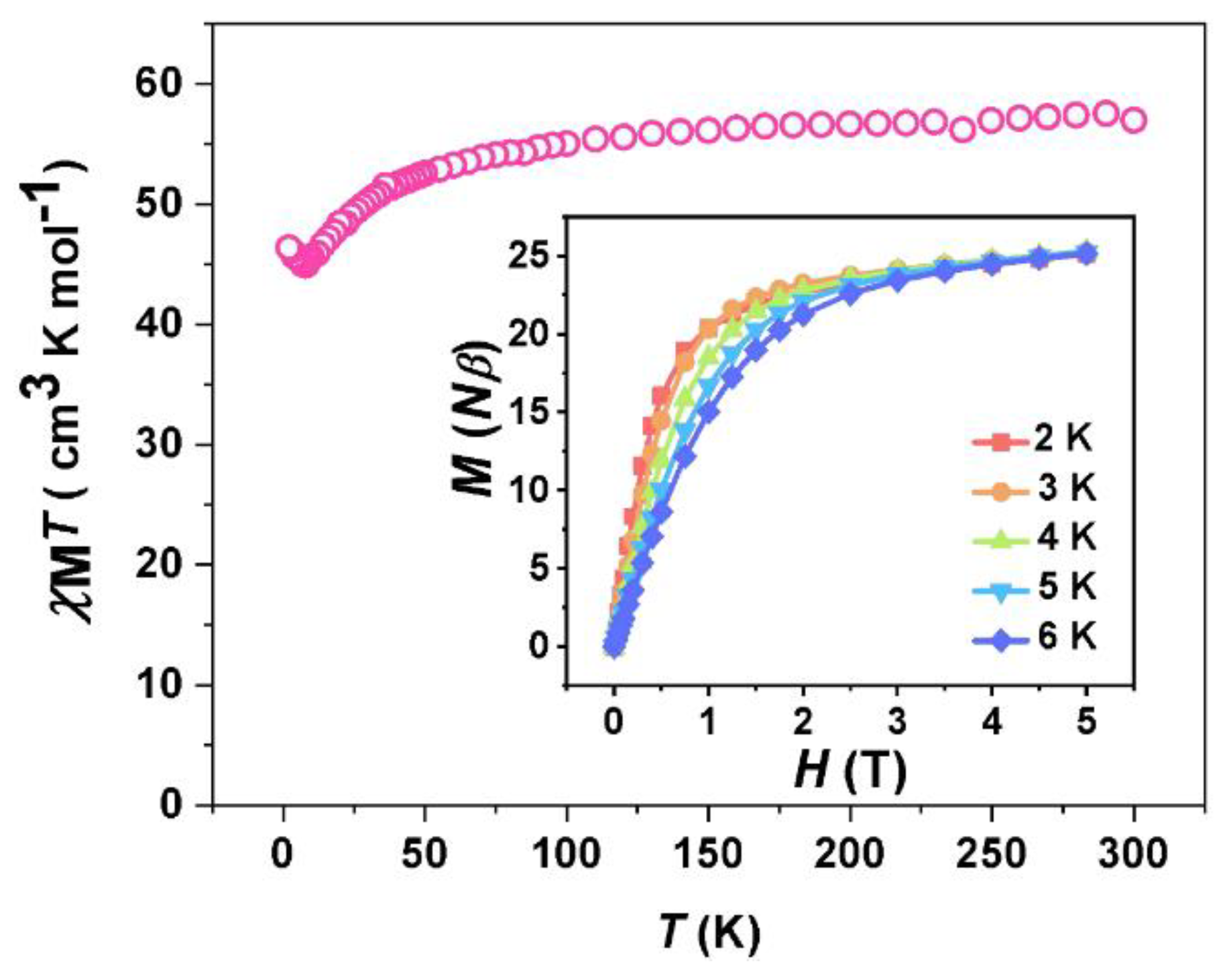
Since complexes 1a and 1b are mirror-related kryptoracemates with similar magnetic properties, only the magnetic properties of complex 1 (a mixture of 1a and 1b) are measured. As shown in Figure 4, the room temperature χMT value for complex 1 is 56.98 cm3 K mol−1, which is consistent with the theoretical value of 56.68 cm3 K mol−1 for four DyIII ions (g = 4/3, 6H15/2) and four diamagnetic ZnII ions. Upon decreasing the temperature, the χMT value was almost unchanged in the range of 300–100 K, and slowly decreased to the lowest value of 44.92 cm3 K mol−1 in the range of 100–8 K. The plot of 1/χM vs. T in the range of 8–300 K conforms to the Curie–Weiss law with θ = –4.54 K (Figure S2), which is due to the existence of crystal-field splitting of Dy(III) in complex 1. However, as the temperature continues to decrease to 2 K, the χMT value slightly increases to 46.38 cm3 K mol−1, which may be attributed to the intermolecular ferromagnetic interaction. The MH curve of complex 1 at 2 K rapidly increases from 0 to 20.38 , and then slowly increases to the maximum value of 25.26 , which is much lower than the theoretical saturation value of 40 for four DyIII ions due to the magnetic anisotropy of the DyIII ion. In addition, the MH curves at 2, 3, 4, 5 and 6 K are not overlapped, which again confirms the existence of magnetic anisotropy [28].
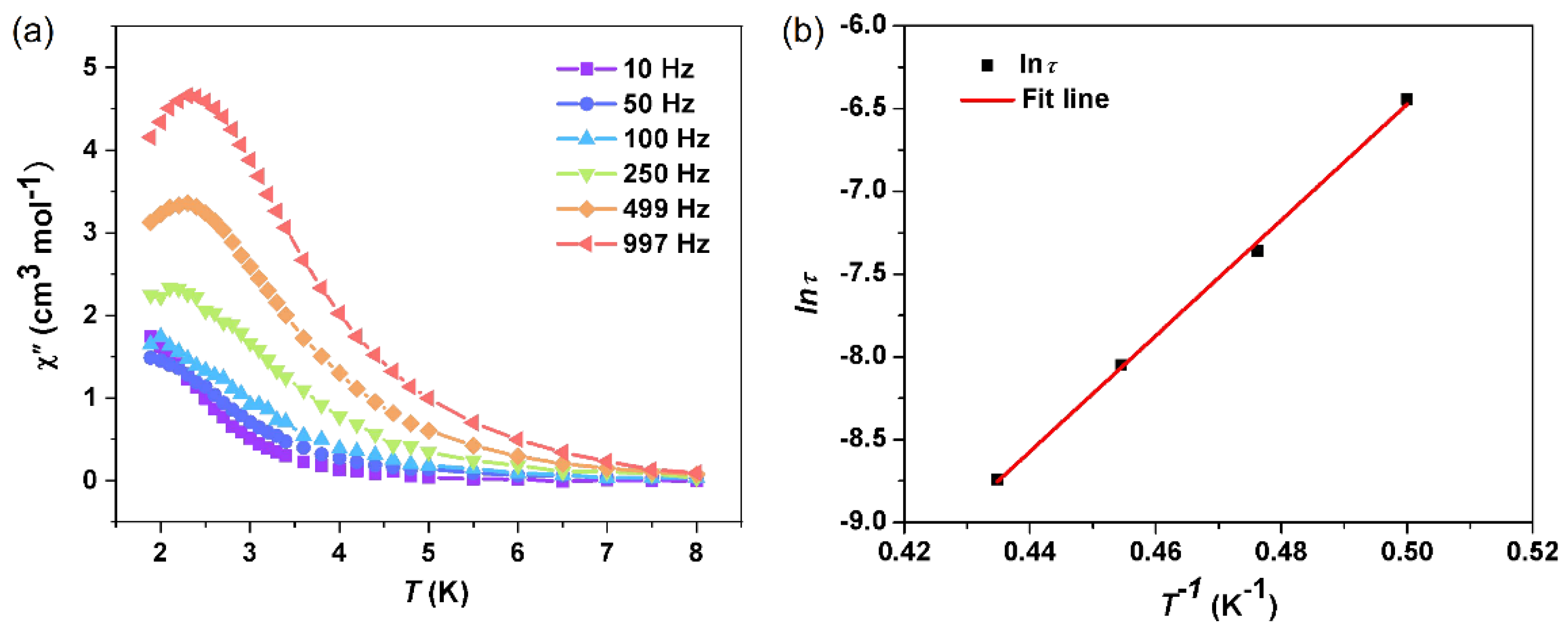
In order to study the dynamic magnetic relaxation of complex 1, the temperature dependence of ac magnetic susceptibility was investigated in an oscillating field of 2.5 Oe. As shown in Figure S3, the plot of χ″ versus H for complex 1 at 997 Hz shows that 600 Oe is the best dc field, while the plot of χ″ vs. T shows no peak at 600 Oe. Then, the dc fields of 0, 1500 and 2000 Oe were selected, respectively, to obtain the plots of χ″ vs. T (Figure S4). The plot of χ″ vs. T at 1500 Oe shows a more obvious peak than that of a 2000 Oe dc field, therefore, a dc field of 1500 Oe was applied to suppress the quantum tunnelling of magnetization (QTM). Under the dc field of 1500 Oe, both the in-phase χ′ and the out-of-phase χ″ exhibit obvious frequency dependence in the range of 2–8 K (Figure S5 and Figure 5a), which indicates the field-induced slow magnetic relaxation of complex 1. As shown in Figure 5b, the scatter plot of lnτ vs. T−1 was derived from the peak temperature values of the χ″ vs. T plots at the frequencies of 100, 250, 499 and 997 Hz, which was fitted according to the Arrhenius’ law of lnτ = lnτ0 + Ueff/kT (τ0 is the relaxation time, Ueff/k is the relaxation energy barrier), resulting in τ0 = 3.9(4) × 10−11 s and Ueff/k = 35.0(9) K. A τ0 value within the range of 10−12–10−6 s further indicates that complex 1 is a typical field-induced SMM. The relatively low energy barrier may be due to the poor axiality of the ligand field: the Dy-Ophenoxy bond length is in the range of 2.268–2.321(4) Å, while the Ophenoxy-Dy-Ophenoxy bond angle is in the range of 70.12.48(13)–72.17(14)°, which is far away from the perfect 180°. As shown in Figure S6, the Cole–Cole plots show a characteristic double magnetic relaxation for complex 1, which can be fitted to obtain α1 = 0.163–0.251 and α2 = 0.299–0.348 (Table S2), which correspond to two crystallographically independent DyIII ions [29]. Of course, theoretical calculations could provide more accurate information on the relaxation mechanism of such ZnDy SMMs, which deserves future investigations.

2.3. Luminescent Properties

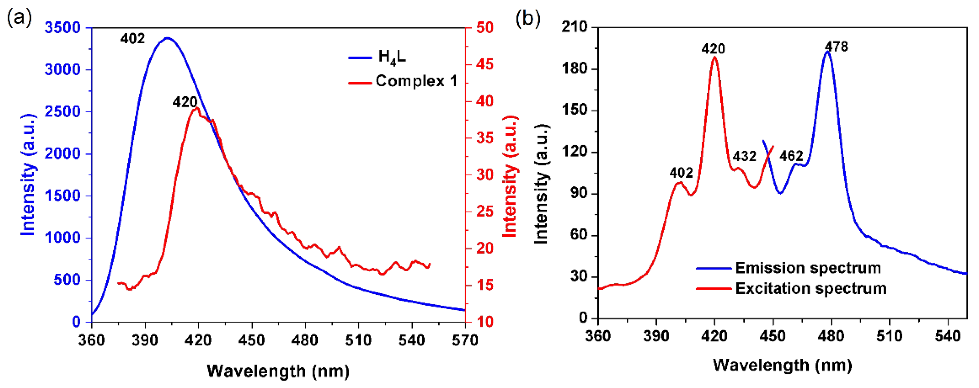
The luminescent properties of complex 1 in MeCN were investigated, as shown in Figure 6a. Upon excitation at 350 nm, complex 1 exhibits a broad band similar to H4L, which can be attributed to the characteristic peak of the H2L2− moiety in 1. However, the band appears at 420 nm for complex 1 and 402 nm for H4L, and the emissive intensity of complex 1 is nearly 85 times weaker than that of H4L. The weakened intensity may be due to the inner-filter effect (IFE) in complex 1 in the presence of the overlap between the emission and the visible absorption at ca. 402 nm (Figure 2). As shown in Figure 6b, the excitation spectrum of complex 1 was recorded by monitoring the characteristic emission band of DyIII ions at 478 nm. The peaks at 402 and 420 nm originate from the intra-ligand π→π * transitions, while the peak at 432 nm is due to the f→f * transition of the DyIII ion from the 6H15/2 ground state to the 4G11/2 excited state [30]. Therefore, the emission spectrum of complex 1 was recorded upon the excitation of 420 nm. There are two peaks at 462 and 478 nm, corresponding to the 4I15/26H15/2 and 4F9/26H15/2 transitions of DyIII [31,32].

3. Materials and Methods

3.1. Materials

Methanol (MeOH), acetonitrile (MeCN), 2-methoxy-4-methylphenol, N,N′-Bis(2-hydroxyethyl)ethylenediamine, formaldehyde, dibenzoylmethane (HDBM), Zn(ClO4)2⋅6H2O, Dy(ClO4)3⋅6H2O and triethylamine were purchased from commercial sources and used without further purification.

3.2. The Preparation of Complex 1

H4L was prepared according to the method we reported previously [23]. The mixture of H4L (45 mg, 0.1 mmol), HDBM (45 mg, 0.2 mmol), Zn(ClO4)2⋅6H2O (37 mg, 0.1 mmol) and 96 μL Dy(ClO4)3⋅6H2O aqueous solution (50%, 0.1 mmol) was dissolved in acetonitrile (5 mL) and methanol (10 mL). Then, triethylamine (100 μL) was slowly added and stirred for 3 min. Subsequently, the above solution was filtered and slowly evaporated without disturbance at room temperature. Colorless needle crystals of complex 1 were obtained after 2 days. Yield: about 74%. Two types of crystal structures were obtained by single crystal X-ray diffraction analysis on several randomly selected single crystals. Anal. calcd (%) for C225H262Cl4Dy4N8O66Zn4 (1a): C, 52.09; H, 5.09; and N, 2.16. Anal. calcd (%) for C224H257Cl4Dy4N8O64.5Zn4 (1b): C, 52.27; H, 5.03; and N, 2.18. Found for 1a + 1b: C, 52.9; H, 5.0; and N, 2.4. Main IR peaks (KBr, cm−1): 1593 (s), 1544 (vs), 1521 (vs), 1481 (vs), 1452 (m), 1390 (vs), 1315 (m), 1250 (m), 1087 (vs), 816 (m), 728 (m), 694 (w), and 617 (m).

3.3. Physical Measurements

IR spectra was recorded on a Beijing Rayleigh WQF-510A FTIR spectrometer. CHN element analysis was tested on a thermal Flash EA 1112 elemental analyzer. Powder X-ray diffraction (PXRD) was measured on a Rigaku D/max 2500 diffractometer. Circular dichroism (CD) spectra were measured on a JASCO J-1500 spectrometer. Luminescence spectra were tested on a Hitachi F-7000 fluorescence spectrometer. Magnetic measurements were carried on a SQUID MPMS-XL5 magnetometer, and the Pascal’s constants were used to correct the diamagnetism. Single-crystal X-ray diffraction was performed on a Rigaku Super Nova diffractometer. The crystal structures were solved and refined by OLEX-2 software. To resolve the severe disorder of the lattice solvent molecules of complexes 1a and 1b, the SQUEEZE function of PLATON software was applied. For complex 1a, 104 electrons in the hole of 509 Å3 correspond to 5CH3OH and 1H2O for each formula (100 electrons). For 1b, 133 electrons in the hole of 822 Å3 correspond to 7CH3OH and 0.5H2O for each formula (131 electrons).

4. Conclusions

In summary, we have developed a novel multi-channel barcode module based on chiral co-crystal complexes 1a and 1b, which contain field-induced SMM behavior and different emission bands. Complexes 1a and 1b are the first chiral kryptoracemate complexes composed of four independent binuclear Zn-Dy groups, two of which are ∆-configuration and two are Λ-configuration. These two chiral complexes have mirror-related structures and mirror-symmetric CD spectra. Complex 1 shows field-induced SMM behavior with an energy barrier of 35.0(9) K. Moreover, luminescence measurements show that complex 1 exhibits different luminescence emissions under the excitation of UV or visible light. The present work provides some valuable information for the construction of novel chiral complexes from achiral ligands, and describes a new strategy for creating multi-channel barcode modules for high-density information storage.

Supplementary Materials

The following are available online at https://www.mdpi.com/article/10.3390/magnetochemistry8120166/s1, Figure S1: PXRD patterns for complex 1. Figure S2: Plot of the 1/χM versus T for 1. The red solid line represents the linear fit of the data. Figure S3: (left) Plot of χ″ versus H for complex 1 at 997 Hz and (right) plot of χ″ versus T for complex 1 at 997 Hz under 600 Oe dc field. Figure S4: Plots of χ″ versus T for complex 1 at 997 Hz under 0, 1500 and 2000 Oe dc field, respectively. Figure S5: Plots of temperature dependent χ′ ac susceptibility for complex 1 (Hdc = 1500 Oe). Figure S6: Cole-Cole curves at 1.9−2.3 K (Hdc = 1500 Oe, Hac = 2.5 Oe). Table S1: The coordination geometry of Dy1 in 1a calculated by SHAPE software. Table S2: Cole-Cole curve fitting parameters for complex 1.

Author Contributions

All authors participated in the writing and editing of the article, M.Z., L.M., X.-R.W., C.-M.L. and H.-Z.K. All authors have read and agreed to the published version of the manuscript.

Funding

This research was funded by the National Natural Science Foundation of China, grant numbers 21971142 and 21771115.

Informed Consent Statement

Not applicable.

Conflicts of Interest

The authors declare no conflict of interest.

References

  1. White, K.A.; Chengelis, D.A.; Gogick, K.A.; Stehman, J.; Rosi, N.L.; Petoud, S. Near-Infrared Luminescent Lanthanide MOF Barcodes. J. Am. Chem. Soc. 2009, 131, 18069–18071. [Google Scholar] [CrossRef]
  2. Zhang, Y.; Zhang, L.; Deng, R.; Tian, J.; Zong, Y.; Jin, D.; Liu, X. Multicolor barcoding in a single upconversion crystal. J. Am. Chem. Soc. 2014, 136, 4893–4896. [Google Scholar] [CrossRef] [PubMed]
  3. Lu, Y.; Yan, B. Luminescent lanthanide barcodes based on postsynthetic modified nanoscale metal–organic frameworks. J. Mater. Chem. C 2014, 2, 7411–7416. [Google Scholar] [CrossRef]
  4. Zhao, Y.; Shum, H.C.; Chen, H.; Adams, L.L.; Gu, Z.; Weitz, D.A. Microfluidic generation of multifunctional quantum dot barcode particles. J. Am. Chem. Soc. 2011, 133, 8790–8793. [Google Scholar] [CrossRef] [PubMed]
  5. Zhang, F.; Haushalter, R.C.; Haushalter, R.W.; Shi, Y.; Zhang, Y.; Ding, K.; Zhao, D.; Stucky, G.D. Rare-earth upconverting nanobarcodes for multiplexed biological detection. Small 2011, 7, 1972–1976. [Google Scholar] [CrossRef] [PubMed] [Green Version]
  6. Pan, M.; Zhu, Y.X.; Wu, K.; Chen, L.; Hou, Y.J.; Yin, S.Y.; Wang, H.P.; Fan, Y.N.; Su, C.Y. Epitaxial Growth of Hetero-Ln-MOF Hierarchical Single Crystals for Domain- and Orientation-Controlled Multicolor Luminescence 3D Coding Capability. Angew. Chem. Int. Ed. Engl. 2017, 56, 14582–14586. [Google Scholar] [CrossRef] [PubMed]
  7. Chen, C.; Zhang, P.; Gao, G.; Gao, D.; Yang, Y.; Liu, H.; Wang, Y.; Gong, P.; Cai, L. Near-infrared-emitting two-dimensional codes based on lattice-strained core/(doped) shell quantum dots with long fluorescence lifetime. Adv. Mater. 2014, 26, 6313–6317. [Google Scholar] [CrossRef]
  8. Shikha, S.; Salafi, T.; Cheng, J.; Zhang, Y. Versatile design and synthesis of nano-barcodes. Chem. Soc. Rev. 2017, 46, 7054–7093. [Google Scholar] [CrossRef] [Green Version]
  9. Ishii, A.; Miyasaka, T. Direct detection of circular polarized light in helical 1D perovskite-based photodiode. Sci. Adv. 2020, 6, eabd3274. [Google Scholar] [CrossRef]
  10. Zeng, M.; Ren, A.; Wu, W.; Zhao, Y.; Zhan, C.; Yao, J. Lanthanide MOFs for inducing molecular chirality of achiral stilbazolium with strong circularly polarized luminescence and efficient energy transfer for color tuning. Chem. Sci. 2020, 11, 9154–9161. [Google Scholar] [CrossRef]
  11. Lu, J.; Guo, M.; Tang, J. Recent Developments in Lanthanide Single-Molecule Magnets. Chem.-Asian J. 2017, 12, 2772–2779. [Google Scholar] [CrossRef] [PubMed]
  12. Marin, R.; Brunet, G.; Murugesu, M. Shining New Light on Multifunctional Lanthanide Single-Molecule Magnets. Angew. Chem.-Int. Ed. 2020, 60, 1728–1746. [Google Scholar] [CrossRef]
  13. Meng, Y.-S.; Jiang, S.-D.; Wang, B.-W.; Gao, S. Understanding the Magnetic Anisotropy toward Single-Ion Magnets. Acc. Chem. Res. 2016, 49, 2381–2389. [Google Scholar] [CrossRef] [PubMed]
  14. Zhu, Z.; Zhao, C.; Feng, T.; Liu, X.; Ying, X.; Li, X.-L.; Zhang, Y.-Q.; Tang, J. Air-Stable Chiral Single-Molecule Magnets with Record Anisotropy Barrier Exceeding 1800 K. J. Am. Chem. Soc. 2021, 143, 10077–10082. [Google Scholar] [CrossRef] [PubMed]
  15. Chen, Y.-C.; Liu, J.-L.; Ungur, L.; Liu, J.; Li, Q.-W.; Wang, L.-F.; Ni, Z.-P.; Chibotaru, L.F.; Chen, X.-M.; Tong, M.-L. Symmetry-Supported Magnetic Blocking at 20 K in Pentagonal Bipyramidal Dy(III) Single-Ion Magnets. J. Am. Chem. Soc. 2016, 138, 2829–2837. [Google Scholar] [CrossRef] [PubMed]
  16. Li, Z.-H.; Zhai, Y.-Q.; Chen, W.-P.; Ding, Y.-S.; Zheng, Y.-Z. Air-Stable Hexagonal Bipyramidal Dysprosium(III) Single-Ion Magnets with Nearly Perfect D-6h Local Symmetry. Chem.-A Eur. J. 2019, 25, 16219–16224. [Google Scholar] [CrossRef]
  17. Yang, Q.Y.; Pan, M.; Wei, S.C.; Li, K.; Du, B.B.; Su, C.Y. Linear Dependence of Photoluminescence in Mixed Ln-MOFs for Color Tunability and Barcode Application. Inorg. Chem. 2015, 54, 5707–5716. [Google Scholar] [CrossRef]
  18. Zhou, Y.; Yan, B. Ratiometric multiplexed barcodes based on luminescent metal–organic framework films. J. Mater. Chem. C 2015, 3, 8413–8418. [Google Scholar] [CrossRef]
  19. Gao, M.L.; Wang, W.J.; Liu, L.; Han, Z.B.; Wei, N.; Cao, X.M.; Yuan, D.Q. Microporous Hexanuclear Ln(III) Cluster-Based Metal-Organic Frameworks: Color Tunability for Barcode Application and Selective Removal of Methylene Blue. Inorg. Chem. 2017, 56, 511–517. [Google Scholar] [CrossRef]
  20. Yao, Y.; Gao, Z.; Lv, Y.; Lin, X.; Liu, Y.; Du, Y.; Hu, F.; Zhao, Y.S. Heteroepitaxial Growth of Multiblock Ln-MOF Microrods for Photonic Barcodes. Angew. Chem.-Int. Ed. 2019, 58, 13803–13807. [Google Scholar] [CrossRef]
  21. Du, B.B.; Zhu, Y.X.; Pan, M.; Yue, M.Q.; Hou, Y.J.; Wu, K.; Zhang, L.Y.; Chen, L.; Yin, S.Y.; Fan, Y.N.; et al. Direct white-light and a dual-channel barcode module from Pr(III)-MOF crystals. Chem. Commun. (Camb.) 2015, 51, 12533–12536. [Google Scholar] [CrossRef] [PubMed]
  22. Nguyen, H.Q.; Baxter, B.C.; Brower, K.; Diaz-Botia, C.A.; DeRisi, J.L.; Fordyce, P.M.; Thorn, K.S. Programmable Microfluidic Synthesis of Over One Thousand Uniquely Identifiable Spectral Codes. Adv. Opt. Mater. 2017, 5, 1600548. [Google Scholar] [CrossRef] [Green Version]
  23. Liu, M.-J.; Yuan, J.; Wang, B.-L.; Wu, S.-T.; Zhang, Y.-Q.; Liu, C.-M.; Kou, H.-Z. Spontaneous Resolution of Chiral Co(III)Dy(III) Single-Molecule Magnet Based on an Achiral Flexible Ligand. Cryst. Growth Des. 2018, 18, 7611–7617. [Google Scholar] [CrossRef]
  24. Sunatsuki, Y.; Fujita, K.; Maruyama, H.; Suzuki, T.; Ishida, H.; Kojima, M.; Glaser, R. Chiral Crystal Structure of a P212121 Kryptoracemate Iron(II) Complex with an Unsymmetric Azine Ligand and the Observation of Chiral Single Crystal Circular Dichroism. Cryst. Growth Des. 2014, 14, 3692–3695. [Google Scholar] [CrossRef]
  25. Tshuva, E.Y.; Gendeziuk, N.; Kol, M. Single-step synthesis of salans and substituted salans by Mannich condensation. Tetrahedron Lett. 2001, 42, 6405–6407. [Google Scholar] [CrossRef]
  26. Zeng, M.; Zhou, Z.-Y.; Wu, X.-R.; Liu, C.-M.; Kou, H.-Z. Assembly of a Heterotrimetallic Zn2Dy2Ir Pentanuclear Complex toward Multifunctional Molecular Materials. Inorg. Chem. 2022, 61, 14275–14281. [Google Scholar] [CrossRef]
  27. Zeng, M.; Ji, S.-Y.; Wu, X.-R.; Zhang, Y.-Q.; Liu, C.-M.; Kou, H.-Z. Magnetooptical Properties of Lanthanide(III) Metal-Organic Frameworks Based on an Iridium(III) Metalloligand. Inorg. Chem. 2022, 61, 3097–3102. [Google Scholar] [CrossRef]
  28. Liu, C.-M.; Sun, R.; Hao, X.; Wang, B. Chiral Co-Crystals of (S)- or (R)-1,1′-Binaphthalene-2,2′-diol and Zn2Dy2 Tetranuclear Complexes Behaving as Single-Molecule Magnets. Cryst. Growth Des. 2021, 21, 4346–4353. [Google Scholar] [CrossRef]
  29. Guo, Y.-N.; Xu, G.-F.; Gamez, P.; Zhao, L.; Lin, S.-Y.; Deng, R.; Tang, J.; Zhang, H.-J. Two-Step Relaxation in a Linear Tetranuclear Dysprosium(III) Aggregate Showing Single-Molecule Magnet Behavior. J. Am. Chem. Soc. 2010, 132, 8538. [Google Scholar] [CrossRef]
  30. Li, L.; Qin, F.; Zhou, Y.; Zheng, Y.; Miao, J.; Zhang, Z. Photoluminescence and time -resolved -luminescence of CaWO 4: Dy 3+phosphors. J. Lumin. 2020, 224, 117308. [Google Scholar] [CrossRef]
  31. Zeng, M.; Zhan, C.; Yao, J. Novel bimetallic lanthanide metal-organic frameworks (Ln-MOFs) for colour-tuning through energy-transfer between visible and near-infrared emitting Ln3+ ions. J. Mater. Chem. C 2019, 7, 2751–2757. [Google Scholar] [CrossRef]
  32. Cui, Y.; Yue, Y.; Qian, G.; Chen, B. Luminescent Functional Metal-Organic Frameworks. Chem. Rev. 2012, 112, 1126–1162. [Google Scholar] [CrossRef] [PubMed]
Scheme 1. Schematic representation of using chiral co-crystals as a multi-channel barcode module.
Magnetochemistry 08 00166 sch001
Figure 1. The formation process of complexes 1a (P43) and 1b (P41).
Magnetochemistry 08 00166 g001
Figure 2. Solid-state CD spectra of 1a and 1b (KCl pellets).
Magnetochemistry 08 00166 g002
Figure 3. Coordination environments of Zn1-Dy1, Zn2-Dy2, Zn3-Dy3 and Zn4-Dy4 in complexes 1a (left) and 1b (right). Some hydrogen atoms are omitted for clarity.
Magnetochemistry 08 00166 g003
Figure 4. Temperature dependence of magnetic susceptibility for complex 1 (Hdc = 1000 Oe). Inset: Field dependence of magnetization for complex 1 at 2, 3, 4, 5 and 6 K.
Magnetochemistry 08 00166 g004
Figure 5. (a) Temperature dependent χac susceptibility for complex 1 (Hdc = 1500 Oe); (b) the ln(τ) vs. T−1 plots based on the Arrhenius relationship for complex 1.
Magnetochemistry 08 00166 g005
Figure 6. (a) Emission spectra of H4L and complex 1 in MeCN with λex = 350 nm; (b) excitation (λem = 478 nm) and emission spectra (λex = 420 nm) for complex 1 in MeCN.
Magnetochemistry 08 00166 g006
Table 1. Crystal data for complexes 1a and 1b.
Complexes1a1b
formulaC225H262Cl4Dy4N8O66Zn4C224H257Cl4Dy4N8O64.5Zn4
formula weight5187.955146.90
T/K173(2)173(2)
crystal systemtetragonaltetragonal
space groupP43P41
a, b29.7479(2)29.7491(2)
c25.3023(3)25.3017(2)
α, β, γ9090
V322,391.0(4)22,392.2(3)
Z44
ρcalc/g⋅cm−31.5391.527
μ/mm−18.5838.562
F(000)10,19210,500
data/restraints/parameters29,484/102/272434,446/103/2666
GOF on F21.0191.013
Rint0.02530.0253
R1, wR2 [I ≥ 2σ (I)]0.0338, 0.08600.0420, 0.1092
R1, wR2 (all data)0.0357, 0.08720.0442, 0.1111
Flack parameter−0.0025(14)0.0012(16)
CCDC2,209,0582,209,059
Publisher’s Note: MDPI stays neutral with regard to jurisdictional claims in published maps and institutional affiliations.