Article

Magnetic Properties of Bi-Magnetic Core/Shell Nanoparticles: The Case of Thin Shells

1
nM2-Lab., Department of Chemistry and Industrial Chemistry, University of Genova, 16146 Genova, Italy
2
REC “Smart Materials and Biomedical Applications”, Immanuel Kant Baltic Federal University, 236041 Kaliningrad, Russia
3
Department of Biomedical Engineering and Sydney Nano Institute, The University of Sydney, Sydney, NSW 2008, Australia
4
nM2-Lab., Consiglio Nazionale delle Ricerche, Istituto di Struttura della Materia, Monterotondo Scalo, 00015 Rome, Italy
*
Authors to whom correspondence should be addressed.
Magnetochemistry 2021, 7(11), 146; https://doi.org/10.3390/magnetochemistry7110146
Submission received: 17 September 2021 / Revised: 25 October 2021 / Accepted: 29 October 2021 / Published: 8 November 2021

Abstract

:
Bi-magnetic core/shell nanoparticles were synthesized by a two-step high-temperature decomposition method of metal acetylacetonate salts. Transmission electron microscopy confirmed the formation of an ultrathin shell (~0.6 nm) of NiO and NiFe2O4 around the magnetically hard 8 nm CoFe2O4 core nanoparticle. Magnetization measurements showed an increase in the coercivity of the single-phase CoFe2O4 seed nanoparticles from ~1.2 T to ~1.5 T and to ~2.0 T for CoFe2O4/NiFe2O4 and CoFe2O4/NiO, respectively. The NiFe2O4 shell also increases the magnetic volume of particles and the dipolar interparticle interactions. In contrast, the NiO shell prevents such interactions and keeps the magnetic volume almost unchanged.

1. Introduction

Bi-magnetic exchange coupled systems consisting of ferro(i)-/antiferromagnetic (F(i)M/AFM) or magnetically hard/soft (h-FM/s-FM) materials provide enhanced magnetic properties compared to their individual counterparts (i.e., single-phase systems) [1,2,3,4,5]. AFM/FM systems may cause an increase of magnetic anisotropy, that is exploited in different applications such as recording media, spintronics and permanent magnets [3,6,7,8]. Aside from the Exchange Bias phenomenon, resulting in unidirectional exchange anisotropy, an increase of the effective anisotropy energy of the FM phase has also been observed in AFM/FM systems [8,9]. On the other hand, bi-magnetic systems consisting of two FM or FiM interfaced phases, such as h-FM/s-FM core/shell nanoparticles (NPs), with tunable saturation magnetization (MS), and magnetic anisotropy (K) are beneficial to achieve, for example, contrast-enhanced magnetic resonance imaging or a high thermomagnetic effect [1,2,4,10,11,12]. Indeed, the magnetic properties of core/shell systems depend on the intrinsic parameters of h-FM and s-FM phases, as well as on the coupling strength and nanoparticle architecture which are controlled by the synthesis procedure (size of layers, quality of interface, shape, etc.) [4].
Depending on the size/thickness of h-FM and s-FM phases, the bi-magnetic system can act in a rigidly coupled regime if the s-FM phase is thinner than the double domain wall thickness of the h-FM [5]. This regime is characterized by a squared hysteresis loop of the soft phase being strongly exchange-coupled with the hard phase and the magnetization reversal process of the two phases occurring at the same field. In the case of the thicker s-FM phase, its reversal occurs at significantly lower fields than the switching of the h-FM. However, with regard to nanoparticles, a wider phenomenology may be observed due to the large dispersion of magnetic properties arising from size distributions or divergences in the structural properties (i.e., stoichiometry and unit-cell parameters) of nanomaterials compared to bulk analog materials [13]. Special attention has been paid to the formation of the ultrathin shell of a few atomic layer thicknesses, exhibiting an enhanced spin canting (ESC) effect [14] which brings out a larger magnetic surface anisotropy, allowing exchange coupled systems to increase, for example, magnetic energy product (BH)max or specific loss power.
This paper discusses the effect of thin AFM and s-FiM shells on the magnetic properties of CoFe2O4 nanoparticles. The cobalt ferrite is one of the most studied oxides with a spinel structure that presents (semi-)hard magnetic properties [15,16]. To tune its magnetic anisotropy, a thin shell (~0.6-nm) of s-FiM-NiFe2O4 or AFM-NiO has been deposited on the nanoparticle’s surface. Both core/shell systems showed an increase of coercivity (HC) with an almost unchanged saturation magnetization compared to cobalt ferrite NPs.

2. Materials and Methods

2.1. Synthesis of NPs

Iron (III) acetylacetonate (97%), Cobalt (II) acetylacetonate (97%) and Nickel (II) acetylacetonate (95%) were purchased from Alfa Aesar (Kandel, Germany). Oleic acid (AO) (technical grade, 90%), Oleylamine (OLA) (technical grade, 70%), benzyl ether (99%), toluene, 2-propanol, and acetone were purchased from Sigma Aldrich (Darmstadt, Germany) and used without further purification. All the syntheses were conducted under oxygen-free conditions in a Schlenk line.
The single-phase core system was synthesized following a modified procedure reported elsewhere [17,18]. To synthesize the CoFe2O4 core, (CFO sample), Fe(acac)3 (2 mmol), Co(acac)2 (1 mmol), 1,2-hexadecanediol (10 mmol), OA (6 mmol), OLA (6 mmol), and benzyl ether (20 mL) were mixed and magnetically stirred under a flow of nitrogen. The mixture was heated to 200 °C for 100 min (5 °C/min) and then heated to reflux at 300 °C (5 °C/min) for 60 min. The black-colored mixture was left to cool to room temperature overnight and washed with acetone, toluene and isopropanol.
A seed-mediated growth technique at high temperature was used to achieve the core/shell structure. For example, for CoFe2O4/NiFe2O4 NPs (CFO/NFO sample), cobalt ferrite seeds were sonicated in 5 mL benzyl ether in the presence of OA (600 μL). Then, the suspension was added to the round bottom flask containing Ni(acac)2 (0.33 mmol), Fe(acac)3 (0.67 mmol), OA (600 μL) in benzyl ether (15 mL). After 1h vacuum at RT, the solution was heated to reflux at 290 °C (5 °C/min) under an Argon atmosphere and was then kept at this temperature for 30 min. A similar protocol was used to prepare the CoFe2O4/NiO NPs (CFO/NiO sample).

2.2. Morphostructural Properties

The X-ray diffraction (XRD) patterns were obtained from a Bruker DaVinci2 diffractometer (Bruker, MA, USA) with a Cu Kα (λ = 1.54056 Å) source in the 10°–75° 2θ range. Crystal phase identification was performed with QualX software. The mean size of crystallites, dXRD, was calculated by using Scherrer’s equation [19]:
d XRD = 0.9 λ β cos θ ,
where β is the full width at half-maximum (FWHM) of the peaks of the corresponding reflections at 2θ geometry.
Scanning Transmission Electron Microscopy (STEM) Hitachi S5500, operating at 30 kV, was used to investigate the size distribution and morphology of the particles. The specimens for the TEM analyses were prepared by depositing one or a few drops of the sample on 300 mesh cupper carbon grids. The particle size distribution obtained by the TEM image analysis was fitted by normal and log-normal functions:
P = A σ TEM 2 π exp 1 2 D σ TEM < d TEM > 2 ;       normal   distribution
P l = A D σ TEM 2 π exp ln 2 D < d TEM > 2 σ TEM 2 ,       log - normal   distribution
where <dTEM> is the mean value of particle diameter D (variable) and σ is its standard deviation.

2.3. Magnetic Properties

The DC magnetization measurements were performed by a Quantum Design superconducting quantum interference device (SQUID) magnetometer. The sample, in the form of a powder, was immobilized in epoxy resin to avoid the movement of the nanoparticles during the measurements. Hysteresis loops were obtained in the ±5 T applied magnetic field range at different temperatures from 5 to 300 K. The value of saturation magnetization, MS, was estimated from the Law of Approach to Saturation (LAS) by fitting the high-field region of hysteresis with the function
M = M S 1 A / H   B / H 2 ,
where A and B are free parameters [20].
The magnetization versus temperature measurements were performed using the zero-field-cooled MzFC(T) and field-cooled MFC(T) protocols in a low measuring magnetic field of 2.5 mT [21,22]. MZFC(T) magnetization was carried out by firstly cooling the sample from room temperature to 5 K in a zero magnetic field. Then, a small measuring magnetic field was applied and the MZFC(T) was measured during warming from 5 to 300 K, whereas MFC(T) was recorded during the subsequent cooling.
For an assembly of non-interacting particles, the difference in MFC−MZFC is related to the distribution of the anisotropy energy barriers [22,23,24]:
M FC M ZFC   ~ α Δ E c f ( Δ E a )   d E ,
where α is a parameter proportional to the initial susceptibility of non-relaxing particles, measuring field and time; ∆Ec is a critical value of energy, above which the particles are blocked. In other words, ∆Ec = KV = 25kBTB, thus, Equation (5) shows the distribution of blocking temperatures:
f T B ~ d ( M FC M ZFC )   d T .
The f(ΔTB) can be fitted by a (log-)normal function because TB is proportional to the particle volume, which is well-described by a (log-)normal function of the particle size distribution (Equations (2) and (3)) [22,25,26]; for the determination of TB in the case of unmerged ZFC/FC, the d(MFC−MZFC)/dT is fitted by the normal function, then the average <TB> is determined as a temperature at which d2(MFC − MZFC)/dT2 = 0, to the left of the MZFC peak position [24].

3. Results

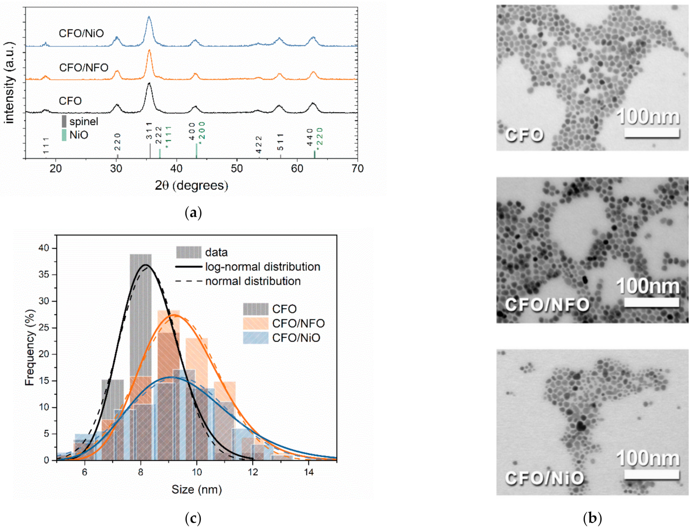
3.1. Morphostructural Properties

The XRD patterns suggest the presence of only spinel and rock salt phases in the bi-magnetic core/shell NPs (Figure 1a). No extra phases have been detected. Scanning transmission electron microscopy (STEM) images show a nearly spherical core and core/shell NPs (Figure 1b), for which the mean diameter (dTEM) has been evaluated using Equations (2) and (3) by counting more than 200 NPs.
On the other hand, the mean crystallites’ size (dXRD), has been calculated using Equation (1) by averaging 5 the most intense XRD reflections. The size of the XRD and STEM measurements, reveals an increase in the diameter after the deposition of a thin shell around the CFO seed NPs, thus confirming the formation of core/shell structures (Figure 1c and Table 1). In particular, the size increase of the dXRD for the CFO/NiO samples is found to be less than the size increase of the dXRD for the CFO/NFO sample. This can probably be ascribed to the partial mismatch of spinel phase planes of the CoFe2O4 with those of the rock salt structure of the NiO phase. However, the dTEM values confirm the growth of a thin shell (~0.6 nm) in both core/shell systems, thus indicating a similar increase in the physical size for both systems. In all cases, these core/shell NPs exhibit a narrow size distribution characterized by the low values of the standard deviation σTEM of the log-normal distribution; the formation of a NiO shell induces a slightly higher dispersion with respect to the CFO/NFO sample (Table 1).

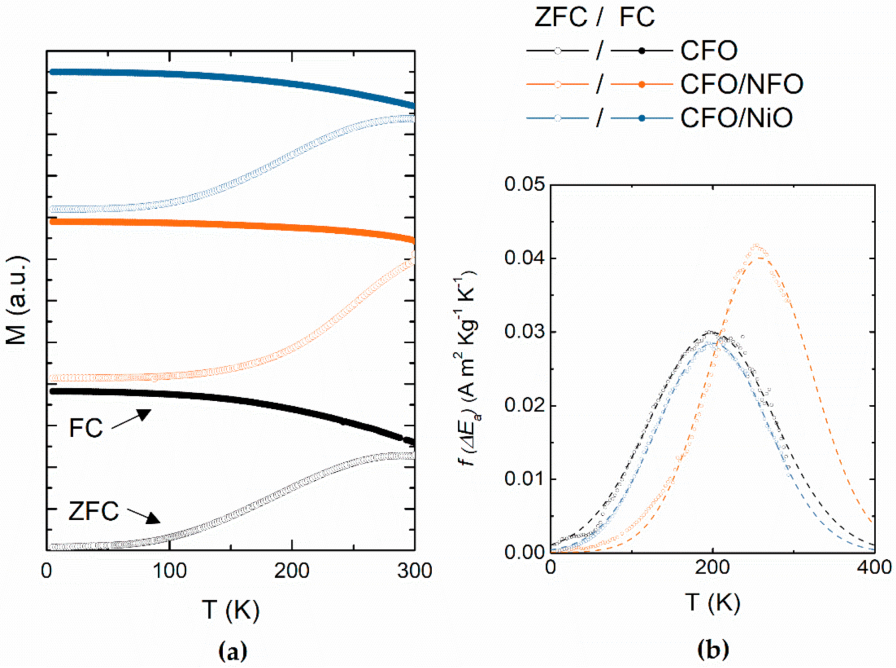
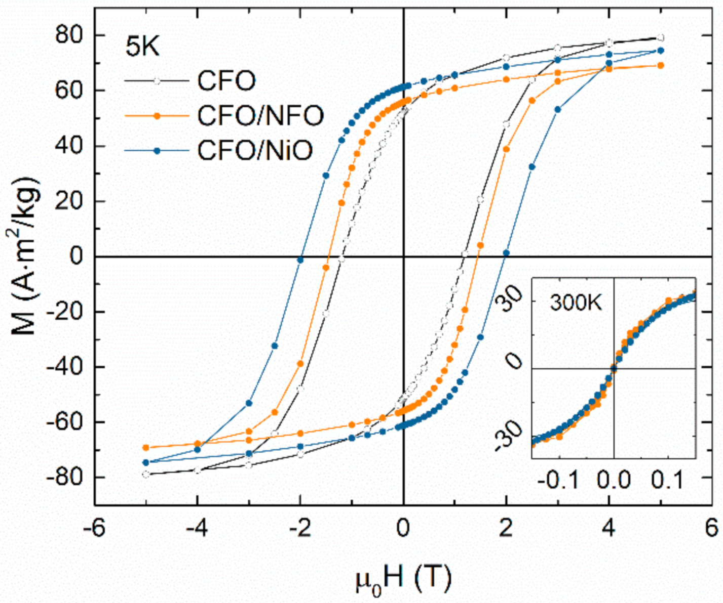
3.2. Magnetic Anisotropy

The temperature dependence of magnetization, which was investigated by the ZFC/FC protocol, shows the typical behavior of single-domain magnetic nanoparticles (Figure 2a). In all samples, MZFC and MFC show irreversibility in the entire explored temperature range. Nevertheless, the average blocking temperature <TB>, which is determined as the temperature at which the magnetic anisotropy energy distribution fEa) reaches 50% (Figure 2b), is below 300 K. To define this value, we performed an extrapolation of the experimental data with the normal function fit following the procedure explained in p.2.3. In the literature, the log-normal distribution is mainly used to describe uncentered distributions of particle size [27]. The distribution of particle sizes is reflected in a log-normal distribution of the magnetic anisotropy energy barriers fEa)~KV [22,25,26]. However, in our case, the best fit of fEa) was achieved with the center-symmetrical normal distribution (Figure 2b). This may be attributed to the specific particle growth process. Fitting the particle size distribution with both Equations (2) and (3) provided similar results. Furthermore, fEa) is proportional to the particle volume but not to the diameter [28]. The volumetric distribution is three-times higher, thus it is expected σMAG = 3σTEM, where σMAG is the standard deviation of the magnetic anisotropy distribution fEa) [21]. The average value of the blocking temperature <TB> was found to be ~200 K for the CFO and CFO/NiO samples, while it is significantly higher than the value of ~260K for the CFO/NFO samples (Table 2).
Considering the Stoner–Wohlfarth model, the temperature dependence of coercivity can be expressed as follows: H C = ξ   2 K / M S 1 T / T B 1 / 2 , where ξ is a coefficient, depending on the type of magnetic anisotropy and distribution of anisotropy axes (for an assembly of non-interacting randomly distributed magnetic particles with a dominating uniaxial anisotropy ξ = 0.48) [23]. Thus, at temperatures above TB, all samples exhibit non-hysteretic behavior confirming the superparamagnetic state at 300 K (inset of Figure 3). M-H hysteresis loops recorded at 5 K show a typical shape for h-F(i)M magnetic material with a high value of both the coercivity (µ0HC) and the reduced remanent magnetization (MR/MS) for all samples (Figure 3 and Table 2). The one-step hysteresis loop confirms the rigid exchange coupling between the core and the shell phases. The values of saturation magnetization (MS) at 5 K extrapolated using Equation (4) are listed in Table 2. The highest value of MS was found in the CFO sample (~84 Am2/kg) while the value of MS was slightly reduced for both core/shell samples. Overall, the magnetic properties, in terms of MS, µ0HC and <TB>, that were observed for the CFO single-phase system are close to those previously reported for an ~8-nm [29] and ~7.5-nm [30] CoFe2O4 NPs prepared using the high-temperature decomposition method.
Notably, both core/shell samples showed increased µ0HC values, i.e., 1.5 and 2.0 T for CFO/NFO and CFO/NiO, respectively, compared to the CFO sample (µ0HC = 1.2 T). An enhancement of the coercive field in exchange-coupled nanostructures, consisting of NiO AFM with h-FM CoPt [8] or h-FiM CoFe2O4 [31] counterparts, had already been observed and was attributed to the interface exchange interaction. Moon et al. showed an increase of the anisotropy of h-FiM CoFe2O4 covered with the ultrathin NiFe2O4, MnFe2O4 and Fe3O4 s-FiM shells with a shell volume fraction (fshell = Vshell/Vtotal) lower than 0.25 [14]. This effect was attributed to an enhanced spin canting (ESC) regime of the surface. Indeed, the cobalt ions with the high anisotropy are found to have been less subjected to the canting, while in softer ferrites the spin canting may be stronger [32,33]. According to the literature, the surface and interface terms contribute to the uniaxial magnetic anisotropy of magnetic NPs [34,35,36]. In this case, the surface/interface effects in both core/shell systems demonstrated a tendency towards cubic anisotropy with MR/MS value of around 0.75, which is close to the theoretical value of ~0.8, while in the case of the uniaxial anisotropy, the lower value of ~0.5 is expected [20].
A typical fingerprint of exchange-coupled AFM/F(i)M systems with the Néel temperature (TN) of an AFM phase below that of the Curie (Néel) temperature (TC) of an F(i)M phase is the horizontal shift of the hysteresis cycle (i.e., Exchange Bias) that may be observed under specific conditions [37,38]. Indeed, in a typical experiment to induce a preferential orientation of interface anisotropy, the material is cooled in the presence of a saturating magnetic field from a temperature of above TN to lower temperatures. Indeed, in the TN < T < TC temperature range, the AFM phase is in the paramagnetic state while F(i)M spins are aligned along the direction of the external magnetic field; according to the classical phenomenological description of the Exchange Bias phenomena [6], when cooling down to T < TN, the AFM interface spins become aligned due to the exchange interaction with the F(i)M spins. At low temperatures, interfacial spins are pinned along the field cooling direction and act as a torque, thereby inducing a preferential direction to the magnetization of the F(i)M phase with a resulting horizontal shift of the hysteresis loop. In the case of the NiO AFM phase, although the TN is relatively high (525 K), the loop shift can often be observed even after cooling from a temperature in the range of 300–380 K [8,32,39,40]. This result is explained as an effect of the disordered spins anchored at the interface as follows: since those spins are frustrated, their freezing occurs within a large temperature range which depends on the distribution of the anisotropy energy barrier; such a distribution depends on the local surroundings and the resulting freezing temperature can be significantly lower than the bulk TN [38]. Nevertheless, in our systems, M-H cycles measured at 5 K after cooling from room temperature in a magnetic field of 3 T do not show any exchange bias shift (data are not presented). This can be explained as a result of an extremely thin AFM shell (tshell~0.6 nm) that is not able to maintain the antiferromagnetic order, as well as due to a possible divergence in the stoichiometry of the shell material [13]. However, the core/shell exchange coupling of CFO/NiO nanoparticles leads to an increase of the effective anisotropy at 5 K.

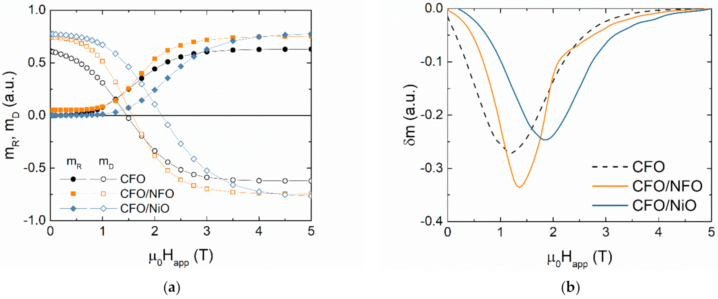
3.3. Intra- and Interparticle Interactions

The effect of interparticle interactions was studied via a remanence plot technique by collecting the values of the remanent magnetization versus the external applied magnetic field µ0Happ obtained from two different initial states of the sample, i.e.,: (i) being demagnetized (mR) and (ii) being saturated in the negative field (mD) [41,42]. The mR (Happ) curve was obtained beginning with a demagnetized sample by applying a positive field µ0Happ, which was then removed, and the remanence mR(Happ) was measured; the process was repeated after increasing the field of µ0Happ up to +5 T (Figure 4a). Similarly, the mD(Happ) curve was measured by saturating the sample in a field of +5 T and then measuring the remanence mD(Happ) after applying a gradually increasing reverse field of µ0Happ up to −5 T.
For a non-interacting assembly of uniaxial single-domain magnetic particles, mR(Happ) and mD(Happ) are connected according to the Wohlfarth model [43]. To quantify the level of interparticle interactions, the Kelly plot was used [44]:
δm = mD − (1 − 2·mR)
where the peak value of the δm(Happ) function can be considered as a quantitative parameter of interparticle interactions (δm = 0 for the ideal, non-interacting case). This method was extended to the case of particles characterized by a cubic anisotropy [42].
A negative value of δm indicates the dominant role of dipolar interactions among particles (Figure 4b). The higher magnitude of |δm| observed in CFO/NFO NPs may be attributed to the greater size of the particles compared to the CFO seeds. For CFO/NiO NPs the value of |δm| was the lowest among all samples because the AFM shell prevents the interactions among the FiM cores, by increasing the average core-to-core distance l (dipolar field ∝ MS2V2l−3), in agreement with the previously observed decrease of |δm| in the nanocomposite of the CoFe2O4 and NiO NPs [31].
To further disclose the effect of the thin shell on the magnetic behavior of the NPs, the “magnetic size” of the particles was studied using three different approaches:
  • Activation volume through magnetic viscosity coefficient [12,45,46];
  • Fit of superparamagnetic M-H curve with Langevin–Chantrell method [47];
  • Fit of superparamagnetic M-H curve with numerical inversion method [48,49].
The magnetic viscosity coefficient was found through the following relaxation measurements [12,32,46]: the sample was cooled down to 5 K; the sample was saturated in the magnetic field of −5 T; a positive field (µ0Happ) was applied and magnetization as a function of time t was recorded during 120 min. In this way, a set of M(t) curves was obtained at different µ0Happ around the coercivity field of the irreversible component of magnetization mD0HCR) [26,50]. The µ0HCR values were defined as fields at which mD(Happ) crosses zero and were found to be ~1.5, 1.6 and 2.1 T for CFO, CFO/NFO and CFO/NiO, respectively (the same fields corresponding to maximum position of dmD/dHapp). A logarithmic character for M(t) was also observed (Figure 5a):
M t = c o n s t   ±   S ln t t 0 ,
where S is the magnetic viscosity coefficient and t0 is the reference time. The descending slope of M(t) changes to ascending when the µ0Happ reaches the value ~µ0HCR (Figure 5b).
According to the Néel theory [51], the fluctuation field (µ0Hf) is defined as µ0Hf = Sirr, where χirr is the irreversibility susceptibility, defined as a peak value of the first derivative MS × (dmD/dHapp). This field describes the effect of the thermal fluctuations on magnetization reversal processes, leading to an equilibrium thermodynamic state. Based on this concept, the magnetic activation volume (Vact) was defined as a volume of magnetic material involved in the process of overcoming the energy barrier for magnetization reversal. For single-domain particles with uniaxial anisotropy, Vact is:
V a c t = k B T χ i r r M S S ,
where kB is the Boltzmann constant. In the case of the dominating cubic anisotropy, the magnetic particles have three easy axes and thus the energy barrier separating the minimums of the potential energy of magnetization states is lower than in the case of uniaxial anisotropy. It was shown in the first approximation that Vactcubic = 4 × Vactuniaxial [46]. Following this procedure and considering the dominant cubic anisotropy, the activation volumes were found to be comparable to the expected values for all the samples; the magnetic volume of the seeds of CFO and CFO/NiO was equal within the experimental error, while it was larger for the CFO/NFO system. As shown in Table 3, Vact was converted into a “magnetic size” dmagI accounting for the spherical shape of particles. For a weakly interacting particles system, the magnetic size is expected to be equal to the physical size of the particles as estimated by the TEM analysis.
Additionally, a determination of the magnetic size was performed by the fit of the superparamagnetic M-H curves with a Langevin function [52]:
M H M S b = coth V M S b H k B T k B T V M S b H ,
where MSb is the saturation magnetization of bulk material. In the Langevin–Chantrell method [47], V is considered to follow a log-normal distribution (Equation (3) with the appropriate replacement of size to volume or magnetic moment variables). The implementation of the fit procedure using Equation (10), with the substituted variable of the particle volume to its distribution, provides the magnetic size dmagII with its standard deviation σmagII of the log-normal distribution. The fitted M-H curves at 300 K are presented in Figure 6.
An approach proposed to fit M-H is based on the numerical inversion method using the MINORIM software [49]. This method is also based on the fit of the M-H superparamagnetic curve with Equation (10) but does not assume a particular shape of the particle size distribution. To perform the fit for the CFO/NFO sample, an averaged value of MSb for both CoFe2O4 and NiFe2O4, with regard to their content, has been considered. In the case of the fitting of the magnetization curve of the CFO/NiO sample, the value of MSb of the bulk CoFe2O4 was obtained, considering that only the core contributes to the magnetization (Table 3).
By comparing the results from three different methods for the estimation of the magnetic size, a qualitative agreement among the three methods can be observed. The magnetic size increases when the h-FM CFO seeds are covered with the s-FiM NFO shell, while it remains the same in the case of an AFM NiO shell. Moreover, the magnetic size determined through magnetic viscosity was in better quantitative agreement with the result obtained from the numerical inversion method. This fact can be attributed to the non-log-normal distribution of the magnetic properties of the investigated samples. All the magnetic sizes are slightly lower than the physical particle sizes, which can be related to the following surface effects: disordered spins on the surface form a non-magnetic shell with a thickness in the range of 0.5–1 nm or some internal spin canting.

4. Conclusions

A two-step seed-mediated thermal decomposition strategy was employed to fabricate bi-magnetic core/shell CFO/NFO and CFO/NiO NPs. The deposition of an ultrathin layer of ~0.6 nm NFO (s-FiM) and NiO (AFM) on ~8 nm CFO (h-FiM) seed particles strongly affected the magnetic reversal process of core/shell NPs. Both of the shells increased in their magnetic anisotropy and prevented reversible processes (residual thermal fluctuations) at low temperatures. The coercive field increased from ~1.2 T (CFO sample) to ~1.5 T for CFO/NFO and to ~2.0 T for CFO/NiO. The increased anisotropy results from the interface exchange interaction between the FiM core and AFM shell (CFO/NiO) or the disordered spins in the ESC regime (CFO/NFO). Nevertheless, the MR/MS ratio increases from ~0.61 for CFO to ~0.75 for core/shell samples, suggesting that the cubic-type magnetocrystalline anisotropy of the cobalt ferrite core is stabilized by the presence of the thin shell.
A more accurate description of the magnetic structure of synthesized particles was obtained via an analysis of the magnetic size of the particles, determined by three alternative approaches (i.e., magnetic viscosity, Langevin–Chantrell and numerical inversion methods). The magnetic size of the CFO seeds was slightly lower than the physical size, suggesting the presence of canted spins on the surface. In the CFO/NiO sample, the magnetic size was unaltered while the physical diameter increased (Figure 7). Thus, the AFM material on the particle’s surface is not involved in the magnetization reversal processes, but, due to the strong exchange coupling, it strengthens the anisotropy and stabilizes the spins of the core against the thermal fluctuations. The increase in the magnetic size of the CFO/NFO sample confirms the coupling of the FiM shell spins with the core, while the shell affects the ESC regime, and increases the effective magnetic anisotropy. The s-FiM material of the NiFe2O4 shell is more affected by the magnetic frustration than the effect of the frustration in the h-FiM material of the core. Co2+ ions with stronger single-ion anisotropy are less subjected to the canting than Fe3+ and Ni2+ with the lower anisotropy energy [32].
Interestingly, despite the very low thickness, the presence of different shells can affect not only single-particle behavior but also has a notable effect on the interparticle interaction strength. Indeed, the s-FiM shell increases the negative value of δm, suggesting a higher impact of the dipolar interactions, while the NiO shell prevents such interactions. The <TB> of the CFO/NiO sample was approximately equal to that of the CFO; however, given that it is proportional to the anisotropy barrier (<TB> ∝ KV), and considering that both systems have the same magnetic size, one expects the higher <TB> value for the sample with the higher anisotropy. This can be attributed to the significant effect of temperature on the interface component of magnetic anisotropy as well as the effect of interparticle interactions on the anisotropy energy barrier.

Author Contributions

Conceptualization, A.O. and D.P.; methodology, S.V., G.S., D.P., F.C., V.R.; validation, D.P., S.L. and F.C.; formal analysis, D.P., S.L. and F.C.; investigation, A.O., S.V., G.S. and D.P.; resources, D.P. and F.C.; data curation, D.P. and A.O.; writing—original draft preparation, A.O.; writing—review and editing, D.P., S.L. and G.S.; visualization, A.O.; supervision, D.P., F.C. and V.R.; project administration, D.P.; funding acquisition, D.P. All authors have read and agreed to the published version of the manuscript.

Funding

This work was partially supported by the European Union’s Horizon 2020 Research and Innovation Programme: under grant agreement No. 731976 (MAGENTA). A.O., V.R. and D.P. gratefully acknowledge the financial support of the Russian Science Foundation grant No. 21-72-30032 (magnetic measurements).

Conflicts of Interest

The authors declare no conflict of interest.

References

  1. Lee, J.-H.; Jang, J.; Choi, J.; Moon, S.H.; Noh, S.; Kim, J.; Kim, J.-G.; Kim, I.-S.; Park, K.I.; Cheon, J. Exchange-coupled magnetic nanoparticles for efficient heat induction. Nat. Nanotechnol. 2011, 6, 418–422. [Google Scholar] [CrossRef] [PubMed]
  2. Noh, S.; Moon, S.H.; Shin, T.-H.; Lim, Y.; Cheon, J. Recent advances of magneto-thermal capabilities of nanoparticles: From design principles to biomedical applications. Nano Today 2017, 13, 61–76. [Google Scholar] [CrossRef]
  3. Vasilakaki, M.; Trohidou, K.N.; Nogués, J. Enhanced magnetic properties in antiferromagnetic-core/ferrimagnetic-shell nanoparticles. Sci. Rep. 2015, 5, 1–7. [Google Scholar] [CrossRef] [PubMed] [Green Version]
  4. Sanna Angotzi, M.; Mameli, V.; Cara, C.; Musinu, A.; Sangregorio, C.; Niznansky, D.; Xin, H.L.; Vejpravova, J.; Cannas, C. Coupled hard–soft spinel ferrite-based core–shell nanoarchitectures: Magnetic properties and heating abilities. Nanoscale Adv. 2020, 2, 3191–3201. [Google Scholar] [CrossRef]
  5. López-Ortega, A.; Estrader, M.; Salazar-Alvarez, G.; Roca, A.G.; Nogués, J. Applications of exchange coupled bi-magnetic hard/soft and soft/hard magnetic core/shell nanoparticles. Phys. Rep. 2015, 553, 1–32. [Google Scholar] [CrossRef] [Green Version]
  6. Nogués, J.; Schuller, I.K. Exchange bias. J. Magn. Magn. Mater. 1999, 192, 203–232. [Google Scholar] [CrossRef]
  7. Lottini, E.; López-Ortega, A.; Bertoni, G.; Turner, S.; Meledina, M.; Van Tendeloo, G.; De Julián Fernández, C.; Sangregorio, C. Strongly Exchange Coupled Core|Shell Nanoparticles with High Magnetic Anisotropy: A Strategy toward Rare-Earth-Free Permanent Magnets. Chem. Mater. 2016, 28, 4214–4222. [Google Scholar] [CrossRef]
  8. Laureti, S.; Del Bianco, L.; Detlefs, B.; Agostinelli, E.; Foglietti, V.; Peddis, D.; Testa, A.M.; Varvaro, G.; Fiorani, D. Interface exchange coupling in a CoPt/NiO bilayer. Thin Solid Films 2013, 543, 162–166. [Google Scholar] [CrossRef]
  9. Meiklejohn, W.H.; Bean, C.P. New magnetic anisotropy. Phys. Rev. 1957, 105, 904–913. [Google Scholar] [CrossRef]
  10. Yang, M.; Ho, C.; Ruta, S.; Chantrell, R.; Krycka, K.; Hovorka, O.; Chen, F.; Lai, P.; Lai, C. Magnetic Interaction of Multifunctional Core–Shell Nanoparticles for Highly Effective Theranostics. Adv. Mater. 2018, 30, 1802444. [Google Scholar] [CrossRef] [Green Version]
  11. Socoliuc, V.; Peddis, D.; Petrenko, V.I.; Avdeev, M.V.; Susan-Resiga, D.; Szabó, T.; Turcu, R.; Tombácz, E.; Vékás, L. Magnetic Nanoparticle Systems for Nanomedicine—A Materials Science Perspective. Magetochemistry 2019, 6, 2. [Google Scholar] [CrossRef] [Green Version]
  12. Fabris, F.; Lohr, J.; Lima, E.; de Almeida, A.A.; Troiani, H.E.; Rodríguez, L.M.; Vásquez Mansilla, M.; Aguirre, M.H.; Goya, G.F.; Rinaldi, D.; et al. Adjusting the Néel relaxation time of Fe3O4/ZnxCo1−xFe2O4 core/shell nanoparticles for optimal heat generation in magnetic hyperthermia. Nanotechnology 2021, 32, 065703. [Google Scholar] [CrossRef] [PubMed]
  13. Estrader, M.; López-Ortega, A.; Golosovsky, I.V.; Estradé, S.; Roca, A.G.; Salazar-Alvarez, G.; López-Conesa, L.; Tobia, D.; Winkler, E.; Ardisson, J.D.; et al. Origin of the large dispersion of magnetic properties in nanostructured oxides: FexO/Fe3O4 nanoparticles as a case study. Nanoscale 2015, 7, 3002–3015. [Google Scholar] [CrossRef] [PubMed] [Green Version]
  14. Moon, S.H.; Noh, S.-H.; Lee, J.-H.; Shin, T.-H.; Lim, Y.; Cheon, J. Ultrathin Interface Regime of Core–Shell Magnetic Nanoparticles for Effective Magnetism Tailoring. Nano Lett. 2017, 17, 800–804. [Google Scholar] [CrossRef]
  15. Muzzi, B.; Albino, M.; Innocenti, C.; Petrecca, M.; Cortigiani, B.; de Julián Fernández, C.; Bertoni, G.; Fernández-Pacheco, R.; Ibarra, A.; Marquina, C.; et al. Unraveling the mechanism of the one-pot synthesis of exchange coupled Co-based nano-heterostructures with high energy product. Nanoscale 2020, 12, 14076–14086. [Google Scholar] [CrossRef]
  16. Quesada, A.; Granados-Miralles, C.; López-Ortega, A.; Erokhin, S.; Lottini, E.; Pedrosa, J.; Bollero, A.; Aragón, A.M.; Rubio-Marcos, F.; Stingaciu, M.; et al. Energy Product Enhancement in Imperfectly Exchange-Coupled Nanocomposite Magnets. Adv. Electron. Mater. 2016, 2, 1500365. [Google Scholar] [CrossRef]
  17. Sun, S.; Zeng, H.; Robinson, D.B.; Raoux, S.; Rice, P.M.; Wang, S.X.; Li, G. Monodisperse MFe2O4 (M=Fe, Co, Mn) Nanoparticles. J. Am. Chem. Soc. 2004, 126, 273–279. [Google Scholar] [CrossRef]
  18. Omelyanchik, A.; Villa, S.; Vasilakaki, M.; Singh, G.; Ferretti, A.M.; Ponti, A.; Canepa, F.; Margaris, G.; Trohidou, K.N.; Peddis, D. Interplay between inter- and intraparticle interactions in bi-magnetic core/shell nanoparticles. Nanoscale Adv. 2021. [Google Scholar] [CrossRef]
  19. Muscas, G.; Singh, G.; Glomm, W.; Mathieu, R.; Kumar, P.A.; Concas, G.; Agostinelli, E.; Peddis, D. Tuning the Size and Shape of Oxide Nanoparticles by Controlling Oxygen Content in the Reaction Environment: Morphological Analysis by Aspect Maps. Chem. Mater. 2015, 27, 1982–1990. [Google Scholar] [CrossRef]
  20. Bertotti, G. Hysteresis in Magnetism; Academic Press: Cambridge, MA, USA, 1998; pp. 3–30. [Google Scholar]
  21. Concas, G.; Congiu, F.; Muscas, G.; Peddis, D. Determination of Blocking Temperature in Magnetization and Mössbauer Time Scale: A Functional Form Approach. J. Phys. Chem. C 2017, 121, 16541–16548. [Google Scholar] [CrossRef]
  22. Micha, J.; Dieny, B.; Régnard, J.; Jacquot, J.; Sort, J. Estimation of the Co nanoparticles size by magnetic measurements in Co/SiO2 discontinuous multilayers. J. Magn. Magn. Mater. 2004, 272–276, E967–E968. [Google Scholar] [CrossRef]
  23. Knobel, M.; Nunes, W.C.; Socolovsky, L.M.; De Biasi, E.; Vargas, J.M.; DeNardin, J.C. Superparamagnetism and Other Magnetic Features in Granular Materials: A Review on Ideal and Real Systems. J. Nanosci. Nanotechnol. 2008, 8, 2836–2857. [Google Scholar] [CrossRef] [PubMed]
  24. Livesey, K.L.; Ruta, S.; Anderson, N.R.; Baldomir, D.; Chantrell, R.W.; Serantes, D. Beyond the blocking model to fit nanoparticle ZFC/FC magnetisation curves. Sci. Rep. 2018, 8, 1–9. [Google Scholar] [CrossRef] [Green Version]
  25. Bruvera, I.J.; Zélis, P.M.; Calatayud, M.P.; Goya, G.F.; Sánchez, F.H. Determination of the blocking temperature of magnetic nanoparticles: The good, the bad, and the ugly. J. Appl. Phys. 2015, 118, 184304. [Google Scholar] [CrossRef]
  26. Omelyanchik, A.; Salvador, M.; D’orazio, F.; Mameli, V.; Cannas, C.; Fiorani, D.; Musinu, A.; Rivas, M.; Rodionova, V.; Varvaro, G.; et al. Magnetocrystalline and surface anisotropy in CoFe2O4 nanoparticles. Nanomaterials 2020, 10, 1288. [Google Scholar] [CrossRef]
  27. Peddis, D.; Orrù, F.; Ardu, A.; Cannas, C.; Musinu, A.; Piccaluga, G. Interparticle Interactions and Magnetic Anisotropy in Cobalt Ferrite Nanoparticles: Influence of Molecular Coating. Chem. Mater. 2012, 24, 1062–1071. [Google Scholar] [CrossRef]
  28. El-Hilo, M.; Chantrell, R. Rationalisation of distribution functions for models of nanoparticle magnetism. J. Magn. Magn. Mater. 2012, 324, 2593–2595. [Google Scholar] [CrossRef]
  29. Muscas, G.; Cobianchi, M.; Lascialfari, A.; Cannas, C.; Musinu, A.; Omelyanchik, A.; Rodionova, V.; Fiorani, D.; Mameli, V.; Peddis, D. Magnetic Interactions Versus Magnetic Anisotropy in Spinel Ferrite Nanoparticles. IEEE Magn. Lett. 2019, 10, 1–5. [Google Scholar] [CrossRef]
  30. Mameli, V.; Musinu, A.; Ardu, A.; Ennas, G.; Peddis, D.; Niznansky, D.; Sangregorio, C.; Innocenti, C.; Thanh, N.T.K.; Cannas, C. Studying the effect of Zn-substitution on the magnetic and hyperthermic properties of cobalt ferrite nanoparticles. Nanoscale 2016, 8, 10124–10137. [Google Scholar] [CrossRef] [PubMed] [Green Version]
  31. Peddis, D.; Laureti, S.; Mansilla, M.; Agostinelli, E.; Varvaro, G.; Cannas, C.; Fiorani, D. Exchange Bias in CoFe2O4/NiO nanocomposites. Superlattices Microstruct. 2009, 46, 125–129. [Google Scholar] [CrossRef]
  32. Peddis, D.; Cannas, C.; Piccaluga, G.; Agostinelli, E.; Fiorani, D. Spin-glass-like freezing and enhanced magnetization in ultra-small CoFe2O4 nanoparticles. Nanotechnology 2010, 21, 125705. [Google Scholar] [CrossRef]
  33. Kodama, R.H.; Berkowitz, A.E.; McNiff, E.J.; Foner, S. Surface spin disorder in NiFe2O4 nanoparticles. Phys. Rev. Lett. 1996, 77, 394–397. [Google Scholar] [CrossRef]
  34. Oyarzún, S.; Tamion, A.; Tournus, F.; Dupuis, V.; Hillenkamp, M. Size effects in the magnetic anisotropy of embedded cobalt nanoparticles: From shape to surface. Sci. Rep. 2015, 5, 14749. [Google Scholar] [CrossRef] [Green Version]
  35. Respaud, M.; Broto, J.M.; Rakoto, H.; Fert, A.R.; Thomas, L.; Barbara, B.; Verelst, M.; Snoeck, E.; Lecante, P.; Mosset, A.; et al. Surface effects on the magnetic properties of ultrafine cobalt particles. Phys. Rev. B 1998, 57, 2925–2935. [Google Scholar] [CrossRef]
  36. Cabreira Gomes, R.; G da Silva, F.; Silva, T.-Q.; Gomide, G.; Pilati, V.; Aquino, R.; Geshev, J.; Perzynski, R.; Depeyrot, J. Magnetic irreversibility and saturation criteria in ultrasmall bi-magnetic nanoparticles. J. Alloys Compd. 2020, 824, 153646. [Google Scholar] [CrossRef]
  37. Nogués, J.; Sort, J.; Langlais, V.; Skumryev, V.; Suriñach, S.; Muñoz, J.S.; Baró, M.D. Exchange bias in nanostructures. Phys. Rep. 2005, 422, 65–117. [Google Scholar] [CrossRef]
  38. Omelianchik, A.; Singh, G.; McDonagh, B.H.; Rodionova, V.; Fiorani, D.; Peddis, D.; Laureti, S.; Omelyanchik, A. From Mn3O4/MnO core–shell nanoparticles to hollow MnO: Evolution of magnetic properties. Nanotechnology 2017, 29, 055703. [Google Scholar] [CrossRef] [PubMed]
  39. Dai, J.; Feng, W.; Cheng, C.; Wen, X. The exchange bias effects of CoFe2O4@NiO nanofibers fabricated by electrospinning. Mater. Res. Express 2020, 7, 045009. [Google Scholar] [CrossRef]
  40. Mohan, R.; Ghosh, M.; Mukherjee, S. The exchange bias effect in CoFe2O4/NiO nanocomposites prepared by chemical co-precipitation method. Mater. Res. Express 2019, 6, 056105. [Google Scholar] [CrossRef]
  41. Chantrell, R.W.; O’Grady, K.; Search, H.; Journals, C.; Contact, A.; Iopscience, M.; Address, I.P. Magnetic characterization of recording media. J. Phys. D. Appl. Phys. 1992, 25, 1–23. [Google Scholar] [CrossRef]
  42. García-Otero, J.; Porto, M.; Rivas, J. Henkel plots of single-domain ferromagnetic particles. J. Appl. Phys. 2000, 87, 7376. [Google Scholar] [CrossRef] [Green Version]
  43. Wohlfarth, E.P. Relations between Different Modes of Acquisition of the Remanent Magnetization of Ferromagnetic Particles. J. Appl. Phys. 1958, 29, 595–596. [Google Scholar] [CrossRef]
  44. Kelly, P.E.; O’Grady, K.; Mayo, P.I. Switching mechanisms in cobalt phosphorus thin films. In Proceedings of the International Magnetics Conference; IEEE: New York, NY, USA, 1989; Volume 25, p. HA5. [Google Scholar]
  45. Wohlfarth, E.P. The coefficient of magnetic viscosity. J. Phys. F Met. Phys. 1984, 14, L155–L159. [Google Scholar] [CrossRef]
  46. Laureti, S.; Varvaro, G.; Testa, A.M.; Fiorani, D.; Agostinelli, E.; Piccaluga, G.; Musinu, A.; Ardu, A.; Peddis, D. Magnetic interactions in silica coated nanoporous assemblies of CoFe2O4 nanoparticles with cubic magnetic anisotropy. Nanotechnology 2010, 21, 315701. [Google Scholar] [CrossRef] [PubMed]
  47. Chantrell, R.; Popplewell, J.; Charles, S. Measurements of particle size distribution parameters in ferrofluids. IEEE Trans. Magn. 1978, 14, 975–977. [Google Scholar] [CrossRef]
  48. Bender, P.; Balceris, C.; Ludwig, F.; Posth, O.; Bogart, L.K.; Szczerba, W.; Castro, A.; Nilsson, L.; Costo, R.; Gavilán, H.; et al. Distribution functions of magnetic nanoparticles determined by a numerical inversion method. New J. Phys. 2017, 19, 073012. [Google Scholar] [CrossRef] [Green Version]
  49. van Rijssel, J.; Kuipers, B.W.; Erné, B.H. Non-regularized inversion method from light scattering applied to ferrofluid magnetization curves for magnetic size distribution analysis. J. Magn. Magn. Mater. 2014, 353, 110–115. [Google Scholar] [CrossRef]
  50. Huerta, J.M.M.; Medina, J.D.L.T.; Piraux, L.; Encinas, A. Self consistent measurement and removal of the dipolar interaction field in magnetic particle assemblies and the determination of their intrinsic switching field distribution. J. Appl. Phys. 2012, 111, 083914. [Google Scholar] [CrossRef] [Green Version]
  51. Néel, L. Théorie du traînage magnétique des substances massives dans le domaine de Rayleigh. J. Phys. Le Radium 1950, 11, 49–61. [Google Scholar] [CrossRef]
  52. Sandler, S.; Fellows, B.D.; Mefford, O.T. Best Practices for Characterization of Magnetic Nanoparticles for Biomedical Applications. Anal. Chem. 2019, 91, 14159–14169. [Google Scholar] [CrossRef] [Green Version]
Figure 1. (a) XRD patterns and (b) STEM images for CFO seeds and core/shell systems; (c) diameter distribution obtained from STEM images.
Magnetochemistry 07 00146 g001
Figure 2. (a) Zero-field cooling and field cooling (ZFC/FC) magnetizations recorded in 2.5 mT measuring field; (b) distribution of magnetic anisotropy energy: measured (dots) and extrapolated by fit with the normal function (dashed lines).
Magnetochemistry 07 00146 g002
Figure 3. M-H hysteresis loop at 5 K for all samples. Inset shows low field region of M-H loops at 300 K.
Magnetochemistry 07 00146 g003
Figure 4. (a) mR and mD remanent magnetizations and (b) δm-plots for CFO, CFO/NFO and CFO/NiO systems at 5 K.
Magnetochemistry 07 00146 g004
Figure 5. (a) normalized M(t) measured at different values of µ0Happ after saturation at −5 T for CFO/NFO sample; (b) magnetic viscosity S as a function of the applied field µ0Happ for all samples.
Magnetochemistry 07 00146 g005
Figure 6. Measured and fitted with the Langevin–Chantrell method M-H cycles at 300 K.
Magnetochemistry 07 00146 g006
Figure 7. Schematic representation of the magnetic structure of CFO/NFO and CFO/NiO core/shell samples.
Magnetochemistry 07 00146 g007
Table 1. Morpho-structural properties of samples: crystallite size (dXRD), particle size (dTEM) and standard deviation of the particle size distribution (σTEM) after fit with normal and log-normal functions.
SampledXRD, nmNormalLog-Normal
dTEM, nmσTEMdTEM, nmσTEM
CFO7.2 ± 0.48.3 ± 0.10.25 ± 0.038.3 ± 0.10.13 ± 0.02
CFO/NFO8.9 ± 0.99.3 ± 0.10.31 ± 0.039.5 ± 0.10.15 ± 0.02
CFO/NiO7.8 ± 0.79.3 ± 0.10.38 ± 0.039.6 ± 0.20.19 ± 0.02
Error for dXRD is the standard deviation over values for all peaks; the error for dTEM and σTEM is the error of fitting with normal and log-normal functions.
Table 2. Mean value of blocking temperature (<TB>) and its dispersion (σMAG), saturation magnetization (MS), reduced remanent magnetization (MR/MS) and coercivity field (µ0HC).
Sample<TB>, KσmagMS, Am2kg−1MR/MSµ0HC, T
CFO201 ± 80.76 ± 0.0584 ± 40.61 ± 0.011.2 1
CFO/NFO259 ± 100.50 ± 0.0575 ± 40.76 ± 0.011.5 ± 0.1
CFO/NiO200 ± 80.65 ± 0.0580 ± 40.74 ± 0.012.0 ± 0.1
1 Error for this value is lower than reported significant digits.
Table 3. Physical particle size determined from TEM analysis after log-normal fit (dTEMlog), magnetic size from the magnetic viscosity (dMAGI), Langevin–Chantrell fit (dMAGII) and MINORIM software (dMAGIII), standard deviation of log-normal distribution of physical (σTEMlog) and magnetic (σMAGII) size.
SampledTEMlog, nmdMAGI, nmdMAGII, nmdMAGIII, nmσTEMlogσmagII
CFO8.3 ± 0.16.9 ± 0.25.5 ± 0.26.90.13 ± 0.020.22 ± 0.02
CFO/NFO9.5 ± 0.17.3 ± 0.26.0 ± 0.27.70.15 ± 0.020.20 ± 0.02
CFO/NiO9.6 ± 0.26.5 ± 0.55.4 ± 0.26.90.19 ± 0.020.23 ± 0.02
Publisher’s Note: MDPI stays neutral with regard to jurisdictional claims in published maps and institutional affiliations.

