Optical-Based (Bio) Sensing Systems Using Magnetic Nanoparticles


Abstract

1. Introduction
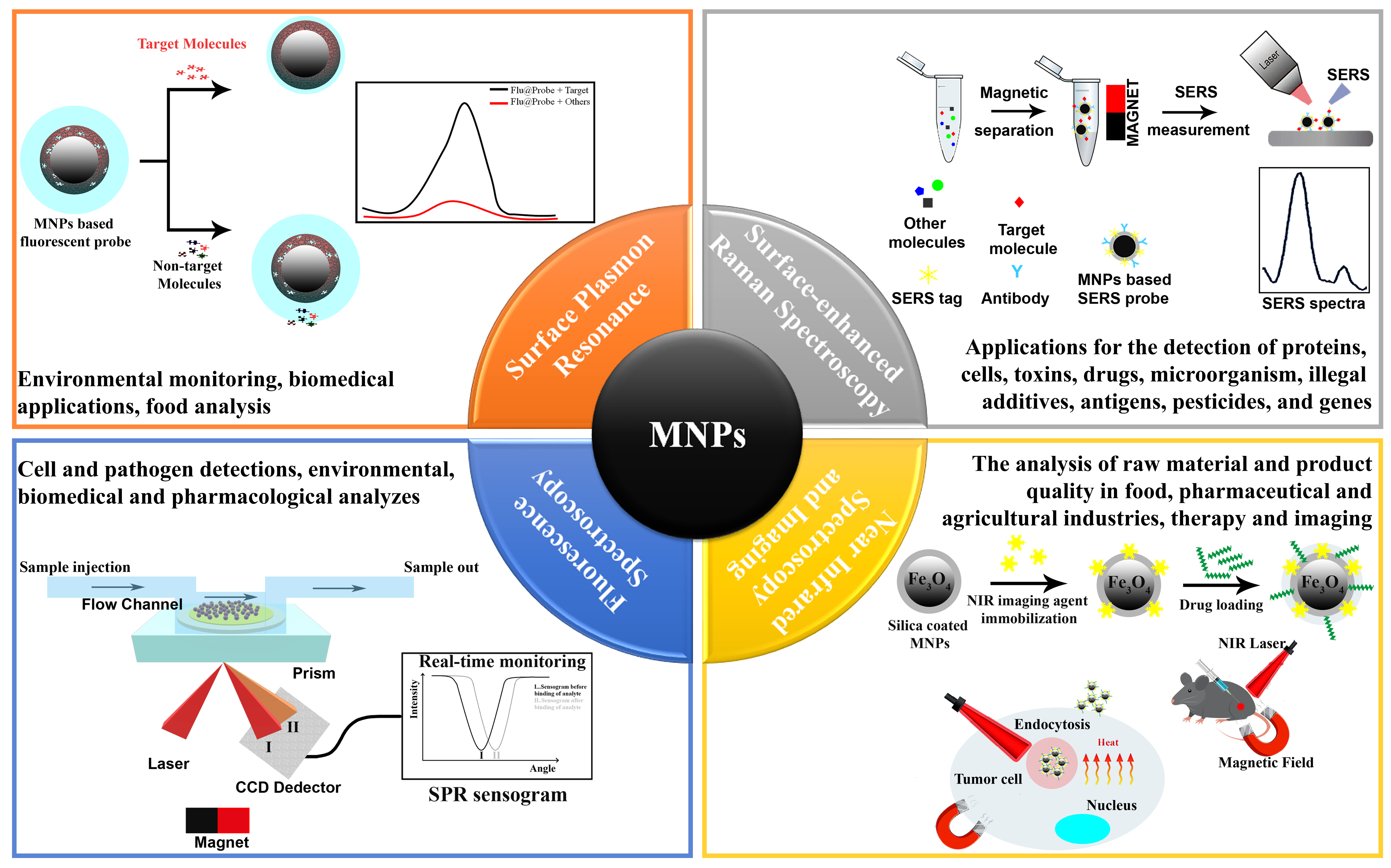
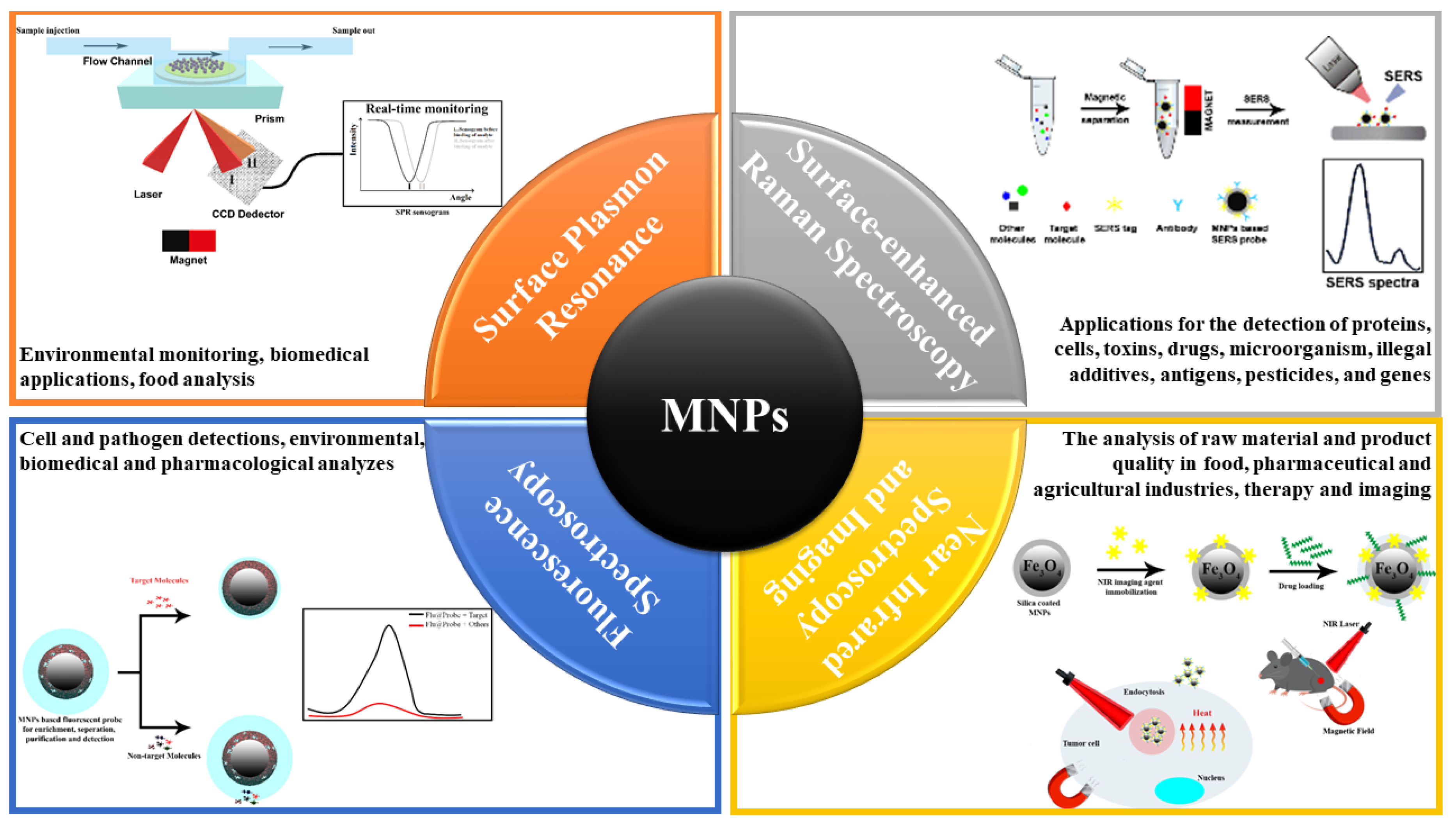
2. Surface Plasmon Resonance (SPR)
3. Surface-Enhanced Raman Spectroscopy (SERS)
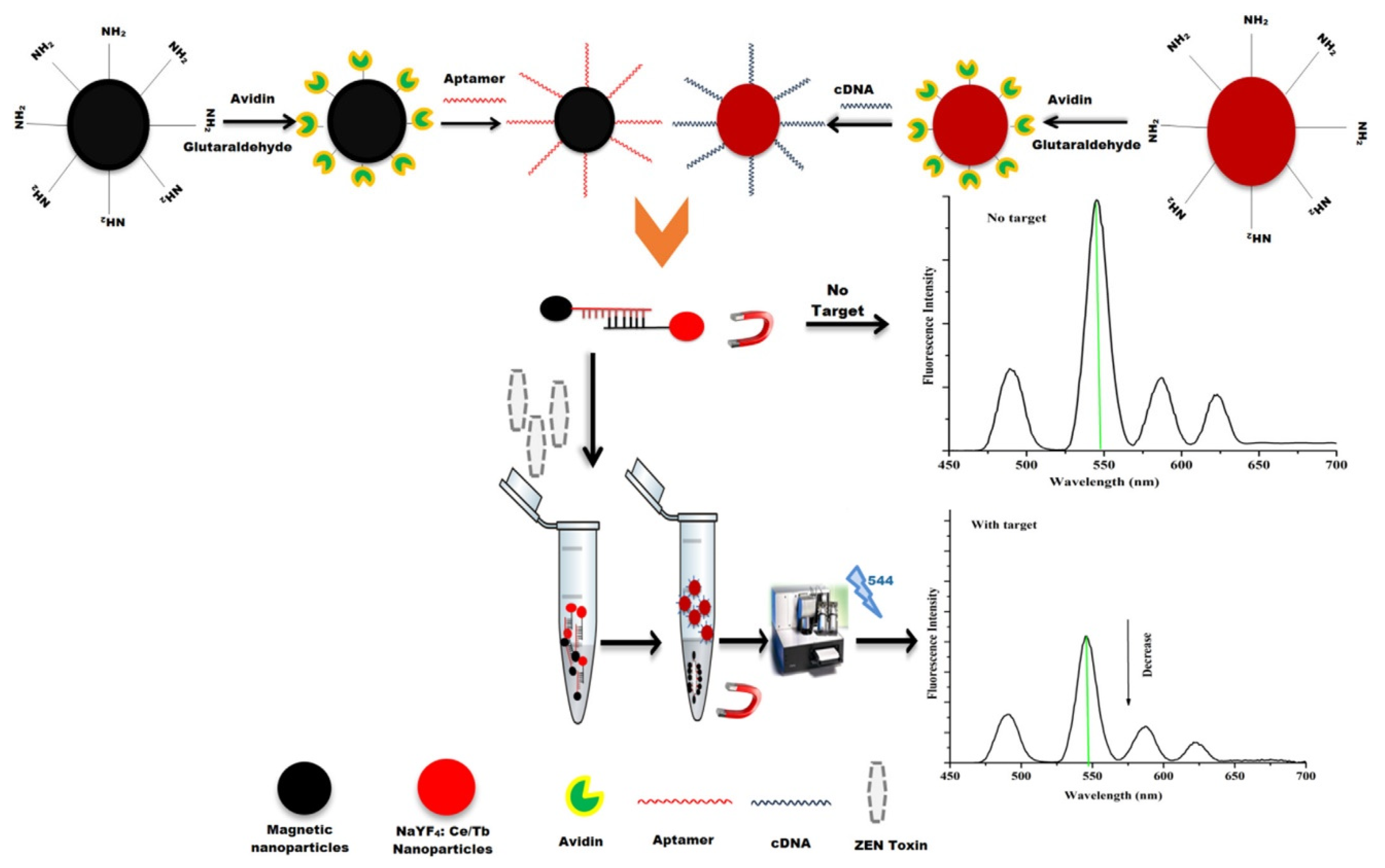
4. Fluorescence Spectroscopy
5. Near Infrared Spectroscopy (NIRS) and Imaging
6. Conclusions and Future Challenges
Author Contributions
Funding
Conflicts of Interest
References
- Koh, I.; Josephson, L. Magnetic nanoparticle sensors. Sensors (Basel) 2009, 9, 8130–8145. [Google Scholar] [CrossRef]
- Nikiforov, V.N.; Filinova, E.Y. Biomedical Applications of Magnetic Nanoparticles. In Magnetic Nanoparticles; Gubin, S.P., Ed.; Wiley-VCH Verlag GmbH & Co. KGaA: Weinheim, Germany, 2009; pp. 393–444. [Google Scholar]
- Mou, X.; Ali, Z.; Li, S.; He, N. Applications of Magnetic Nanoparticles in Targeted Drug Delivery System. J. Nanosci. Nanotechnol. 2015, 15, 54–62. [Google Scholar] [CrossRef]
- Bao, Y.; Wen, T.; Samia, A.C.; Khandhar, A.; Krishnan, K.M. Magnetic Nanoparticles: Material Engineering and Emerging Applications in Lithography and Biomedicine. J. Mater. Sci. 2016, 51, 513–553. [Google Scholar] [CrossRef]
- Rocha-Santos, T.A.P. Sensors and biosensors based on magnetic nanoparticles. Trends Anal. Chem. 2014, 62, 28–36. [Google Scholar] [CrossRef]
- Colombo, M.; Carregal-Romero, S.; Casula, M.F.; Gutierrez, L.; Morales, M.P.; Bohm, I.B.; Heverhagen, J.T.; Prosperi, D.; Parak, W.J. Biological applications of magnetic nanoparticles. Chem. Soc. Rev. 2012, 41, 4306–4334. [Google Scholar] [CrossRef] [PubMed]
- Mejías, R.; Pérez-Yagüe, S.; Roca, A.G.; Pérez, N.; Villanueva, Á.; Cañete, M.; Mañes, S.; Ruiz-Cabello, J.; Benito, M.; Labarta, A. Liver and brain imaging through dimercaptosuccinic acid-coated iron oxide nanoparticles. Nanomedicine 2010, 5, 397–408. [Google Scholar] [CrossRef] [PubMed]
- Anbarasu, M.; Anandan, M.; Chinnasamy, E.; Gopinath, V.; Balamurugan, K. Synthesis and characterization of polyethylene glycol (PEG) coated Fe3O4 nanoparticles by chemical co-precipitation method for biomedical applications. Spectrochim. Acta A Mol. Biomol. Spectrosc. 2015, 135, 536–539. [Google Scholar] [CrossRef] [PubMed]
- Figuerola, A.; Di Corato, R.; Manna, L.; Pellegrino, T. From iron oxide nanoparticles towards advanced iron-based inorganic materials designed for biomedical applications. Pharmacol. Res. 2010, 62, 126–143. [Google Scholar] [CrossRef] [PubMed]
- Li, X.; Wei, J.; Aifantis, K.E.; Fan, Y.; Feng, Q.; Cui, F.Z.; Watari, F. Current investigations into magnetic nanoparticles for biomedical applications. J. Biomed. Mater. Res. A 2016, 104, 1285–1296. [Google Scholar] [CrossRef]
- Chen, Q.; Rondinone, A.J.; Chakoumakos, B.C.; Zhang, Z.J. Synthesis of superparamagnetic MgFe2O4 nanoparticles by coprecipitation. J. Magn. Magn. Mater. 1999, 194, 1–7. [Google Scholar] [CrossRef]
- Puntes, V.F.; Krishnan, K.M.; Alivisatos, A.P. Colloidal nanocrystal shape and size control: The case of cobalt. Science 2001, 291, 2115–2117. [Google Scholar] [CrossRef] [PubMed]
- Weller, D.; Moser, A. Thermal effect limits in ultrahigh-density magnetic recording. IEEE Trans. Magn. 1999, 35, 4423–4439. [Google Scholar] [CrossRef]
- Andres, R.P.; Bein, T.; Dorogi, M.; Feng, S.; Henderson, J.I.; Kubiak, C.P.; Mahoney, W.; Osifchin, R.G.; Reifenberger, R. “Coulomb Staircase” at Room Temperature in a Self-Assembled Molecular Nanostructure. Science 1996, 272, 1323–1325. [Google Scholar] [CrossRef] [PubMed]
- Sun, S.; Murray, C.B.; Weller, D.; Folks, L.; Moser, A. Monodisperse FePt nanoparticles and ferromagnetic FePt nanocrystal superlattices. Science 2000, 287, 1989–1992. [Google Scholar] [CrossRef] [PubMed]
- Chen, X.J.; Zhu, J.W.; Chen, Z.X.; Xu, C.B.; Wang, Y.; Yao, C. A novel bienzyme glucose biosensor based on three-layer Au-Fe3O4@SiO2 magnetic nanocomposite. Sens. Actuators B Chem. 2011, 159, 220–228. [Google Scholar] [CrossRef]
- Baghayeri, M.; Nazarzadeh Zare, E.; Mansour Lakouraj, M. A simple hydrogen peroxide biosensor based on a novel electro-magnetic poly (p-phenylenediamine) @ Fe3O4 nanocomposite. Biosens. Bioelectron. 2014, 55, 259–265. [Google Scholar] [CrossRef]
- Baghayeri, M.; Veisi, H.; Ghanei-Motlagh, M. Amperometric glucose biosensor based on immobilization of glucose oxidase on a magnetic glassy carbon electrode modified with a novel magnetic nanocomposite. Sens. Actuators B Chem. 2017, 249, 321–330. [Google Scholar] [CrossRef]
- Zhao, Y.; Zhang, W.; Lin, Y.; Du, D. The vital function of Fe3O4@Au nanocomposites for hydrolase biosensor design and its application in detection of methyl parathion. Nanoscale 2013, 5, 1121–1126. [Google Scholar] [CrossRef]
- Liu, J.; Qiao, S.Z.; Hu, Q.H.; Lu, G.Q. Magnetic nanocomposites with mesoporous structures: Synthesis and applications. Small 2011, 7, 425–443. [Google Scholar] [CrossRef]
- Corma, A. From microporous to mesoporous molecular sieve materials and their use in catalysis. Chem. Rev. 1997, 97, 2373–2419. [Google Scholar] [CrossRef]
- Lu, A.H.; Schuth, F. Nanocasting: A versatile strategy for creating nanostructured porous materials. Adv. Mater. 2006, 18, 1793–1805. [Google Scholar] [CrossRef]
- Ying, J.Y.; Mehnert, C.P.; Wong, M.S. Synthesis and applications of supramolecular-templated mesoporous materials. Angew. Chem. Int. Ed. 1999, 38, 56–77. [Google Scholar] [CrossRef]
- Kudr, J.; Haddad, Y.; Richtera, L.; Heger, Z.; Cernak, M.; Adam, V.; Zitka, O. Magnetic Nanoparticles: From Design and Synthesis to Real World Applications. Nanomaterials (Basel) 2017, 7, 243. [Google Scholar] [CrossRef] [PubMed]
- Leena, M.; Gomaa, H.; Ragab, D.; Zhu, J. Magnetic nanoparticles for environmental and biomedical applications. Particuology 2017, 30, 1–14. [Google Scholar]
- Willard, M.A.; Kurihara, L.K.; Carpenter, E.E.; Calvin, S.; Harris, V.G. Chemically prepared magnetic nanoparticles. Int. Mater. Rev. 2004, 49, 125–170. [Google Scholar] [CrossRef]
- Reddy, L.H.; Arias, J.L.; Nicolas, J.; Couvreur, P. Magnetic nanoparticles: design and characterization, toxicity and biocompatibility, pharmaceutical and biomedical applications. Chem. Rev. 2012, 112, 5818–5878. [Google Scholar] [CrossRef]
- Kango, S.; Kalia, S.; Celli, A.; Njuguna, J.; Habibi, Y.; Kumar, R. Surface modification of inorganic nanoparticles for development of organic–inorganic nanocomposites—A review. Prog. Polymer Sci. 2013, 38, 1232–1261. [Google Scholar] [CrossRef]
- Mahmoudi, M.; Sant, S.; Wang, B.; Laurent, S.; Sen, T. Superparamagnetic iron oxide nanoparticles (SPIONs): development, surface modification and applications in chemotherapy. Adv. Drug Deliv. Rev. 2011, 63, 24–46. [Google Scholar] [CrossRef]
- da Silva, B.F.; Pérez, S.; Gardinalli, P.; Singhal, R.; Mozeto, A.A.; Barceló, D. Analytical chemistry of metallic nanoparticles in natural environments. Trends Anal. Chem. 2011, 30, 528–540. [Google Scholar] [CrossRef]
- Madkour, L.H. Biogenic–biosynthesis metallic nanoparticles (MNPs) for pharmacological, biomedical and environmental nanobiotechnological applications. Chron. Pharm. Sci. J. 2018, 2, 384–444. [Google Scholar]
- Coulet, P.R. What is a Biosensor. In Biosensor Principles and Applications; Coulet, P.R., Blum, L.J., Eds.; CRC Press: New York, NY, USA, 1991; pp. 1–8. [Google Scholar]
- Suvarnaphaet, P.; Pechprasarn, S. Graphene-Based Materials for Biosensors: A Review. Sensors (Basel) 2017, 17, 2161. [Google Scholar] [CrossRef] [PubMed]
- Tereshchenko, A.; Bechelany, M.; Viter, R.; Khranovskyy, V.; Smyntyna, V.; Starodub, N.; Yakimova, R. Optical biosensors based on ZnO nanostructures: advantages and perspectives. A review. Sens. Actuators B Chem. 2016, 229, 664–677. [Google Scholar] [CrossRef]
- Fan, X.; White, I.M.; Shopova, S.I.; Zhu, H.; Suter, J.D.; Sun, Y. Sensitive optical biosensors for unlabeled targets: a review. Anal. Chim. Acta 2008, 620, 8–26. [Google Scholar] [CrossRef] [PubMed]
- Wang, C.; Irudayaraj, J. Multifunctional magnetic-optical nanoparticle probes for simultaneous detection, separation, and thermal ablation of multiple pathogens. Small 2010, 6, 283–289. [Google Scholar] [CrossRef]
- Englebienne, P.; Van Hoonacker, A.; Verhas, M. Surface plasmon resonance: principles, methods and applications in biomedical sciences. Spectrosc. Int. J. 2003, 17, 255–273. [Google Scholar] [CrossRef]
- Tudos, A.J.; Schasfoort, R.B.M. Introduction to Surface Plasmon Resonance. In Handbook of Surface Plasmon Resonance; Royal Society of Chemistry: London, UK, 2008; pp. 1–14. [Google Scholar] [CrossRef]
- Van Der Merwe, P.A. Surface plasmon resonance. In Protein-Ligand Interactions: Hydrodynamics Calorimetry; Oxford University Press: Oxford, UK, 2001; Volume 1, pp. 137–170. [Google Scholar]
- Olaru, A.; Bala, C.; Jaffrezic-Renault, N.; Aboul-Enein, H.Y. Surface plasmon resonance (SPR) biosensors in pharmaceutical analysis. Crit. Rev. Anal. Chem. 2015, 45, 97–105. [Google Scholar] [CrossRef]
- Nguyen, H.H.; Park, J.; Kang, S.; Kim, M. Surface plasmon resonance: A versatile technique for biosensor applications. Sensors (Basel) 2015, 15, 10481–10510. [Google Scholar] [CrossRef]
- Yang, D.; Ma, J.; Peng, M.; Zhang, Q.; Luo, Y.; Hui, W.; Jin, T.; Cui, Y. Building nanoSPR biosensor systems based on gold magnetic composite nanoparticles. J. Nanosci. Nanotechnol. 2013, 13, 5485–5492. [Google Scholar] [CrossRef]
- Liu, X.; Li, L.; Liu, Y.Q.; Shi, X.B.; Li, W.J.; Yang, Y.; Mao, L.G. Ultrasensitive detection of deltamethrin by immune magnetic nanoparticles separation coupled with surface plasmon resonance sensor. Biosens. Bioelectron. 2014, 59, 328–334. [Google Scholar] [CrossRef]
- Lee, J.R.; Bechstein, D.J.; Ooi, C.C.; Patel, A.; Gaster, R.S.; Ng, E.; Gonzalez, L.C.; Wang, S.X. Magneto-nanosensor platform for probing low-affinity protein-protein interactions and identification of a low-affinity PD-L1/PD-L2 interaction. Nat. Commun. 2016, 7, 12220. [Google Scholar] [CrossRef]
- Jia, Y.T.; Peng, Y.; Bai, J.L.; Zhang, X.H.; Cui, Y.G.; Ning, B.A.; Cui, J.S.; Gao, Z.X. Magnetic nanoparticle enhanced surface plasmon resonance sensor for estradiol analysis. Sens. Actuators B Chem. 2018, 254, 629–635. [Google Scholar] [CrossRef]
- Lou, Z.; Han, H.; Zhou, M.; Wan, J.; Sun, Q.; Zhou, X.; Gu, N. Fabrication of Magnetic Conjugation Clusters via Intermolecular Assembling for Ultrasensitive Surface Plasmon Resonance (SPR) Detection in a Wide Range of Concentrations. Anal. Chem. 2017, 89, 13472–13479. [Google Scholar] [CrossRef] [PubMed]
- Lee, K.S.; Lee, M.; Byun, K.M.; Lee, I.S. Surface plasmon resonance biosensing based on target-responsive mobility switch of magnetic nanoparticles under magnetic fields. J. Mater. Chem. 2011, 21, 5156–5162. [Google Scholar] [CrossRef]
- Li, S.; Wu, Q.; Ma, P.; Zhang, Y.; Song, D.; Wang, X.; Sun, Y. A sensitive SPR biosensor based on hollow gold nanospheres and improved sandwich assay with PDA-Ag@Fe3O4/rGO. Talanta 2018, 180, 156–161. [Google Scholar] [CrossRef] [PubMed]
- Chen, H.X.; Qi, F.J.; Zhou, H.; Jia, S.S.; Gao, Y.M.; Koh, K.; Yin, Y.M. Fe3O4@Au nanoparticles as a means of signal enhancement in surface plasmon resonance spectroscopy for thrombin detection. Sens. Actuators B Chem. 2015, 212, 505–511. [Google Scholar] [CrossRef]
- Ekariyani, N.Y.; Wardani, D.P.; Suharyadi, E.; Daryono, B.S.; Abraha, K. The use of Fe3O4 magnetic nanoparticles as the active layer to detect plant’s DNA with surface plasmon resonance (SPR) based biosensor. In Proceedings of the 1st International Conference on Science and Technology, Yogyakarta, Indonesia, 11–13 November 2015; p. 150016. [Google Scholar]
- Liu, X.; Hu, Y.X.; Zheng, S.; Liu, Y.; He, Z.; Luo, F. Surface plasmon resonance immunosensor for fast, highly sensitive, and in situ detection of the magnetic nanoparticles-enriched Salmonella enteritidis. Sens. Actuators B Chem. 2016, 230, 191–198. [Google Scholar] [CrossRef]
- Zou, F.; Wang, X.X.; Qi, F.J.; Kohn, K.; Lee, J.; Zhou, H.J.; Chen, H.X. Magneto-plamonic nanoparticles enhanced surface plasmon resonance TB sensor based on recombinant gold binding antibody. Sens. Actuators B Chem. 2017, 250, 356–363. [Google Scholar] [CrossRef]
- Wu, Q.; Sun, Y.; Zhang, D.; Li, S.; Wang, X.; Song, D. Magnetic field-assisted SPR biosensor based on carboxyl-functionalized graphene oxide sensing film and Fe3O4-hollow gold nanohybrids probe. Biosens. Bioelectron. 2016, 86, 95–101. [Google Scholar] [CrossRef]
- Wu, Q.; Sun, Y.; Zhang, D.; Li, S.; Zhang, Y.; Ma, P.; Yu, Y.; Wang, X.; Song, D. Ultrasensitive magnetic field-assisted surface plasmon resonance immunoassay for human cardiac troponin I. Biosens. Bioelectron. 2017, 96, 288–293. [Google Scholar] [CrossRef]
- Ying, Y.; Zhao, Y.; Lv, R.-Q.; Hu, H.-F. Magnetic field measurement using surface plasmon resonance sensing technology combined with magnetic fluid photonic crystal. IEEE Trans. Instrum. Meas. 2015, 65, 170–176. [Google Scholar] [CrossRef]
- Reiner, A.T.; Ferrer, N.G.; Venugopalan, P.; Lai, R.C.; Lim, S.K.; Dostalek, J. Magnetic nanoparticle-enhanced surface plasmon resonance biosensor for extracellular vesicle analysis. Analyst 2017, 142, 3913–3921. [Google Scholar] [CrossRef] [PubMed]
- Yuan, C.; Lou, Z.; Wang, W.; Yang, L.; Li, Y. Synthesis of Fe(3)C@C from Pyrolysis of Fe(3)O(4)-Lignin Clusters and Its Application for Quick and Sensitive Detection of PrP(Sc) through a Sandwich SPR Detection Assay. Int. J. Mol. Sci. 2019, 20, 741. [Google Scholar] [CrossRef] [PubMed]
- Nurrohman, D.; Oktivina, M.; Suharyadi, E.; Suyono, E.; Abraha, K. Monitoring Microalgae Population Growth by using Fe3O4 Nanoparticles-based Surface Plasmon Resonance (SPR) Biosensor. In Proceedings of the IOP Conference Series: Materials Science and Engineering, Universitas Negeri Malang, Malang, Indonesia, 27–28 September 2016; p. 012077. [Google Scholar]
- Pal, M.K.; Rashid, M.; Bisht, M. Multiplexed magnetic nanoparticle-antibody conjugates (MNPs-ABS) based prognostic detection of ovarian cancer biomarkers, CA-125, β-2M and ApoA1 using fluorescence spectroscopy with comparison of surface plasmon resonance (SPR) analysis. Biosens. Bioelectron. 2015, 73, 146–152. [Google Scholar] [CrossRef] [PubMed]
- Campion, A.; Kambhampati, P. Surface-enhanced Raman scattering. Chem. Soc. Rev. 1998, 27, 241–250. [Google Scholar] [CrossRef]
- Stiles, P.L.; Dieringer, J.A.; Shah, N.C.; Van Duyne, R.P. Surface-enhanced Raman spectroscopy. Annu. Rev. Anal. Chem. (Palo Alto Calif.) 2008, 1, 601–626. [Google Scholar] [CrossRef]
- Sharma, B.; Frontiera, R.R.; Henry, A.I.; Ringe, E.; Van Duyne, R.P. SERS: Materials, applications, and the future. Mater. Today 2012, 15, 16–25. [Google Scholar] [CrossRef]
- Pilot, R.; Signorini, R.; Durante, C.; Orian, L.; Bhamidipati, M.; Fabris, L. A Review on Surface-Enhanced Raman Scattering. Biosensors (Basel) 2019, 9, 57. [Google Scholar] [CrossRef]
- Fan, M.; Andrade, G.F.; Brolo, A.G. A review on the fabrication of substrates for surface enhanced Raman spectroscopy and their applications in analytical chemistry. Anal. Chim. Acta 2011, 693, 7–25. [Google Scholar] [CrossRef]
- Zengin, A.; Tamer, U.; Caykara, T. A SERS-based sandwich assay for ultrasensitive and selective detection of Alzheimer’s tau protein. Biomacromolecules 2013, 14, 3001–3009. [Google Scholar] [CrossRef]
- Zong, S.; Wang, Z.; Zhang, R.; Wang, C.; Xu, S.; Cui, Y. A multiplex and straightforward aqueous phase immunoassay protocol through the combination of SERS-fluorescence dual mode nanoprobes and magnetic nanobeads. Biosens. Bioelectron. 2013, 41, 745–751. [Google Scholar] [CrossRef]
- Zong, S.; Wang, Z.; Chen, H.; Hu, G.; Liu, M.; Chen, P.; Cui, Y. Colorimetry and SERS dual-mode detection of telomerase activity: combining rapid screening with high sensitivity. Nanoscale 2014, 6, 1808–1816. [Google Scholar] [CrossRef] [PubMed]
- Mo, A.H.; Landon, P.B.; Gomez, K.S.; Kang, H.; Lee, J.; Zhang, C.; Janetanakit, W.; Sant, V.; Lu, T.; Colburn, D.A.; et al. Magnetically-responsive silica-gold nanobowls for targeted delivery and SERS-based sensing. Nanoscale 2016, 8, 11840–11850. [Google Scholar] [CrossRef] [PubMed]
- Ruan, H.M.; Wu, X.X.; Yang, C.C.; Li, Z.H.; Xia, Y.Z.; Xue, T.; Shen, Z.Y.; Wu, A.G. A Supersensitive CTC Analysis System Based on Triangular Silver Nanoprisms and SPION with Function of Capture, Enrichment, Detection, and Release. ACS Biomater. Sci. Eng. 2018, 4, 1073–1082. [Google Scholar] [CrossRef]
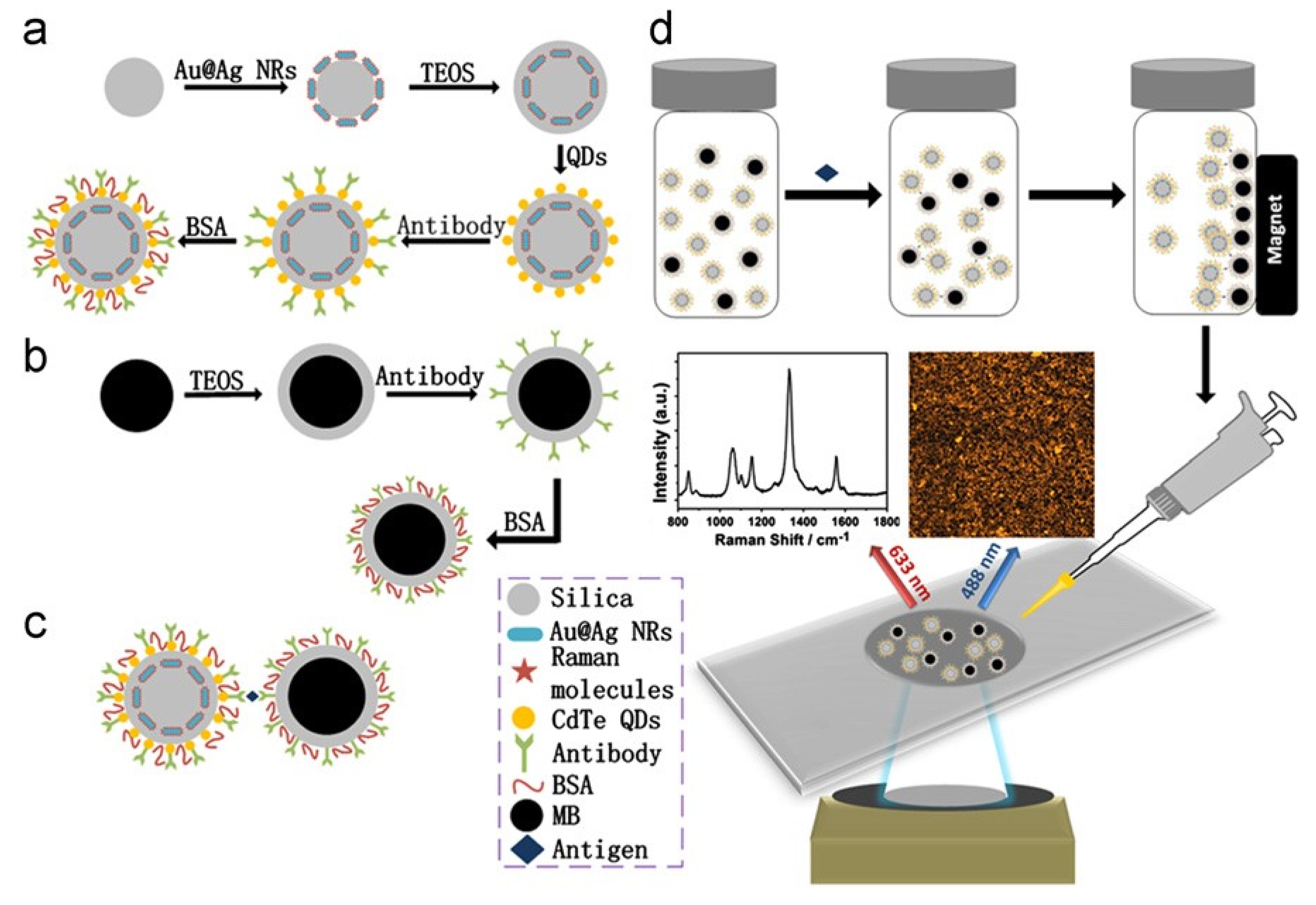
- Kim, H.M.; Kim, D.M.; Jeong, C.; Park, S.Y.; Cha, M.G.; Ha, Y.; Jang, D.; Kyeong, S.; Pham, X.H.; Hahm, E.; et al. Assembly of Plasmonic and Magnetic Nanoparticles with Fluorescent Silica Shell Layer for Tri-functional SERS-Magnetic-Fluorescence Probes and Its Bioapplications. Sci. Rep. 2018, 8, 13938. [Google Scholar] [CrossRef]
- Chen, L.; Hong, W.; Guo, Z.; Sa, Y.; Wang, X.; Jung, Y.M.; Zhao, B. Magnetic assistance highly sensitive protein assay based on surface-enhanced resonance Raman scattering. J. Colloid Interface Sci. 2012, 368, 282–286. [Google Scholar] [CrossRef]
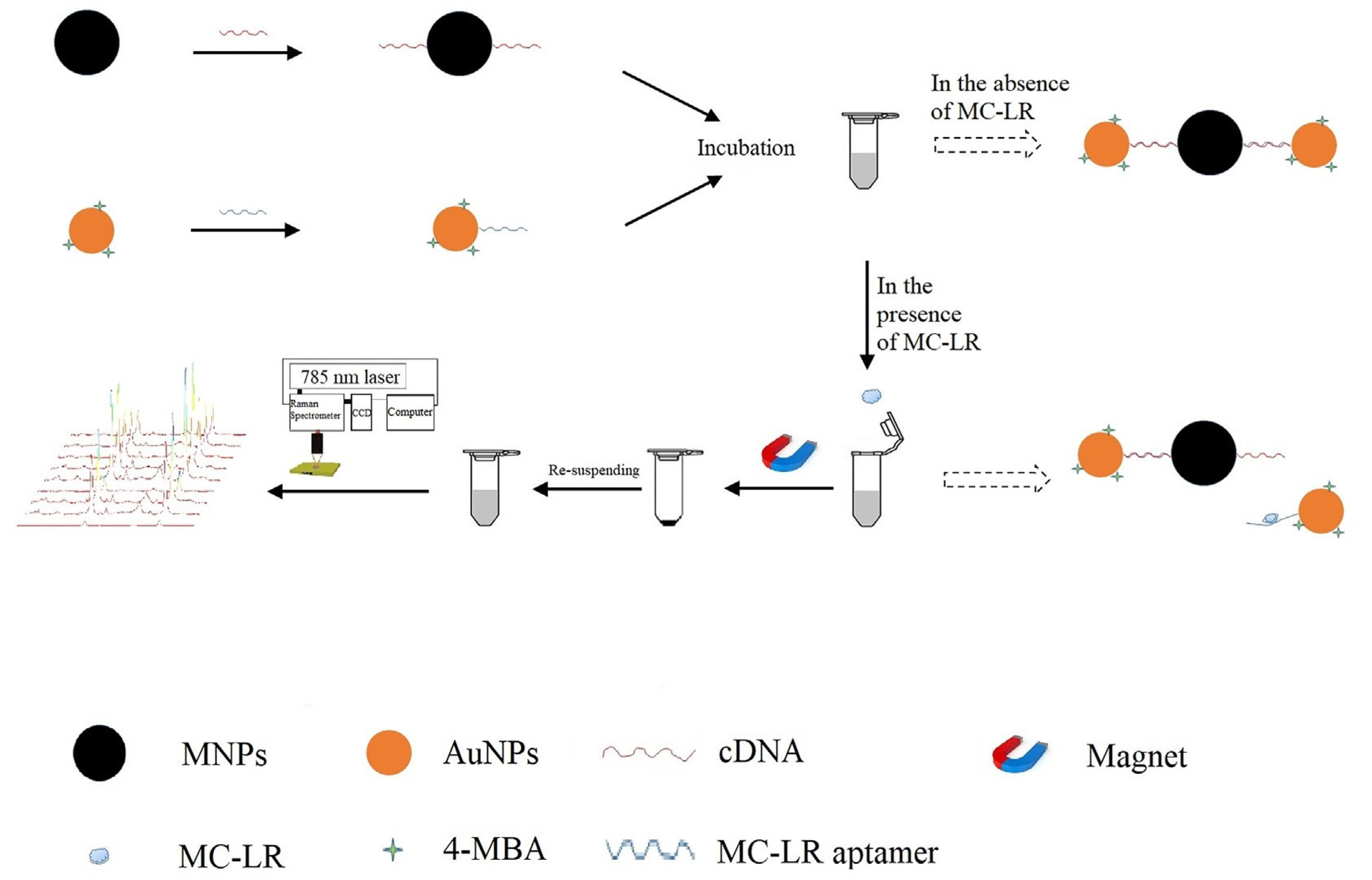
- He, D.; Wu, Z.; Cui, B.; Jin, Z. A novel SERS-based aptasensor for ultrasensitive sensing of microcystin-LR. Food Chem. 2019, 278, 197–202. [Google Scholar] [CrossRef]
- Hwang, H.; Kim, S.H.; Yang, S.M. Microfluidic fabrication of SERS-active microspheres for molecular detection. Lab Chip 2011, 11, 87–92. [Google Scholar] [CrossRef]
- Yu, S.; Liu, Z.; Wang, W.; Jin, L.; Xu, W.; Wu, Y. Disperse magnetic solid phase microextraction and surface enhanced Raman scattering (Dis-MSPME-SERS) for the rapid detection of trace illegally chemicals. Talanta 2018, 178, 498–506. [Google Scholar] [CrossRef]
- Hardiansyah, A.; Chen, A.-Y.; Liao, H.-L.; Yang, M.-C.; Liu, T.-Y.; Chan, T.-Y.; Tsou, H.-M.; Kuo, C.-Y.; Wang, J.-K.; Wang, Y.-L. Core-shell of FePt@SiO2-Au magnetic nanoparticles for rapid SERS detection. Nanoscale Res. Lett. 2015, 10, 412. [Google Scholar] [CrossRef]
- Wang, C.; Gu, B.; Liu, Q.; Pang, Y.; Xiao, R.; Wang, S. Combined use of vancomycin-modified Ag-coated magnetic nanoparticles and secondary enhanced nanoparticles for rapid surface-enhanced Raman scattering detection of bacteria. Int. J. Nanomed. 2018, 13, 1159–1178. [Google Scholar] [CrossRef]
- Chattopadhyay, S.; Sabharwal, P.K.; Jain, S.; Kaur, A.; Singh, H. Functionalized polymeric magnetic nanoparticle assisted SERS immunosensor for the sensitive detection of S. typhimurium. Anal. Chim. Acta 2019, 1067, 98–106. [Google Scholar] [CrossRef] [PubMed]
- Kim, K.; Choi, J.Y.; Lee, H.B.; Shin, K.S. Silanization of Ag-deposited magnetite particles: an efficient route to fabricate magnetic nanoparticle-based Raman barcode materials. ACS Appl. Mater. Interfaces 2010, 2, 1872–1878. [Google Scholar] [CrossRef]
- Yang, K.; Hu, Y.; Dong, N. A novel biosensor based on competitive SERS immunoassay and magnetic separation for accurate and sensitive detection of chloramphenicol. Biosens. Bioelectron. 2016, 80, 373–377. [Google Scholar] [CrossRef] [PubMed]
- Yu, S.; Liu, Z.; Li, H.; Zhang, J.; Yuan, X.X.; Jia, X.; Wu, Y. Combination of a graphene SERS substrate and magnetic solid phase micro-extraction used for the rapid detection of trace illegal additives. Analyst 2018, 143, 883–890. [Google Scholar] [CrossRef]
- Yang, K.; Hu, Y.; Dong, N.; Zhu, G.; Zhu, T.; Jiang, N. A novel SERS-based magnetic aptasensor for prostate specific antigen assay with high sensitivity. Biosens. Bioelectron. 2017, 94, 286–291. [Google Scholar] [CrossRef]
- Neng, J.; Harpster, M.H.; Wilson, W.C.; Johnson, P.A. Surface-enhanced Raman scattering (SERS) detection of multiple viral antigens using magnetic capture of SERS-active nanoparticles. Biosens. Bioelectron. 2013, 41, 316–321. [Google Scholar] [CrossRef]
- Alula, M.T.; Lemmens, P.; Bo, L.; Wulferding, D.; Yang, J.; Spende, H. Preparation of silver nanoparticles coated ZnO/Fe3O4 composites using chemical reduction method for sensitive detection of uric acid via surface-enhanced Raman spectroscopy. Anal. Chim. Acta 2019, 1073, 62–71. [Google Scholar] [CrossRef]
- Liang, Y.; Gong, J.L.; Huang, Y.; Zheng, Y.; Jiang, J.H.; Shen, G.L.; Yu, R.Q. Biocompatible core-shell nanoparticle-based surface-enhanced Raman scattering probes for detection of DNA related to HIV gene using silica-coated magnetic nanoparticles as separation tools. Talanta 2007, 72, 443–449. [Google Scholar] [CrossRef]
- Pang, Y.F.; Wang, C.W.; Wang, J.; Sun, Z.W.; Xiao, R.; Wang, S.Q. Fe3O4@Ag magnetic nanoparticles for microRNA capture and duplex-specific nuclease signal amplification based SERS detection in cancer cells. Biosens. Bioelectron. 2016, 79, 574–580. [Google Scholar] [CrossRef]
- Song, J.; Chen, Z.P.; Jin, J.W.; Chen, Y.; Yu, R.Q. Quantitative surface-enhanced Raman spectroscopy based on the combination of magnetic nanoparticles with an advanced chemometric model. Chemom. Intell. Lab. Syst. 2014, 135, 31–36. [Google Scholar] [CrossRef]
- Zengin, A.; Tamer, U.; Caykara, T. Extremely sensitive sandwich assay of kanamycin using surface-enhanced Raman scattering of 2-mercaptobenzothiazole labeled gold@silver nanoparticles. Anal. Chim. Acta 2014, 817, 33–41. [Google Scholar] [CrossRef] [PubMed]
- Hou, X.M.; Zhang, X.L.; Chen, S.T.; Kang, H.Z.; Tan, W.H. Facile synthesis of Ni/Au, Ni/Ag hybrid magnetic nanoparticles: New active substrates for surface enhanced Raman scattering. Colloids Surf. A Physicochem. Eng. Asp. 2012, 403, 148–154. [Google Scholar] [CrossRef]
- Hassanain, W.A.; Izake, E.L.; Schmidt, M.S.; Ayoko, G.A. Gold nanomaterials for the selective capturing and SERS diagnosis of toxins in aqueous and biological fluids. Biosens. Bioelectron. 2017, 91, 664–672. [Google Scholar] [CrossRef] [PubMed]
- Qu, H.; Lai, Y.; Niu, D.; Sun, S. Surface-enhanced Raman scattering from magneto-metal nanoparticle assemblies. Anal. Chim. Acta 2013, 763, 38–42. [Google Scholar] [CrossRef] [PubMed]
- Kim, M.; Ko, S.M.; Chungyeon, L.; Son, J.; Kim, J.; Kim, J.-M.; Nam, J.-M. Hierarchic Interfacial Nanocube Assembly for Sensitive, Selective and Quantitative DNA Detection with Surface-Enhanced Raman Scattering. Anal. Chem. 2019. [Google Scholar] [CrossRef]
- Li, D.; Jiang, L.; Piper, J.A.; Maksymov, I.S.; Greentree, A.D.; Wang, E.; Wang, Y. Sensitive and Multiplexed SERS Nanotags for the Detection of Cytokines Secreted by Lymphoma. ACS Sens. 2019, 4, 2507–2514. [Google Scholar] [CrossRef]
- Cao, J.; Wang, W.; Bo, B.; Mao, X.; Wang, K.; Zhu, X. A dual-signal strategy for the solid detection of both small molecules and proteins based on magnetic separation and highly fluorescent copper nanoclusters. Biosens. Bioelectron. 2017, 90, 534–541. [Google Scholar] [CrossRef]
- Qin, J.; Li, K.; Peng, C.; Li, X.; Lin, J.; Ye, K.; Yang, X.; Xie, Q.; Shen, Z.; Jin, Y.; et al. MRI of iron oxide nanoparticle-labeled ADSCs in a model of hindlimb ischemia. Biomaterials 2013, 34, 4914–4925. [Google Scholar] [CrossRef]
- Mahmoudi, M.; Shokrgozar, M.A. Multifunctional stable fluorescent magnetic nanoparticles. Chem. Commun. (Camb.) 2012, 48, 3957–3959. [Google Scholar] [CrossRef]
- Ebrahiminezhad, A.; Ghasemi, Y.; Rasoul-Amini, S.; Barar, J.; Davaran, S. Preparation of novel magnetic fluorescent nanoparticles using amino acids. Colloids Surf. B Biointerfaces 2013, 102, 534–539. [Google Scholar] [CrossRef]
- Lee, H.U.; Jung, D.U.; Lee, J.H.; Song, Y.S.; Park, C.; Kim, S.W. Detection of glyphosate by quantitative analysis of fluorescence and single DNA using DNA-labeled fluorescent magnetic core-shell nanoparticles. Sens. Actuators B Chem. 2013, 177, 879–886. [Google Scholar] [CrossRef]
- Liu, C.H.; Sahoo, S.L.; Tsao, M.H. Acridine orange coated magnetic nanoparticles for nucleus labeling and DNA adsorption. Colloids Surf. B Biointerfaces 2014, 115, 150–156. [Google Scholar] [CrossRef] [PubMed]
- Chen, O.; Riedemann, L.; Etoc, F.; Herrmann, H.; Coppey, M.; Barch, M.; Farrar, C.T.; Zhao, J.; Bruns, O.T.; Wei, H.; et al. Magneto-fluorescent core-shell supernanoparticles. Nat. Commun. 2014, 5, 5093. [Google Scholar] [CrossRef] [PubMed]
- Wei, Z.; Wu, Y.; Zhao, Y.; Mi, L.; Wang, J.; Wang, J.; Zhao, J.; Wang, L.; Liu, A.; Li, Y.; et al. Multifunctional nanoprobe for cancer cell targeting and simultaneous fluorescence/magnetic resonance imaging. Anal. Chim. Acta 2016, 938, 156–164. [Google Scholar] [CrossRef]
- Ruan, J.; Ji, J.; Song, H.; Qian, Q.; Wang, K.; Wang, C.; Cui, D. Fluorescent magnetic nanoparticle-labeled mesenchymal stem cells for targeted imaging and hyperthermia therapy of in vivo gastric cancer. Nanoscale Res. Lett. 2012, 7, 309. [Google Scholar] [CrossRef]
- Icten, O.; Kose, D.A.; Matissek, S.J.; Misurelli, J.A.; Elsawa, S.F.; Hosmane, N.S.; Zumreoglu-Karan, B. Gadolinium borate and iron oxide bioconjugates: Nanocomposites of next generation with multifunctional applications. Mater. Sci. Eng. C 2018, 92, 317–328. [Google Scholar] [CrossRef]
- Syed, M.A. Advances in nanodiagnostic techniques for microbial agents. Biosens. Bioelectron. 2014, 51, 391–400. [Google Scholar] [CrossRef]
- Bhaisare, M.L.; Gedda, G.; Khan, M.S.; Wu, H.F. Fluorimetric detection of pathogenic bacteria using magnetic carbon dots. Anal. Chim. Acta 2016, 920, 63–71. [Google Scholar] [CrossRef]
- Wu, Z.; Xu, E.; Chughtai, M.F.J.; Jin, Z.; Irudayaraj, J. Highly sensitive fluorescence sensing of zearalenone using a novel aptasensor based on upconverting nanoparticles. Food Chem. 2017, 230, 673–680. [Google Scholar] [CrossRef]
- Clemente, C.S.; Ribeiro, V.G.P.; Sousa, J.E.A.; Maia, F.J.N.; Barreto, A.C.H.; Andrade, N.F.; Denardin, J.C.; Mele, G.; Carbone, L.; Mazzetto, S.E.; et al. Porphyrin synthesized from cashew nut shell liquid as part of a novel superparamagnetic fluorescence nanosystem. J. Nanopart. Res. 2013, 15, 1739. [Google Scholar] [CrossRef]
- Wang, F.; Xu, L.; Zhang, Y.; Petrenko, V.A.; Liu, A. An efficient strategy to synthesize a multifunctional ferroferric oxide core@ dye/SiO 2@ Au shell nanocomposite and its targeted tumor theranostics. J. Mater. Chem. B 2017, 5, 8209–8218. [Google Scholar] [CrossRef]
- Myklatun, A.; Cappetta, M.; Winklhofer, M.; Ntziachristos, V.; Westmeyer, G.G. Microfluidic sorting of intrinsically magnetic cells under visual control. Sci. Rep. 2017, 7, 6942. [Google Scholar] [CrossRef]
- Yin, M.; Li, Z.; Liu, Z.; Ren, J.; Yang, X.; Qu, X. Photosensitizer-incorporated G-quadruplex DNA-functionalized magnetofluorescent nanoparticles for targeted magnetic resonance/fluorescence multimodal imaging and subsequent photodynamic therapy of cancer. Chem. Commun. (Camb.) 2012, 48, 6556–6558. [Google Scholar] [CrossRef] [PubMed]
- Wang, W.; Jiang, X.; Chen, K. CePO 4: Tb, Gd hollow nanospheres as peroxidase mimic and magnetic–fluorescent imaging agent. Chem. Commun. 2012, 48, 6839–6841. [Google Scholar] [CrossRef] [PubMed]
- Zheng, J.; Nie, Y.; Hu, Y.; Li, J.; Li, Y.; Jiang, Y.; Yang, R. Time-resolved fluorescent detection of Hg 2+ in a complex environment by conjugating magnetic nanoparticles with a triple-helix molecular switch. Chem. Commun. 2013, 49, 6915–6917. [Google Scholar] [CrossRef]
- Xu, Y.H.; Zhou, Y.; Li, R.X. Simultaneous fluorescence response and adsorption of functionalized Fe3O4@SiO2 nanoparticles to Cd2+, Zn2+ and Cu2+. Colloids Surf. A Physicochem. Eng. Asp. 2014, 459, 240–246. [Google Scholar] [CrossRef]
- Chen, Q.; Hu, W.; Sun, C.; Li, H.; Ouyang, Q. Synthesis of improved upconversion nanoparticles as ultrasensitive fluorescence probe for mycotoxins. Anal. Chim. Acta 2016, 938, 137–145. [Google Scholar] [CrossRef]
- Liu, Z.; Koczera, P.; Doleschel, D.; Kiessling, F.; Gatjens, J. Versatile synthetic strategies for PBCA-based hybrid fluorescent microbubbles and their potential theranostic applications to cell labelling and imaging. Chem. Commun. (Camb.) 2012, 48, 5142–5144. [Google Scholar] [CrossRef]
- Chang, L.; Chen, S.; Jin, P.; Li, X. Synthesis of multifunctional fluorescent magnetic graphene oxide hybrid materials. J. Colloid Interface Sci. 2012, 388, 9–14. [Google Scholar] [CrossRef]
- Hua, X.; You, H.; Luo, P.; Tao, Z.; Chen, H.; Liu, F.; Wang, M. Upconversion fluorescence immunoassay for imidaclothiz by magnetic nanoparticle separation. Anal. Bioanal. Chem. 2017, 409, 6885–6892. [Google Scholar] [CrossRef]
- Jin, M.; Liu, X.; van den Berg, A.; Zhou, G.; Shui, L. Ultrasensitive DNA detection based on two-step quantitative amplification on magnetic nanoparticles. Nanotechnology 2016, 27, 335102. [Google Scholar] [CrossRef] [PubMed]
- Li, N.; Gao, Z.F.; Kang, B.H.; Li, N.B.; Luo, H.Q. Sensitive mutant DNA biomarker detection based on magnetic nanoparticles and nicking endonuclease assisted fluorescence signal amplification. RSC Adv. 2015, 5, 20020–20024. [Google Scholar] [CrossRef]
- Quarta, A.; Bernareggi, D.; Benigni, F.; Luison, E.; Nano, G.; Nitti, S.; Cesta, M.C.; Di Ciccio, L.; Canevari, S.; Pellegrino, T.; et al. Targeting FR-expressing cells in ovarian cancer with Fab-functionalized nanoparticles: a full study to provide the proof of principle from in vitro to in vivo. Nanoscale 2015, 7, 2336–2351. [Google Scholar] [CrossRef] [PubMed]
- Portnoy, E.; Polyak, B.; Inbar, D.; Kenan, G.; Rai, A.; Wehrli, S.L.; Roberts, T.P.; Bishara, A.; Mann, A.; Shmuel, M.; et al. Tracking inflammation in the epileptic rat brain by bi-functional fluorescent and magnetic nanoparticles. Nanomedicine 2016, 12, 1335–1345. [Google Scholar] [CrossRef]
- Wan, Y.; Sun, Y.; Qi, P.; Wang, P.; Zhang, D. Quaternized magnetic nanoparticles-fluorescent polymer system for detection and identification of bacteria. Biosens. Bioelectron. 2014, 55, 289–293. [Google Scholar] [CrossRef]
- Wang, S.; Zheng, L.; Cai, G.; Liu, N.; Liao, M.; Li, Y.; Zhang, X.; Lin, J. A microfluidic biosensor for online and sensitive detection of Salmonella typhimurium using fluorescence labeling and smartphone video processing. Biosens. Bioelectron. 2019, 140, 111333. [Google Scholar] [CrossRef]
- Wang, Y.; Sun, H.; Li, R.; Ke, P.; Zhu, H.; Guo, H.; Liu, M.; Sun, H. An immunomagnetic separation based fluorescence immunoassay for rapid myoglobin quantification in human blood. Anal. Methods 2016, 8, 7324–7330. [Google Scholar] [CrossRef]
- Niazi, S.; Wang, X.; Pasha, I.; Khan, I.M.; Zhao, S.; Shoaib, M.; Wu, S.; Wang, Z. A novel bioassay based on aptamer-functionalized magnetic nanoparticle for the detection of zearalenone using time resolved-fluorescence NaYF4: Ce/Tb nanoparticles as signal probe. Talanta 2018, 186, 97–103. [Google Scholar] [CrossRef]
- Shen, T.; Yue, Q.; Jiang, X.; Wang, L.; Xu, S.; Li, H.; Gu, X.; Zhang, S.; Liu, J. A reusable and sensitive biosensor for total mercury in canned fish based on fluorescence polarization. Talanta 2013, 117, 81–86. [Google Scholar] [CrossRef]
- Shrivastava, S.; Lee, W.-I.; Lee, N.-E. Culture-free, highly sensitive, quantitative detection of bacteria from minimally processed samples using fluorescence imaging by smartphone. Biosens. Bioelectron. 2018, 109, 90–97. [Google Scholar] [CrossRef]
- Yao, Q.; Zheng, Y.; Cheng, W.; Chen, M.; Shen, J.; Yin, M. Difunctional fluorescent HSA modified CoFe 2 O 4 magnetic nanoparticles for cell imaging. J. Mater. Chem. B 2016, 4, 6344–6349. [Google Scholar] [CrossRef]
- Wen, D.; Liu, Q.; Cui, Y.; Kong, J.; Yang, H.; Liu, Q. DNA based click polymerization for ultrasensitive IFN-γ fluorescent detection. Sens. Actuators B Chem. 2018, 276, 279–287. [Google Scholar] [CrossRef]
- De La Rosa-Romo, L.M.; Oropeza-Guzmán, M.T.; Olivas-Sarabia, A.; Pina-Luis, G. Flavone functionalized magnetic nanoparticles: A new fluorescent sensor for Cu2+ ions with nanomolar detection limit. Sens. Actuators B Chem. 2016, 233, 459–468. [Google Scholar] [CrossRef]
- Luo, S.; Liu, Y.; Rao, H.; Wang, Y.; Wang, X. Fluorescence and magnetic nanocomposite Fe3O4@ SiO2@ Au MNPs as peroxidase mimetics for glucose detection. Anal. Biochem. 2017, 538, 26–33. [Google Scholar] [CrossRef] [PubMed]
- Amini, B.; Kamali, M.; Salouti, M.; Yaghmaei, P. Fluorescence bio-barcode DNA assay based on gold and magnetic nanoparticles for detection of Exotoxin A gene sequence. Biosens. Bioelectron. 2017, 92, 679–686. [Google Scholar] [CrossRef]
- Ma, L.; Sun, N.; Meng, Y.; Tu, C.; Cao, X.; Wei, Y.; Chu, L.; Diao, A. Harnessing the affinity of magnetic nanoparticles toward dye-labeled DNA and developing it as an universal aptasensor revealed by lipopolysaccharide detection. Anal. Chim. Acta 2018, 1036, 107–114. [Google Scholar] [CrossRef]
- Kim, S.; Ko, J.; Lim, H.B. Application of magnetic and core-shell nanoparticles to determine enrofloxacin and its metabolite using laser induced fluorescence microscope. Anal. Chim. Acta 2013, 771, 37–41. [Google Scholar] [CrossRef]
- Zarei-Ghobadi, M.; Mozhgani, S.H.; Dashtestani, F.; Yadegari, A.; Hakimian, F.; Norouzi, M.; Ghourchian, H. A genosensor for detection of HTLV-I based on photoluminescence quenching of fluorescent carbon dots in presence of iron magnetic nanoparticle-capped Au. Sci. Rep. 2018, 8, 15593. [Google Scholar] [CrossRef]
- Ma, Y.; Zheng, B.; Zhao, Y.; Yuan, H.; Cai, Y.; Du, J.; Xiao, D. A sensitive and selective chemosensor for GSSG detection based on the recovered fluorescence of NDPA-Fe(3)O(4)@SiO(2)-Cu(II) nanomaterial. Biosens. Bioelectron. 2013, 48, 138–144. [Google Scholar] [CrossRef]
- Xue, Q.; Wang, L.; Jiang, W. A versatile platform for highly sensitive detection of protein: DNA enriching magnetic nanoparticles based rolling circle amplification immunoassay. Chem. Commun. (Camb.) 2012, 48, 3930–3932. [Google Scholar] [CrossRef]
- Yu, X.; Wen, C.Y.; Zhang, Z.L.; Pang, D.W. Control of magnetic field distribution by using nickel powder@PDMS pillars in microchannels. RSC Adv. 2014, 4, 17660–17666. [Google Scholar] [CrossRef]
- Song, E.Q.; Hu, J.; Wen, C.Y.; Tian, Z.Q.; Yu, X.; Zhang, Z.L.; Shi, Y.B.; Pang, D.W. Fluorescent-magnetic-biotargeting multifunctional nanobioprobes for detecting and isolating multiple types of tumor cells. ACS Nano 2011, 5, 761–770. [Google Scholar] [CrossRef] [PubMed]
- Xie, H.Y.; Xie, M.; Zhang, Z.L.; Long, Y.M.; Liu, X.; Tang, M.L.; Pang, D.W.; Tan, Z.; Dickinson, C.; Zhou, W. Wheat germ agglutinin-modified trifunctional nanospheres for cell recognition. Bioconjug. Chem. 2007, 18, 1749–1755. [Google Scholar] [CrossRef] [PubMed]
- Xie, H.Y.; Zuo, C.; Liu, Y.; Zhang, Z.L.; Pang, D.W.; Li, X.L.; Gong, J.P.; Dickinson, C.; Zhou, W. Cell-targeting multifunctional nanospheres with both fluorescence and magnetism. Small 2005, 1, 506–509. [Google Scholar] [CrossRef]
- Reich, G. Near-infrared spectroscopy and imaging: basic principles and pharmaceutical applications. Adv. Drug Deliv. Rev. 2005, 57, 1109–1143. [Google Scholar] [CrossRef]
- Sakudo, A. Near-infrared spectroscopy for medical applications: Current status and future perspectives. Clin. Chim. Acta 2016, 455, 181–188. [Google Scholar] [CrossRef]
- Roggo, Y.; Chalus, P.; Maurer, L.; Lema-Martinez, C.; Edmond, A.; Jent, N. A review of near infrared spectroscopy and chemometrics in pharmaceutical technologies. J. Pharm. Biomed. Anal. 2007, 44, 683–700. [Google Scholar] [CrossRef]
- Büning-Pfaue, H. Analysis of water in food by near infrared spectroscopy. Food Chem. 2003, 82, 107–115. [Google Scholar] [CrossRef]
- Yang, H.M.; Park, C.W.; Park, S.; Kim, J.D. Cross-linked magnetic nanoparticles with a biocompatible amide bond for cancer-targeted dual optical/magnetic resonance imaging. Colloids Surf. B Biointerfaces 2018, 161, 183–191. [Google Scholar] [CrossRef]
- Wu, H.; Cheng, K.; He, Y.; Li, Z.; Su, H.; Zhang, X.; Sun, Y.; Shi, W.; Ge, D. Fe3O4-based multifunctional nanospheres for amplified magnetic targeting photothermal therapy and Fenton reaction. ACS Biomater. Sci. Eng. 2018, 5, 1045–1056. [Google Scholar] [CrossRef]
- Gollavelli, G.; Ling, Y.C. Magnetic and fluorescent graphene for dual modal imaging and single light induced photothermal and photodynamic therapy of cancer cells. Biomaterials 2014, 35, 4499–4507. [Google Scholar] [CrossRef] [PubMed]
- Wu, L.; Zong, L.; Ni, H.; Liu, X.; Wen, W.; Feng, L.; Cao, J.; Qi, X.; Ge, Y.; Shen, S. Magnetic thermosensitive micelles with upper critical solution temperature for NIR triggered drug release. Biomater. Sci. 2019, 7, 2134–2143. [Google Scholar] [CrossRef] [PubMed]
- Jin, X.; Zhang, M.; Gou, G.; Ren, J. Synthesis and Cell Imaging of a Near-Infrared Fluorescent Magnetic “CdHgTe–Dextran-Magnetic Layered Double Hydroxide–Fluorouracil” Composite. J. Pharm. Sci. 2016, 105, 1751–1761. [Google Scholar] [CrossRef] [PubMed]
- Zhou, H.; Hou, X.; Liu, Y.; Zhao, T.; Shang, Q.; Tang, J.; Liu, J.; Wang, Y.; Wu, Q.; Luo, Z.; et al. Superstable Magnetic Nanoparticles in Conjugation with Near-Infrared Dye as a Multimodal Theranostic Platform. ACS Appl. Mater. Interfaces 2016, 8, 4424–4433. [Google Scholar] [CrossRef]
- Zhou, Z.; Chen, H.; Lipowska, M.; Wang, L.; Yu, Q.; Yang, X.; Tiwari, D.; Yang, L.; Mao, H. A dual-modal magnetic nanoparticle probe for preoperative and intraoperative mapping of sentinel lymph nodes by magnetic resonance and near infrared fluorescence imaging. J. Biomater. Appl. 2013, 28, 100–111. [Google Scholar] [CrossRef]
- Huang, P.; Li, Z.; Lin, J.; Yang, D.; Gao, G.; Xu, C.; Bao, L.; Zhang, C.; Wang, K.; Song, H.; et al. Photosensitizer-conjugated magnetic nanoparticles for in vivo simultaneous magnetofluorescent imaging and targeting therapy. Biomaterials 2011, 32, 3447–3458. [Google Scholar] [CrossRef]
- Cho, S.H.; Kim, A.; Shin, W.; Heo, M.B.; Noh, H.J.; Hong, K.S.; Cho, J.H.; Lim, Y.T. Photothermal-modulated drug delivery and magnetic relaxation based on collagen/poly(gamma-glutamic acid) hydrogel. Int. J. Nanomedicine 2017, 12, 2607–2620. [Google Scholar] [CrossRef]
- Lee, H.; Choi, H.; Lee, M.; Park, S. Preliminary study on alginate/NIPAM hydrogel-based soft microrobot for controlled drug delivery using electromagnetic actuation and near-infrared stimulus. Biomed. Microdevices 2018, 20, 103. [Google Scholar] [CrossRef]


| Magnetic Part | SPR System | Applications | Advantages | [R] |
|---|---|---|---|---|
| Active layer consisting of Fe3O4 + Polyethylene glycol (PEG4000) | Silver-coated prism-coupled SPR System | Plant DNA | Target analyte could not be detected without using a magnetic part. | [50] |
| Polydopamine-Ag capped Fe3O4 NPs reduced with graphene oxide | Gold film of SPR chip was electromagnetically coupled with hollow gold nanoparticles | Rabbit IgG | Detection limit 132 times lower than conventional SPR and 8 times lower than immunosandwich assay. | [48] |
| Gold capped Fe3O4 nanoparticles (GMNPs) | Conventional SPR system with gold chip | Thrombin | SPR angle shift is enlarged for 5 times comparing with that of control group without GMNPs | [49] |
| MNPs conjugated with antibody | Conventional SPR system with gold chip | Pathogenic Bacteria | MNPs offer a sensing enhancement of 4 orders of magnitude. | [51] |
| Magneto plasmonic nanoparticles (core shell gold capped MNPs) | Conventional SPR system | Antigen (Tuberculosis marker protein) | Implementation of magneto-plasmonic NPs outcomes in 30-fold extension of the SPR signal at the limit of detection. | [52] |
| Nanohybrids containing Fe3O4 NPs and hollow gold sphere nanoparticles. | SPR chip coated with carboxyl functionalized graphene oxide sheet. | Human IgG | Detection limit is approximately 260-fold lower than that acquired with sandwich assay by routine SPR biosensors. | [53] |
| Polydopamine-wrapped magnetic multi-walled carbon nanotubes | Capture antibody-immobilized SPR-sensing film modified by hollow gold nanoparticles and polydopamine | Protein (Human cardiac troponin I, CTnI) | Minimum detectable SPR response for the concentration of target molecule is 1000 times lower than that achieved by the traditional SPR immunoassay. | [54] |
| Magnetic fluid photonic crystal (i.e. kind of colloid compassed of MNPs dispersed in carrier liquid) | Kretschmann configuration (prism coupling) SPR system. | -------------- | The excellent benefit of elevated sensitivity is with the combination of magnetic fluid photonic crystal and SPR. | [55] |
| Aptamer-immobilized Fe3O4 nanoparticles with organic clusters | Kretschmann configuration (prism coupling) SPR system. | Protein (prion disease associated isoform, PrPSC) | The SPR scheme involves magnetic NPs-organic clusters that allow for a 215-fold rise in the immediate SPR signal. | [46] |
| Streptavidin-coated MNPs | Grating-coupled SPR (GC-SPR) with wavelength interrogation. | Lipid (Extracellular vesicles, EVs) | Target could not be detected with the direct SPR detection platform at such low concentrations. | [56] |
| Fe-C core shell–aptamer conjugation | Prism-coupling SPR system | Protein (prion disease associated isoform, PrPSC) | The detection sensitivity of PrPSC has been improved by about 10 times relative to the direct format of SPR detection. | [57] |
| Antibody-functionalized MNPs | Antibody-immobilized SPR immunoassay | Hormone (Estradiol) | The MNPs showed outstanding ability to amplify the SPR signal. | [45] |
| PEG-4000 functionalized Fe3O4 MNPs | SPR set up device in Kretschmann configuration and He/Ne laser beam. | Microalgae | Microalgae population has been successfully monitored with the proposed system. | [58] |
| MNPs antibody conjugates | Conventional SPR spectroscopy | Cancer Biomarkers | To differentiate ovarian cancer, this multiplexed scheme accomplished sensitivity and specificity of up to 94% and 98%, respectively. | [59] |
| Type of PNS | Size of PNS, nm | Forms of MNPs | Size of MNPs, nm | Reporter 1 | Analyte | Detection limit | [R] |
|---|---|---|---|---|---|---|---|
| Au NPs | 15 | γ-Fe2O3 covered with silica shell and polymer shell | 64 | DTNB | Tau protein | 25 fM | [65] |
| Au NPs | 20 | γ-Fe2O3 MNPs covered with silica and Au shell | 160 | DTNB | Telomerase activity | 1 cell/mL | [67] |
| Triangular Ag nanoprisms | 40 | γ-Fe2O3 MNPs MNPs | 21 | MBA | Tumor cells | 1 cell/mL | [69] |
| Au NPs | 20 | γ-Fe2O3 covered with silica shell | 25 | MBA | Microcystin-LR | 2.0pg/mL. | [72] |
| Au NPs | 20 | Fe3O4 magnetic microspheres covered with SiO2 shell and Au nanoparticles | 500 | Cyanine | Sildenafil citrate | 10 nM | [74] |
| Au NPs | 31 | γ-Fe2O3 encapsulated with polymer | 330 | MBA and DSNB | S. typhimurium | 10 cells/mL | [77] |
| Au NPs | 30 | γ-Fe2O3 MNPs | 500 | DP | Chloramphenicol | 1.0 pg/mL | [79] |
| Ag NPs | 20 | Fe3O4 MNPs covered with GO and Ag NPs | 500 | - | Chloramphenicol | 0.1 nM | [80] |
| Au NPs | 60 | ParaMNPs | 200 | IR-792 and NB | West Nile virus Antigen and Rift Valley fever virus Antigen | 5 fg/mL | [81] |
| Au NPs | 30 | γ-Fe2O3 MNPs | 500 | DP | PSA antigen | 5 pg/mL | [82] |
| Ag/SiO2 core-shell NPs | 35 | γ-Fe2O3 MNPs covered with silica shell | 50 | Rhodamine B | DNA | 5 µM | [84] |
| Ag shell | 30 | γ-Fe2O3 MNPs covered with Ag shell | 300 | Cyanine | miRNA | 0.3 fM | [85] |
| Ag shell | 8 | γ-Fe2O3 MNPs covered with Ag shell | 300 | PATP | Thiram | 1.0 nM | [86] |
| Au@Ag core-shell NPs | 32 | γ-Fe2O3 MNPs covered with polymer shell | 36 | MBT | Kanamycin | 2 pg/mL | [87] |
| Ni@Au and Ni@Ag NPs | 212 and 222 | Ni MNPs | 89 | rhodamine 6G | rhodamine 6G | 1 mM | [88] |
| Au Shell | 35 | γ-Fe2O3 MNPs covered with Au shell | 50 | - | Microcystin-LR | 3 fM | [89] |
| Au Shell | 15 | Fe3O4 MNPs covered with Au shell | 17-30 | pthiocresol | Pthiocresol | 4.5 pM | [90] |
| Au nanocubes and nanospheres | 51 | DNA modified Fe3O4 magnetic beads | 1000 | Cyanine | DNA | 1pM | [91] |
| Silver layers on reporter-coated AuNPs | 60 | Protein G modified Fe3O4 magnetic beads | 1000 | MBA, DTNB, and TFMBA | Cytokines | 4.5 pg mL | [92] |
| Types of NPs | Capture probe | Size nm | Signal Probe | Size nm | Method | Target | LOD | [R] |
|---|---|---|---|---|---|---|---|---|
| MNPs@CuNCs | Folate receptor functionalized MNPs | 300 | Fluorescent copper nanoclusters | 20 | Sandwich assay | Streptavidin and biotin | 0.47 nM and 3.1 nM | [93] |
| MNPs@FMs | Monoclonal antibodies (MAbs) immobilized Fe3O4 MNPs | 150 | PAbs functionalized fluorescent microspheres (FMs) | 150 | Microfluidic biosensor- smartphone based fluorescent microscopic system | Salmonella typhimurium | 58 CFU/mL | [122] |
| MNP@SiO2@BSA@Au-Myo-SNP@RhX | Fe3O4 MNP@SiO2@BSA@Au@antibody | 292 | SNP@RhX@antibody conjugates | 85 | Sandwich assay | myoglobin | 0.28 ng/mL | [123] |
| MNPs-aptamer/TRFLNPs-cDNA | Fe3O4 MNPs functionalized with ZEN aptamers | 55 | NaYF4: Ce/Tb modified complementary DNA (TRFLNPs-cDNA) | 34 | Sandwich assay | Zearalenone (ZEN) | 0.21 pg/mL | [124] |
| MNPs@ssDNA-FAM | Fe3O4 MNPs functionalized with ssDNA | 50 | carboxyfluorescein (FAM) | - | Sandwich assay | Total mercury (Hg) | 0.49 nM | [125] |
| Aptamer-conjugated FMNPs | Specific aptamer modified fluorescent magnetic nanoparticles (FMNPs) | 100 | - | - | Smartphone-based detection | Staphylococcus aureus | 10 CFU/mL | [126] |
| CoFe2O4@dopamine@HSA@PDI-4NH2 | CoFe2O4@dopamine@HSA@PDI-4NH2 | 190 | PDI-4NH2 (Perylene diimide) | - | Fluorescence imaging | Cell imaging | - | [127] |
| Fe3O4@aptamer 1@ IFN-γ@aptamer- FAMs@dsDNA | Fe3O4–aptamer 1/ IFN-γ/ aptamer | 100-300 | carboxyfluorescein modified aptamer and dsDNA (FAMs@dsDNA) | - | Aptamer/protein/aptamerpolymer supersandwich fluorescence sensor | Interferon gamma (IFN-γ) | 0.175 fM | [128] |
| Fe3O4@SiO2-NH2-morin | Fe3O4@SiO2-NH2 | 78 | Morin | - | Fluorescence titrations | Cu2+ | 7.5 nM | [129] |
| Fe3O4@SiO2@Au MNPs | Fe3O4@SiO2@Au MNPs | 11 | BSA-Au NCs | - | H2O2 quenching | glucose | 3.0 µM | [130] |
| MNP-2nd DNA probe-target DNA-1st DNA probe-GNP-barcode DNA | Second target specific DNA probe modified Fe3O4 MNPs | 45 | First target-specific DNA probe 1 (1pDNA) and bio-barcode DNA coated Au NPs (GNP) | 23 | Sandwich assay | Exotoxin A gene sequence | 1.2 ng/mL | [131] |
| MNPs-PEI-ssDNA | MNPs-PEI-ssDNA | 78 | dye-labeled DNA | - | Sandwich assay | lipopolysaccharide | 35 ng/mL | [132] |
| Types of NPs* | Size, nm | MNPs form | NIR agent | Target | Methods | Advantages | [R] |
|---|---|---|---|---|---|---|---|
| CMNPs | 45 | Fe3O4 | Cy5.5 | Tumor cells | Specific cancer-targeting and magnetic resonance/ near-infrared(MR/NIR) imaging | Dual imaging, excellent structural stability with biocompatible and biodegradable | [145] |
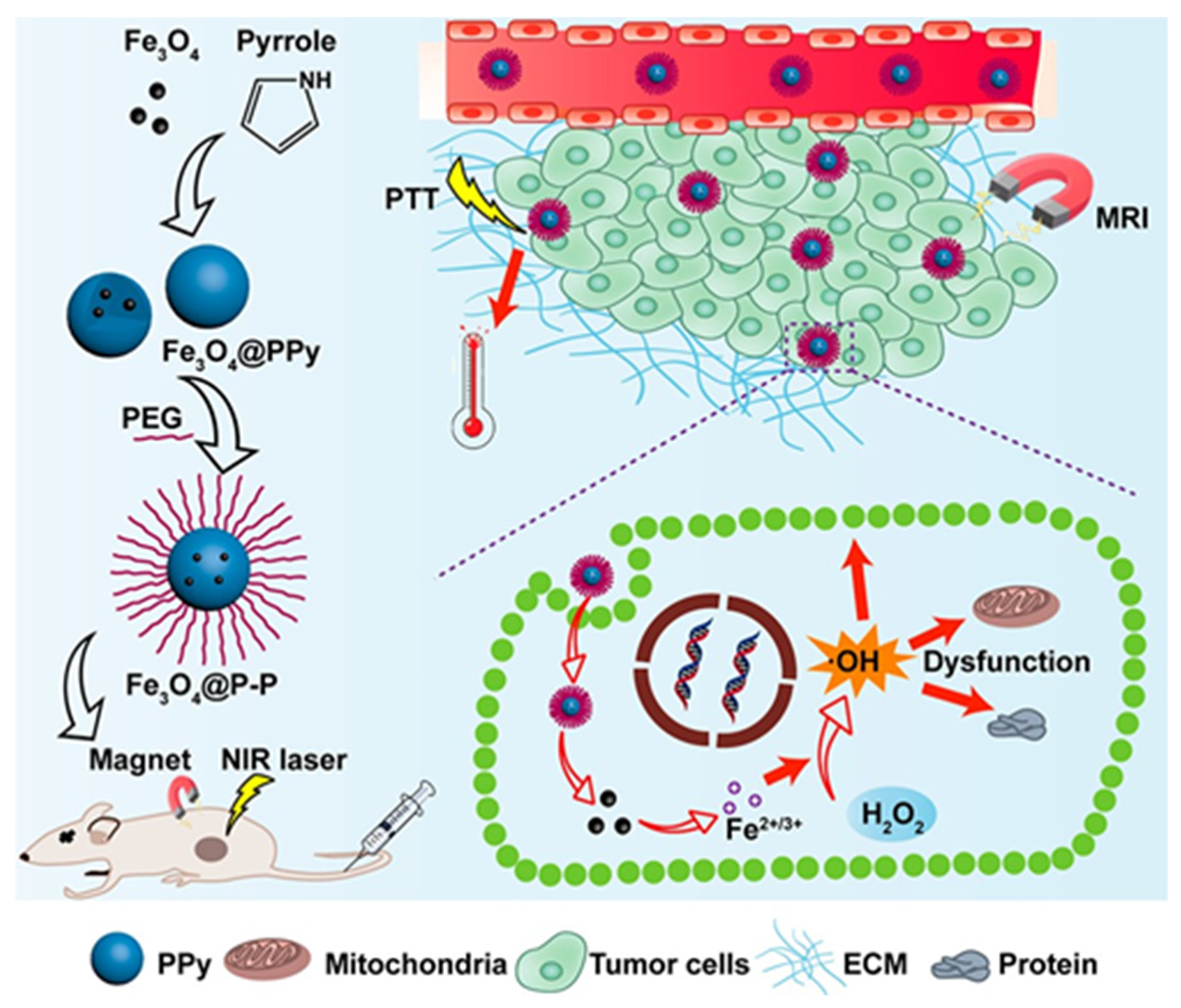
| Fe3O4@PPy-PEG | 89 | Fe3O4 | Polypyrrole (PPy) | Tumor cells | Photothermal therapy, magnetic targeting | Combination of combined the Fenton reaction and photothermal and magnet-guided cancer therapy | [146] |
| MFG-SiNc4 | 40 | Magnetic graphene | SiNc4 and fluorescein | Tumor cells | Photodynamic/photothermal therapeutic | Time and cost effective treatments with a minimal therapy dose | [147] |
| DOX-Fe3O4@PAAP | 170 | Fe3O4 | Hydrophobic Fe3O4 | Doxorubicin (DOX) | In situ drug release and combined photothermal-chemotherapy | Fast and effective thermosensitive drug delivery | [148] |
| CdHgTe@DMF | 100 | Magnetic layered double hydroxide | Quantum dots of CdHgTe | Tumor cells | Drug delivery, optical bioimaging and magnetic targeted therapy | Slow-release curative effect and good cell imaging | [149] |
| Fe3O4-Aurods-Fe3O4 nanodumbbells | 70 | Fe3O4 | Au Nanorods (Aurods) | Multiple pathogens | Detection, magnetic separation, and photokilling of multiple pathogens | Tunable nanoprobes for multiplex detection | [36] |
© 2019 by the authors. Licensee MDPI, Basel, Switzerland. This article is an open access article distributed under the terms and conditions of the Creative Commons Attribution (CC BY) license (http://creativecommons.org/licenses/by/4.0/).
Share and Cite
Üzek, R.; Sari, E.; Merkoçi, A. Optical-Based (Bio) Sensing Systems Using Magnetic Nanoparticles. Magnetochemistry 2019, 5, 59. https://doi.org/10.3390/magnetochemistry5040059
Üzek R, Sari E, Merkoçi A. Optical-Based (Bio) Sensing Systems Using Magnetic Nanoparticles. Magnetochemistry. 2019; 5(4):59. https://doi.org/10.3390/magnetochemistry5040059
Chicago/Turabian StyleÜzek, Recep, Esma Sari, and Arben Merkoçi. 2019. "Optical-Based (Bio) Sensing Systems Using Magnetic Nanoparticles" Magnetochemistry 5, no. 4: 59. https://doi.org/10.3390/magnetochemistry5040059
APA StyleÜzek, R., Sari, E., & Merkoçi, A. (2019). Optical-Based (Bio) Sensing Systems Using Magnetic Nanoparticles. Magnetochemistry, 5(4), 59. https://doi.org/10.3390/magnetochemistry5040059