Review

Why Magnetic Nanoparticles Still Struggle to Translate: A Systematic Analysis of Structural Gaps in Nanobiotechnology


by
Fernando Gomes de Souza, Jr.
1,2,3,*,
Carolina de Souza Cardoso Delfino
2 and
Yuri Ranieri de Medeiros Camargo
2
1
Instituto de Macromoléculas Professora Eloisa Mano, Universidade Federal do Rio de Janeiro (UFRJ), Centro de Tecnologia, Cidade Universitária, Rio de Janeiro 21941-598, Brazil
2
Instituto Alberto Luiz Coimbra de Pós-Graduação e Pesquisa de Engenharia—COPPE, Universidade Federal do Rio de Janeiro (UFRJ), Centro de Tecnologia, Cidade Universitária, Rio de Janeiro 21941-972, Brazil
3
Department of Electrical and Computer Engineering, Florida International University (FIU), 10555 West Flagler Street, EC3900, Miami, FL 33174, USA
*
Author to whom correspondence should be addressed.
Magnetochemistry 2026, 12(6), 65; https://doi.org/10.3390/magnetochemistry12060065
Submission received: 26 December 2025 / Revised: 22 May 2026 / Accepted: 29 May 2026 / Published: 5 June 2026
(This article belongs to the Special Issue Magnetic Nano- and Microparticles in Biotechnology)

Abstract

This review offers an in-depth look at the diagnostic and therapeutic potential of MNPs as superparamagnetic and high-surface-area-to-volume entities, considering their applications in MRI, magnetic hyperthermia, and targeted drug delivery. Based on an integrative approach, which includes systematic searches in 3 main bibliographic databases, 870 articles, semantic network analysis, Retrieval-Augmented Generation (RAG), and gap classification (Miles’ taxonomy), our analysis identifies a constant gap between lab performances and in vivo applications, described through eight critical challenges. The development of MNP-based biotechnologies is largely hindered by open issues in terms of safety, standardization, and control of the nanobio interface, mainly incomplete physicochemical characterization and poor methodological harmonization, because the high sensitivity of MNPs to synthesis routes and scale is a major bottleneck for GMP-compatible translation. Moreover, the analysis of in vivo data suggests that, on average, less than 1% of the injected dose accumulates in solid tumors, whereas a substantial fraction is diverted to non-target organs, particularly those associated with the mononuclear phagocyte system, reinforcing concerns regarding off-target sequestration, incomplete clearance, and long-term safety. Other critical challenges include complex interactions with biofluids, lack of unifying conceptual frameworks, limited experimental validation, underexploited methodological integration, and geographical and biological biases. Consequently, successfully overcoming these challenges will require the early and deliberate integration of rigorous materials engineering, mechanistic biological insight, and application-oriented validation for robust, reproducible, and translatable magnetic nanoplatforms.

1. Introduction

Magnetic nanoparticles (MNPs), particularly iron-oxide-based systems such as magnetite and maghemite, have emerged as highly versatile nanoplatforms at the interface of biotechnology, chemistry, and medicine due to their ability to be remotely manipulated by external magnetic fields together with their favorable biocompatibility profiles. These characteristics have enabled broad exploration of MNPs in biomedical and biotechnological applications, including magnetic resonance imaging enhancement, magnetic targeting, controlled drug delivery, biosensing, and hyperthermia-based cancer therapy [1,2,3,4,5,6].
Furthermore, the ability of MNPs to respond to weak external magnetic fields is primarily associated with their intrinsic magnetic properties, particularly high magnetization and superparamagnetic behavior, which are strongly dependent on material composition and crystal structure. These characteristics enable remote magnetic manipulation, magnetic resonance imaging enhancement, targeted drug delivery, and hyperthermia-based therapeutic approaches. In parallel, their high surface-to-volume ratios provide elevated loading capacity and extensive surface functionalization possibilities, allowing magnetic cores to be coated with polymers, biomolecules, or inorganic shells to improve colloidal stability, physicochemical robustness, biocompatibility, and targeting specificity. Such multifunctionality has positioned MNPs as promising theranostic platforms capable of integrating diagnostic and therapeutic functions within a single nanosystem, particularly in precision oncology applications [4,5,6,7,8,9].
Non-magnetic environments, particularly aqueous and biological media, provide favorable signal-to-noise conditions for magnetic detection and manipulation, enabling the broad exploration of MNPs across diverse bioanalytical and biomedical technologies. These characteristics have supported the development of highly sensitive magnetic biosensing systems, efficient bioseparation strategies, enhanced magnetic resonance imaging approaches, and magnetic particle imaging platforms with minimal background interference. In addition, the ability of MNPs to be remotely guided by external magnetic fields has enabled targeted drug delivery and hyperthermia-based therapeutic strategies, particularly in cancer-related applications [10,11,12,13,14,15,16]. Such versatility has consolidated magnetic nanoparticles as multifunctional “workhorse” platforms in modern nanobiotechnology, although important challenges associated with long-term biocompatibility, toxicity, and clinical translation still remain under active investigation [17,18].
MNPs are used to support a growing range of diagnostic and therapeutic modalities in biomedical biotechnology, including magnetic resonance imaging (MRI), image-guided drug delivery, magnetic hyperthermia, and magnetically assisted tissue engineering. Superparamagnetic iron-oxide nanoparticles (SPIONs) have been developed as MRI contrast agents and tested in a variety of clinical settings, with ongoing efforts to improve their core size, crystallinity, and surface chemistry for safe and efficient organ-specific imaging and cell tracking [19,20,21]. MNPs convert electromagnetic energy into heat, enabling minimally invasive cancer treatments that are often combined with various therapy strategies, such as chemotherapy, radiotherapy, or immunomodulation in theranostic designs [22,23,24]. In addition to therapies, MNP-based carriers and scaffolds improve targeted delivery and controlled release of nucleic acids, proteins, and small molecules, while magnetic beads and nanoscale colloids are now standard tools for high-throughput bioseparation of nucleic acids, proteins, and cells in diagnostic and bioprocess workflows [25,26].
Concurrently, MNPs can be integrated into microfluidic lab-on-a-chip devices, organoid and organ-on-chip platforms, and magnetogenetic or mechano-responsive constructs. Magnetically responsive nanostructures are being embedded in those platforms, where they serve as remotely addressable actuators, labels, and local heat sources to modulate biochemical signaling, mechanotransduction, and cellular fate with high spatiotemporal precision [27,28]. Continuous-flow synthesis and biosynthetic routes using magnetosomes allow for more precise control over size distributions, anisotropy, and surface patterning. These are required for consistent performance in complex biological media.
Even with all this progress, the development of nanoproducts remains constrained by unresolved issues in safety, standardization, and nanobio interface control. Studies focused on the biodistribution, clearance, and long-term fate of MNPs are essential. Highly sensitive to core composition, size, shape, and surface functionalization, these nanoparticles need a profound pharmacological investigation, as well as administration route and dose, necessitating systematic and toxicological assessment [29,30,31].
The moment nanomaterials are introduced into the body, they rapidly adsorb proteins, and so a protein corona is formed. Further layers may then form at the surface and change the stability of the particles, their cellular uptake, and the way they are recognized by the immune system, all of which affect the balance between efficacy and side effects, and for that reason the materials have to be designed rather than generated uncontrolledly [32,33,34].
In this scenario, variations in synthesis protocols, magnetic field conditions, and characterization standards among laboratories hinder the comparability of studies and regulatory acceptance. Creating integrated frameworks that combine materials engineering, quantitative magnetism, and biotechnology is essential to develop platforms that are not only functionally advanced but also clinically usable.
A central difficulty in the development of integrated review frameworks is that of defining what gaps in the literature exist and how to categorize them in a consistent way. The taxonomy developed by Miles [24] provides a theory-driven and straightforward means of doing so by differentiating evidence, knowledge, practical–knowledge, methodological, empirical, theoretical, and population or geographical gaps, such that limitations are not reported anecdotally or by opinion, but rather as functional categories that emerge through systematic comparative analysis of what studies do and do not demonstrate, which provides a clear rationale for research priorities and enables the comparison of findings across studies in a reproducible manner. Such structure is of particular utility in the rapidly developing, multidisciplinary field of magnetic nanobiotechnology, where limitations are diverse and often cross-cutting, in terms of experimental practice, core concepts, and translational applications [35,36,37].
This technical report aims to address, in an integrated and critical manner, eight central questions about magnetic nanoparticles applied to biotechnology: (i) whether there is consensus or conflict in the literature; (ii) which relevant topics remain underexplored; (iii) how and why discrepancies arise between laboratory results and industrial or clinical applicability; (iv) which methodological approaches are insufficient or obsolete; (v) which potentially powerful combinations of methods are missing; (vi) where experimental validation is scarce or absent; (vii) how the field lacks unifying conceptual models; and (viii) to what extent specific populations, organisms, and geographical regions are under-represented. Based on this analysis, we identify key obstacles and potential pathways that need to be addressed to facilitate the translational application of these nanomaterials in advanced biotechnology [38,39,40,41,42].
The originality of this work lies in its integrated, end-to-end nature, which incorporates large-scale bibliometric consolidation, semantic supergraph modeling, Retrieval-Augmented Generation, and systematic gap classification using the Miles taxonomy. To our knowledge, no previous review of magnetic nano- and microparticles in biotechnology has jointly utilized quantitative bibliometrics, graph-based semantic representations, Large Language Model (LLM)-assisted synthesis, and a formal taxonomy of research gaps to investigate methodological, conceptual, translational, and geographical limitations concurrently. Thus, this study advances the state of the art beyond descriptive or theme-based reviews by offering a reproducible, data-driven methodology that not only identifies where gaps exist, but also why they persist and how they impede technological translation.
The clinical and biotechnological translation of magnetic nanoparticles remains limited by challenges associated with toxicity, material instability, and complex biointeractions. Although these materials exhibit remarkable potential for applications such as drug delivery, imaging, and cancer therapy, further advances in safety standardization, colloidal stability, and biological predictability are still required for reliable clinical implementation. In addition, the biological responses to magnetic nanoparticles are strongly dependent on physicochemical parameters including core composition, size, shape, surface chemistry, dose, and protein corona formation. These factors directly influence biodistribution, circulation time, cellular uptake, immune engagement, accumulation behavior, and long-term biological fate. In particular, protein corona formation plays a critical role in defining nanoparticle behavior in biological environments, often exerting a greater influence on in vivo interactions than core size or hydrodynamic diameter alone. Therefore, understanding the interplay between nanoparticle physicochemical properties and biological systems remains essential for the rational design of safer and more effective magnetic nanomaterials for biomedical applications [43,44,45,46,47,48]. From this foundation, we propose eight gap-driven analytical questions aimed at identifying the structural limitations that still hinder the reliable translation of magnetic nanoparticles from laboratory proof-of-concept studies to clinically and industrially relevant applications [49,50,51,52]. These questions were formulated based on Miles’ taxonomy of research gaps and are fully described in the Supplementary Material (Section S2), including their thematic focus, associated semantic nodes, and analytical retrieval criteria. Addressing these eight areas is required to systematically optimize the physicochemical, magnetic, and functional properties of magnetic nanoparticles, ultimately supporting the development of safer, more efficient, and clinically viable therapeutic and diagnostic platforms. Real-world performance will be achieved only through the integration of rigorous materials engineering, mechanistic biological understanding, and application-oriented validation across in vitro studies, in vivo models, and industrial or clinical workflows. Such integration will form the basis for next-generation advances in targeted drug delivery, regenerative biotechnology, precision medicine, and translational nanobiotechnology.

2. Methods Overview and Study Design

The present integrative review combined systematic literature searching, large-scale metadata harmonization, semantic network analysis and Retrieval-Augmented Generation (RAG) to map the conceptual, methodological and translational gaps in the research of magnetic nano- and microparticles in biotechnology from 2010 to 2025, because it utilized data from three major bibliographic databases, Web of Science (Clarivate Analytics), Scopus (Elsevier), and PubMed (NIH), using combinations of controlled vocabulary and free-text keywords on magnetic nanoparticles, magnetic microparticles, and biotechnology. A total of 1434 records from Web of Science, 4062 from Scopus, and 241 from PubMed were retrieved, and complete metadata were exported from each source with their native tools (Excel, CSV, RIS, or MEDLINE), including Title, Abstract, Authors, Publication year, Journal, and DOI when available. The metadata were subsequently processed using TesserScope v1.0 [53], a reproducible software ecosystem for scientific text mining, semantic supergraph construction, FAISS (Facebook AI Similarity Search)-based vector indexing, node-guided semantic retrieval, and Retrieval-Augmented Generation (RAG)-based analytical workflows developed at the Federal University of Rio de Janeiro (UFRJ). TesserScope is formally registered at the Brazilian National Institute of Industrial Property (INPI) under process BR 51 2026 001567-7 (“TesserScope–Integrated Platform for Scientific Mining, Semantic Supergraphs, Vector Indexing, and RAG Analysis to Support Knowledge Production”) and was implemented in Python 3.11. Additional methodological details, including semantic supergraph construction, taxonomy-driven analytical logic, node-guided corpus filtering, constrained RAG retrieval design, traceability procedures, and post hoc inspection workflows, are provided in Supplementary Material S1 (“Overview of the Semantic Mining Strategy: The TesserScope Ecosystem”), particularly in Sections S1–S3. Briefly, the workflow integrates semantic co-occurrence supergraphs, predefined semantic nodes extracted from network topology, and taxonomy-driven analytical prompting based on Miles’ classification of research gaps. Retrieved text blocks were processed under constrained retrieval conditions designed to minimize thematic drift, hallucination, and interpretative bias while preserving traceability between retrieved evidence and synthesized analytical outputs. The heterogeneous metadata were converted into a unified schema containing the following fields: Title, Abstract, DOI, Year, Authors, Journal, Volume, Issue, Pages, and SourceDB. Duplicate records were subsequently removed through a hierarchical filtering strategy that prioritized exact DOI matching, followed by exact title matching when DOI information was unavailable. After consolidation and quality-control procedures, a curated corpus comprising 870 unique articles was obtained.
Text preprocessing and semantic supergraph construction involved the preprocessing of titles and abstracts through tokenization, lemmatization, removal of stopwords, and elimination of other non-informative terms. From the resulting vocabulary, high-frequency and semantically relevant terms were retained to construct a co-occurrence-based semantic supergraph. Additional technical details regarding the semantic topology, centrality metrics, node selection strategy, and gap-oriented analytical mapping are provided in Supplementary Material S3 (“Semantic Supergraph Topology, Centrality Patterns, and Gap-Oriented Analytical Mapping”).
In this network, nodes represent key scientific concepts, whereas weighted edges reflect their co-occurrence within individual documents. The final semantic supergraph was composed of approximately 100 highly connected nodes and was subsequently analyzed to identify dominant research axes, peripheral themes, and emerging semantic clusters based on centrality and connectivity patterns. The unified corpus and its metadata were indexed in FAISS to enable embedding-based semantic search, and a structured set of eight analytical questions was developed on the basis of Miles’ taxonomy of seven research gaps, including evidence, knowledge, practical–theoretical, methodological, empirical, theoretical, and geographical/population, by explicitly splitting the methodological gap into shortcomings of existing methods and absent/untested method combinations. For each question, we selected specific nodes from the semantic supergraph as contextual filters and crafted focused RAG prompts to search, compare, and synthesize evidence across the corpus, and Retrieval-Augmented Generation was run with a large language model for scientific synthesis with prompts aligned to Miles’ taxonomy and conditioned on the selected supergraph nodes. Prompt objectives included revealing consensus vs. conflict, uncovering under-researched topics, contrasting theoretical expectations with practical/translation limitations, and highlighting methodological absences/inconsistencies, and the LLM (Llama 3,1:8b) was not used to generate de novo interpretations, but instead operated strictly on the basis of retrieved textual evidence to ensure traceable and corpus-grounded synthesis and to minimise interpretative bias. Supplementary Materials S1 presents further information regarding selected semantic nodes and queries conducted in TesserScope.
Synthesis and validation were performed iteratively, and bibliometric indicators were reviewed, the semantic supergraph was inspected, and RAG-derived outputs were analyzed to ensure internal consistency and conceptual coherence. The final interpretation merges quantitative signals, such as publication dynamics, network structure, and node centrality, with qualitative insights from structured semantic retrieval, and together, these constitute a single, transparent, and reproducible framework for the identification, comparison, and prioritization of research gaps in magnetically driven biotechnologies, while avoiding the methodological opacity that can accompany purely narrative or AI-only reviews.

3. Results and Discussion

Numerous studies have demonstrated that controlled synthesis techniques are required for reproducible synthesis of MNPs with the right size, composition, and surface chemistry for biomedical applications, and particularly for imaging and cell labeling [19,54,55,56,57]. Figure 1 illustrates the principles and application of Surface Plasmon Resonance (SPR) in biosensors, especially with signal amplification using MNPs, and details the mechanics of SPR and how magnetic nanoparticles are employed to increase sensitivity in biomolecule detection, presenting both the setups and typical results.
Mamani [38] demonstrated that the magnetization of MNPs is affected by small variations in core size, crystallinity, and colloidal stability. Related work on water-dispersible Fe3O4 colloids also found evidence showing that aggregation and in vivo fate are also affected by these alterations in the physicochemical properties of MNPs [56,58]. Likewise, Pachouri and co-workers [41] show in the tuberculosis context that magnetic and other nanoparticle-based carriers exhibit a strong sensitivity to drug loading. The release kinetics and therapeutic index are related to particle diameter, polymer/magnetic composition, and surface ligands [59,60]. These findings are consistent with broader reviews indicating that rational design of MNPs for theranostics demands simultaneous optimization of magnetic response, colloidal stability, and biointerface engineering to match specific diagnostic or therapeutic tasks [61,62,63].
This illustration of SPR sensing principle in the Kretschmann prism-coupling configuration shows the incident beam, reflected beam, prism coupler, gold sensing layer, sample medium, and evanescent plasmon field at the metal–dielectric interface. The functionalized gold surface is represented with immobilized capture probes interacting with the target analyte. The amplified sandwich assay is depicted by the subsequent binding of antibody-functionalized magnetic nanoparticles to the captured analyte, increasing the effective refractive index variation at the sensing interface. The analytical response is represented by resonance shifts in the reflectance curve under baseline, analyte-bound, and MNP-amplified conditions, as well as by real-time monitoring of the sensor response using angular variation, Δθ, or wavelength variation, Δλ. Source: Created by the author.
At the molecular level, recent work highlights the complexity of interactions between nanoparticles and biomolecules, including plasma proteins and extracellular matrix components, which govern protein corona formation, cellular recognition, trafficking, and biodistribution. Ural [46] demonstrated how human serum albumin, one of the most important proteins in the transport of biomolecules such as hormones and drugs, binds to polymeric and hybrid nanocapsules. This binding in carrier nanosystems remodels colloidal stability and biological identity in ways directly relevant to magnetic systems with applications in imaging and drug delivery [64,65].
Studies with magnetite nanoparticles have shown that the composition of the medium and the adsorbed protein layers alter the cytotoxicity profiles and absorption pathways of these nanoparticles. This surface modification, even when the chemical composition of the core is nominally identical, directly influences the characteristics of the corona protein [65].
Advances in sustainable syntheses and biogenic routes for iron oxide nanoparticles have been increasingly explored as viable alternatives to conventional chemical methods, particularly due to their potential to enhance biocompatibility while reducing environmental and toxicological burdens. For instance, Fe3O4 nanoparticles synthesized via plant-mediated or microbial-assisted pathways have demonstrated promising biological performance and, in several cases, improved catalytic and theranostic behavior when compared to chemically synthesized counterparts [66,67,68]. These observations are not merely empirical but can be mechanistically rationalized based on the intrinsic physicochemical characteristics imparted by biogenic synthesis.
Biologically produced iron oxide nanoparticles exhibit enhanced biocompatibility due to a combination of structural uniformity, surface chemistry, and biomolecular functionalization that emerges during biosynthesis. Magnetosomes produced by magnetotactic bacteria, for example, are characterized by high crystallinity, narrow size distributions, and well-defined morphologies, which contribute to stable magnetic behavior and predictable interactions in biological environments [69]. These features are critical for maintaining colloidal stability, minimizing aggregation, and ensuring reproducible biological responses.
A key distinguishing factor of biogenic nanoparticles is the presence of naturally derived capping agents. During biosynthesis, biomolecules such as proteins, polysaccharides, flavonoids, and other phytochemicals act simultaneously as reducing, stabilizing, and functionalizing agents, forming hydrophilic and biologically compatible surface layers. These coatings modulate surface reactivity, reduce nonspecific protein adsorption, and mitigate direct exposure of the inorganic core to cellular components, thereby decreasing cytotoxicity and improving dispersion in physiological media [70,71]. Kabiru [51] and related studies further reinforce that phytochemical capping plays a central role in tuning nanoparticle biointerface interactions, supporting the integration of eco-design principles into magnetic nanomaterial development [72].
From a biological perspective, multiple studies have demonstrated that biogenic iron oxide nanoparticles exhibit favorable interactions with human-derived cells and blood components. These nanoparticles have shown compatibility with immune and hematological systems, including low complement activation, minimal hemolysis, and absence of coagulation effects under controlled conditions [69,73]. Additionally, iron oxide nanoparticles may exhibit intrinsic enzyme-like activity, such as catalase-mimetic behavior, which contributes to the reduction in reactive oxygen species and attenuation of oxidative stress, further enhancing cellular compatibility [74].
The sustainability dimension also plays a critical role in the observed biocompatibility. Green synthesis approaches, which utilize plant extracts, microorganisms, or agricultural residues, eliminate or significantly reduce the use of hazardous solvents, strong reducing agents, and synthetic surfactants. This not only minimizes residual toxicity but also aligns nanoparticle production with environmentally responsible practices [75,76]. The resulting materials are often more compatible with biological systems due to their biomimetic surface characteristics and reduced chemical contamination [77].
Despite these advantages, it is important to emphasize that biogenic origin does not inherently guarantee safety. Variability in biological synthesis conditions, incomplete purification, and the potential presence of endotoxins, particularly lipopolysaccharides from Gram-negative bacteria, can introduce significant risks for in vivo applications [69]. Furthermore, batch-to-batch variability and limited control over surface composition may affect reproducibility and functional performance. Therefore, claims of superior biocompatibility must be supported by rigorous physicochemical characterization, endotoxin assessment, protein corona analysis, and comprehensive in vitro and in vivo validation.
Collectively, these findings demonstrate that the improved biocompatibility of biogenic iron oxide nanoparticles arises from an interplay of controlled structural features, biologically derived surface chemistry, reduced synthetic toxicity, and intrinsic functional properties. However, these advantages must be critically evaluated within standardized frameworks to ensure safe and reproducible translation into biomedical applications.
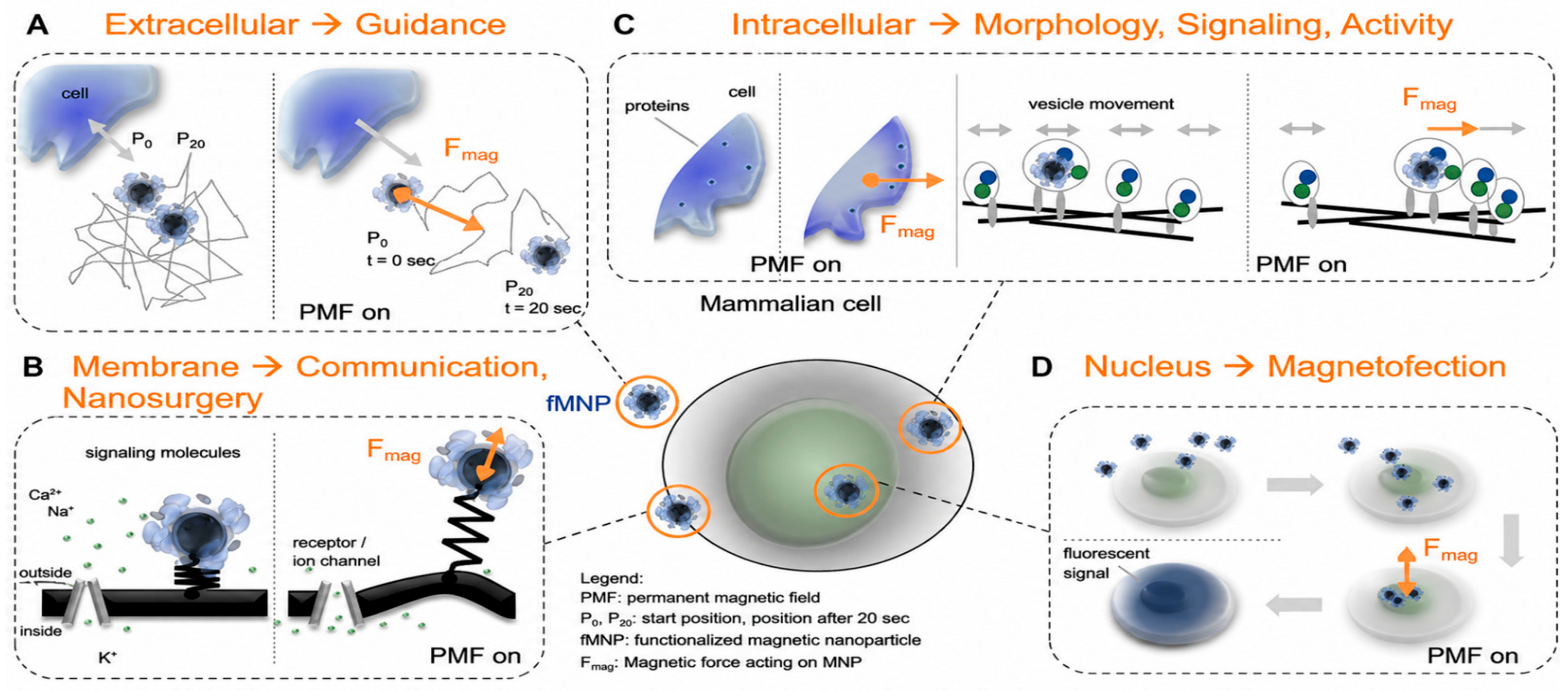
The use of MNPs, although widely explored for imaging and diagnosis, is demonstrated in Figure 2, showing how the interaction between magnetically mediating nanoparticles (FMNPs) and mammalian cells can be exploited to modulate different cellular functions. The schematic details the role of FMNPs in directing cell guidance in the extracellular space (A), controlling receptor functionality and cell communication when associated with the cell membrane (B), establishing protein gradients and modulating vesicle dynamics inside cells (C), and genetically modifying cells when localized to the nucleus (D) [78].
Despite this progress, there is still no explicit consensus on which combinations of magnetic and interfacial properties should be considered “ideal” for distinct biotechnological scenarios, from biosensing and separations to hyperthermia and targeted delivery. Reiss and coworkers [55] already emphasized that device performance in biotechnology depends sensitively on particle size, coating strategy, and the strength, frequency, and geometry of applied magnetic fields—points that remain highly relevant in current MNP-based biosensing and actuation platforms [79,80].
Recent reviews on iron-oxide nanomaterials similarly emphasize that discrepancies among studies frequently stem not from direct conceptual conflict but from variations in synthesis methods, dispersion media, surface functionalization, and magnetization protocols, which hinder the cross-comparison of in vitro and in vivo results [55,62,63,81].
In line with the present technical report, this plurality of approaches, while scientifically fertile, translates into methodological gaps, limited experimental validation under clinically relevant conditions, and an incomplete set of unifying conceptual models, all of which must be addressed to reconcile laboratory performance with robust industrial and clinical translation of magnetic nanoparticle technologies.

3.1. Stability of Magnetic Nanoparticles

The stability of MNPs under environmentally and physiologically relevant conditions is a primary design constraint for any biotechnology or therapeutic application. Preservation of hydrodynamic size, dispersibility, aggregation state, and surface-functional integrity determines not only colloidal behavior but also bioavailability, selective engagement with molecular or cellular targets, and spatiotemporally controlled biodistribution in vivo [82,83].
Stable magnetic colloidosomes fabricated by click-mediated crosslinking at liquid–liquid interfaces exemplify how covalent interparticle coupling can maintain size and permeability while enabling magnetically triggered response [82]. Likewise, dendritic-polyglycerol–stabilized iron-oxide nanoparticles have shown that dense, hydrophilic shell architectures can suppress aggregation, prolong circulation, and support selective imaging of inflamed tissues by MRI, underscoring the central role of surface engineering in long-term stability [84]. More recent reviews on surface modification of iron-oxide nanomaterials converge on the notion that only a narrow stability window in size, charge, and ligand density is compatible with robust performance in nanomedicine [85].
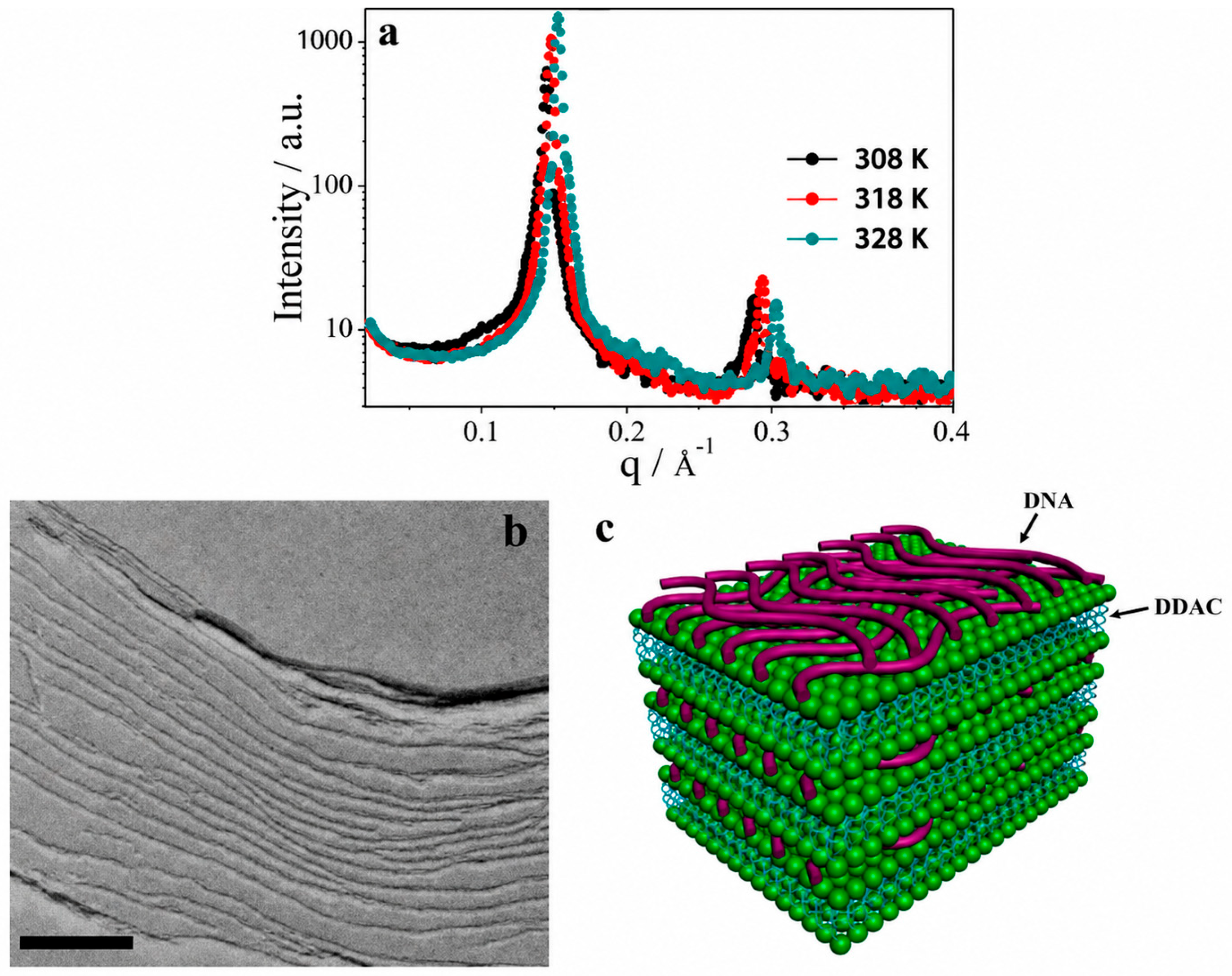
Figure 3 elucidates the microstructures of solvent-free thermotropic liquid crystal (LC) ferrofluids, and they are formed by the self-assembly of ssDNA and paramagnetic lipids. Figure 3a displays the small-angle X-ray scattering (SAXS) profiles, which exhibit two distinct Bragg peaks. Ordered multilamellar structures are observed—these peaks are typical with a periodicity of 43.5 Å (4.35 nm). These findings show that the structures are made up of ssDNA sublayers (about 10 Å) and fully extended DDACe lipid bilayers (about 33.5 Å), but there is no sign of ordered intralayer DNA packing. The freeze-fracture transmission electron microscopy (FF-TEM) in Figure 3b complements these findings, confirming the multilamellar nature and the repeat distance observed by SAXS (4.27 ± 0.70 nm). The images also suggest that the DNA−DDACe layers are not perfectly flat, with nucleic acids randomly oriented on the lipid bilayers. These findings enabled the development of the schematic diagram depicted in Figure 3c, which depicts the proposed microstructures for ferrofluids in the LC phase [83].
When destabilizing processes, such as agglomeration, surface oxidation, ligand desorption, and coating degradation, are strongly driven by variations in pH, ionic strength, temperature, and biomolecular composition of the surrounding medium, they can rapidly compromise colloidal stability, reduce tissue penetration, and alter magnetization dynamics, leading to reduced drug-delivery efficiency and loss of imaging contrast [86,87]. Research on iron-oxide dispersions has demonstrated that transitioning from low-molecular-weight ligands to multidentate polymers significantly enhances colloidal stability while concurrently altering cellular uptake, highlighting the strong relationship between physical stability and bio-interactions [88,89]. On the other hand, structural or chemical instability can trigger the release of metal ions or degraded coating fragments. These effects are particularly problematic for long-circulating or repeatedly dosed MNP formulations intended for chronic or precision therapies and are frequently associated with oxidative stress, membrane damage, and pro-inflammatory responses in vitro and in vivo [21,90].
Current convergent studies suggest that the stability requires more than just varying one parameter at a time. In physiological microenvironments, MNPs undergo concurrent variations in pH, gradients in ionic strength, and intricate mixtures of serum proteins, lipids, and metabolites, such as localized redox/oxidative conditions. Nonetheless, most routine stability assays are still carried out in simplified buffers or protein-poor media, which only partially capture this complexity. Recent reviews point out that testing nanoparticle coatings in the unrealistic conditions of the lab makes them seem more stable than they really are, because their problems only become apparent in real-world fluids like serum, whole blood or the fluid in the tumor microenvironment [88,91,92]. This discrepancy between laboratory conditions and in vivo environments is now acknowledged as a significant impediment to the clinical translation of magnetic nanosystems, in accordance with the current technical evaluation.
At a more detailed level, the proteins that rapidly form a coating on the surface of magnetic nanoparticles are critical in determining the stability and biological fate of these particles. The proteins that adsorb from blood or tissue can either stabilise or aggregate particles depending on which proteins adsorb, the orientation of adsorption and the coverage of the surface, and the same proteins also determine the detection of particles by the immune system, uptake by cells, and distribution in the body. For example, certain proteins from blood make iron-oxide nanoparticles appear smaller and more dispersed, while others bind the particles together and accelerate their clearance from the body. Detailed studies have also shown that even small changes to the particle core or surface coating can result in changes to the proteins that adsorb, affecting stability and safety in ways that cannot be easily predicted in advance, as demonstrated by studies such as those conducted by Zhu et al. [61], and cited works by Wiogo et al. [68] and Safi et al. in [65,85,89,90,92].
This implies a more holistic approach to studying MNP stability, one that brings together surface chemistry, colloids and interfaces, and cell biology as a single, integrated system rather than as individual components. Multi-parameter stability maps—constructed under systematically varied pH, ionic strength, protein/lipid content, and oxidative load that are combined with advanced in situ characterization (e.g., magnetic relaxometry, DLS–SAXS coupling, high-resolution MRI phantoms) and mechanistic biological readouts—can begin to reveal quantitative design rules for robust magnetic nanoplatforms [17]. Such multi-scale integration is essential for the rational engineering of MNPs that maintain consistent functional performance under realistic biomedical use conditions, closing the gap between proof-of-concept in vitro data and reliable operation in complex tissues and organs.

3.2. Discrepancy Between Laboratory Findings and Industrial/Clinical Practice

The main obstacles to the clinical use of MNPs—and nanomedicines more broadly—are multifactorial, tightly interconnected, and collectively responsible for the extremely slow pace of practical implementation. At the biological level, inorganic MNPs engage in complex, context-dependent interactions with proteins, membranes, immune cells, and tissue microenvironments, leading to biodistribution and toxicity profiles that are only partially predictable from conventional preclinical models; meta-analyses show that, on average, <1% of the injected nanoparticle dose actually reaches solid tumors, sharply constraining therapeutic index and cost-effectiveness [93]. Technically, magnetic control itself is not trivial, mainly due to problems such as strong dipole–dipole interactions between particles that promote aggregation and consequently alter rheology, as well as magnetic relaxation and clearance. Achieving deep three-dimensional guidance and actuation requires highly heterogeneous field gradients, which are difficult to generate safely in humans. The use of stents or ferromagnetic implants to target magnetic nanoparticles sacrifices non-invasiveness and introduces additional safety and regulatory burdens. One of the major challenges is to not compromise reproducible in vivo performance and achieve safety for translational use [94].
On the research side, several authors argue that early-stage development is often dominated by incremental optimization of physicochemical properties and “beautiful” in vitro data, with insufficient clinical input and disease-driven design, a dynamic captured by Park’s “invisible gorilla” metaphor for drug delivery, in which the field obsessively tracks secondary metrics while missing the clinically relevant endpoint [95,96]. This is exacerbated by publication bias towards positive outcomes and lack of standardized methods for nanoparticle characterization that would allow for cross-study comparison or development of robust design rules [97,98]. And in the clinic, the results are often disappointing. Although some nanoformulations, including magnetic nanoparticle systems, have been reported to improve drug exposure, safety, or quality of life, definite improvements in overall survival or durable disease control are scarce, because many programs fail due to insufficient magnitude of effect, inability to replicate findings, or appearance of unexpected toxicities upon assessment across a wide range of patients and co-medications [99,100]. Together, these scientific, technological, and translational hurdles explain why MNPs, despite their compelling physics and rich preclinical literature, still occupy only a narrow niche in everyday industrial and clinical practice.
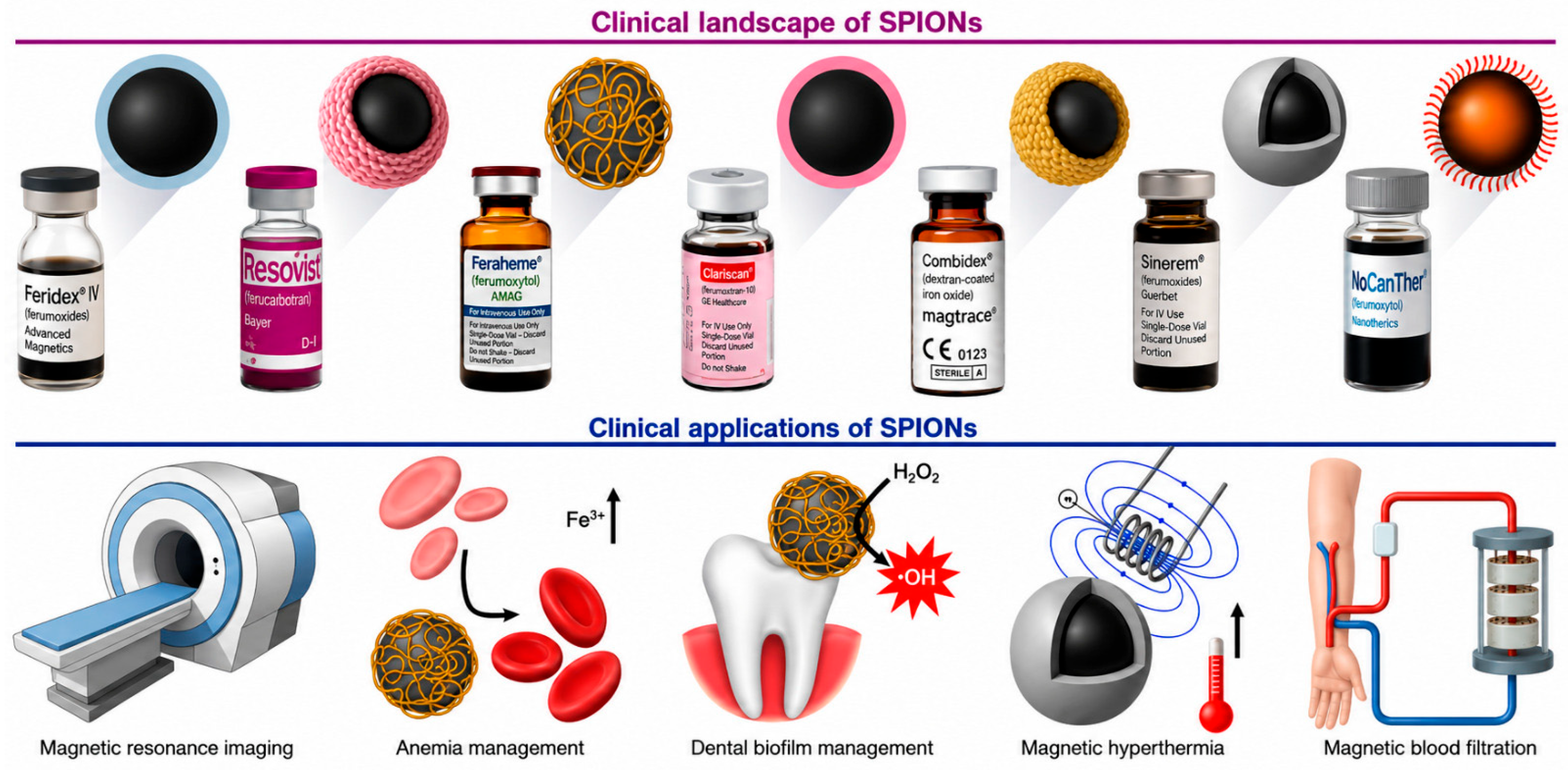
The contrast between the promising performance of magnetic nanoparticles in carefully controlled laboratory settings and the relative paucity of their translation into practical industrial or clinical applications has been observed in broad reviews of SPIONs and similar systems, which note that while hundreds of preclinical studies in model environments have demonstrated strong performance, only a few formulations have progressed to clinical trials or the market, and some of those approved have since been withdrawn [20,38,101,102,103] (see Figure 4).
There are so many translational bottlenecks across applications, as well as in diagnostic imaging, image-guided drug delivery, hyperthermia, and environmental remediation, where issues of reproducibility, long-term safety, and large-scale manufacturability systematically erode the apparent promise seen in proof-of-concept studies [17,104,105]. Within this context, seminal translational analyses and reviews had already anticipated that the gap between bench-scale optimization and real-world deployment would become one of the defining challenges for MNP-based technologies [97,99,101].
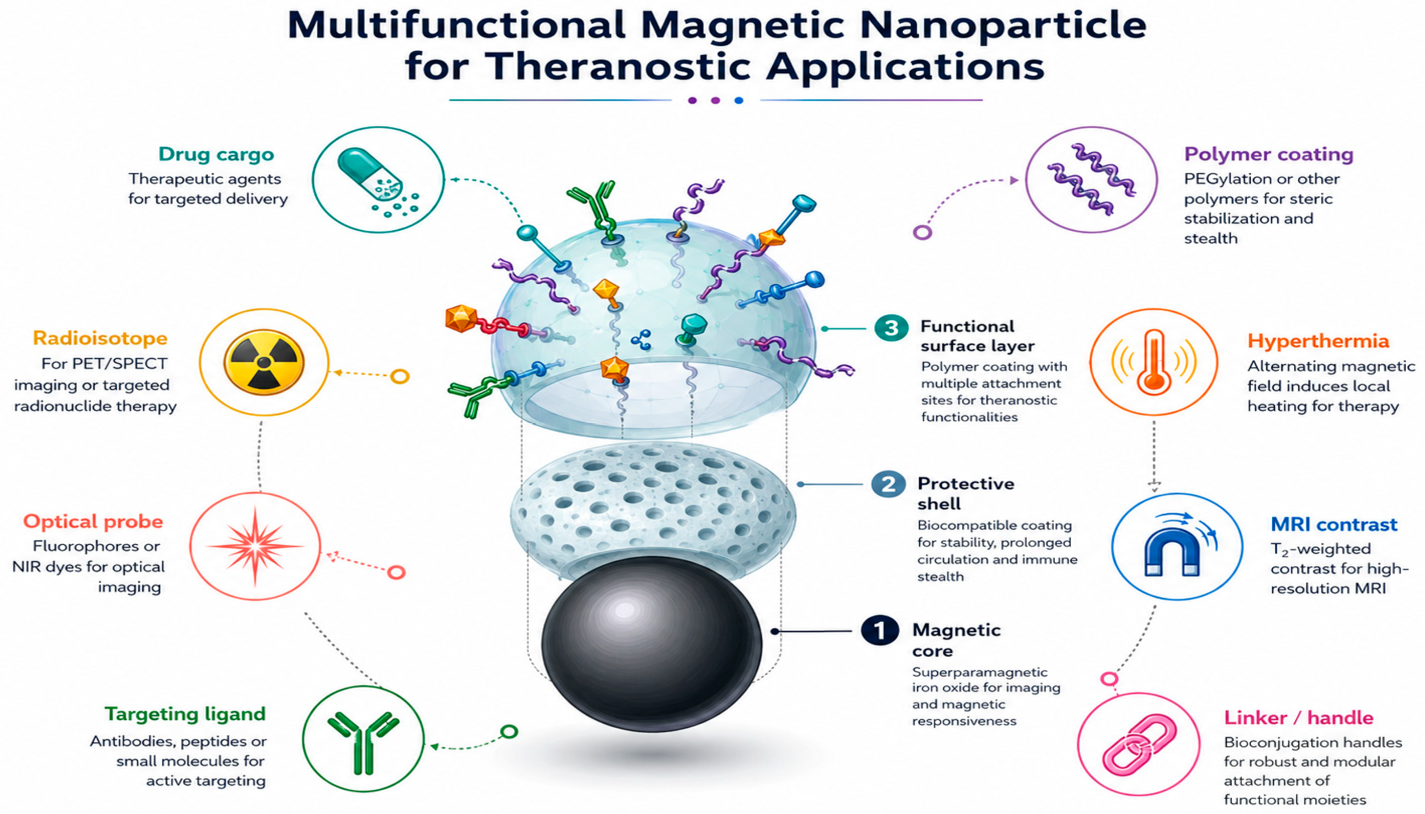
Figure 5 schematically illustrates the possible types of functionalized MNPs. Koksharov et al. [77] explain that this functionalization is a crucial task, as it involves modifying MNPs to improve existing properties or to obtain special specific properties for each practical medical application. For instance, it’s necessary for the MNP drug nanocarrier to freely exist in biological fluids and then specifically interact with its target. When MNPs are used for diagnostic purposes, radioactive or fluorescent molecules must be pre-attached to their surface, which is an example of such functionalization.
A central contributor to this discrepancy is the extreme sensitivity of MNP physicochemical properties to the synthesis route and its scale. Parameters that are straightforward to tune in small batches—temperature ramp rates, precursor concentration, reagent addition kinetics, reaction atmosphere, and purification sequence—can be tightly correlated with core size, polydispersity, magnetic anisotropy, saturation magnetization, and surface chemistry, all of which underpin biological performance. Upscaling, however, alters mixing regimes, heat and mass transfer, and residence-time distributions so that nominally identical recipes often yield particles with measurably different size distributions, aggregation behavior, and magnetic signatures. Detailed studies on nanobead and MNP upscaling, as well as on continuous-flow and microfluidic synthesis, make clear that even modest deviations in mixing or thermal profiles can propagate into clinically relevant changes in relaxivity, heating efficiency, and biodistribution [106,107,108,109]. In the absence of rigorously standardized synthesis and processing protocols, it is difficult to design robust processes compatible with Good Manufacturing Practice (GMP) and regulatory expectations [110].
Experimental and theoretical studies increasingly indicate that manufacturing constraints become more severe—not less—when MNP formulations are engineered with the sophisticated architectures required for modern theranostics (e.g., multi-component shells, targeting ligands, stimuli-responsive linkers). Nanomedicine reviews and translational frameworks consistently identify batch-to-batch variability, incomplete characterization across scales, and limited process analytical technologies (PAT) as major obstacles to clinical approval [111,112,113].
Biological performance is rarely governed by a single descriptor such as hydrodynamic diameter or zeta potential; rather, it emerges from high-dimensional parameter spaces that are difficult to control when moving from gram-scale to kilogram-scale production. Advanced strategies based on quality-by-design (QbD), continuous manufacturing, and real-time monitoring are only beginning to be implemented for magnetic nanomaterials, but studies show that they can markedly reduce variability and improve the reliability of industrial-scale production [114,115]. The complexity and challenges in transitioning from laboratory approaches to industrial-scale continuous manufacturing of nanomaterials highlight the need to overcome obstacles such as scalability, post-processing stability, reproducibility, and a complete understanding of the impact of process parameters on product quality and safety [114].
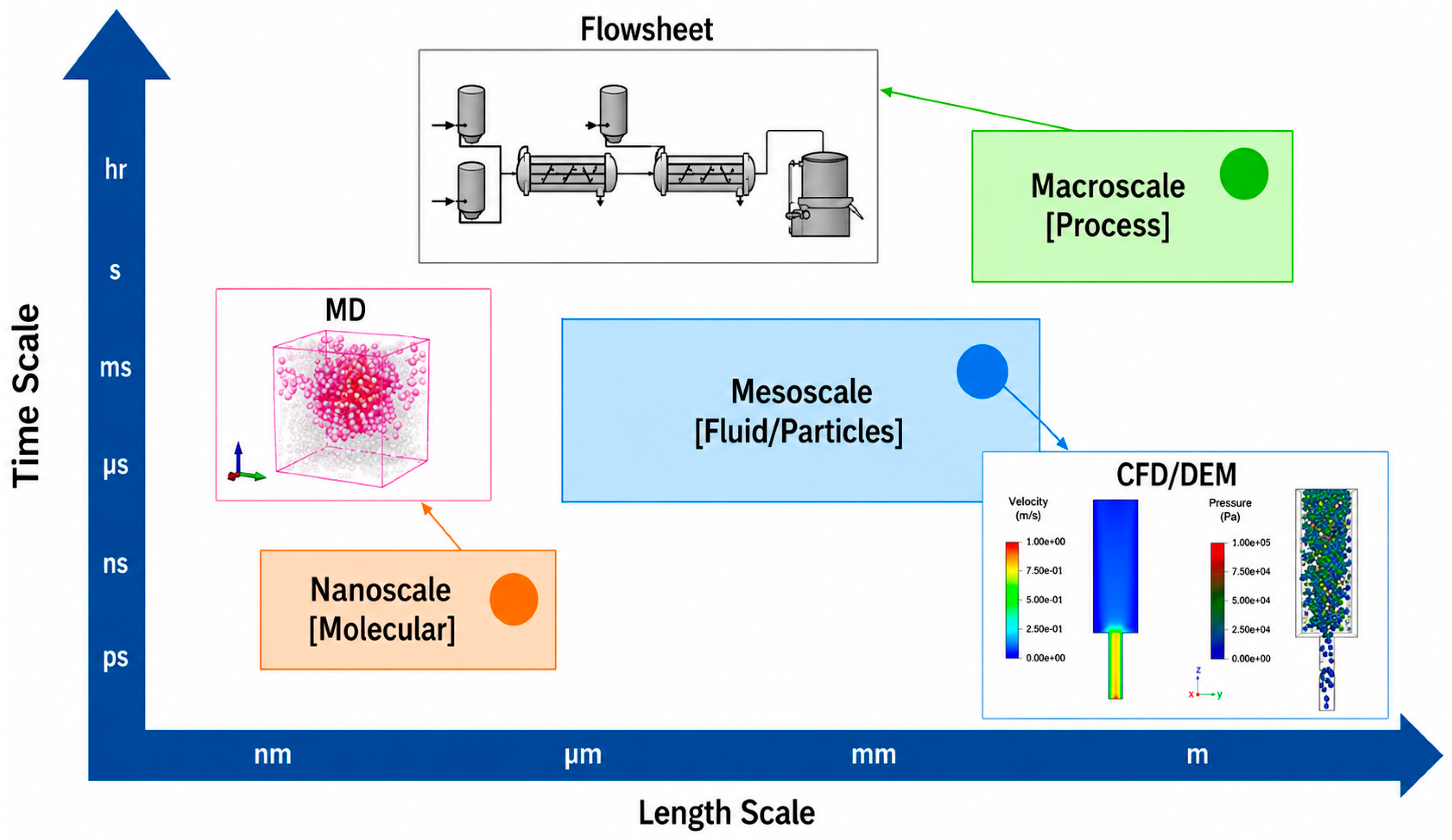
A “digital twin,” as illustrated in Figure 6, is a virtual representation of a physical process that operates in parallel with the physical plant. It integrates nanoscale, mesoscale, and macroscale models to enable real-time monitoring, parameter optimization, process modeling, and prediction. Its main purpose is to support decision-making and process control in the manufacturing of nanomaterials without directly impacting physical operations. It also contributes to reducing time-to-market [114].
The main differences observed between the simplified models used in preclinical studies and the complexity of real clinical or environmental settings widen the translational gap. In vitro assays, indispensable for this process, often use static monolayers, protein-poor media, or idealized flow conditions, which only partially mimic the heterogeneous microenvironments found in living systems or polluted matrices. The use of more complex analyses of high-impact nanomaterials for biomedical applications has the potential to help understand these differences. Furthermore, it is necessary to establish a greater capacity to collectively remodel the stability of MNPs, as they remain challenges to be overcome due to aspects such as shear stress, non-equilibrium protein coronas, dynamic pH and redox gradients, and immune surveillance [101,105,116].
In environmental and industrial biotechnology, analogous discrepancies arise when MNPs optimized in small-volume reactors fail to maintain reactivity, selectivity, or recyclability in complex effluents or soil matrices, where multicomponent ionic and organic backgrounds perturb colloidal and magnetic behavior [117].
Accumulating evidence also indicates that regulatory and safety frameworks, while essential, can unintentionally widen the laboratory–clinic divide if they are engaged only at late development stages. The current debate about the safety and regulation of nanomedicines is a testament to the importance of carefully evaluating the accumulation of drug carriers over time in organs, the immunogenic potential of drug carriers, the potential for off-target accumulation of drug carriers, and the toxic breakdown products of drug carriers under clinically relevant dosing schedules and in combination with clinically relevant chemotherapeutic agents, because the risks associated with drug carriers should not be inferred based on short-duration studies in mice alone [117,118]. Case studies using iron-oxide formulations, even after initial approval, have shown that these promising agents were discontinued due to safety concerns or manufacturing problems. This validates the need for alignment between formulation design, analytical capabilities, and regulatory expectations, aiming to advance nanosafety and nanotoxicology studies [20,101].
When considered collectively, these considerations emphasize the value of aligning the development of magnetic nanoparticles with industrial and clinical constraints. Therefore, instead of being an afterthought, scalability, standard protocols, GMP preparedness, strict quality control, toxicology, and reliability must be considered from the very beginning of materials discovery, because this approach is supported by various studies [17,105,109].
If this alignment is not achieved, the field will be doomed to be a fancy demonstration technology in the lab with little impact in practice and will fail to deliver the progress in environmental remediation, drug targeting, diagnostics, and image-guided therapy for which magnetic nanotechnologies are uniquely placed. In this sense, addressing the laboratory–practice discrepancy is not merely a matter of scale-up engineering but a systems-level problem that spans synthesis, characterization, biology, regulation, and economics.

Biodistribution, Clearance, and Long-Term Biological Fate of Magnetic Nanoparticles

The safe and predictable removal of magnetic nanoparticles (MNPs), and specifically magnetic iron oxide nanoparticles (MiONPs), from the body following completion of their diagnostic or therapeutic tasks is a leading unresolved barrier to clinical translation. The biological fate of MNPs is influenced by a complex interplay of factors, including core composition, hydrodynamic diameter, surface charge, coating chemistry, aggregation state, protein corona formation, route of administration, dose, and frequency of exposure [119,120]. Recent studies on biodistribution, clearance mechanisms, and long-term biological fate of MiONPs have concentrated on four closely related areas: the role of the mononuclear phagocyte system (MPS), renal elimination, the role of the protein corona, and quantitative pharmacokinetic and toxicokinetic (PK/TK) characterization. These areas are central to clinical translation because they explain how MiONPs are processed through and interact with biological systems, providing a rational basis for the design of safer and more effective theranostic platforms [121].
Mononuclear phagocyte system uptake. Following systemic administration, a significant fraction of iron-oxide nanoparticles is rapidly recognized by the MPS (circulating monocytes and tissue-resident macrophages) and is sequestered primarily in macrophage-rich organs such as the liver and spleen. Particle size, shape, surface charge, and coating chemistry are strong modulators of this uptake [122]. To prevent premature clearance or redirect particles to specific tissues, surface engineering strategies have employed stealth coatings such as polyethylene glycol (PEG) or zwitterionic polymers, as well as biomimetic ligands such as immunoglobulins or CD47-mimetic peptides, which can extend circulation time or promote active targeting, as reported in recent reviews on nano-bio interactions [122,123].
Renal elimination pathways. Smaller or more degradable fractions of MiONPs may be cleared, at least in part, through the kidneys or via progressive biotransformation and incorporation into endogenous iron-handling pathways. Renal clearance is largely controlled by hydrodynamic diameter and surface charge, favoring smaller, neutrally or negatively charged particles for glomerular filtration. Current design efforts are increasingly directed towards tuning these parameters to promote renal excretion, reduce long-term organ accumulation, and mitigate chronic toxicity, as noted in recent studies on nanoparticle clearance dynamics [123].
Impact of the protein corona. Upon exposure to biological fluids, MiONPs rapidly adsorb proteins and other biomolecules to form a dynamic protein corona, which effectively redefines their biological identity [122,124]. The composition, orientation, and density of the corona can stabilize or destabilize colloids, modulate cellular uptake pathways, and modify recognition by the immune system [123]. Critically, the corona can mask targeting ligands or accelerate MPS clearance, effects that are difficult to predict from in vitro assays alone [121,122].
Quantitative pharmacokinetic and toxicokinetic studies. Robust development of MNP-based nanomedicines requires quantitative PK/TK analyses that track circulation half-life, organ distribution, degradation products, iron metabolism, urinary and fecal elimination, tissue retention, immune activation, and long-term histopathology over timeframes relevant to clinical use [119,121]. Approaches that radiolabel the 59Fe in the nanoparticle core allow for the prolonged monitoring of biodistribution and the recycling of iron into endogenous pools. Long-term studies have confirmed predominant accumulation in the liver and spleen with gradual entrance into endogenous iron pools, as reported by Ahmad et al. [125] and Walton et al. [126] utilizing isotopic tracing. Advanced imaging modalities, such as magnetic particle imaging (MPI) and multimodal MRI/PET protocols, as well as intravital microscopy, are providing greater spatial and temporal resolution of these events at the organ and cellular levels [119,121]. Long-term fate studies have shown that the transformation of MiONPs into ferritin or hemosiderin, along with the upregulation of erythropoietic markers, occurs without overt tissue toxicity under controlled dosing regimens [125,126].
Unwanted particle accumulation during therapy represents an additional and clinically relevant dimension of this problem. During therapeutic use, particularly under repeated-dose regimens or prolonged theranostic protocols, MiONPs may accumulate in non-target organs such as the liver, spleen, lungs, kidneys, and bone marrow, where persistent retention can extend beyond the intended therapeutic window. This off-target sequestration may compromise treatment specificity and contribute to macrophage overload, altered iron homeostasis, oxidative stress, chronic inflammatory signaling, and organ-specific toxicity. Accordingly, the success of magnetic nanotherapies depends not only on maximizing target-site delivery, but also on minimizing unintended accumulation and ensuring controlled biodegradation and elimination [127,128,129,130,131,132,133,134].
Critical considerations and remaining challenges. However, clearance should not be considered intrinsically safe simply because the core material is an iron oxide. Particle persistence, coating degradation, local iron overload, lysosomal processing, inflammatory activation, and accumulation under repeated dosing can disrupt tissue homeostasis and lead to delayed adverse effects that are not captured by short-term in vitro assays or acute animal studies [119,120,121,122,123,124,125,126]. Consequently, clearance should be treated as a primary translational endpoint, not just a secondary toxicological observation.
Several major challenges remain:
  • Physiological relevance: There is a need for more physiologically relevant systems, such as 3D organoids, humanized mouse models, and microfluidic “organ-on-chip” platforms, to represent the combined dynamics of protein corona evolution, MPS activity, and tissue-specific barriers [121].
  • Multidimensional optimization: Surface modifications that decrease MPS uptake can also affect targeting efficiency, colloidal stability, and manufacturability at large scales [122,123].
  • Standardization: The lack of standardized protocols for PK/TK studies hampers comparison of data across laboratories and hinders regulatory evaluation [119,120].
  • Long-term safety data: Data regarding chronic exposure, inter-individual variability, and the potential for perturbation of iron metabolism in susceptible populations are lacking [125,126].
Continued advancements in this area will require the ongoing integration of rigorous materials engineering with mechanistic biological insight and application-driven validation. Only through such coordinated, multidisciplinary efforts can MiONPs be developed as safe, effective, and clinically translatable nanoplatforms for precision diagnostics and therapy.

3.3. Methods and Techniques That Are Insufficient, Inadequate or Missing

A critical reading reveals persistent methodological gaps that constrain both the depth and reproducibility of current studies. A first axis of limitation lies in the mathematical and computational modeling of magnetically driven fluids—particularly when MNPs are embedded in complex media containing gyrotactic or chemotactic microorganisms. While recent bioconvection models by Sarkar, Das, and co-workers (2024) represent a step forward in describing magneto-thermo-bioconvection of nanofluids with gyrotactic microbes, they typically rely on highly idealized geometries, effective single-phase approximations, and reduced non-linear couplings [135,136,137].
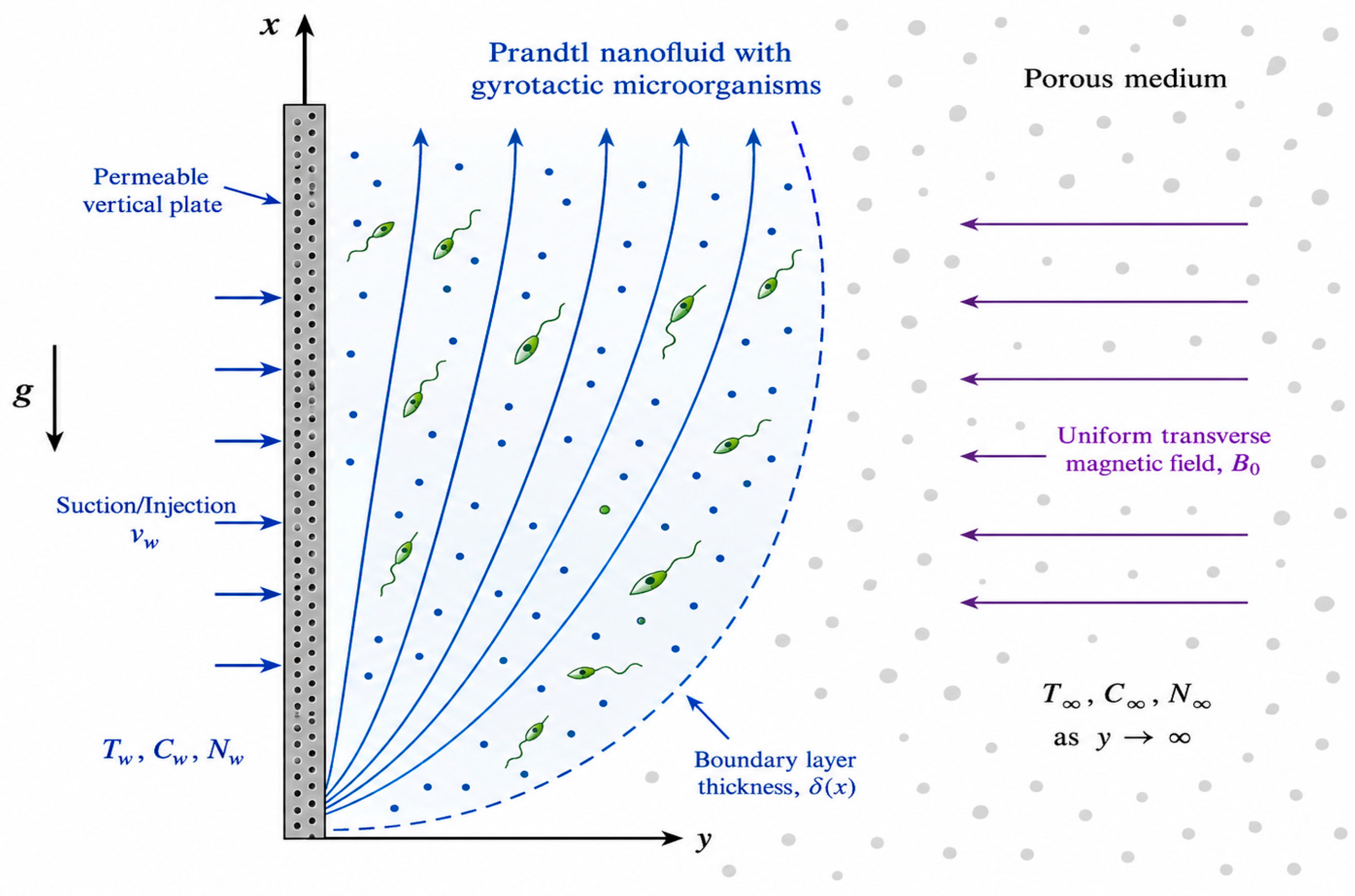
Only a handful of studies begin to address fully coupled multiphase flow, non-Newtonian rheology, reactive mass transport, and magnetization dynamics in the same framework—let alone under boundary conditions that resemble real biotechnological systems such as biofilters, packed beds, or microfluidic bioreactors [138,139]. As a result, many predictions remain qualitative and difficult to translate into design rules for scalable devices. Figure 7 represents a two-dimensional, time-independent, and incompressible flow of a non-Newtonian Prandtl nanofluid, which contains gyrotactic microorganisms. This flow occurs over a permeable vertical plate and through a porous medium. The system is subject to a uniform transverse magnetic field of strength B0. The figure, therefore, establishes the physical scenario and the coordinate system (with x and y axes) used for the mathematical formulation of the problem.
From a characterization standpoint, there is a recurrent lack of essential physicochemical data: hydrodynamic size distributions by dynamic light scattering (DLS), morphology by transmission electron microscopy (TEM), zeta potential, and structural analysis by X-ray diffraction (XRD) or complementary spectroscopies are still absent or superficially reported in a substantial fraction of MNP-based biotechnology studies. Yet these parameters are fundamental to assessing composition, crystallinity, polydispersity, purity, and batch homogeneity—prerequisites for any robust correlation between structure and biological response. For decades, there has been discussion about the minimum amount of information needed for bionanomaterial investigations. Attempts to align the characterization of nanomaterials are frequent. Determining the size of nanoparticles (TEM/DLS) and their surface properties, including charge and colloidal stability, is critical for their safety. But, in practice, these suggestions do not always work [140,141,142]. This disparity is evident in the editorial standards and position papers of ACS Nano, as well as in projects associated with Nature and ISO working groups. There is a demand for standardized processes and checklists of “minimum information” for the characterization of nanomaterials. From this perspective, regarding requirements, this stance is crucial for the advancement of nanotechnology. They also point out that factors that are not sufficiently published or consistently monitored hinder reproducibility and regulatory acceptability [143,144,145].
Even when basic physicochemical characterization is reported, advanced magnetic and dynamic readouts remain underutilized. In many biotechnology papers, MNPs are still described solely by static magnetometry (e.g., VSM) and generic relaxation times, which provide only a partial picture of their behavior under oscillating fields or in crowded biological matrices. In contrast, more specialized modalities—magnetic particle spectroscopy (MPS), frequency-mixing or dual-frequency detection schemes, AC susceptometry (ACS), and related techniques—can resolve non-linear magnetization dynamics, Brownian vs. Néel relaxation, hydrodynamic mobility, and surface binding events with high sensitivity [146,147,148,149,150].
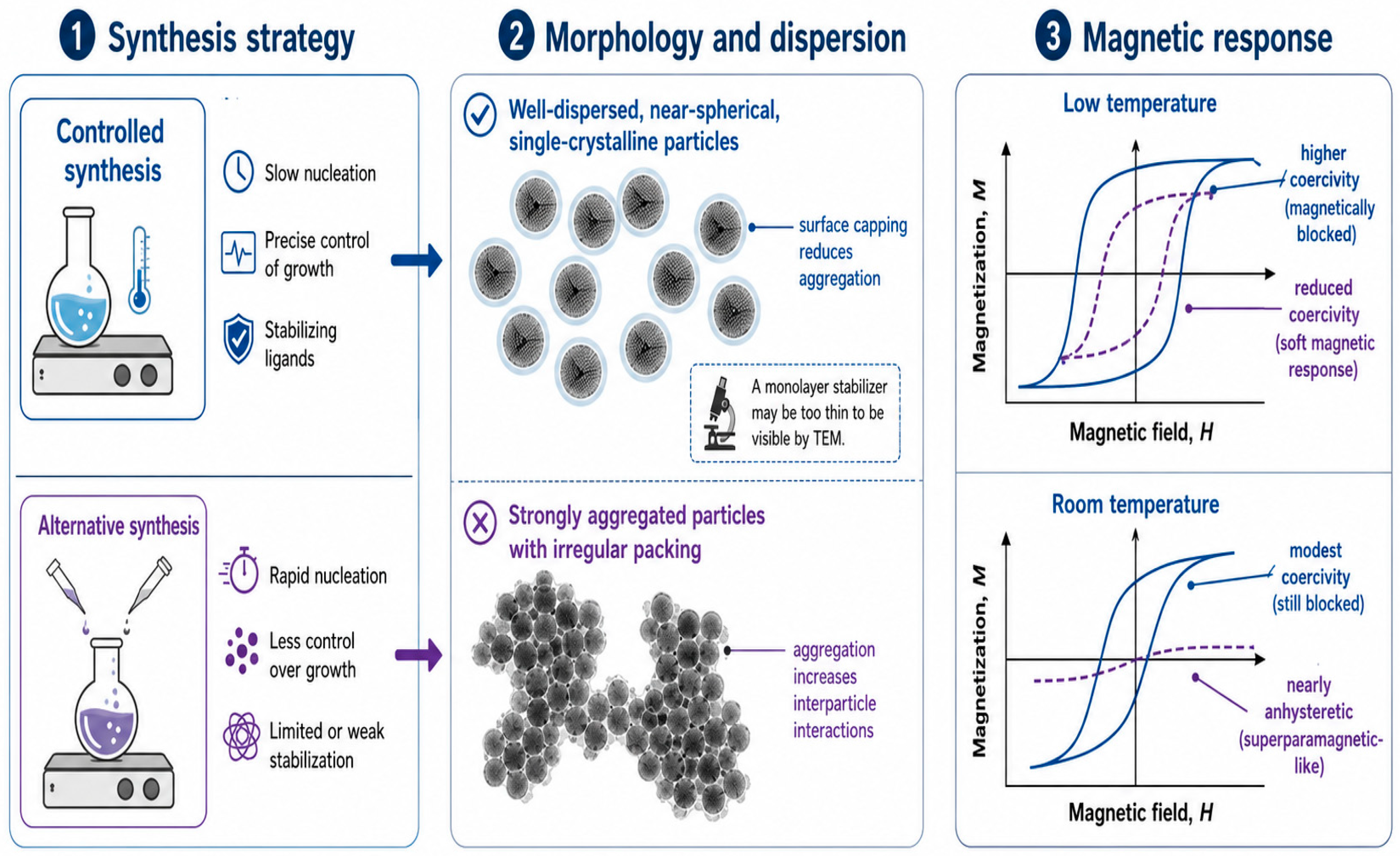
Nanoparticles prepared by the thermal decomposition method (CF-td and ZF-td) exhibit a single-crystalline, near-spherical character. Their TEM inspection suggests they are capped with oleic acid/oleylamine, which prevents aggregation and creates spacing between individual particles. In contrast, hydrothermally prepared CF-h nanoparticles were highly aggregated. Individual citrate-stabilized ZF-s@cit particles were indistinguishable from their bare ZF-s counterparts, as the citrate forms only a monolayer that is not observable by TEM. The DC hysteresis curves of “bare” magnetic cores and dry Resovist®, measured at 5 K and 300 K. Cobalt ferrite nanoparticles (CF-td and CF-h) exhibit high coercivities at 5 K (1.57 T and 0.95 T, respectively), attributed to the high magnetocrystalline anisotropy of Co2+ cations. Even at 300 K, although their coercivities decrease (to 57 mT and 16 mT), these particles remain magnetically blocked. In contrast, zinc ferrite nanoparticles (ZF-td and ZF-s) show coercivities of 19 mT and 24 mT at 5 K, which are typical values for soft magnetic materials. The materials are comparable to the 17 mT magnetization of Resovist®. At 300 K, the hysteresis curves of zinc ferrite are “seemingly anhysteretic,” suggesting either a very low coercivity below the experimental detection limit or a superparamagnetic regime on the SQUID magnetometry timescale. Resovist®’s magnetization was normalized by its iron concentration for comparison [110].
For example, recent work has shown that MPS-based platforms can detect subtle changes in MNP mobility associated with biomolecular binding or microenvironmental viscosity, enabling point-of-care assays and in situ monitoring that are directly relevant to bioprocess control [107,109]. Yet these techniques are rarely integrated systematically into biological studies, leading to an under-characterization of the very magnetic behaviors that distinguish MNPs from other nanomaterials.
Methodological limitations are also evident in experimental strategies that combine MNPs with gyrotactic microorganisms or other living agents in magnetically driven fluids. Protocols for infusing, dispersing, and sustaining microbial populations in magnetic nanofluids are often optimized for small-scale, short-term experiments, with limited attention to scalability, long-term viability, or operational robustness under variable magnetic fields, temperature, and medium composition. The absence of standardized approaches to control cell density, orientation, motility, and spatial heterogeneity in such hybrid systems complicates the interpretation of observed bio-convection and transport phenomena and makes cross-study comparison difficult [97,98,99].
This situation is exacerbated by frequent inconsistencies in key experimental parameters—field strength and frequency (especially for AC fields), exposure time, MNP concentration and functionalization, shear conditions—which are sometimes underreported or only qualitatively described. Collectively, these gaps underscore the urgency of a more rigorous methodological infrastructure for MNP-based biotechnology. Standardized, fully reported characterization pipelines; multi-physics models that couple realistic geometries, transport, magnetization, and biological response; and carefully designed protocols for living, gyrotactic, or biofilm-forming systems are all needed to move beyond isolated proof-of-concept studies. Emerging community frameworks—such as minimum-information reporting guidelines for bio-nano experiments and nanomaterial biocorona studies—offer concrete templates for such standardization and should be explicitly adopted and adapted for magnetic nanoparticle platforms [105,111]. Only with this level of methodological maturity will it be possible to build reproducible structure–function relationships and to translate magnetic nanotechnologies into robust, scalable biotechnological applications. The Figure 8 summarizes the proposed relationship between synthesis strategy, particle morphology/dispersion, and magnetic response, highlighting how changes in structural organization may influence the magnetic behavior of the system.

3.4. Underexplored Combinations of Methods

A second critical dimension in the development of MNPs for biotechnology is the relative scarcity of truly multitechnique and multidisciplinary approaches. In much of the current literature, synthesis, physicochemical characterization, and evaluation of biointeractions are still performed in a fragmented manner, with limited integration of complementary methods. Comprehensive reviews emphasize that robust design of bioactive MNPs requires concurrent access to structural, topographical, magnetic, and chemical information across scales, yet many studies still rely on minimal toolsets, such as TEM plus basic magnetometry, with only rudimentary colloidal or surface analyses [7,9,151].
Multimodal workflows that systematically combine TEM, AFM, and spectroscopies such as UV–Vis, FTIR, Raman, and fluorescence—under conditions close to the physiological state—remain comparatively rare, even though they have proven powerful in reconstructing core–shell architectures, nanoscale roughness, ligand conformation, and local chemical environment in a way that directly links morphology, surface chemistry, and protein-corona formation [7,152].
A particularly promising, yet underdeveloped, area concerns the deliberate combination of MNP functionalities with the selective delivery and detection of proteins. While MNPs are widely explored as MRI contrast agents and drug carriers, rational conjugation with protein vectors, disease-specific peptides, and engineered binding motifs is only beginning to be addressed systematically for complex pathologies. In neurodegenerative disease, for example, magnetic nanoplatforms have been proposed as dual imaging–therapeutic systems for amyloid and tau pathology, integrating iron-oxide cores with ligands that recognize misfolded proteins and, in some designs, co-deliver neuroprotective cargo [7,153].
Targeted MNPs functionalized with β-amyloid-binding tetrapeptides and fluorescent labels illustrate how magnetic, optical, and molecular-recognition elements can be co-engineered for early Alzheimer’s disease diagnosis, providing MR-visible and optically traceable probes for amyloid plaques [153,154]. Similar strategies are emerging for other proteinopathies, but these systems are still largely explored as isolated case studies rather than as part of a consolidated design framework for MNP–protein conjugates in precision diagnostics and therapy.
Related opportunities arise in oncology and advanced biotechnology, where integration of magnetic targeting, molecular recognition, and protein or peptide therapeutics remains far from fully exploited. In glioblastoma and other high-grade brain tumors, iron-oxide nanoparticles conjugated to antibodies or peptides against EGFR and related targets have demonstrated enhanced accumulation across the blood–brain barrier and improved MRI contrast, while co-encapsulating chemotherapeutics or radiosensitizers in lipid or polymer shells around the magnetic core [7,9]. These examples highlight how magnetically guided, stimulus-responsive release can be combined with disease-specific molecular targeting, yet they rarely leverage the full combinatorial space of protein ligands, field geometries, and microenvironment-responsive chemistries. More generally, systematic efforts to map how combinations of magnetic field parameters, protein design, and local tissue mechanics jointly determine targeting, retention, and controlled release are still in their infancy.
A third, often overlooked, axis concerns hybrid architectures that combine magnetic and non-magnetic supports in heterogeneous enzymatic cascades and other complex biomolecular systems, with clear implications for biofiltration, bioseparation, and magnetically addressable bioreactors. Recent work in biocatalysis shows that co-immobilizing multiple enzymes, or enzymes with chemocatalysts, on architectured supports can yield multicatalytic materials with tunable spatial organization, enhanced stability, and straightforward recycling [155,156].
Within this framework, the introduction of magnetic components allows remote manipulation and rapid separation, enabling cascades in which one or more catalytic steps are anchored to MNPs while others reside on non-magnetic carriers, thereby decoupling local microenvironments and kinetics [155,157]. Hybrid magnetic catalytic systems on micro- and nanoscale supports further illustrate how highly active, magnetically recoverable materials can operate in complex reaction media, suggesting analogous strategies for magnetically retrievable biosensors, enzyme-loaded filters, and smart delivery devices for labile biomolecules [156,158].
Taken together, these developments underscore that the field has only begun to explore the combinatorial design space linking synthesis, multimodal characterization, and engineered biointeractions. Systematic integration of high-resolution microscopy (TEM, AFM), advanced magnetic readouts, and spectroscopic probes with protein- and enzyme-level engineering would enable genuinely multiscale views of how MNP structure and microenvironment govern function in realistic biological contexts [7,152]. Moving from fragmented, single-technique studies to explicitly multitechnique and multidisciplinary protocols—supported by standardized reporting and cross-comparable workflows—is therefore essential to transform isolated proof-of-concept demonstrations into rationally designed magnetic nanoplatforms with robust clinical and biotechnological potential.

3.5. Lack of Experimental Validation of the Proposed Mechanisms

Despite an extensive body of theoretical and numerical work on MNP-based systems, there is a persistent and substantial gap between mechanistic models and systematic experimental validation in biologically relevant settings. Reviews on MNP hyperthermia show that sophisticated simulations of specific loss power, bio-heat transfer, and intratumoral temperature distributions are often calibrated on simplified phantoms or homogeneous media, with limited confrontation against quantitative in vivo data or physiomimetic 3D models [159,160]. At the same time, translational overviews of nanomedicine emphasize that mechanistic narratives about targeting, controlled release, or immune modulation frequently rest on sparse or indirect data, which is a key reason why many “highly promising” platforms do not advance beyond preclinical proof-of-concept [98,161].
Bifunctional magnetic–fluorescent nanocomposites are a paradigmatic example of this imbalance. Huang et al. [122] synthesized Fe3O4/CdSe/CdS nanocomposites with well-defined architectures and carefully characterized magnetic and optical properties, demonstrating robust control over core–shell construction and photophysical behavior [162]. Sun et al. [123] extended this concept to Fe3O4/CdTe systems, achieving detailed structural, spectroscopic, and magnetometric characterization and showing proof-of-principle immunolabeling of HeLa cells [163]. However, these and related studies typically fail to implement comprehensive in vitro and in vivo programs that measure cellular internalization pathways, subcellular trafficking, long-term biodistribution, pharmacokinetics, chronic toxicity, or therapeutic efficacy in complex physiological microenvironments. As a result, the proposed multifunctional mechanisms—simultaneous imaging, targeting, and therapy—remain only partially validated beyond the level of structural and spectroscopic plausibility.
A similar pattern is observed in the thermal and fluidic domains. Analytical and finite-element models describing heat deposition, magnetically driven flow, and nanoparticle transport in tissues or bioengineered constructs are now highly sophisticated, but their experimental confirmation is often limited to homogeneous gels, simple flow cells, or static 2D monolayers [159,160]. Only a minority of studies move systematically from modelling to calibrated in vivo validation, where predicted temperature fields and force distributions are directly compared with intratumoral thermometry, histopathology, and longitudinal outcome metrics such as tumor regression, recurrence, and systemic toxicity [160,161]. In many cases, the discrepancy between model assumptions (homogeneous media, isotropic perfusion, static vasculature) and the complexity of real tissues is acknowledged but not quantitatively resolved.
Emerging work with physiomimetic 3D tumor models and organotypic systems points to a concrete path for closing this gap. Soeiro and coworkers (2024) systematically reviewed the use of engineered 3D tumor models to screen hyperthermic nanomedicines, emphasizing how controlled gradients of oxygen, nutrients, extracellular matrix, and flow can be leveraged to test photo- and magnetically driven nanoparticles under conditions that more closely recapitulate the tumor microenvironment [164]. Still, integration of such platforms into the development pipelines of magnetic nanotechnologies is incipient, and many proposed mechanisms—field-guided accumulation, magneto-mechanical disruption, selective heating of specific cell subpopulations—remain under-tested in these more realistic models.
Altogether, this landscape underscores the need for broader, more rigorous experimental programs that explicitly link mechanistic hypotheses to quantitative data across scales: from 2D cell monolayers and physiomimetic 3D constructs to well-controlled animal models and early preclinical studies that jointly address toxicity, pharmacokinetics, immunogenicity, and efficacy [98,159,160,161,164]. Only on this basis will it be possible to consolidate the credibility of proposed mechanisms for magnetic nanoparticle action and to transform elegant theoretical descriptions into robust, deployable biotechnological and clinical platforms.

3.6. Gap in Unifying Conceptual Models

Magnetic nanoporous systems lack a cohesive conceptual framework that integrates the various interaction mechanisms involved. Substantial progress has been achieved in synthesis and functionalization over the last decade, including research aimed at improving the delivery and safeguarding of hydrophobic drugs and proteins through rough mesoporous silica (organo) nanocapsules [141,165,166] and electrochemical platforms utilizing magnetic nanoparticles for the sensitive detection of colorectal cancer biomarkers, such as methylated SEPT9 [167]. The integration of sensing and therapy through nanostructured platforms in therapeutically pertinent scenarios was also demonstrated, exemplified by enzyme-activated in situ self-assembled theranostic probes [168]. Nonetheless, these advancements are often characterized as distinct “verticals”—drug administration, sensing, imaging—rather than elements of a cohesive paradigm that links synthesis decisions, colloidal and interfacial behavior, and system-level performance under magnetic actuation.
Consequently, the literature remains fragmented into relatively separate threads: (i) optimization of coatings and in vivo degradation kinetics; (ii) delivery efficiency and therapeutic response; and (iii) biomarker detection and monitoring.
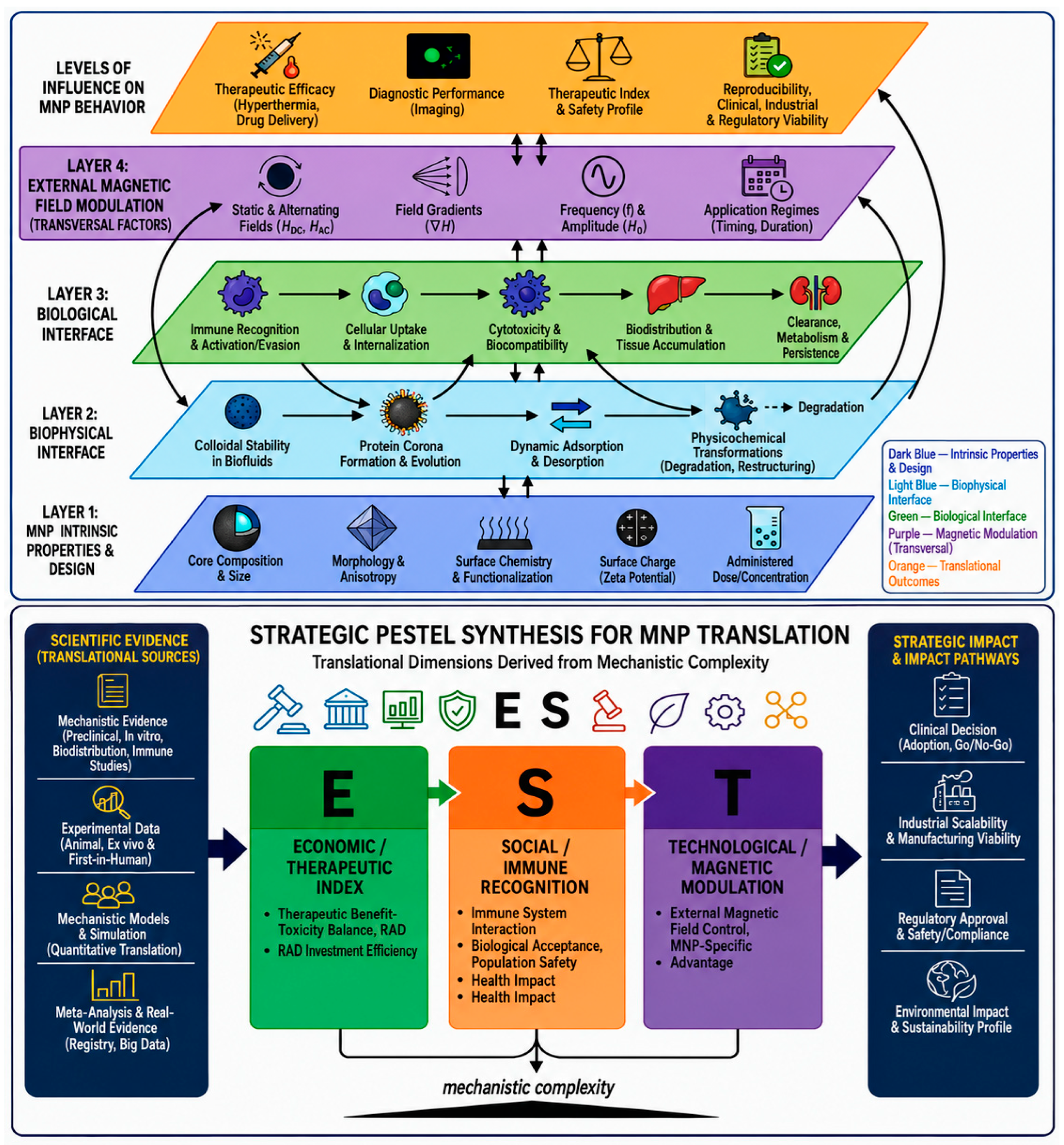
To explicitly address this conceptual fragmentation, we propose an integrative, multiscale framework that unifies physicochemical design variables, nanobio interface dynamics, biological responses, magnetic-field modulation, and translational constraints (Figure 9).
Reviews on protein corona formation and immune modulation show in detail how adsorbed biomolecular layers reshape nanoparticle biodistribution, targeting, and immunogenicity [169,170,171,172]. In parallel, multi-scale modelling efforts in nano-cancer drug delivery, biomolecular corona formation, and nanotoxicology provide mathematical descriptions that couple systemic transport, tissue penetration, and cellular uptake, often enhanced by data-driven or AI/ML approaches [173,174]. However, explicit theoretical constructs that tie together colloidal stability, dynamic protein corona evolution, degradation and clearance, immune-cell engagement, tissue-level transport, and modulation by time-varying magnetic fields are still scarce, especially for MNP platforms.
This lack of integrated models hinders the generalization of results from simpler systems to authentic physiological contexts and varied patient demographics. In vitro–in vivo correlation (IVIVC) studies for nanomedicines demonstrate that drug release kinetics, coronal remodeling, and microenvironmental heterogeneity (pH, ionic strength, oxidative stress) collectively govern pharmacokinetics and efficacy in ways that single-parameter descriptors such as size or zeta potential fail to encompass [175,176,177,178]. Established conceptual frameworks for magnetic nanoparticles (MNPs), such as the “unified view” of magnetophoresis separation, primarily focus on field–particle interactions and hydrodynamics, yet they insufficiently address the biointerfacial and immunological factors that ultimately affect safety and clinical efficacy [179].
To close this gap, conceptual frameworks must explicitly integrate data streams spanning synthesis, advanced characterization, in vitro and in vivo experiments, and multi-scale analyses at the molecular, tissue, and organ levels. Conceptually, such models should at least include (i) degradation kinetics and dissolution pathways for various coating and core compositions; (ii) cell type-specific and species-dependent responses, such as immunotoxicity and tissue repair; (iii) microenvironmental variability in pH, ionic strength, protein and lipid composition, and reactive species; and (iv) the spatiotemporal structure of dynamic magnetic fields used for targeting, actuation, or imaging. Multimodal toxicity frameworks for iron-oxide nanoparticles and imaging-enhanced drug-delivery models are now appearing in multiscale corona-prediction pipelines [171,172,176,180]. Nonetheless, these components are rarely combined to form a cohesive, MNP-focused framework capable of reconciling disparate datasets and guiding the rational design of next-generation magnetic nanoplatforms.
It is only through such unifying, data-driven conceptual models that we can systematically resolve inconsistencies between studies, forecast MNP behavior across diverse biological microenvironments and patient variability, and transition from predominantly empirical optimization to genuinely rational, mechanism-based engineering of magnetic nanomaterials with authentic translational significance. In this context, the development of integrative frameworks that amalgamate colloidal and interfacial physics, corona and immune dynamics, multiscale transport, and magnetically driven actuation emerges as a principal theoretical and computational challenge for the forthcoming decade of MNP-based biotechnology [173,174,178,179].

3.7. Underrepresentation of Populations, Organisms, and Geographic Regions

Currently, it has been reported that studies of MNPs in biotechnology are marked by a pronounced geographical asymmetry. Most experimental and translational work is concentrated in North America, Western Europe, and some Asian countries with high investment (China, Japan, and Republic of Korea). On the other hand, the African continent, Latin America, and large parts of the Global South remain underrepresented in primary data and clinical or environmental case studies. Clinical trials and patenting are heavily biased towards high-income regions, raising concerns about global health equity and context-appropriate innovation, as demonstrated in bibliometric analysis studies, as well as in policy-oriented work in both nanomedicine and nanotechnology, and consistently show that research capacity behaves unevenly around the world. Recent studies on MNP-based diagnostics, drug delivery, and bioseparation highlight that much of the initial preclinical and translational work is being conducted in a few centers with advanced infrastructure. This inequality directly affects both the prioritized diseases and the physicochemical design criteria and regulatory pathways considered viable [181,182,183].
A parallel imbalance exists at the biological level. The performance and safety of MNPs are still evaluated predominantly in a narrow repertoire of immortalized human cancer cell lines (such as HeLa, MCF-7, and U87), primary rodent cells, and murine models, with far less systematic attention to species and strains that capture the genetic, metabolic, and immunological diversity of under-represented human populations or to non-mammalian systems that are ecologically and agriculturally relevant. Nowadays, biomedical applications consistently emphasize that dosing, biodistribution, biotransformation, clearance, and toxicity are highly sensitive to subtle differences in physiology, immune status, and tissue microenvironment, yet genuinely comparative cross-population and cross-species studies remain rare and typically exploratory [90,184,185,186,187,188].
In a Latin American academic context, Lorenzato et al. [149] examined thermosensitive magnetoliposomes as a way to tune contrast in magnetic resonance imaging. When local groups manage to combine magnetic cores with flexible nanocapsules to address concrete problems in imaging and drug delivery, it’s a clear sign that innovation in Southern Hemisphere countries is not just theoretical—it can produce practical, locally relevant solutions [189,190,191].
A similar “local problem, local resource” logic appears in the study by Vijayalakshmi and colleagues [152], who immobilized cellulase on magnetite nanoparticles to valorize agricultural residues from Allamanda schottii in Brazil. Their results support a straightforward point: magnetic recovery can make enzyme reuse simpler and more scalable, and bioethanol production can be designed around the biomass and waste streams that a region actually has [175,192].
Jarhad et al. [153] offers another useful illustration with cinnamon-assisted CdS nanoparticles. These particles are not magnetic, but the lesson still translates: using native plant species and low-cost extraction methods can be a smart way to develop nanobiotechnological platforms that fit local constraints and opportunities. Conceptually, that same mindset can guide how MNP systems are engineered for regional needs, rather than copied from “one-size-fits-all” models. Finally, studies that combine magnetite-based nanoparticles with microalgae cultivation and harvesting for biofuel production reinforce the feasibility of pairing MNP technology with localized bioenergy efforts—especially when those efforts are grounded in local microbial diversity and production realities [103,193,194].
This paper provides the field with a direct contextualization and validation of its main limitations. By applying a strict and integrative methodological framework, this work moves beyond fragmented and isolated reports on the therapeutic and diagnostic potential of MNPs, and shows structural weaknesses that currently prevent scientific rigor and translational progress, because through a systematic analysis of a consolidated corpus of 870 studies, a semantic supergraph model, and a taxonomy-driven gap assessment, this work shows that many of the challenges faced by MNP-based biotechnologies are not isolated faults but systemic problems stemming from the lack of characterization protocols, validation, and conceptual integration.
This paper’s strong focus on safety, standardization, and control of the nanobio interface highlights how biological responses are sensitive to factors such as particle composition, size, surface chemistry, and dose, revealing the need for rational and reproducible design strategies. Consequently, the limitations of the field arise from unaddressed methodological, translational, conceptual, and geographical gaps—gaps that this analysis makes explicit and organizes into a coherent structure to guide future advances.

3.8. Structural Limitations and Systemic Gaps in Magnetic Nanoparticle Research

The main limitation of the existing MNP literature is the absence of rigor and quality in reporting methodologies, because most studies omit one or more of the fundamental physicochemical descriptors, such as hydrodynamic size distribution by DLS, zeta potential, and TEM-derived morphological parameters, necessary for structure-function inference and cross-study comparability. Magnetic characterization is often confined to static readouts, such as VSM, while dynamic readouts, like MPS and ACS, required to elucidate time-dependent magnetic responses and binding-mediated effects, remain underutilized. Moreover, integrative, multimethod study designs that enable concurrent probing of magnetic functionality together with selective protein delivery, hybrid magnetic-nonmagnetic architectures, and enzyme cascade systems within the same experimental framework are rare. Consequently, these knowledge gaps hinder mechanistic resolution, reproducibility, and translational relevance.
Another major issue is the disparity between bench and bedside, and the physicochemical properties of MNPs are highly dependent on the synthesis method—including temperature, precursor stoichiometry, and reaction kinetics. This results in significant batch-to-batch variation and hinders reproducibility, because this complicates scale-up and poses challenges for compliance with industrial standards and regulations. Furthermore, research indicates that, on average, less than 1% of an administered dose of MNP-based therapeutics actually accumulates in solid tumors in vivo, significantly decreasing the delivered drug payload and efficacy. Thus, biological parameters, particularly the dynamic protein corona, also influence the stability of such particles in vivo, their biodistribution, cellular uptake, and immune response. Moreover, biological parameters, particularly the dynamic protein corona, also influence the stability of such particles in vivo, their biodistribution, cellular uptake, and immune response, further contributing to translational barriers and limiting clinical implementation; therefore, this results in significant challenges for the development of MNP-based therapeutics.
The field is further limited by the lack of unifying conceptual frameworks that meaningfully integrate the basic phenomena of colloidal stability, protein corona dynamics, degradation pathways, immune interactions, and tissue-level transport under magnetic actuation, while theoretical models and simulations are increasingly sophisticated, but they are insufficiently anchored to systematic experimental validation, which prevents the translation of conceptual advances into mature, predictive, and deployable biotechnologies.
Finally, structural biases in the global research landscape undermine the external validity of the current evidence, although the geographical distribution of academic output has become more equitable in some fields, academic contributions are still dominated by North America, Europe, and China, while African and Latin American countries, among other regions of the Global South, are underrepresented in the research output. Moreover, the biological evidence base is built on a limited set of cell lines and animal models, which limits extrapolation across diverse biological, environmental, and epidemiological contexts, and therefore the reduced representativeness increases the likelihood that MNP-based technologies will be poorly matched to local conditions and global health priorities, leading to inequitable translation and impact.
Hence, the establishment of public policies to mitigate these barriers needs to be discussed urgently by the scientific community, and we initiate this discussion with the following propositions.

3.9. Public Policy Proposal for the Translational Advancement of Magnetic Nanoparticles

Public policies should prioritize the elimination of recurring methodological weaknesses and insufficient systematic characterization of MNPs.
  • Mandatory adoption of minimum information guidelines: We propose that all funded projects and scientific publications formally adopt MNPs’ minimum information reporting standards, which should at a minimum require disclosure of the following key physicochemical parameters: hydrodynamic size distribution by DLS, morphology and dispersion by TEM, zeta potential and crystallinity and phase composition by XRD. This is because such requirements are critical for interlaboratory comparability and batch-level traceability.
  • Integration of dynamic magnetic characterization: Funding calls should explicitly encourage and incentivize the use of advanced magnetic characterization methods as a standard practice, such as magnetic particle spectroscopy (MPS) and AC susceptometry (ACS), because these methods can provide valuable information about Néel/Brownian relaxation dynamics, hydrodynamic mobility, and surface-mediated interactions, which cannot be reasonably probed by static measurements such as vibrating sample magnetometry (VSM).
  • Promotion of integrated, multitechnique workflows: The funding policy should preferentially support research programs that include, in a combined and contemporaneous fashion, controlled synthesis, high-resolution physicochemical characterization (e.g., TEM, AFM, and appropriate spectroscopic methods), and functional biological assessment, and such an integrated approach should replace segmented or strictly serial approaches that preclude thorough structure–function correlation.
Given the pronounced “translational gap” identified, scientific development must be aligned with industrial and regulatory constraints from the earliest stages.
  • Early incorporation of scalability criteria: Projects funded under this FOA must provide, from the outset, a plan that clearly describes how scalable, reproducible, GMP-compliant material and process designs will be integrated into material and process development, and examples of the type of information that would be required in an application include a plan for how the scale-up will be achieved, statistically powered process controls and acceptance criteria, batch-to-batch variability and quality attributes, as well as a plan for how the synthesis, purification, and formulation steps will be integrated and documented with GMP-ready documentation and validation protocols.
  • Support for advanced manufacturing strategies: Targeted funding should be directed to continuous manufacturing paradigms, implementation of Quality-by-Design (QbD) frameworks, and the deployment of digital twins for real-time process monitoring and control, because these paradigms in concert are required to reduce batch-to-batch variability, increase process robustness, and enhance industrial reliability.
  • Comprehensive and longitudinal toxicological assessment: We advocate for policy to dictate the obligatory and systematic toxicological profiling across in vitro, ex vivo, and in vivo models (e.g., biodistribution, clearance kinetics, long-term accumulation, immunogenicity, and toxicity of degradation products) of nanomaterials under clinically relevant dosing regimens and exposure periods, and such studies should be rigorously protocolized with the inclusion of standardized endpoints, dose–response characterization, and longitudinal follow-up to achieve translational relevance and regulatory readiness.
Public policy should address the imbalance between sophisticated physicochemical characterization and comparatively weak functional validation.
  • Requirement for robust experimental validation: Experimental validation programmes of commensurate scope and depth to the theoretical and computational models should be proposed, and validation should be performed under physiologically relevant conditions, such as in protein-rich media, whole blood, or tumour interstitial fluid, because this will capture dynamic phenomena, including protein corona formation and evolution, adsorption–desorption kinetics, and matrix-dependent changes in colloidal stability and functional performance.
  • Prioritization of unifying conceptual frameworks: Funding mechanisms should be put in place to encourage the development of integrated conceptual frameworks that couple in a mechanistic fashion colloidal stability, protein corona formation and evolution, degradation pathways, immune recognition and response, tissue-scale transport, and modulation by static and dynamic magnetic fields, because these models are necessary for the predictive, mechanism-informed, and rational design of magnetic nanoplatforms.
  • Elevation of sustainability to a core policy pillar: While scalability, GMP compliance, and Quality-by-Design are well embedded in the current policy landscape, sustainability and eco-design remain at the periphery because environmental responsibility should be clearly recognized as a third pillar of public policy influencing the development of magnetic nanoparticles.
  • Mandatory integration of eco-design principles: Funding mechanisms should require eco-design and sustainable-by-design, so that environmental consideration is integrated into the development of materials and processes from the outset.
  • Life-cycle assessment (LCA): Projects should require projects to conduct a full life cycle assessment to evaluate environmental impact across the full value chain of magnetic nanomaterials, including synthesis, processing, use-phase performance, and end-of-life management.
  • Adoption of low-impact synthesis routes: Particular emphasis should be placed on biogenic, green or other low-energy synthetic routes that minimize the use of hazardous reagents and minimize waste generation.
  • Systematic evaluation of environmental safety: Policy should require detailed assessment of environmental toxicity, persistence, degradation pathways, and post-use fate of magnetic nanoparticles to limit long-term ecological risks.
  • Alignment with global sustainability agendas: The combination of safe-by-design and sustainable-by-design approaches will ensure that the developed magnetic nanotechnologies are not only applicable for clinical and industrial purposes, but also sustainable for the environment in all stages of the product life cycle, and the developed concepts will be in line with the major global initiatives, such as EU Green Deal, ESG frameworks and Horizon Europe priorities.
Correcting structural biases in scientific production is essential to enhance external validity and global applicability.
  • Support for decentralized international collaborations: Funding programs should emphasize the formation of consortia that include substantial participation of institutions and investigators from historically underrepresented regions of the world, such as Africa, Latin America, and the Global South more broadly, because this will ensure equitable knowledge exchange, sustainable infrastructure development, and the growth of local scientific leadership.
  • Expansion of biological and environmental representativeness: Research agendas must mandate the use of diverse and representative sampling schemes that encompass heterogeneous human populations, multiple model organisms, different biomes, and diverse environmental conditions, because the diversity of sampling is necessary to prevent the development of technologies that are inappropriate for local epidemiological profiles, environmental conditions, and socio-economic realities. This will enhance external validity, equity, and real-world applicability.
Collectively, these policy directives are intended to transform the magnetic nanoparticle research field by strengthening scientific rigor, reproducibility, equity, and translational efficiency in order to enable sustainable clinical, industrial, and environmental applications.

3.10. Translational Fragmentation and Structural Limitations in Magnetic Nanobiotechnology

In this section, we explain how translational fragmentation, semantic topology, and systemic bottlenecks affect the real-world translation of magnetic nanoparticles (MNPs) in biotechnology, and the review maintains that the limited clinical and industrial translation of MNPs stems less from a lack of scientific imagination than from structural weaknesses in how knowledge is produced, reported, connected, and validated across the field. By integrating 870 unique studies, constructing a semantic supergraph, and using taxonomy-guided Retrieval-Augmented Generation (RAG), the study demonstrates a research ecosystem that is fragmented in terms of methodology, inferentially incomplete, and repeatedly unable to transition from proof-of-concept experiments to clinically or industrially relevant applications, because reproducibility appears as one of the most fundamental limitations. Key physicochemical descriptors such as hydrodynamic size distribution, zeta potential, crystallinity, morphology, aggregation state, colloidal stability, and batch-to-batch consistency are frequently missing, incompletely reported, or measured under nonstandard conditions, precluding robust interstudy benchmarking and the derivation of reliable structure–property–function relationships, which are further complicated by the predominance of static magnetic measurements and the relative scarcity of dynamic characterization under oscillating or otherwise biologically relevant fields. The very magnetic properties that differentiate MNP systems are thus rarely evaluated under realistic biomedical or biotechnological conditions; therefore, translation becomes a multidimensional bottleneck of biological complexity, manufacturing variability, and regulatory uncertainty, rather than a simple scaling step. Without harmonized reporting standards and minimum information guidelines, it remains difficult to compare synthesis strategies, physicochemical behavior, biological responses, and translational performance in a consistent way; however, the semantic supergraph shows that, even in materials with extensive literature coverage such as magnetite, central positions in the supergraph coincide with strong inferential fragmentation.
The review documents considerable variability in synthesis routes, hydrodynamic size determination, colloidal stabilization procedures, surface functionalization, magnetic field exposure, protein corona analysis, zeta potential characterization, and biological validation workflows in studies focused on magnetite alone, because the different synthesis methods use different reaction conditions, stabilizers, and functionalization steps that can significantly modify nanoparticles morphology, colloidal stability, crystallinity, and magnetic properties. Corresponding differences in DLS protocols, colloidal chemistry, magnetic exposure schemes, and biological assays typically prevent meaningful cross-study comparison, suggesting that translational fragmentation is present not only across different material systems, but also within heavily studied subclasses such as magnetite itself [195,196,197,198,199].
The present analysis therefore focuses on identifying recurrent translational and methodological patterns emerging across multiple magnetic nanoparticle systems, allowing the investigation of whether similar structural bottlenecks persist despite differences in material composition, synthesis routes, characterization workflows, and biomedical applications. Within this perspective, the intrinsic heterogeneity of magnetic nanobiotechnology becomes an analytical dimension of the study itself, enabling distinction between isolated material-specific limitations and broader organizational and inferential pathologies affecting the field as a whole. Accordingly, Miles’ taxonomy functions here as a higher-order inferential and topology-guided analytical architecture capable of organizing fragmented evidence into semantically coherent categories while preserving the diversity of nanoparticle platforms, experimental protocols, and translational contexts represented throughout the literature [200,201,202,203,204].
Integrated with the semantic supergraph and node-guided retrieval architecture, this framework enables the identification of recurrent methodological fragmentation, reproducibility limitations, weak translational integration, and inferential instability across otherwise disconnected research trajectories. Within this framework, controlled heterogeneity becomes an analytical asset, because by restricting each RAG query to semantically coherent subspaces derived from the supergraph topology, the workflow minimizes thematic dilution and systematically elucidates methodological, empirical, translational, theoretical and geographical fragmentation patterns across the literature. On the experimental side, MNP synthesis is extremely sensitive to temperature, stoichiometry, precursor purity, reaction kinetics and post-functionalization conditions, such that even minor deviations can introduce batch-to-batch variability incompatible with Good Manufacturing Practice (GMP). Biologically, the formation and dynamic remodeling of the protein corona decisively influence colloidal stability, biodistribution, immune recognition, clearance and therapeutic efficacy; however, meta-analyses consistently demonstrate that, on average, less than one percent of administered nanoparticles accumulate in solid tumors in vivo, resulting in severely compromised therapeutic performance, because most preclinical studies still rely on simplified buffers, low-protein media, static exposure systems or two-dimensional monocultures that fail to recapitulate the physicochemical and biomechanical complexity of blood, tumor interstitial fluid, immune-active tissues or heterogeneous extracellular matrices.
Conceptually, the field is missing multiscale frameworks that link colloidal physics, nanobio interface dynamics, immune modulation, tissue-scale transport, magnetic field interactions and long-term biological response into predictive, experimentally anchored models, because theoretical and computational advances are significant, but loosely coupled to systematic validation under realistic physiological conditions, which favors proof-of-concept demonstrations that excel in controlled settings but fail in translational progression. The multiscale framework proposed here seeks to bridge this gap by directly linking material design variables to biological readouts, translational behavior, regulatory endpoints, economic drivers, and sustainability metrics, within a PESTEL-informed perspective; however, epistemologically, the supergraph analysis reveals that magnetic nanobiotechnology is characterized by a dense accumulation of experimental reports but limited translational convergence, pointing to a disconnect between experimental productivity and clinically actionable knowledge. This disconnect becomes apparent when trying to correlate physicochemical descriptors with biological outcomes across independent studies, because differences in colloidal preparation, magnetic field exposure, medium composition, protein adsorption dynamics, cell models, and validation methods frequently preclude the establishment of robust, reproducible structure–property–function relationships; thus, the translational bottleneck in MNP-enabled biotechnology should be seen not only as a technological barrier but as an inferential problem, exacerbated by fragmented experimental frameworks and semantic isolation of research subfields. One of the key conceptual shifts proposed by this work is to treat the nanobio interface as a regulatory bottleneck as much as a materials science challenge, because protein adsorption, shifts in biological identity and interfacial instability directly affect toxicological predictability, the reproducibility of biodistribution and regulatory decision-making.
Instability at this interface propagates through the translational chain, threatening safety assessment, validation reproducibility, and approval pathways; therefore, nanobio interface control becomes essential not only for efficacy but also for regulatory acceptance and industrial scalability, while sustainability arises similarly as a strategic translational variable rather than a late-stage optimization target, because biogenic, low-impact, and sustainability-oriented synthesis routes can decrease toxicological uncertainty, simplify degradation pathways, improve life-cycle performance, and facilitate alignment with regulatory frameworks such as REACH and EPA guidance. Early incorporation of safe-by-design and sustainable-by-design principles can reduce regulatory friction, shorten validation timelines, and enhance long-term translational prospects; however, the bibliometric and semantic analyses reveal pronounced geographical and biological biases limiting the generalizability of current MNP research, because scientific production and validation data are concentrated in North America, Europe, and East Asia, with Africa, Latin America, and other Global South regions largely absent. Biologically, the literature is based on a narrow selection of immortalized cell lines and murine models, with limited consideration of genetic, immunological, environmental, and socio-economic diversity, which increases the risk that emerging nanobiotechnologies will be poorly suited to the diverse disease burdens, environmental conditions, healthcare infrastructures, and population realities encountered worldwide, thereby undermining global equity and translational impact. Taken together, the data suggest that genuine advances in MNP-enabled biotechnology are unlikely to arise from the incremental optimisation of single parameters, but will instead require integrated, standardized, multiscale and systems-oriented strategies that explicitly interlink synthesis, characterization, biological validation, regulatory planning, sustainability and clinical applicability within consistent inferential frameworks, because key priority actions include rigorous and harmonised physicochemical characterization, experimentally validated multiscale predictive models, systematic incorporation of sustainability-by-design and safe-by-design principles, topology-guided approaches to reproducibility and comparability, and more globally inclusive, biologically diverse validation strategies. Only through the early reconciliation of materials engineering with biological complexity, regulatory strategy and translational design can current proof-of-concept demonstrations be translated into reproducible, scalable and socially relevant biotechnological platforms.

4. Conclusions

The conclusions of this work, based on the integrated analysis of conceptual, methodological, empirical, and translational gaps in the field of magnetic nanoparticles (MNPs) applied to biotechnology, unequivocally indicate the need for a paradigm shift in the research and development of these technologies. Although MNPs have shown promising therapeutic and diagnostic potential in MRI, magnetic hyperthermia, and targeted drug delivery, a large and persistent gap between laboratory success and clinical or industrial deployment remains because unresolved structural problems, such as those related to safety, methodological standardization, and precise control of the nanobio interface, maintain this gap. This gap is further complicated by evidence of recurrent methodological heterogeneity and incomplete physicochemical reporting, which affect the reproducibility and cross-study comparability of the research; in particular, the absence of key parameters such as hydrodynamic size distributions from DLS, TEM-resolved morphology, zeta potential, and robust structural metrics makes it difficult to establish reliable structure–function relationships. Moreover, the field underemploys dynamic magnetic characterization; for example, magnetic particle spectroscopy and AC susceptometry are less favored compared to static approaches, which do not capture magnetization dynamics and interaction phenomena in complex biological environments. Additionally, the field is challenged by the extreme sensitivity of MNP properties to the synthesis route and scale, resulting in batch-to-batch variability that hinders GMP-compatible scale-up. Consequently, this fragility is reflected in the low in vivo delivery efficiency, with meta-analyses indicating that, on average, <1% of the injected dose accumulates in solid tumors, while a relevant proportion undergoes unwanted sequestration in healthy tissues and macrophage-rich organs. This persistent off-target accumulation narrows the therapeutic window and may contribute to oxidative stress, inflammatory signaling, altered iron homeostasis, and organ-specific toxicity, especially under repeated-dose or prolonged theranostic regimens.
The swift formation and evolution of the protein corona, when exposed to biofluids, will further modulate colloidal stability, biodistribution, cellular uptake, and immune recognition while calling for a systematic pharmacological and toxicological evaluation. Moreover, the lack of unifying conceptual frameworks remains a major bottleneck, as the literature is highly fragmented and lacks cohesive integration across colloidal stability, protein corona dynamics, degradation pathways, immune interactions, tissue transport, and magnetic-field modulation. Additionally, this is further compounded by the lack of experimental validation of theoretical models and simulations, which illustrates the importance of comprehensive programs spanning advanced in vitro systems, in vivo models, and preclinical assessments of safety, pharmacokinetics, and efficacy. Therefore, based on the findings reported here, the future of MNPs hinges on the explicit alignment of fundamental research, regulatory expectations, and industrial constraints, including not only scalability, standardization, GMP-compliant processing, rigorous quality control, toxicological safety, and functional reproducibility, but also systematic monitoring of biodistribution, clearance, long-term organ retention, and unwanted particle accumulation during therapy, which should be incorporated from the earliest design stages following an application-oriented, rational design paradigm. However, bridging the laboratory-to-practice divide is a systemic challenge that spans synthesis, characterization, biology, regulation, economics, and science policy; furthermore, persistent geographical and biological underrepresentation, particularly from Africa, Latin America, and other regions of the Global South, together with the reliance on a limited set of cell lines and animal models, limits the external validity and global applicability of the research.
Increasing geographic and biological diversity via more representative sampling strategies is crucial for the development of scientifically valid, socially relevant, and truly translatable MNP platforms, but this progress will remain incomplete unless the field also addresses the central translational challenge of ensuring that magnetic nanoparticles are not only effective at the target site, but also predictably cleared from the organism without persistent off-target retention. Thus, it is essential to address these challenges to advance the field of MNPs.

Supplementary Materials

The following supporting information can be downloaded at: https://www.mdpi.com/article/10.3390/magnetochemistry12060065/s1.

Author Contributions

F.G.d.S.J.: Conceptualization, Writing—original draft, Supervision, Programming; C.d.S.C.D.: Methodology, Writing—original draft, Writing—review and editing; Y.R.d.M.C.: Figure preparation, Writing—review and editing. All authors have read and agreed to the published version of the manuscript.

Funding

This work was supported by the following agencies and scholarships: Agência Nacional de Petróleo (PRH 16.1); Conselho Nacional de Desenvolvimento Científico e Tecnológico (CNPq–BRICS 440090/2022-9, PQ-1D 302508/2022-8, SiBEN 446377/2023-6, Universal 402901/2023-1, CoopInternacional 441135/2023-4 and 201304/2024-4); Coordenação de Aperfeiçoamento de Pessoal de Nível Superior (CAPES–Finance Code 001); Fundação Carlos Chagas Filho de Amparo à Pesquisa do Estado do Rio de Janeiro (FAPERJ–E-26/210.800/2021 Energy, E-26/211.122/2021 COVID, E-26/210.511/2021 ConBraPA2022, E-26/201.154/2021 and E-26/204.115/2024 CNE, E-26/210.080/2023 Thematic, E-26/210.806/2023 ConBraPA2024, E-26/210.267/2023 SiBEN, and E-26/210.080/2023 Microplastics); and REPSOL (IMA 25485).

Institutional Review Board Statement

Not applicable.

Informed Consent Statement

Not applicable.

Data Availability Statement

No new data were created or analyzed in this study. Data sharing is not applicable to this article.

Acknowledgments

The authors gratefully acknowledge the generous support and scholarships provided by the Agência Nacional de Petróleo (ANP); the Conselho Nacional de Desenvolvimento Científico e Tecnológico (CNPq); the Coordenação de Aperfeiçoamento de Pessoal de Nível Superior (CAPES); the Fundação Carlos Chagas Filho de Amparo à Pesquisa do Estado do Rio de Janeiro (FAPERJ); and REPSOL. Artificial intelligence-assisted tools were used exclusively as supportive instruments during specific stages of manuscript preparation, computational analysis, and semantic organization. Large Language Models (LLMs) were employed only for language refinement, grammar checking, sentence restructuring, and improvement of textual clarity. No LLM was used to autonomously generate scientific hypotheses, experimental data, analytical conclusions, or independent scientific interpretations. For large-scale literature processing and semantic analysis, the study employed TesserScope, a scientific text-mining and semantic-analysis ecosystem developed at UFRJ and formally registered at the Brazilian National Institute of Industrial Property (INPI; Process BR 51 2026 001567-7). Within this framework, AI-assisted components supported metadata harmonization, semantic embeddings, co-occurrence network construction, vector indexing, and Retrieval-Augmented Generation (RAG)-assisted synthesis. RAG operations were performed under taxonomy-constrained and evidence-grounded conditions. All prompts were explicitly aligned with Miles’ taxonomy of research gaps and restricted to predefined semantic regions derived from the semantic supergraph topology. All LLM-assisted syntheses were exclusively based on retrieved corpus content and did not incorporate external knowledge sources beyond the indexed literature dataset. All AI-assisted outputs, including textual refinements, semantic syntheses, graph interpretations, and computational analyses, were critically reviewed, validated, curated, and interpreted by the authors. Human oversight was maintained throughout all stages of the research process. The authors assume full responsibility for the methodological integrity, analytical coherence, scientific interpretation, accuracy, and compliance of the manuscript with the ethical and transparency principles established by CNPq Ordinance No. 2.664/2026. AI-assisted design tools were additionally used for figure layout optimization and schematic organization exclusively for visual communication purposes. No scientific images, experimental data, numerical results, references, or analytical findings were fabricated, manipulated, or generated without human validation.

Conflicts of Interest

The authors declare no conflicts of interest.

References

  1. Rezaei, B.; Yari, P.; Sanders, S.M.; Wang, H.; Chugh, V.K.; Liang, S.; Mostufa, S.; Xu, K.; Wang, J.-P.; Gómez-Pastora, J.; et al. Magnetic Nanoparticles: A Review on Synthesis, Characterization, Functionalization, and Biomedical Applications. Small 2024, 20, 2304848. [Google Scholar] [CrossRef] [Scilit]
  2. Reddy, L.H.; Arias, J.L.; Nicolas, J.; Couvreur, P. Magnetic Nanoparticles: Design and Characterization, Toxicity and Biocompatibility, Pharmaceutical and Biomedical Applications. Chem. Rev. 2012, 112, 5818–5878. [Google Scholar] [CrossRef] [Scilit]
  3. Maheshwari, S.; Singh, A.; Verma, A.; Shariq, M.; Akhtar, J.; Alsaidan, O.A.; Prajapati, B.G. Superparamagnetic Iron Oxide Nanoparticles (SPIONs) in Targeting Brain Tumors: Advances and Challenges. Med. Oncol. 2025, 42, 338. [Google Scholar] [CrossRef] [Scilit]
  4. Baki, A.; Wiekhorst, F.; Bleul, R.; Baki, A.; Wiekhorst, F.; Bleul, R. Advances in Magnetic Nanoparticles Engineering for Biomedical Applications—A Review. Bioengineering 2021, 8, 134. [Google Scholar] [CrossRef] [Scilit]
  5. Ali, A.; Shah, T.; Ullah, R.; Zhou, P.; Guo, M.; Ovais, M.; Tan, Z.; Rui, Y. Review on Recent Progress in Magnetic Nanoparticles: Synthesis, Characterization, and Diverse Applications. Front. Chem. 2021, 9, 629054. [Google Scholar] [CrossRef] [Scilit] [PubMed]
  6. Chand, K.; Vasquez-Guardado, E.S. Magnetic Nanoparticles: Synthesis and Applications in Life Sciences. ChemistryOpen 2025, 14, e202500214. [Google Scholar] [CrossRef] [Scilit] [PubMed]
  7. Tran, N.; Webster, T.J. Magnetic Nanoparticles: Biomedical Applications and Challenges. J. Mater. Chem. 2010, 20, 8760–8767. [Google Scholar] [CrossRef] [Scilit]
  8. Materón, E.M.; Miyazaki, C.M.; Carr, O.; Joshi, N.; Picciani, P.H.S.; Dalmaschio, C.J.; Davis, F.; Shimizu, F.M. Magnetic Nanoparticles in Biomedical Applications: A Review. Appl. Surf. Sci. Adv. 2021, 6, 100163. [Google Scholar] [CrossRef] [Scilit]
  9. Guo, Z.; Liu, D.; Zhang, H.; Yu, W.; Li, Y.; Gu, N. Superparamagnetic Iron Oxide Nanoparticles in Clinical Applications: Current Status and Future Perspectives. eBioMedicine 2025, 122, 106054. [Google Scholar] [CrossRef] [Scilit]
  10. Aboushoushah, S.F.O. Iron Oxide Nanoparticles Enhancing Magnetic Resonance Imaging: A Review of the Latest Advancements. J. Sci. Adv. Mater. Devices 2025, 10, 100875. [Google Scholar] [CrossRef] [Scilit]
  11. Baldea, I.; Iacoviță, C.; Gurgu, R.A.; Vizitiu, A.S.; Râzniceanu, V.; Mitrea, D.R.; Baldea, I.; Iacoviță, C.; Gurgu, R.A.; Vizitiu, A.S.; et al. Magnetic Hyperthermia with Iron Oxide Nanoparticles: From Toxicity Challenges to Cancer Applications. Nanomaterials 2025, 15, 1519. [Google Scholar] [CrossRef] [Scilit] [PubMed]
  12. Ghazi, R.; Ibrahim, T.K.; Nasir, J.A.; Gai, S.; Ali, G.; Boukhris, I.; Rehman, Z. Iron Oxide Based Magnetic Nanoparticles for Hyperthermia, MRI and Drug Delivery Applications: A Review. RSC Adv. 2025, 15, 11587–11616. [Google Scholar] [CrossRef] [Scilit]
  13. Zimina, T.M.; Sitkov, N.O.; Gareev, K.G.; Fedorov, V.; Grouzdev, D.; Koziaeva, V.; Gao, H.; Combs, S.E.; Shevtsov, M.; Zimina, T.M.; et al. Biosensors and Drug Delivery in Oncotheranostics Using Inorganic Synthetic and Biogenic Magnetic Nanoparticles. Biosensors 2022, 12, 789. [Google Scholar] [CrossRef] [Scilit]
  14. Das, A.; Sengupta, P.; Khanam, J.; Augustine, R.; Thakor, A.S.; Thomas, S.; Roy, S.; Ghosal, K. Magnetic Nanoparticle as a New Cutting-Edge Drug Delivery and Diagnostic Platform: A Review on Its Properties, Synthesis, Surface Modification and Applications. Carbohydr. Polym. Technol. Appl. 2025, 11, 100905. [Google Scholar] [CrossRef] [Scilit]
  15. Anderson, S.D.; Gwenin, V.V.; Gwenin, C.D. Magnetic Functionalized Nanoparticles for Biomedical, Drug Delivery and Imaging Applications. Nanoscale Res. Lett. 2019, 14, 188. [Google Scholar] [CrossRef] [Scilit] [PubMed]
  16. Takahashi, M.; Le, T.S.; Maenosono, S. Modern Biomedical Applications of Magnetic Nanoparticles. In Modern Biomedical Applications of Magnetic Nanoparticles; Takahashi, M., Le, T.S., Maenosono, S., Eds.; Springer Nature: Singapore, 2023; pp. 25–91. [Google Scholar]
  17. Rarokar, N.; Yadav, S.; Saoji, S.; Bramhe, P.; Agade, R.; Gurav, S.; Khedekar, P.; Subramaniyan, V.; Wong, L.S.; Kumarasamy, V. Magnetic Nanosystem a Tool for Targeted Delivery and Diagnostic Application: Current Challenges and Recent Advancement. Int. J. Pharm. X 2024, 7, 100231. [Google Scholar] [CrossRef] [Scilit]
  18. Nowak-Jary, J.; Machnicka, B.; Nowak-Jary, J.; Machnicka, B. Toxicity of Magnetic Nanoparticles in Medicine: Contributing Factors and Modern Assessment Methods. Int. J. Mol. Sci. 2025, 26, 8586. [Google Scholar] [CrossRef] [Scilit]
  19. Soares, G.A.; Faria, J.V.C.; Pinto, L.A.; Prospero, A.G.; Pereira, G.M.; Stoppa, E.G.; Buranello, L.P.; Bakuzis, A.F.; Baffa, O.; Miranda, J.R.A.; et al. Long-Term Clearance and Biodistribution of Magnetic Nanoparticles Assessed by AC Biosusceptometry. Materials 2022, 15, 2121. [Google Scholar] [CrossRef] [Scilit]
  20. Sindhwani, S.; Chan, W.C.W. Nanotechnology for Modern Medicine: Next Step towards Clinical Translation. J. Intern. Med. 2021, 290, 486–498. [Google Scholar] [CrossRef] [Scilit]
  21. Yallapu, M.M.; Chauhan, N.; Othman, S.F.; Khalilzad-Sharghi, V.; Ebeling, M.C.; Khan, S.; Jaggi, M.; Chauhan, S.C. Implications of Protein Corona on Physico-Chemical and Biological Properties of Magnetic Nanoparticles. Biomaterials 2015, 46, 1–12. [Google Scholar] [CrossRef] [Scilit]
  22. Sun, Y.; Zhou, Y.; Rehman, M.; Wang, Y.-F.; Guo, S. Protein Corona of Nanoparticles: Isolation and Analysis. Chem. Bio. Eng. 2024, 1, 757–772. [Google Scholar] [CrossRef] [Scilit]
  23. Chakraborty, D.; Ethiraj, K.R.; Mukherjee, A. Understanding the Relevance of Protein Corona in Nanoparticle-Based Therapeutics and Diagnostics. RSC Adv. 2020, 10, 27161–27172. [Google Scholar] [CrossRef] [Scilit]
  24. Miles, D.A. A Taxonomy of Research Gaps: Identifying and Defining the Seven Research Gaps. J. Res. Methods Strateg. 2017, 1, 1–10. [Google Scholar]
  25. Chigbu, U.E.; Atiku, S.O.; Plessis, C.C.D.; Chigbu, U.E.; Atiku, S.O.; Plessis, C.C.D. The Science of Literature Reviews: Searching, Identifying, Selecting, and Synthesising. Publications 2023, 11, 2. [Google Scholar] [CrossRef] [Scilit]
  26. Askarizad, R.; Lamíquiz Daudén, P.J.; Garau, C. The Application of Space Syntax to Enhance Sociability in Public Urban Spaces: A Systematic Review. ISPRS Int. J. Geo-Inf. 2024, 13, 227. [Google Scholar] [CrossRef] [Scilit]
  27. Flores-Rojas, G.G.; López-Saucedo, F.; Vera-Graziano, R.; Mendizabal, E.; Bucio, E.; Flores-Rojas, G.G.; López-Saucedo, F.; Vera-Graziano, R.; Mendizabal, E.; Bucio, E. Magnetic Nanoparticles for Medical Applications: Updated Review. Macromol 2022, 2, 374–390. [Google Scholar] [CrossRef] [Scilit]
  28. Anik, M.I.; Hossain, M.K.; Hossain, I.; Mahfuz, A.M.U.B.; Rahman, M.T.; Ahmed, I. Recent Progress of Magnetic Nanoparticles in Biomedical Applications: A Review. Nano Sel. 2021, 2, 1146–1186. [Google Scholar] [CrossRef] [Scilit]
  29. Yang, X.; Kubican, S.E.; Yi, Z.; Tong, S. Advances in Magnetic Nanoparticles for Molecular Medicine. Chem. Commun. 2025, 61, 3093–3108. [Google Scholar] [CrossRef] [Scilit]
  30. Metselaar, J.M.; Lammers, T. Challenges in Nanomedicine Clinical Translation. Drug Deliv. Transl. Res. 2020, 10, 721–725. [Google Scholar] [CrossRef] [Scilit] [PubMed]
  31. Zheng, C.; Li, M.; Ding, J. Challenges and Opportunities of Nanomedicines in Clinical Translation. BIO Integr. 2021, 2, 57–60. [Google Scholar] [CrossRef] [Scilit]
  32. Ezealigo, U.S.; Ezealigo, B.N.; Aisida, S.O.; Ezema, F.I. Iron Oxide Nanoparticles in Biological Systems: Antibacterial and Toxicology Perspective. JCIS Open 2021, 4, 100027. [Google Scholar] [CrossRef] [Scilit]
  33. Arias, L.S.; Pessan, J.P.; Vieira, A.P.M.; de Lima, T.M.T.; Delbem, A.C.B.; Monteiro, D.R. Iron Oxide Nanoparticles for Biomedical Applications: A Perspective on Synthesis, Drugs, Antimicrobial Activity, and Toxicity. Antibiotics 2018, 7, 46. [Google Scholar] [CrossRef] [Scilit]
  34. Nosrati, H.; Salehiabar, M.; Fridoni, M.; Abdollahifar, M.-A.; Kheiri Manjili, H.; Davaran, S.; Danafar, H. New Insight about Biocompatibility and Biodegradability of Iron Oxide Magnetic Nanoparticles: Stereological and In Vivo MRI Monitor. Sci. Rep. 2019, 9, 7173. [Google Scholar] [CrossRef] [Scilit]
  35. Böhmert, L.; Voß, L.; Stock, V.; Braeuning, A.; Lampen, A.; Sieg, H. Isolation Methods for Particle Protein Corona Complexes from Protein-Rich Matrices. Nanoscale Adv. 2020, 2, 563–582. [Google Scholar] [CrossRef] [Scilit]
  36. Zhang, C.; Liu, T.; Gao, J.; Su, Y.; Shi, C. Recent Development and Application of Magnetic Nanoparticles for Cell Labeling and Imaging. Mini Rev. Med. Chem. 2010, 10, 193–202. [Google Scholar] [CrossRef] [Scilit] [PubMed]
  37. Fatmawati, T.; Shiddiq, M.; Armynah, B.; Tahir, D. Synthesis Methods of Fe3O4 Nanoparticles for Biomedical Applications. Chem. Eng. Technol. 2023, 46, 2356–2366. [Google Scholar] [CrossRef] [Scilit]
  38. Mamani, J.B.; Gamarra, L.F.; de Souza Brito, G.E. Synthesis and Characterization of Fe3O4 Nanoparticles with Perspectives in Biomedical Applications. Mater. Res. 2014, 17, 542–549. [Google Scholar] [CrossRef] [Scilit]
  39. Akbarzadeh, A.; Samiei, M.; Davaran, S. Magnetic Nanoparticles: Preparation, Physical Properties, and Applications in Biomedicine. Nanoscale Res. Lett. 2012, 7, 144. [Google Scholar] [CrossRef] [Scilit]
  40. Srivastava, M.; Singh, J.; Yashpal, M.; Gupta, D.K.; Mishra, R.K.; Tripathi, S.; Ojha, A.K. Synthesis of Superparamagnetic Bare Fe3O4 Nanostructures and Core/Shell (Fe3O4/Alginate) Nanocomposites. Carbohydr. Polym. 2012, 89, 821–829. [Google Scholar] [CrossRef] [Scilit]
  41. Pachouri, C. Recent Trends in Nanoparticles Based Drug Delivery for Tuberculosis Treatment. Int. J. Med. Nano Res. 2021, 8, 035. [Google Scholar] [CrossRef] [Scilit]
  42. Nabi, B.; Rehman, S.; Aggarwal, S.; Baboota, S.; Ali, J. Nano-Based Anti-Tubercular Drug Delivery: An Emerging Paradigm for Improved Therapeutic Intervention. Drug Deliv. Transl. Res. 2020, 10, 1111–1121. [Google Scholar] [CrossRef] [Scilit]
  43. Banerjee, R.; Katsenovich, Y.; Lagos, L.; McIintosh, M.; Zhang, X.; Li, C.-Z. Nanomedicine: Magnetic Nanoparticles and Their Biomedical Applications. Curr. Med. Chem. 2010, 17, 3120–3141. [Google Scholar] [CrossRef] [Scilit]
  44. Ghazanfari, M.R.; Kashefi, M.; Shams, S.F.; Jaafari, M.R. Perspective of Fe3O4 Nanoparticles Role in Biomedical Applications. Biochem. Res. Int. 2016, 2016, 7840161. [Google Scholar] [CrossRef] [Scilit]
  45. Shabatina, T.I.; Vernaya, O.I.; Shabatin, V.P.; Melnikov, M.Y.; Shabatina, T.I.; Vernaya, O.I.; Shabatin, V.P.; Melnikov, M.Y. Magnetic Nanoparticles for Biomedical Purposes: Modern Trends and Prospects. Magnetochemistry 2020, 6, 30. [Google Scholar] [CrossRef] [Scilit]
  46. Ural, M.S.; Joseph, J.M.; Wien, F.; Li, X.; Tran, M.-A.; Taverna, M.; Smadja, C.; Gref, R. A Comprehensive Investigation of the Interactions of Human Serum Albumin with Polymeric and Hybrid Nanoparticles. Drug Deliv. Transl. Res. 2024, 14, 2188–2202. [Google Scholar] [CrossRef] [Scilit]
  47. Mbeh, D.A.; Javanbakht, T.; Tabet, L.; Merhi, Y.; Maghni, K.; Sacher, E.; Yahia, L.H. Protein Corona Formation on Magnetite Nanoparticles: Effects of Culture Medium Composition, and Its Consequences on Superparamagnetic Nanoparticle Cytotoxicity. J. Biomed. Nanotechnol. 2015, 11, 828–840. [Google Scholar] [CrossRef] [Scilit]
  48. Khatun, R.; Mamun, M.S.A.; Islam, S.; Khatun, N.; Hakim, M.; Hossain, M.S.; Dhar, P.K.; Barai, H.R.; Khatun, R.; Mamun, M.S.A.; et al. Phytochemical-Assisted Synthesis of Fe3O4 Nanoparticles and Evaluation of Their Catalytic Activity. Micromachines 2022, 13, 2077. [Google Scholar] [CrossRef] [Scilit] [PubMed]
  49. Piro, N.S.; Hamad, S.M.; Mohammed, A.S.; Barzinjy, A.A. Green Synthesis Magnetite (Fe3O4) Nanoparticles from Rhus Coriaria Extract: A Characteristic Comparison with a Conventional Chemical Method. IEEE Trans. Nanobiosci. 2023, 22, 308–317. [Google Scholar] [CrossRef] [Scilit]
  50. Haji, A.A.; Abduljabar, R.S.; Yasin, S.A.; Omar, Z.A.; Ahmed, H.A.; Assiri, M.A.; Ali, G.A.M.; Haji, A.A.; Abduljabar, R.S.; Yasin, S.A.; et al. Green Synthesis of Magnetite Nanoparticles Mediated Fumaria officinalis L. Plant as Sustainable and Renewable Adsorbing Materials. Separations 2023, 10, 518. [Google Scholar] [CrossRef] [Scilit]
  51. Kabiru, H.D.; Ahmad, K.B.; Bello, N.M.; Chiegero, C.E. Biogenic Silver Nanoparticles from Stereospermum kunthianum Cham Stem Bark Extract: Synthesis, Characterization, In Vitro Antimicrobial Study and Cytotoxicity Effects Against Brine Shrimp Artemia. Int. J. Sci. Glob. Sustain. 2022, 8, 82. [Google Scholar] [CrossRef] [Scilit]
  52. Nasseri, M.; Behravesh, S.; Allahresani, A.; Kazemnejadi, M. Green Synthesis of Silver and Magnetite Nanoparticles Using Cleome heratensis (Capparaceae) Plant Extract. Org. Chem. Res. 2019, 5, 190–201. [Google Scholar] [CrossRef] [Scilit]
  53. Kona, N.; Islam, B.; Alam, J.; Mohammad Hridoy, H.; Hossain, P.; Khanam, A.; Uzzaman Chouduri, A.; Mosaddik, A.; Hasan, I. Antiproliferative, Pharmacological and Antibacterial Activities of Synthesized Silver Nanoparticles from the Combined Extract of Pandanus foetidus and Alangium salvifolium. Results Chem. 2024, 7, 101547. [Google Scholar] [CrossRef] [Scilit]
  54. Gahl, T.J.; Kunze, A. Force-Mediating Magnetic Nanoparticles to Engineer Neuronal Cell Function. Front. Neurosci. 2018, 12, 299. [Google Scholar] [CrossRef] [Scilit] [PubMed]
  55. Reiss, G.; Brueckl, H.; Huetten, A.; Schotter, J.; Brzeska, M.; Panhorst, M.; Sudfeld, D.; Becker, A.; Kamp, P.B.; Puehler, A.; et al. Magnetoresistive Sensors and Magnetic Nanoparticles for Biotechnology. J. Mater. Res. 2005, 20, 3294–3302. [Google Scholar] [CrossRef] [Scilit]
  56. Haun, J.B.; Yoon, T.-J.; Lee, H.; Weissleder, R. Magnetic Nanoparticle Biosensors. WIREs Nanomed. Nanobiotechnol 2010, 2, 291–304. [Google Scholar] [CrossRef] [Scilit]
  57. Akintelu, S.A.; Oyebamiji, A.K.; Olugbeko, S.C.; Folorunso, A.S. Green Synthesis of Iron Oxide Nanoparticles for Biomedical Application and Environmental Remediation: A Review. Eclética Quím. 2021, 46, 17–37. [Google Scholar] [CrossRef] [Scilit]
  58. Samanta, B.; Patra, D.; Subramani, C.; Ofir, Y.; Yesilbag, G.; Sanyal, A.; Rotello, V.M. Stable Magnetic Colloidosomes via Click-Mediated Crosslinking of Nanoparticles at Water–Oil Interfaces. Small 2009, 5, 685–688. [Google Scholar] [CrossRef] [Scilit]
  59. Xu, L.; Chen, M.; Hao, J. Ferrofluids of Thermotropic Liquid Crystals by DNA–Lipid Hybrids. J. Phys. Chem. B 2017, 121, 420–425. [Google Scholar] [CrossRef] [Scilit]
  60. Nordmeyer, D.; Stumpf, P.; Gröger, D.; Hofmann, A.; Enders, S.; Riese, S.B.; Dernedde, J.; Taupitz, M.; Rauch, U.; Haag, R.; et al. Iron Oxide Nanoparticles Stabilized with Dendritic Polyglycerols as Selective MRI Contrast Agents. Nanoscale 2014, 6, 9646–9654. [Google Scholar] [CrossRef] [Scilit]
  61. Zhu, N.; Ji, H.; Yu, P.; Niu, J.; Farooq, M.U.; Akram, M.W.; Udego, I.O.; Li, H.; Niu, X.; Zhu, N.; et al. Surface Modification of Magnetic Iron Oxide Nanoparticles. Nanomaterials 2018, 8, 810. [Google Scholar] [CrossRef] [Scilit]
  62. Loos, C.; Syrovets, T.; Musyanovych, A.; Mailänder, V.; Landfester, K.; Nienhaus, G.U.; Simmet, T. Functionalized Polystyrene Nanoparticles as a Platform for Studying Bio–Nano Interactions. Beilstein J. Nanotechnol. 2014, 5, 2403–2412. [Google Scholar] [CrossRef] [Scilit]
  63. Qu, J.; Cheah, P.; Adams, D.; Collen, C.; Zhao, Y. The Influence of the Polyol Solvents on the Continuous Growth of Water-Dispersible Iron Oxide Nanoparticles. J. Mater. Res. 2024, 39, 165–175. [Google Scholar] [CrossRef] [Scilit]
  64. Falahati, M.; Attar, F.; Sharifi, M.; Haertlé, T.; Berret, J.-F.; Khan, R.H.; Saboury, A.A. A Health Concern Regarding the Protein Corona, Aggregation and Disaggregation. Biochim. Biophys. Acta Gen. Subj. 2019, 1863, 971–991. [Google Scholar] [CrossRef] [Scilit]
  65. Safi, M.; Courtois, J.; Seigneuret, M.; Conjeaud, H.; Berret, J.-F. The Effects of Aggregation and Protein Corona on the Cellular Internalization of Iron Oxide Nanoparticles. Biomaterials 2011, 32, 9353–9363. [Google Scholar] [CrossRef] [Scilit]
  66. Jacinto, C.; Javed, Y.; Lavorato, G.; Tarraga, W.A.; Conde, B.I.C.; Manuel Orozco, J.; S. Picco, A.; Garcia, J.; Baraldi Dias, C.S.; Malik, S.; et al. Biotransformation and Biological Fate of Magnetic Iron Oxide Nanoparticles for Biomedical Research and Clinical Applications. Nanoscale Adv. 2025, 7, 2818–2886. [Google Scholar] [CrossRef] [Scilit]
  67. Ly, P.-D.; Ly, K.-N.; Phan, H.-L.; Nguyen, H.H.T.; Duong, V.-A.; Nguyen, H.V. Recent Advances in Surface Decoration of Nanoparticles in Drug Delivery. Front. Nanotechnol. 2024, 6, 1456939. [Google Scholar] [CrossRef] [Scilit]
  68. Wiogo, H.T.R.; Lim, M.; Bulmus, V.; Yun, J.; Amal, R. Stabilization of Magnetic Iron Oxide Nanoparticles in Biological Media by Fetal Bovine Serum (FBS). Langmuir 2011, 27, 843–850. [Google Scholar] [CrossRef] [Scilit]
  69. Wilhelm, S.; Tavares, A.J.; Dai, Q.; Ohta, S.; Audet, J.; Dvorak, H.F.; Chan, W.C.W. Analysis of Nanoparticle Delivery to Tumours. Nat. Rev. Mater. 2016, 1, 16014. [Google Scholar] [CrossRef] [Scilit]
  70. Bohara, R.A.; Leporatti, S.; Bohara, R.A.; Leporatti, S. Translational Hurdles with Magnetic Nanoparticles and Current Clinical Scenario in Hyperthermia Applications. Magnetochemistry 2022, 8, 123. [Google Scholar] [CrossRef] [Scilit]
  71. Park, K. Drug Delivery of the Future: Chasing the Invisible Gorilla. J. Control. Release 2016, 240, 2–8. [Google Scholar] [CrossRef] [Scilit]
  72. Anchordoquy, T.J.; Simberg, D. Watching the Gorilla and Questioning Delivery Dogma. J. Control. Release 2017, 262, 87–90. [Google Scholar] [CrossRef] [Scilit]
  73. Hua, S.; de Matos, M.B.C.; Metselaar, J.M.; Storm, G. Current Trends and Challenges in the Clinical Translation of Nanoparticulate Nanomedicines: Pathways for Translational Development and Commercialization. Front. Pharmacol. 2018, 9, 790. [Google Scholar] [CrossRef] [Scilit]
  74. Đorđević, S.; Gonzalez, M.M.; Conejos-Sánchez, I.; Carreira, B.; Pozzi, S.; Acúrcio, R.C.; Satchi-Fainaro, R.; Florindo, H.F.; Vicent, M.J. Current Hurdles to the Translation of Nanomedicines from Bench to the Clinic. Drug Deliv. Transl. Res. 2022, 12, 500–525. [Google Scholar] [CrossRef] [Scilit] [PubMed]
  75. Singh, D.; McMillan, J.M.; Kabanov, A.V.; Sokolsky-Papkov, M.; Gendelman, H.E. Bench-To-Bedside Translation of Magnetic Nanoparticles. Nanomedicine 2014, 9, 501–516. [Google Scholar] [CrossRef] [Scilit] [PubMed]
  76. Liu, G.; Li, R.-W.; Chen, Y.; Wu, Y.; Yang, X.; Yi, X.; Liu, Y. Magnetic Nanoparticle for Biomedicine Applications. Nanotechnol. Nanomed. Nanobiotechnol. 2015, 2, 003. [Google Scholar] [CrossRef] [Scilit]
  77. Koksharov, Y.A.; Gubin, S.P.; Taranov, I.V.; Khomutov, G.B.; Gulyaev, Y.V. Magnetic Nanoparticles in Medicine: Progress, Problems, and Advances. J. Commun. Technol. Electron. 2022, 67, 101–116. [Google Scholar] [CrossRef] [Scilit]
  78. Hassan, S.; Prakash, G.; Ozturk, A.; Saghazadeh, S.; Sohail, M.F.; Seo, J.; Dockmeci, M.; Zhang, Y.S.; Khademhosseini, A. Evolution and Clinical Translation of Drug Delivery Nanomaterials. Nano Today 2017, 15, 91–106. [Google Scholar] [CrossRef] [Scilit]
  79. Li, Y.; Barmin, R.A.; Zhang, R.; Kiessling, F.; Lammers, T.; Pallares, R.M. Clinical Translation and Landscape of Superparamagnetic Iron Oxide Nanoparticles. Adv. Drug Deliv. Rev. 2026, 229, 115756. [Google Scholar] [CrossRef] [Scilit]
  80. Schleich, N.; Danhier, F.; Préat, V. Iron Oxide-Loaded Nanotheranostics: Major Obstacles to in Vivo Studies and Clinical Translation. J. Control. Release 2015, 198, 35–54. [Google Scholar] [CrossRef] [Scilit]
  81. Jijo, H.; A, S.; Abraham, A.; A, F.X.P.; Chahal, S.; Numan, A.; Simon, S.M. Harnessing the Potential of Iron Oxide Nanoparticles: A Comprehensive Review on Their Synthesis, Functionalization and Pharmaceutical Innovations. Next Nanotechnol. 2025, 8, 100270. [Google Scholar] [CrossRef] [Scilit]
  82. Bigall, N.C.; Rodio, M.; Avugadda, S.; Leal, M.P.; Di Corato, R.; Conteh, J.S.; Intartaglia, R.; Pellegrino, T. Scaling Up Magnetic Nanobead Synthesis with Improved Stability for Biomedical Applications. J. Phys. Chem. A 2022, 126, 9605–9617. [Google Scholar] [CrossRef] [Scilit] [PubMed]
  83. Göpfert, L.; Schoenen, M.; Reisen, O.; Buhl, E.M.; Mues, B.; Schmitz-Rode, T.; Slabu, I. Enabling Continuous Flow Manufacturing of Magnetic Nanoparticles with a Millifluidic System. J. Magn. Magn. Mater. 2022, 563, 169985. [Google Scholar] [CrossRef] [Scilit]
  84. Zimbitas, G.; Norfolk, L.; Sefcik, J.; Staniland, S. Enhanced Control and Production Rates for a Green Continuous Flow Synthesis of Magnetite Nanoparticles: A Comparative Study of Ethylenediamine Additives. Nanoscale Adv. 2025, 7, 7798–7810. [Google Scholar] [CrossRef] [Scilit]
  85. Stepien, G.; Moros, M.; Pérez-Hernández, M.; Monge, M.; Gutiérrez, L.; Fratila, R.M.; Las Heras, M.; Menao Guillén, S.; Puente Lanzarote, J.J.; Solans, C.; et al. Effect of Surface Chemistry and Associated Protein Corona on the Long-Term Biodegradation of Iron Oxide Nanoparticles In Vivo. ACS Appl. Mater. Interfaces 2018, 10, 4548–4560. [Google Scholar] [CrossRef] [Scilit]
  86. Türeli, N.G.; Türeli, A.E. Good Manufacturing Practices (GMP) of Magnetic Nanoparticles. In Clinical Applications of Magnetic Nanoparticles; CRC Press: Boca Raton, FL, USA, 2018. [Google Scholar]
  87. Herdiana, Y. Bridging the Gap: The Role of Advanced Formulation Strategies in the Clinical Translation of Nanoparticle-Based Drug Delivery Systems. Int. J. Nanomed. 2025, 20, 13039–13053. [Google Scholar] [CrossRef] [Scilit] [PubMed]
  88. Joyce, P.; Allen, C.J.; Alonso, M.J.; Ashford, M.; Bradbury, M.S.; Germain, M.; Kavallaris, M.; Langer, R.; Lammers, T.; Peracchia, M.T.; et al. A Translational Framework to DELIVER Nanomedicines to the Clinic. Nat. Nanotechnol. 2024, 19, 1597–1611. [Google Scholar] [CrossRef] [Scilit]
  89. de Souza Cardoso Delfino, C.; de Paula Pereira, M.C.; dos Santos Oliveira, M.; de Carvalho Favareto, I.; Valladão, V.S.; de Oliveira Mota, M.; Costa, M.V.B.; Sousa-Batista, A.J.; Balbino, T.A. Scaling Nanopharmaceutical Production for Personalized Medicine: Challenges and Strategies. J. Nanopart. Res. 2025, 27, 108. [Google Scholar] [CrossRef] [Scilit]
  90. VandenBerg, M.A.; Dong, X.; Smith, W.C.; Tian, G.; Stephens, O.; O’Connor, T.F.; Xu, X. Learning from the Future: Towards Continuous Manufacturing of Nanomaterials. AAPS Open 2025, 11, 7. [Google Scholar] [CrossRef] [Scilit]
  91. Kudr, J.; Haddad, Y.; Richtera, L.; Heger, Z.; Cernak, M.; Adam, V.; Zitka, O.; Kudr, J.; Haddad, Y.; Richtera, L.; et al. Magnetic Nanoparticles: From Design and Synthesis to Real World Applications. Nanomaterials 2017, 7, 243. [Google Scholar] [CrossRef] [Scilit]
  92. Gujjar, S.; Kukal, S.; Jayabal, P.; Balaji, N.; Sainger, S.; Roy, S.; Rallapalli, S.; Mahadevappa, R.; Minocha, S.; Kumar, S.; et al. Nanomaterials for Biomedical Applications: Addressing Regulatory Hurdles and Strategic Solutions. Nano Trends 2025, 11, 100127. [Google Scholar] [CrossRef] [Scilit]
  93. Dahiya, P.; Bhatia, A.; Chandra, A. Safety and Regulatory Considerations: Navigating the Use of Multifunctional Magnetic Nanoparticles; Royal Society of Chemistry: London, UK, 2025. [Google Scholar]
  94. Sammasagi, S.S.; Sutar, K.P.; Hooli, S. Scale-Up and Quality Control Challenges in the Industrial Manufacturing of Nanoformulations: Current Trends and Future Perspectives. Int. J. Sci. Technol. 2025, 16, 1–27. [Google Scholar] [CrossRef] [Scilit]
  95. Sarkar, S.; Das, S. Magneto-Thermo-Bioconvection of a Chemically Sensitive Cross Nanofluid with an Infusion of Gyrotactic Microorganisms over a Lubricious Cylindrical Surface: Statistical Analysis. Int. J. Model. Simul. 2023, 43, 980–1001. [Google Scholar] [CrossRef] [Scilit]
  96. Sarkar, S.; Das, S. Gyrotactic Microorganisms Swimming in Magneto-Sutterby-Nanofluid over a Sliding Cylinder Set in a Darcy-Forchheimer Porous Space with Arrhenius Kinetics. Int. J. Ambient. Energy 2024, 45, 2258896. [Google Scholar] [CrossRef] [Scilit]
  97. Hussain, Z.; Khan, W.A.; Ali, M.; Saleem, S.; Azees, N.A. Significance of Gyrotactic Microorganism’s Analysis for Magnetized Convectively Heat 3D Sisko Fluid Flow with Bioconvection Phenomenon. J. Radiat. Res. Appl. Sci. 2024, 17, 101140. [Google Scholar] [CrossRef] [Scilit]
  98. Mekheimer, K.S.; Ramadan, S.F. New Insight into Gyrotactic Microorganisms for Bio-Thermal Convection of Prandtl Nanofluid over a Stretching/Shrinking Permeable Sheet. SN Appl. Sci. 2020, 2, 450. [Google Scholar] [CrossRef] [Scilit]
  99. Umavathi, J.C.; Bég, O.A.; Khan, U.F.; Bég, T.A.; Kadir, A. Computation of Swirling Hydromagnetic Nanofluid Flow Containing Gyrotactic Microorganisms from a Spinning Disk to a Porous Medium with Hall Current and Anisotropic Slip Effects. ZAMM-J. Appl. Math. Mech. 2023, 103, e202100575. [Google Scholar] [CrossRef] [Scilit]
  100. Ntolia, A.; Chatzigiannakou, T.; Michailidis, N.; Aggeli, A.; Ntolia, A.; Chatzigiannakou, T.; Michailidis, N.; Aggeli, A. A Comprehensive Physicochemical Characterization of Silver Nanoparticles as a Prerequisite for Their Successful Biomedical Applications. Inorganics 2025, 13, 341. [Google Scholar] [CrossRef] [Scilit]
  101. Xu, C.; Lei, C.; Yu, C. Mesoporous Silica Nanoparticles for Protein Protection and Delivery. Front. Chem. 2019, 7, 290. [Google Scholar] [CrossRef] [Scilit]
  102. Lin, P.-C.; Lin, S.; Wang, P.C.; Sridhar, R. Techniques for Physicochemical Characterization of Nanomaterials. Biotechnol. Adv. 2014, 32, 711–726. [Google Scholar] [CrossRef] [Scilit]
  103. Mulvaney, P.; Parak, W.J.; Caruso, F.; Weiss, P.S. Standardizing Nanomaterials. ACS Nano 2016, 10, 9763–9764. [Google Scholar] [CrossRef] [Scilit]
  104. Aublant, J.-M.; Clifford, C.A.; Frejafon, E.; Fujimoto, T.; Hackley, V.A.; Herrmann, J.; Hight Walker, A.R.; Kaiser, D.L.; Koltsov, D.K.; Michael, D.J.; et al. Response to ACS Nano Editorial “Standardizing Nanomaterials”. ACS Nano 2020, 14, 14255–14257. [Google Scholar] [CrossRef] [Scilit] [PubMed]
  105. Sharifi, S.; Mahmoud, N.N.; Voke, E.; Landry, M.P.; Mahmoudi, M. Importance of Standardizing Analytical Characterization Methodology for Improved Reliability of the Nanomedicine Literature. Nano-Micro Lett. 2022, 14, 172. [Google Scholar] [CrossRef] [Scilit]
  106. Wu, K.; Su, D.; Saha, R.; Liu, J.; Chugh, V.K.; Wang, J.-P. Magnetic Particle Spectroscopy: A Short Review of Applications Using Magnetic Nanoparticles. ACS Appl. Nano Mater. 2020, 3, 4972–4989. [Google Scholar] [CrossRef] [Scilit]
  107. Yari, P.; Rezaei, B.; Dey, C.; Chugh, V.K.; Veerla, N.V.R.K.; Wang, J.-P.; Wu, K.; Yari, P.; Rezaei, B.; Dey, C.; et al. Magnetic Particle Spectroscopy for Point-of-Care: A Review on Recent Advances. Sensors 2023, 23, 4411. [Google Scholar] [CrossRef] [Scilit]
  108. Krause, H.-J.; Engelmann, U.M. Fundamentals and Applications of Dual-Frequency Magnetic Particle Spectroscopy: Review for Biomedicine and Materials Characterization. Adv. Sci. 2025, 12, 2416838. [Google Scholar] [CrossRef] [Scilit]
  109. Vogel, P.; Rückert, M.A.; Friedrich, B.; Tietze, R.; Lyer, S.; Kampf, T.; Hennig, T.; Dölken, L.; Alexiou, C.; Behr, V.C. Critical Offset Magnetic PArticle SpectroScopy for Rapid and Highly Sensitive Medical Point-of-Care Diagnostics. Nat. Commun. 2022, 13, 7230. [Google Scholar] [CrossRef] [Scilit]
  110. Kaman, O.; Kubániová, D.; Kubíčková, L.; Herynek, V.; Veverka, P.; Jirák, Z.; Pashchenko, M.; Kmječ, T.; Veverka, M.; Štorkán, M.; et al. Magnetic Particle Spectroscopy and Magnetic Particle Imaging of Zinc and Cobalt Ferrite Nanoparticles: Distinct Relaxation Mechanisms. J. Alloys Compd. 2024, 978, 173022. [Google Scholar] [CrossRef] [Scilit]
  111. Chetwynd, A.J.; Wheeler, K.E.; Lynch, I. Best Practice in Reporting Corona Studies: Minimum Information about Nanomaterial Biocorona Experiments (MINBE). Nano Today 2019, 28, 100758. [Google Scholar] [CrossRef] [Scilit] [PubMed]
  112. Stiufiuc, G.F.; Stiufiuc, R.I.; Stiufiuc, G.F.; Stiufiuc, R.I. Magnetic Nanoparticles: Synthesis, Characterization, and Their Use in Biomedical Field. Appl. Sci. 2024, 14, 1623. [Google Scholar] [CrossRef] [Scilit]
  113. Pansieri, J.; Gerstenmayer, M.; Lux, F.; Mériaux, S.; Tillement, O.; Forge, V.; Larrat, B.; Marquette, C.; Pansieri, J.; Gerstenmayer, M.; et al. Magnetic Nanoparticles Applications for Amyloidosis Study and Detection: A Review. Nanomaterials 2018, 8, 740. [Google Scholar] [CrossRef] [Scilit] [PubMed]
  114. Chmelyuk, N.S.; Nikitin, A.A.; Vadekhina, V.V.; Mitkevich, V.A.; Abakumov, M.A.; Chmelyuk, N.S.; Nikitin, A.A.; Vadekhina, V.V.; Mitkevich, V.A.; Abakumov, M.A. Targeted Magnetic Nanoparticles for Beta-Amyloid Detection. Pharmaceutics 2024, 16, 1395. [Google Scholar] [CrossRef] [Scilit]
  115. Júnior, A.A.d.T.; Ladeira, Y.F.X.; França, A.d.S.; Souza, R.O.M.A.d.; Moraes, A.H.; Wojcieszak, R.; Itabaiana, I.; Miranda, A.S.d. Multicatalytic Hybrid Materials for Biocatalytic and Chemoenzymatic Cascades—Strategies for Multicatalyst (Enzyme) Co-Immobilization. Catalysts 2021, 11, 936. [Google Scholar] [CrossRef] [Scilit]
  116. Fernandez-Lafuente, R. Multienzymatic Catalysis and Enzyme Co-Immobilization. Catalysts 2023, 13, 1488. [Google Scholar] [CrossRef] [Scilit]
  117. Lau, E.C.; Cowan, R.M.; Dodds, K.C.; McKenna, C.; Ganin, A.Y.; Campopiano, D.J.; Yiu, H.H. Development of Heterogeneous Enzymatic Cascades with a Case Study for a Separable and Recyclable System Using a Combination of Magnetic and Nonmagnetic Supports. J. Chem. Technol. Biotechnol. 2024, 99, 759–768. [Google Scholar] [CrossRef] [Scilit]
  118. Ganjali, F.; Kashtiaray, A.; Zarei-Shokat, S.; Taheri-Ledari, R.; Maleki, A. Functionalized Hybrid Magnetic Catalytic Systems on Micro- and Nanoscale Utilized in Organic Synthesis and Degradation of Dyes. Nanoscale Adv. 2022, 4, 1263–1307. [Google Scholar] [CrossRef] [Scilit] [PubMed]
  119. Raouf, I.; Khalid, S.; Khan, A.; Lee, J.; Kim, H.S.; Kim, M.-H. A Review on Numerical Modeling for Magnetic Nanoparticle Hyperthermia: Progress and Challenges. J. Therm. Biol. 2020, 91, 102644. [Google Scholar] [CrossRef] [Scilit]
  120. Rodrigues, H.F.; Capistrano, G.; Bakuzis, A.F. In Vivo Magnetic Nanoparticle Hyperthermia: A Review on Preclinical Studies, Low-Field Nano-Heaters, Noninvasive Thermometry and Computer Simulations for Treatment Planning. Int. J. Hyperth. 2020, 37, 76–99. [Google Scholar] [CrossRef] [Scilit]
  121. Liu, Y.; Zhang, Y.; Li, H.; Hu, T.Y. Recent Advances in the Bench-to-Bedside Translation of Cancer Nanomedicines. Acta Pharm. Sin. B 2025, 15, 97–122. [Google Scholar] [CrossRef] [Scilit] [PubMed]
  122. Huang, M.; Wang, X.; Liu, J. Synthesis and Characterization of Bifunctional Fe3O4/CdSe/CdS Fluorescent Magnetic Nanocomposites. Acta Chim. Sin. 2010, 68, 1623–1628. [Google Scholar]
  123. Sun, P.; Zhang, H.; Liu, C.; Fang, J.; Wang, M.; Chen, J.; Zhang, J.; Mao, C.; Xu, S. Preparation and Characterization of Fe3O4/CdTe Magnetic/Fluorescent Nanocomposites and Their Applications in Immuno-Labeling and Fluorescent Imaging of Cancer Cells. Langmuir ACS J. Surf. Colloids 2010, 26, 1278–1284. [Google Scholar] [CrossRef] [Scilit]
  124. Soeiro, J.F.; Sousa, F.L.; Monteiro, M.V.; Gaspar, V.M.; Silva, N.J.O.; Mano, J.F. Advances in Screening Hyperthermic Nanomedicines in 3D Tumor Models. Nanoscale Horiz. 2024, 9, 334–364. [Google Scholar] [CrossRef] [Scilit] [PubMed]
  125. Ahmad Nor, Y.; Niu, Y.; Karmakar, S.; Zhou, L.; Xu, C.; Zhang, J.; Zhang, H.; Yu, M.; Mahony, D.; Mitter, N.; et al. Shaping Nanoparticles with Hydrophilic Compositions and Hydrophobic Properties as Nanocarriers for Antibiotic Delivery. ACS Cent. Sci. 2015, 1, 328–334. [Google Scholar] [CrossRef] [Scilit]
  126. Walton, N.I.; Brozek, E.M.; Gwinn, C.C.; Zharov, I.; Walton, N.I.; Brozek, E.M.; Gwinn, C.C.; Zharov, I. The Synthesis and Reactivity of Mesoporous and Surface-Rough Vinyl-Containing ORMOSIL Nanoparticles. Colloids Interfaces 2024, 8, 18. [Google Scholar] [CrossRef] [Scilit]
  127. Hanoglu, S.B.; Man, E.; Harmanci, D.; Ruzgar, S.T.; Sanli, S.; Keles, N.A.; Ayden, A.; Tuna, B.G.; Duzgun, O.; Ozkan, O.F.; et al. Magnetic Nanoparticle-Based Electrochemical Sensing Platform Using Ferrocene-Labelled Peptide Nucleic Acid for the Early Diagnosis of Colorectal Cancer. Biosensors 2022, 12, 736. [Google Scholar] [CrossRef] [Scilit]
  128. Wen, X.; Zhang, C.; Tian, Y.; Miao, Y.; Liu, S.; Xu, J.-J.; Ye, D.; He, J. Smart Molecular Imaging and Theranostic Probes by Enzymatic Molecular In Situ Self-Assembly. JACS Au 2024, 4, 2426–2450. [Google Scholar] [CrossRef] [Scilit] [PubMed]
  129. Digiacomo, L.; Pozzi, D.; Palchetti, S.; Zingoni, A.; Caracciolo, G. Impact of the Protein Corona on Nanomaterial Immune Response and Targeting Ability. Wiley Interdiscip. Rev. Nanomed. Nanobiotechnol. 2020, 12, e1615. [Google Scholar] [CrossRef] [Scilit]
  130. Giulimondi, F.; Digiacomo, L.; Pozzi, D.; Palchetti, S.; Vulpis, E.; Capriotti, A.L.; Chiozzi, R.Z.; Laganà, A.; Amenitsch, H.; Masuelli, L.; et al. Interplay of Protein Corona and Immune Cells Controls Blood Residency of Liposomes. Nat. Commun. 2019, 10, 3686. [Google Scholar] [CrossRef] [Scilit]
  131. Rampado, R.; Crotti, S.; Caliceti, P.; Pucciarelli, S.; Agostini, M. Recent Advances in Understanding the Protein Corona of Nanoparticles and in the Formulation of “Stealthy” Nanomaterials. Front. Bioeng. Biotechnol. 2020, 8, 166. [Google Scholar] [CrossRef] [Scilit]
  132. Navarro-Marchal, S.A.; Martín-Contreras, M.; Castro-Santiago, D.; del Castillo-Santaella, T.; Graván, P.; Jódar-Reyes, A.B.; Marchal, J.A.; Peula-García, J.M.; Navarro-Marchal, S.A.; Martín-Contreras, M.; et al. Effect of the Protein Corona Formation on Antibody Functionalized Liquid Lipid Nanocarriers. Int. J. Mol. Sci. 2023, 24, 16759. [Google Scholar] [CrossRef] [Scilit]
  133. Debnath, G.; Vasu, B.; Gorla, R.S.R. Current State-of-the-Art in Multi-Scale Modeling in Nano-Cancer Drug Delivery: Role of AI and Machine Learning. Cancer Nanotechnol. 2025, 16, 45. [Google Scholar] [CrossRef] [Scilit]
  134. Moradi Kashkooli, F.; Bhandari, A.; Gu, B.; Kolios, M.C.; Kohandel, M.; Zhan, W. Multiphysics Modelling Enhanced by Imaging and Artificial Intelligence for Personalised Cancer Nanomedicine: Foundations for Clinical Digital Twins. J. Control. Release 2025, 386, 114138. [Google Scholar] [CrossRef] [Scilit] [PubMed]
  135. Liu, S.; Fu, S.; Wen, Z.; Wang, X.; Jiang, T.; Liu, H. Efficient Harvesting of Green Microalgae Cells by Magnetic Flocculated Fe3O4 Nanoparticles Combined with Chitosan. Int. J. Agric. Biol. Eng. 2023, 16, 215–221. [Google Scholar] [CrossRef] [Scilit]
  136. Ewii, U.E.; Attama, A.A.; Olorunsola, E.O.; Onugwu, A.L.; Nwakpa, F.U.; Anyiam, C.; Chijioke, C.; Ogbulie, T. Nanoparticles for Drug Delivery: Insight into In Vitro and In Vivo Drug Release from Nanomedicines. Nano TransMed 2025, 4, 100083. [Google Scholar] [CrossRef] [Scilit]
  137. Hare, J.I.; Lammers, T.; Ashford, M.B.; Puri, S.; Storm, G.; Barry, S.T. Challenges and Strategies in Anti-Cancer Nanomedicine Development: An Industry Perspective. Adv. Drug Deliv. Rev. 2017, 108, 25–38. [Google Scholar] [CrossRef] [Scilit]
  138. Cicha, I.; Chauvierre, C.; Texier, I.; Cabella, C.; Metselaar, J.M.; Szebeni, J.; Dézsi, L.; Alexiou, C.; Rouzet, F.; Storm, G.; et al. From Design to the Clinic: Practical Guidelines for Translating Cardiovascular Nanomedicine. Cardiovasc. Res. 2018, 114, 1714–1727. [Google Scholar] [CrossRef] [Scilit]
  139. Leong, S.S.; Ahmad, Z.; Low, S.C.; Camacho, J.; Faraudo, J.; Lim, J. Unified View of Magnetic Nanoparticle Separation under Magnetophoresis. Langmuir 2020, 36, 8033–8055. [Google Scholar] [CrossRef] [Scilit]
  140. Rouse, I.; Power, D.; Subbotina, J.; Lobaskin, V. NPCoronaPredict: A Computational Pipeline for the Prediction of the Nanoparticle–Biomolecule Corona. J. Chem. Inf. Model. 2024, 64, 7525–7543. [Google Scholar] [CrossRef] [Scilit]
  141. Kumah, E.A.; Fopa, R.D.; Harati, S.; Boadu, P.; Zohoori, F.V.; Pak, T. Human and Environmental Impacts of Nanoparticles: A Scoping Review of the Current Literature. BMC Public Health 2023, 23, 1059. [Google Scholar] [CrossRef] [Scilit] [PubMed]
  142. Gunnarsson, J.S.; Castillo, L.E. Ecotoxicology in Tropical Regions. Environ. Sci. Pollut. Res. 2018, 25, 13203–13206. [Google Scholar] [CrossRef] [Scilit]
  143. Naidu, S.; Singh, I.K.; Singh, A. Microbial Synthesis of Magnetic Nanoparticles for Plant Science and Agriculture. Plant Nano Biol. 2023, 4, 100036. [Google Scholar] [CrossRef] [Scilit]
  144. Cardoso, V.F.; Francesko, A.; Ribeiro, C.; Bañobre-López, M.; Martins, P.; Lanceros-Mendez, S. Advances in Magnetic Nanoparticles for Biomedical Applications. Adv. Healthc. Mater. 2018, 7, 1700845. [Google Scholar] [CrossRef] [Scilit]
  145. Dadfar, S.M.; Roemhild, K.; Drude, N.I.; von Stillfried, S.; Knüchel, R.; Kiessling, F.; Lammers, T. Iron Oxide Nanoparticles: Diagnostic, Therapeutic and Theranostic Applications. Adv. Drug Deliv. Rev. 2019, 138, 302–325. [Google Scholar] [CrossRef] [Scilit]
  146. Akhtar, M.J.; Ahamed, M.; Alhadlaq, H.A. Challenges Facing Nanotoxicology and Nanomedicine Due to Cellular Diversity. Clin. Chim. Acta Int. J. Clin. Chem. 2018, 487, 186–196. [Google Scholar] [CrossRef] [Scilit]
  147. Malhotra, N.; Lee, J.-S.; Liman, R.A.D.; Ruallo, J.M.S.; Villaflores, O.B.; Ger, T.-R.; Hsiao, C.-D.; Malhotra, N.; Lee, J.-S.; Liman, R.A.D.; et al. Potential Toxicity of Iron Oxide Magnetic Nanoparticles: A Review. Molecules 2020, 25, 3159. [Google Scholar] [CrossRef] [Scilit] [PubMed]
  148. Nowak-Jary, J.; Machnicka, B. In Vivo Biodistribution and Clearance of Magnetic Iron Oxide Nanoparticles for Medical Applications. Int. J. Nanomed. 2023, 18, 4067–4100. [Google Scholar] [CrossRef] [Scilit]
  149. Lorenzato, C.; Cernicanu, A.; Meyre, M.-E.; Germain, M.; Pottier, A.; Levy, L.; de Senneville, B.D.; Bos, C.; Moonen, C.; Smirnov, P. MRI Contrast Variation of Thermosensitive Magnetoliposomes Triggered by Focused Ultrasound: A Tool for Image-Guided Local Drug Delivery. Contrast Media Mol. Imaging 2013, 8, 185–192. [Google Scholar] [CrossRef] [Scilit]
  150. Sumathi, Y.; Kumar, P.; Singhania, R.R.; Chen, C.-W.; Gurunathan, B.; Dong, C.-D.; Patel, A.K. Harnessing Fe3O4 Nanoparticles for Sustainable Harvesting of Astaxanthin-Producing Microalgae: Advancing Industrial-Scale Biorefinery. Sep. Purif. Technol. 2025, 353, 128408. [Google Scholar] [CrossRef] [Scilit]
  151. Guillén, A.; Ardila, Y.; Noguera, M.J.; Campaña, A.L.; Bejarano, M.; Akle, V.; Osma, J.F. Toxicity of Modified Magnetite-Based Nanocomposites Used for Wastewater Treatment and Evaluated on Zebrafish (Danio Rerio) Model. Nanomaterials 2022, 12, 489. [Google Scholar] [CrossRef] [Scilit] [PubMed]
  152. Vijayalakshmi, S.; Govindarajan, M.; Al-Mulahim, N.; Ahmed, Z.; Mahboob, S. Cellulase Immobilized Magnetic Nanoparticles for Green Energy Production from Allamanda schottii L: Sustainability Research in Waste Recycling. Saudi J. Biol. Sci. 2021, 28, 901–910. [Google Scholar] [CrossRef] [Scilit]
  153. Jarhad, K.V.R.; Pawanoji, A.A.; Parab, P.S.; Pawar, A.S. Green Synthesis of Multifunctional CdS Nanoparticles from Cinnamon (Cinnamomum verum) Extract-Functionalized for In Vitro Biomedical, Photocatalytic Degradation and Photoelectrochemical Applications. BioNanoScience 2024, 15, 155. [Google Scholar] [CrossRef] [Scilit]
  154. Zhu, L.-D.; Hiltunen, E.; Li, Z. Using Magnetic Materials to Harvest Microalgal Biomass: Evaluation of Harvesting and Detachment Efficiency. Environ. Technol. 2019, 40, 1006–1012. [Google Scholar] [CrossRef] [Scilit] [PubMed]
  155. Bié, J.; Sepodes, B.; Fernandes, P.C.B.; Ribeiro, M.H.L. Enzyme Immobilization and Co-Immobilization: Main Framework, Advances and Some Applications. Processes 2022, 10, 494. [Google Scholar] [CrossRef] [Scilit]
  156. Liang, L.; Xu, S.-S.; Jiang, Y.-J. Ergothioneine Biosynthesis: The Present State and Future Prospect. Synth. Syst. Biotechnol. 2025, 10, 314–325. [Google Scholar] [CrossRef] [Scilit] [PubMed]
  157. Sheen Kumar, N.; Rajesh, P.; Shajin, F.H. Development and Optimisation of Fe3O4/PEG Magnetic Nanomaterials for Nano-Biocatalysis Systems Using the MSNN–BOA Approach. Biocatal. Biotransform. 2026, 44, 75–86. [Google Scholar] [CrossRef] [Scilit]
  158. Rawat, S.; Tauseef, S.M.; Sharma, M.; Rautela, A.; Gurav, R.G. A Comprehensive Review on Emerging Nanoparticle-Based Sustainable Water Treatments: Synthesis, Characterisation, and Treatment Efficiency. Desalin. Water Treat. 2026, 326, 101724. [Google Scholar] [CrossRef] [Scilit]
  159. Zeinali, B.; Mojra, A.; Vafai, K. A Genetically Optimized Physics-Informed Neural Network for Multiphysics Modeling of Magnetic Hyperthermia in Brain Cancer. Comput. Biol. Med. 2026, 206, 111621. [Google Scholar] [CrossRef] [Scilit]
  160. Sampath, B.; Charles, J. Theranostic Magnetic Hydrogels for Treatment, Recurrence Prevention, Tumour Modelling and Emerging Applications in Breast Cancer Management: A Review. Int. J. Biol. Macromol. 2026, 359, 151874. [Google Scholar] [CrossRef] [Scilit]
  161. Tiwari, A.; Widodo; Krisnawati, D.I.; Chen, C.-Y.; Kuo, T.-R. AI-Driven Nanomedicine for Cancer Theranostics. Mol. Cancer 2026, 25, 78. [Google Scholar] [CrossRef] [Scilit]
  162. Chen, J.P.; Shu, X.; Hong, Y.; Jiang, F.Q.; Gong, W.Z.; Zhang, Y.; Wu, Y.C. Preparation and Fluorescent Composite Modification of Micronano-Fe3O4 Magnetic Powders. China Powder Sci. Technol. 2020, 26. [Google Scholar] [CrossRef]
  163. Belew, A.A.; Assege, M.A. Solvothermal Synthesis of Metal Oxide Nanoparticles: A Review of Applications, Challenges, and Future Perspectives. Results Chem. 2025, 16, 102438. [Google Scholar] [CrossRef] [Scilit]
  164. Javan, M.; Zareian, D.A.; Mojarad-Jabali, S. Deep Tumor Penetration Using Nanoparticle Delivery Systems: Programmed Design Strategies and Emerging Evaluation Platforms. Int. J. Nanomed. 2026, 21, 563233. [Google Scholar] [CrossRef] [Scilit]
  165. Cheng, D.; Gu, Y.; Yu, C. Interfacial Chemistry-Tailored Silica&Metal-Based Heterostructures: From Rational Design to Antibacterial Applications. Small 2026, 22, e00038. [Google Scholar] [CrossRef] [Scilit] [PubMed]
  166. Hosseini, M.; Jafari, R. Hierarchical Cotton Surface Engineering via Mesoporous Silica Integration and In Situ Copper Stearate Growth for Durable Superhydrophobicity and Enhanced Antibacterial Activity. Chem. Eng. J. 2026, 539, 177042. [Google Scholar] [CrossRef] [Scilit]
  167. Sarac, B.; Adıgüzel, B.; Yücer, S.; Karabag, D.G.O.; Sahin, B.G.; Ciftci, F. Nanocomposite Biosensors for Cancer Biomarker Detection: From Sensing Mechanisms to Prognostic Applications. Nano-Struct. Nano-Objects 2026, 46, 101657. [Google Scholar] [CrossRef] [Scilit]
  168. Hasan, M.S.; Seu, M.-S.; Lee, J.; Gothwal, S.; Dhasaiyan, P.; Ryu, J.-H. Self-Assembled Nanomaterials for ER-Targeted Cancer Therapy: From Molecular Design to Therapeutic Applications. Biomacromolecules 2026, 27, 2430–2448. [Google Scholar] [CrossRef] [Scilit] [PubMed]
  169. Sayad, U.; Rahman, S.U.; Khalil, A.A.K.; Ullah, A.; Ullah, S.; Büyüker, S.M. Nanorobotic Approaches Against Multidrug-Resistant Infections: Design, Principle, Mechanistic Innovation, Translational Challenges and Biomedical Applications. Molecules 2026, 31, 1268. [Google Scholar] [CrossRef] [Scilit] [PubMed]
  170. Anwar, M.Z.; Kathuria, H.; Er, J.X.; Wang, Y.; Chiu, G.N.C. Hybrid Hydrogels for Biomedical Applications: Addressing Challenges in Drug Delivery through Advanced Crosslinking and Nanocarrier Integration. J. Control. Release 2026, 392, 114719. [Google Scholar] [CrossRef] [Scilit]
  171. Saini, A.; Li, S.; Wang, Z.; Plebanski, M.; Ju, Y. Navigating the Biomolecular Corona: Biological Interactions and Therapeutic Implications of RNA–Lipid Nanoparticles. ACS Nano 2026. [Google Scholar] [CrossRef] [Scilit]
  172. Pancaro, A.; Mailänder, V.; Landfester, K.; Ethirajan, A. Tuning the Functional Properties of Nanocarriers Using Tunable Nano-Biointerfaces. Adv. Funct. Mater. 2026, 36, 2508121. [Google Scholar] [CrossRef] [Scilit]
  173. Yang, C.-W.; Fang, H.-M.; Gong, X.-Y.; Wang, J.; Zou, T. Nanoparticles for Colorectal Cancer Therapy: A Review. Biomater. Adv. 2026, 184, 214926. [Google Scholar] [CrossRef] [Scilit]
  174. Bai, L.; Su, J. Artificial Intelligence Virtual Organoids (AIVOs). Bioact. Mater. 2026, 59, 45–68. [Google Scholar] [CrossRef] [Scilit]
  175. Michael, M.; Vanness, V.; Henry, H.; Tjandra, T.M.; Kristian, K.; Nasution, J.A.; Masytha, M.; Sidabutar, R.; Trisakti, B.; Irvan, I.; et al. Microalgae for Sustainable Biodiesel and Omega-3: A Comprehensive Review of Production, Processing, and Implementation. Renew. Sustain. Energy Rev. 2026, 226, 116327. [Google Scholar] [CrossRef] [Scilit]
  176. Perfeito, F.G.; Oliveira, M.B.; Mano, J.F. Shaping Function: Polymeric 3D Systems with Unconventional Geometries for Biomedical Applications. Small 2026, e14666. [Google Scholar] [CrossRef] [Scilit] [PubMed]
  177. Jiang, X.; Chen, Y.; Gao, Y.; Rong, R.; Lai, Y.; Zhu, Z.; Xu, Z. Stimuli-Activatable Protein Degraders for Targeted Cancer Therapy: From Prodrug Design to Nanoplatform Applications. Adv. Drug Deliv. Rev. 2026, 232, 115808. [Google Scholar] [CrossRef] [Scilit]
  178. Ma, Y.; Lan, Z.; Zhong, L.; Zhao, M.; Luo, Y.; Xiang, T.; Zhou, S. Application of Nanomaterials in the Treatment of Diabetes and Its Complications: As Nanocarriers or with Inherent Therapeutic Functions. Biomater. Adv. 2026, 183, 214783. [Google Scholar] [CrossRef] [Scilit] [PubMed]
  179. Kushwaha, N.; Patel, A.; Akarte, K.; Patel, S.; Panjwani, D.; Yadav, M.R.; Kesarla, R. Process and Purification Synergies with Data Analytics Driving Clinical Translation in Nanomedicine. Nanomedicine 2026, 21, 1195–1222. [Google Scholar] [CrossRef] [Scilit]
  180. Song, X.; Liu, P.; Zhang, X.; Zhou, Y.; Qian, H.; Yang, K.; Zhang, X.; Gao, X.; Zheng, J.-J.; Shi, X.; et al. In Silico Methods, Simulations, and Design of Drug-Delivery Nanocarriers. Adv. Funct. Mater. 2026, 36, e23068. [Google Scholar] [CrossRef] [Scilit]
  181. Kirubakaran, D.; Wahid, J.B.A.; Karmegam, N.; Jeevika, R.; Sellapillai, L.; Rajkumar, M.; SenthilKumar, K.J. A Comprehensive Review on the Green Synthesis of Nanoparticles: Advancements in Biomedical and Environmental Applications. Biomed. Mater. Devices 2026, 4, 388–413. [Google Scholar] [CrossRef] [Scilit]
  182. Nguyen, T.-D.; Huang, Y.-C.; Pham, T.L.; Le, V.A.; Tran, V.Q.; Dao, T.-S.; Arias, M.; Zhang, Q.; Liu, Y.; Luo, Q.-M.; et al. Beyond Insects: High Sensitivity of the Water Fleas to Insecticides and Implications for Ecotoxicological Assessment. Environ. Toxicol. Pharmacol. 2026, 124, 104993. [Google Scholar] [CrossRef] [Scilit]
  183. Atanda, S.A.; Shaibu, R.O.; Agunbiade, F.O. Nanoparticles in Agriculture: Balancing Food Security and Environmental Sustainability. Discov. Agric. 2025, 3, 26. [Google Scholar] [CrossRef] [Scilit]
  184. Wu, D.; Chen, Q.; Chen, X.; Han, F.; Chen, Z.; Wang, Y. The Blood–Brain Barrier: Structure, Regulation and Drug Delivery. Signal Transduct. Target. Ther. 2023, 8, 217. [Google Scholar] [CrossRef] [Scilit]
  185. Ding, Q.; Li, W.; Mei, L.; Luo, S.; Tao, J.; Shi, C.; Yang, H.; Feng, W.; Chen, Y.; Chen, Y.; et al. Tailoring Advanced Metal-Based Nanomedicines for Adaptable Nanodynamic Disease Therapies and Theranostics. Adv. Mater. 2026, 38, e13609. [Google Scholar] [CrossRef] [Scilit]
  186. Santos, N.; Alarcon, J.F.; Ahumada, M. Understanding the Toxicity of Metal-Based Nanomaterials: Current Insights, Challenges, and Future Directions. ACS Mater. Lett. 2025, 7, 2547–2564. [Google Scholar] [CrossRef] [Scilit]
  187. Wang, Y.; Fu, J.; Zou, M.; Xia, P.; Zheng, X.; Huang, X.; Liu, Y.; Yang, P.; Liu, X.; Zhang, D.; et al. Programming the Beating Heart with Polymer Catalysis: A Therapeutic Microenvironment Revolution. J. Nanobiotechnol. 2026, 24, 516. [Google Scholar] [CrossRef] [Scilit]
  188. Singaravelu, S.; Motsoene, F. Green-Synthesized Metal Nanoparticles: A Promising Approach for Accelerated Wound Healing. Front. Bioeng. Biotechnol. 2025, 13, 1637589. [Google Scholar] [CrossRef] [Scilit]
  189. Estelrich, J.; Escribano, E.; Queralt, J.; Busquets, M.A. Iron Oxide Nanoparticles for Magnetically-Guided and Magnetically-Responsive Drug Delivery. Int. J. Mol. Sci. 2015, 16, 8070–8101. [Google Scholar] [CrossRef] [Scilit]
  190. Fu, Y.; Zhang, Q.; Tan, Z.; Yu, S.; Zhang, Y. Predicting Harvesting Efficiency of Microalgae with Magnetic Nanoparticles Using Machine Learning Models. J. Environ. Chem. Eng. 2025, 13, 115406. [Google Scholar] [CrossRef] [Scilit]
  191. Prochaska, C.; Tzitzios, V.; Basina, G. Clay-Supported Fe3O4 Magnetic Nanocomposites as Adsorbents for Heavy Metal Removal from Water and Wastewater: A Mini Review on Trends and Future Perspectives. Sustainability 2026, 18, 1745. [Google Scholar] [CrossRef] [Scilit]
  192. Sulman, A.M.; Matveeva, V.G.; Bronstein, L.M. Cellulase Immobilization on Nanostructured Supports for Biomass Waste Processing. Nanomaterials 2022, 12, 3796. [Google Scholar] [CrossRef] [Scilit]
  193. Al-Hadid, K.J.; Salhieh, W.; Abdallah, H.; Sharbaji, R.; Ahmad, R.M.; Alshaer, W.; Alqudah, D.A.; AlQuaissi, B.; Hamadneh, I.; Araj, S.-E.; et al. Green Synthesized Silver-Doped Cadmium Sulfide Nanoparticles: Molecular Docking, Antimicrobial & Insecticidal Activities. Nano-Struct. Nano-Objects 2026, 46, 101633. [Google Scholar] [CrossRef] [Scilit]
  194. Kumar, P.; Singhania, R.R.; Kedves, A.; Kónya, Z.; Patel, A.K. Smart Nanomaterials for Efficient Microalgae Harvesting: Bridging Laboratory Innovation and Sustainable Biorefinery. Environ. Technol. Innov. 2026, 41, 104813. [Google Scholar] [CrossRef] [Scilit]
  195. Vera Moreno, F.S.; Ormaza Hugo, R.M.; Coello Cabezas, J.R.; Yanchapanta Bastidas, V.N.; Gusqui Macas, S.E. Application of a Completely Randomized Experimental Design to Determine Size Variability in the Synthesis of Magnetic Iron Nanoparticles. Cienc. Digit. 2018, 2, 140–153. [Google Scholar] [CrossRef] [Scilit]
  196. Sun, J.; Zhang, Y.; Chen, Z.; Zhou, J.; Gu, N. Fibrous Aggregation of Magnetite Nanoparticles Induced by a Time-Varied Magnetic Field. Angew. Chem. Int. Ed. 2007, 46, 4767–4770. [Google Scholar] [CrossRef] [Scilit] [PubMed]
  197. Koo, K.N.; Ismail, A.F.; Othman, M.H.D.; Bidin, N.; A Rahman, M. Preparation and Characterization of Superparamagnetic Magnetite (Fe3O4) Nanoparticles: A Short Review. Malays. J. Fundam. Appl. Sci. 2019, 15, 23–31. [Google Scholar] [CrossRef] [Scilit]
  198. Modan, E.M.; Cursaru, L.M.; Piticescu, R.M.; Negrea, D.A.; Moga, S.G.; Ducu, C.M. Synthesis and Characterization of Magnetite Nanoparticles. Smart Energy Sustain. Environ. 2023, 26, 5–12. [Google Scholar] [CrossRef] [Scilit]
  199. Pul’kova, N.V.; Tonevitskaya, S.A.; Gerasimov, V.M.; Rudakovskaya, P.G.; Mazhuga, A.G.; Sakharov, D.A. Synthesis and Optimization of Methods for the Production of Magnetite Nanoparticles with Different Sizes and Morphology for Biological Application. Nanotechnol. Russ. 2015, 10, 570–575. [Google Scholar] [CrossRef] [Scilit]
  200. Yazdani, S.; Shirvani, A.; Heidarpoor, P. A Model for the Taxonomy of Research Studies: A Practical Guide to Knowledge Production and Knowledge Management. Arch. Pediatr. Infect. Dis. 2021, 9, e112456. [Google Scholar] [CrossRef] [Scilit]
  201. Hendrawan, M.R.; Mat Isa, A.; Samsudin, A.Z.H. Evolving Taxonomies: Fifty Years of Taxonomy Development in Library and Information Science. J. Librariansh. Inf. Sci. 2026, 58, 597–618. [Google Scholar] [CrossRef] [Scilit]
  202. Murtagh, F.; Orlov, M.; Mirkin, B. Qualitative Judgement of Research Impact: Domain Taxonomy as a Fundamental Framework for Judgement of the Quality of Research. arXiv 2016, arXiv:1607.03200. [Google Scholar] [CrossRef] [Scilit]
  203. Julianti, J.; Fitrisia, A.; Fatimah, S. Taxonomy of Science: Science Consists of Diverse Specializations and Interdisciplinary Disciplines. CENDEKIA J. Ilmu Pengetah. 2024, 4, 623–632. [Google Scholar] [CrossRef] [Scilit]
  204. Ioachimescu, O.C.; Shaker, R. Translational Science and Related Disciplines. J. Investig. Med. 2025, 73, 3–26. [Google Scholar] [CrossRef] [Scilit] [PubMed]
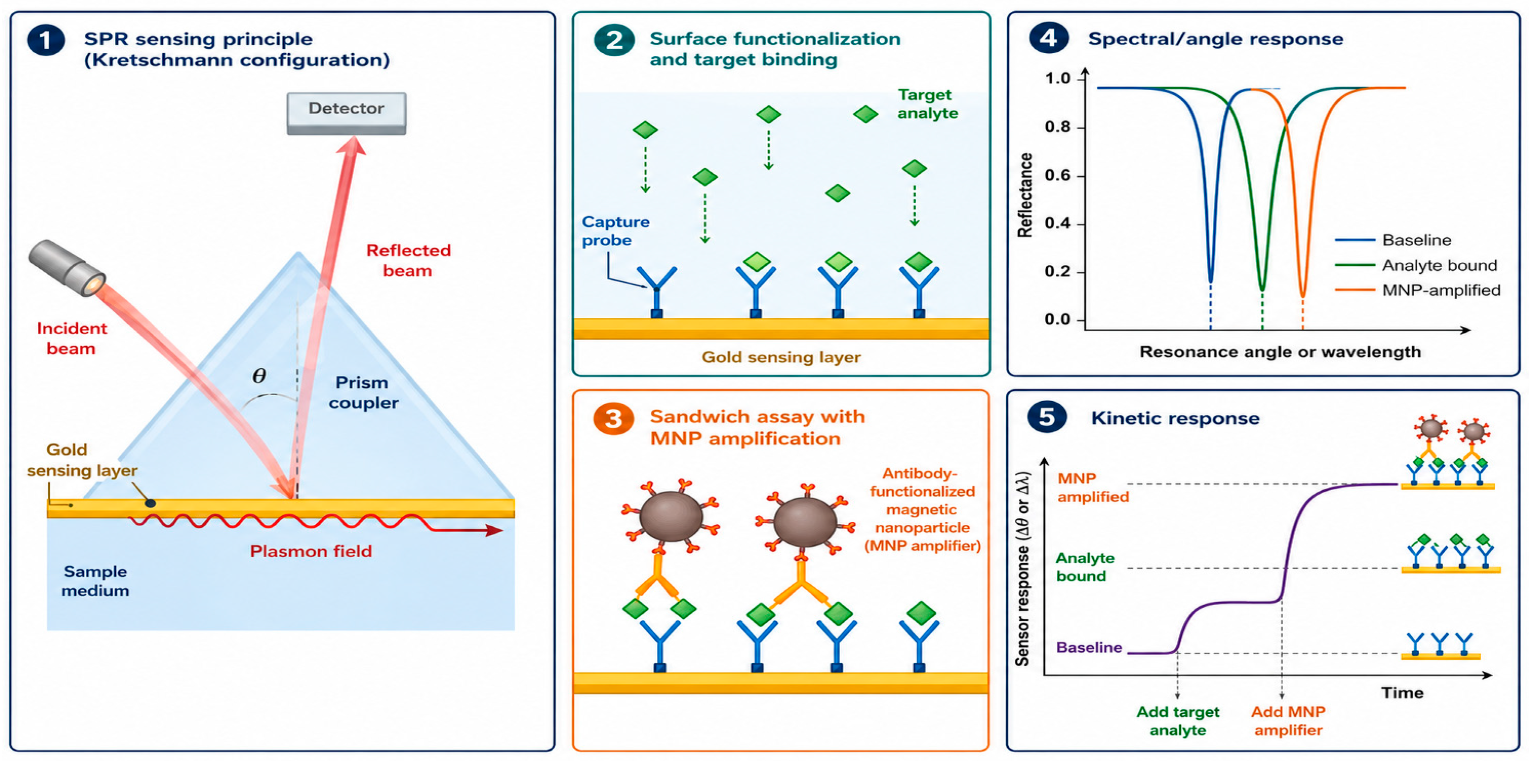
Figure 1. Schematic representation of the SPR biosensing platform with magnetic nanoparticles amplification.
Magnetochemistry 12 00065 g001
Figure 2. Nanoparticles that mediate force and how they affect the operation of mammalian cells. (A) FMNPs as intermediaries to steer cell navigation through the extracellular matrix when subjected to a constant magnetic field gradient (t: time). (B) FMNPs are linked to the cell membrane and influence how receptors work, boost communication between cells, and do local cell operations. (C) Putting fMNPs within cells can create protein gradients and change how vesicles move. (D) Localizing FMNPs to the cell nucleus is employed for genetic modification of cells, exemplified by the translation of a fluorescent protein (e.g., GFP or eGFP). Reprinted from Ref. [54].
Magnetochemistry 12 00065 g002
Figure 3. (a) SAXS profiles of the solvent-free DNA−DDACe ferrofluids in the LC phase at various temperatures. (b) The FF-TEM image shows the ferrofluids in the LC phase at 35 °C. (c) The suggested model depicts the microstructures of the solvent-free ferrofluids at temperatures between 35 and 65 °C. The bar is 50 nanometers long. Reprinted with permission from Ref. [59]. Copyright 2016 American Chemical Society.
Magnetochemistry 12 00065 g003
Figure 4. The potential applications of superparamagnetic iron-oxide nanoparticles are far-ranging, but the ones that have made it to the clinic are primarily being used as MRI contrast agents and a few other medical applications. The nanoparticles that successfully reach the clinic are primarily the simplest in design, consisting of an iron-oxide core covered with a biocompatible layer of dextran or silane; this simplicity facilitates large-scale manufacturing and more accurate predictions of in vivo behavior. These designs exploit the relationship between the size, surface, and movement of the particles and their biodistribution and residence times, as noted by Li et al. in 2026 [79]. Reprinted with permission from Ref. [79]. Copyright 2026 Elsevier.
Magnetochemistry 12 00065 g004
Figure 5. Potential modalities of MNP functionalization. Linker is a chemically manufactured double-stranded DNA molecule. An anchor is the ligand that is unique to a designated target (molecule or chemical complex).
Magnetochemistry 12 00065 g005
Figure 6. Multi-scale models within ‘digital twins’. Nanoscale, mesoscale, and macroscale modeling, accompanied by examples and their respective temporal and spatial scales. Reprinted with permission from Ref. [90]. Copyright 2025 Springer Nature.
Magnetochemistry 12 00065 g006
Figure 7. Geometric configuration of the physical model and coordinate framework.
Magnetochemistry 12 00065 g007
Figure 8. Schematic representation of the relationship between synthesis strategy, particle morphology/dispersion, and magnetic response.
Magnetochemistry 12 00065 g008
Figure 9. Unifying conceptual framework for magnetic nanoparticle (MNP) behavior and translation in biological systems.
Magnetochemistry 12 00065 g009
Disclaimer/Publisher’s Note: The statements, opinions and data contained in all publications are solely those of the individual author(s) and contributor(s) and not of MDPI and/or the editor(s). MDPI and/or the editor(s) disclaim responsibility for any injury to people or property resulting from any ideas, methods, instructions or products referred to in the content.