Article

Inhibitory Activity and Mechanism of Action with Thymol against the Blueberry Pathogenic Fungi Caused by Neopestalotiopsis clavispora

1
Food and Pharmaceutical Engineering Institute, Guiyang University, Guiyang 550005, China
2
School of Chemistry and Food Science, Nanchang Normal University, Nanchang 330023, China
*
Author to whom correspondence should be addressed.
Horticulturae 2023, 9(9), 983; https://doi.org/10.3390/horticulturae9090983
Submission received: 20 July 2023 / Revised: 20 August 2023 / Accepted: 28 August 2023 / Published: 31 August 2023
(This article belongs to the Section Postharvest Biology, Quality, Safety, and Technology)

Abstract

:
Decay caused by Neopestalotiopsis clavispora is an important postharvest disease of blueberries that seriously affects the commercial value of blueberry fruit. In this paper, we studied the inhibitory activity and mode of action of thymol against the pathogenic fungus of blueberries caused by Neopestalotiopsis clavispora. The results demonstrated that thymol administration could limit mycelial growth in vitro; the inhibitory effect was positively connected with thymol mass concentrations, and the minimal inhibitory concentration (MIC) was 100 mg/L. Further investigations revealed that MIC thymol treatment dramatically reduced the germination of pathogenic spores and led to an increase in the conductivity of the pathogen, leakage of contents, and a decrease in pH. Propidium iodide (PI) staining experiments demonstrated that MIC thymol caused damage to mycelial cell membranes. Additionally, MIC thymol treatment promoted mycelium malondialdehyde content accumulation, inhibited superoxide dismutase (SOD) and catalase (CAT) enzyme activities, decreased adenosine triphosphate (ATP), adenosine diphosphate (ADP), and adenosine monophosphate (AMP) content and energy charge levels, and the fluorescence intensity of mycelium caused by MIC thymol treatment was significantly increased by the 2,7-Dichlorodi-hydrofluorescein diacetate (DCFH-DA) assay. The results of this study indicate that thymol suppresses the proliferation of Neopestalotiopsis clavispora by compromising the integrity of their cell membranes, promoting the accumulation of cellular reactive oxygen species (ROS), and interfering with energy metabolism.

1. Introduction

Blueberries (Vaccinium ssp.) are cultivated commercially on an enormous scale around the world and are popular among consumers due to the fact that they are rich in a variety of health-promoting bioactive substances, such as anthocyanins, flavonoids, polyphenols, and folic acid [1]. However, blueberry fruit is prone to mechanical damage after harvest due to its thin skin, juicy pulp, and soft texture, which are infected by pathogenic microorganisms. Pathogenic microbial infestations can cause rapid decay and deterioration of blueberry fruit, which can seriously curb the development of the blueberry industry [2]. It is reported that there are many species of pathogenic fungus that can cause postharvest rot of blueberry fruits, mainly Botrytis cinerea, Penicillium spp., Aspergillus spp., Alternaria spp., and Colletotrichum spp. [3]. In addition, Neopestalotiopsis clavispora is a pathogen that occurs in postharvest blueberries in the Majiang area of Guizhou Province. It can cause the softening and decay of blueberries and cause economic losses. Recent studies have demonstrated that postharvest decay of blueberry fruit may be significantly decreased by employing methods utilizing exogenous substances such as SO2 [4], ClO2 [5], cold plasma (CP), ultraviolet irradiation (UV) therapy, and microbial antagonism [6]. Nowadays, chemical fungicides are widely used to control pre-harvest and post-harvest plant diseases of fruits and vegetables, but excessive use of chemical fungicides leads to pathogen resistance, chemical residues, and environmental pollution. Therefore, it is very important to find a safe and effective antifungal technology to maintain the postharvest quality of blueberry fruit [7].
Thymol (2-isopropyl-5-methylphenol) is the main monoterpene phenol occurring in essential oils isolated from plants belonging to the Lamiaceae family (Thymus, Ocimum, Origanum, and Monarda genera) and other plants such as those belonging to the Verbenaceae, Scrophulariaceae, Ranuncolaceae, and Apiaceae families [8]. Because of its high safety, wide range of sources, lack of chemical residues, broad-spectrum antifungal properties, and other characteristics [9,10], it has been authorized by the Federal Drug Administration (FDA) for food and as a food additive, and it has since been widely utilized in the field of food preservation [11]. Thymol has been demonstrated to be capable of being transformed into microencapsulation [12] and antimicrobial films [13] in order to effectively extend the freshness period of fruits and vegetables. Furthermore, thymol can be produced as an individual or combination aqueous solution for postharvest preservation of fruits and vegetables. As a natural, secure active ingredient, thymol offers a wide variety of prospective applications.
Before utilizing natural fungicides, it is essential to comprehend their mode of action and fungicidal activity. Currently, thymol has been shown to have a better inhibitory effect on common pathogenic fungi of fruits and vegetables, such as Penicillium spp. [14,15], Fusarium spp. [16], and Botrytis cinerea [17]. Its inhibitory effect is mainly attributed to the inhibition of ergosterol biosynthesis by disrupting the cell membrane and cell wall structural integrity of pathogenic fungus, and the inhibitory effect was mainly attributed to the inhibition of membrane lipid peroxidation caused by disrupting pathogenic cell membranes and, cell wall structural integrity [18], inhibiting ergosterol biosynthesis, and promoting cellular reactive oxygen species (ROS) radical accumulation. In addition, Li et al. [19] discovered that NOX2 was involved in maintaining the intracellular reduced nicotinamide adenine dinucleotide phosphate/oxidized nicotinamide adenine dinucleotide phosphate (NADPH/NADP+) balance in Staphylococcus aureus and caused a decrease in intracellular NADPH and ATP and an increase in NADP+ through thymol treatment, thus interfering with mycelial energy metabolism to exert antifungal activity.
To date, the mechanism of inhibition of Neopestalotiopsis clavispora by thymol has not been reported. This study was therefore designed to examine if thymol could exhibit antifungal activity against N. clavispora, the causative agent of postharvest blueberry. The specific objectives included determining (A) the ability of thymol to inhibit mycelial growth of N. clavispora on agar plates, (B) the ability of thymol to inhibit the germination of N. clavispora spores, and (C) how thymol would manifest its antifungal behavior against N. clavispora.

2. Materials and Methods

2.1. Fungal Strain and Culture Conditions

A strain of Neopestalotiopsis clavispora was found in post-harvest blueberries showing decay in Majiang, Guizhou, China. This strain underwent morphological and molecular assessment at Guiyang University’s Laboratory of Biology and Medicine and was preserved. It was cultured on potato dextrose agar (PDA, sourced from Bo Microbiology Technology Corporation, Shanghai, China) and maintained at a temperature of 25 ± 2 °C. A spore suspension was prepared to a concentration of 1 × 10⁶ CUF/mL using a hematocytometer. This suspension was then added to 20 mL of potato dextrose broth (PDB) and incubated for 48 h. After incubation, the mycelium underwent three washes with a 0.1 mol/L phosphate buffer sodium (PBS) solution, maintaining a pH of 7.0, before further experimental use.

2.2. Effect of Thymol on Colony Growth

Thymol’s stock solution (99%, sourced from Genye Biological Corporation, Shanghai, China) was formulated at 10 mg/mL. This stock was further diluted to achieve PDA plates with varying thymol concentrations: 0 (control), 20, 40, 60, 80, and 100 mg/L. Mycelial disks from N. clavispora, 5 mm in diameter, were centrally positioned on these PDA media. Daily, colony diameters were gauged using the cross method and a standard ruler. The radial mycelial growth approach assessed the antifungal potency. The formula [(C − T)/(C − 0.6)] × 100% was employed to determine the inhibition rate, where C denotes the mycelial diameter in the control and T stands for that in the treated plate [20]. The concentration at which there was no observed mycelial growth of Neopestalotiopsis clavispora disks after 48 h was designated as the minimum inhibitory concentration (MIC) [21].

2.3. Determination of Thymol on N. clavispora Spore Germination

Adopting the procedure delineated by Run [22], the spore germination of N. clavispora was determined. A spore suspension of N. clavispora at a concentration of 50 μL (1 × 10⁶ CUF/mL) was formulated and applied atop PDA medium enhanced with thymol and ethanol. Subsequent incubation was performed in a light-excluded environment at 25 °C across intervals of 0, 4, 8, and 12 h. Sections, measuring 8 mm × 8 mm, were procured from the four corners and the center of the medium. Using a light microscope (BX53, OLYMPUS), spores within these sections were enumerated. The germination rate was ascertained by contrasting the number of germinated spores to the total spore count.

2.4. Effect of Thymol on the Dry Weight of N. clavispora Mycelium

Using the approach detailed by Wan et al. [23] for assessing mycelial dry weight, the mycelium underwent treatment with thymol at concentrations ranging from 0 to 100 mg/L. Following a 25 °C, 24-h incubation, the mycelium received three sodium phosphate buffer washes. Subsequently, drying was carried out at 70 °C over a 12-h period. An analytical balance (Ms105, Mettler Toledo, Greifensee, Switzerland) provided the dry weight measurements, with findings presented in terms of g/100 mL PDB.

2.5. Effect of Thymol on the Relative Conductivity, Nucleic Acid Release, and pH of N. clavispora

Adapting the method from Shao et al. [24], both the relative conductivity and pH of N. clavispora mycelia were assessed. The mycelia, acquired post-centrifugation of a 20 mL PDB culture at 4000× g for 20 min, were then washed and reintroduced into a 20 mL pH 7.0 phosphate buffer. Upon treatment with thymol at final mass concentrations of both 0 (acting as the control) and 100 mg/L, the measurements of pH and external conductivity at intervals of 0, 2, 4, 6, and 8 h were captured using a portable pH/conductivity device (PHBJ-260, 0.01 pH precision, Precision Scientific Instruments, Shanghai, China) and a specific conductivity meter (DDS-11A, Yidian Scientific Instruments Corporation, Shanghai, China) in an oscillating environment. From these, the relative conductivity was deduced. Meanwhile, leakage from the mycelial nucleic acid was gauged utilizing a UV-visible spectrophotometer (60UV-Vis, Agilent) following the process outlined by Shi et al. [25], with outcomes indicated as 260 nm absorbance values.

2.6. Determination of Thymol on Extracellular Soluble Protein and Reducing Sugar Content of N. clavispora

Following the methodology presented by Chen et al. [26], suspensions underwent treatment with thymol at distinct concentrations, specifically 0 and MIC. The testing durations were spread over intervals of 0, 2, 4, 6, and 8 h. Utilizing the ultraviolet-visible spectrophotometer (60UV-Vis, Agilent), the contents of both extracellular soluble protein and reducing sugars were quantified. Outcomes were presented in terms of mg/g.

2.7. Effect of Thymol on the Integrity of N. clavispora Cell Membrane

Suspensions underwent thymol treatment at specified concentrations, specifically 0 and MIC, for a duration of 24 h. Adhering to the protocol delineated by Tao et al. [27], mycelium that had been exposed to the treatment was then mixed with 50 μL of PI (1 g/L, sourced from Aladdin Biochemical Technology Corporation, Shanghai, China). This mixture was subsequently incubated in dark conditions for 15 min at a temperature of 30 °C. To remove any surplus dye, a thorough washing with PBS was performed. Finally, the samples were visualized and captured using a fluorescence microscope, model BX53 by OLYMPUS.

2.8. Determination of Intracellular Malondialdehyde (MDA) Content

Mycelium underwent thymol treatment at intervals of 0, 2, 4, 6, and 8 h using concentrations of 0 (control) and 100 mg/L. The thiobarbituric acid procedure, as detailed by Pasquariello et al. [28], was employed to assess MDA content. To initiate, 5 mL of trichloroacetic acid (100 g/L) was introduced to 2.0 g of the rinsed mycelium samples. This was followed by a thorough homogenization under cold conditions with a chilled mortar and pestle. Post-homogenization, centrifugation was executed at 2000 g for a span of 10 min at 4 °C Following this, a mixture of thiobarbituric acid solution (2 mL, 6.7 g/L) and 2 mL of the sample was prepared and incubated in a boiling water environment for 20 min, subsequently allowing it to cool to ambient temperature. From this, the supernatant’s absorbance was evaluated at wavelengths of 450, 532, and 600 nm, offering the MDA content. The entirety of this process was replicated three times, and the outcomes are presented in mmol/L.

2.9. Determination of Superoxide Dismutase and Catalase Enzyme Activities of N. clavispora Mycelium

The activities of superoxide dismutase (SOD) and catalase (CAT) were gauged with slight alterations from the protocol established by Lai et al. [29]. For SOD, a single enzyme activity unit (U) is identified as the protein quantity that impedes the reduction of nitrogen blue tetrazolium (NBT) by half each minute for every mass. Conversely, one unit of CAT corresponds to the protein volume that shifts the absorbance by 0.01 in order to convert hydrogen peroxide by 1 mmol every minute for its mass. The data collected is represented in terms of U/mg.

2.10. Detection of Intracellular Reactive Oxygen Species (ROS) Levels in N. clavispora

Following an 8-h treatment with control and thymol at 100 mg/L concentrations, the mycelia underwent a specific process. Drawing inspiration from the technique outlined by Pang [30], 1 mL of 1 × 10⁵ mol/L 2,7-Dichlorodi-hydrofluorescein diacetate (DCFH-DA, sourced from Aladdin Biochemical Technology Corporation, Shanghai, China) was introduced to the treated samples. This mixture was then subjected to a 37 °C environment for 20 min. Subsequent to three cleansing cycles in sodium phosphate buffer, these cells were inspected via a BX53 OLYMPUS fluorescent microscope. Visual representations of the observed mycelia were then captured.

2.11. N. clavispora Energy Metabolism Analysis

Mycelium samples, post-exposure to thymol control and a concentration of 100 mg/L for intervals of 0, 2, 4, 6, and 8 h, had their ATP content analyzed. Building upon the procedures detailed by Peng and colleagues [31], we employed high-performance liquid chromatography (HPLC) for detection. A quick outline: 0.6 g of the mycelium was isolated, followed by the introduction of a 3% perchloric acid solution. After a brief grinding session in an iced setting, it was centrifuged. Post-extraction, a 20% KOH solution was incorporated and thoroughly mixed. This mixture then rested at 4 °C for half an hour before undergoing another round of centrifugation to prepare for HPLC scrutiny. Analytical parameters comprised: column inert sil ODS-SPC18 (250 mm × 4.6 mm, 5 pm); Solvent A being KH2PO4-K2HPO4 buffer; and solvent B being 3% methanol, with a 0.8 mL/min flow, 30 °C column temperature, 20 μL injection volume, and a 230 nm detection wavelength.

2.12. Statistical Analysis

Each experimental procedure was executed in triplicate. We used SPSS 25.0 for the interpretation and analysis of the gathered data. Subsequent to variance analysis, Duncan’s multiple range tests were employed to differentiate between the means. Results with a p-value less than 0.05 were deemed to be of statistical significance.

3. Results

3.1. Effect of Thymol on Colony Growth

The minimum inhibitory concentration (MIC) is a key metric for assessing and evaluating a substance’s fungus-inhibitory activity [32]. Variable concentrations of thymol inhibited N. clavispora in various manners, and the inhibitory effect was concentration-dependent (Figure 1A–C). The ethanol concentration utilized in this experiment had no influence on the growth of N. clavispora (Figure 1A). At 2 days of incubation, the colony diameter of the control group was 23.62 mm, and the inhibition rates of the 20 mg/L and 80 mg/L treatment groups were 17.85% and 95.31%, respectively, and the colony diameter was 19.46 mm and 1.10 mm, respectively. After two days of treatment, the inhibition rate of the 100 mg/L treatment group reached 100%, the growth was completely sterile, and the minimum inhibitory concentration (MIC) was 100 mg/L. The control group had a colony diameter of 80.00 mm on the 7th day of incubation; the 100 mg/L thymol-treated group had a colony diameter of 8.86 ± 0.72 mm, which was substantially smaller than the other treatment groups (p < 0.05). MIC values were used in further tests.

3.2. Effect of Thymol on the Germination of N. clavispora Spores

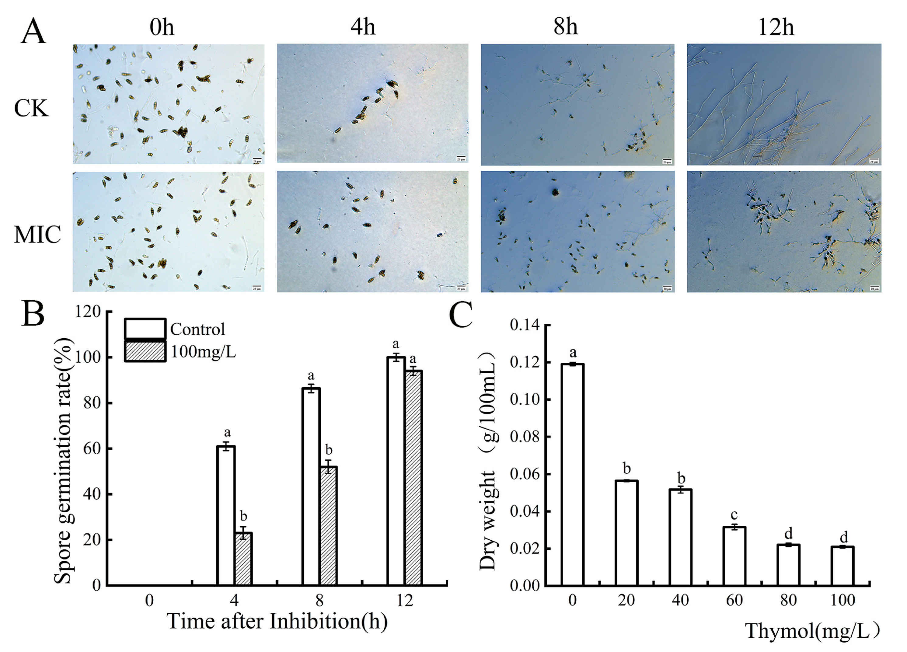
Thymol had an inhibitory effect on the spore germination of N. clavispora, and the effect was more pronounced with time (Figure 2A). At 4 h, the thymol treatment and control groups were 23.67% and 61.33%, respectively. At 8 h of treatment, the spore germination rate of the control group was 1.65 times that of the treatment group. There was no significant difference in spore germination between the treatment group and the control group at 12 h (p > 0.05), but the elongation length of the control group was significantly longer than that of the treatment group. It can be seen that thymol can delay the germination process of conidia.

3.3. Effect of Thymol on the Dry Weight of N. clavispora Mycelium

The mycelial weights of N. clavispora in the thymol treatment and control groups are shown in Figure 2C. The data showed that as the concentration of thymol increased, the biomass of mycelial growth was strongly inhibited. Initially, the dry weight was 0.1191 g/100 mL at the no-thymol concentration. At higher thymol concentrations (20, 40, 60, and 80 mg/mL), the effect on mycelial weights was recorded at a significant level (p < 0.05) in comparison with the control group.

3.4. Effect of Thymol on the Relative Conductivity, Nucleic Acid Release, and pH of N. clavispora

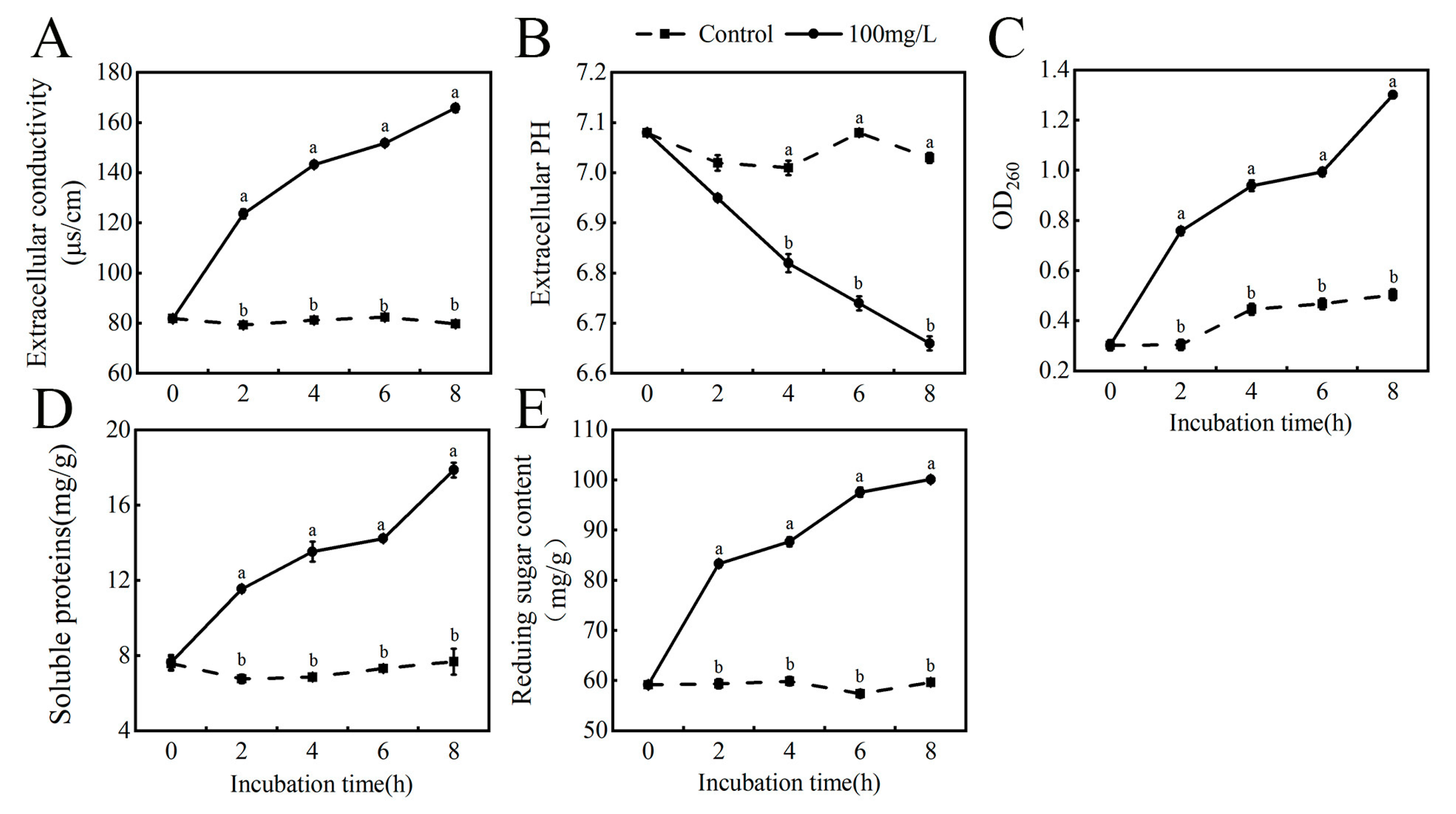
The damage to membrane permeability can be reflected in the relative conductivity. After treatment with thymol, the extracellular conductivity of N. clavispora increased gradually with the prolongation of the treatment time (Figure 3A). After a 0–2 h sharp rise in the whole process, the treatment group was significantly higher than the control group (p < 0.05). The extracellular conductivity of the treatment group was 123.62, 143.28, 151.83, and 165.86 S/cm at 2, 4, 6, and 8 h, respectively, increasing by 33.97%, 42.86%, 45.53%, and 50.17% compared to the control group.
Extracellular pH reflects extracellular acid-base balance (Figure 3B). Thymol treatment of N. clavispora resulted in acid leakage from the cell, resulting in a substantial fall in extracellular pH. At 2, 4, 6, and 8 h, the pH of the treatment group was 6.95, 6.82, 6.74, and 6.66, respectively, which was significantly different (p < 0.05) from that of the control group (7.08 ± 0.033). This demonstrates that the N. clavispora cell membrane was disrupted as a result of the massive leaking of intracellular nucleic acid material, which resulted in a fall in extracellular pH.
Nucleic acid leakage can indirectly assess cell membrane integrity. After cell damage, intracellular substances are prone to extravasation (Figure 3C). Thymol therapy increased mycelial nucleic acid leakage significantly (p < 0.05). Compared with the control group, the thymol treatment group increased by 59.79%, 52.41%, 52.89%, and 61.17% at 2, 4, 6, and 8 h, respectively.

3.5. Effect of Thymol on the Extracellular Soluble Protein and Reducing Sugar Content of N. clavispora

The extracellular soluble proteins of thymol-treated N. clavispora increased significantly (p < 0.05) with the treatment period (Figure 3D). The extracellular soluble protein content of the thymol-treated group increased by 1.70-fold, 1.97-fold, 1.94-fold, and 2.32-fold at 2, 4, 6, and 8 h compared with the control group, respectively. The extracellular reducing sugars of N. clavispora showed a significant upward trend (p < 0.05) with the increase in treatment time (Figure 3E). It rose rapidly after 2 h of treatment. Thymol-treated groups increased 28.73%, 32.06%, 41.15%, and 40.40% at 2, 4, 6, and 8 h, respectively, compared with the control group. In conclusion, thymol caused the leakage of extracellular soluble proteins and reducing sugars to be excreted and damaged the N. clavispora cell membrane.

3.6. Effect of Thymol on N. clavispora Cell Membrane Integrity

Propidium iodide (PI) staining was utilized to determine whether thymol can cause damage to N. clavispora cell membranes (Figure 4). When the integrity of the cell membrane is disturbed, PI may enter the cytoplasm and bind to DNA to create red fluorescence [33]. Red fluorescence was found in mycelium after treatment with MIC thymol, but no fluorescence was observed in the control group, suggesting that the cell membrane of mycelium treated with thymol was destroyed, which is consistent with the preceding findings.

3.7. Changes in Intracellular Malondialdehyde (MDA) Content

The MDA content of N. clavispora increases with increasing treatment duration, with a significant difference (p < 0.05) at 6–8 h (Figure 5A). At 8 h, the MDA concentration was 1.32 mmol/L, 1.28 times greater than the control. It is possible that the cells underwent enhanced membrane lipid peroxidation due to the accumulation of reactive oxygen species caused by the early addition of thymol.

3.8. Changes in Superoxide Dismutase and Catalase Enzyme Activities of N. clavispora Mycelium

The CAT content of N. clavispora decreased significantly (p < 0.05) as treatment duration increased (Figure 5B). When compared to the control group, the CAT content of the thymol-treated group declined 1.92, 2.24, 3.8, and 4.65 times at 2, 4, 6, and 8 h, respectively. The SOD content of Neopestalotiopsis clavispora tended to decrease significantly (p < 0.05) with increasing treatment time (Figure 5C). It increased rapidly after 2 h of treatment. Thymol-treated groups increased by 30.93%, 40.20%, 64.67%, and 70.71% at 2, 4, 6, and 8 h, respectively, compared with the control group. The aforementioned findings could be explained by the fact that mycelium’s ROS generation outpaced its ability to scavenge ROS, upsetting the dynamic balance between ROS production and scavenging and causing a rapid drop in antioxidant enzyme activity. It may be because prolonged usage of thymol resulted in N. clavispora apoptosis, the buildup of MDA, the occurrence of free radicals and reactive oxygen species, as well as the disruption of the antioxidant system, all of which eventually caused significant oxidative damage and loss of cell integrity.

3.9. Intracellular Reactive Oxygen Species (ROS) Levels in N. clavispora Cells

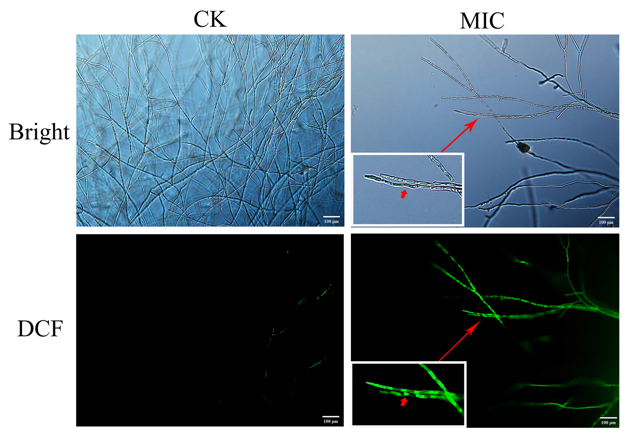
To evaluate the impact of thymol on intracellular ROS levels in N. clavispora cells, DCFH-DA fluorescent dye was utilized (Figure 6). Mycelium fluoresced after thymol treatment, whereas no fluorescence was seen in the control group. This shows that thymol administration dramatically increased the production of ROS in N. clavispora cells.

3.10. Effect of Thymol Treatment on Energy of Pathogenic Mycelium

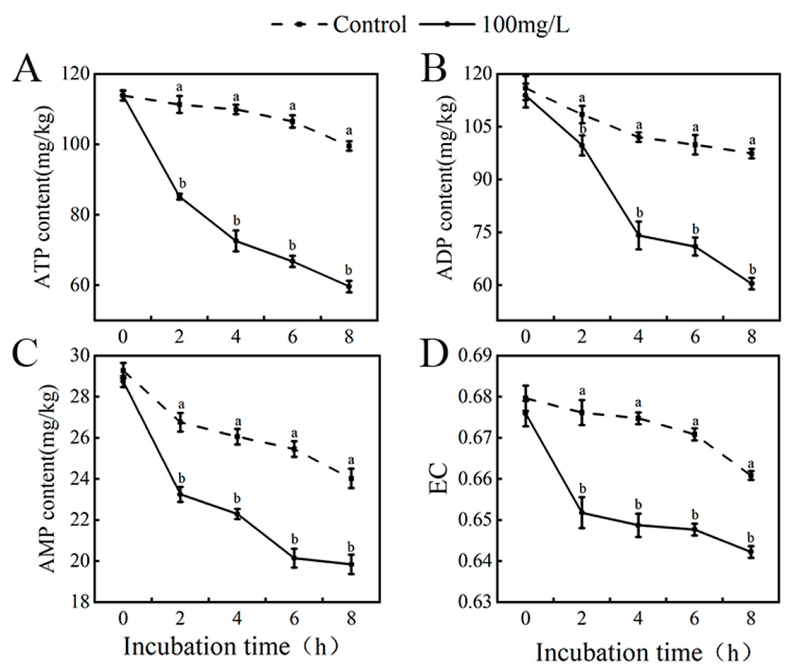
The ATP, ADP, and AMP contents in mycelium treated or untreated at various time intervals were evaluated to study the impact of thymol on energy metabolism (Figure 7). It was seen that ATP, ADP, and AMP contents in both control and MIC-treated groups showed a significant decrease with time, and both treated groups were significantly lower than the control group (p < 0.05). Furthermore, the energy charge remained lower at varied treatment times. This could be due to thymol-induced decreased ATP, altered mitochondrial activity, apoptotic, necrotic, or toxic cells, or aberrant energy metabolism in the mycelium. In conclusion, thymol disrupted the normal energy metabolism and physiological activities of the pathogenic blueberry fungus.

4. Discussion

In recent years, China’s blueberry planting area has ranked first in the world, but the development of plant infections has harmed the fruit’s quality, significantly limiting export volume. To avoid economic losses, adequate disease defense measures must be implemented. Nowadays, it is difficult to distinguish the pathogenic fungi that produce rot in blueberries according to their disease symptoms, and new pathogens of postharvest blueberry diseases have emerged due to changes in climate, environment, varieties, and cultivation methods. Therefore, it is critical to control novel strains of blueberry pathogens discovered in recent years. In this study, the pathogenic fungus Neopestalotiopsis clavispora was identified in naturally deteriorated blueberries after harvesting in the Majiang district of Guizhou, and it was able to infest blueberries and induce blueberry fruit decline. Neopestalotiopsis clavispora has been associated with fruit rot in kiwi [34], strawberry [35], loquat [36], and Rosa sterilis [37]. The results presented indicate that pathogenic fungi may have a diverse host range and may constitute a substantial danger to the fruit and vegetable business.
Effective control of blueberry postharvest diseases is a major challenge worldwide, so it is important to study thymol inhibition of blueberry postharvest diseases. The in vitro inhibitory action of thymol revealed that it inhibited N. clavispora at mass concentrations ranging from 20 to 100 mg/L, with a MIC of 100 mg/L. Although thymol had a low effect on the final germination rate of N. clavispora conidia at this time, it could effectively delay the germination process of conidia, and the results indicated that thymol could inhibit mycelial growth and spore germination, affect mycelial dry weight, and increase the sensitivity of N. clavispora to stress. This is consistent with the results of Liu [38], who found that thymol completely inhibited the mycelial growth and spore germination rate of the potato dry rot fungus (Fusarium oxysporum). To verify these observations and elucidate the underlying mechanisms, we conducted a series of experiments to analyze the effects of thymol on N. clavispora cell membrane integrity, antioxidant systems, and energy metabolism.
The cell membrane is a part of the physiological structure of fungus and is an important channel for material exchange and information transmission between the cell and the external environment, which can effectively regulate the homeostasis of the internal and external environment and guarantee the normal life activities of fungus. It is also an important target for the action of common antifungal agents [39]. Numerous investigations have revealed that thymol damages pathogenic fungus cell membranes and affects the equilibrium of their internal and external environments, functioning as an inhibitor [14,15,16,17]. There is growing evidence that, among other things, the lipophilic properties of volatile substances enhance membrane permeability and fluidity, limit respiration, disturb intramembrane proteins, trigger intracellular contents like ion leakage, and modify ion transport processes in fungi [40]. A range of regulatory activities in the cell, such as enzyme activity, protein synthesis, and homeostasis maintenance, are inextricably linked to pH [41]. Relative conductivity and extracellular pH are usually used to indicate severe and irreversible damage that occurs at the plasma membrane. According to the study’s findings, mycelium’s extracellular relative conductivity increased noticeably and its extracellular pH fell after being exposed to thymol, indicating that the cell membrane had been damaged. PI staining results also confirmed that thymol caused N. clavispora to lose cell membrane integrity. Soluble proteins and reducing sugars are the primary components of the cytoplasm, and an increase in their amounts typically signals a loss in mycelial intracellular membrane integrity and an increase in cell permeability [42]. It has been reported that the loss of cell membrane integrity may lead to the release of cytoplasmic contents in cells. Thus, in order to determine whether the loss of membrane integrity caused by thymol treatment leads to the release of intracellular contents in fungal cells, we found that intracellular nucleic acids, extracellular soluble proteins, and extracellular reducing sugars in mycelium increased with the lengthening of treatment time. This finding was in line with the hypothesis that thymol could disrupt cell membrane permeability and cause leakage of intracellular contents, thus interfering with the growth of other organisms. Similar results have been found by Zhang et al. [43], who discovered that thymol damaged pathogenic fungi and ruined cell membranes by allowing intracellular chemicals to flow out.
Reactive oxygen species (ROS) are byproducts of oxidative metabolism in living things, but too much of them can have negative consequences [44]. The permeability of the fungus cell membrane increases, and intracellular oxidative stress is increased after damage to the fungus biofilm. Therefore, we suggest that the damage to the cell membrane caused by antifungal agents may lead to the accumulation of ROS in cells, and then the oxidative damage to cell macromolecules leads to cell death [45]. DCFH-DA staining results demonstrate that thymol could lead to ROS accumulation in N. clavispora. The main components of the cell membranes are lipids and proteins, and lipids contain unsaturated double bonds that predispose to lipid peroxidation reactions when elevated ROS bind to the cell membrane, which can lead to malondialdehyde (MDA) accumulation [46]. The results of this study showed that the level of the lipid peroxidation index MDA confirmed that thymol-induced oxidative stress led to the oxidative damage of N. clavispora. This is consistent with observations [47] that the action of thymol prevented the development of Fusarium oxysporum by accumulating MDA and generating a lot of reactive oxygen species. In addition, it was discovered that fungi may activate both enzymatic and non-enzymatic defense mechanisms in response to ROS stress [48]. In addition to reflecting the process of lipid peroxidation through changes in their activity, superoxide dismutase (SOD) and catalase (CAT), two crucial antioxidant active enzymes in fungus, may also react quickly to oxidative stress [49]. In agreement with the results of this paper, the mycelium’s oxidative defense mechanism helped it fend off attacks from reactive oxygen species (ROS). However, as treatment time increased, the burst of ROS caused MDA to build up and the antioxidant activities of catalase and superoxide dismutase enzymes to decline, ultimately inhibiting the growth of pathogenic fungi. Particularly, the activity of catalase was significantly lower than that of the control group, indicating that CAT is more susceptible to attack by ROS and to inactivation as a result. The outcomes of Pang’s work [30] on the impact of curcumin-based photodynamic inactivation (PDI) treatment affected the SOD and CAT activity of Penicillium expansum (PAT), with similar results to the present study.
Mitochondria, a membrane organelle composed of proteins and lipids, have been reported to be another important target of essential oils in plants [50]. Mitochondria are the main site of ROS production in fungal cells and the most important organelle for cellular energy metabolism. When the balance between the production of reactive oxygen species and the antioxidant defense system is lost, mitochondria are damaged by lipid peroxidation, leading to disruption of energy metabolism [51]. Adenosine triphosphate is central to energy metabolism, storage, and utilization and is an essential energy substance for the vital activities of living organisms [52]. As a crucial component of energy metabolism, adenosine triphosphate is a necessary energy source for maintaining organisms’ vital activity, and changes in its concentration in cells can directly reflect the level of that activity [53]. The ratio of adenosine triphosphate, adenosine diphosphate, and adenosine monophosphate reflects the cellular energy charge, and many metabolic activities in cells, such as glycolysis, the tricarboxylic acid cycle, the electron transport system, and oxidative phosphorylation, are dependent on the regulation of energy charge changes [54]. In the present study, fluctuating differences in ATP, ADP, AMP, and energy charge at different treatment times may be due to thymol disrupting the balance of intra- and extracellular ATP by disrupting the cell membrane, leading to ATP loss as well as inhibition of metabolic activities, resulting in cell membrane damage and abnormalities in the antioxidant defense system.

5. Conclusions

In summary, thymol significantly inhibited Neopestalotiopsis clavispora mycelial growth and spore germination in vitro. When the MIC value was 100 mg/L, the mycelial growth inhibition rate was only (88.925 ± 0.9)%. In addition, 100 mg/L thymol treatment resulted in increased extraspore conductivity, caused exocytosis of contents, and lowered the pH of the pathogenic fungi, thereby destroying the integrity of N. clavispora cell membranes. Thymol’s potential antifungal mechanism of action may depend on its capacity to increase excessive intracellular ROS production and interfere with energy metabolism. The above results provide a new idea for thymol as a plant antifungal agent and fruit and vegetable preservative for effective prevention and treatment of postharvest pathogens in blueberries.

Author Contributions

Conceptualization, S.Y. and L.S.; methodology, S.Y.; software, S.Y. and D.L.; validation, L.B. and L.S.; formal analysis, S.Y.; writing—original draft preparation, S.Y.; supervision, L.B. All authors have read and agreed to the published version of the manuscript.

Funding

This research was funded by the Science and Technology Foundation of Guizhou Province (Grant No.[2021]122), and the Sixth Batch of Guizhou Province High-level Innovative Talent Training Program [2022], and Guiyang University Graduate Research Fund Project (2023-YJS02). This achievement also has received special support from the Guizhou Provincial Department of Science and Technology for academic seedling cultivation and free exploration and innovation.

Data Availability Statement

Datasets generated and/or analyzed during this study are available on request from the corresponding author.

Conflicts of Interest

The authors declare no conflict of interest.

References

  1. Silva, S.; Costa, E.M.; Veiga, M.; Morais, R.M.; Calhau, C.; Pintado, M. Health promoting properties of blueberries: A review. Crit. Rev. Food Sci. Nutr. 2020, 60, 181–200. [Google Scholar] [CrossRef] [PubMed]
  2. Hernández-Ceja, A.; Loeza-Lara, P.D.; Espinosa-García, F.J.; García-Rodríguez, Y.M.; Medina-Medrano, J.R.; Gutiérrez-Hernández, G.F.; Ceja-Torres, L.F. In Vitro Antifungal Activity of Plant Extracts on Pathogenic Fungi of Blueberry (Vaccinium sp.). Plants 2021, 10, 852. [Google Scholar] [CrossRef] [PubMed]
  3. Bell, S.R.; Montiel, L.G.H.; Estrada, R.R.G.; Martínez, P.G. Main diseases in postharvest blueberries, conventional and eco-friendly control methods: A review. LWT 2021, 149, 112046. [Google Scholar] [CrossRef]
  4. Abeli, P.J.; Fanning, P.D.; Issaacs, R.; Randolph, M.B. Blueberry fruit quality and control of blueberry maggot (Rhagoletis mendax Curran) larvae after fumigation with sulfur dioxide. Postharvest Biol. Tec. 2021, 179, 11568. [Google Scholar] [CrossRef]
  5. Jiang, T.; Guo, F.; Fang, T.; Hwang, C.A.; Huang, L. Efficacy of gaseous chlorine dioxide generated by sodium chlorite-carbon dioxide reaction on safety and quality of blueberries, cherry tomatoes, and grapes. Food Control. 2023, 143, 109288. [Google Scholar] [CrossRef]
  6. Dukare, A.S.; Paul, S.; Nambi, V.E.; Gupta, R.K.; Vishwakarma, R.K. Exploitation of microbial antagonists for the control of postharvest diseases of fruits: A review. Crit. Rev. Food Sci. Nutr. 2018, 59, 1–16. [Google Scholar] [CrossRef]
  7. Radi, M.; Ahmadi, H.; Amiri, S. Effect of Cinnamon Essential Oil-Loaded Nanostructured Lipid Carriers (NLC) Against Penicillium Citrinum and Penicillium Expansum Involved in Tangerine Decay. Food Biobroc. Tech. 2022, 15, 306–318. [Google Scholar] [CrossRef]
  8. Marchese, A.; Orhan, I.E.; Daglia, M.; Barbieri, R.; Di Lorenzo, A.; Nabavi, S.F.; Gortzi, O.; Izadi, M.; Nabavi, S.M. Antibacterial and antifungal activities of thymol: A brief review of the literature. Food Chem. 2016, 210, 402–414. [Google Scholar] [CrossRef]
  9. Cruz, J.N.; Silva, S.G.; Pereira, D.S.; Filho, A.P.D.S.S.; Oliveira, M.S.D.; Lima, R.R.; Andrade, E.H.A. In Silico Evaluation of the Antimicrobial Activity of Thymol-Major Compounds in the Essential Oil of Lippia thymoides Mart. & Schauer (Verbenaceae). Molecules 2022, 27, 4768. [Google Scholar]
  10. Bagy, H.M.M.K.; Abo-Elyousr, K.A.M. Antibacterial activity of some essential oils on bacterial spot disease of tomato plant caused by xanthomonas axonopodis pv. vesicatoria. Int. J. Phytopathol. 2019, 8, 53–61. [Google Scholar] [CrossRef]
  11. Prakash, B.; Kediak, K.; Misra, P.K.; Dubey, N.K. Plant essential oils as food preservatives to control moulds, mycotoxin contamination and oxidative deterioration of agri-food commodities - potentials and challenges. Food Control. 2015, 47, 381–391. [Google Scholar] [CrossRef]
  12. Tomazelli Júnior, O.; Kuhn, F.; Padilha, P.J.M.; Vicente, L.R.M.; Costa, S.W.; Boligon, A.A.; Scapinello, J.; Nesi, C.N.; Dal Magro, J.; Castellví, S.L. Microencapsulation of essential thyme oil by spray drying and its antimicrobial evaluation against vibrio alginolyticus and vibrio parahaemolyticus. Braz. J. Biol. 2017, 78, 311–317. [Google Scholar] [CrossRef] [PubMed]
  13. Guedes, J.; Medeiros, J.; Silva, R.; Sousa, J.; Lúcia da Conceio, M..; Souza, E. The efficacy of Mentha arvensis L. and M. piperita L. essential oils in reducing pathogenic bacteria and maintaining quality characteristics in cashew, guava, mango, and pineapple juices. Int. J. Food Microbiol. 2016, 238, 183–192. [Google Scholar] [CrossRef] [PubMed]
  14. Ranjbar, A.; Ramezanian, A. Antifungal Activity of Thymol against the Main Fungi Causing Fruit Rot in In Vitro Conditions. Chem. Proc. 2022, 10, 79. [Google Scholar]
  15. Abdel-Rahim, I.R.; Abo-Elyousr, K.A.M. Using of endophytic Saccharomycopsis fibuligera and thyme oil for management of gray mold rot of guava fruits. Biol. Control. 2017, 110, 124–131. [Google Scholar] [CrossRef]
  16. Venturini, T.P.; Rossato, L.; Chassot, F.; Azevedo, M.I.D.; Alves, S.H. Activity of cinnamaldehyde, carvacrol and thymol combined with antifungal agents against Fusarium spp. J. Essent. Oil Res. 2021, 33, 502–508. [Google Scholar] [CrossRef]
  17. Lvarez-Hernández, M.H.; Martínez-Hernández, G.B.; Castillejo, N.; Martínez, J.A.; Artés-Hernández, F. Development of an antifungal active packaging containing thymol and an ethylene scavenger. Validation during storage of cherry tomatoes. Food Packag. Shelf Life. 2021, 29, 100734. [Google Scholar] [CrossRef]
  18. Sallam, N.M.A.; Ali, E.F.; Abo-Elyousr, K.A.M.; Bereika, M.F.F.; Seleim, M.A.A. Thyme oil treatment controls bacterial wilt disease symptoms by inducing antioxidant enzyme activity in solanum tuberosum. J. Plant Patho. 2021, 103, 563–572. [Google Scholar] [CrossRef]
  19. Li, Q.; Huang, K.X.; Pan, S.; Su, C.; Bi, J.; Lu, X. Thymol Disrupts Cell Homeostasis and Inhibits the Growth of Staphylococcus aureus. Contrast Media Mol. Imaging. 2022, 192, 1–12. [Google Scholar] [CrossRef]
  20. Shumin, Z.; Xianzhe, Z.; Reiter, R.J.; Shun, F.; Ying, W.; Sen, L. Melatonin Attenuates Potato Late Blight by Disrupting Cell Growth, Stress Tolerance, Fungicide Susceptibility and Homeostasis of Gene Expression in Phytophthora infestans. Front. Plant Sci. 2017, 8, 1993. [Google Scholar]
  21. Xu, Y.; Chen, L.; Zhang, Y.; Huang, Y.; Cao, J.; Jiang, W. Antimicrobial and controlled release properties of nanocomposite film containing thymol and carvacrol loaded UiO-66-NH2 for active food packaging. Food Chem. 2022, 404, 134427. [Google Scholar] [CrossRef] [PubMed]
  22. Ren, J.; Tong, J.; Li, P.; Huang, X.; Ren, M. Chitosan is an effective inhibitor against potato dry rot caused by Fusarium oxysporum. Physiol. Mol. Plant Pathol. 2021, 113, 101601. [Google Scholar]
  23. Wan, C.; Shen, Y.; Nisar, M.F.; Qi, W.; Chen, J. The Antifungal Potential of Carvacrol against Penicillium Digitatum through 1H-NMR Based Metabolomics Approach. Appl. Sci. 2019, 9, 2240. [Google Scholar] [CrossRef]
  24. Shao, X.; Cheng, S.; Wang, H.; Yu, D.; Mungai, C. The possible mechanism of antifungal action of tea tree oil on Botrytis cinerea. J. Appl. Microbiol. 2013, 114, 1642–1649. [Google Scholar] [CrossRef]
  25. Shi, Y.; Zhu, Y.J.; Shao, S.Y.; Zhang, R.R.; Wu, Y.; Zhu, C.M.; Liang, X.R.; Cai, W.Q. Alkyl Ferulate Esters as Multi-functional Food Additives: Antibacterial Activity and Mode of Action against Escherichia coli in Vitro. J. Agric. Food Chem. 2018, 66, 12088–12101. [Google Scholar] [CrossRef] [PubMed]
  26. Chen, C.; Qi, W.; Peng, X.; Chen, J.; Wan, C. Inhibitory Effect of 7-Demethoxytylophorine on Penicillium italicum and its Possible Mechanism. Microorganisms 2019, 7, 36. [Google Scholar] [CrossRef]
  27. Bo, T.; Liu, M.; Zhong, C.; Zhang, Q.; Jia, S.R. Metabolomic Analysis of Antimicrobial Mechanisms of ε-Poly-l-lysine on Saccharomyces cerevisiae. J. Agric. Food Chem. 2014, 62, 4454–4465. [Google Scholar] [CrossRef]
  28. Pasquariello, M.S.; Patre, D.D.; Mastrobuoni, F.; Zampella, L.; Scortichini, M.; Petriccione, M. Influence of postharvest chitosan treatment on enzymatic browning and antioxidant enzyme activity in sweet cherry fruit. Postharvest Biol. Tec. 2015, 109, 45–56. [Google Scholar]
  29. Lai, T.; Li, B.; Qin, G.; Tian, S. Oxidative damage involves in the inhibitory effect of nitric oxide on spore germination of Penicillium expansum. Curr. Microbiol. 2010, 62, 229–234. [Google Scholar] [CrossRef]
  30. Pang, J.; Zhang, F.; Wang, Z.; Wu, Q.; Liu, B.; Meng, X. Inhibitory effect and mechanism of curcumin-based photodynamic inactivation on patulin secretion by Penicillium expansum. Innov. Food Sci. Emerg. Technol. 2022, 80, 103078. [Google Scholar] [CrossRef]
  31. Peng, L.; Yang, S.; Cheng, Y.J.; Chen, F.; Pan, S.; Fan, G. Antifungal activity and action mode of pinocembrin from propolis against Penicillium italicum. Food Sci. Biotech. 2012, 21, 1533–1539. [Google Scholar]
  32. Pesavento, G.; Calonico, C.; Bilia, A.R.; Barnabei, M.; Calesini, F.; Addona, R.; Mencarelli, L.; Carmagnini, L.; Di Martino, M.C.; Lo Nostro, A. Antibacterial activity of Oregano, Rosmarinus and Thymus essential oils against Staphylococcus aureus and Listeria monocytogenes in beef meatballs. Food Control. 2015, 54, 188–199. [Google Scholar] [CrossRef]
  33. Tenn, C.C.; Wang, Y. VX-induced cell death involves activation of caspase-3 in cultured rat cortical neurons. Neurosci. Lett. 2005, 417, 155–159. [Google Scholar]
  34. Li, K.; Zhang, C.; Wang, W.; Chen, C.; Liu, Q.; Yin, H. First Report of Neopestalotiopsis clavispora Causing Postharvest Fruit Rot on Actinidia arguta in Liaoning Province, China. Plant Dis. 2022, 107, 217. [Google Scholar] [CrossRef] [PubMed]
  35. Mahapatra, S.; Banerjee, J.; Kumar, K.; Pramanik, S.; Pramanik, K.; Islam, S. Leaf spot and fruit rot of strawberry caused by Neopestalotiopsis clavispora in Indo-gangetic plains of india. Indian Phytopathol. 2018, 71, 1–5. [Google Scholar] [CrossRef]
  36. Abbas, M.F.; Batool, S.; Khan, T.; Rashid, M. First report of Neopestalotiopsis clavispora causing postharvest fruit rot of loquat in Pakistan. J. Plant Pathol. 2022, 104, 459. [Google Scholar] [CrossRef]
  37. Shi, T.; Pan, T.; Guo, M. First Isolation and Identification of Neopestalotiopsis clavispora Causing Postharvest Rot of Rosa sterilis and Its Control with Methyl Jasmonate and Calcium Chloride. Horticulturae 2013, 8, 190. [Google Scholar]
  38. Liu, Y.; Liu, S.; Luo, X.; Wu, X.; Ren, J.; Huang, X.; Dong, P. Antifungal activity and mechanism of thymol against Fusarium oxysporum, a pathogen of potato dry rot, and its potential application. Postharvest Biol. Tec. 2022, 192, 112025. [Google Scholar] [CrossRef]
  39. Gunduz, G.T.; Tuncel, G. Biofilm formation in an ice cream plant. Antonie Van Leeuwenhoek 2006, 89, 329–336. [Google Scholar] [CrossRef]
  40. Fadli, M.; Saad, A.; Sayadi, S.; Chevalier, J.; Hassani, L. Antibacterial activity of Thymus maroccanus and Thymus broussonetii essential oils against nosocomial infection -bacteria and their synergistic potential with antibiotics. Phytomedicine 2021, 19, 464–471. [Google Scholar] [CrossRef]
  41. Martinière, A.; Gibrat, R.; Sentenac, H.; Dumont, X.; Gaillard, I.; Paris, N. Uncovering pH at both sides of the root plasma membrane interface using noninvasive imaging. Proc. Natl. Acad. Sci. USA 2018, 115, 6488–6493. [Google Scholar] [CrossRef] [PubMed]
  42. Yang, S.; Liu, L.; Li, D.; Xia, H.; Su, X.; Peng, L. Use of active extracts of poplar buds against Penicillium italicum and possible modes of action. Food Chem. 2016, 196, 610–618. [Google Scholar] [CrossRef] [PubMed]
  43. Zhang, J.; Ma, S.; Du, S.; Sun, H. Antifungal activity of thymol and carvacrol against postharvest pathogens Botrytis cinerea. J. Food Sci. Technol. 2019, 56, 2611–2620. [Google Scholar] [CrossRef] [PubMed]
  44. Pallavi, S.; Bhushan, J.A.; Shanker, D.R.; Mohammad, P. Reactive oxygen species, oxidative damage, and antioxidative defense mechanism in plants under stressful conditions. J. Botany. 2012, 2012, 1–26. [Google Scholar]
  45. Yang, F.; Mi, J.; Huang, F.; Pienpinijtham, P.; Guo, Y.; Cheng, Y. Trans-cinnamaldehyde inhibits Penicillium italicum by damaging mitochondria and inducing apoptosis mechanisms. Food Sci. Hum. Well. 2022, 11, 975–981. [Google Scholar]
  46. Xin, Z.; Ouyang, Q.; Wan, C.; Che, J.; Tao, N. Isolation of antofine from Cynanchum atratum BUNGE (Asclepiadaceae) and its antifungal activity against Penicillium digitatum. Postharvest Biol. Tec. 2019, 157, 110961. [Google Scholar] [CrossRef]
  47. Zhang, M.; Ge, J.; Yu, X. Transcriptome Analysis Reveals the Mechanism of Fungicidal of Thymol Against Fusarium oxysporum f. sp. niveum. Curr. Microbiol. 2017, 75, 410–419. [Google Scholar] [CrossRef]
  48. Zou, X.; Wei, Y.; Jiang, S.; Xu, F.; Wang, H.; Zhan, P.; Shao, X. ROS Stress and Cell Membrane Disruption are the Main Antifungal Mechanisms of 2-Phenylethanol against Botrytis cinerea. J. Agric. Food Chem. 2022, 70, 14468–14479. [Google Scholar] [CrossRef]
  49. Pan, C.; Yang, K.; Famous, E.; Li, Y.X.; Liu, M.; Pan, S.; Yang, S.; Lu, G.; Ma, D.; Tian, J. Inhibitory effect of cinnamaldehyde on Fusarium solani and its application in postharvest preservation of sweet potato. Food Chem. 2023, 408, 135213. [Google Scholar] [CrossRef]
  50. Burt, S.A. Essential oils: Their antibacterial properties and potential applications in foods-A review. Int. J. Food Microbiol. 2004, 94, 223–253. [Google Scholar]
  51. Helmerhorst, E.J.; Troxler, R.F.; Oppenheim, F.G. The human salivary peptide histatin 5 exerts its antifungal activity through the formation of reactive oxygen species. Proc. Natl. Acad. Sci. USA 2022, 98, 14637–14642. [Google Scholar] [CrossRef] [PubMed]
  52. Okayama, S.; Kopelovich, L.; Balmus, G.; Weiss, R.S.; Subbaramaiah, K. P53 protein regulates Hsp90 ATPase activity and thereby wnt signaling by modulating aha1 expression. J. Biol. Chem. 2014, 289, 6513–6525. [Google Scholar] [CrossRef] [PubMed]
  53. Li, C.; Sun, L.; Zh, J.; Ji, X.; Huang, R.; Fan, Y.; Ge, Y. Trehalose Regulates Starch, Sorbitol, and Energy Metabolism to Enhance Tolerance to Blue Mold of “Golden Delicious” Apple Fruit. J. Agric. Food Chem. 2022, 70, 5658–5667. [Google Scholar] [CrossRef]
  54. Saquet, A.A.; Streif, J.; Bangerth, F. Energy metabolism and membrane lipid alterations in relation to brown heart development in ‘conference’ pears during delayed controlled atmosphere storage. Postharvest Biol. Tec. 2003, 30, 123–132. [Google Scholar] [CrossRef]
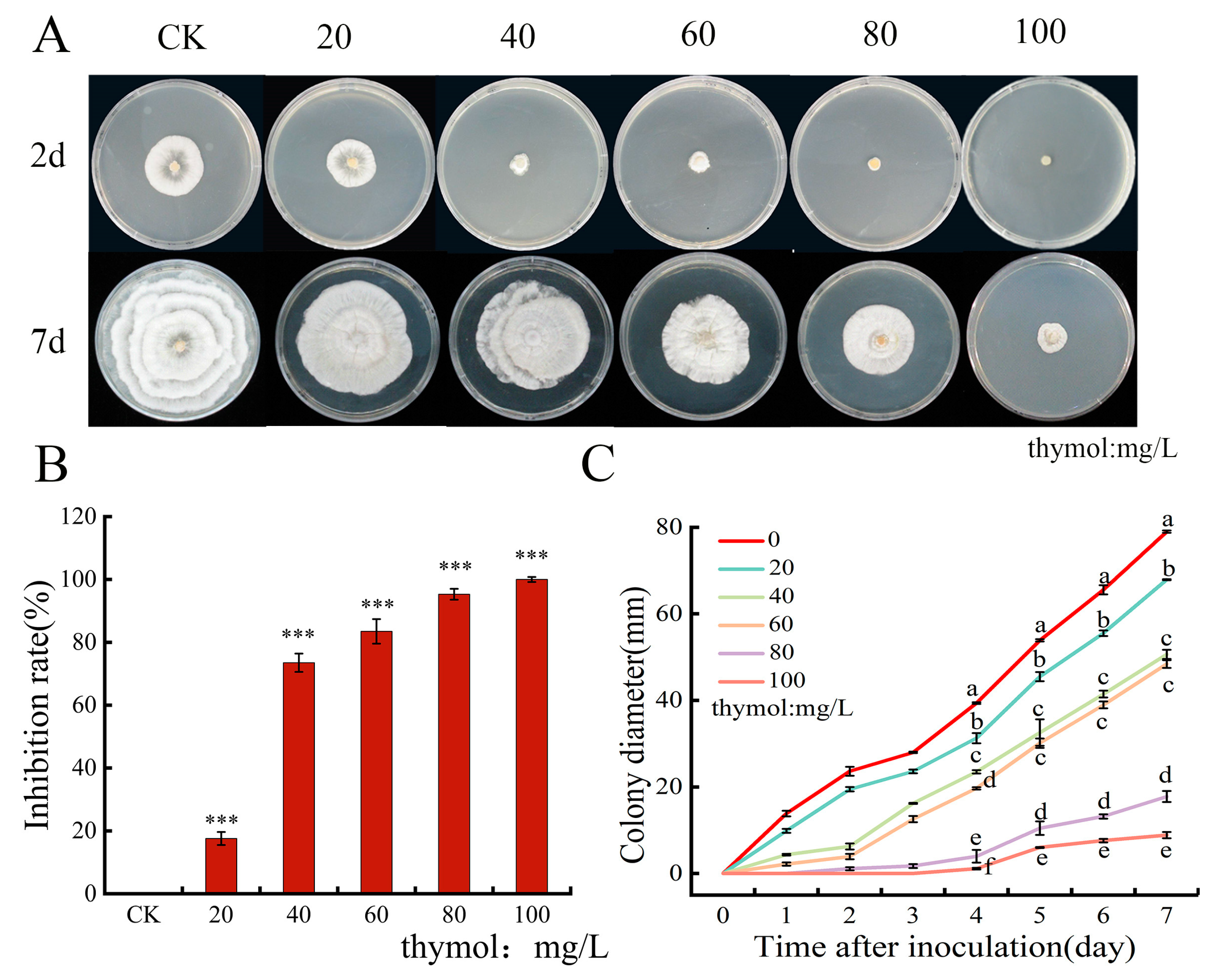
Figure 1. Inhibitory effect of thymol on Neopestalotiopsis clavispora. (A) in vitro growth of N. clavispora under the effect of different concentrations of thymol; (B) thymol inhibition rate on N. clavispora (* p < 0.05, *** p < 0.001); (C) colony diameter of N. clavispora; Significant changes in the mean for that sampling period are indicated by different letters above the bars (p < 0.05).
Horticulturae 09 00983 g001
Figure 2. (A): Effect of 0 and 100 mg/L thymol treatments on spore germination at different times; (B): inhibition of spore germination for treatments 0, 4, 8, and 12 h; (C): change in mycelial dry weight for different concentrations of thymol treatments. The standard errors of the means are shown by the error bars. Significant changes in the mean for that sampling period are indicated by different letters above the bars (p < 0.05).
Horticulturae 09 00983 g002
Figure 3. Extracellular conductivity (A), extracellular pH (B), intracellular nucleic acid release (C), extracellular soluble protein (D), and reducing sugars (E) of N. clavispora affected by different thymol concentrations. Values are expressed as the mean ± Standard deviation and error bars represent standard deviations of the means, and significant changes in the mean for that sampling period are indicated by different letters above the bars (p < 0.05).
Horticulturae 09 00983 g003
Figure 4. Fluorescence microscopy of Neopestalotiopsis clavispora after treatment with thymol. Effect of thymol treatment on mycelium cell membrane integrity analyzed by PI staining. (Bright: Bright field; PI: propidium iodide).
Horticulturae 09 00983 g004
Figure 5. Effect of thymol on malondialdehyde (MDA) (A), catalase (CAT) (B), and superoxide dismutase (SOD) (C) activity of Neopestalotiopsis clavispora. The standard errors of the means are shown by the error bars. Significant changes in the mean for that sampling period are indicated by different letters above the bars (p < 0.05).
Horticulturae 09 00983 g005
Figure 6. Fluorescence microscopy images showing the accumulation of reactive oxygen species in Neopestalotiopsis clavispora. Green fluorescence indicates the presence of ROS; red arrows in Figure point to the clearly visible septae separating constituent cells within the filaments (bright: bright field; DCF: 2,7-Dichlorodi-hydrofluorescein diacetate).
Horticulturae 09 00983 g006
Figure 7. Effect of thymol on intracellular adenosine triphosphate (ATP) (A), adenosine diphosphate (ADP) (B), adenosine monophosphate (AMP) (C) and energy charge (EC) levels (D) of Neopestalotiopsis clavispora. Values are expressed as the mean ± Standard deviation and error bars represent standard deviations of the means, and significant changes in the mean for that sampling period are indicated by different letters above the bars (p < 0.05).
Horticulturae 09 00983 g007
Disclaimer/Publisher’s Note: The statements, opinions and data contained in all publications are solely those of the individual author(s) and contributor(s) and not of MDPI and/or the editor(s). MDPI and/or the editor(s) disclaim responsibility for any injury to people or property resulting from any ideas, methods, instructions or products referred to in the content.