Article

Use of Light Spectra for Efficient Production of PLBs in Temperate Terrestrial Orchids

by
Hossein Naderi Boldaji
,
Shirin Dianati Daylami
* and
Kourosh Vahdati
Department of Horticulture, College of Aburaihan, University of Tehran, Tehran 3391653755, Iran
*
Author to whom correspondence should be addressed.
Horticulturae 2023, 9(9), 1007; https://doi.org/10.3390/horticulturae9091007
Submission received: 9 July 2023 / Revised: 24 August 2023 / Accepted: 4 September 2023 / Published: 7 September 2023

Abstract

:
Wild orchids, especially the terrestrial temperate ones are endangered species due to challenges in their natural habitats. Therefore, there is an urgent need to introduce efficient propagation methods to overcome the natural reproduction problems of these orchids. In this study, the effects of different light spectrums, explant types, wounding, and combinations of different plant growth regulators (PGRs) on direct somatic embryogenesis (DSE) of two species of these endangered orchids listed in the conservation category, were studied. The highest percentages of DSE formation and embryo germination were observed in Dactylorhiza umberosa protocorm explants exposed to white light (400–730 nm) and in Epipactis veratifolia protocorm explants exposed to a combination of red and far-red spectra (R: FR = 70:30). This occurred while red (610–700) alone and in combination with far-red (710–730 nm) spectrum induced embryogenesis more than the blue spectrum and dark condition in E. veratifolia. Thidiazuron (TDZ, 3 mg L−1), produced the highest percentage of protocorm-like bodies (PLBs) on protocorm explants in both orchids. Kinetin (Kin, 2 mg L−1) and Benzyladenine (BA 3 mg L−1) had the most effect on the survival and growth of PLBs, respectively, in D. umberosa and E. veratifolia. Species did not show similar embryogenesis responses under light spectrums. In a medium containing 3 mg L−1 TDZ, white light and R-FR spectra produced the most PLBs on wounded protocorm explants of D. umberosa and E. veratifolia respectively. The developmental stage of apical meristem of PLBs in both species was more advanced under R-B spectra, compared to others.

1. Introduction

Orchids are high-price ornamental crops that have attracted the attention of their production as pot plants and cut flowers [1]. Of the wild orchids, native orchids such as saleps have traditionally been harvested from their natural habitats for their use in the medicinal and food industries. The tubers of some terrestrial orchids like Orchis mascula, Dactylorhiza umberosa, and some other species are called salep [2]. Salep also refers to the powder of dried tubers that are used in the production of ice cream, drinks, medicines, confectionery, and hot drinks [3]. The scarcity of resources for salep has led to the uncontrolled harvesting of these plants from their natural habitats [4], making them an endangered species. Epipactis veratrifolia is one of the wild orchids used in traditional medicine and has a high potential for breeding as an ornamental plant [5]. Low seed germination, slow vegetative growth, and lack of proper coexistence have sometimes made their propagation by conventional methods impossible [6,7]. To achieve successful regeneration, in vitro techniques are now efficiently practiced and used for sexual and non-sexual propagation of orchids, especially for endangered orchid species [8,9]. There are various methods for mass in vitro culture of orchids such as culture of seeds [10], shoot tip and axillary bud culture [11], and using protocorm-like bodies (PLBs) [12], and flower buds [13]. Direct and indirect somatic embryogenesis (DSE), (ISE) and protocols have been offered for the induction of protocorm-like bodies (PLBs) in orchids [5,6,7]. These micropropagation methods have been used for somatic embryogenesis (SE) in many orchids such as Cymbidium [14], Oncidium [15], Phalaenopsis [1], and Xenikophyton smeeanum [16], however, there is not a wealth of information for in vitro micropropagation of temperate terrestrial orchids [17]. Explant type, genotype, medium composition, plant growth regulators (PGRs), and light regime can be mentioned as factors influencing somatic embryogenesis (SE) in Oncidium orchids [18]. Light is a determinant factor for the tissue culture of plants. Different light spectra have been used to study their effects on plant growth and organogenesis [19,20,21]. Light-emitting diodes (LEDs) have emerged as a new light source for in vitro culture. Different kinds of artificial light can play a key role in successful in vitro plant production, besides other factors such as gas exchange in the culture vessel, temperature, and composition of the culture medium [22]. The effects of the light spectrum have been investigated on the in vitro growth of many plant species such as Lilium ‘Pesaro’ [23], Dianthus caryophyllus [24], and Zantedeschia jucunda [25]. The emission of light in LED lighting systems allows the selection of spectra quality and provides the opportunity for the regulation of photosynthetic and photomorphogenic reactions required for an in vitro culture of plants [26]. Significant improvements have been achieved in increasing the fresh and dry weight of shoots and proliferation rate by changing the photoperiod regime from 16 h to a 4 h photoperiod, thereby allowing explants to do a better exchange of CO2 [27,28]. Furthermore, the light has been introduced as one of the important inducers for the generation of SE [29]. Effects of various spectra of LED lights have been reported in some orchids such as Oncidium [18], Phalaenopsis [30], and Cymbidium [31], as well as other plant species such as China Rose [32], and Agave tequilana [33]. The induction of SE and callus was obtained under different light conditions in different plant species. For instance, red, red + far-red lights in Phalaenopsis, red light in Cymbidium, red and white light in Agave tequilana, and red light in Rosa chinensis [30,31,32,33]. PGRs are among the most important factors that influence SE. Auxins and cytokinins can be mentioned specifically as the most used ones [34]. Applying PGRs can effectively improve DSE or ISE [24]. Positive effects of PGRs on embryogenesis have also been reported in plant species such as Phalaenopsis amabilis [34], Anthurium [35], Lilium ledebourii [36], and Cyclamen [37]. It was reported that wounding of the explants can lead to faster SE induction in soybeans [38], while there is little information on the effects of wounding on embryogenesis in plants such as orchids [39]. Most studies have been limited to tropical orchids, and only a few studies have been conducted on SE and regeneration in temperate terrestrial orchids. To the best of our knowledge, there are no studies on the effects of light spectra and wounding on somatic embryogenesis and regeneration in terrestrial orchids. In the present study, we have successfully established an in vitro propagation protocol for D. umberosa and E. veratifolia by proliferation through PLBs to facilitate the conservation, cultivation, and introduction of these plants into future flower markets.

2. Materials and Methods

2.1. Plant Materials and Explant Preparation

In this study, two native Iranian orchids were utilized; namely the rhizomatous species, E. veratrifolia and the tuberous species D. umberosa. Capsules containing mature brown seeds of these plants were collected from their natural habitats in the Alborz Mountains. Capsules were sterilized for 20 min in 20% sodium hypochlorite (NaOCl), followed by 3 rinses with sterilized distilled water; then about 100 tiny, dusty seeds (There are more than a thousand seeds in each capsule) were cultured on modified FAST (MFAST) as a basal medium in each Petri dish [40]. Four types of explants were used. Protocorm was the first kind of explant. At the end of the germination process of orchid seeds, embryos form small spherical tuber-like bodies referred to as protocorms and were used in two forms: wounded and non-wounded in both species (Figure 1f). After seedling growth, three other types of explants were prepared, which included leaf segment (in both species Figure 1d), single node (in E. veratrifolia-Figure 1e), and crown of the plantlet (in D. umberosa). The explants were cultured on a basal medium, containing different concentrations of PGRs (including TDZ, NAA, BA, and Kin) according to Table 1.

2.2. Media and Culture Conditions

Cultivated seeds (Figure 1) were grown using a hormone-free modified FAST medium (MFAST) as the basal medium containing both macro and micro elements (Merck, Hesse-Darmstadt, Germany). The medium was supplemented with myo-inositol 100 mg L−1, nicotinic acid 0.5 mg L−1, pyridoxine HCl 0.5 mg L−1, thiamine HCl 0.1 mg L−1, glycine 2 mg L−1, sucrose 3 g L−1, peptone 2% and agar 4.8 g L−1. [40]. The pH was adjusted to 5.5 ± 0.1 and the solution was autoclaved at 121 °C for 20 min. The same medium was used for continuing growth of explants for a period of 6 to 8 weeks. To study the effects of four kinds of growth regulators (PGRs), including thidiazuron (Sigma Aldrich, UAS) (TDZ 0, 2 and 3 mg L−1), N6-benzyleadenine (BA 0, 1, 2 and 3 mg L−1), ∞-naphthaleneacetic acid (NAA 0, 0.5 and 1 mg L−1), kinetin (KIN 0, 1, 2 and 3 mg L−1) and four kinds of explants (including five explants of each type of crown, node, leaf segment, and protocorm) were cultured in separate Petri dishes (five micro-samples in each of three replicates) and placed in a growth room under white LED lights (Figure 1). Then the interaction between TDZ (0 and 3 mg L−1), wounding, and light spectra was investigated on SE of protocorm explants (wounded or un-wounded) in both species. The light treatments including white light (W, as the control range at 400–700 nm), blue (B) range at 460 nm, red (R) range at 660 nm, green (G) range at 530 nm, combination of red and blue (R:B = 70:30), also a combination of red and far-red (R:FR = 70:30) were provided using LED lamps at light intensity of 80 µmol m−2 s−1 as well as darkness condition, as the control. The cultures were placed in a growth room with a temperature of 22 ± 2 °C, a light period of 16 h and, a relative humidity of 70%. Wavelengths were measured using a Sekonic C7000 spectrometer (Sekonic Corp., Tokyo, Japan) within the range of 300–800 nm. After four weeks, induction of DSE (globular type) was evaluated using a microscope. Following SE induction, the cultures were transferred to the hormone-free MFAST medium and placed under the light with the same environmental conditions as previously described.

2.3. Germination and Acclimatization

Embryos produced via DSE were counted as germinated (Table 1) and transferred to a hormone-free basal medium for plant growth under the environmental conditions described above. For acclimatization, the plantlets (developed from DSE) were transferred to plastic pots filled with a sterile coco-peat: perlite mixture (3:1), and placed in an adapted chamber at a temperature of 28 ± 2 °C.

2.4. Statistical Analysis

Experiments were arranged as factorial in a completely randomized design. The data were subjected to analysis of variance (ANOVA) and means were compared using Duncan’s multiple range tests at p < 0.05 probability level using the SAS 9.3 software.

3. Results

3.1. Effects of PGRs, Explant Type, and Wounding on DSE

Embryo formation was observed three weeks after placing the explants in the growth medium. The results of the statistical analysis showed a significant difference between the interaction of treatments (growth regulators, the type of explant, and the light spectrum). The percentage of DSE formation, PLBs germination, and final plantlets formation, especially on protocorm explants, in both species, had significant differences (p < 0.05) under various light spectra (Figure 2, Figure 3, Figure 4, Figure 5, Figure 6, Figure 7 and Figure 8). The type of PGR caused a significant effect on embryo formation in both species (Table 1, Figure 4). Between the PGR treatments, E. veratifolia showed the highest embryogenesis frequency (100%) when grown on a medium supplemented with 3 mg L−1 TDZ, using protocorms and followed by 1 mg L−1 NAA (38%) in node explants (Table 1). The medium supplemented with 3 mg L−1 BA had the lowest SE rate at 13%. Table 1 shows an increase in DSE production with increasing NAA concentration (from 0 to 1 mg L−1) in combination with BA. NAA treatment (0.5 mg L−1) alone and in combination with 1 mg L−1 BA, caused 25% PLB formation on the node explants in Epipactis veratifolia, but in combination with BA, the survival of these somatic embryos (PLBs growth %) was 50% more (Table 1). The positive effect of BA on the survival of somatic embryos is also observed in combination with 1 mg L−1 of NAA (Table 1).
Different concentrations of the BA alone or in combination with NAA did not affect the leaf explants of Epipactis veratifolia (Table 1). Three weeks after placing the explants in the growth medium, somatic embryos formed on protocorm explants of D. umberosa, under all light spectra. Embryo formation and germination rate, percentage of DSE, embryo germination, and plantlet production were significantly (p < 0.05) influenced by the type of explant, the combination of PGRs, light spectrums, and wounding (Table 1, Figure 4, Figure 5, Figure 6 and Figure 8). The kind of PGRs utilized had a considerable effect on the DSE percentage (Table 1). Only protocorm explants that developed globular embryos displayed somatic embryo initiation in D. umberosa (Table 1). The DSE was induced on the protocorm explants after three to four weeks. The maximum rate (100%) of embryo formation (DSE) was observed in a medium containing 3 mg L−1 TDZ (Table 1). In contrast, no embryos were observed on crown explants of D. umberosa in all PGR treatments. Kin in 1.5 and 2 mg L−1 concentrations, also caused 25% and 50% direct embryogenesis in protocorm explants of D. umberosa, respectively. All concentrations of PGRs (TDZ, BA, and NAA) did not affect the crown explant. SE was observed on both protocorm (100%) and stem node (38%) explants of E. veratifolia (Table 1). R-FR spectra caused most DSE on non-wounded protocorm explants of E. veratifolia (Figure 2 and Figure 5a). In D. umberosa only one form of the explant (protocorm) produced a number of somatic embryos (Figure 3) with a high frequency of embryo germination (Table 1). Therefore, the protocorm explant was selected to proceed with the experiment. The most somatic embryo of D. umberosa was obtained under white light and wounding, which had a positive effect on the DSE of this species (Figure 5b).

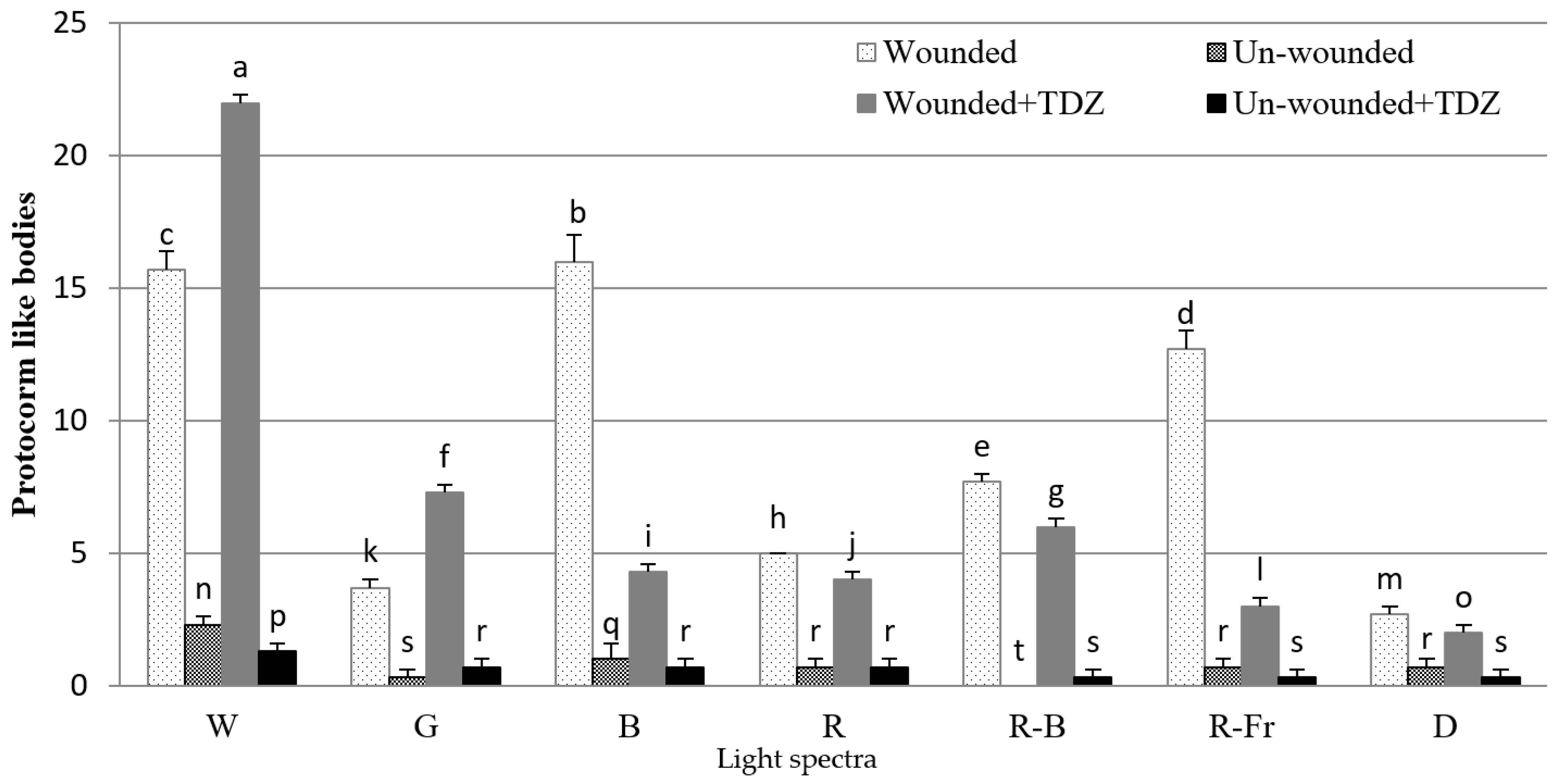
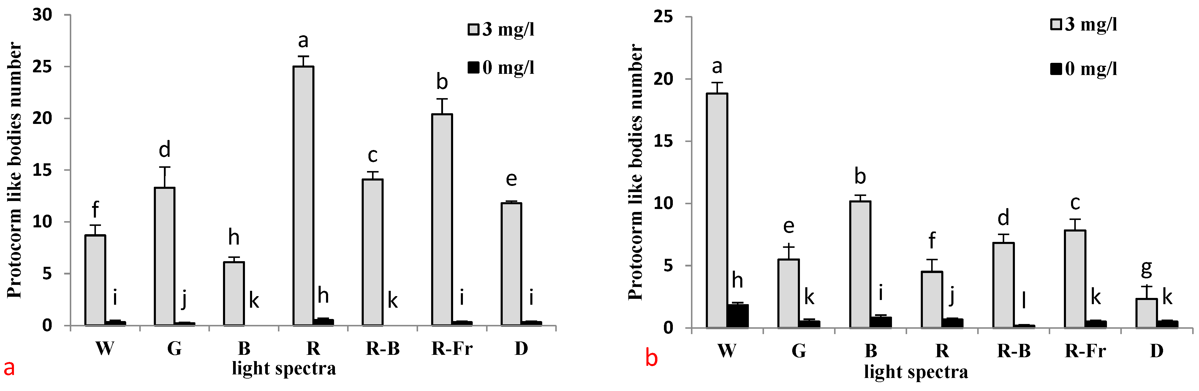
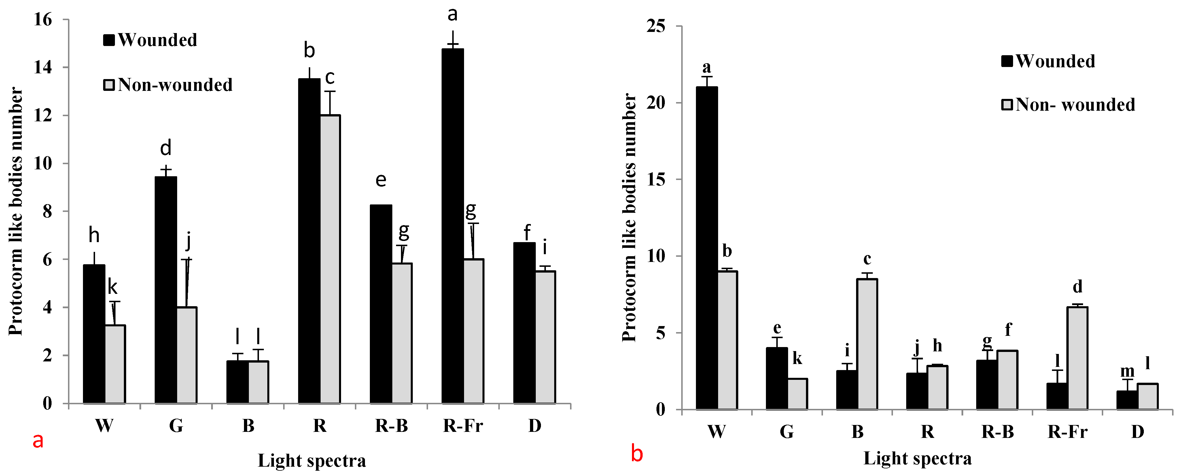
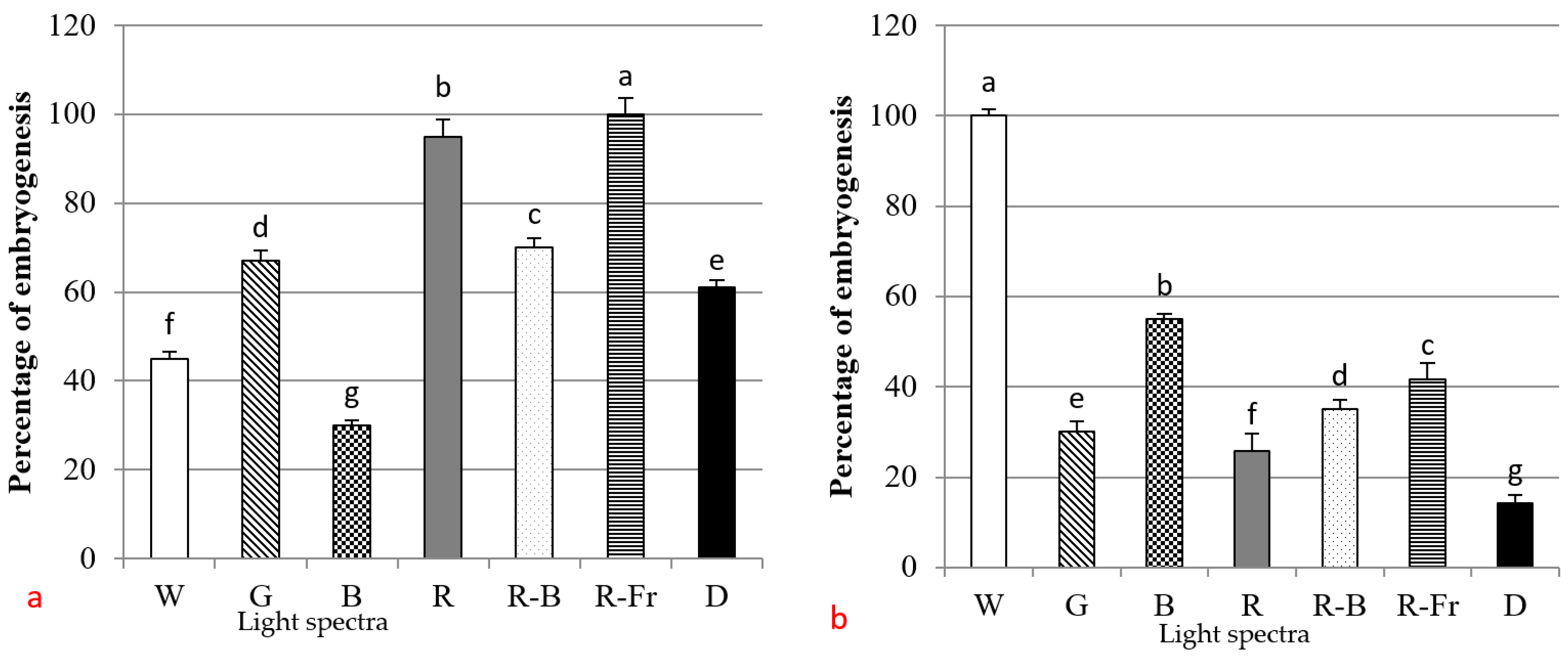
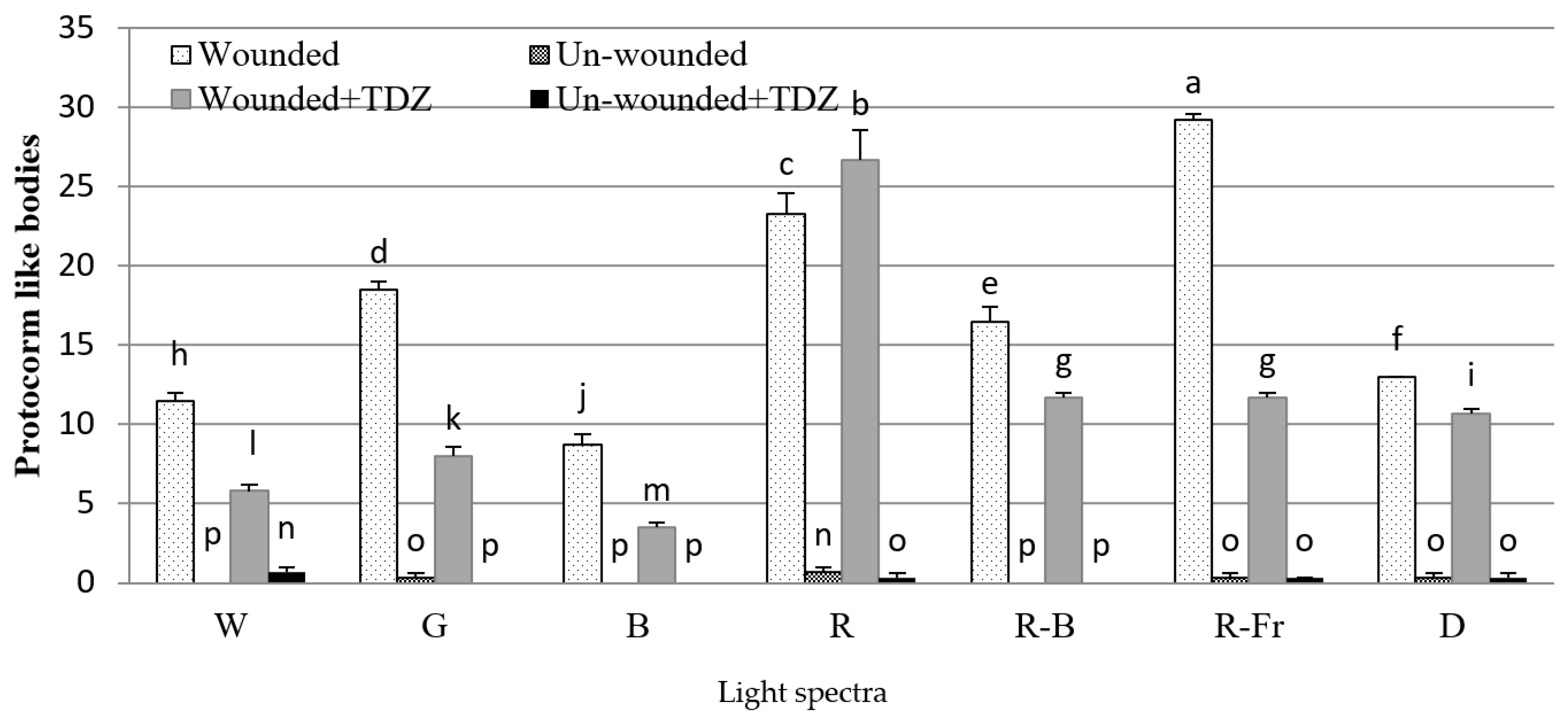
3.2. Induction of SE under Different Light Spectra

Different light spectrums had a notable impact on DSE in both orchid types. Red light led to a higher percentage (100%) of DSE and a number of PLBs (25 PLB) in E. veratifolia were observed under red light (Figure 4). The percentages of DSE were also affected by the light spectrums (p < 0.05). The greatest DSE percentages were observed under R: FR (100%), R (95%), RB (70%), and G (67%) spectra, with the lowest DSE seen under B (30%), W (45%), and darkness (61%), respectively (Figure 6a). FR (100%), R (95%), RB (70%), and G (67%) spectra, with the lowest DSE seen under B (30%), W (45%), and darkness (61%), respectively (Figure 6a). In D. umberosa, W light induced the highest DSE (100%), followed by B (55%), R: FR (41.7%), RB (35%), G (30%), R (25.8%), and D (14.2%) (Figure 6b). The size of SEs was significantly larger when explants of E. veratifolia were exposed to B, R-B, and G spectra. Both white light and RB spectra led to greater development of PLBs with three-leaf primordia (Figure 2). Bigger PLBs of D. umberosa were observed under the G spectrum and dark conditions. RB spectra resulted in the development of more PLBs with three-leaf primordia in this species (Figure 3). Explants of E. veratifolia turned brown and subsequently exhibited necrosis when exposed to RB, W, and R lights. As a result, the survival rate of plantlets decreased (Figure 9). Furthermore, the light spectrum had an effect on embryo germination in both orchids. The embryos generated under R-Fr and W lights resulted in the highest percentage of embryo germination and plantlet development in E. veratifolia (100%) and D. umberosa (100%), respectively (not shown).

4. Discussion

The effect of different light spectra on the direct embryogenesis of temperate terrestrial native orchids and their combination with plant growth regulators, explant type, and wounding has not been previously investigated. Some researchers have studied the embryogenesis of native orchids, for example, an in vitro somatic embryogenesis method and regeneration for Anoectochilus elatus Lindley, an endangered orchid, by studying some PGRs and vitamins [7]. Similar methods have been developed for some of the other endemic terrestrial orchids such as Anoectochilus elatus and Caladenia latifolia [41]. A number of studies conducted in this field on orchids have used different types of explants for DSE; for example, stem nodes [5,6,42], protocorm [41,42,43], apical buds, crowns, and leaves [5]. In the current study, we focused on introducing an efficient method for DSE in D. umberosa and E. veratifolia using a novel combination of light spectra, PGRs, and wounding. Leaf explants of E. veratifolia did not produce any SE when placed in different media with different PGRs, but protocorm and nodal explants efficiently induced SE (Table 1 and Figure 2a–h). Wounding had effects on the genes encoding the induction of cell-wall proteins [44]. In the current research, we found that in D. umberosa, wounded protocorm explants efficiently induced DSE compared to non-wounded ones especially under white light and G spectrum (Figure 5b). This phenomenon is observed in E. veratifolia under all light conditions (Figure 5a). This finding is consistent with previous findings in soybean [38] and in tomato shoot regeneration by wounding of cotyledonary explants [45]. The results suggest that intact explants are more efficient for embryo formation in D. umberosa under B, R, RB, RFR spectra, and dark conditions (Figure 5b). It has been previously reported that the wounding of explants causes browning, which increases the phenolic compounds and eventually leads to the death of explants [46]. It can be said that wounding is an effective treatment to induce embryogenesis and the light spectrum may affect its efficiency.
PGRs are known to stimulate cell division and play an important role in the induction of SE [47]. The majority of previous studies have required auxins to induce SE in various plant species, while some reports have shown that cytokinins promote the formation of embryogenic cells with a role similar to that of auxins [48,49], for example, in orchids [5,15,50]. We used three concentrations of Kin (1, 1.5, and 2 mg L−1), which had not previously been used for DSE in terrestrial orchids. Two concentrations of Kin (1.5 and 2 mg L−1) resulted in positive effects on the DSE of D. umberosa (Table 1 and Figure 3). In other studies, the growth regulator kinetin has been used to induce protocorm formation in Dactylorhiza majalis [51,52]. The effect of these kinds of PGRs on embryogenesis has been reported on black iris [53], Phalaenopsis [54], and Rhynchostylis retusa [55]. Effective PGR treatments were 3 mg L−1 TDZ on protocorm and 1 mg L−1 NAA in nodal explant of E. veratifolia and 3 mg L−1 TDZ and 2 mg L−1 Kin on protocorm explant of D. umberosa (Table 1). The high concentrations of Kin and NAA (3 and 1 mg L−1, respectively) had no positive effect on SE. It was reported that the exogenous addition of a high concentration of BA and Kin significantly inhibited SE formation in orchard grass [56]. Our results showed that TDZ had the highest efficiency for DSE than BA and NAA in E. veratifolia. This was consistent with the previous results in Dendrobium [50,57] and Cymbidium [33]. In general, we found that the application of TDZ and NAA on wounded explant was more effective for DSE in E. veratifolia and TDZ, Kin on wounded explant induced DSE in D. umberosa. There are only a few reports that mention the role of light spectra on SE induction in native orchid species. In the present experiment, the highest percentage of DSE and embryo germination rate in E. veratifolia was observed in protocorm explants exposed to R-Fr (100%) and then R (95%) spectra (Figure 6), while B spectra, W light, and D condition had less effect on embryo formation on explants (Figure 6a). This finding is in agreement with the results of previous studies on the initiation and development of SE by R and R-Fr spectra in Araujia sericifera [29], Oncidium [18], and Rosa chinensis [34]. In E. veratifolia, R and R-Fr spectra produced the highest rate of embryogenesis and proliferation of embryos (PLBs); although, the highest percentage of embryo proliferation was obtained under R-Fr light (Figure 5a and Figure 7). The positive effects of the red light spectrum have been reported on the formation of 100% protocorms in orchids [58]. Consistently, R light has been reported to promote the induction and proliferation of SE in Oncidium [18]. Red light has been shown to affect plant reproduction through phytochrome, which in its active form increases the endogenous hormonal balance, increasing the amount of cytokinin in the tissues and counteracting the action of auxin [59]. In our study, in addition to R and R-Fr, G, R-B, B, W light and D conditions were used on explants to induce embryogenesis in E. veratifolia, but most of the explants produced a small number of embryos that also turned brown and necrotic under these conditions during three to four weeks (Figure 9a,b). This shows that G, R-B, B spectra, W light, and D conditions delayed the germination of SE in E. veratifolia. In the current study, the maturation of embryos as well as the germination of embryos was significantly different in different light treatments. In some of the previous studies, it has been reported that callus formation was high in the blue spectrum, which proves that rapid cell division occurred, although the organized center of cell division required for primordia formation was reduced and growth was delayed [60]. Interestingly, we found that G light increased the formation and germination of SE in protocorm explants of E. veratifolia (Figure 4a and Figure 6a). The effect of the G spectrum was significant on the percentage of explants that had embryogenesis and on the number of embryos (Figure 5a and Figure 6a). A study on carrot embryogenesis showed that G light caused the highest SE compared to the other light spectra [61]. The highest PLBs formation, root formation, and shoot formation rate in Cymbidium insigne was reported under the G spectrum [62]. The G light may have active photoreceptors [63], but unlike red and blue light, green light photoreceptors have not been discovered yet [64]. Embryos germinated when sub-cultured in the hormone-free medium. In both orchids, the somatic embryos had advanced developmental stages under the B and R-B spectrum, which rapidly increased the size of the embryos (Figure 9d). This result shows the positive effects of the B spectrum on the development of PLBs and it is in agreement with the previous report on the promotive effects of the B spectrum on embryo size in carnation [24].
The results of this study show that the highest percentage of DSE (100%) was observed in the explants exposed to W light, while the dark condition had the lowest effect on embryo induction rate in D. umberosa (Figure 6b). Researchers have also reported that SE induction and promotion are reduced in darkness in olive [65], soybean [66], and Coffea Arabica [67]. The highest percentage of embryogenesis in D. umberosa was produced under the W light and B spectrum (Figure 6b). The B spectrum had an efficient effect on DSE and ISE in carnation [24]. The highest number of the torpedo, globular, and heart embryos was observed under the B spectrum; in comparison to the effect of other light treatments that had no embryo formation [24]. The B light can affect plant auxin content (specifically indoleacetic acid-IAA). Therefore, it can affect PGR and morphotomorphogenesis [68]. Similar to the previous species, in D. umberosa, the apical meristem of PLBs had the most development under R-B spectra and produced two to three-leaf primordia (Figure 2e). This issue shows the positive and promotive effects of the B spectrum. The result showed that when the G, R spectra and D conditions were used to induce embryogenesis in D. umberosa, most of the explants did not produce embryos or a high number of embryos died (Figure 9). The PLB with larger diameters were observed under the G spectrum and D conditions (Figure 3d,f), but these conditions were not suitable for their survival. In D. umberosa, the embryos germinated when they were transferred and cultured on the PGR-free basal medium, and the germination level of embryos in different light spectra was significantly different. After exposure to white light, the B spectrum increased the induction and germination of SE from the protocorm explant (Figure 6b and Figure 8). The B spectrum is important for photomorphogenesis [68]. Consistent with this, it has been reported in Oncidium that the B spectrum increases differentiation and enzyme activities in embryos [18]. Previous studies have already mentioned the relationship between SE production and photo-equilibrium and the increase in SE formation by increasing photo-equilibrium. There is a higher photo-equilibrium under W light compared to the B spectrum [69]. Our results indicate that LEDs are a forward light source that increases DSE induction. Formation with high efficiencies of plant regeneration in both terrestrial orchids studied. We recommend the use of white light to produce the most PLBs on wounded protocorm explants of D. umberosa (Figure 8) and R-Fr light for producing the highest number of PLBs on wounded protocorm explants of E. veratifolia (Figure 7), both in a culture medium containing 3 mg L−1 TDZ.

5. Conclusions

Some native species like D. umberosa have been exposed to extinction because of uncontrolled harvest from their natural habitats. Mass propagation of E. veratrifolia and D. umberosa is difficult and sometimes impossible by using conventional in vitro culture methods because of low seed germination, lack of suitable symbionts, slow vegetative growth, and absence of efficient methods for asexual reproduction. In the present study, for the first time, a successful method and an efficient in vitro propagation protocol were established for these species by using light spectra through DSE. This protocol will have a significant impact on commercial micropropagation and genetic resource conservation of these native orchids. Wounding on the protocorm explants of D. umberosa with the use of a medium containing 3 mg L−1 TDZ caused a 94.1% increase in the number of PLBs under white light. In E. veratrifolia, the use of wounded protocorm explants in the medium containing 3 mg L−1 TDZ under the R spectrum caused an increase in production by 98.8%.

Author Contributions

Conceptualization, H.N.B. and S.D.D.; Data curation, H.N.B.; Formal analysis, H.N.B. and S.D.D.; Investigation, H.N.B. and S.D.D.; Methodology, S.D.D.; Project administration, S.D.D.; Resources, S.D.D.; Software, H.N.B. and S.D.D.; Supervision, S.D.D.; Validation, S.D.D.; Writing—original draft, H.N.B.; Writing—review and editing, H.N.B., S.D.D. and K.V. All authors have read and agreed to the published version of the manuscript.

Funding

This research received no external funding.

Data Availability Statement

Not applicable.

Acknowledgments

The authors wish to thank the Orchid Breeding and Propagation Laboratory of the Horticultural Department, University of Tehran for the opportunity to carry out their research.

Conflicts of Interest

No potential conflict of interest were reported in this study.

References

  1. Gow, W.P.; Chen, J.T.; Chang, W.C. Influence of growth regulators on direct embryo formation from leaf explants of Phalaenopsis orchids. Acta Physiol. Plant. 2008, 30, 500–507. [Google Scholar] [CrossRef]
  2. Ghorbani, A.; Barbara, G.; Farzaneh, N.; Hugo, B. Wild orchid tuber collection in Iran: A wake-up call for conservation. Biodivers. Conserv. 2014, 23, 2749–2760. [Google Scholar] [CrossRef]
  3. Starin, D. Salepi extinction, salepi survival: How a change in ingredients could help safeguard orchids. Am. Orch. Soc. Bull. 2012, 81, 490–494. [Google Scholar]
  4. Sezik, E. Destroying of Ophrys species to obtain salep in Turkey. J. Eur. Orch. 2006, 38, 290–295. [Google Scholar]
  5. Moradi, S.; Dianati Daylami, S.; Arab, M.; Vahdati, K. Direct somatic embryogenesis in Epipactis veratrifolia, a temperate terrestrial orchid. J. Hortic. Sci. Biotechnol. 2017, 92, 88–97. [Google Scholar] [CrossRef]
  6. Jalal, J.S. Endemic orchids of peninsular India: A review. J. Threat. Taxa 2012, 15, 3415–3425. [Google Scholar] [CrossRef]
  7. Sherif, N.; Ahamed, J.H.; Franklin, B.; Senthil, K.; Rao, M.V. Somatic embryogenesis, acclimatization and genetic homogeneity assessment of regenerated plantlets of Anoectochilus elatus Lindl an endangered terrestrial jewel orchid. Plant Cell Tissue Organ Cult. 2018, 132, 303–316. [Google Scholar] [CrossRef]
  8. Kumar, S.; Narender, S.; Manisha, M. Micropropagation of Simmondsia chinensis (Link) Schneider through enhanced axillary branching from nodal segments. J. Plant Biol. 2009, 36, 75–81. [Google Scholar]
  9. Dogan, Y.; Suleyman, B.; Gungor, A.; Hasan, H.M. the use of wild edible plants in western and central Anatolia (Turkey). Econ. Bot. 2004, 58, 684–690. [Google Scholar] [CrossRef]
  10. Stewart, S.L.; Kane, M.E. Asymbiotic seed germination and in vitro seedling development of Habenaria macroceratitis (Orchidaceae) a rare Florida terrestrial orchid. Plant Cell Tissue Organ Cult. 2006, 86, 147–158. [Google Scholar] [CrossRef]
  11. Pant, B.; Thapa, D. In vitro mass propagation of an epiphytic orchid, Dendrobium primulinum lindl through shoot tip culture. Afr. J. Biotechnol. 2012, 11, 9970–9974. [Google Scholar] [CrossRef]
  12. Tanaka, M.; Hasegawa, A.; Goi, M. Studies on the clonal propagation of monopodial orchids by tissue culture I. Formation of protocorm-like bodies from leaf tissues in Phalaenopsis and Vanda. J. Jap. Soc. Hort. Sci. 1975, 44, 47–58. [Google Scholar]
  13. Kosir, P.; Skof, S.; Luthar, Z. Direct shoot regeneration from nodes of Phalaenopsis orchids. Acta Agric. Slov. 2004, 83, 233–242. [Google Scholar]
  14. Mahendran, G.; Bai, V.N. Direct somatic embryogenesis and plant regeneration from seed derived protocorms of Cymbidium bicolor Lindl. Sci. Hortic. 2012, 135, 40–44. [Google Scholar] [CrossRef]
  15. Chen, J.T.; Chang, W.C. Effects of auxins and cytokinins on direct somatic embryogenesis on leaf explants of Oncidium ‘Gower Ramsey’. J. Plant Growth Regul. 2001, 34, 229–232. [Google Scholar] [CrossRef]
  16. Mulgund, G.S.; Nataraja, K.; Malabadi, R.B.; Kumar, S.V. TDZ induced in vitro propagation of an epiphytic orchid Xenikophyton smeeanum. Plant Biol. 2011, 1, 235–241. [Google Scholar]
  17. Arditti, J. Micropropagation of Orchids; John Wiley & Sons: Hoboken, NJ, USA, 2009. [Google Scholar]
  18. Mengxi, L.; Zhigang, X.; Yang, Y.; Yijie, F. Effects of different spectral lights on Oncidium PLBs induction, proliferation, and plant regeneration. Plant Cell Tissue Organ Cult. 2011, 106, 1–10. [Google Scholar] [CrossRef]
  19. Nguyen, Q.T.; Kozai, T.; Niu, G.; Van Nguyen, U. Photosynthetic characteristics of coffee (Coffea arabusta) plantlets in vitro in response to different CO2 concentrations and light intensities. Plant Cell Tissue Organ Cult. 1998, 55, 133–139. [Google Scholar] [CrossRef]
  20. Nhut, D.T.; Takamura, T.; Watanabe, H. Responses of strawberry plantlets cultured in vitro under superbright red and blue lightemitting diodes (LEDs). Plant Cell Tissue Organ Cult. 2003, 73, 43–52. [Google Scholar] [CrossRef]
  21. Cybularz-Urban, T.; Hanus-Fajerska, E.; Swiderski, A. Effect of light wavelength on in vitro organogenesis of a Cattleya hybrid. Acta Biol. Crac. Ser. Bot. 2007, 49, 113–118. [Google Scholar]
  22. Cavallaro, V.; Pellegrino, A.; Muleo, R.; Forgione, I. Light and plant growth regulators on in vitro proliferation. Plants 2022, 11, 844. [Google Scholar] [CrossRef] [PubMed]
  23. Lian, M.L.; Murthy, H.N.; Paek, K.Y. Effects of light emitting diodes (LEDs) on the in vitro induction and growth of bulblets of Lilium oriental hybrid ‘Pesaro’. Sci. Hortic. 2002, 94, 365–370. [Google Scholar] [CrossRef]
  24. Aalifar, M.; Arab, M.; Aliniaeifard, S.; Dianati, S.; Mehrjerdi, M.Z.; Limpens, E.; Serek, M. Embryogenesis efficiency and genetic stability of Dianthus caryophyllus embryos in response to different light spectra and plant growth regulators. Plant Cell Tissue Organ Cult. 2019, 139, 479–492. [Google Scholar] [CrossRef]
  25. Jao, R.C.; Chien-Chou, L.; Wei, F.; Sen-Fuh, C. Effects of red light on the growth of Zantedeschia plantlets in vitro and tuber formation using light-emitting diodes. HortScience 2005, 40, 436–438. [Google Scholar] [CrossRef]
  26. Dutta Gupta, S.; Jatothu, B. Fundamentals and applications of light-emitting diodes (LEDs) in in vitro plant growth and morphogenesis. Plant Biotechnol. Rep. 2013, 7, 211–220. [Google Scholar] [CrossRef]
  27. Biswal, A.K.; Pattanayak, G.K.; Pandey, S.S.; Leelavathi, S.; Reddy, V.S.; Govindjee; Tripathy, B.C. Light intensity-dependent modulation of chlorophyll b biosynthesis and photosynthesis by overexpression of chlorophyllide a oxygenase in Tobacco. Plant Physiol. 2012, 159, 433–449. [Google Scholar] [CrossRef]
  28. Zhou, M.; Guan, Q.; Wei, Y.; Zhang, Z. Effects of Sucrose Concentration and light intensity on growth and photosynthesis of ginger plantlets in vitro. Chin. J. Appl. Environ. Biol. 2008, 14, 356–361. [Google Scholar]
  29. Torne, J.M.; Moysset, L.; Santos, M.; Simón, E. Effects of light quality on somatic embryogenesis in Araujia sericifera. Physiol. Plant. 2001, 111, 405–411. [Google Scholar] [CrossRef] [PubMed]
  30. Park, S.Y.; Yeung, E.C.; Paek, K.Y. Endoreduplication in Phalaenopsis is affected by light quality from light-emitting diodes during somatic embryogenesis. Plant Biotechnol. Rep. 2010, 4, 303–309. [Google Scholar] [CrossRef]
  31. Le, V.T.; Tanaka, M. Effects of red and blue light-emitting diodes on callus induction, callus proliferation, and protocorm-like body formation from callus in Cymbidium orchid. Environ. Control. Biol. 2004, 42, 57–64. [Google Scholar]
  32. Chen, J.R.; Wu, L.; Hu, B.W.; Yi, X.; Liu, R.; Deng, Z.N.; Xiong, X.Y. The influence of plant growth regulators and light quality on somatic embryogenesis in China rose. J. Plant Growth Regul. 2014, 33, 295–304. [Google Scholar] [CrossRef]
  33. Rodríguez-Sahagún, A.; Acevedo-Hernández, G.; Rodríguez-Domínguez, J.M.; Rodríguez-Garay, B.; Cervantes-Martínez, J.; Castellanos-Hernández, O.A. Effect of light quality and culture medium on somatic embryogenesis of Agave tequilana Weber, Azul. Plant Cell Tissue Organ Cult. 2011, 104, 271–275. [Google Scholar] [CrossRef]
  34. Mose, W.; Indrianto, A.; Purwantoro, A.; Semiarti, E. The influence of thidiazuron on direct somatic embryo formation from various types of explant in Phalaenopsis amabilis (L.) blume orchid. Hayati J. Biosci. 2017, 24, 201–205. [Google Scholar] [CrossRef]
  35. Bhattacharya, C.; Dam, A.; Karmakar, J.; Bandyopadhyay, T.K. Direct somatic embryogenesis and genetic homogeneity assessment of regenerated plants of Anthurium andraeanum Linden cv. Fantasia. Vitr. Cell. Dev. Biol. Plant 2016, 52, 512–519. [Google Scholar] [CrossRef]
  36. Bakhshaie, M.; Babalar, M.; Mirmasoumi, M.; Khalighi, A. Somatic embryogenesis and plant regeneration of Lilium ledebourii (Baker) Boiss an endangered species. Plant Cell Tissue Organ Cult. 2010, 102, 229–235. [Google Scholar] [CrossRef]
  37. Prange, A.N.S.; Bartsch, M.; Serek, M.; Winkelmann, T. Regeneration of different Cyclamen species via somatic embryogenesis from callus, suspension cultures and protoplasts. Sci. Hortic. 2010, 125, 442–450. [Google Scholar] [CrossRef]
  38. Santarem, E.R.; Pelissier, B.; Finer, J.J. Effect of explant orientation, pH, solidifying agent and wounding on initiation of soybean somatic embryos. Vitr. Cell. Dev. Biol. Plant 1997, 33, 13–19. [Google Scholar] [CrossRef]
  39. Rojas-Herrera, R.; Loyola-Vargas, V. Induction of a class acidic chitinase in foliar explants of (Coffea arabica L.) during somatic embryogenesis and wounding. Plant Sci. 2002, 163, 705–711. [Google Scholar] [CrossRef]
  40. Dianati, S.; Kafi, M.; Mirmaoumi, M.; Mozaffarian, V.; Salami, A.R. Introduce the suitable medium for asymbiotic germination of Epipactis veratrifolia. J. Crops Improv. 2014, 15, 161–177. [Google Scholar] [CrossRef]
  41. Bustam, B.M.; Dixon, K.; Bunn, E. Proliferation and harvesting of secondary protocorms as a novel means for improving propagation of terrestrial orchids. Aust. J. Bot. 2015, 62, 614–621. [Google Scholar] [CrossRef]
  42. Gangaprasad, A.N.; Decruse, W.S.; Seeni, S.; Menon, S. Micropropagation and restoration of the endangered Malabar daffodil orchid Ipsea malabarica. Lindleyana 1999, 14, 38–46. [Google Scholar]
  43. Romero, C.; Cuba-Díaz, M.; Silva, R. In vitro culture of Chloraea gavilu Lindl., an endemic terrestrial orchid from Chile. Plant Biosyst. 2018, 152, 612–620. [Google Scholar] [CrossRef]
  44. Showalter, A.M.; Bell, J.N.; Cramer, C.L.; Bailey, J.A.; Varner, J.E.; Lamb, C.J. Accumulation of hydroxyproline-rich glycoprotein mRNAs in response to fungal elicitor and infection. Proc. Natl. Acad. Sci. USA 1985, 82, 6551–6555. [Google Scholar] [CrossRef] [PubMed]
  45. Bhatia, P.; Ashwath, N.; Midmore, D.J. Effects of genotype, explant orientation, and wounding on shoot regeneration in tomato. Vitr. Cell. Dev. Biol. Plant. 2005, 41, 457–464. [Google Scholar] [CrossRef]
  46. Chamandoosti, F. The relationship between plant growth regualtors for organogenesis and phenolic compound in cotton. Asian J. Dev. Biol. 2010, 2, 16–22. [Google Scholar]
  47. Vahdati, K.; Jariteh, M.; Niknam, V.; Mirmasoumi, M.; Ebrahimzadeh, H. Somatic embryogenesis and embryo maturation in Persian walnut. Acta Hortic. 2006, 705, 199–205. [Google Scholar] [CrossRef]
  48. Thomas, C.; Jiménez, V.M. Mode of action of plant hormones and plant growth regulators during induction of somatic embryogenesis: Molecular aspects. Somat. Embryog. 2005, 23, 157–175. [Google Scholar] [CrossRef]
  49. Singh, C.; Raj, S.R.; Jaiswal, P.; Patil, V.; Punwar, B.; Chavda, J.; Subhash, N. Effect of plant growth regulators on in vitro plant regeneration of sandalwood Santalum album L. via organogenesis. Agrofor. Syst. 2016, 90, 281–288. [Google Scholar] [CrossRef]
  50. Gow, W.P.; Jen-Tsung, C.; Wei-Chin, C. Enhancement of direct somatic embryogenesis and plantlet growth from leaf explants of Phalaenopsis by adjusting culture period and explant length. Acta Physiol. Plant. 2010, 32, 621–627. [Google Scholar] [CrossRef]
  51. Ponert, J.; Vosolsobě, S.; Kmecová, K.; Lipavská, H. European orchid cultivation–from seed to mature plant. Eur. J. Environ. Sci. 2011, 26, 5–15. [Google Scholar] [CrossRef]
  52. Chen, L.R.; Chen, J.T.; Chang, W.C. Efficient production of protocorm-like bodies and plant regeneration from flower stalk explants of the sympodial orchid Epidendrum radicans. Vitr. Cell. Dev. Biol. Plant. 2002, 38, 441–445. [Google Scholar] [CrossRef]
  53. Shibli, R.A.; Ajlouni, M. Somatic embryogenesis in the endemic black iris. Plant Cell Tissue Organ Cult. 2000, 61, 15–21. [Google Scholar] [CrossRef]
  54. Kuo, H.L.; Chen, J.T.; Chang, W.C. Efficient plant regeneration through direct somatic embryogenesis from leaf explants of Phalaenopsis ‘Little Steve’. Vitr. Cell. Dev. Biol. Plant. 2005, 41, 453–461. [Google Scholar] [CrossRef]
  55. Bhattacharjee, B.; Shahinul Islam, S.M. Assessment of antibacterial and antifungal activities of the extracts of Rhynchostylis retusa Blume-A Medicinal Orchid. WJPPS 2015, 2, 74–87. [Google Scholar]
  56. Wenck, A.R.; Conger, B.V.; Trigiano, R.N.; SAMS, C.E. Inhibition of somatic-embryogenesis in orchardgrass by endogenous cytokinins. Plant Physiol. 1988, 88, 990–992. [Google Scholar] [CrossRef] [PubMed]
  57. Chung, H.H.; Chen, J.T.; Chang, W.C. Plant regeneration through direct somatic embryogenesis from leaf explants of Dendrobium. Biol. Plant. 2007, 2, 346–350. [Google Scholar] [CrossRef]
  58. Kamal, M.M.; Shimasaki, K.; Akter, N. Effect of light emutting diode (LED) lamps and N-Acetylglucosamine (NAG) on organogenesis in protocorm-like bodies (PLBs) of a Cymbidium hybrid cultured in vitro. Plant Tissue Cult. Biotechnol. 2014, 24, 273–297. [Google Scholar] [CrossRef]
  59. Finlayson, S.A.; Krishnareddy, S.R.; Kebrom, T.H.; Casal, J.J. Phytochrome regulation of branching in Arabidopsis. Plant Physiol. 2010, 152, 1914–1921. [Google Scholar] [CrossRef]
  60. Lotfi, M.; Mars, M.; Werbrouck, S. Optimizing pear micropropagation and rooting with light emitting diodes and trans-cinnamic acid. Plant Growth Regul. 2019, 88, 173–180. [Google Scholar] [CrossRef]
  61. Michler, C.H.; Lineberger, R.D. Effects of light on somatic embryo development and abscisic levels in carrot suspension cultures. Plant Cell Tissue Organ Cult. 1987, 11, 189–207. [Google Scholar] [CrossRef]
  62. Golovatskaya, I.F.; Karnachuk, R.A. Role of green light in physiological activity of plants. Russ. J. Plant Physiol. 2015, 62, 727–740. [Google Scholar] [CrossRef]
  63. Shinomura, T.; Nagatani, A.; Hanzawa, H.; Kubota, M.; Watanabe, M.; Furuya, M. Action spectra for phytochrome A-and B-specific photoinduction of seed germination in Arabidopsis thaliana. Proc. Natl. Acad. Sci. USA 1996, 93, 8129–8133. [Google Scholar] [CrossRef] [PubMed]
  64. Nahar, S.J.; Haque, S.M.; Kazuhiko, S. Application of chondroitin sulfate on organogenesis of two Cymbidium spp. under different sources of lights. Not. Sci. Biol. 2016, 8, 156–160. [Google Scholar] [CrossRef]
  65. Lavee, S.; Glenda, M. The effect of growth-regulating substances and light on olive callus growth in vitro. J. Exp. Bot. 1969, 20, 604–614. [Google Scholar] [CrossRef]
  66. Lazzeri, P.; David, F.H.; Glenn, B.C. Soybean somatic embryogenesis: Effects of hormones and culture manipulations. Plant Cell Tissue Organ Cult. 1987, 10, 197–208. [Google Scholar] [CrossRef]
  67. Gatica, A.; Andrés, M.; Griselda, A.; Ana, M.E. direct somatic embryogenesis in Coffea arabica L. Caturra and Catuaí: Effect of tricontanol, light condition, and medium consistency. Agron. Costarric. 2008, 5, 203–212. [Google Scholar]
  68. Schuerger, C.; Christopher, S.; Brown, C. Anatomical features of pepper plants Capsicum annuum grown under red light-emitting diodes supplemented with blue or far-red light. Ann. Bot. 1997, 79, 273–282. [Google Scholar] [CrossRef]
  69. D’Onofrio, C.; Morini, S.; Bellocchi, G. Effect of light quality on somatic embryogenesis of quince leaves. Plant Cell Tissue Organ Cult. 1998, 53, 91–98. [Google Scholar] [CrossRef]
Figure 1. The seed germination stage of Epipactis veratifolia; kinds of explants and seedlings (a) cultivated seeds, (b) swelling of the embryos (arrow) in the first stage of germination, (c) rupture of seed testa by swelled embryos, (d) leaf explant (basal part of leaf), (e) stem node explant, (f) intact and wounded protocorm explants; the end part without shoot apical meristem (arrow) used as explants, (g) in vitro rooted seedlings; their leaf and stem nodes (N) used as explants, (h) a protocorm resulting from the development of a Dactylorhiza umberosa seed embryo at the stage of converting apical meristem (AP) to leaf primotdia (LP), with many hairy rhizoids (R) at the bottom end.
Horticulturae 09 01007 g001
Figure 2. PLBs formation of Epipactis veratifolia after direct somatic embryogenesis under different light spectra; on protocorm explants ((ag). in medium contain 3 mg L−1 TDZ) and node explant ((h), in medium contain 0.5 mg L−1 NAA); (a) Cluster of PLBs at different developmental stages, including rudimentary PLBs (RP), PLBs with shoot apical meristem (SM), PLBs with first leaf primordia (LP) and PLBs with first leaf (FL) under blue spectrum. Each red circle represents a PLB which its stage or morphology is explained by arrow, (b) PLBs with huge diameter under green spectrum, (c) large PLBs with two leaf primordia (2LP) under red + blue spectra, (d) deformed PLBs with branched first leaf primordia under red spectrum, (e) many tiny PLBs with one leaf primordia without chlorophyll under red + far spectra, (f) PLBs with three leaf primordia (3LP) without chlorophyll under dark conditions, (g) many PLBs with three leaf primordia (3LP) under white light, (h) direct somatic embryo (one PLB) formation on node explant under dark condition by using 0.5 mg L−1 NAA, (i) growth of somatic plantlet on hormone-free medium.
Horticulturae 09 01007 g002
Figure 3. Direct somatic embryogenesis (formation of PLBs) on protocorm explants of Dactylorhiza umberosa under different light conditions including; (a) Cluster of PLBs at different stages of development stages with many rhizoids (R) under blue spectrum (in media containing 1.5 mg L−1 Kin), (b) under white light (in media containing 2 mg L−1 Kin), (c) red + far-red spectra (in media containing 3 mg L−1 TDZ), (d) dark condition (in media containing 1.5 mg L−1 Kin), (e) red + blue spectra (in media containing 3 mg L−1 TDZ), (f) green spectra (in media containing 3 mg L−1 TDZ), (g) red spectra (in media contain 3 mg L−1 TDZ), (h) germination of somatic embryos (PLBs), (i) growth of somatic plantlets.
Horticulturae 09 01007 g003
Figure 4. Interaction effects of TDZ concentrations (0 and 3 mg L−1) and different light spectra on DSE of protocorm explants of Epipactis Veratifolia (a) and Dactylorhiza umberosa (b). The horizontal axis shows the light treatment (W = white, G = green, R = red, R-B = red + blue, RFR = red + far-red, and D = dark). The columns show somatic embryogenesis (the mean number of embryos (PLBs) per treatment). Values are the mean of three replicates and bars represent the standard errors. Data were recorded two months after embryo formation. Different letters indicate significant differences at p < 0.05 using Duncan’s multiple range test.
Horticulturae 09 01007 g004
Figure 5. Interaction between different light spectra and wounding on embryogenesis (PLBs formation on protocorm explants) in Epipactis veratifolia (a) and Dactylorhiza umberosa (b) on media containing 3 mg L−1 TDZ. The horizontal axis shows the light treatment (W = white, G = green, R = red, R-B = red + blue, RFR = red + far-red, and D = dark). The vertical columns show the means of somatic embryogenesis with wounding/unwounding explants. Values are the means of three replicates and bars represent the standard errors. Data were recorded two months after the formation of embryos Different letters indicate significant differences at p < 0.05 using Duncan’s multiple range test.
Horticulturae 09 01007 g005
Figure 6. Influence of different light spectra on the induction of somatic embryos% on protocorm wounded explants from Epipactis veratifolia (a) and Dactylorhiza umberosa (b) in media containing TDZ. The horizontal axis shows the light treatment (W = white, G = green, R = red, R-B = red + blue, RFR = red + far-red, and D = dark). The vertical columns show the means of somatic embryogenesis under the wounding/unwounding and PGR-free/TDZ conditions. Values are the means of three replicates and bars represent the standard errors. Different letters indicate significant differences at p < 0.05 using Duncan’s multiple range test.
Horticulturae 09 01007 g006
Figure 7. Interaction of different light spectra, TDZ application, and wounding on the induction of somatic embryos (PLBs) in Epipactis Veratifolia. The horizontal axis shows the light treatment (W = white, G = green, R = red, R-B = red + blue, RFR = red + far-red, and D = dark). The vertical columns show the means of somatic embryogenesis under the wounding/unwounding and PGR-free/TDZ conditions. Values are the means of three replicates and bars represent the standard errors. Different letters indicate significant differences at p < 0.05 using Duncan’s multiple range test.
Horticulturae 09 01007 g007
Figure 8. Interaction of different light spectra, TDZ application, and wounding on the induction of somatic embryos (PLBs) in Dactylorhiza umberosa. The horizontal axis shows the light treatment (W = white, G = green, R = red, R-B = red + blue, RFR = red + far-red, and D = dark). The vertical columns show the means of somatic embryogenesis under the wounding/unwounding and PGR-free/TDZ conditions. Values are the means of three replicates and bars represent the standard errors. Different letters indicate significant differences at p < 0.05 using Duncan’s multiple range test.
Horticulturae 09 01007 g008
Figure 9. Growth and survival of PLBs after direct somatic embryogenesis in Epipactis veratifolia. (a) Production of the cluster with a number of large PLBs which became brown and necrotic under a blue and white LED light spectra. (b) Red + blue and green light spectra produced the cluster with a few numbers of PLBs that also turned brown and necrotic. (c) Red light produced the highest embryogenesis and PLBs proliferation rate. (d) Significantly larger PLBs with a higher developmental stage were observed in the cluster when explants were exposed to blue and red-blue light. In most of the PLBs, the first leaflet (FL) and in some others the second leaflet (SL) were visible under these spectra (arrow).
Horticulturae 09 01007 g009
Table 1. Effects of PGRs and explant types on the mean formation of somatic embryos (PLBs) and their germination rate, in Epipactis veratifolia and Dactylorhiza umberosa. (×) in column Shows the kinds of explant. Within each column, different letters indicate significant differences at p < 0.05 using ANOVA and Duncan’s multiple range tests.
PlantConcentration of PGRs (mg/L)Explant TypePLBs Formation (%)PLBs Growth (%)
TDZNAABAKinProtocormLeafNodeCrown
tiEpipactis verafolia0000×---00
3000×---100 a100 a
0010-×--00
0010--×-00
0020-×--00
0020--×-00
0030-×--00
0030--×-00
00.500-×--00
00.500--×-25 b25 d
00.510-×--00
00.510--×-25 b75 b
00.520-×--00
00.520--×-00
00.530-×--00
00.530--×-00
0100-×--00
0100--×-38 a20 d
0110-×--00
0110--×-25 b50 c
0120-×--00
0120--×-18 c75 b
0130-×--00
0130--×-00
0010-×--00
0010--×-00
0020-×--00
0020--×-25 b50 c
0030-×--00
0030--×-13 c100 a
Dactylorhiza umberosa0000×---00
3000×---100 a90 b
0001×---00
0001.5×---25 c75 b
0002×---50 b100 a
2020---×00
10.500---×00
00.520---×00
Disclaimer/Publisher’s Note: The statements, opinions and data contained in all publications are solely those of the individual author(s) and contributor(s) and not of MDPI and/or the editor(s). MDPI and/or the editor(s) disclaim responsibility for any injury to people or property resulting from any ideas, methods, instructions or products referred to in the content.

