Article

The Development of Molecular Markers for Peach Skin Blush and Their Application in Peach Breeding Practice

1
Zhengzhou Fruit Research Institute, Chinese Academy of Agricultural Sciences, Zhengzhou 450009, China
2
The National and Local Joint Engineering Laboratory of High Efficiency and High Quality Cultivation and Deep Processing Technology of Characteristic Fruit Trees in Southern Xinjiang, Tarim University, Alaer 843300, China
3
Zhongyuan Research Center, Chinese Academy of Agricultural Sciences, Xinxiang 453599, China
4
Western Research Institute, Chinese Academy of Agricultural Sciences, Changji 831100, China
*
Authors to whom correspondence should be addressed.
Horticulturae 2023, 9(8), 887; https://doi.org/10.3390/horticulturae9080887
Submission received: 9 June 2023 / Revised: 15 July 2023 / Accepted: 29 July 2023 / Published: 4 August 2023
(This article belongs to the Special Issue New Advances in Genetic Improvement and Breeding of Fruit Trees)

Abstract

:
Peach is an economically important fruit tree crop worldwide. The external color of the fruit governs the peach price, especially in fruits with different degrees of blush. Molecular marker-assisted breeding has become a necessary part of modern breeding practices, increasing their efficiency. Although the key related genes responsible for peel coloration have been found in peach, corresponding molecular markers have not been widely used in peach breeding. The development of molecular markers for peach peel color needs to be advanced and implemented in practice. This study aimed to explore the variation related to peach skin color and to develop molecular markers linked to these variants that can be used in breeding. By analyzing the expression of anthocyanin synthesis-related and regulatory genes, we confirmed that MYB10.1 is a key gene controlling skin color. We further identified that 5243 bp insertion and 483 bp deletion in the MYB10.1 promoter was highly associated with peach skin color phenotypes. In addition, we identified one transposon insertion mutation at the −2706 bp position of the MYB10.1 promoter associated with the non-red fruit skin trait and developed a molecular marker for validation. The insertion size amplified from the ‘ShiYuBaiTao’ genome DNA was approximately 3.5 kb. However, it explained a lower percentage of the non-red skin phenotype variance in peach, at 36.1%, compared to MYB10.1-2/MYB10.1-2 in this study. Based on these results, we propose that MYB10.1-2/MYB10.1-2 should not only be the only non-red skin genotype assessed but should also be combined with other molecular makers to increase the prediction accuracy of peach skin color.

1. Introduction

Fruit color is a major peach trait that determines consumer preference. The skin color can vary from white to red due to differences in anthocyanin accumulation. The anthocyanins constitute an important subgroup of flavonoids, as they can provide flower and fruit coloration to attract pollinators and seed dispersers [1]. The degree of the red blush in the peach skin has usually been considered to be controlled by multiple genes, and several associated QTLs have been identified [2,3,4]. In previous studies, a segregation ratio of 3:1 between red to non-red genotypes was observed in five F2 populations derived from the ‘Contender’ × ‘PI65977’ (‘Giallo di Padova’) and ‘Mexico Selection’ × ‘Oro A’ cross. Therefore, it was proposed that a single recessive gene controls the highlighter (H/h) phenotype, whereas the anthocyaninless or highlighter phenotype, characterized by the absence of red pigmentation in the peach fruit, including skin and flesh, is controlled by the recessive h allele [5]. The presence of red color over the entire fruit epidermis, the ‘full red’ (Fr) phenotype, is controlled in peach by the single locus (Fr/fr) [6]. Tuan investigated the relationship between three peach MYB10 genes, namely, PpMYB10.1, PpMYB10.2, and PpMYB10.3, and showed that PpMYB10.1-1/PpMYB10.1-2 is a major allelic variant contributing to anthocyanin accumulation in red-skinned peach cultivars [7]. An F2 population of 276 individuals segregating for this trait was analyzed and the H gene was mapped to a 5 cM region in chromosome 3 [8]. Although Tuan et al. suggested that two allelic variants were the key regulators of peach skin coloring, this did not explain the relationship between other variants or SNPs in the MYB10.1 promoter region and the observed phenotypes. Recently, an elongated hypocotyl 5- gene homolog, PpHYH, has been shown to be involved in regulating anthocyanin pigmentation in peach fruit peel [9]. Many studies have confirmed the genotype that is most highly associated with peach skin color, especially the MYB10.1-1/MYB10.1-2 allele variants proposed by Tuan. Still, as only three varieties have a non-red skin phenotype, the phenotype–genotype associations remain unclear. In our varieties, non-red phenotypes include white, green, and yellow variants, and other variations may also be discovered.
Gene variations, including the promoter and coding regions, greatly affect plant phenotypes [10]. In red-fleshed apples, the two red-fleshed types I and II, are caused by variation in different transcription factors. The type I red-fleshed phenotype results from the rearrangement of a 23 bp sequence containing an MYB-binding site in the promoter region of MdMYB10 [11,12]. The mutations underlying the type II red-fleshed phenotype are unclear and may be related to variation in the MdMYB110a promoter, which is not observed in type I red-fleshed apples [13]. A candidate molecular marker (Marker2175442) was identified on this QTL locus, significantly associated with the flesh anthocyanin content in red flesh apple [14]. In red-skinned apples, the red coloration was shown to be a result of the retrotransposon TE insertion in the MdMYB1 promoter [15]. Moreover, it has been suggested that all Mediterranean blood oranges are derived from a single event involving the insertion of a Copia-like retrotransposon adjacent to an MYB transcription factor, Ruby, which regulates anthocyanin production [16]. Similarly, a 14 bp deletion mutation was found in the coding region of the PpBBX24 gene of ‘ZaoSuRed,’ a variant of Red Pear, which was responsible for the red coloration of its pericarp [17]. The above studies showed that genomic variation, especially mutation in the coding or promoter regions, was responsible for phenotypic changes in fruit flesh or skin color.
To improve the efficiency of fruit tree breeding, the marker-assisted selection (MAS) of desirable traits has been implemented in young seedings. Some studies have comprehensively analyzed how different genotypic variants affect the anthocyanin synthesis of fruit skin color in peach. Previous studies suggest that the peach skin color varies mainly due to multiple mutations on PpMYB10.1 [7,8]. However, some phenomena cannot be explained by the existing results, such as in some particularly late-maturing or green skin varieties, during peel maturation. Specifically, the MYB10.1-1/MYB10.1-2 variants are not highly associated with these specific phenotypes (late-maturing or green skin varieties). Based on previous studies, we selected 36 different types of non-red (white, yellow, and green) skin-colored varieties to identify novel mutations controlling peach fruit skin color. We further validated the variance in fruit skin color explained by the MYB10.1-1/MYB10.1-2 genotype and provided a theoretical basis for the breeding of new peach varieties with increased pigmentation and health-promoting properties.

2. Materials and Methods

2.1. Plant Materials

A total of 61 peach cultivars were used in this study (Table 1). They were collected from tress grown in the national peach germplasm resources repository of the Zhengzhou Fruit Research Institute, Chinese Academy of Agricultural Sciences, Zhengzhou, Henan Province, China. The varieties with red and non-red peel in the sample were ‘ZhongTaoJinMi’ (ZTJM) and ‘ShiYuBaiTao’ (SYBT), respectively. Fruit samples of ‘ZTJM’ and ‘SYBT’ were harvested at 50, 60, 68, 75, 82, and 88 days after full bloom (DAF). The skin samples were frozen in liquid nitrogen and stored at −80 °C until use.

2.2. Genomic PCR

Genomic DNA was extracted from leaves using a DN15-Plant DNA Mini Kit (Aialab, Shenzhen, China). PCR was carried out using 2 × Phanta® Max Master Mix (Vazyme, Nanjing, China). The sequences of the primes were designed using the NCBI website (https://www.ncbi.nlm.nih.gov/tools/primer-blast/, accessed on 10 March 2020) (Table 2). The PCR condition was as follows: 95 °C for 3 min initially; 35 cycles of 95 °C for 15 s, 58 °C for 15 s, 58 for 90 s; and 72 °C for 5 min. The PCR products were separated on 1.5 × PrimeGel Agarose LE (Takara Bio, Beijing, China) and stained with Ultra Gelred Nucleic Acid Stain (Vazyme, Nanjing, China). Signals were detected by a Gel Doc EZ (Bio-Rad, Hercules, CA, USA). Reaction products were purified with a DR02-PCR Purification Kit (Vazyme, Nanjing, China) and electrophoresed using a DNA Sequencer ABI PRISM 3130xl (Thermo Fisher Scientific, Waltham, MA, USA).

2.3. Measurement of the Total Anthocyanins

The anthocyanin content in peach skin was quantified as described by the method of [18] with minor modifications. In brief, fruit skin tissues were ground in a mortar with 2.0 g of weighed liquid nitrogen. Anthocyanins were extracted at 25 °C in a solid–liquid ratio of 1:4, in a 1% hydrochloric acid ethanol solution. After 60 min, the content of anthocyanins was determined on the extracted supernatant; 2.0 mL of the sample solution was obtained and diluted to 20 mL with a buffer solution of pH = 1.0 and buffer solution of pH = 4.5. After mixing well, the absorbance at 510 nm and 700 nm was determined using 2 mL of the solvent and 18 mL of the corresponding buffer solution as a blank, respectively. Three independent replicates were performed for each measurement.

2.4. Quantitative RT-PCR

The peach skin samples were ground in a mortar with liquid nitrogen, and total RNA was extracted using the Quick RNA isolation Kit (Huayueyang, Beijing, China), and its concentration was adjusted to 400 ng/μL. For first-strand cDNA synthesis, the FastQuant RT Kit (Tiangen, Beijing, China) was used. RT-PCR products were confirmed by fragment sizes, melting curves, and sequencing. Expression levels of the target genes were analyzed by relative quantification, using the elongation factor 2 housekeeping gene (EF2, ppa001368) as the reference gene. The sequences of the primers used in RT-PCR were designed using the Primer 5.0 suite (Table S1). The qRT-PCR reaction mixture (20 μL) contained 2 μL of diluted cDNA sample (80 ng/μL), 0.5 μL of each primer (10 μM), 10 μL of SYBR Green PCR Master Mix (Nippon Gene, Tokyo, Japan), and ddH2O to 20 μL. qRT-PCR was performed using the 7500 Real-Time PCR System (Applied Biosystems, Foster City, CA, USA), and the results were analyzed with the 7500 System Sequence Detection Software ver. 1.4. The relative gene expression was calculated based on three biological replicates with three technical replicates for each sample.

3. Results

3.1. Comparison of Anthocyanin Accumulation Patterns in Different Peach Skin Color Types

We compared the anthocyanin accumulation in the ‘SYBT’ cultivar compared to the red skin cultivar ‘ZTJM’. The fruit skin of ‘SYBT’ is white throughout the ripening stages. ‘ZTJM’ fruit skin is white in the early stages, and becomes nearly red in stages 4, 5, and 6 (Figure 1A). Total anthocyanin content was measured in the fruit skin. As expected, white-skinned ‘SYBT’ did not accumulate anthocyanin in the skin throughout fruit development (Figure 1B). Anthocyanins were not detected at the initial stages of the ‘ZTJM’ fruit development. Their synthesis and accumulation began at stage 3 and increased to a great extent at stage 4. At this stage, the anthocyanin content was about 10-fold higher in ‘ZTJM’ skin than in ‘SYBT’ skin. This is in accordance with the red coloration observed in the ‘ZTJM’ skin at the maturity stages 4, 5, and 6.

3.2. Expression Analysis of Genes Associated with Peach Skin Blush

The expression profiles of structural genes involved in anthocyanin biosynthesis were examined using quantitative real-time PCR. We measured the expression of three PpMYB10 genes, namely, PpMYB10.1, PpMYB10.2, and PpMYB10.3, which are highly correlated with anthocyanin biosynthesis in peach. Expression levels of PpMYB10.1/2/3 were extremely low in the skin during all six developmental stages of ‘SYBT’ fruits (Figure 2A). In the fruit skin of ‘ZTJM’, the expression levels of PpMYB10.1/2/3 were also low at the beginning of the fruit development. Then, the expression levels of PpMYB10.1 increased dramatically at stages 3 to 6, while expression levels of PpMYB10.2/3 remained low throughout fruit maturation. High transcription levels of PpMYB10.1 were observed in stages 4, 5, and 6, and were tightly correlated with anthocyanin content, which occurred only during these three ripening stages of the ‘ZTJM’ fruit. These results suggest that PpMYB10.1 is almost exclusively responsible for anthocyanin accumulation in the skin of ‘ZTJM’. We also measured the expression of downstream structural genes in the anthocyanin biosynthesis pathway, including PpCHS, PpF3H, PpDFR, PpANS, PpCHI, and PpUFGT. All showed similar expression patterns in the skin of ‘ZTJM’ and compared to ‘SYBT’ during the fruit development, being expressed at lower levels in the ‘SYBT’ cultivar than in the ‘ZTJM’ cultivar throughout the fruit development (Figure 2B).

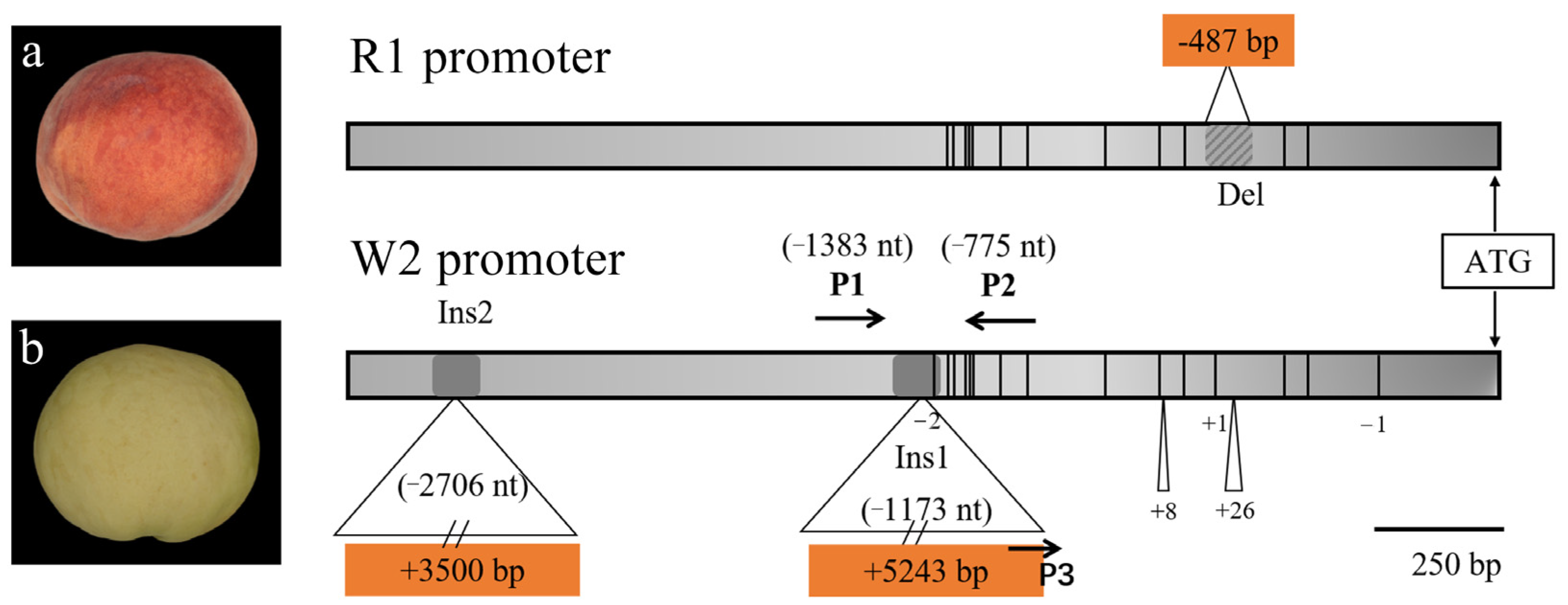
3.3. Sequence Variation in PpMYB10.1 Promoter Region

Mutation in the MYB10.1 promoter may inhibit anthocyanin synthesis and accumulation in ‘SYBT’ fruit skin. We investigated the genomic structure of the MYB10.1 promoter in the ‘SYBT’ cultivar. As shown in Figure 3, we found many single nucleotide polymorphisms (SNPs) and short insertion/deletions in the sequences of the promoter compared with the ‘Lovell’ peach cultivar, which was homozygous or heterozygous [19]. In particular, the MYB10.1 promoter of the ‘SYBT’ cultivar had two long fragment insertions. One was a 5243 bp insertion at 1173 bp upstream of ATG, considered an allelic variant of MYB10.1 and named Ins1 in this study. The other allelic variant was discovered in this study. Specifically, we found that the size of the MYB10.1 fragments amplified from the ‘SYBT’ genome was approximately 3.5 kb longer than those amplified from the ‘Lovell’ peach genome, which is due to a 3500 bp insertion at 2706 bp upstream of ATG and was named Ins2 in this study. The other SNPs found in the promoter region were also detected in red-skinned peach cultivars, indicating that these SNPs do not control the non-red skin phenotype of peach cultivars.

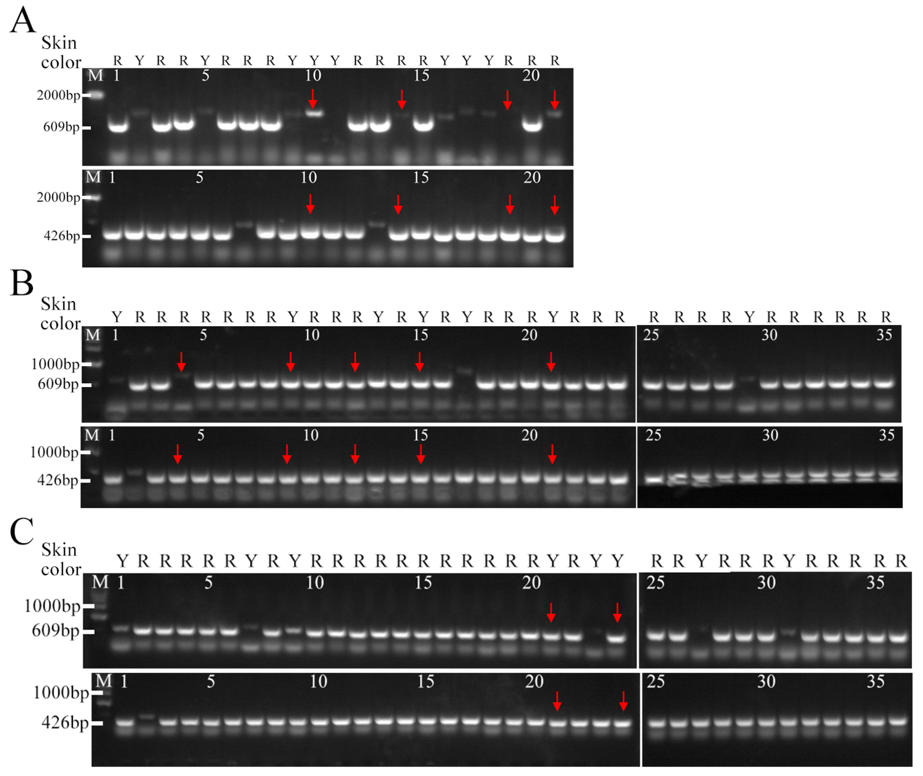
3.4. Development and Validation of Molecular Markers for Peach Skin Blush

Two allelic types of MYB10.1, MYB10.1-1/MYB10.1-2, were identified in Japanese cultivars, and those with two MYB10.1-2 alleles had white-skinned fruits. However, we investigated the transposon insertion in the MYB10.1 promoter of 61 peach cultivars, and found that the genotype and phenotype were not consistent in some cultivars (Figure 4A). Among 36 non-red skin peach cultivars, 15 cultivars carried two MYB10.1-2 alleles, and 2 cultivars carried MYB10.1-1/MYB10.1-2. Moreover, 19 cultivars carried two MYB10.1-1 alleles. The genotypes of the remaining 25 red skin peach cultivars were MYB10.1-1/MYB10.1-1 or MYB10.1-1/MYB10.1-2 (Figure 4A). Interestingly, 21 cultivars carried the MYB10.1-1 allele, which results in no anthocyanin accumulation in 36 non-red skin peach cultivars. For example, the skin color phenotype of lines 11, 12, 13, 14, 15, and 16 were green/yellow, but the Ins1 insertion was absent from the PpMYB10.1 promoter. The MYB10.1-2/MYB10.1-2 insertion explained the non-red phenotype of some cultivars. In contrast, other phenotypes were not associated with these variants, suggesting that deletions or other sequence variations may cause the non-red phenotype.
Although transposon insertion may have been the key mutation leading to skin color differences, there was a deletion at 570 nt upstream of the ATG on the PpMYB10.1 promoter, which would affect the transcriptional activity of the MYB10.1 transcription factor. In the study, we assessed the presence of this deletion in 61 cultivars, which results in amplified bands of 1053 bp or 487 bp, when the deletion is present (Figure 4B). The results showed that most of the 25 cultivars with red skin (lanes 37 to 61) had homozygous or heterozygous deletion mutations, which were present in 88.0% of the cultivars. Among the 36 non-red skin (lanes 1 to 36) cultivars, 24 did not have the deletion genotype (1053 bp/1053 bp), accounting for 66.7% of the non-red skin cultivars. This indicates that the deletion in the PpMYB10.1 promoter is associated with the red color indexes in the skin of these cultivars. The combination of the transposon insertion and deletion mutation genotypes exhibited the highest association with the skin coloration variation and basically suggested that transposon insertion and non-deletion mutation were the keys to causing the non-red coloration of the skin.
After determining the relationship between Ins1 and the deletion mutations and phenotypes, certain varieties had genotypes that were inconsistent with the phenotype. Other mutations may result from these inconsistencies, so the upstream of the MYB10.1 promoter sequence was amplified to identify additional mutations. The promoter region was extended and amplified up to the region of 2700 bp upstream of ATG. Among the various cultivars, we found a large fragment insertion of approximately 3500 bp (Ins2) upstream of the MYB10.1 promoter. By genotyping this insertion in 61 peach cultivars, we identified 14 non-red skin color cultivars that carried a homozygous insertion, consistent with the MYB10.1-2/MYB10.1-2 insertion, and only two cultivars that were heterozygous for the insertion differed. All other red-colored cultivars were homozygous or heterozygous without the Ins2 insertion (Figure 4C), and 96.7% carried the Ins1 mutation. The results were generally consistent with Ins1, indicating Ins2 is a variation on the MYB10.1 promoter linked to Ins1, and may be related to the Ins1 transposon. However, it cannot further explain the variance that Ins1 does not explain. These results suggest that other variants are controlling the non-red skin trait except the transposon Ins1 and Ins2 mutations.

3.5. Molecular Identification of the Hybrid Populations in the Promoter of MYB10.1

The peach fruit red/non-red peel trait is important, and red skin is one of the most economically important and distinguishable traits. Segregating populations and gene structures have been analyzed in previous studies, and variations in the MYB10.1 promoter were found to lead to anthocyanin accumulation, but these variants were not assessed in the breeding process [5,7,8]. Therefore, to verify that the MYB10.1 promoter variation is the major determinant of anthocyanin accumulation, we assessed 92 offspring from three segregating populations (Table S2). The results showed that 4 of the 21 lines in the segregating population did not show an association between the genotype and the phenotype, and thus the percentage of the explained phenotypic variance was 81% (Figure 5A). In segregating population 2, the phenotypes of five strains were not consistent with the genotypes, and the percentage of the explained phenotypic variance was 85% (Figure 5B). Finally, the phenotype of two lines in segregating population 3 did not match the genotype, and the percentage of the explained phenotypic variance reached 94% (Figure 5C).

4. Discussion

The rapid development of molecular marker and resequencing technologies is conducive to genetic research related to important traits by breeders, and it is also helpful in mining genetic variation information in different species and filling sequence gaps in the reference genome [20,21]. Molecular marker-assisted selection can be conducted in two ways: (1) phenotypic identification can be directly undertaken by developing molecular markers closely linked to the gene variants controlling the traits of interest; (2) using the parental genotypes, molecular markers can be developed for the different allelic variants of the target trait loci to achieve phenotypic identification [22]. In recent years, multi-disciplinary studies have reported phenotypic variants caused by mutations in the coding region or promoter of key candidate genes (SNPs, In/Dels, CNVs). Using these variations to develop markers, we can achieve the direct molecular identification of phenotypic traits.
In this study, the 5243 bp transposon inserted in the MYB10.1 promoter, discovered by previous studies to control the peel color pericarp, was identified in different materials. Most non-red color materials had the 5243 bp transposon inserted in the MYB10.1 promoter, consistent with previous results [7]. However, we found that the peel color of some varieties was green and devoid of anthocyanin accumulation until maturity. Their genotype was MYB10.1/MYB10.1, which did not correspond to their phenotypes, and their maturity was rather late and was reached in mid and late August. Potentially, the green peach peel is controlled by other genes [9]. In apple fruit, ethylene significantly induced the expression of the R2R3-MYB gene MdMYB17 [23]. It was speculated that the genotype might be only responsible for the pericarp coloration in early and middle-maturing varieties, and there might be another variant controlling the pericarp color in late-maturing varieties. The homolog of this ethylene response factor may induce PpMYB10.1 in peach. As a result, this may explain the differences in color formation of the peach fruit at different maturity stages.
At the same time, other mutations in the MYB10.1 promoter were identified, and we identified that a 487 bp deletion was highly correlated with the red color of the pericarp. From a molecular biology perspective, the closer the promoter variation is to the starting codon, the greater the impact on the phenotype. The 487 bp deletion in the MYB10.1 promoter contributes to differences in peach flesh color formation around the stone among varieties of the same species [24]. Therefore, the 487 bp deletion may upregulate anthocyanin synthesis in peach skin. However, there were also very few cases, such as in lines 11, 16, 24, 25, and 34, which carried the deletion mutation, but their skin color was white, and they all had a late-maturity phenotype. Therefore, the phenotype may be affected by other unidentified mutations and other factors.
In addition, we identified a 3500 bp long fragment insertion at 2600 bp upstream of the MYB10.1 promoter. The insertion was also closely associated with the non-red color phenotype of the fruit peel, and the overlap rate with the results of MYB10.1-2 genotyping and phenotype association was 96%. Due to the special structure, the sequence is not clear. It was found that alternative splicing triggered by the insertion of a 7433 bp CACTA transposon could lead to the development of pale-green leaves in lettuce [25]. Obviously, MYB10.1 is a key gene for anthocyanin biosynthesis in peach skin, and there are many Indels and SNPs in its promoter. However, the relationship between these natural variations and the phenotypic variation of pericarp coloration is complex and variable. Of course, many genes regulate anthocyanin accumulation in other horticultural plants. The expression of a heat-induced (32 °C) inhibitor, MdMYB16, inhibited anthocyanin biosynthesis [26], and high temperature can affect anthocyanin accumulation by inhibiting the expression of structural genes of anthocyanin biosynthesis [27]. Gene methylation has a huge impact on anthocyanin synthesis and has been studied in peach [28], pear [29], and grape [30].
The key sequence differences between MYB10.1-1 and MYB10.1-2 types that lead to the differential regulation of MYB10.1-1 have not yet been determined [7]. In this study, we reported a new variant of the MYB10.1-1 promoter controlling fruit skin color in peach, using 61 different cultivars (lines). A large insertion of approximately 3500 bp at −2706 bp in the MYB10.1 promoter was identified as another variant that might lead to the non-red type of peach skin color phenotype, and it was only present in local peach varieties grown in China but not in newly developed cultivated varieties. We believe that this variation was not favored by natural selection and its frequency was reduced. However, it was found to contain many complex sequence motifs, and further experiments are needed to explore the functional significance of this Indel. The pleiotropic effect on anthocyanin accumulation in the peach fruit of other genes controlling traits, such as genes involved in ethylene regulation during fruit ripening and fruit pigmentation sensitivity to light and temperature, and DNA methylation changes, should also be considered. We will continue to explore this insertion variation, especially focusing on the development of functional markers that can be implemented in the breeding of peach fruit color.

5. Conclusions

One insertion (Ins1) and one deletion were verified as candidate variants, and their association with fruit skin color was validated using PCR amplification of variant bands in different varieties. We found that an insertion fragment, Ins2, was identified in the PpMYB10.1 gene promoter, which may also be related to the peach skin color. These molecular markers can be applied to the molecular-assisted breeding of peach skin color. The candidate gene variants could be further exploited with genetic engineering to improve fruit color traits.

Supplementary Materials

The following supporting information can be downloaded at: https://www.mdpi.com/article/10.3390/horticulturae9080887/s1, Table S1: Primers used in RT-PCR experiments; Table S2: Peach skin color phenotype and genotype in three hybrid populations.

Author Contributions

L.W. conceived the research idea and plans. T.G. and L.W. prepared the manuscript. L.W. and J.W. (Jinlong Wu) participated in the experiment. X.L. conducted a group validation experiment. T.G. and J.W. (Jiao Wang) contributed equally to this paper. T.G. and J.W. (Jinlong Wu) contributed equally to this paper. Please direct all correspondence to J.W. (Jinlong Wu) or L.W. All authors have read and agreed to the published version of the manuscript.

Funding

This work was supported by the study on the breeding of new peach varieties and labor-saving standardized cultivation techniques (201300110500), the Agricultural Science and Technology Innovation Program (CAAS-ASTIP-2022-ZFRI-01) and Natural Science Foundation of Henan (212300410311).

Data Availability Statement

All data supporting the conclusions of this article are provided within the article (and its Supplementary Materials).

Acknowledgments

We wish to thank Jinlong Wu for suggestions on the paper and Jiao Wang, Xinxin Lu for their help in tissue collection, DNA extraction, and PCR amplification.

Conflicts of Interest

The authors declare there is no conflict of interest.

References

  1. Kong, J.-M.; Chia, L.-S.; Goh, N.-K.; Chia, T.-F.; Brouillard, R. Analysis and biological activities of anthocyanins. Phytochemistry 2003, 64, 923–933. [Google Scholar] [CrossRef] [PubMed]
  2. Frett, T.J.; Reighard, G.L.; Okie, W.R.; Gasic, K. Mapping quantitative trait loci associated with blush in peach [Prunus persica (L.) Batsch]. Tree Genet. Genomes 2014, 10, 367–381. [Google Scholar] [CrossRef]
  3. Eduardo, I.; Pacheco, I.; Chietera, G.; Bassi, D.; Pozzi, C.; Vecchietti, A.; Rossini, L. QTL analysis of fruit quality traits in two peach intraspecific populations and importance of maturity date pleiotropic effect. Tree Genet. Genomes 2011, 7, 323–335. [Google Scholar] [CrossRef]
  4. Cantín, C.; Crisosto, C.; Ogundiwin, E.; Gradziel, T.; Torrents, J.; Moreno, M.; Gogorcena, Y. Chilling injury susceptibility in an intra-specific peach [Prunus persica (L.) Batsch] progeny. Postharvest Biol. Technol. 2010, 58, 79–87. [Google Scholar] [CrossRef] [Green Version]
  5. Beckman, T.; Alcazar, J.R.; Sherman, W.; Werner, D. Evidence for qualitative suppression of red skin color in peach. HortScience 2005, 40, 523–524. [Google Scholar] [CrossRef] [Green Version]
  6. Beckman, T.; Sherman, W. Probable qualitative inheritance of full red skin color in peach. HortScience 2003, 38, 1184–1185. [Google Scholar] [CrossRef]
  7. Tuan, P.A.; Bai, S.; Yaegaki, H.; Tamura, T.; Hihara, S.; Moriguchi, T.; Oda, K. The crucial role of PpMYB10.1 in anthocyanin accumulation in peach and relationships between its allelic type and skin color phenotype. BMC Plant Biol. 2015, 15, 280. [Google Scholar] [CrossRef] [Green Version]
  8. Bretó, M.; Cantín, C.M.; Iglesias, I.; Arús, P.; Eduardo, I. Mapping a major gene for red skin color suppression (highlighter) in peach. Euphytica 2017, 213, 14. [Google Scholar] [CrossRef]
  9. Zhao, L.; Sun, J.; Cai, Y.; Yang, Q.; Zhang, Y.; Ogutu, C.O.; Liu, J.; Zhao, Y.; Wang, F.; He, H.; et al. PpHYH is responsible for light-induced anthocyanin accumulation in fruit peel of Prunus persica. Tree Physiol. 2022, 42, 1662–1677. [Google Scholar] [CrossRef]
  10. Chen, Z.J. Genetic and epigenetic mechanisms for gene expression and phenotypic variation in plant polyploids. Annu. Rev. Plant Biol. 2007, 58, 377–406. [Google Scholar] [CrossRef] [Green Version]
  11. Espley, R.V.; Brendolise, C.; Chagne, D.; Kutty-Amma, S.; Green, S.; Volz, R.; Putterill, J.; Schouten, H.J.; Gardiner, S.E.; Hellens, R.P. Multiple repeats of a promoter segment causes transcription factor autoregulation in red apples. Plant Cell 2009, 21, 168–183. [Google Scholar] [CrossRef] [PubMed] [Green Version]
  12. Chagné, D.; Carlisle, C.M.; Blond, C.; Volz, R.K.; Whitworth, C.J.; Oraguzie, N.C.; Crowhurst, R.N.; Allan, A.C.; Espley, R.V.; Hellens, R.P. Mapping a candidate gene (MdMYB10) for red flesh and foliage colour in apple. BMC Genom. 2007, 8, 212. [Google Scholar] [CrossRef] [Green Version]
  13. Chagné, D.; Lin-Wang, K.; Espley, R.V.; Volz, R.K.; How, N.M.; Rouse, S.; Brendolise, C.; Carlisle, C.M.; Kumar, S.; De Silva, N. An ancient duplication of apple MYB transcription factors is responsible for novel red fruit-flesh phenotypes. Plant Physiol. 2013, 161, 225–239. [Google Scholar] [CrossRef] [PubMed] [Green Version]
  14. Yang, C.; Sha, G.; Wei, T.; Ma, B.; Li, C.; Li, P.; Zou, Y.; Xu, L.; Ma, F. Linkage map and QTL mapping of red flesh locus in apple using a R1R1× R6R6 population. Hortic. Plant J. 2021, 7, 393–400. [Google Scholar] [CrossRef]
  15. Zhang, L.; Hu, J.; Han, X.; Li, J.; Gao, Y.; Richards, C.M.; Zhang, C.; Tian, Y.; Liu, G.; Gul, H.; et al. A high-quality apple genome assembly reveals the association of a retrotransposon and red fruit colour. Nat. Commun. 2019, 10, 1494. [Google Scholar] [CrossRef] [Green Version]
  16. Butelli, E.; Licciardello, C.; Zhang, Y.; Liu, J.; Mackay, S.; Bailey, P.; Reforgiato-Recupero, G.; Martin, C. Retrotransposons control fruit-specific, cold-dependent accumulation of anthocyanins in blood oranges. Plant Cell 2012, 24, 1242–1255. [Google Scholar] [CrossRef] [Green Version]
  17. Ou, C.; Zhang, X.; Wang, F.; Zhang, L.; Zhang, Y.; Fang, M.; Wang, J.; Wang, J.; Jiang, S.; Zhang, Z. A 14 nucleotide deletion mutation in the coding region of the PpBBX24 gene is associated with the red skin of “Zaosu Red” pear (Pyrus pyrifolia White Pear Group): A deletion in the PpBBX24 gene is associated with the red skin of pear. Hortic. Res. 2020, 7, 39. [Google Scholar] [CrossRef]
  18. Cheng, G.W.; Breen, P.J. Activity of phenylalanine ammonia-lyase (PAL) and concentrations of anthocyanins and phenolics in developing strawberry fruit. J. Am. Soc. Hortic. Sci. 1991, 116, 865–869. [Google Scholar] [CrossRef]
  19. Verde, I.; Abbott, A.G.; Scalabrin, S.; Jung, S.; Shu, S.; Marroni, F.; Zhebentyayeva, T.; Dettori, M.T.; Grimwood, J.; Cattonaro, F.; et al. The high-quality draft genome of peach (Prunus persica) identifies unique patterns of genetic diversity, domestication and genome evolution. Nat. Genet. 2013, 45, 487–494. [Google Scholar] [CrossRef] [Green Version]
  20. Fuentes, A.P.; Ruzzante, D.E. Whole-genome sequencing approaches for conservation biology: Advantages, limitations and practical recommendations. Mol. Ecol. 2017, 26, 5369–5406. [Google Scholar] [CrossRef] [Green Version]
  21. Bentley, D.R. Whole-genome re-sequencing. Curr. Opin. Genet. Dev. 2006, 16, 545–552. [Google Scholar] [CrossRef]
  22. Lu, Z.H.; Shen, Z.J.; Niu, L.; Pan, L.; Cui, G.C.; Zeng, W.F.; Wang, Z.Q. Molecular Marker-Assisted Identification of Yellow/White Flesh Trait for 122 Peach Cultivars (Lines). Sci. Agric. Sin. 2020, 53, 2929–2940. [Google Scholar]
  23. Wang, S.; Li, L.-X.; Zhang, Z.; Fang, Y.; Li, D.; Chen, X.-S.; Feng, S.-Q. Ethylene precisely regulates anthocyanin synthesis in apple via a module comprising MdEIL1, MdMYB1, and MdMYB17. Hortic. Res. 2022, 9, uhac034. [Google Scholar] [CrossRef] [PubMed]
  24. Guo, J.; Cao, K.; Deng, C.; Li, Y.; Zhu, G.; Fang, W.; Chen, C.; Wang, X.; Wu, J.; Guan, L.; et al. An integrated peach genome structural variation map uncovers genes associated with fruit traits. Genome Biol. 2020, 21, 258. [Google Scholar] [CrossRef] [PubMed]
  25. Zhang, L.; Qian, J.; Han, Y.; Jia, Y.; Kuang, H.; Chen, J. Alternative splicing triggered by the insertion of a CACTA transposon attenuates LsGLK and leads to the development of pale-green leaves in lettuce. Plant J. 2022, 109, 182–195. [Google Scholar] [CrossRef] [PubMed]
  26. Wang, K.L.; Diego, M.; John, P.; Richard, V.; Lidia, L.; Richard, E.; Roger, P.H.; David, C.; Daryl, D.R.; Michela, T.; et al. High temperature reduces apple fruit colour via modulation of the anthocyanin regulatory complex. Plant Cell Environ. 2011, 34, 1176–1190. [Google Scholar] [CrossRef]
  27. Dela, G.; Or, E.; Ovadia, R.; Nissim-Levi, A.; Weiss, D.; Oren-Shamir, M. Changes in anthocyanin concentration and composition in ‘Jaguar’ rose flowers due to transient high-temperature conditions. Plant Sci. 2003, 164, 333–340. [Google Scholar] [CrossRef]
  28. Wu, X.; Zhou, Y.; Yao, D.; Iqbal, S.; Gao, Z.; Zhang, Z. DNA methylation of LDOX gene contributes to the floral colour variegation in peach. J. Plant Physiol. 2020, 246–247, 153116. [Google Scholar] [CrossRef]
  29. Qian, M.; Sun, Y.; Allan, A.C.; Teng, Y.; Zhang, D. The red sport of ‘Zaosu’ pear and its red-striped pigmentation pattern are associated with demethylation of the PyMYB10 promoter. Phytochemistry 2014, 107, 16–23. [Google Scholar] [CrossRef]
  30. Xia, H.; Shen, Y.; Hu, R.; Wang, J.; Deng, H.; Lin, L.; Lv, X.; Deng, Q.; Xu, K.; Liang, D. Methylation of MYBA1 is Associated with the Coloration in “Manicure Finger” Grape Skin. J. Agric. Food Chem. 2021, 69, 15649–15659. [Google Scholar] [CrossRef]
Figure 1. Different anthocyanin accumulation in different cultivars. (A) Fruit developmental stages of the non-red skin cultivar ‘SYBT’ and the red skin cultivar ‘ZTJM’. (B) Total anthocyanin content of fruit skins at different developmental stages. Error bars correspond to the SE of the mean. *, a significant difference (p < 0.05) based on Student’s t-test.
Horticulturae 09 00887 g001
Figure 2. Expression levels of genes involved in anthocyanin biosynthesis pathways in peach skin. (A) Expression profiles of PpMYB10.1/2/3 in peach skin at different stages of fruit development. (B) Expression profiles of structural genes involved in anthocyanin biosynthesis in peach skin at different stages of fruit development. Error bars correspond to ±standard error (SE) of three biological replicates.
Horticulturae 09 00887 g002
Figure 3. Multiple mutation sites in MYB10.1 promoter in peach with different skin color. Schematic representation of the differences between the R1 (found in both white- and red-skinned peach varieties) and the W2 promoter showing positions of the ATG translation start site, the 487 bp deletion mutation, the Ins1 transposon insertion, and the Ins2 insertion in R1 and W2. ‘|’, ‘−n’, and ‘+n’ indicate the single nucleotide polymorphism, number of nucleotide insertions, and number of nucleotide deletions (nt) indicate the nucleotide position relative to the start codon ATG. Representative phenotypes are shown on the left of the schematic: (a) the red-skinned cultivar ‘ZTJM’ and (b) the white-skinned cultivar ‘SYBT’. Th ATG codon corresponds to the predicted transcription start site.
Horticulturae 09 00887 g003
Figure 4. Detection of the transposon insertion and deletion in 61 peach cultivars. (A) Genomic PCR was performed using the Mpro1 F/R, Mpro2 F/R primers, and M, the DNA ladder. All evaluated peach cultivars are listed in Table 1. The red and white circles and red semicircles, respectively, indicate the skin color phenotype of the peach cultivars. Black and white triangles indicate the PCR products amplified from genomic DNA with or without transposon insertion. (B) Detection of the promoter deletion in various peach cultivars. Genomic PCR was performed using Mpro3 F/R primers and M, the DNA ladder. All peach cultivars evaluated are shown in Table 1. The red and white circles and red semicircles, respectively, indicate the skin color phenotype of the peach cultivars. White and black triangles indicate PCR products amplified from genomic DNA with or without the promoter deletion mutation. (C) Genomic PCR was performed using the CR F/R primers and M, the DNA ladder. All evaluated peach cultivars are shown in Table 1. The red and white circles and red semicircles, respectively, indicate the skin color phenotype of the peach cultivars. Black and white triangles indicate PCR products amplified from genomic DNA with or without transposon insertion.
Horticulturae 09 00887 g004
Figure 5. Identification of the MYB10.1pro insertion in three segregating populations. A total of 92 hybrid progeny plants were selected from three segregating populations using Mpro1 F/R, and Mpro2 F/R for PCR genotyping. (A) 21 hybrids of the ‘Zhongyoupan 7′ × ‘NJF 16′cross; (B) 35 hybrids of ‘09-5-29′ × ‘09-10-East-22′cross; (C) 36 hybrids of ‘Wen 09-5-29′ × ‘South 2nd District East 2-45′ cross. R corresponds to red peel color, and Y to yellow peel color. The red arrows in the figure indicates a single plant whose genotype does not match the skin color phenotype.
Horticulturae 09 00887 g005
Table 1. Information of 61 peach varieties. The red marker in the table is that the genotype is not consistent with the peel color phenotype.
No.Accession NameResource TypeSampled LocationOriginSkin ColorMYB10.1 Allelic Insertion/bpMYB10.1 Allelic Deletion/bp
P1Gui Zhou Shui MiLandrace varietyGuizhouChinaWhite426/4261053/1053
P2Yu BaiImproved varietyZhengzhou, HenanChinaWhite426/4261053/1053
P3Qing Zhou Bai Pi Mi TaoLandrace varietyQingzhou, ShandongChinaWhite426/4261053/1053
P4Da Xue TaoLandrace varietyMancheng, HebeiChinaWhite426/4261053/1053
P5Shui bai TaoLandrace varietyNajing, JiangsuChinaWhite426/4261053/1053
P6Shi Yu Bai TaoImproved varietyShijiazhuang, HebeiChinaWhite426/4261053/1053
P7NJC77Improved varietyXinzexizhouAmericaYellow426/4261053/1053
P8Maria SerenaImproved varietyFlorenceItalyYellow426/4261053/1053
P9KXN43-37Superior lineZhengzhou, HenanChinaWhite426/4261053/1053
P10KXN43-52Superior lineZhengzhou, HenanChinaWhite426/4261053/1053
P11Mi Yang ShanLandrace varietyHetian, XinjiangChinaGreen609/609487/487
P12Bai Li HuLandrace varietyMengzi, YunnanChinaGreen609/6091053/1053
P13Tu-2Landrace varietyYecheng, XinjiangChinaGreen609/609487/1053
P14Tie 4-1Landrace varietyYecheng, XinjiangChinaGreen609/6091053/1053
P15Da Li He Huang RouLandrace varietyHetian, XinjiangChinaYellow609/6091053/1053
P16Gua TaoLandrace varietyNapo, GuangxiChinaYellow609/609487/487
P17Yang Zhou 3 Improved varietyYangzhou, JiangsuChinaWhite426/4261053/1053
P18Yun Shu 2 Improved varietyHangzhou, ZhejiangChinaGreen426/4261053/1053
P19Pin Ding You Pan TaoImproved varietyZhengzhou, HenanChinaWhite426/4261053/1053
P20Suan TaoLandrace varietyFeicheng, ShandongChinaGreen609/426487/1053
P21Chi Yuan MiImproved varietyNanjing, JiangsuChinaWhite426/426487/1053
P22Tai Yuan Shui MiLandrace varietyTaiyuan, ShanxiChinaWhite609/426487/1053
P23Gao Tai 1 Landrace varietyGaotai, GansuChinaGreen609/6091053/1053
P24Ying XueLandrace varietyBeijingChinaGreen609/609487/487
P25Han Lu MiLandrace varietyQingdao, ShandongChinaGreen609/609487/487
P26Dunhuang Dong TaoLandrace varietyDunhuang, GansuChinaWhite609/6091053/1053
P27Xiang TaoLandrace varietyDalian, LiaoningChinaGreen609/6091053/1053
P28Bai He TaoLandrace varietyYunguigaoyuanChinaWhite609/609487/1053
P29Bai Nian HeLandrace varietyMengzi, YunnanChinaWhite609/609487/1053
P30Golden QueenImproved varietyUnknowNew ZealandYellow609/6091053/1053
P31EvertsImproved varietyUnknowAmericaYellow609/6091053/1053
P32Huang YanLandrace varietyJingning, UunnanChinaYellow609/6091053/1053
P33Nan Shan 1 Landrace varietyNingxian, GansuChinaYellow609/6091053/1053
P34Bei Jing Wan Pan TaoLandrace varietyBeijingChinaGreen609/609487/487
P35Feicheng Bai Li 10 Landrace varietyFeicheng, ShandongChinaWhite426/4261053/1053
P36Anlong Bai TaoLandrace varietyAnlong, GuizhouChinaWhite609/609487/1053
P37KXN43-04Superior lineZhengzhou, HenanChinaRed609/426487/1053
P38KXN42-125Superior lineZhengzhou, HenanChinaRed609/426487/1053
P39KXN43-57Superior lineZhengzhou, HenanChinaRed609/609487/487
P40KXN43-59Superior lineZhengzhou, HenanChinaRed609/609487/487
P41Da Hong TaoLandrace varietyYuncheng, YunchengChinaRed609/609487/487
P42An Nong Shui MiImproved varietyShouxian, AnhuiChinaRed609/609487/487
P43Xi Nong Shui MiImproved varietyWugong, ShanxiChinaRed609/609487/487
P44Ba HuaLandrace varietyWuxi, JiangsuChinaRed609/609487/487
P45Bao LuImproved varietyHangzhou, ZhejiangChinaRed609/609487/487
P46Hu Jing Mi LuLandrace varietyWuxi, JiangsuChinaRed609/609487/1053
P47Yi Xian HongLandrace varietyChangli, HebeiChinaRed609/6091053/1053
P48Qi TaoLandrace varietyTianshui, GansuChinaRed609/609487/487
P49Ji Zui BaiLandrace varietyShangshui, HenanChinaRed609/6091053/1053
P50HatsukamiImproved varietyUnknowJapanRed609/609487/487
P51Nagasawa HakuhoImproved varietyShanlixianJapanRed609/609487/487
P52Gan Xuan 4 Landrace varietyLanzhou, GansuChinaRed609/609487/487
P53Hong Gan LuImproved varietyDalian, LiaoningChinaRed609/609487/487
P54Liquan 54 Landrace varietyLiquan, ShanxiChinaRed609/609487/487
P55Datuan Mi LuImproved varietyNanhui, ShanghaiChinaRed609/609487/487
P56Er Zao TaoLandrace varietyChenggon, YunnanChinaRed609/609487/487
P57Wu Yue BaiLandrace varietyXihua, HenanChinaRed609/609487/487
P58Zhong Tao Jin MiImproved varietyZhengzhou, HenanChinaRed609/609487/487
P59FantasiaImproved varietyJiazhou, FresnoAmericaRed609/609487/487
P60Su HongLandrace varietyJiangsuChinaRed609/4261053/1053
P61Shi Tou TaoLandrace varietyZhengzhou, HenanChinaRed609/426487/1053
Table 2. Primers used in genomic PCR.
Prime NamePrimer SequenceAnnealing Temperature (°C)Size (bp)
Mpro1 FGGGAAACGATGTAAAGCCAC55 °C609
Mpro1 RCGAATATCAATGCAGCATCGTG
Mpro2 FCGAATATCAATGCAGCATCGTG55 °C426
Mpro2 RCGGTTTTGGTCTTGCGCTAT
Mpro3 FGTGGCTACGTACGGTTCTCC55 °C487/1053
Mpro3 RTTTTATGCCCTGCCTGCTCA
CR FCCTTCTCCTATGGACTTTCTCCC58 °C269/3500
CR RAGCGTCGTGGTTTATGAGGG
Disclaimer/Publisher’s Note: The statements, opinions and data contained in all publications are solely those of the individual author(s) and contributor(s) and not of MDPI and/or the editor(s). MDPI and/or the editor(s) disclaim responsibility for any injury to people or property resulting from any ideas, methods, instructions or products referred to in the content.

