Article

The Composition and the Content of ∆-5 Sterols, Fatty Acids, and the Activity of Acyl-Lipid Desaturases in the Shoots of Ephedra monosperma, Introduced in the Botanical Garden of the Cryolithozone of Yakutia

by
Vasiliy V. Nokhsorov
1,*,
Luybov V. Dudareva
2,
Natalia V. Semenova
2 and
Valentina E. Sofronova
1
1
Institute for Biological Problems of the Cryolithozone, Siberian Branch of Russian Academy of Sciences, 41 Lenina Av., 677000 Yakutsk, Russia
2
Siberian Institute of Plant Physiology and Biochemistry, Siberian Branch of Russian Academy of Sciences, 132 Lermontova Str., 664033 Irkutsk, Russia
*
Author to whom correspondence should be addressed.
Horticulturae 2023, 9(8), 858; https://doi.org/10.3390/horticulturae9080858
Submission received: 22 June 2023 / Revised: 14 July 2023 / Accepted: 24 July 2023 / Published: 27 July 2023
(This article belongs to the Section Floriculture, Nursery and Landscape, and Turf)

Abstract

:
Evergreen plants in permafrost ecosystems survive unfavorable autumn cooling and extremely low winter temperatures by maintaining optimal physiological activity of tissue cell membranes. To some extent, these features are due to the properties of shoot lipids performing a number of functions during adaptation. Sterols (STs) play a key role in regulating the fluidity and permeability of plant membranes (phytosterols) with a wide structural diversity. The composition of neutral lipids, STs, and fatty acids (FAs) in shoots of the evergreen shrub Ephedra monosperma growing in the Botanical Garden cryolithozone was first studied with HPTLC-UV/Vis/FLD and GC-MS. Twenty FAs were found, from C14:0 to C23:0; they included mono-, di-, tri-, and tetraene FAs. The high content of β-sitosterol among other ∆-5 sterols and an increased amount of C18:2(∆9,12) linoleic acid in lipids composition during the autumn–winter period was found to play an important role in the adaptation of ephedra shoots to the autumn–winter period, providing the cell membrane with greater plasticity, fluidity, and flexibility. The important role of diene linoleic fatty acid C18:2(∆9,12) in ephedra shoot lipids in the processes of low-temperature adaptation was shown.

1. Introduction

Plants are sources of valuable biologically active compounds. Phytochemical substances that are present in plant extracts are generally low toxic and have high physiological activity at low concentrations. In regions with extreme climatic conditions, there are several plant species that are excellent sources of phytochemicals [1]. Ephedra species are known as sources of biologically active compounds with potential pharmaceutical, cosmetic, food, and agricultural purposes [2]. While studying secondary metabolites of different ephedra species, alkaloids, amino acids and their derivatives, volatile substances, and phenolic compounds were revealed [3]. All ephedra species contain biologically significant alkaloids: ephedrine, pseudoephedrine, norephedrine, norepseudoephedrine, methylephedrine, and methylpseudoephedrine. The alkaloids ephedrine and pseudoephedrine have been found to have a significant effect on the central nervous system [4], and the content of these compounds in E. monosperma fluctuates and reaches 34.7 mg/g DW [5]. Generally, the genus Ephedra (Ephedraceae) includes 69 species, four subspecies, and two accepted varieties, all of which are widely spread in arid and semi-arid regions of Asia, Europe, North Africa (Sahara), southwestern North America, and South America [2]. There are 12 species of the genus Ephedra in Siberia which inhabit arid areas [6]. Under permafrost conditions (Republic of Sakha (Yakutia)), in the steppe plots and dry rocky and rubbly hillsides Ephedra monosperma J.G. Gmel. ex C.A. Mey grows (division Gymnospermae, class Gnetopsida, subclass Pinidae, family Ephedraceae Wettst, genus Ephedra) is a survivor from the Pre-Glacial Period and is one of the few evergreens shrubs grown in permafrost conditions. This species can be assigned to the group of light-loving species adapted to growth in open well-lit areas. The habitats of E. monosperma are marked by severe continental arid climate characterized by extremely low winter (to −40–−45 °C) and very high summer (up to 38 °C) temperatures with an annual precipitation of less than 200 mm, resulting in water deficit in air and soil. Its natural distribution areas are also characterized as sandy or calcareous rocks soils. In the permafrost ecosystems of the Botanical Garden of the Institute of Biological Problems of the Cryolitozone of the Siberian Branch of the Russian Academy of Sciences, which is located on the second above floodplain terrace of the Lena River valley (62°15′ N, 129°37′ E), there are areas where native, rare, and valuable plant species grow, including the pine conifer E. monosperma.
Lipophilic metabolites, which include phytosterols, are one of the important classes of compounds that play a significant role in plant adaptation to unfavorable environmental conditions [7,8,9]. STs are known to be essential components of membranes, being responsible not only for structural but also for regulatory functions in many key cellular processes. The most common ∆-5 plant STs are β-sitosterol, stigmasterol (24-ethyl sterols), and campesterol (24-methyl sterol), as well as avenasterol and cholesterol, which are contained in relatively small amounts in most species [10]. STs are found in plant tissues both in the free state and in conjunction with fatty acids, as well as in the form of sterylglycosides and acylsterylglycosides. It is assumed that β-sitosterol and 24-methyl cholesterol can regulate the fluidity and permeability of plant membranes by limiting the mobility of fatty acyl chains. STs can be involved in the adaptation of plant membranes to changes in temperature and humidity, as well as in the modulation of the activity of membrane-bound enzymes [11].
STs are structural components of the membrane, therefore, they can affect the physical state of the membrane under stress not only through quantitative changes in the sum of STs but also through changes in the ratio of their molecular species [12]. The ratio of molecular forms of STs, particularly β-sitosterol/stigmasterine, can affect the physicochemical properties of ordered microdomains, the so-called “lipid rafts” enriched with sphingolipids and STs [13,14]. Consequently, changes in the ratio of ST molecular species can modulate plant signaling and defense responses. Moreover, among the medically useful compounds, plant STs attract special interest as new functional products for the treatment of metabolic disorders in humans and animals, since they can reduce cholesterol levels and possess anti-inflammatory and antioxidant properties [15,16].
Previously, it was shown that phosphatidic acid (PA) was the dominant lipid class of shoots E. monosperma [17]. In Ephedra alata lipids, PA was also the dominant class compared to phosphatidylcholine and phosphatidylethanolamine under control conditions [18]. A large proportion of PAs in the phospholipid profile is characteristic of some extremophile organisms [19]. The lipids of seeds of four ephedra species (E. nevadensis, E. viridis, E. przewalskii, and E. gerardiana) contain delta5-unsaturated polymethylene-interrupted fatty acids (∆5-UPIFA): ∆5,9- and ∆5,11-C18:2; ∆5,9,12-C18:3; ∆5,9,12,15-C18:4; ∆5,11-C20:2; ∆5,11,14-C20:3; and ∆5,11,14,17-C20:4 acids [20]. Our studies revealed that ∆5-UPIFA was detected in frost-tolerant coniferous [21] and in the aboveground parts of wintergreen species of horsetail [22].
However, the profiling of STs and fatty acids, which play a crucial role in the cold response of E. monosperma at low temperatures, has not been reported. In the present study, we studied for the first time the absolute and relative content and composition of fatty acids as well as free and esterified STs as the main components of cell membranes isolated from the assimilating shoots of E. monosperma. In addition to the fundamental interest, the study of lipids, including the ST and FA composition of evergreen shrub E. monosperma, has also an important applied value, since it gives an idea at the physiological and biochemical levels of the adaptive responses of introduced valuable, rare and medicinal plants growing in the Botanical Garden in permafrost ecosystem conditions.

2. Materials and Methods

2.1. Plant Material and Growth Conditions

The plants of Ephedra monosperma J.G. Gmel. ex C.A. Mey. grow naturally in a well-lit area in a clearing of a pine forest in the Botanical Garden of the Institute of Biological Problems of the Cryolithozone, Siberian Branch, Russian Academy of Sciences. The garden area is located on the second terrace above the flood plain of the Lena Valley (62°1′27″ N, 129°36′4″ E). The assimilating partial shoots of E. monosperma of the current year served as plant material for biochemical analyses (Figure 1).

2.2. Field Experiment

Samples were collected in the morning (9:00–11:00). The samples of ephedra shoots were collected from introduced plants that grew on the territory of the Botanical Garden in two points of 5−7 samples each. To reveal the seasonal dynamics of lipid composition, E. monosperma shoot samples were collected in different periods: July 12 (23.9 °C), September 25 (5.1 °C), October 30 (−8.3 °C) and December 11 (−38.4 °C). The samples were immediately fixed in liquid nitrogen and transported in Dewar vessels to the laboratory.
The samples were stored in the freezer at −80 °C prior to analysis (Panasonic, Japan). For biochemical studies, the samples of the aerial parts of E. monosperma fixed in liquid nitrogen were dried in the lyophilizer (VirTis, New York, NY, USA). The experiments were conducted in 2019. The air temperature at the experimental site was recorded with an accuracy of ±0.5 °C at 1 h intervals using a DS 1922L iButton thermograph (Dallas Semiconductor, Dallas, TX, USA). The average air temperature during the growing season (May–September) in 2019 was between 13.1 and 13.9 °C, and the total liquid precipitation was between 123 and 127 mm. The beginning of weak frosts was noted from the beginning of the second half of September, and a stable decrease in night temperatures below the 0 °C level occurred after 30 September and 9 October 2019. Minimum winter air temperatures were −44 and −54 °C. The snow cover was established on 13 October and 10 October 2019. The thickness of snow cover was 2–10 cm by the end of October, 15–21 cm in the middle of November, 18–26 cm in December, and 24–34 cm in January.

2.3. Lipid Extraction

For lipid extraction, a weighed portion of plant material (0.5 g) was fixed in liquid nitrogen and ground until a homogeneous mass was obtained. Cooled laboratory glassware and reagents were used. Therefore, 10 mL of a 1:2 chloroform/methanol mixture was added. Ionol was added to the mixture as an antioxidant (0.00125 g per 100 mL of the mixture). It was thoroughly mixed and left for 30 min until the complete diffusion of lipids into the solvent. The solution was transferred quantitatively into a separatory funnel through a filter. The mortar and filter were washed three times with the same solvent mixture. With the aim of separating the non-lipid components, some water was added.

2.4. Neutral Lipid Composition Analysis by HPTLC-UV/Vis/FLD Method

For the analysis of neutral lipids, the lower chloroform fraction was separated. Chloroform (high purity grade, stabilized with 0.005% amylene) was removed from the lipid extract under vacuum using the UL-2000 rotary evaporator (Ulab, Shanghai, China). To separate neutral lipids, we used the method of one-dimensional high-performance thin-layer chromatography (HPTLC) on silica gel plates 60 (10 × 10 cm) (Merck, Darmstadt, Germany). The plate was placed in a chamber filled with hexane:diethyl ether:acetic acid eluent (80:20:1 v/v/v). For this purpose, chromatograms were developed in 10% sulfuric acid in methanol followed by heating at 140 °C. The plate was sprinkled with reagent using a sprayer (Lenchrom, Russia) connected to a JAS 1202 compressor (JAS-AIR, Hong Kong, China), then dried and transferred to a desiccator and heated for 20 min at 140 °C. The amount of neutral lipids was determined densitometrically using the Sorbfil TLC View (Imid, Krasnodar, Russia) and documented at white light illumination (Vis), UV 254 nm, and FLD 366 nm. The calculation of the content of individual classes of lipids in chromatograms was carried out using the Sorbfil TLC Videodensitometer 2.3 program. The calculated Rf values for free sterols (Rf = 0.19) and sterol esters (Rf = 0.87–0.92) coincided with the Rf values for standard samples (cholesterol, stigmasterine, campesterol (Sigma, St. Louis, MO, USA)), β-sitosterol (European Pharmacopoeia Reference Standard, France) and the tabulated values.

2.5. Separation of Free and Bound Sterols

For lipid extraction, a weight of plant material (30 mg of lyophilically dried sample) was fixed in liquid nitrogen by adding the antioxidant 0.001% ionol. The lipids were extracted with 10 mL of chloroform/methanol mixture (2:1 v/v), thoroughly stirred, and left for 30 min until the lipids were completely diffused into the solvent. The lower chloroform fraction was separated for analysis. Chloroform was removed from the lipid extract under vacuum using an RVO-64 rotary evaporator (Czech Republic). To separate neutral lipids, the method of one-dimensional thin-layer chromatography (TLC) on Sorbfil plates (PTC-AF-B, Imid, Russia) was used. The plate was placed in a chamber filled with the eluent hexane:diethyl ether:acetic acid (80:20:1 v/v/v). To visualize the zone of STs and their esters, the edge of the plate was treated with a 10% solution of sulfuric acid in ethanol and heated on a hotplate. Then, sterols and their esters were extracted sequentially with chloroform and ethyl acetate. At each of these steps, the sample in the solvent was placed in an ultrasonic bath and centrifuged at 3000× g. The ethyl acetate fraction was taken with a 1 mL pipette, transferred to glass tubes, and evaporated to dryness in a nitrogen current [7].

2.6. Silylation

For the analysis, trimethylsilyl derivatives of the target components were prepared by heating the sample for 30 min at 70 °C with the addition of 150 µL N,O-bis (trimethylsilyl) acetamide, 50 µL hexamethyl disilazane (Sigma-Aldrich, Burlington, MA, USA), and 300 µL ethyl acetate (pure for chromatography, Component Reactiv, Moscow, Russia). Ergosterol (Sigma-Aldrich, Burlington, MA, USA) was used as an internal standard.

2.7. GC-MS Analysis of Sterols and Their Ethers

Free and bound STs were analyzed by gas chromatography using a 7777QQQ/7890N MSD/DS Agilent Technologies (Santa Clara, CA, USA) chromato-mass spectrometer. The detector mass spectrometer was used in single quadrupole mode, the method of ionization was electronic impact EI, ionization energy was 70 eV, and full ionic current registration mode was used for the analysis. An HP5-MS capillary column (30 m × 250 μm × 0.25 μm) with a stationary phase of 5% phenyl-methyl-polysiloxane was used for separation. The mobile phase was helium, gas flow rate 1 mL/min, temperature gradient: 150 to 300 °C at a rate of 10 °C/min and incubation for 23 min at this temperature. Evaporator temperature: 250 °C, ion source temperature: 230 °C, detector temperature: 150 °C, line temperature between chromatograph and mass spectrometer: 280 °C. The scanning range was 50–700 A.E.M. Sample injection volume was 1 µL, and the flow divider was 5:1 [7].

2.8. Determination of FAs Content

The extraction of lipids was performed as described above. To control the lipid extractability (%), the known amount of 10 µg of nonadecanoic acid (C19:0) was added at the homogenization stage. Fatty acid methyl esters (FAMEs) were obtained using the Christie method [23]. Additional FAME purification was carried out by thin-layer chromatography (TLC) on glass plates with KSK silica gel (Reachem, Moscow, Russia). Benzene was used as the mobile phase. To visualize the FAME zone (Rf = 0.71–0.73), the plates were sprayed with 10% H2SO4 in methanol and heated in an oven at 100 °C. The FAME zone was removed from the plate with a spatula and eluted from silica gel with n-hexane. The FAME analysis was performed by GC-MS using the 5973/6890N MSD/DS gas chromatograph–mass spectrometer (Agilent Technologies, Santa Clara, CA, USA). The detector was a quadrupole mass spectrometer. The method of ionization was electronic impact EI, ionization energy of 70 eV. The analysis was performed in the mode of the total ion current recording. An HP-INNOWAX capillary column (30 m × 250 μm × 0.50 μm) with a stationary phase (PEG) was used to separate the FAME mixture. The carrier gas was helium, and the gas flow rate was 1 mL/min. The evaporator temperature was 250 °C, the ion source temperature was 230 °C, and the detector temperature was 150 °C. The temperature of the line connecting the chromatograph with the mass spectrometer was 280 °C. The scanning range was 41–450 amu. The volume of the injected sample was 1 μL, and the flow divider was 5:1. The separation of the FAME mixture was carried out in isothermal mode at 200 °C. To identify FAs, the NIST 08 mass spectral library, and the Christie FAME mass spectral archive were used [24]. The relative FA content was determined by the method of internal normalization in weight percent (wt.%) of the total content in the test sample, taking into account the FAs response coefficient.
The absolute content of FAs was calculated by the internal standard method (C19:0) according to the formula:
Cd = Cint.st. × Sx/Sint.st,
Cd: content of the component to be determined; Cint.st.: known content of internal standard; Sx and Sint.st.: areas of the corresponding peaks on the chromatogram.

2.9. Identification and Quantitative Analysis

The identification of sterols and FAMEs was performed using standards for the target components by comparing their retention times. Libraries of mass spectra NIST 08 and Wiley 7 were used if necessary. The quantitative analysis of the target components was performed using an external calibration method, taking into account the response of the internal standard.

2.10. Activity of Sterol Methyltransferase and Acyl-Lipid Desaturase Enzymes

The activity of the sterol methyltransferase (SMT) enzymes that catalyze the methylation reaction at the 24th carbon atom (C24) in the side chain was determined as the ratio 24-methyl sterols/24-ethyl sterols [25]. The activity of sterol C-22 desaturase was determined as the ratio of β-sitosterol/stigmasterine.
To characterize the unsaturation level of lipid FAs, the unsaturation index (UI) [26] and double-bond index were calculated (DBI = (2 × %C18:2 + 3 × %C18:3)/(%C16:0 + %C18:0 + %C18:1) [27,28]. The activity efficiencies of the ∆9-, ∆6-, and ∆3-desaturases were calculated as the palmitic desaturation ratio (PDR = %C16:1/(%C16:0 + %C16:1)), stearic desaturation ratio (SDR), oleic desaturation ratio (ODR), and linoleic desaturation ratio (LDR) [29]. Desaturation and elongation partitioning ratios were calculated based on the ratios of the following FAs: C16 desaturation—%C16:1/(%C16:0 + %C18:0 + %C18:1 + %C18:2 + %C18:3 + %C20:0 + %C22:0); C18 desaturation—(%C18:1 + %C18:2 + %C18:3)/(%C18:0 + %C20:0 + %C22:0); C16 elongation—(%C18:0 + %C18:1 + %C18:2 + %C18:3 + %C20:0 + %C22:0)/(%C16:0 + %C16:1) [30].

2.11. Statistical Processing

The table and figures show the average data from three biological replicates and their standard deviations. The experimental data were statistically processed using the statistical analysis package in the Microsoft Office Excel 2017 environment. The statistical significance of the differences between the compared mean values was assessed using Kruskal–Wallis ANOVA by ranks (p < 0.05).

3. Results

3.1. Composition of Neutral Lipids in E. monosperma Shoots by HPTLC−UV/Vis/FLD Profiling Method

The experiments were carried out from July 2019 to January 2020. The average daily air temperature decreased during this period from 24–25 °C to the range between −30 and −46 °C, and the duration of light exposure during the day shortened from 17.4 to 5.1 h. Using HPTLC-UV/Vis/FLD, the composition of neutral lipids in E. monosperma shoots was established, and the following types of lipids were identified: polar lipids (PLs), sterols (STs), fatty acid methyl esters (FAMEs), triglycerides (TAGs), sterol esters (SEs), and squalene (Sq) (Figure 2). The detection zones of sterol components appeared as pink-blue spots on the plate.

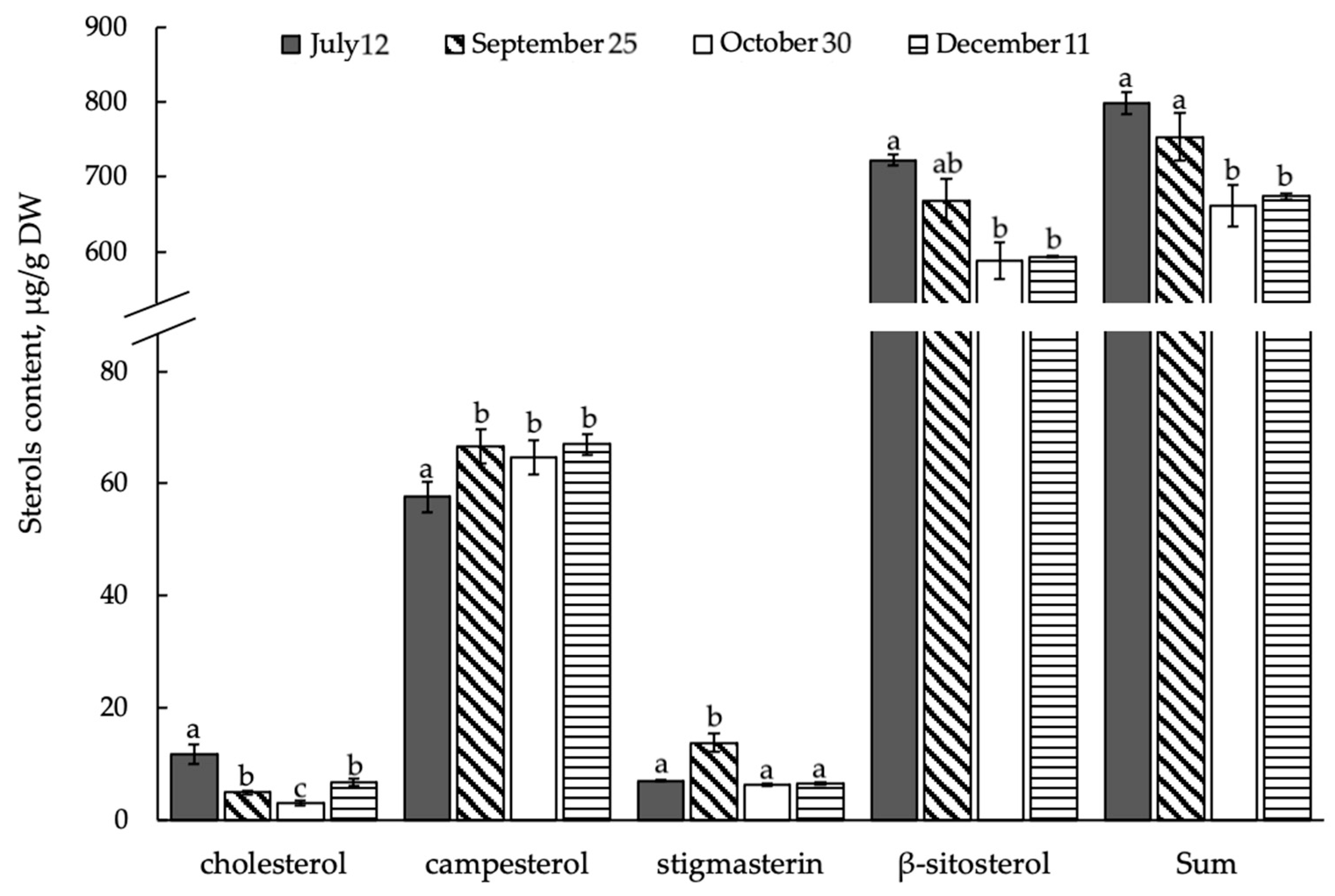
3.2. Composition and Content of Membrane Sterols in the Shrub E. monosperma Shoots

The total absolute content of all STs ranged from 661.9–798.6 μg/g DW (Figure 3). According to our data, the proportion of free STs remained comparable with the summer months until the average daily temperature decreased to 3.6 ± 1.2 °C from 18 to 24 September. However, with the onset of negative low temperatures in October, their proportion decreased by 1.2 times compared to the summer period. The absolute content of free ∆5-sterols exceeded the amount of bound STs (sterol esters) in ephedra shoots, so in summer this difference was 12.4 times in favor of free ∆5-sterols, while in autumn, there was an increase in this difference of 16–17 times compared with bound STs.

3.3. Composition and Content of Free ∆5-Sterols in Shoots of E. monosperma

Phytosterols such as cholesterol, campesterol, stigmasterine, and β-sitosterol were identified as free ∆5-sterols by GC-MS (Figure 4). In the shoots of the evergreen shrub E. monosperma, the amount of β-sitosterol was higher than that of other ∆5-sterols. The content of this ST decreased with the onset of stable negative temperatures in October and December. The content of campesterol increased 1.1-fold with the onset of low hardening temperatures in September compared to the summer period. With a further decrease in the ambient temperature in the cryolithozone at the end of October and in December, the amount of campesterol remained at the same level. The content of cholesterol decreased significantly by 2.4 times in response to a decrease in average daily air temperature compared with the summer period. The absolute content of stigmasterin increased 1.9-fold compared to the summer period; later (30 October, 11 December), the content of this ∆5-sterol was comparable to the summer values. In general, the total content of ∆5-sterols decreased significantly during the establishment of persistent negative low air temperatures in late October and December.

3.4. Composition and Content of Bound STs (Sterol Esters) in Shoots of E. monosperma

The composition of bound STs (sterol esters) fraction, which included ∆5-sterol esters (cholesterol, campesterol, and β-sitosterol) was studied (Figure 5). The ST ester fraction in ephedra shoots was dominated by β-sitosterol. In response to low-temperature stress caused by a decrease in average daily ambient temperature, the β-sitosterol content in ST ester fraction significantly decreased. The most pronounced decrease was revealed during cold hardening in September and October (by 1.7 times) compared to the summer period. During the studies, the content of another phytosterol-campesterol in E. monosperma shoots did not change. The content of cholesterol in bound ST esters was minimal compared to other sterols. Seasonal dynamics of cholesterol content in ephedra shoots was as follows: in September the amount of this ST increased 1.5-fold as compared with July, then with decreasing temperature we found that cholesterol content was comparable with summer indices. The analysis showed that the content of cholesterol in ST esters in the shoots of the evergreen ephedra shrub during winter was comparable with the September values. The total content of bound STs in ephedra shoots repeated the dynamics of β-sitosterol content, which confirms the dominance of this phytosterol in the composition of bound STs.

3.5. Activity of Sterol Methyltransferase (SMT) Enzymes in Ephedra Shoots during Quenching to Low Cryolithozone Temperatures

Having analyzed the composition and content of STs, we indirectly estimated the seasonal dynamics of sterol methyltransferase (SMT) enzyme activity in free STs and in bound STs occurring in E. monosperma shoots (Figure 6) by calculating the ratio of 24-methyl sterols/24-ethyl sterols. SMT activity for free STs increased significantly in late October and December compared with the summer period. SMT activity for ST esters was most consistently higher during the autumn–winter period compared to the summer period. A comparative analysis of enzyme activity for free and bound STs revealed that SMT activity was the highest in bound esters.

3.6. Seasonal Dynamics of Saturated and Unsaturated Fatty Acids in Ephedra Shoots

The absolute and relative content of FAMEs in ephedra shoots introduced into the Botanical Garden conditions was investigated. The study showed that in ephedra shoots, the relative content of FA groups changed depending on the season of the year, which were divided by the number of double bonds into monoene, diene, triene, and tetraene FAs (Figure 7). The qualitative composition of FAs in ephedra shoots practically did not change in all seasons; thus, 19 FAs were identified in July, September, and December, and 20 FAs in October. Unsaturated FAs dominated in ephedra shoots in all seasons, their relative content increasing in the autumn–winter period. Among unsaturated FAs, triene FAs dominated in the FAs profile. The most noticeable increase in relative content in response to the fall temperature decrease was found among diene FAs. It was shown that, in general, the total content of saturated FAs gradually decreased from the summer period to the winter one.

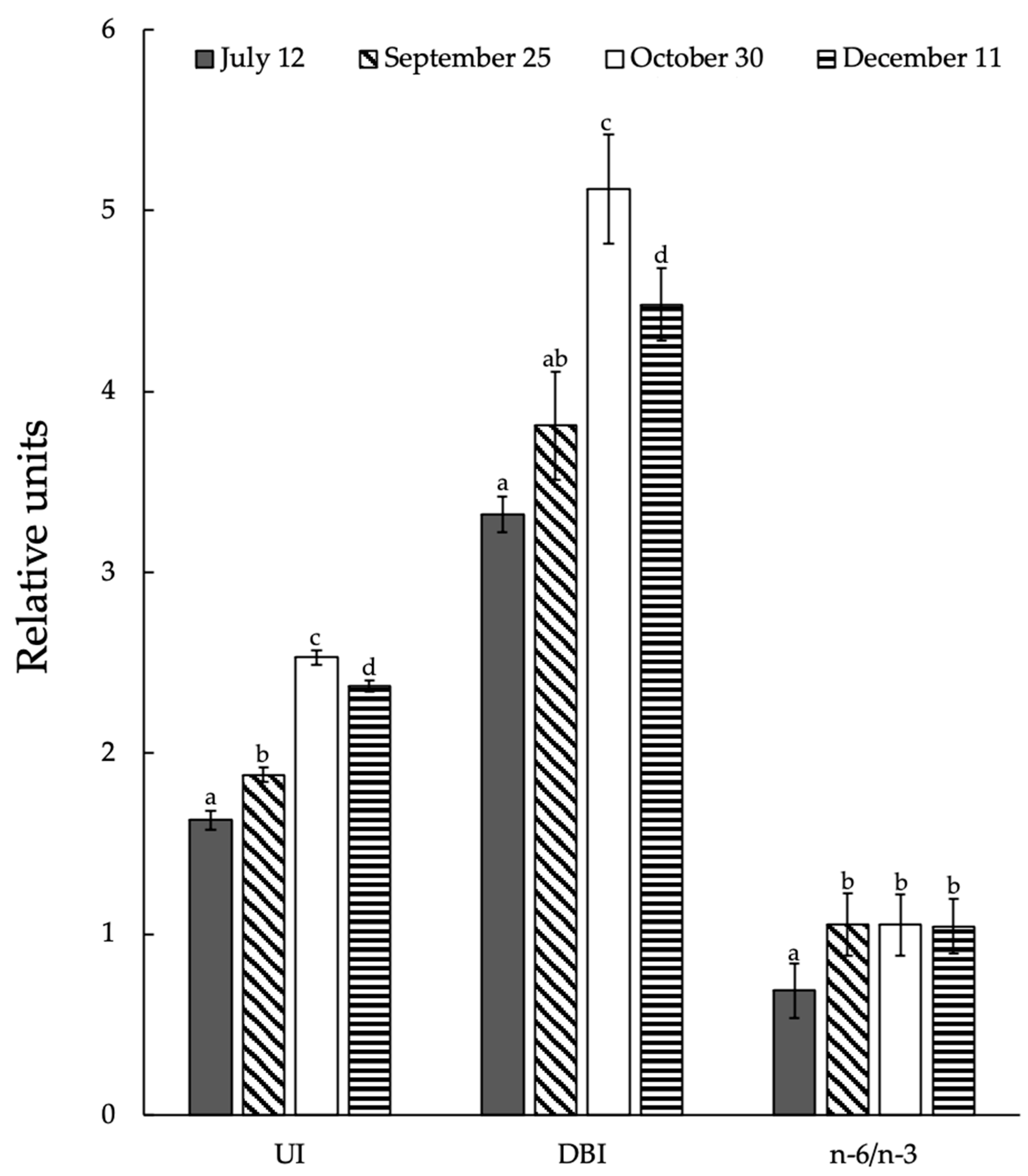
3.7. Seasonal Changes in Integral Indexes of Lipid Unsaturated Levels and Activity of Acyl-Lipid Desaturases in Ephedra Shoots

During the study period, two integral indices of the degree of FA unsaturation were evaluated: the level of unsaturation proper, and the number of double bonds (UI and DBI) (Figure 8). It was found that the unsaturated level of lipid FAs in ephedra shoots increased during the vegetation period; this was reflected in the number of double bonds in FAs. This integral index for the whole period of the study increased when the temperature decreased during the autumn–winter period. The ratio of n-6/n-3 acids content increased with the onset of autumn and remained high throughout late autumn and winter.
The synthesis of polyunsaturated FAs is a series of sequential desaturation reactions of the acyl residue of FAs, with the end product of the previous reaction serving as a substrate for the subsequent one. Analysis of desaturase activity performed for ephedra shoots growing under natural conditions of the Botanical Garden cryolithozone showed that this index (PDR, SDR, ODR) remained practically unchanged and remained at the same level during the growing season (Table 1). The activity of Δ3-desaturase (LDR) was higher in summer, then the activity of this enzyme decreased by fall. At the same time, the desaturation process significantly increased only for 18-FAs (Table 1) against the background of a monotonic increase in the elongation level of 16-FAs.

4. Discussion

It was found that the content of free STs exceeded the number of bound STs (sterol esters) in the shoots of ephedra plants. It is known that ST esters are contained in plant cells in much smaller amounts than free STs [11]. In the microalgae Pavlova gyrans, a native haptophyte from the southwestern coast of the Atlantic Ocean, it was shown that the highest percentage of bound sterols was found at the early stages of its cultivation (on the 2nd day). However, by the end of cultivation (the 9th day), under the influence of low-temperature stress, the proportion of free sterols increased significantly [31]. The properties of plant cell membranes, including those under low-temperature stress, are affected by changes in the ratio between campesterol, stigmasterine, and β-sitosterol [32]. These changes may concern the plasma membrane to a greater extent since it contains the main part of free cell sterols. However, it was shown for beets that under oxidative stress the level of sterols increases compared to the control, in particular stigmasterol. The authors of this work believe that, in addition to the plasmalemma, the tonoplast plays a significant role in the protection of plant cells from stress [33]. Moreover, the increase in campesterol may also be associated with protective functions in response to cold stress. The authors of the study conducted by Ozolina et al. [34] conclude that the increase in campesterol content in plasmalemma rafts isolated from beets indicates their involvement in the protective response of plant cells against oxidative stress. The increase in cholesterol content in December, as compared to October, can probably be explained by the fact that increased cholesterol content causes intensive synthesis and accumulation of spermine in cells, thus having a negative effect on plant growth [35,36]. In ephedra shoots, we observed that the content of the “stress” sterol- stigmasterine increases with the onset of low quenching temperatures in September. This may be due to a simultaneous decrease in the amount of β-sitosterol, since it is known that β-sitosterol is a precursor in stigmasterine biosynthesis [37]. However, a further decrease in average daily air temperature to −8.3 °C at the end of October and to the extremely low temperature of −38.4 °C in December did not result in an increase in stigmasterine content in ephedra shoots. The same results were obtained when wheat (Triticum aestivum) leaves were exposed to low temperatures; short-term exposure led to an increase in stigmasterine content and longer exposure to low temperatures was accompanied by restoration of indicators to control values [8]. The results obtained provide new information in support of the known thesis on the universal role of stigmasterine in the plant response to low negative ambient temperatures. The ST profile of ephedra shoots was dominated by β-sitosterol in all seasons. It is known that β-sitosterol has high antioxidant activity in the plant cell [38,39,40,41]. Cold stress in plants is characterized by the activation of oxidative processes in the plant cell [42]. Earlier it was revealed that β-sitosterol is able to neutralize free radicals of diphenylpicrylhydrazyl (superoxide anion donor) [43]. It is possible that ephedra shoots contain a high content of β-sitosterol due to the fact that the presence of the ethyl group in β-sitosterol may increase Van der Waals interactions, which leads to greater membrane adhesion and, consequently, less temperature sensitivity [44]. Changes in the composition and content of ST components entail significant changes in the functioning of membranes, cell metabolism, and the body as a whole. SMT is an integral protein localized in the EPR [45]. This enzyme is characteristic of plants and fungi and is absent in animal cells. The ratio of β-sitosterol and stigmasterol contents, in turn, is determined by the activity of C22-sterol desaturase. The activity of C22-sterol desaturase in ephedra shoots was highest in summer, which explains the high content of β-sitosterol (Table 1). It was previously shown that β-sitosterol is an important participant in the process of cell elongation [46] and also participates in their proliferation processes [47] and differentiation [48], the high content of β-sitosterol in the shoots of the evergreen shrub is most likely due to the fact that ephedra plants are in the phase of shoot growth, flowering, fruit ripening, and bud setting during the summer period. Some authors believe that plant biomass accumulation also correlates with high β-sitosterol [49]. It was shown earlier that tissues of non-embryogenic lines of Siberian larch (Larix sibirica Ledeb.) had higher levels of β-sitosterol than other sterols [50], and the analysis of the STs composition of ephedra shoots under natural conditions confirmed this tendency: in the sterol ester fraction, the content of β-sitosterol esters was maximal in summer, i.e., during active biomass accumulation, as compared to the autumn–winter period (Figure 5). Sq (squalene), a triterpene that is an intermediate in sterol biosynthesis, was identified in neutral lipids along with sterol components (Figure 2) [49].
The increase in the proportion of unsaturated FAs in September and October coincided with the prevailing outflow of metabolites and with the preparation of the photosynthetic apparatus of ephedra shoots for the dormancy period, which is initiated by a decrease in the photoperiod and lower air temperature in the fall period (Figure 7). It was found that the accumulation of unsaturated FAs in ephedra shoots, especially diene FAs such as C18:2(∆5,9) and C18:2(∆9,12), occurs at the expense of saturated levels decrease during the growing season. This increase is explained by the fact that ephedra are frost-resistant evergreen shrubs (extremophiles). It was shown earlier that cold-resistant rice genotypes are characterized by the accumulation of C18:2(∆5,9) acids after cold exposure, when C16:0 content decreases [51]. Earlier, by conducting experiments with conifers, we found that during the fall, the level of light, along with low temperatures, are stress factors for the needles of evergreen trees growing in cryolithozone conditions, which leads to an increase in the content of almost all unsaturated FAs [21]. A similar increase in diene FAs, along with a decrease in saturated FAs, was also noted for the rare evergreen fern Asplenium scolopendrium, whose buds can tolerate freezing during the fall and winter [52]. It is known that the accumulation of unsaturated FAs in cell membranes, especially polyunsaturated ones, contributes to plant resistance to low temperatures [53]. The gradual decrease in air temperature in the autumn revealed that the level of integral indexes UI and DBI in ephedra shoots increases, which indicates that active biosynthesis of unsaturated GI occurs in the shoots. It is known that the synthesis of unsaturated fatty acids in plant cells occurs with the participation of desaturase enzymes, which catalyze the introduction of a double bond into the acyl chain of FAs (desaturation reaction) [54]. As a rule, the first double bond is formed at position ∆9, followed by reactions at positions ∆12 and n-3 [55]. In this regard, the method of assessing desaturase activity by the content of end products was used. In ephedra shoots, in response to cooling temperatures in autumn, we found that C18 desaturation increased, which is explained by an active desaturation reaction of C18 acids, namely C18:2(∆5,9); C18:2(∆9,12); C18:3(isomer); C18:3(∆9,12,15); and C18:4(∆5,9,12.15). The level of the ratio of n-6/n-3 of the acids content increased during the period of autumn cooling, which is explained by the increased level of C18:2(∆9,12) linoleic acid. It is logical to assume that the presence of large amounts of unsaturated FAs in the plant cell membrane, especially diene C18:2(∆5,9), C18:2(∆9,12), whose proportion in ephedra shoots increased markedly in response to a decrease in ambient temperature (Figure 8), leads to an increase in membrane fluidity.

5. Conclusions

Seasonal changes in ∆5-sterols, ST esters, and FA groups in the shoots of the evergreen shrub E. monosperma growing in the territory of the Botanical Garden in the cryolithic zone have been revealed and analyzed. Significant qualitative and quantitative changes in the composition and content of membrane ∆5-sterols and unsaturated FAs caused by the formation of resistance to low-temperature influences during the autumn–winter period have been shown. It was revealed that the unsaturated FAs of lipids, especially linoleic acid C18:2(∆9,12), play an important role in the adaptation of ephedra shoots to the seasonal decrease in ambient temperature in the autumn–winter period. It is logical to assume that the high content of polyunsaturated FAs in ephedra shoots during the autumn–winter period gives the lipid bilayer a looser packing in the area of phospholipid-protein contact, which provides the membrane with greater plasticity, fluidity, and flexibility. The unique ability of plants with high β-sitosterol content in ∆5-sterols and a high degree of lipid FAs unsaturation may be part of the evolutionary process of plant adaptation to a wide range of temperature fluctuations and serve to maintain membrane-bound metabolic processes.
Thus, the results obtained on the seasonal dynamics of lipids of ephedra shoots provide useful information at the physiological and biochemical levels on the adaptation reactions of introduced valuable, rare, and medicinal plants growing in the Botanical Garden in permafrost ecosystem conditions.

Author Contributions

Conceptualization, V.V.N. and L.V.D.; methodology, V.V.N., L.V.D. and V.E.S.; software, V.V.N., L.V.D. and N.V.S.; validation, L.V.D. and N.V.S.; formal analysis, V.V.N., L.V.D. and N.V.S.; investigation, V.V.N. and L.V.D.; resources, V.V.N. and L.V.D.; data curation, V.V.N. and L.V.D.; writing—original draft preparation, V.V.N.; writing—review and editing, V.V.N., L.V.D. and V.E.S.; visualization, V.V.N.; supervision, V.V.N. and L.V.D.; project administration, V.V.N. and V.E.S.; funding acquisition, V.V.N. and V.E.S. All authors have read and agreed to the published version of the manuscript.

Funding

Lipids analysis was supported by Russian Science Foundation (project No. 22-76-00043). Fieldwork was carried out in the framework of the State Task of the Ministry of Education and Science of the Russian Federation FWRS-2021-0024.

Data Availability Statement

All data are contained within the article.

Acknowledgments

We are grateful to Svetlana Petrova for the linguistic corrections and improvements.

Conflicts of Interest

The authors declare no conflict of interest. The funders had no role in the design of the study; in the collection, analyses, or interpretation of data; in the writing of the manuscript; or in the decision to publish the results.

References

  1. Eslamdoust, J. Bark content variations and the production of Populus deltoides as a source of bioenergy under temperate climate conditions. J. For. Res. 2022, 33, 1807–1815. [Google Scholar] [CrossRef]
  2. González-Juárez, D.E.; Escobedo-Moratilla, A.; Flores, J.; Hidalgo-Figueroa, S.; Martínez-Tagüeña, N.; Morales-Jiménez, J.; Muñiz-Ramírez, A.; Pastor-Palacios, G.; Pérez-Miranda, S.; Ramírez-Hernández, A.; et al. A Review of the Ephedra genus: Distribution, Ecology, Ethnobotany, Phytochemistry and Pharmacological Properties. Molecules 2020, 25, 3283. [Google Scholar] [CrossRef] [PubMed]
  3. Abourashed, E.A.; El-Alfy, A.T.; Khan, I.A.; Walker, L. Ephedra in perspective—A current review. Phytother. Res. 2003, 17, 703–712. [Google Scholar] [CrossRef] [PubMed]
  4. EFSA ANS Panel (EFSA Panel on Food Additives and Nutrient Sources). Scientific opinion on safety evaluation of Ephedra species in food. EFSA J. 2013, 11, 3467–3546. [Google Scholar] [CrossRef]
  5. Ibragic, S.; Sofic, E. Chemical composition of various Ephedra species. Bosn. J. Basic Med. Sci. 2015, 15, 21–27. [Google Scholar] [CrossRef] [Green Version]
  6. Ephedra // Plantarium. Plants and Lichens of Russia and Neighboring Countries: Open Online Galleries and Plant Identification Guide. Available online: https://www.plantarium.ru/lang/en/page/view/item/44768.html (accessed on 11 June 2023).
  7. Dudareva, L.V.; Semenova, N.V.; Nochsorov, V.V.; Rudikovskaya, E.G.; Petrov, K.A. The component composition of the phytosterols of the aerial part of the horsetail variegated Equisétum variegatum Schleich. ex. Web. growing in North-East Yakutia. Khimiya Rastit. Syr’ya 2020, 2, 133–139. [Google Scholar] [CrossRef]
  8. Valitova, J.; Renkova, A.; Mukhitova, F.; Dmitrieva, S.; Beckett, R.P.; Minibayeva, F.V. Membrane sterols and genes of sterol biosynthesis are involved in the response of Triticum aestivum seedlings to cold stress. Plant Physiol. Biochem. 2019, 142, 452. [Google Scholar] [CrossRef]
  9. Xie, D.; Ye, J.; Lu, M.; Wang, S.; You, C.; Li, Y. Comparsion of Activities of Fatty Acyl Desaturases and Elongases among Six Teleosts with Different Feeding and Ecological Habits. Front. Mar. Sci. 2020, 7, 117. [Google Scholar] [CrossRef] [Green Version]
  10. Rozentsvet, O.A.; Kotlova, E.R.; Bogdanova, E.S.; Nesterov, V.N.; Senik, S.V.; Shavarda, A.L. Balance of Δ5-and Δ7-sterols and stanols in halophytes in connection with salinity tolerance. Phytochemistry 2022, 198, 113156. [Google Scholar] [CrossRef]
  11. Valitova, J.N.; Sulkarnayeva, A.G.; Minibayeva, F.V. Plant sterols: Diversity, biosynthesis, and physiological functions. Biochem. Mosc. 2016, 81, 819–834. [Google Scholar] [CrossRef]
  12. Senthil-Kumar, M.; Wang, K.; Mysore, K.S. AtCYP710A1 gene-mediated stigmasterol production plays a role in imparting temperature stress tolerance in Arabidopsis thaliana. Plant Signal. Behav. 2013, 8, e23142. [Google Scholar] [CrossRef] [Green Version]
  13. Zauber, H.; Burgos, A.; Garapati, P.; Schulze, W.X. Plasma membrane lipid-protein interactions affect signaling processes in sterol-biosynthesis mutants in Arabidopsis thaliana. Front. Plant Sci. 2014, 5, 78. [Google Scholar] [CrossRef] [Green Version]
  14. Ozolina, N.V.; Kapustina, I.S.; Gurina, V.V.; Nurminsky, V.N. Role of tonoplast microdomains in plant cell protection against osmotic stress. Planta 2022, 255, 65. [Google Scholar] [CrossRef]
  15. Piironen, V.; Lindsay, D.G.; Miettinen, T.A.; Toivo, J.; Lampi, A.-M. Plant sterols: Biosynthesis, biological function and their importance to human nutrition. J. Sci. Food Agric. 2000, 80, 939–966. [Google Scholar] [CrossRef]
  16. Vezza, T.; Canet, F.; de Marañón, A.M.; Bañuls, C.; Rocha, M.; Víctor, V.M. Phytosterols: Nutritional Health Players in the Management of Obesity and Its Related Disorders. Antioxidants 2020, 9, 1266. [Google Scholar] [CrossRef]
  17. Nokhsorov, V.V.; Senik, S.V.; Sofronova, V.E.; Kotlova, E.R.; Misharev, A.D.; Chirikova, N.K.; Dudareva, L.V. Role of Lipids of the Evergreen Shrub Ephedra monosperma in Adaptation to Low Temperature in the Cryolithozone. Plants 2023, 12, 15. [Google Scholar] [CrossRef]
  18. Alqarawi, A.A.; Hashem, A.; Abd Allah, E.F.; Alshahrani, T.S.; Huqail, A.A. Effect of salinity on moisture content, pigment system, and lipid composition in Ephedra alata Decne. Acta Biol. Hung. 2014, 65, 61–71. [Google Scholar] [CrossRef]
  19. Bondarenko, S.A.; Ianutsevich, E.A.; Danilova, O.A.; Grum-Grzhimaylo, A.A.; Kotlova, E.R.; Kamzolkina, O.V.; Bilanenko, E.N.; Tereshina, V.M. Membrane lipids and soluble sugars dynamics of the alkaliphilic fungus Sodiomyces tronii in response to ambient pH. Extremophiles 2017, 21, 743–754. [Google Scholar] [CrossRef]
  20. Wolff, R.L.; Christie, W.W.; Pedrono, F.; Marpeau, A.M.; Tsevegsüren, N.; Aitzetmüller, K.; Gunstone, F.D. Δ5-olefinic acids in the seed lipids from four Ephedra species and their distribution between the α and β positions of triacylglycerols. Characteristics common to coniferophytes and cycadophytes. Lipids 1999, 58, 101–115. [Google Scholar] [CrossRef]
  21. Nokhsorov, V.V.; Dudareva, L.V.; Petrov, K.A. Content and Composition of Lipids and Their Fatty Acids in Needles of Pinus sylvestris L. and Picea obovata Ledeb. upon Cold Hardening in the Cryolithozone of Yakutia. Russ. J. Plant Physiol. 2019, 66, 548–555. [Google Scholar] [CrossRef]
  22. Nokhsorov, V.V.; Dudareva, L.V.; Senik, S.V.; Chirikova, N.K.; Petrov, K.A. Influence of Extremely Low Temperatures of the Pole of Cold on the Lipid and Fatty-Acid Composition of Aerial Parts of the Horsetail Family (Equisetaceae). Plants 2021, 10, 996. [Google Scholar] [CrossRef] [PubMed]
  23. Christie, W.W.; Han, X. Lipid Analysis: Isolation, Separation, Identification and Lipidomic Analysis, 4th ed.; Woodhead Publishing: Bridgwater, UK, 2010; p. 448. [Google Scholar] [CrossRef]
  24. Christie, W.W. The AOCS Lipid Library: Methyl Esters of Fatty Acids—Archive of Mass Spectra. Available online: https://www.lipidmaps.org/resources/lipidweb/index.php?page=ms/methesters/me-arch/index.htm (accessed on 31 March 2023).
  25. Nes, W.D. Sterol methyltransferase: Enzymology and inhibition. Biochim. Biophys. Acta, Mol. Cell Biol. Lipids 2000, 1529, 63–88. [Google Scholar] [CrossRef]
  26. Lyons, J.M.; Wheaton, T.A.; Pratt, H.K. Relationship between the Physical Nature of Mitochondrial Membranes and Chilling Sensitivity in Plants. Plant Physiol. 1964, 39, 262–268. [Google Scholar] [CrossRef] [PubMed] [Green Version]
  27. Wismer, W.V.; Worthing, W.M.; Yada, R.Y.; Marangoni, A.G. Membrane lipid dynamics and lipid peroxidation in the early stages of low-temperature sweetening in tubers of Solanum tuberosum. Physiol. Plant. 1998, 102, 396–410. [Google Scholar] [CrossRef]
  28. Lee, S.H.; Ahn, S.J.; Im, Y.J.; Cho, K.; Chung, G.C.; Cho, B.H.; Han, O. Differential impact of low temperature on fatty acid unsaturation and lipoxygenase activity in figleaf gourd and cucumber roots. Biochem. Biophys. Res. Commun. 2005, 330, 1194–1198. [Google Scholar] [CrossRef]
  29. Voronkov, A.S.; Ivanova, T.V. Fatty Acids Composition of the Epiphytic Ferns, Platycerium bifurcatum and Asplenium nidus, and the Terrestrial Fern, Asplenium trichomanes. Am. Fern J. 2021, 111, 117–128. [Google Scholar] [CrossRef]
  30. O’Hare, T.J.; Trieu, H.H.; Topp, B.; Russell, D.; Pun, S.; Torrisi, C.; Liu, D. Assessing Fatty Acid Profiles of Macadamia Nuts. HortScience 2019, 54, 633–637. [Google Scholar] [CrossRef] [Green Version]
  31. Almeyda, M.D.; Scodelaro Bilbao, P.G.; Sanchez-Puerta, M.V.; Constenla, D.; Leonardi, P.I. Pavlova gyrans as a potential source of essential fatty acids, sterols and pigments: Culture under low temperature. J. Appl. Phycol. 2023, 35, 1073–1089. [Google Scholar] [CrossRef]
  32. Schaller, H. The role of sterols in plant growth and development. Prog. Lipid Res. 2003, 42, 163–175. [Google Scholar] [CrossRef]
  33. Ozolina, N.V.; Gurina, V.V.; Kapustina, I.S.; Spiridonova, E.V.; Nurminsky, V.N. Comparison of Changes in the Content of Plasma Membrane and Tonoplast Sterols under Oxidative and Osmotic Stress. Biochem. Mosc. Suppl. Ser. A 2023, 17, 180–182. [Google Scholar] [CrossRef]
  34. Ozolina, N.; Kapustina, I.; Gurina, V.; Spiridonova, E.; Nurminsky, V. The microdomains (rafts) of plasmalemma in the protection of the plant cell under oxidative stress. Protoplasma 2023, 1–10. [Google Scholar] [CrossRef]
  35. Chen, M.; Chen, J.; Luo, N.; Qu, R.; Guo, Z.; Lu, S. Cholesterol accumulation by suppression of SMT1 leads to dwarfism and improved drought tolerance in herbaceous plants. Plant Cell Environ. 2018, 41, 1417–1426. [Google Scholar] [CrossRef]
  36. Renkova, A.G.; Khabibrakhmanova, V.R.; Chasov, A.V.; Valitova, J.N.; Galeeva, E.I.; Minibayeva, F.V. Changes in Composition and Content of Lipophilic Compounds in the Seedlings of Triticum aestivum L. Treated with Stress Phytohormones. Russ. J. Plant Physiol. 2023, 70, 6. [Google Scholar] [CrossRef]
  37. Renkova, A.G.; Khabibrakhmanova, V.R.; Valitova, J.N.; Mukhitova, F.K.; Minibayeva, F.V. Effects of Stress Phytohormones on Sterol Metabolism of Triticum aestivum L. Russ. J. Plant Physiol. 2021, 68, 474–482. [Google Scholar] [CrossRef]
  38. Weng, X.C.; Wang, W. Antioxidant activity of compounds isolated from Salvia plebeian. Food Chem. 2000, 71, 489–493. [Google Scholar] [CrossRef]
  39. Wang, T.; Hicks, K.B.; Moreau, R. Antioxidant activity of phytosterols, oryzanol, and other phytosterol conjugates. J. Am. Oil Chem. Soc. 2002, 79, 1201–1206. [Google Scholar] [CrossRef]
  40. Vivancos, M.; Moreno, J.J. Beta-sitosterol modulates antioxidant enzyme response in RAW 264.7 macrophages. Free Radic. Biol. Med. 2005, 39, 91–97. [Google Scholar] [CrossRef]
  41. Pose, D.; Castanedo, I.; Borsani, O.; Nieto, B.; Rosado, A.; Taconnat, L.; Ferrer, A.; Dolan, L.; Valpuesta, V.; Botella, M.A. Identification of the Arabidopsis dry2/sqe1-5 mutant reveals a central role for sterols in drought tolerance and regulation of reactive oxygen species. Plant J. 2009, 59, 63–76. [Google Scholar] [CrossRef]
  42. Kendall, E.J.; McKersie, B.D. Free radical and freezing injury to cell membranes of winter wheat. Physiol. Plant. 1989, 76, 86–94. [Google Scholar] [CrossRef]
  43. Wegener, A.; Gimbel, W.; Werner, T.; Hani, J.; Ernst, D.; Sandermann, H., Jr. Molecular cloning of ozone-inducible protein from Pinus sylvestris L. with high sequence similarity to vertebrate 3-hydroxy-3-methylglutaryl-CoA-synthase. Biochim. Biophys. Acta Gene Struct. Expr. 1997, 1350, 247–252. [Google Scholar] [CrossRef]
  44. Dufourc, E.J. The role of phytosterols in plant adaptation to temperature. Plant Signal. Behav. 2008, 3, 133–134. [Google Scholar] [CrossRef] [PubMed] [Green Version]
  45. Boutte, Y.; Grebe, M. Cellular processes relying on sterol function in plants. Curr. Opin. Plant Biol. 2009, 12, 705–713. [Google Scholar] [CrossRef]
  46. Deng, S.; Wei, T.; Tan, K.; Hu, M.; Li, F.; Zhai, Y.; Ye, S.; Xiao, Y.; Hou, L.; Pei, Y.; et al. Phytosterol content and the campesterol: Sitosterol ratio influence cotton fiber development: Role of phytosterols in cell elongation. Sci. China Life Sci. 2016, 59, 183–193. [Google Scholar] [CrossRef] [PubMed] [Green Version]
  47. Qian, P.; Han, B.; Forestier, E.; Hu, Z.; Gao, N.; Lu, W.; Shaller, H.; Li, J.; Hou, S. Sterols are required for cell fate commitment and maintenance of the stomatal lineage in Arabidopsis. Plant J. 2013, 74, 1029–1044. [Google Scholar] [CrossRef] [PubMed]
  48. Carland, F.M.; Fujioka, S.; Takatsuto, S.; Yoshida, S.; Nelson, T. The identification of CVP1 reveals a role for sterols in vascular patterning. Plant Cell 2002, 14, 2045–2058. [Google Scholar] [CrossRef] [Green Version]
  49. Cunha, A.; Ferreira, M.F. Differences in free sterols content and composition associated with somatic embryogenesis, shoot organogenesis and calli growth of flax. Plant Sci. 1997, 124, 97–105. [Google Scholar] [CrossRef] [Green Version]
  50. Semenova, N.V.; Shmakov, V.N.; Konstantinov, Y.M.; Dudareva, L.V. Comparative Analysis of Sterol Composition of Embryogenic and Nonembryogenic Cell Lines of Larix sibirica Ledeb. Russ. J. Plant Physiol. 2023, 70, 181–191. [Google Scholar] [CrossRef]
  51. Cruz, R.P.D.; Golombieski, J.I.; Bazana, M.T.; Cabreira, C.; Silveira, T.F.; Silva, L.P.D. Alterations in fatty acid composition due to cold exposure at the vegetative stage in rice. Braz. J. Plant Physiol. 2010, 22, 199–207. [Google Scholar] [CrossRef] [Green Version]
  52. Voronkov, A.; Ivanova, T. Significance of Lipid Fatty Acid Composition for Resistance to Winter Conditions in Asplenium scolopendrium. Biology 2022, 11, 507. [Google Scholar] [CrossRef]
  53. Nejadsadeghi, L.; Maali-Amiri, R.; Zeinali, H.; Ramezanpour, S.; Sadeghzade, B. Membrane fatty acid compositions and cold-induced responses in tetraploid and hexaploid wheats. Mol. Biol. Rep. 2015, 42, 363–372. [Google Scholar] [CrossRef]
  54. Starikov, A.Y.; Sidorov, R.A.; Goriainov, S.V.; Los, D.A. Acyl-Lipid Δ6-Desaturase May Act as a First FAD in Cyanobacteria. Biomolecules 2022, 12, 1795. [Google Scholar] [CrossRef]
  55. Sato, N.; Wada, H. Lipid Biosynthesis and Its Regulation in Cyanobacteria. Lipids in Photosynthesis: Essential and Regulatory Functions; Wada, H., Murata, N., Eds.; Springer Science + Business Media B.V.: Amsterdam, The Netherlands, 2009; Volume 30, pp. 157–177. [Google Scholar] [CrossRef]
Figure 1. Ephedra monosperma shoots growing on the territory of the Botanical Garden in the cryolithozone of Yakutia (photo taken by Nokhsorov, May 2019).
Horticulturae 09 00858 g001
Figure 2. HPTLC chromatograms of the neutral lipid extracts extracted from shoots E. monosperma. Images were taken at (a) white light after derivatization; (b) 366 nm; (c) 254 nm; Track 1—12 July, Track 2—25 September, Track 3—30 October, and Track 4—11 December.
Horticulturae 09 00858 g002
Figure 3. Total content of free and bound STs in shoots of E. monosperma (μg/g DW). Values are presented as the means ± SEMs. The significance of differences between the compared mean values was assessed by Kruskal–Wallis ANOVA by ranks (p < 0.05). Different superscript letters indicate significant differences in analyzed parameters.
Horticulturae 09 00858 g003
Figure 4. Seasonal changes in the most abundant ∆5-sterols in shoots of the evergreen shrub E. monosperma (μg/g DW). Values are presented as the means ± SEMs. The significance of differences between the compared mean values was assessed by Kruskal–Wallis ANOVA by ranks (p < 0.05). Different superscript letters indicate significant differences in analyzed parameters.
Horticulturae 09 00858 g004
Figure 5. Seasonal changes in the ST ester fraction in shoots of the evergreen shrub E. monosperma (μg/g DW). Values are presented as the means ± SEMs. The significance of differences between the compared mean values was assessed by Kruskal–Wallis ANOVA by ranks (p < 0.05). Different superscript letters indicate significant differences in analyzed parameters.
Horticulturae 09 00858 g005
Figure 6. Activity of sterol methyltransferase (SMT) enzymes in shoots of E. monosperma. Values are presented as the means ± SEMs. The significance of differences between the compared mean values was assessed by Kruskal–Wallis ANOVA by ranks (p < 0.05). Different superscript letters indicate significant differences in analyzed parameters.
Horticulturae 09 00858 g006
Figure 7. The percentage of saturated and unsaturated (mono-, di-, tri-, tetraene) FA components in shoots E. monosperma Values are presented as the means ± SEMs. The significance of differences between the compared mean values was assessed by Kruskal–Wallis ANOVA by ranks (p < 0.05). Different superscript letters indicate significant differences in analyzed parameters.
Horticulturae 09 00858 g007
Figure 8. Unsaturation index (UI) and double-bond index (DBI), as well as the ratio n-6/n-3 in shoots E. monosperma. Values are presented as the means ± SEMs. The significance of differences between the compared mean values was assessed by Kruskal–Wallis ANOVA by ranks (p < 0.05). Different superscript letters indicate significant differences in analyzed parameters.
Horticulturae 09 00858 g008
Table 1. Desaturation ratios and desaturation and elongation of E. monosperma shoots in summer (12 July), autumn (25 September and 30 October), and winter (11 December).
Parameters12 July25 September30 October11 December
PDR0.03 ± 0.01 a0.03 ± 0.01 a0.02 ± 0.00 a0.03 ± 0.01 a
SDR0.60 ± 0.18 a0.50 ± 0.16 a0.60 ± 0.13 a0.60 ± 0.13 a
ODR0.90 ± 0.09 a0.90 ± 0.13 a0.90 ± 0.13 a0.90 ± 0.07 a
LDR0.60 ± 0.04 a0.50 ± 0.03 b0.50 ± 0.03 b0.50 ± 0.04 b
C16 desaturation0.01 ± 0.00 a0.01 ± 0.00 a*,a0.01 ± 0.00 a
C18 desaturation3.81 ± 0.24 a3.69 ± 0.18 a5.57 ± 0.31 b5.20 ± 0.22 b
C16 elongation2.90 ± 0.12 a3.40 ± 0.26 b3.99 ± 0.17 b3.78 ± 0.19 b
Sterol C-22 desaturase103.19 ± 10.3 a48.43 ± 5.8 b93.32 ± 7.6 a89.84 ± 8.9 a
Note: PDR—palmitic desaturation ratio; SDR—stearic desaturation ratio; ODR—oleic desaturation ratio; LDR—linoleic desaturation ratio; *—trace. The significance of differences between the compared mean values was assessed by Kruskal–Wallis ANOVA by ranks (p < 0.05). Different superscript letters indicate significant differences in analyzed parameters.
Disclaimer/Publisher’s Note: The statements, opinions and data contained in all publications are solely those of the individual author(s) and contributor(s) and not of MDPI and/or the editor(s). MDPI and/or the editor(s) disclaim responsibility for any injury to people or property resulting from any ideas, methods, instructions or products referred to in the content.

