Article

Ectopic Expression of AGAMOUS-like 18 from Litchi (Litchi chinensis Sonn.) Delayed the Floral Organ Abscission in Arabidopsis

1
Guangdong Laboratory for Lingnan Modern Agriculture, Guangzhou 510642, China
2
Guangdong Litchi Engineering Research Center, College of Horticulture, South China Agricultural University, Guangzhou 510642, China
3
State Key Laboratory for Conservation and Utilization of Subtropical Agro-Bioresources, South China Agricultural University, Guangzhou 510642, China
4
Key Laboratory of Biology and Genetic Improvement of Horticultural Crops (South China), Ministry of Agriculture and Rural Affairs, College of Horticulture, South China Agricultural University, Guangzhou 510642, China
*
Authors to whom correspondence should be addressed.
These authors contributed equally to this work.
Horticulturae 2023, 9(5), 578; https://doi.org/10.3390/horticulturae9050578
Submission received: 24 March 2023 / Revised: 1 May 2023 / Accepted: 11 May 2023 / Published: 12 May 2023

Abstract

:
The regulation of abscission has a significant impact on fruit yield and quality. Thus, understanding the mechanisms underlying abscission, particularly identifying key genes, is critical for improving fruit crop breeding and cultivation practices. Here, to explore the key genes involved in litchi fruitlet abscission, the two closest homologs of AGAMOUS-like 15/18 (LcAGL15 and LcAGL18) were identified. During the litchi fruitlet abscission process, LcAGL15 expression was reduced, whereas LcAGL18 expression was increased at the abscission zone. The abscission of floral organs was unaffected by ectopic expression of LcAGL15 in Arabidopsis. Moreover, high expression of LcAGL18 significantly delayed the abscission process of floral organs, particularly the sepals. Overexpression of LcAGL18 in Arabidopsis consistently repressed the expression of abscission-related genes, including HAESA (HAE) and HAESA-LIKE2 (HSL2), and cell wall remodeling genes at the abscission zone. Furthermore, LcAGL18 was localized in the nucleus and acted as a transcriptional inhibitor. Collectively, these results suggest that AGL18 homologs have conserved functions in Arabidopsis and litchi, and that LcAGL18 might function as a key regulator in litchi fruitlet abscission.
Keywords:
litchi; abscission; AGL18

1. Introduction

Plants rely on the evolutionary fitness mechanism of organ loss, such as the dropping of senescent leaves and diseased fruit, to promote growth and survival. Unfavorable precocious abscission, on the other hand, has a significant impact on crop plant productivity and quality [1]. Thus, understanding the mechanism underlying the abscission of a crop plant or fruit crop is a key agricultural concern. The abscission process includes four major stages [2]: (i) differentiation of the abscission zone (AZ) where the separation process occurs; (ii) acquisition of the capabilities to cope with abscission signals; (iii) abscission activation; (iv) formation of a protective layer on the AZ’s main body side. Abscission is thus regarded as a tightly controlled process that occurs in response to various stimuli.
Key regulators controlling abscission have primarily been identified using Arabidopsis and tomato as model systems for abscission of floral organs and flowers, respectively. Arabidopsis floral organ abscission is triggered by the binding of the small peptide INFLORESCENCE DEFICIENT ABSCISION (IDA) to the receptor-like kinases HAE and HSL2 [3,4,5,6,7]. Then, MITOGEN-ACTIVATED PROTEIN KINASE KINASE 4/5 and MPK3/6 constitute the MAP kinase cascade, which can be activated by binding of co-receptors and HAE/HSL2 [4,5,7,8]. In tomatoes, activation of flower abscission can be triggered by interactions between the phytohormones ethylene and auxin, as well as abiotic stresses such as drought and low light intensity [9,10,11,12]. Peptide hormones IDA-like 6 (IDL6) and phytosulfokine (PSK) [13,14], and transcription factors (TFs), including ethylene response factor 52 [15], KNOTTED1-LIKE HOMEOBOX protein KD1 [10], homeodomain-leucine zipper transcription factor SlHB15A [16], and MADS-box proteins [17], have all been revealed to play critical roles in regulation of this process.
MADS-box TFs are crucial in controlling the progression of AZ and the induction of abscission. The knockout of the MADS-box gene JOINTLESS produces the mutant j in tomatoes, which is characterized by the absence of AZ [18]. Expression of target genes essential for AZ development is regulated by a complex formed by JOINTLESS, MACROCALYX, and SEPALLATA MADS-box TF SlLMBP21 [19]. Recent studies in several plant species, including Arabidopsis, tomato, and several species of orchid, have revealed a role for MADS-box TFs in controlling abscission activation. In Arabidopsis, the MADS-box TFs AGL15/18 play essential roles in multiple developmental processes, such as floral transition [20], pollen maturation [21], somatic embryogenesis [22,23], and function as strong negative regulators of floral organ abscission [20]. AGL15 has been shown to function as a direct negative upstream mediator of HAE. AGL15 overexpression leads to a reduction in HAE expression, resulting in a prolonged abscission [24]. Overexpression of FOREVER YOUNG FLOWER (a MADS-box gene) orthologs from Oncidium orchids (OnFYF), Cattleya orchids (CaFYF1/2), and Phalaenopsis orchids (PaFYF1/2) in transgenic Arabidopsis delays floral organ abscission [25,26,27]. These observations suggest the significant roles of MADS-box TFs in organ abscission in model plants and orchid species. However, it is unknown whether MADS-box TFs can regulate abscission during fruit abscission in fruit crops.
Litchi (Litchi chinensis Sonn.) is one of the most desirable and expensive tropical or subtropical fruits available worldwide [28]. Litchi fruit, however, experiences significant economic losses due to 3–5 fruit drop waves depending on varieties during fruit set, roughly three months after pollination [29]. In litchi, each panicle can produce 100–250 female flowers; however, less than 5% of female flowers can develop into mature fruit, mainly due to heavy fruit abscission [30,31]. Wave I occurs near the end of week 1 after full bloom, mainly due to poor pollination and fertilization, which can be regarded as normal abscission. Wave II to wave V happens when the available carbohydrate is limited or imbalanced endogenous hormones occur in fruit, which are regarded as abnormal abscission [29]. Previously, we found that litchi fruitlet abscission can be activated by ethylene and identified several genes that can regulate litchi fruitlet abscission via controlling ethylene biosynthesis and signaling [32,33,34].To further explore the key candidate genes in litchi fruitlet abscission, two closest homologs of AGL15/18 were identified in this study, and we found that heavy ectopic expression of LcAGL18 in Arabidopsis substantially slowed down floral organ abscission, implying that LcAGL18 may function as a key regulator in the regulation of litchi fruitlet abscission.

2. Materials and Methods

2.1. Plant Materials and Treatments

A total of 3 22-year-old “Feizixiao” litchi trees were chosen randomly from the orchard of South China Agricultural University (113°21′ E, 23°9′ N). For this experiment, 30 shoots bearing fruitlets were selected. These shoots ranged in diameter from 5–8 mm and were grown in a variety of directions. At 25 days post-anthesis, 10 shoots were given the girdling and defoliation treatment (abbreviated GPD, in which a 0.5 cm wide ring of bark and cambium was removed from the bottom of the branches). GPD treatment is a rapid and highly reproducible abscission-accelerating approach via decreasing the soluble sugar content and inducing ethylene production in the fruitlet of litchi [35]. A total of 10 individual shoots were dipped in a 250 mg·L−1 ethephon solution for 1 min that was designed for ET treatment, while the rest were used as controls. Three shoots from each treatment were selected to record the abscission rate of the fruitlets, while the rest were used for sampling. Following treatment, the cumulative fruit abscission rate was determined as described earlier [36]. Briefly, the fruit number on the tagged shoots was counted at each sapling date. Cumulative fruit abscission was calculated by subtracting the number of remaining fruitlets from the initial number, dividing by the initial number, and multiplying by 100. Samples of fruitlet AZ were prepared by slicing off about 2 mm on either side of the abscission fracture plane. AZ samples were taken daily from day 0 to day 5 after treatment.
The open reading frames for LcAGL15 (LITCHI004481) and LcAGL18 (LITCHI008820) were cloned into the pCAMBIA1302 vector, and formerly the recombinant constructs were inserted into wild-type Col plants via floral dip for expression in Arabidopsis [37]. For phenotypic analysis, both Col wild-type controls and stable T3 homozygous transgenic lines were grown under the following conditions: 22 °C with a light-dark cycle of 16/8 h.

2.2. Isolation of Genes, Sequence Alignment, and qRT-PCR

The gene sequences of litchi were retrieved from the litchi genome dataset (http://121.37.229.61:82/ (accessed on 1 September 2021)), genes from other species were attained from the plant genomics database (https://phytozome-next.jgi.doe.gov/ (accessed on 1 September 2021). ClustalW and GeneDoc were used for multiple sequence alignments. MEGA 11 was employed to generate a phylogenetic tree by selecting the Poisson correction model and the neighbor-joining (NJ) method.
Total RNA was isolated from AZ tissues of litchi fruitlets or Arabidopsis leaves (20 days old) and floral organs using 1 mL of Trizol reagent (Invitrogen). cDNA synthesis was performed as described earlier [38]. The assay was performed in triplicate for litchi and Arabidopsis, and EF-1a and Ubiquitin 10 (UBQ) were used to serve as internal controls, respectively [38]. A quantitative RT-PCR assay and relative expression level were carried out as described earlier [39].

2.3. Subcellular Compartmentation of LcAGL18

The coding sequence of LcAGL18 was fused into pEAQ-GFP. The fusion constructs 35S:LcLcAGL18-GFP were introduced into Nicotiana benthamiana leaves as described previously [40]. The GFP signal was observed using a confocal laser scanning microscope (LSM 7 DUO, ZEISS, Oberkochen, Germany).

2.4. BCECF Fluorescence Assay

The BCECF fluorescence analysis was performed according to the procedures outlined in the literature [11]. Briefly, inflorescences were separated from the rest of the plant and immersed in a 10 μM BCECF-AM (B1150, Thermo Scientific, Waltham, MA, USA) solution in the dark for 20 min before being washed four times with phosphate-buffered saline (PBS, pH 7.4) to get rid of any remaining BCECE-AM. Using a confocal laser scanning microscope, samples were excited by 488 nm and 633 nm light. Chlorophyll auto-fluorescence and BCECF fluorescence were examined via 647–721 and 494–598 filters, respectively.

2.5. Histochemical GUS Assays

To generate the LcAGL18pro:GUS constructs, the LcAGL18 promoter region (−1 to −2000 bp) was fused into the pCAMBIA1391 vector. As previously stated, the LcAGL18pro:GUS constructs were introduced into Arabidopsis. Flowers of T1 transgenic plants were stained in GUS solution (0.1% Triton X-100, 10 mM EDTA, 2 mM potassium ferrocyanide, 2 mM potassium ferricyanide, 1 mg·mL−1 X-Gluc and 100 μg·mL−1 chloramphenicol in a 50 mM sodium phosphate buffer, pH 7.0) for 6 h at 37 °C and then cleared in 70% ethanol. A Zeiss SV11 stereomicroscope was utilized to observe GUS expression.

2.6. Scanning Electron Microscopy

The floral organ AZs of 35S:LcAGL18 transgenic lines and Col plants were removed and immediately fixed in 4% glutaraldehyde (w/v) for 4 h. Following fixation, the AZ tissues were rinsed 3 times in 0.1 M potassium phosphate buffer (pH 7.5), and afterward dehydrated in graded ethanol. AZ samples were critical-point dried in liquid CO2 and were then sputter-coated with gold palladium. The Zeiss EVO MA 15 scanning electron microscope was employed to examine the samples at an acceleration voltage of 10 K.

2.7. Transcriptional Activity Assays

The LcAGL18 coding sequence was fused into the vector GAL4-BD as the effector. In the double reporter vector, the firefly luciferase gene (GAL4-LUC) is preceded by a GAL4 DNA consensus binding site, and the Renilla luciferase gene (REN) is driven by the 35S promoter in A. tumefaciens strain GV3101. The reporter and effector plasmids were co-delivered into N. benthamiana leaves as explained earlier [35]. Dual-Luciferase® Reporter Assay System kits (Promega, Madison, WI, USA) were employed to examine the activities of LUC and REN luciferases. Each reporter and effector pair was subjected to at least six assays.

2.8. Data Analysis

The results are shown as the mean of three to six independent biological replicates. SPSS22.0 (IBM Corp., Armonk, NY, USA) was utilized to conduct a one-way analysis of variance (ANOVA) with a post hoc Tukey honest significant difference test.

2.9. Primers

All primers were generated by using Primer 3 (https://bioinfo.ut.ee/primer3-0.4.0/ (accessed on 1 September 2021)) and are listed in Table S1.

3. Results

3.1. Identification of the Closest Homologs of AGL15/18 in Litchi

We used the amino acid sequences of Arabidopsis AGL15 and AGL18 to perform a TBLASTN search against the litchi genome to find the closest homologs of AGL15 and AGL18. Two AGL-like genes, designated LcAGL15 and LcAGL18, were subsequently found in the litchi genome. Additionally, we performed alignments between LcAGL15/18 and other MADS-box proteins that have been linked to abscission regulation. Protein sequence alignment showed that LcAGL15/18 contains a conserved MADS-box domain in the N-terminal that can recognize the specific DNA motif CW6G (Theissen et al., 2000) and a conserved K domain that is involved in protein-protein interactions [41] (Figure 1A). As expected, phylogenetic analysis showed that LcAGL15/18 and Arabidopsis AGL15/18 are closely grouped together (Figure 1B).

3.2. Expression Patterns of LcAGL15 and LcAGL18 during the Litchi Fruitlet Abscission

To determine if LcAGL15/18 are associated with the regulation of litchi fruitlet abscission, we first analyzed their expression profiles during the litchi fruitlet abscission activated by girdling plus defoliation (GPD) and ethephon application (ET) [35]. Similar to previous studies, ET and GPD induced fruitlet drops starting one day after treatments (Figure 2A). At five days after treatment, ET and GPD caused 91.6% and 100% fruitlet drop, respectively, while only 15.8% of the fruitlets abscised in control (Figure 2A). qPCR assays showed that LcAGL15 was downregulated (Figure 2B), whereas LcAGL18 was greatly upregulated at the AZ during the ET-/GPD-induced abscission compared with that in control (Figure 2C).

3.3. High Expression of LcAGL18 in Arabidopsis Leads to the Delay of Floral Organ Abscission

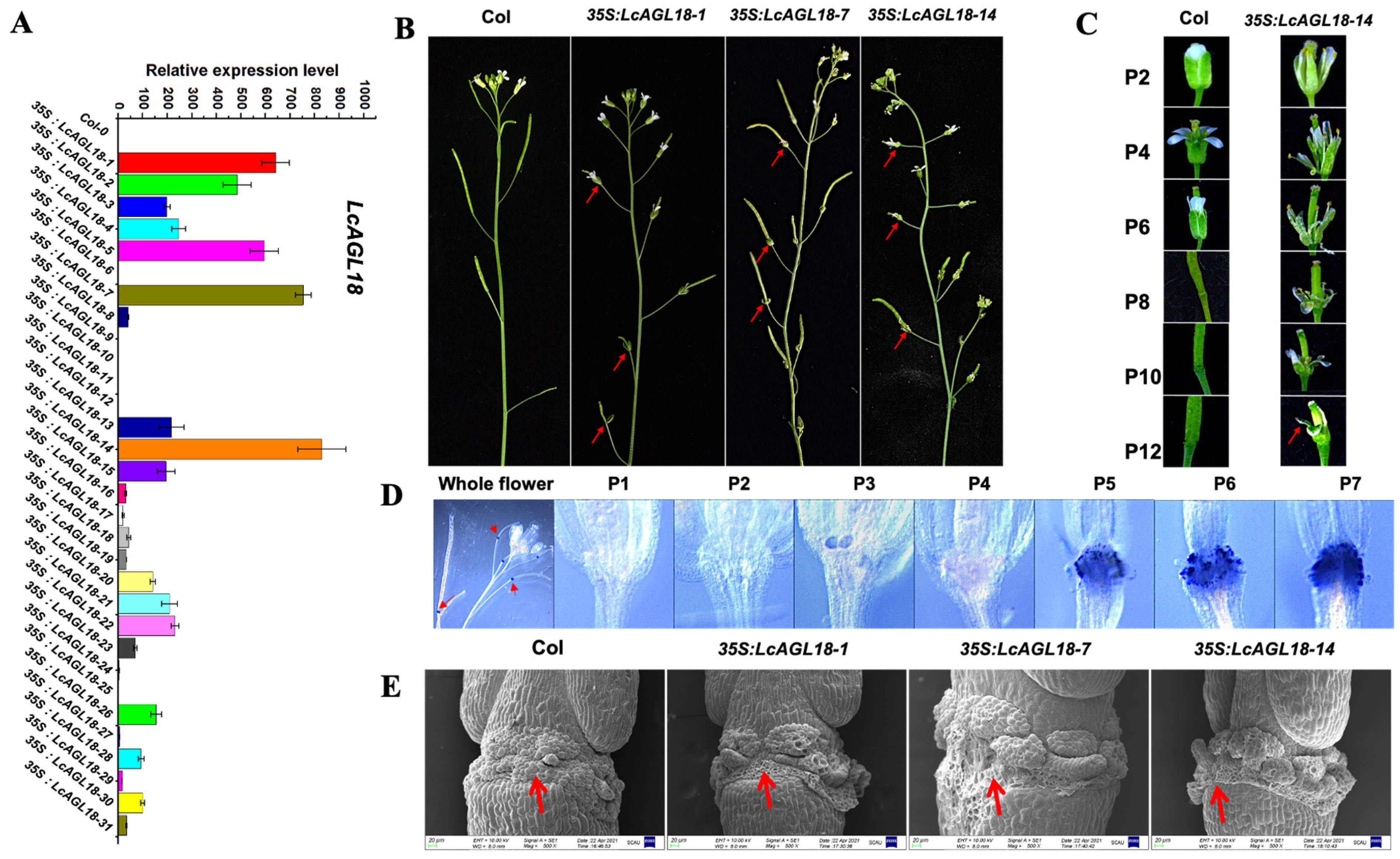
Ectopic expression of LcAGL15/18 in Arabidopsis allowed us to further investigate their function in abscission regulation. To attain this, both constructs were transformed into wild-type Arabidopsis plants to drive the expression of LcAGL15 or LcAGL18 by the strong constitutive cauliflower mosaic virus 35S promoter. As a result, 30 and 31 independent transgenic lines expressing LcAGL15 and LcAGL18, respectively, were generated (Figure 3A and Figure S1A). The floral organs, such as sepals, petals, and stamens, were fully dropped at position 9 for wild type plants (as determined by starting with the first flower in the cluster with white petals). We discovered that, such as wild type, the thirty independent 35S:LcAGL15 transgenic plants did not show any delay or promotion of floral organ abscission, regardless of LcAGL15 expression level in these transgenic plants (Figure S1B). Notably, three transgenic lines, 35S:LcAGL18-1, 35S:LcAGL18-7, and 35S:LcAGL18-14, with relative high expression levels of LcAGL18, showed delayed floral organ abscission, in particular the sepals (Figure 3B,C). However, low expression of LcAGL18, such as that in 35S:LcAGL18-3, 35S:LcAGL18-21, and 35S:LcAGL18-30 transgenic lines, had no effect on the process of floral organ abscission (Figure S2). Moreover, we also observed that most siliques failed to elongate throughout development, especially in transgenic lines 35S:LcAGL18-14 (Figure 3B), indicating that LcAGL18 might also play a role in Arabidopsis fruit development. We also discovered that the GUS signal driven by the LcAGL18 promoter began to deposit primarily at the floral AZ of position-5 flowers in Arabidopsis (Figure 3D). Furthermore, the morphology of the AZ floral organ in wild-type plants was compared to that of highly expressed LcAGL18 transgenic lines using scanning electron microscopy (SEM). Flowers at position 12 were selected for SEM assays (Figure 3C). At position 12, the floral organs in wild-type Arabidopsis had abscised naturally, whereas the floral organs in 35S:LcAGL18 transgenic plants must be forcibly removed before SEM. Consistently, the floral AZ in wild type had fully rounded cells under SEM observation (Figure 3E). However, in 35S:LcAGL18-1, 35S:LcAGL18-7, and 35S:LcAGL18-14 transgenic plants, all cells of the sepal AZ and some cells of the petal and stamen AZ were broken, displaying a flattened cavity (Figure 3E).
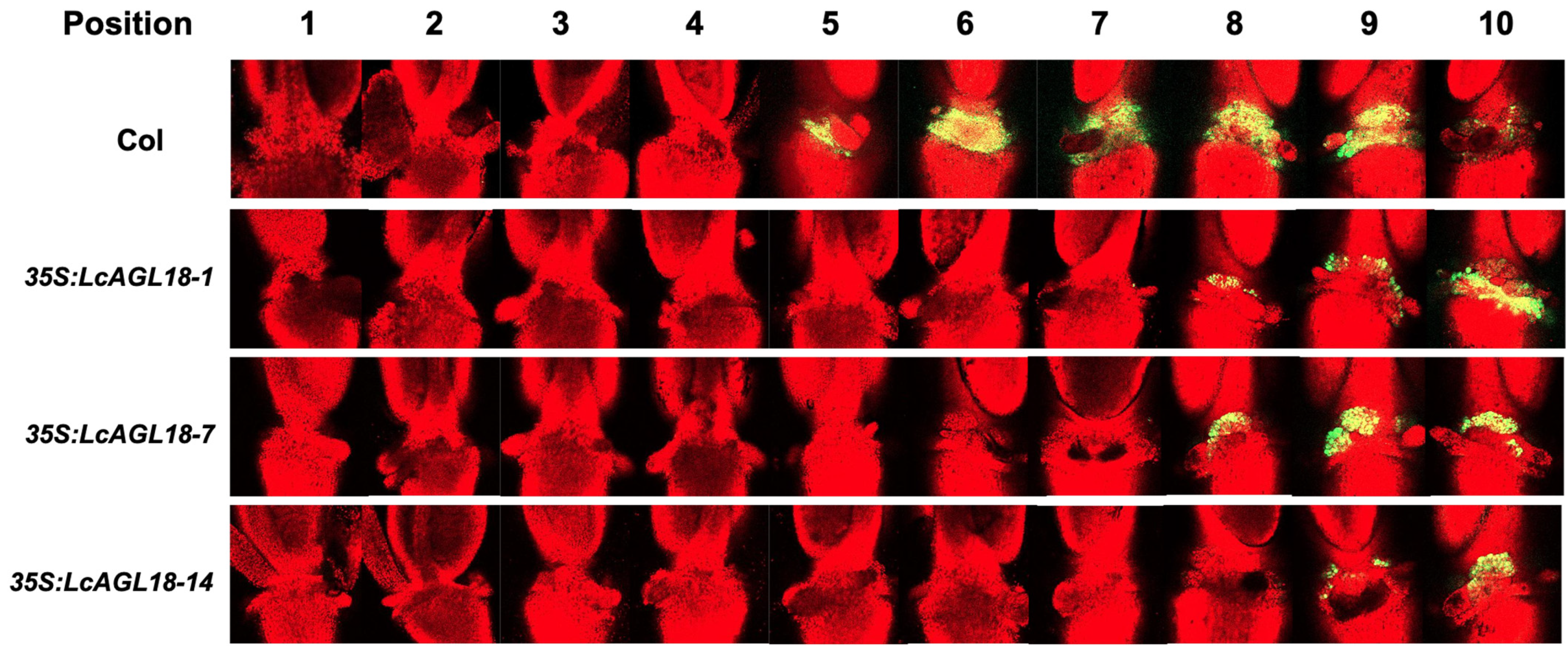
Using BCECF-AM staining, researchers have found that the pH value in the cytoplasm of AZ cells is positively correlated with the process of floral organ abscission in Arabidopsis [11]. The BCECF signals in the floral organ AZ became visible in wild type at position 5. (Figure 4). BCECF signals, on the other hand, were first detected at position 8 in the 35S:LcAGL18-1 and 35S:LcAGL18-7 transgenic lines and at position 9 in the 35S:LcAGL18-14 transgenic line. Moreover, from position 8 to position 10, the BCECF signals only appeared at the petal and stamen AZ but not at the sepal AZ (Figure 4).

3.4. LcAGL18 Inhibits Abscission-Related Gene Expression in Arabidopsis

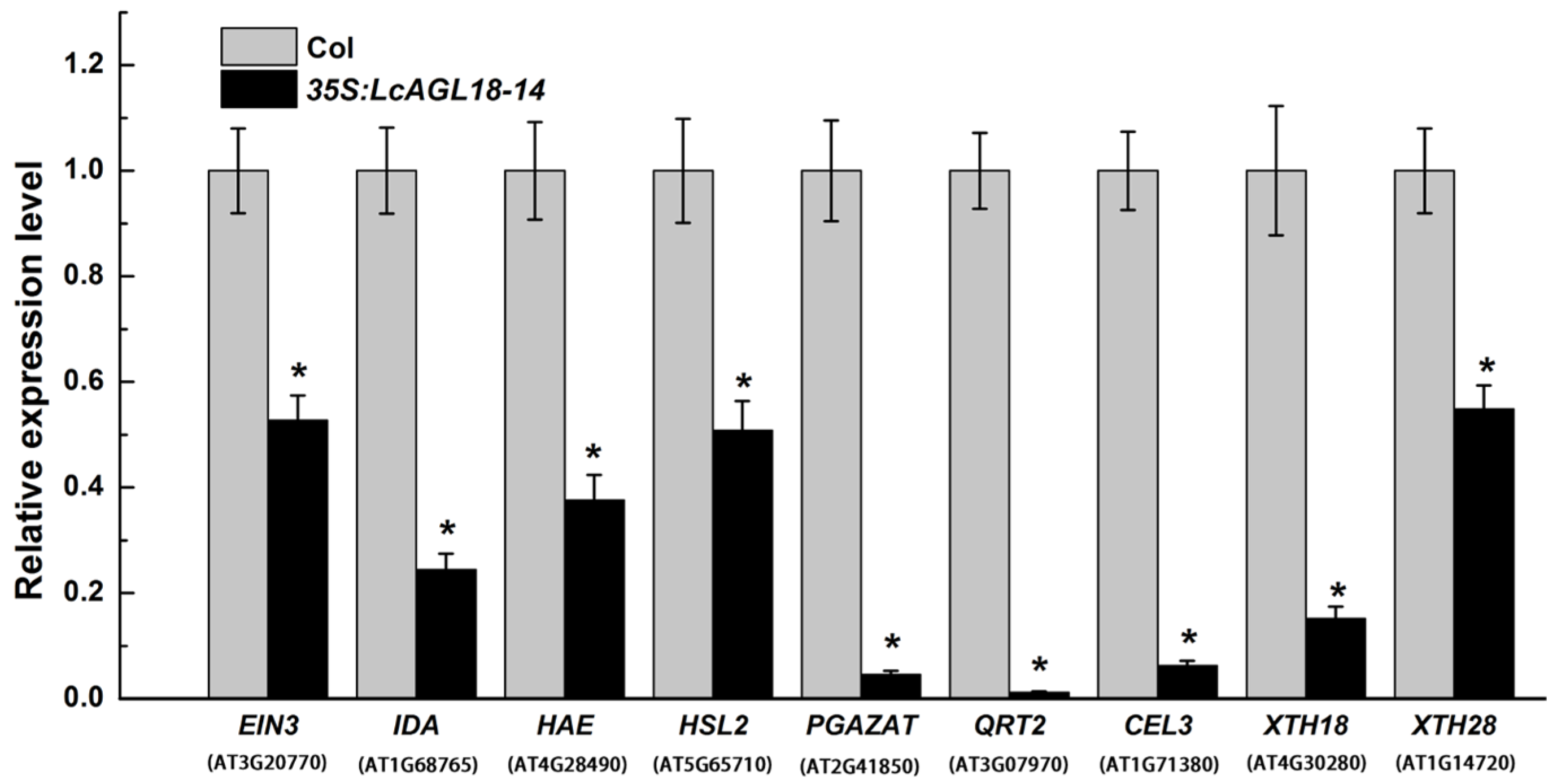
The expression of abscission-related genes at floral AZ was analyzed by qPCR to better understand the abscission defect in LcAGL18 transgenic plants. Areas of the AZ were collected for qPCR analysis at positions 3–8, when the cell walls begin to loosen and the organs begin to separate from one another. ETHYLENE INSENSITIVE 3 (EIN3) is a core TF in ethylene signaling that regulates the timing of floral organ abscission in Arabidopsis [35]. IDA, HAE, and HSL2 are key factors that regulate downstream cell wall hydrolytic enzymes or loosening proteins, including QRT2 (Quartet 2), PGAZAT (Polygalacturonase abscission zone a thaliana), CEL3 (Celluase 3), ADPG2 (Arabidopsis dehiscence zone polygalacturonase 2), XTH18 and XTH28 (Xyloglucan endotransglucosylase/hydrolase 18 and 28), to degrade the middle lamella of AZ cells [42,43,44]. According to our findings, the expression levels of these genes were substantially reduced in 35S:LcAGL18-14 transgenic AZ than in control Col (Figure 5).

3.5. LcAGL18 Is a Transcriptional Repressor

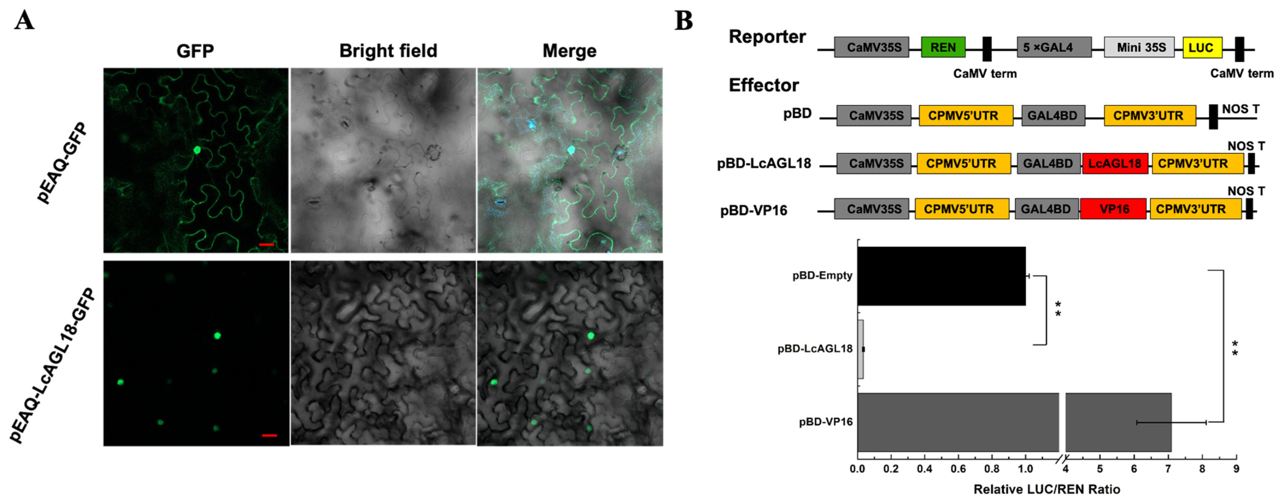
The subcellular localization of LcAGL18 in N. benthamiana leaf was conducted to determine the molecular characterization of LcAGL18. Figure 6A shows that LcAGL18-GFP signals were restricted to the nucleus, whereas GFP signals from the pEAQ-GFP control were evenly distributed throughout the cells. Moreover, we explored the transcriptional activity of LcAGL18 in N. benthamiana leaf epidermal cells using a dual-luciferase reporter system. Positive and negative control effectors, pBD-VP16 and pBD-empty, respectively, were employed in this study (Figure 6B). We found that pBD-VP16 remarkably improved about seven-fold LUC reporter expression beyond that of pBD alone, while pBD-LcAGL18 substantially repressed more than nine-fold LUC reporter expression than that of the pBD empty vector (Figure 6B).

4. Discussion

An increase in crop yield and quality could be achieved through a better understanding of the mechanisms underlying abscission. A variety of gene families, including the MADS-box TFs, are involved in abscission activation [45]. Herein, LcAGL15 and LcAGL18, two putative MADS-box genes, were found in the litchi genome, and their functions in Arabidopsis regarding abscission regulation were examined.
Regulation of organ abscission by MADS-box TFs has been reported in several studies. In Arabidopsis, overexpression of the MADS-box TFs FYF1/2 resulted in a significant delay of floral organ abscission [46]. Similarly, ectopic expression of FYF orthologs in Arabidopsis, including OnFYF, CaFYF1/2, and PaFYF1/2, also delayed the floral organ abscission [25,26,27,47]. FYF orthologs were found to function similarly as repressors in regulating floral organ abscission by repressing ethylene response genes such as ethylene response DNA-binding factors (EDFs) and abscission-associated genes such as BLAD ON PETIOLE (BOP1/2) and IDA [48]. We also found that LcAGL18 acted as a repressor in the regulation of floral organ abscission in Arabidopsis (Figure 3), providing further evidence that MADS-box TFs are involved in abscission regulation. Consistent with the delayed floral organ abscission observed in LcAGL18 transgenic plants, the expression of genes that are required for abscission, such as EIN3, IDA, HAE, HSL2, and genes encoding enzymes for cell wall degradation, was decreased at the floral organ AZ (Figure 5). However, we hypothesized that how LcAGL18 regulates litchi fruitlet abscission might be different with FYF homologs since LcAGL18 and FYF orthologs showed different expression patterns before abscission. For FYF orthologs, such as AtFYF, CaFYF1/2, and PaFYF1/2, which were highly expressed at the young floral buds and mature flowers but were significantly decreased before abscission [26,27,48]. By contrast, the expression of LcAGL18 was greatly enhanced during the litchi fruitlet abscission (Figure 2C). Thus, the mechanism underlying how LcAGL18 regulates litchi fruitlet abscission still requires further study.
AGL15-mediated negative feedback loop regulates floral organ abscission in Arabidopsis. It was found that AGL15 was phosphorylated prior to abscission in a MAPK-dependent manner, and unphosphorylated AGL15 can suppress HAE expression, thereby providing negative feedback in controlling HAE expression [24]. In our study, although ectopically expressing LcAGL15 in Arabidopsis did not alter the floral organ abscission process (Figure S1), LcAGL18, a close homolog of AGL15, exhibited a role in delaying the floral organ abscission, in particular the sepal shedding (Figure 3). According to the findings of this investigation, we propose that, unlike FYF orthologs, LcAGL18 might share similar regulatory mechanisms with Arabidopsis AGL15 in control of abscission. First, LcAGL18 displayed similar expression patterns in the AZ with AtAGL15 prior to abscission. Arabidopsis AGL15 protein peaks just before the floral organ abscission [24], and LcAGL18 was also induced during the litchi fruitlet abscission (Figure 2C). Second, the obvious delayed floral organ abscission was observed only in transgenic plants with strong overexpression of AtAGL15 or LcAGL18 [24] (Figure 3). Of note, the gene expression of LcAGL18 or the protein expression of AtAGL15 in the AZ was positively correlated with the abscission process; however, overexpression of AtAGL15 or LcAGL18 in Arabidopsis retarded floral organ abscission and suppressed the expression of abscission-related genes. These conflicting results suggest complicated mechanisms underlying AtAGL15 or LcAGL18 in regulating abscission. Regarding this phenomenon, it might be possible that in Arabidopsis, the activity of the MAPK pathway might be sufficient to phosphorylate the protein amount of AGL15 that accumulates during the floral organ abscission. However, when LcAGL18 or AtAGL15 was ectopically expressed, especially when the expression level was very high, excess proteins of LcAGL18 or AtAGL15 might not be fully phosphorylated due to the limited activity of the MAPK pathway, leading to a portion of LcAGL18 or AtAGL15 proteins remaining unphosphorylated, which in turn represses abscission-promoting genes such as HAE to delay the abscission. Furthermore, our study provided evidence to prove that LcAGL18 has transcriptional repression activity and found that two putative MADS-box binding sites (CW6G) are present in the promoters of LcHSL2 (Figure 6 and File S1), one close homolog of HAE that has been reported to act as a positive regulator in litchi fruitlet abscission [49]. Additionally, LcAGL18 has one putative MAPK phosphorylation site (S198) (File S1). Thus, it might be possible that a LcAGL18 phosphorylation-mediated feedback loop is conserved in litchi to fine-tune fruitlet abscission.

Supplementary Materials

The following supporting information can be downloaded at: https://www.mdpi.com/article/10.3390/horticulturae9050578/s1, Figure S1: Ectopic expression of LcAGL15 in Arabidopsis has no effect on floral organ abscission; Figure S2: Low expression of LcAGL18 in Arabidopsis has no effect on floral organ abscission; Table S1: List of primers used in the assays; File S1: The promoter sequence of LcHSL2 (LITCHI007137) and the protein sequence of LcAGL18.

Author Contributions

M.Z. and J.L., conceived and designed the experiments; F.W. and Z.L., performed most of the experiments; Z.H. and X.M., provided assistance; M.Z. and J.L., wrote the paper. All authors have read and agreed to the published version of the manuscript.

Funding

This work was supported by grants from the Natural Science Foundation of Guangdong Province, China (2021B1515120082, 2021A1515010462), the National Natural Science Foundation of China (32072544 and 32072514), and the Laboratory of Lingnan Modern Agriculture Project (NZ NT2021004).

Data Availability Statement

All relevant data are included in the article and its Supporting Materials.

Conflicts of Interest

The authors declare no conflict of interest.

References

  1. Estornell, L.H.; Agusti, J.; Merelo, P.; Talon, M.; Tadeo, F.R. Elucidating mechanisms underlying organ abscission. Plant Sci. 2013, 199, 48–60. [Google Scholar] [CrossRef] [PubMed]
  2. Patterson, S.E. Cutting loose. Abscission and dehiscence in Arabidopsis. Plant Physiol. 2001, 126, 494–500. [Google Scholar] [CrossRef]
  3. Jinn, T.L.; Stone, J.M.; Walker, J.C. HAESA, an Arabidopsis leucine-rich repeat receptor kinase, controls floral organ abscission. Genes Dev. 2000, 14, 108–117. [Google Scholar] [CrossRef]
  4. Cho, S.K.; Larue, C.T.; Chevalier, D.; Wang, H.; Jinn, T.L.; Zhang, S.; Walker, J.C. Regulation of floral organ abscission in Arabidopsis thaliana. Proc. Natl. Acad. Sci. USA 2008, 105, 15629–15634. [Google Scholar] [CrossRef] [PubMed]
  5. Stenvik, G.E.; Tandstad, N.M.; Guo, Y.; Shi, C.L.; Kristiansen, W.; Holmgren, A.; Clark, S.E.; Aalen, R.B.; Butenko, M.A. The EPIP peptide of Inflorescence Deficient in Abscission is sufficient to induce abscission in arabidopsis through the receptor-like kinases HAESA and HAESA-LIKE2. Plant Cell 2008, 20, 1805–1817. [Google Scholar] [CrossRef] [PubMed]
  6. Liljegren, S.J.; Leslie, M.E.; Darnielle, L.; Lewis, M.W.; Taylor, S.M.; Luo, R.; Geldner, N.; Chory, J.; Randazzo, P.A.; Yanofsky, M.F.; et al. Regulation of membrane trafficking and organ separation by the NEVERSHED ARF-GAP protein. Development 2009, 136, 1909–1918. [Google Scholar] [CrossRef]
  7. Santiago, J.; Brandt, B.; Wildhagen, M.; Hohmann, U.; Hothorn, L.A.; Butenko, M.A.; Hothorn, M. Mechanistic insight into a peptide hormone signaling complex mediating floral organ abscission. eLife 2016, 5, e15075. [Google Scholar] [CrossRef]
  8. Meng, X.; Zhou, J.; Tang, J.; Li, B.; de Oliveira, M.; Chai, J.; He, P.; Shan, L. Ligand-induced receptor-like kinase complex regulates floral organ abscission in Arabidopsis. Cell Rep. 2016, 14, 1330–1338. [Google Scholar] [CrossRef]
  9. Meir, S.; Philosoph-Hadas, S.; Sundaresan, S.; Selvaraj, K.S.; Burd, S.; Ophir, R.; Kochanek, B.; Reid, M.S.; Jiang, C.Z.; Lers, A. Microarray analysis of the abscission-related transcriptome in the tomato flower abscission zone in response to auxin depletion. Plant Physiol. 2010, 154, 1929–1956. [Google Scholar] [CrossRef]
  10. Ma, C.; Meir, S.; Xiao, L.; Tong, J.; Liu, Q.; Reid, M.S.; Jiang, C.Z. A KNOTTED1-LIKE HOMEOBOX protein regulates abscission in tomato by modulating the auxin pathway. Plant Physiol. 2015, 167, 844–853. [Google Scholar] [CrossRef]
  11. Sundaresan, S.; Philosoph-Hadas, S.; Riov, J.; Belausov, E.; Kochanek, B.; Tucker, M.L.; Meir, S. Abscission of flowers and floral organs is closely associated with alkalization of the cytosol in abscission zone cells. J. Exp. Bot. 2015, 66, 1355–1368. [Google Scholar] [CrossRef] [PubMed]
  12. Cheng, L.; Li, R.; Wang, X.; Ge, S.; Wang, S.; Liu, X.; He, J.; Jiang, C.Z.; Qi, M.; Xu, T.; et al. A SlCLV3-SlWUS module regulates auxin and ethylene homeostasis in low light-induced tomato flower abscission. Plant Cell 2022, 34, 4388–4408. [Google Scholar] [CrossRef] [PubMed]
  13. Reichardt, S.; Piepho, H.P.; Stintzi, A.; Schaller, A. Peptide signaling for drought-induced tomato flower drop. Science 2020, 367, 1482–1485. [Google Scholar] [CrossRef] [PubMed]
  14. Li, R.; Shi, C.L.; Wang, X.; Meng, Y.; Cheng, L.; Jiang, C.Z.; Qi, M.; Xu, T.; Li, T. Inflorescence abscission protein SlIDL6 promotes low light intensity-induced tomato flower abscission. Plant Physiol. 2021, 186, 1288–1301. [Google Scholar] [CrossRef]
  15. Nakano, T.; Fujisawa, M.; Shima, Y.; Ito, Y. The AP2/ERF transcription factor SlERF52 functions in flower pedicel abscission in tomato. J. Exp. Bot. 2014, 65, 3111–3119. [Google Scholar] [CrossRef]
  16. Liu, X.; Cheng, L.; Li, R.; Cai, Y.; Wang, X.; Fu, X.; Dong, X.; Qi, M.; Jiang, C.Z.; Xu, T.; et al. The HD-Zip transcription factor SlHB15A regulates abscission by modulating jasmonoyl-isoleucine biosynthesis. Plant Physiol. 2022, 189, 2396–2412. [Google Scholar] [CrossRef]
  17. Xie, Q.; Hu, Z.; Zhu, Z.; Dong, T.; Zhao, Z.; Cui, B.; Chen, G. Overexpression of a novel MADS-box gene SlFYFL delays senescence, fruit ripening and abscission in tomato. Sci. Rep. 2014, 4, 4367. [Google Scholar] [CrossRef]
  18. Mao, L.; Begum, D.; Chuang, H.W.; Budiman, M.A.; Szymkowiak, E.J.; Irish, E.E.; Wing, R.A. JOINTLESS is a MADS-box gene controlling tomato flower abscission zone development. Nature 2000, 406, 910–913. [Google Scholar] [CrossRef]
  19. Liu, D.; Wang, D.; Qin, Z.; Zhang, D.; Yin, L.; Wu, L.; Colasanti, J.; Li, A.; Mao, L. The SEPALLATA MADS-box protein SLMBP21 forms protein complexes with JOINTLESS and MACROCALYX as a transcription activator for development of the tomato flower abscission zone. Plant J. 2014, 77, 284–296. [Google Scholar] [CrossRef]
  20. Adamczyk, B.J.; Lehti-Shiu, M.D.; Fernandez, D.E. The MADS domain factors AGL15 and AGL18 act redundantly as repressors of the floral transition in Arabidopsis. Plant J. 2007, 50, 1007–1019. [Google Scholar] [CrossRef]
  21. Verelst, W.; Twell, D.; de Folter, S.; Immink, R.; Saedler, H.; Munster, T. MADS-complexes regulate transcriptome dynamics during pollen maturation. Genome Biol. 2007, 8, R249. [Google Scholar] [CrossRef] [PubMed]
  22. Paul, P.; Joshi, S.; Tian, R.; Diogo, J.R.; Chakrabarti, M.; Perry, S.E. The MADS-domain factor AGAMOUS-Like18 promotes somatic embryogenesis. Plant Physiol. 2022, 188, 1617–1631. [Google Scholar] [CrossRef] [PubMed]
  23. Zheng, Q.; Perry, S.E. Alterations in the transcriptome of soybean in response to enhanced somatic embryogenesis promoted by orthologs of Agamous-like15 and Agamous-like18. Plant Physiol. 2014, 164, 1365–1377. [Google Scholar] [CrossRef] [PubMed]
  24. Patharkar, O.R.; Walker, J.C. Floral organ abscission is regulated by a positive feedback loop. Proc. Natl. Acad. Sci. USA 2015, 112, 2906–2911. [Google Scholar] [CrossRef] [PubMed]
  25. Chen, M.K.; Lee, P.F.; Yang, C.H. Delay of flower senescence and abscission in Arabidopsis transformed with a Forever Young Flower homolog from Oncidium orchid. Plant Signal. Behav. 2011, 6, 1841–1843. [Google Scholar] [CrossRef] [PubMed]
  26. Chen, W.H.; Lee, Y.I.; Yang, C.H. Ectopic expression of two FOREVER YOUNG FLOWER orthologues from Cattleya Orchid suppresses ethylene signaling and DELLA results in delayed flower senescence/abscission and reduced flower organ elongation in Arabidopsis. Plant Mol. Biol. Rep. 2018, 36, 710–724. [Google Scholar] [CrossRef]
  27. Chen, W.H.; Jiang, Z.Y.; Hsu, H.F.; Yang, C.H. Silencing of FOREVER YOUNG FLOWER-Like Genes from Phalaenopsis Orchids Promotes Flower Senescence and Abscission. Plant Cell Physiol. 2021, 62, 111–124. [Google Scholar] [CrossRef]
  28. Hu, G.; Feng, J.; Xiang, X.; Wang, J.; Salojärvi, J.; Liu, C.; Wu, Z.; Zhang, J.; Liang, X.; Jiang, Z.; et al. Two divergent haplotypes from a highly heterozygous lychee genome suggest independent domestication events for early and late-maturing cultivars. Nat. Genet. 2022, 54, 73–83. [Google Scholar] [CrossRef]
  29. Zhao, M.; Li, J. Molecular Events Involved in Fruitlet Abscission in Litchi. Plants 2020, 9, 151. [Google Scholar] [CrossRef]
  30. Stern, R.A.; Kigel, J.; Tomer, E.; Gazit, S. ‘Mauritius’ lychee fruit development and reduced abscission after treatment with the auxin 2,4,5-TP. Am. Soc. Hortic. Sci. 1995, 120, 65–70. [Google Scholar] [CrossRef]
  31. Mitra, S.K.; Pereira, L.S.; Pathak, P.K.; Majumdar, D. Fruit abscission pattern of lychee cultivars. In II International Symposium on Lychee, Longan, Rambutan and Other Sapindaceae Plants; ISHS Acta Horticulturae: Chiang Mai, Thailand, 2005; Volume 665, pp. 215–218. [Google Scholar]
  32. Li, C.Q.; Wang, Y.; Ying, P.Y.; Ma, W.Q.; Li, J.G. An improved fruit transcriptome and the identification of the candidate genes involved in fruit abscission induced by carbohydrate stress in litchi. Front. Plant Sci. 2015, 6, 439. [Google Scholar] [CrossRef] [PubMed]
  33. Zhao, M.L.; Li, C.Q.; Ma, X.S.; Xia, R.; Chen, J.Y.; Liu, X.C.; Ying, P.Y.; Peng, M.J.; Wang, J.; Shi, C.L.; et al. KNOX protein KNAT1 regulates fruitlet abscission in litchi by repressing ethylene biosynthetic genes. J. Exp. Bot. 2020, 71, 4069–4082. [Google Scholar] [CrossRef] [PubMed]
  34. Ma, X.S.; Yuan, Y.; Wu, Q.; He, Z.D.; Li, J.G.; Zhao, M.L. Brassinosteroids suppress ethylene-induced fruitlet abscission through LcBZR1/2-mediated transcriptional repression of LcACS1/4 and LcACO2/3 in litchi. Hortic. Res. 2021, 8, 105. [Google Scholar] [CrossRef] [PubMed]
  35. Ma, X.; Yuan, Y.; Wu, Q.; Wang, J.; Li, J.; Zhao, M. LcEIL2/3 are involved in fruitlet abscission via activating genes related to ethylene biosynthesis and cell wall remodeling in litchi. Plant J. 2020, 103, 1338–1350. [Google Scholar] [CrossRef]
  36. Kuang, J.F.; Wu, J.Y.; Zhong, H.Y.; Li, C.Q.; Chen, J.Y.; Lu, W.J.; Li, J.G. Carbohydrate stress affecting fruitlet abscission and expression of genes related to auxin signal transduction pathway in litchi. Int. J. Mol. Sci. 2012, 13, 16084–16103. [Google Scholar] [CrossRef]
  37. Clough, S.J.; Bent, A.F. Floral dip: A simplified method for Agrobacterium-mediated transformation of Arabidopsis thaliana. Plant J. 2010, 16, 735–743. [Google Scholar] [CrossRef]
  38. Zhong, H.Y.; Chen, J.W.; Li, C.Q.; Chen, L.; Wu, J.Y.; Chen, J.Y.; Lu, W.J.; Li, J.G. Selection of reliable reference genes for expression studies by reverse transcription quantitative real-time PCR in litchi under different experimental conditions. Plant Cell Rep. 2011, 30, 641–653. [Google Scholar] [CrossRef]
  39. Ying, P.; Li, C.; Liu, X.; Xia, R.; Zhao, M.; Li, J. Identification and molecular characterization of an IDA-like gene from litchi, LcIDL1, whose ectopic expression promotes floral organ abscission in Arabidopsis. Sci. Rep. 2016, 6, 37135. [Google Scholar] [CrossRef]
  40. Fan, Z.Q.; Tan, X.L.; Chen, J.W.; Liu, Z.L.; Kuang, J.F.; Lu, W.J.; Shan, W.; Chen, J.Y. Characterization of a Transcriptional Regulator BrWRKY6 that Associates with Gibberellin-Suppressed Leaf Senescence of Chinese Flowering Cabbage. J. Agric. Food Chem. 2018, 66, 7b06085. [Google Scholar] [CrossRef]
  41. Immink, R.G.; Gadella, T.J.; Ferrario, S.; Busscher, M.; Angenent, G.C. Analysis of MADS box protein-protein interactions in living plant cells. Proc. Natl. Acad. Sci. USA 2002, 99, 2416–2421. [Google Scholar] [CrossRef]
  42. Ogawa, M.; Kay, P.; Wilson, S.; Swain, S.M. Arabidopsis Dehiscence Zone Polygalacturonase1 (ADPG1), ADPG2, and QUARTET2 are Polygalacturonases required for cell separation during reproductive development in Arabidopsis. Plant Cell 2009, 21, 216–233. [Google Scholar] [CrossRef] [PubMed]
  43. Lee, Y.; Yoon, T.H.; Lee, J.; Jeon, S.Y.; Lee, J.H.; Lee, M.K.; Chen, H.; Yun, J.; Oh, S.Y.; Wen, X.; et al. A Lignin Molecular Brace Controls Precision Processing of Cell Walls Critical for Surface Integrity in Arabidopsis. Cell 2018, 173, 1468. [Google Scholar] [CrossRef] [PubMed]
  44. Crick, J.; Corrigan, L.; Belcram, K.; Khan, M.; Dawson, J.W.; Adroher, B.; Li, S.; Hepworth, S.R.; Pautot, V. Floral organ abscission in Arabidopsis requires the combined activities of three TALE homeodomain transcription factors. J. Exp. Bot. 2022, 73, 6150–6169. [Google Scholar] [CrossRef] [PubMed]
  45. Ma, C.; Jiang, C.Z.; Gao, J. Regulatory Mechanisms Underlying Activation of Organ Abscission. Annu. Plant Rev. 2021, 4, 27–55. [Google Scholar]
  46. Chen, W.H.; Li, P.F.; Chen, M.K.; Lee, Y.I.; Yang, C.H. Forever Young Flower Negatively Regulates Ethylene Response DNA-Binding Factors by Activating an Ethylene-Responsive Factor to Control Arabidopsis Floral Organ Senescence and Abscission. Plant Physiol. 2015, 168, 1666–1683. [Google Scholar] [CrossRef]
  47. Chen, W.H.; Lin, P.T.; Hsu, W.H.; Hsu, H.F.; Li, Y.C.; Tsao, C.W.; Hsu, M.C.; Mao, W.T.; Yang, C.H. Regulatory network for Forever Young Flower-like genes in regulating Arabidopsis flower senescence and abscission. Commun. Biol. 2022, 5, 662. [Google Scholar] [CrossRef]
  48. Chen, M.K.; Hsu, W.H.; Lee, P.F.; Thiruvengadam, M.; Chen, H.I.; Yang, C.H. The MADS box gene, FOREVER YOUNG FLOWER, acts as a repressor controlling floral organ senescence and abscission in Arabidopsis. Plant J. 2011, 68, 168–185. [Google Scholar] [CrossRef]
  49. Wang, F.; Zheng, Z.; Yuan, Y.; Li, J.; Zhao, M. Identification and Characterization of HAESA-Like Genes Involved in the Fruitlet Abscission in Litchi. Int. J. Mol. Sci. 2019, 20, 5945. [Google Scholar] [CrossRef]
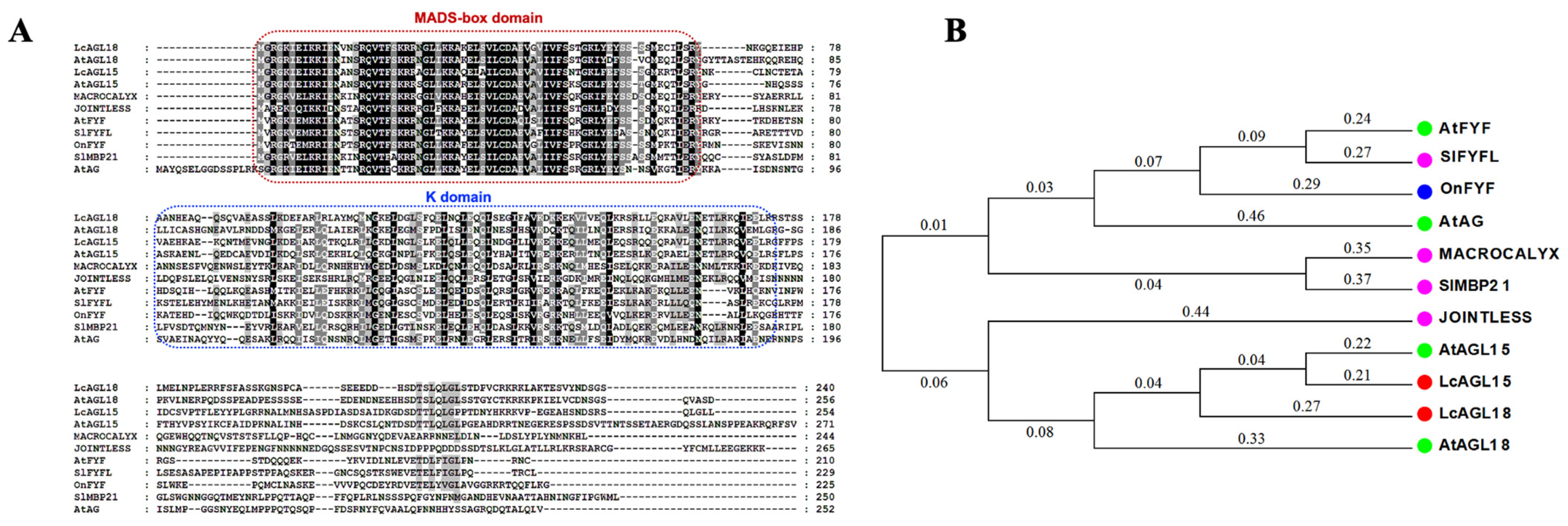
Figure 1. A phylogenetic and sequence analysis of LcAGL15/18. (A) Multiple alignments of LcAGL15/18 with other MADS-box proteins implicated in organ abscission have been reported. The MADS-box domain and K domain, which are the characteristics of MADS-box protein, are indicated by the red and blue box, respectively. (B) Phylogenetic relationships of LcAGL15/18 with other MADS-box proteins, including homologs from Arabidopsis (AtFYF, AtAG, and AtAGL15/18), Solanum lycopersicum (SlFYFL, SlMBP21, MACROCALYX, and JOINTLESS), and Oncidium orchid (OnFYF).
Horticulturae 09 00578 g001
Figure 2. LcAGL15/18 expression profile at the AZ during the litchi fruitlet abscission. (A) Ethephon (ET) and GPD trigger litchi fruitlet abscission. (B) Litchi fruitlet abscission induced by GPD/ET resulted in repression of LcAGL15 at the AZ. (C) During fruitlet abscission induced by GPD/ET in litchi, LcAGL18 was upregulated at the AZ. The results denote the averages of three independent biological samples, and the bars represent standard deviations. Different letters indicate the statistical significance (p < 0.05) determined using one-way analysis of variance followed by post hoc Tukey’s honest significant difference test.
Horticulturae 09 00578 g002
Figure 3. LcAGL18 overexpression delays floral organ abscission in Arabidopsis. (A) Expression analysis of LcAGL18 in transgenic lines. (B,C) The transgenic lines with strong expression of LcAGL18 display delayed floral organ abscission. Red arrows indicate the attached floral organs. (D) The GUS signals driven by the LcAGL18 promoter are detected at the Arabidopsis floral organ AZ. The numbers represent the position of the flowers along the inflorescence. The first flower with visible white petals at the apex of the florets is used to determine position numbers. (E) Scanning electron microscopy (SEM) of wild-type and LcAGL18 transgenic lines of floral organ AZ fracture planes. Organs abscise naturally at position 12 in wild-type Col and are manually removed at position 12 in 35S:LcAGL18-1/-7/-14 transgenic lines. The floral organ AZ in wild type is fully rounded, whereas the entire cells of the sepal AZ display a flattened cavity. Red arrowheads designate sepal AZs.
Horticulturae 09 00578 g003
Figure 4. BCECF signals from the floral organ AZ of LcAGL18 transgenic lines and wild-type Col. Chlorophyll autofluorescence and BCECF fluorescence were combined under a confocal laser scanning microscope (CLSM) to obtain the fluorescence images. The presence of green fluorescence indicates an increase in pH. The photographs shown for each position and plant are representative of 3–4 replicates.
Horticulturae 09 00578 g004
Figure 5. The floral organ AZ of LcAGL18 transgenetic plants shows a reduced expression level of abscission-related genes. For testing, AZ tissues from positions 3–8 were taken. The data represent the mean ± SE for three biological replicates. * denote statistically significant differences (p < 0.05) in the Student’s t-test.
Horticulturae 09 00578 g005
Figure 6. LcAGL18 possesses transcriptional repression activity. (A) Subcellular localization of LcAGL18 in N. benthamiana leaves. After 48 h of incubation, GFP signals were observed using a CLSM. Scale bars show 25 μm. (B) Transcriptional activity of LcAGL18 in tobacco leaves. The reporter and effector vectors are depicted diagrammatically in the top panel. The dual-luciferase reporter assay was used to determine the transcriptional activity of LcAGL18 by measuring the ratio of LUC to REN. pBD-VP16 was used as a transcriptional activator. The LUC/REN ratio from leaves expressing the empty pBD vector (negative control) was used to calibrate fluorescence (set to 1). Each value denotes the mean ±SD of six biological replicates, and an ** shows a significant difference (Student’s t-test: p < 0.01).
Horticulturae 09 00578 g006
Disclaimer/Publisher’s Note: The statements, opinions and data contained in all publications are solely those of the individual author(s) and contributor(s) and not of MDPI and/or the editor(s). MDPI and/or the editor(s) disclaim responsibility for any injury to people or property resulting from any ideas, methods, instructions or products referred to in the content.