Abstract
Common hyssop (Hyssopus officinalis L.) is widely used as an aromatic and medicinal plant. In the component composition of the essential oil from the above-ground mass of H. officinalis f. cyaneus, from the collection of the Nikita Botanical Garden, the bicyclic monoterpene ketones isopinocamphone (50.99–64.41%) and pinocamphone (3.95–18.88%) predominate, which allows us to attribute this form to the isopinocamphone chemotype for use in pharmacology. An essential oil sample with a high content of isopinocamphone (70.74%) in complete absence of pinocamphone was determined, which made it possible to use the plant as a starting material for breeding. The objective of our work was to study the component composition of the essential oil of this form, and the morphology, anatomy and ploidy level of microshoots in vitro on a nutrient medium with BAP. This was compared with ex situ samples to develop a cultivation technique with the preservation of a valuable trait for use in task-oriented selection. Biotechnological methods are used for future mass propagation, study and the preservation of breeding forms. Morphological and anatomical features and ploidy of H. officinalis microshoots were studied in vitro. Using in vitro culturing of microshoots on MS medium containing BAP, a decrease in the cuticular layer and the degree of development of collenchyma near vascular bundles in leaves were shown compared to microshoots ex situ. Significant structural changes were found with a high increase in BAP concentration, while no changes in the ploidy level were detected.
1. Introduction
Hyssop (Hyssopus L.) is a genus of perennial grasses or semi-shrubs containing, according to the Plant List, seven species and three subspecies that are naturally distributed in Southern Europe, Central Asia and North Africa. Common hyssop (Hyssopus officinalis L.) is widely used as an aromatic and medicinal plant in landscape design; today, it is a promising species for the southern regions. It is mainly used for the production of essential oils. Its essential oil is applied in the pharmaceutical (as an antioxidant, expectorant, antiseptic, antibacterial and antifungal agent) and perfume industries, as well as in aromatherapy, where this essential oil with the main components of trans- and cis-pinocamphone has a muscle relaxant effect [1,2]. The species of H. officinalis has three plant forms with white, blue and pink flowers. In the literature, there is information about the morphology of seeds and their germination [3], the micromorphology of vegetative and generative organs [4], the content of essential oil [5] and its component composition [6] and farming standards [7]. In addition, new economically valuable forms and cultivars of this species are being bred [7,8,9]. Due to economic use, hyssop essential oil’s chemical characteristics are of interest [10]. The muscle relaxant effect of Hyssopus officinalis L. (Lamiaceae) essential oil was studied, as well as its main components (isopinocamphone, limonene and β-pinene). It was shown that isopinocamphone, an isomer of pinocamphone, also had muscle-relaxing properties, while limonene and β-pinene had no effect [11]. Earlier, during research in the laboratory of aromatic and medicinal plants of the NBG-NSC, a form (f. cyaneus) with a fairly high biosynthesis of isopinocamphone (up to 61.12%) was isolated among the studied hyssop plants, which allowed the plant to be used as a starting material for breeding [6]. Using generally accepted methods (individual positive selection, hybridization) and methodological developments of the laboratory of aromatic and medicinal plants of the NBG [12], a promising accession of Hyssopus officinalis f. cyaneus with an increased content (up to 70%) of isopinocamphone was selected for use for medicinal purposes. It should be noted that the traditional method of reproduction of hyssop has some limitations for breeding forms, given the possibility of trait loss. Therefore, the valuable material of H. officinalis in most cases increases due to cutting [7]. Currently, biotechnological methods are of great importance for mass reproduction, study and preservation of breeding forms. In the biotechnological aspect, data on in vitro regeneration from axillary buds [13], apical parts of shoots [14], segments of shoots with a node [15], rooting and adaptation [14] are known for H. officinalis. The researchers note that the optimization of the in vitro cultivation process and the detailed study of plants of this species remain an urgent task. The use of morphology and anatomy methods at an early stage makes it possible to optimize the cultivation protocol and eventually obtain plants with high survival in an ex vitro environment. Currently, the emphasis is also on the genetic stability of plant material in vitro [15,16,17], due to the possibility of a number of chromosome variations, their structure, as well as individual DNA sequences [16]. For H. officinalis, there are single published data describing the histoanatomy of shoots obtained in vitro [13]. In the NBG-NSC, an investigation of the structural and genetic stability of three forms of H. officinalis has been started. For H. officinalis cv. Nikitskiy Beliy (f. albus), genetic similarity analysis between donor plants and microshoots obtained in vitro by RAPD- and ISSR-PCR methods was performed [15]. The objective of our work was to study the component composition of the essential oil of the isolated form of Hyssopus officinalis f. cyaneus, and the morphology, anatomy and ploidy level of microshoots in vitro on a nutrient medium with BAP, in comparison with ex situ samples, to develop a cultivation technique with the preservation of a valuable trait and for use in task-oriented selection.
2. Materials and Methods
The object of the study was the seed population of Hyssopus officinalis f. cyaneus Alef. It grows at the collection plots of the Nikita Botanical Gardens, located at an altitude of 200 m above sea level in a subtropical climate of the Mediterranean type.
The mass fraction of essential oil was determined in fresh raw materials by hydrodistillation on Ginsberg apparatuses [18]. The component composition of essential oils was determined using a hardware and software complex based on a chromatograph Chromatek-Crystal 5000.2 (Chromatek, Russia) equipped with a mass spectrometric detector: Capillary column CR—5 ms, length 30 m, inner diameter 0.25 mm; Phase 5% phenyl 95% polysilphenylenesiloxane, film thickness 0.25 microns. The temperature of the thermostat was programmed from 75 °C to 240 °C at a speed of 4 °C/min. The evaporator temperature was 250 °C. The carrier gas was helium; the flow rate was 1 mL/min. The temperature of the transition line was 250 °C. The temperature of the ion source was 200 °C. The electronic ionization was 70 eV. The scanning range was 20–450. The scan duration was 0.2. Identification was performed based on a comparison of the obtained mass spectra with data from the NIST 14 library (National Institute of Standards and Technology, Gaithersburg, MD, USA) using NIST MSSearch software—v. 2.2 (Gaithersburg, MD, USA). Retention indices were obtained by logarithmic interpolation of the reduced retention times using the analytical standard of a mixture of reference n-alkanes (Sigma-Aldrich, Switzerland) and analytical standards (Supelco, Bellefonte, PA, USA). The mass fraction of the components in the sample was determined by the percentage normalization method [19,20].
The biochemical studies of essential oil were carried out on the equipment of the Common Use Center “Physiological and biochemical studies of plant objects” of the Federal State Funded Institution of Science “NBG-NSC” (Yalta, Russia).
Single-node segments of shoots were introduced into culture in vitro. Sterilization of the material was carried out according to the method described by Bulavin et al. [15]. In vitro cultivation was carried out on a modified Murashige–Skoog culture medium (MS), with subsequent sub-cultivations on the same medium with 0.3–0.9 mg/L of BAP. The material in culture vessels was kept in the phytochambers of the Unique Scientific Installation “PHYTOBIOGEN” of the NBG-NSC or growth chamber MLR-352-PE (Panasonic, Oizumi, Japan) at a temperature of 24 ± 1 °C, 16 h photoperiod and 37.5 µm m–2 s–1 light intensity under basic cool daylight lamps (Ledvance, Smolensk, Russia).
For anatomical investigation, the leaves of the shoots ex situ and microshoots in vitro were cut out and immediately fixed in a solution of formalin, alcohol, acetic acid and water (1:5:0.5:3.5), dehydrated in alcohols and embedded into polyester wax (Electron Microscopy Science, USA). Sections (10–15 microns) were made on a microtome MZ-2 (Tochmedpribor, Ukraine), stained with a solution of methylene blue, mounted into sucrose solution and viewed using a light microscope CX-41 (Olympus, Japan) with a digital camera SC 50 (Olympus, Germany) and CellSens image processing software version 1.17. A total of 25 cells were analyzed for each parameter.
The study of the ploidy level was carried out on fresh material. Leaf blades (about 0.5 cm2) of six ex situ shoots and microshoots in vitro were immersed in a modified WPB buffer [21] with 2% polyvinylpyrrolidone (K10) supplemented with propidium iodide (50 µg/mL), RNase (50 µg/mL) and β-mercaptoethanol (0.3%), and ground up with a safety razor. The obtained samples were passed through a 30 mµ CellTrics® filter (Partec, Germany). The analysis was performed using CyFlow® Ploidy Analyzer (Sysmex, Partec, Germany). Ficus benjamina L. plants were used as an external control to determine the DNA content [22]. The measurements were carried out using the same analyzer settings, with at least 10.000 nuclei.
Statistical analysis was carried out using the software Past v. 4.03. Samples were checked for normality of distribution, and either the t-criterion or the U-criterion was used (p ≤ 0.05).
3. Results and Discussion
3.1. The Component Composition of the Essential Oil of the Isolated Form of Hyssopus officinalis f. cyaneus
According to the literature data, the essential oil of the blue-flowered form contains anywhere from 18.0 to 36.37% pinocamphone, and from 40.7 to 57.93% isopinocamphone [6,23,24,25,26]. The study of the component composition of the essential oil of the seed population of the blue-form common hyssop showed that the intraspecific composition of the essential oil from the above-ground mass of raw materials is diverse and consists of the following main components: isopinocamphone and pinocamphone (bicyclic monoterpene ketones), β-pinene, sabinene, β-phellandrene (monoterpene hydrocarbons), linalool, myrtenol (monoterpene alcohols), germacrene D, bicyclogermacrene (sesquiterpene hydrocarbon), elemol (sesquiterpene alcohol), etc. (Table 1, Figure 1).

Table 1.
Component composition of the essential oil of Hyssopus officinalis f. cyaneus Alef.

Figure 1.
Chromatogram of the component composition of the promising accession, 2022.
In total, 68 components were identified in the composition of the oil: isopinocamphone and pinocamphone, β-pinene, sabinene, linalool, β-phellandrene, myrtenol, germacrene D, bicyclogermacrene, etc. (Table 1). The mass fraction of the essential oil of the promising accession was 0.20% of the raw weight and 0.55% of the absolutely dry weight. The main component of isopinocamphone represents up to 70.74% of the compound. The essential oil contained predominantly terpenoids (up to 87.9%), mainly ketones (up to 72.30%), and monoterpene and sesquiterpene hydrocarbon compounds, which accounted for up to 22.92% and 9.15%, respectively. In the published works, there are data showing that hyssop oil from different phenotypes or different areas shows great variability in chemical composition [2]. Our investigation showed that selected H. officinalis f. cyaneus belong to a pinocamphone-rich chemotype, which is consistent with data obtained by other researchers [27].
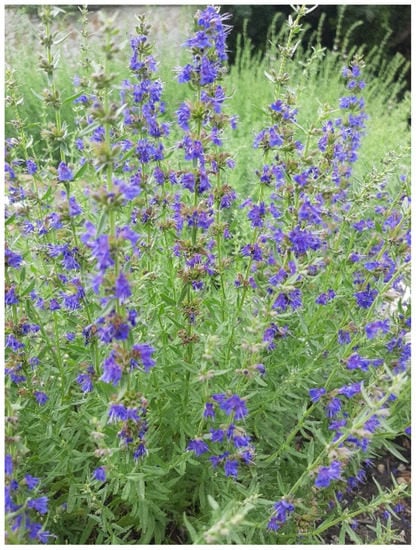
The analysis of essential oil from different accessions for 5 years showed that the content of isopinocamphone was variable (from 50.99 to 70.74) (Table 1). In 2022, an accession was selected (Figure 2); the content of isopinocamphone in its essential oil was 70.74%, with the complete absence of pinocamphone.

Figure 2.
Selected perspective specimen of Hyssopus officinalis f. cyaneus Alef.
The selected promising accession has the following characteristics: a bush with a semi-spreading shape, 60 cm high and 70 cm in diameter. The leaves are dark green. The flowers are blue-purple in color and form a thyrsus-type inflorescence in the upper part of the stem with a length of 11–12 cm. Mass flowering occurs from the third week of July to the first week of August. The duration of flowering is 45–50 days. The above-ground mass collected at the beginning of flowering, and mown at a height of 15–20 cm from the soil surface, is used as the raw material.
The content of isopinocamphone in the selected accession does not meet the requirements of ISO 9841 Standard (25.0 to 45.0%), which limits the use of this form as a food raw material and determines the prospects for use as a medicinal product, since isopinocamphone, like pinocamphone, has muscle-relaxing and antifungal properties.
The main component content of essential oil can be affected by the stage of plant development, plant age, harvesting time of plant material and the part of the plant used. In the process of plant development, the main components of the essential oil are changed: pinocamphone predominates before flowering, and isopinocamphone accumulates during the period of mass flowering [28]. As for the age of plants, in the first year, maximal pinocamphone content is determined, while isopinocamphone predominates in three-year-old plants [29]. Tavakoli and Agadjani [30] found that the main components such as pinocamphone and isopinocamphone varied depending on irrigation degree. The published data also suggest that one of the most important factors affecting the essential oil chemical composition of Hyssopus officinalis is intraspecific diversity as a result of adaptation to habitat conditions and cultivation [24,31].
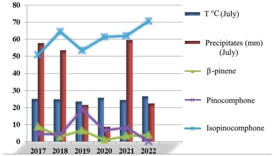
During a period of five years (2017–2022), the average air temperature in May was 16.3 ± 0.6 °C, in June was 22.4 ± 0.7 °C and in July was 24.8 ± 0.4; the precipitates were 26.6 ± 6.3 mm, 60.4 ± 12.7 mm and 37.1 ± 9.1, respectively (according to meteorological observations by the agrometeorological station of the Nikita Botanical Gardens (44°31′ N, 34°15′ E). Based on the evidence that the yield of isopinocamphone increases significantly under dry conditions [24,32], we compared temperatures and precipitates in the flowering period (third week of July) with isopinocamphone, pinocamphone and β-pinene percentage yield. According to the results (Figure 3), there were no robust relationships between investigated parameters. It is supposed that the differences in the main component amounts between accessions are genetically determined. Similar conclusions were made also by Zawislak [28], based on changes in cis-, and tran-pinocamphone portions during vegetative and flowering periods. Interestingly, robust data on essential oil composition genetic determination were obtained by Figueiredo et al. [33] for some aromatic plants from genera such as Mentha, Achillea, Perilla, Salvia, Thymus, etc.

Figure 3.
Relationship between some climate dimensions and main essential oil component amounts.
3.2. Comparative Morphological and Anatomical Characteristics during Ex Situ and In Vitro Cultivation
Microshoots of H. officinalis f. cyaneus with a length of 1.0–1.5 cm, obtained by the induction of shoot development, were transferred to a modified MS nutrient medium with different BAP concentrations. It was found that the process of morphogenesis significantly depended on the concentration of the growth regulator in the nutrient medium. The addition of BAP at the levels of 0.4 and 0.5 mg/L contributed to the formation of numerous microshoots with morphologically normal stems and leaves in the upper part (Figure 4b,c), and with minor structural changes to organs in direct plant material contact with the nutrient medium. An increase in the BAP concentration to 0.8 mg/L provoked hyperhydricity of plant organs in vitro (Figure 4d). Reducing BAP concentration to 0.3 mg/L in the MS medium during subsequent sub-cultivation avoids vitrification of microshoots (Figure 4a).

Figure 4.
Microshoots of H. officinalis f. cyaneus obtained on MS nutrient medium supplemented with 0.1 mg/L IBA and BAP at concentrations of 0.3 mg/L (a), 0.4 mg/L (b), 0.5 mg/L (c) and 0.8 mg/L (d).
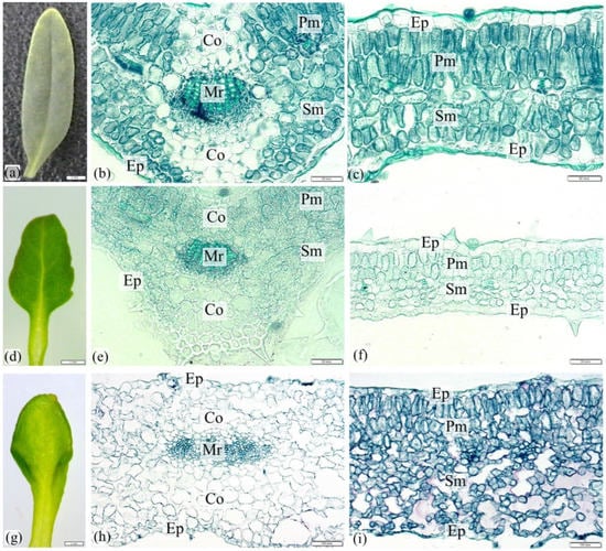
The morphology and anatomy of plant leaves were studied ex situ in comparison with those in vitro. The leaf blades of ex situ plants were green in color, linear-lanceolate in shape, with an obtuse tip, a tapered base with a short petiole (about 1 mm) and entire-edged (Figure 5a). On the upper and lower surface of the epidermis, there were non-glandular and glandular trichomes.

Figure 5.
Morphology and anatomy of H. officinalis f. cyaneus leaf blades ex situ (a–c) and in vitro when cultured on a modified MS nutrient medium supplemented with 0.1 mg/L IBA and BAP at concentrations of 0.4 mg/L (d–f) and 0.8 mg/L (g–i). Co—collenchyma, Ep—epidermis, Mr—midrib, Pm—palisade mesophyll, Sm—spongy mesophyll.
The epidermis with a cuticle, several subepidermal layers of collenchyma in the upper and lower parts, and an oval bundle consisting of xylem and phloem are differentiated on the cross sections of the midrib (Figure 5b). The collenchyma cells were adjacent to the palisade mesophyll (2–3 layers) and spongy mesophyll (3–4 layers), in which lateral veins were found.
When microshoots were cultured on MS nutrient medium with a concentration of 0.3–0.5 mg/L BAP, their morphology was similar. The color of the leaves was green, the shape was ovoid or oblong, the tip was obtuse, the edge was entire or partially crenate (dentate) and the base was tapered with a well-developed petiole (Figure 5d). The presence of varying version degrees on the abaxial side of the organs was typical. On the MS nutrient medium supplemented with 0.8 mg/L BAP, the leaf blades had (1) a spatulate shape with a rounded tip and a tapered base, entire or crenate margin, slightly versed on the adaxial side (Figure 5g); (2) a spatulate shape with bilobular tip and tapered base, slightly versed entire or crenate edge; (3) a heart-shaped form with a bilobular tip and a tapered base, entire edge; (4) an irregular shape with a rounded tip and a tapered base, slightly or significantly versed with an entire edge or sometimes crenate/dentate edge.
Anatomically, the leaves of microshoots obtained in vitro on MS nutrient medium with 0.4 and 0.5 mg/L BAP had a general structure similar to ex situ leaves, while there was a decrease in the degree of development of cuticles and collenchyma cells near the midrib (Figure 5e). When cultivating the material on a nutrient medium with 0.8 mg/L BAP (Figure 5h,i), collenchyma also developed poorly; variants with a modified geometry of the midrib in the form of its elongation were noted and the number of layers of spongy mesophyll increased to 8–10.
Quantitative anatomical measurements of the leaf blade on cross sections (Table 2) revealed a statistically significant decrease in most of the studied parameters during in vitro cultivation on MS medium with the addition of 0.4 mg/L BAP, as well as their increase at 0.8 mg/L BAP.

Table 2.
Quantitative anatomical dimensions of cross sections of leaf blades of H. officinalis f. cyaneus.
It is known that the morphogenesis of plants in vitro depends on the genotype of the material, the type of explant, the hormonal composition of the nutrient medium and some other factors [34,35]. The development and growth analysis of H. officinalis f. cyaneus microshoots showed that their formation was significantly influenced by the BAP concentration in the nutrient medium. A similar dependence on this growth regulator was previously established for H. officinalis cv. Nikitskiy Beliy (f. albus) [15].
In addition, morphoses were also noted at a BAP concentration greater than 0.7 mg/L, which indicates the similarity of the reaction of H. officinalis f. cyaneus and H. officinalis cv. Nikitskiy Beliy. The similarity of morphogenesis pathways is explained by a significant degree of genetic similarity between plants.
The analysis of the anatomical structure of plants in vitro is carried out using the following steps: (1) identification of anomalies, (2) optimization of cultivation conditions and (3) assessment of the possibility of adaptation to ex vitro conditions. Anomalies can be manifested by changes in the morphology and anatomy of organs (for example, hyperhydricity, organ fusion), and when determining the sources of such morphoses, the chemical nature (for example, the influence of components of the nutrient medium, such as growth regulators) and structural nature (e.g., deviations from normal meristematic processes) are indicative. [36,37]. Our studies have shown that when BAP concentration increases to 0.8 mg/L, not only morphological, but also anatomical changes of the leaf blade occur. There is evidence in the literature that plant growth regulators, such as cytokinins, depending on the concentration, can significantly affect the structures of shoot organs in vitro and regenerating plants [36,38]. It should also be noted that minor structural rearrangements of the microshoot’s leaf blade on the MS nutrient medium with 0.4–0.5 BAP are associated with the rejuvenation of the material, which occurs when it is applied in vitro.
3.3. Ploidy Level Investigation of the Nuclei, Isolated from Leaves In Vitro
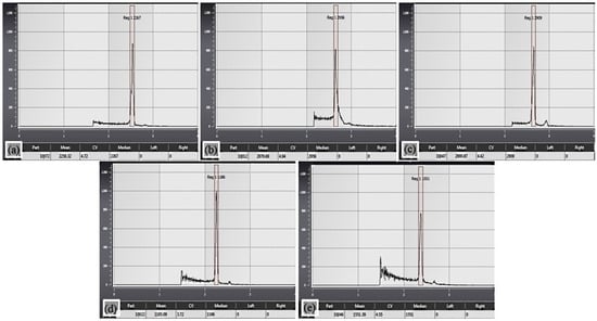
Somaclonal variability is one of the main problems with clonal micropropagation; its appearance is undesirable for obtaining a homogeneous planting material from valuable genotypes. The study of the ploidy level of nuclei of leaves of in vitro material grown on a nutrient medium with a concentration of 0.8 mg/L BAP revealed no changes in the parameter of interest compared to samples obtained from ex situ plants; the relative DNA content and ploidy corresponded to 2C and 2× (Figure 6a–c).

Figure 6.
Histograms of the ploidy level of the analyzed H. officinalis f. cyaneus material: (a,d)—external control, (b)—samples of nuclei from plant leaves ex situ, (c)—samples of nuclei from leaves of microshoots in vitro (0.8 mg/L BAP), (e)—samples of nuclei from leaves of microshoots in vitro (0.3 mg/L BAP).
Similar results were obtained for microshoots sub-cultivated in vitro for five months (Figure 5d,e). In in vitro culture, the genome may also be affected, i.e., somaclonal variability (spontaneous mutagenesis) is possible [39]. Genetic variations can manifest themselves both in DNA sequences and at the chromosomal level (for example, polyploidy) [17,40]. To determine the cellular and tissue degree of sensitivity of plant organs in vitro, forms that deviate from normal development processes are of interest, since during their development the material is in a “critical” condition, which contributes to the detection of limiting reactions. According to the few available data, hyperhydricity may correlate with abnormal flow cytometry profiles, which were shown using three Lathyrus sativus L. genotypes cultured in vitro on a nutrient medium containing BAP. In such morphologically deviant forms, researchers noted the appearance of three peaks corresponding to 2C, 4C and 8C, whereas all phenotypically normal regenerants had diploid profiles, i.e., two peaks corresponding to 2C and 4C [41]. We have shown that in direct morphogenesis, despite the high concentration of BAP which contributed to the formation of structural rearrangements, no changes in the ploidy level were detected. The differences in the results obtained may be related to the species specificity of plants, manifested during the cultivation of the material in vitro. With short-term culturing in vitro on MS medium with a BAP concentration of 0.3 mg/L, no shifts were detected. The researchers note that significant changes can be observed when using tissue cultures in vitro, while during direct morphogenesis or embryogenesis, the activity of genomic rearrangements decreases, and their occurrence is associated with the duration of cultivation of the material in the presence of growth regulators [42].
According to the data obtained, successful in vitro regeneration of H. officinalis f. cyaneus from single-node segments of shoots is observed on a modified MS nutrient medium with the addition of BAP in the range of 0.3–0.5 mg/L and 0.1 mg/L IBA. An increase in BAP to 0.8 mg/L provoked microshoot hyperhydricity. Morphoanatomical studies of leaf blades in vitro revealed their minor structural changes in the MS medium with a concentration of 0.4 mg/L BAP, which is due to the rejuvenation processes occurring during the formation of microshoots. Significant anatomical changes were observed in hyperhydrated organs, while the ploidy level corresponded to diploid material, similarly as with short-term in vitro cultivation with 0.3 mg/L of BAP. The obtained data clearly demonstrate the dependence of structural changes in the lateral photosynthetic organs of H. officinalis f. Cyaneus on the BAP concentration, while maintaining the ploidy level.
4. Conclusions
The mass fraction of the essential oil of the prospective sample was 0.20% of the raw weight and 0.55% of the absolutely dry weight. In total, 68 components were identified in the composition of the oil: isopinocamphone and pinocamphone, β-pinene, sabinene, linalool, β-phellandrene, myrtenol, germacrene D, bicyclogermacrene, etc. The analysis of the essential oil showed that the content of isopinocamphone was variable (from 50.99 to 70.74). A promising accession of Hyssopus officinalis f. cyaneus with an increased content (up to 70%) of isopinocamphone in its essential oil was selected for pharmaceutical use. Biotechnological methods were applied for the propagation of the valuable H. officinalis f. cyaneus breeding form, examining the morphological and anatomical characteristics in vitro and ex situ; additionally, the ploidy levels of microshoots in vitro on a nutrient medium were studied. When cultured in vitro, a decrease in the cuticle layer and the degree of development of the collenchyma near vascular bundles, as well as quantitative indicators of cells when cultured on an MS medium with 0.4 mg/L BAP compared with ex situ were shown. Significant structural changes were revealed at 0.8 mg/L of BAP, while no changes in the ploidy level were detected. For evaluating genetic similarity between ex situ and in vitro plants, subsequent analysis based on molecular markers is recommended.
Author Contributions
Conceptualization—Y.V.P., O.M.S. and I.V.B. Methodology—S.A.F., I.V.B. and N.N.I. Formal analysis—N.N.I., N.N.M., N.M.S. and S.A.F. Writing, draft preparation—I.V.B.; writing, review and editing—I.V.B., T.S.N. and O.M.S. All authors have read and agreed to the published version of the manuscript.
Funding
The study was carried out within the framework of the State Task No. FNNS-2022-0010 (registration number 122011700347-4) of the Federal State Funded Institution of Science “NBG-NSC” as well as within the program ‘Priority-2030’ of Sevastopol State University (strategic project No. 3, No. 121121700318-1), financed by the Ministry of Science and Higher Education of the Russian Federation.
Data Availability Statement
Data are available upon reasonable request to the corresponding author.
Acknowledgments
The authors thank the Senior Researcher of the South Siberian Botanical Garden of the Altai State University, Mikhail Skaptsov, for comprehensive assistance in conducting studies on the ploidy level of plant raw materials; and Head of the Laboratory of Aromatic and medicinal plants of the “NBG-NSC”, Senior Researcher, Tatyana Sakhno for providing plant material.
Conflicts of Interest
The authors declare no conflict of interest.
References
- Pirbalouti, A.G.; Bajalan, I.; Malekpoor, F. Chemical compositions and antioxidant activity of essential oils from inflorescences of two landraces of hyssop [Hyssopus officinalis L. subsp. angustifolius (Bieb.)] cultivated in Southwestern, Iran. J. Essent. Oil Bear. Plants 2019, 22, 1074–1081. [Google Scholar] [CrossRef]
- Sharifi-Rad, J.; Quispe, C.; Kumar, M.; Akram, M.; Amin, M.; Iqbal, M.; Koirala, N.; Sytar, O.; Kregiel, D.; Nicola, S.; et al. Hyssopus essential oil: An update of its phytochemistry, biological activities, and safety profile. Oxid. Med. Cell. Longev. 2022, 2022, 8442734. [Google Scholar] [CrossRef] [PubMed]
- Shibko, A.N. Biomorphologycal peculiarities of Hyssopus officinalis L. seeds under the cultivation in the conditions of the Pre-mountain Crimea. Sci. Notes Taurida V.I. Vernadsky Natl. Univ. Ser. Biol. Chem. 2011, 24, 371–377. (In Russian) [Google Scholar]
- Kotyuk, L.A. Features of micromorphological structure of medicinal hyssop. Mod. Phytomorphol. 2016, 10, 59–67. (In Ukrainian) [Google Scholar]
- Kalinichenko, L.V.; Malankina, E.L.; Kozlovskay, L.N. Comparative productivity assessment of common hyssop (Hyssopus officinalis L.) depending on the sample’s variety and origin. Izv. TAA 2013, 5, 171–176. (In Russian) [Google Scholar]
- Rabotyagov, V.D.; Shibko, A.N. Investigations of essential oil component composition of Hyssopus officinalis L. Work. State Nikit. Botan. Gard. 2014, 139, 88–100. (In Russian) [Google Scholar]
- Bespalyko, L.V.; Kharchenko, V.A.; Shevchenko, Y.P.; Ushakova, I.T. Common hyssop (Hyssopus officinalis L.). Veg. Crops Russ. 2016, 2, 60–63. [Google Scholar] [CrossRef]
- Plugatar, Y.V.; Shevchuk, O.M. Results and directions of breeding of aromatic and medicinal plants in the Nikitsky Botanical Gardens. Bull. State Nikitsk. Bot. Gard. 2019, 130, 9–17. [Google Scholar] [CrossRef]
- Chernyavskikh, V.I. Selection and seed production of Hyssopus officinalis L. in the Central Black Soil (Chernozem) region. Taurida Her. Agrar. Sci. 2018, 3, 137–146. (In Russian) [Google Scholar]
- Kizil, S.; Hasimi, N.; Tolan, V.; Kilinc, E.; Karatas, H. Chemical composition, antimicrobial and antioxidant activities of hyssop (Hyssopus officinalis L.) essential oil. Not. Bot. Horti Agrobot. Cluj 2010, 38, 99–103. [Google Scholar]
- Lu, M.; Battinelli, L.; Daniele, C.; Melchioni, C.; Salvatore, G.; Mazzanti, G. Muscle relaxing activity of Hyssopus officinalis essential oil on isolated intestinal preparations. Planta Med. 2002, 68, 213–216. [Google Scholar] [CrossRef] [PubMed]
- Isikov, V.P.; Rabotyagov, V.D.; Khlypenko, L.A.; Logvinenko, I.E.; Logvinenko, L.A.; Kutko, S.P.; Bakova, N.N.; Marko, N.V. Introduction and Breeding of Aromatic and Medicinal Plants (Methodological and Procedural Aspects); NBG-NSC: Yalta, Ukraine, 2009; 110p. (In Russian) [Google Scholar]
- Toma, I.; Toma, C.; Ghiorghita, G. Histo-anatomy and in vitro morphogenesis in Hyssopus officinalis L. (Lamiaceae). Acta Bot. Croat. 2004, 63, 59–68. [Google Scholar]
- Nanova, Z.; Slavova, Y.; Nenkova, D.; Ivanova, I. Microclonal propagation of hyssop (Hyssopus officinalis L.). Bulg. J. Agric. Sci. 2007, 13, 213–219. [Google Scholar]
- Bulavin, I.V.; Ivanova, N.N.; Mitrofanova, I.V. In vitro regeneration of Hyssopus officinalis L. and plant genetic similarity. Dokl. Biol. Sci. 2021, 499, 109–112. [Google Scholar] [CrossRef] [PubMed]
- Kritskaya, T.A.; Kashin, A.S.; Kasatkin, M.Y. Micropropagation and somaclonal variation of Tulipa suaveolens (Liliaceae) in vitro. Ontogenesis 2019, 50, 270–277. [Google Scholar] [CrossRef]
- Parab, A.R.; Lynn, C.B.; Subramaniam, S. Assessment of genetic stability on in vitro and ex vitro plants of Ficus carica var. black jack using ISSR and DAMD markers. Mol. Biol. Rep. 2021, 48, 7223–7231. [Google Scholar] [CrossRef] [PubMed]
- Shevchuk, O.M.; Isikov, V.P.; Logvinenko, L.A. Methodological and Procedural Aspects of Introduction and Breeding of Aromatic and Medicinal Plants; PH “Arial”: Simferopol, Ukraine, 2022; 140p, ISBN 978-5-907587-95-3. (In Russian) [Google Scholar]
- Tkachev, A.V. Study of Volatile Compounds of Plants; Ofset: Novosibirsk, Russia, 2008; 969p. (In Russian) [Google Scholar]
- Adams, R.P. Identification of Essential Oil Compounds by Gas Chromatography/Quadrupole Mass Spectroscopy; Allured Pub. Corp.: Carol Stream, IL, USA, 2007; 804p. [Google Scholar]
- Loureiro, J.; Rodriguez, E.; Doležel, J.; Santos, C. Two new nuclear isolation buffers for plant DNA flow cytometry: A test with 37 species. Ann. Bot. 2007, 100, 875–888. [Google Scholar] [CrossRef]
- Skaptsov, M.V.; Smirnov, S.V.; Kutsev, M.G.; Shmakov, A.I. Problems of standardization in flow cytometry of plants. Turczaninowia 2016, 19, 120–122. (In Russian) [Google Scholar] [CrossRef]
- Raboyagov, V.D.; Paliy, A.E.; Kurdyukova, O.N. Essential Oils of Aromatic Plants; PH “ARIAL”: Simferopol, Ukraine, 2017; 208p. (In Russian) [Google Scholar]
- Acimovic, M.; Pezo, L.; Zeremski, T.; Loncar, B.; Marjanovic Jeromela, A.; Stankovic Jeremic, J.; Cvetkovic, M.; Sikora, V.; Ignjatov, M. Weather conditions influence on hyssop essential oil quality. Processes 2021, 9, 1152. [Google Scholar] [CrossRef]
- Kerrola, K.; Galambosi, B.; Kallio, H. Volatile components and odor intensity of four phenotypes of hyssop (Hyssopus officinalis L.). J. Agric. Food Chem. 1994, 42, 776–781. [Google Scholar] [CrossRef]
- Zawislak, G. Morphological characters of Hyssopus officinalis L. and chemical composition of its essential oil. Mod. Phytomorphol. 2013, 4, 93–95. [Google Scholar] [CrossRef]
- Acimovic, M.; Stankovic, J.; Cvetkovic, M.; Kiprovski, B.; Marjanovic-Jeromela, A.; Rat, M.; Malencic, D. Essential oil analysis of different hyssop genotypes from IFVCNS medicinal plant collection garden. LetopisNaučnihRadova 2019, 43, 38–45. [Google Scholar]
- Zawislak, G. The chemical composition of essential hyssop oil depending on plant growth stage. Acta Sci. Pol. Hortum Cultus 2013, 12, 161–170. [Google Scholar]
- Kotyuk, L.A. Hyssop composition depending on age and plants development phases. Biotechnol. Acta 2015, 8, 55–63. [Google Scholar] [CrossRef]
- Tavakoli, M.; Aghajani, Z. The effects of drought stress on the components of the essential oil of Hyssopus officinalis L. and determining the antioxidative properties of its water extracts. J. Appl. Environ. Biol. Sci. 2016, 6, 31–36. [Google Scholar]
- Salvatore, G.; D’Andrea, A.; Nicoletti, M. A pinocamphone poor oil of Hyssopus officinalis L. var. decumbens from France (Banon). J. Essent. Oil. Res. 1998, 10, 563–567. [Google Scholar] [CrossRef]
- Ahmadi, H.; Babalar, M.; Sarcheshmeh, M.A.A.; Morshedloo, M.R.; Shokrpour, M. Effects of exogenous application of citrulline on prolonged water stress damages in hyssop (Hyssopus officinalis L.): Antioxidant activity, biochemical indices, and essential oils profile. Food Chem. 2020, 333, 127433. [Google Scholar] [CrossRef] [PubMed]
- Figueiredo, A.C.; Barroso, J.G.; Pedro, L.G.; Scheffer, J.J.C. Factors affecting secondary metabolite production in plants: Volatile components and essential oil. Flavour Fragr. J. 2008, 23, 213–226. [Google Scholar] [CrossRef]
- Bulavin, I.; Brailko, V.; Zhdanova, I. In vitro rhizogenesis of the Lavandula angustifolia cultivars. BIO Web Conf. 2020, 24, 00017. [Google Scholar] [CrossRef]
- Mitrofanova, I.V.; Lesnikova-Sedoshenko, N.P.; Chelombit, S.V.; Zhdanova, I.V.; Ivanova, N.N.; Mitrofanova, O.V. The effect of plant growth regulators on the in vitro regeneration capacity in some horticultural crops and rare endangered plant species. Acta Hortic. 2022, 1339, 181–189. [Google Scholar] [CrossRef]
- Bairu, M.W.; Kane, M.E. Physiological and developmental problems encountered by in vitro cultured plants. Plant Growth Regul. 2011, 63, 101–103. [Google Scholar] [CrossRef]
- Matushkina, O.V.; Pronina, I.N.; Tkachev, E.N. Vitrification of the shoots in vitro: Anatomical structure and possible solutions to the problem. Achiev. Sci. Technol. Agric. 2012, 1, 58–60. (In Russian) [Google Scholar]
- Manjula, R.; Jholgiker, P.; Subbaiah, K.V.; Prabhuling, G.; Swamy, G.S.K.; LeninKumar, Y. Morphological abnormality among hardened shoots of banana cv. Rajapuri (AAB) after in vitro multiplication with TDZ and BAP from excised shoot tips. Int. J. Agric. Environ. Biotechnol. 2014, 7, 465–470. [Google Scholar] [CrossRef]
- Lebedev, V.G.; Azarova, A.B.; Shestibratov, K.A.; Demenko, V.I. Manifestation of somaclonal variability in micro-propagated and transgenic plants. Izv. TAA 2012, 2, 153–163. (In Russian) [Google Scholar]
- Ryabushkina, N.A. Clonality and somaclonal variations in plants. Biotechnol. Theory Pract. 2014, 2, 17–27. [Google Scholar] [CrossRef]
- Ochatt, S.J.; Muneaux, E.; Machado, C.; Jacas, L.; Pontécaille, C. The hyperhydricity of in vitro regenerants of grass pea (Lathyrus sativus L.) is linked with an abnormal DNA content. J. Plant Physiol. 2002, 159, 1021–1028. [Google Scholar] [CrossRef]
- Skaptsov, M.V.; Krasnoborodkina, M.A.; Kutsev, M.G.; Smirnov, S.V.; Shmakov, A.I.; Matsyura, A.V. Ploidy level and relative nuclear DNA content in the plant cell and tissue culture in vitro. Biol. Bull. Bogdan Chmelnitskiy Melitopol State Pedagog. Univ. 2016, 6, 33–38. [Google Scholar] [CrossRef]
Disclaimer/Publisher’s Note: The statements, opinions and data contained in all publications are solely those of the individual author(s) and contributor(s) and not of MDPI and/or the editor(s). MDPI and/or the editor(s) disclaim responsibility for any injury to people or property resulting from any ideas, methods, instructions or products referred to in the content. |
© 2023 by the authors. Licensee MDPI, Basel, Switzerland. This article is an open access article distributed under the terms and conditions of the Creative Commons Attribution (CC BY) license (https://creativecommons.org/licenses/by/4.0/).