Article

In Vitro Propagation and Phytochemical Composition of Centratherum punctatum Cass—A Medicinal Plant

1
Cellular Differentiation and Molecular Genetics Section, Department of Botany, Jamia Hamdard, New Delhi 110062, India
2
Plant Production Department, College of Food and Agriculture Sciences, King Saud University, Riyadh 11451, Saudi Arabia
3
Research Institute of Nyíregyháza, Institutes for Agricultural Research and Educational Farm (IAREF), University of Debrecen, P.O. Box 12, 4400 Nyíregyháza, Hungary
*
Author to whom correspondence should be addressed.
Horticulturae 2023, 9(11), 1189; https://doi.org/10.3390/horticulturae9111189
Submission received: 4 September 2023 / Revised: 25 October 2023 / Accepted: 27 October 2023 / Published: 30 October 2023

Abstract

:
An effective and reproducible micropropagation protocol was developed for Centratherum punctatum Cass. Successful in vitro initiation of callus and subsequent plant regeneration were obtained on nodal explants cultured on MS medium supplemented with plant growth regulators (PGRs). The maximum frequency of callus formation (98.3%) was noted on MS containing 4.0 mg/L 6-Benzylaminopurine (BAP) and 3.5 mg/L Kinetin with a maximum callus weight of 2.02 g. The best shoot induction frequency (100%) with an average of 30.2 shoots per explant was achieved when 4.5 mg/L BAP and 4.0 mg/L Kinetin were added to the MS. The same PGR combination resulted in the best callus-mediated shoot formation (8.3 shoots/callus mass). The highest rhizogenic response (95.3%) with an average 26.1 roots per shoot and root length of 6.2 cm was obtained with 1.0 mg/L Indole-3-acetic acid (IAA)-supplemented MS medium. The gas chromatography–mass spectrometry (GC-MS) technique was applied in the present study to analyze the methanolic extracts of the leaf, callus, and root of regenerated C. punctatum shoots to detect the different phytochemical constituents. The leaf extract of the regenerated C. punctatum showed 37 phytocompounds; some important bioactive compounds were the Phytol,1,6-Octadien 3,5-Dimethyl-Cis, 4,8-Dimethylnona-3,8-dien-2-one, 2,6-Octadiene, Stigmasterol, Chondrillasterol, Lanosteryl acetate, etc. In the callus, the extract had a total of 57 phytocompounds; among them, the Stigmasterol, Guanosine, and Tri-decanoic acid were the major ones. In the root extract, the GC-MS revealed a low number of 23 phytocompounds, the important compounds of which were Stigmasterol, Trimethylsilyl (TMS) derivative, Chrysantenyl 2-methuylbutanoate, 4-tert-Butoxybutan-1-ol, etc. The order in terms of numbers of phytocompounds present in tissue sources are callus > leaf > root.

1. Introduction

Medicinal plants are a vital resource for billions around the world, serving as the foundation of both traditional remedies and modern pharmaceuticals. These plants not only offer cultural and economic benefits to communities but they may also be of significant interest in scientific research and the thriving herbal pharmaceutical sector. The World Health Organization (WHO) notes that a majority of individuals in developing nations, ranging from 70 to 95%, primarily depend on these plants for their basic healthcare needs [1]. Herbal medicine has made a significant contribution in healthcare as it has been in use since the dawn of mankind [2]. Centratherum punctatum Cass. is a herbaceous plant which belongs to the family Asteraceae and has been regarded as a species from the Vernonieae tribe. C. punctatum is found in Australia, Central and South America, the West Indies, the Philippines, India, and Java [3]. It is a fragrant, bushy, perennial plant and attains a height up to 45–60 cm, well-branched with purple flowers at the terminal ends, with aromatic leaves [4]. It is commonly known as Lark daisy, Kesavardhini, and Brazilian Bachelor’s Button [4,5]. It is known for its medicinal value and is used to treat various ailments. It was highlighted that C. punctatum (being rich in secondary metabolites) serves as a health-caring plant with various beneficial properties including antimicrobial, anti-oxidant, anti-allergic, anti-inflammatory, and cytotoxic anti-tumor effects [5]. Due to these important facts, the plant is referred to as a traditional wound healer.
Plant tissue culture is a technique utilized to propagate and cultivate plants in an artificial environment. This method allows for the production of a sufficient number of plants with uniform traits. The basic steps include selection and preparation of plant material, surface sterilization, and placement of plant parts (explants) onto nutrient medium supplemented by plant growth regulators for inducing callus, shoots, and roots. The obtained plantlets can then be transferred to the soil. The whole technique has revolutionized plant biotechnology and has become an essential tool for plant scientists and researchers [6,7].
In vitro cultures have been involved in plant breeding and the conservation of endangered species. The conservation of the biodiversity, often affected by natural phenomena, may heavily depend on plant tissue culture. This technique allows for the cultivation and fast propagation of endangered plant species, thus promoting conservation [8]. Several medicinally important plant species like American ginseng (Panax quinquefolius L.) [9], Madagascar periwinkle (Catharanthus roseus (L.) G. Don) [10], and common yew (Taxus baccata L.) [11] have successfully been cultured using tissue culture.
Moreover, the plant cell and tissue cultures are widely utilized in gene expression analysis, genetic transformation, and functional genomics research [12]. It is possible to harvest secondary metabolites from a variety of medicinal plants by increasing the yield using a cell/tissue culture technique [13]. In C. punctatum, a medicinal ornamental plant, a 14-day-old in vitro-derived leaf was noted to be responsive in 0.5 mg/L Thidiazuron-supplemented media for direct and indirect shoot regeneration [14]. The same research group earlier described a C. punctatum micropropagation protocol through transverse thin cell layers technology (tTCLs) in which 1.0 mm wide leaves and 2.0 mm thick nodal explants were observed to be effective [15]. However, biotechnological methods involving other explants or regeneration mode and/or in vitro morphogenesis using PGRs was still not found to be sufficient.
After the establishment of a plant cell culture, the researchers tried to analyze the extract in order to identify potential bioactive molecules present in it using GC-MS [2,16]. GC-MS is widely used in various fields like chemistry, biology, pharmacology, and environmental science to examine complex mixtures and to identify unknown compounds [17]. The technology makes it possible to separate a mixture of compounds into individual components using gas chromatography, followed by the analysis of separated components using mass spectrometry, which may detect the ionized fragments. The resulting mass spectra provide valuable information about the molecular weight, structure, and identification of compounds [18].
In plant tissue culture context, GC-MS can be used for multiple applications, such as in the analysis of volatile organic compounds, the identification and quantification of plant hormones, the characterization of secondary metabolites, and the metabolic profiling of other compounds. GC-MS was recently applied for the phytochemical profiling of Hibiscus asper Hook. fil. [19], and those of several medicinal plants such as carnation (Dianthus caryophyllus L.), common thyme (Thymus vulgaris L.), and common basil (Ocimum basilicum L.) [20].
The aim of this study was to investigate the effect of different plant growth regulators on callus formation, regeneration, and root induction in the successful culture of C. punctatum. This study also evaluated metabolite profiles and detected valuable phytocompounds present in tissues through GC-MS. Moreover, a comparative account of a metabolomic map, which may be the first of its kind, was prepared based on the results from the in vitro-developed callus, leaf, and roots.

2. Material and Methods

2.1. Plant Materials, Surface Disinfection, and In Vitro Culture Establishment

Explants of Centratherum punctatum Cass were collected from the campus of Jamia Hamdard and were used as experimental materials. These explants were treated with 2–3 drops of Teepol (a surfactant solution) which was followed by immersion in water for 10 min. Subsequent rinsing was conducted under running tap water. The explants were immersed in 70% ethanol for two min, followed by two rinses with double-distilled autoclaved water. This process was repeated twice. Subsequently, the explants were treated with 0.1% mercuric chloride (HgCl2) for one min and then rinsed thrice with double-distilled autoclaved water. Murashige–Skoog (MS) [21] medium containing 30 g/L sucrose and various plant growth regulators (PGRs) such as 2,4-Dichlorophenoxyacetic acid (2,4-D) (1.0–3.0 mg/L), α-naphthalene acetic acid (NAA) (0.5–2.5 mg/L), BAP (2.0–4.0 mg/L), Kinetin (Kn) (1.5–3.5 mg/L), and IAA (0.5–1.5 mg/L) were used. The pH of the medium was adjusted to 5.6–5.8 using a pH meter with 0.1 N solution of HCl and/or NaOH. Subsequently, 0.7% agar was incorporated into the MS medium as a solidifying agent. The medium (10 mL) was then poured into glass culture tubes or conical flasks (100 mL), which were autoclaved at 121 °C for 25–30 min and 15 psi. The cultures were incubated at a temperature of 25 ± 2 °C, under a photoperiod regime of 16 h light and 8 h darkness, with a light intensity of 100 μ mol m−2s−1 PPFD.

2.2. Callus Induction Using Nodal Explant

The surface-sterilized nodal explants were cultured on MS medium, supplemented by auxins (2-4-D (1.0–3.0 mg/L) + NAA (0.5–2.5 mg/L)) and cytokinins (BAP (2.0–4.0 mg/L) + Kn (1.5–3.5 mg/L)), for induction of callus. The callus development was observed within 7–10 days and the callus was sub-cultured within 4 weeks. Callus biomass weight was measured after 4 weeks of incubation.

2.3. Adventitious Shoot Induction from Callus

Nodal callus of about one gram was placed onto MS medium, supplemented with the same level of PGRs, i.e., 2.0–4.5 mg/L BAP and 1.0–4.0 mg/L Kinetin for caulogenesis. The shoot induction percentage (the number of callus clumps with at least one shoot, %) and the mean number of shoots per explant or callus clump with mean shoot length were noted after 8 weeks of incubation.

2.4. Axillary Shoot Induction Using Nodal Explants

Nodal explants were placed horizontally (attached throughout length) on MS medium enriched with BAP (2.0–4.5 mg/L) and Kinetin (1.0–4.0 mg/L) for shoot formation. The shoot regeneration frequency (%) and the mean number of shoots per explant were recorded after 8 weeks of culture, with mean shoot length. Eight-to-ten test tubes contained nodal explants (each containing one explant) in each PGR treatment and each experiment was repeated three times.

2.5. In Vitro Rooting and Acclimatization

The regenerated shoots were separated and were sub-cultured on media supplemented by varying IAA (0.5–1.5 mg/L) concentrations. The shoots showing root induction percentage and the mean number of roots with their root length were counted after four weeks. To acclimatize the plantlets with induced roots, the plants were carefully removed from the culture medium and cleaned with sterile double-distilled water. The plants were planted in plastic pots containing 3:1 blend of soil and vermiculite for acclimatization. This process lasted for approximately 3–4 weeks, and to maintain humidity, the pots were covered with cellophane wrap. Subsequently, the acclimatized plantlets were relocated to a greenhouse environment, maintaining a relative humidity from 60% to 70% and a constant temperature of 25 ± 2 °C, following a 16/8 h day/night cycle.

2.6. Preparation of Plant Methanolic Extract for GC-MS Analysis and Identification of Chemical Constituents

In vitro-derived calluses, leaves, and roots of adventitious origin, harvested from best growth media (callus from 4.0 mg/L BAP+ 3.5 mg/L Kn; shoot leaf from 4.5 mg/L BAP+ 4.0 mg/L Kn; and root from 1.0 mg/L IAA) were dried in sterilized condition and finely ground. The powdered samples of the above-mentioned tissues were soaked in 100% methanol (1.0 gm in 5.0 mL) for 12 h [22]. Syringe filter of pore size 0.22 µm was used to filter the extracts. The extracts were centrifuged at 8000 rpm and at a temperature of 4 °C for 10 min. Later, the supernatant was collected (leaving the debris) into new tubes and 1.0 µL of each sample was used for analysis via GC-MS. The sample was taken to University Science Instrumentation Center, AIRF, Jawaharlal Nehru University, Delhi for GC-MS analysis. Plant extracts were analyzed using a GC-MS QP2010 Plus system (Shimadzu, Kyoto, Japan). The system was equipped with an auto-injector (AOC-20i), a head space sampler (AOC-20s), a mass selective detector with an ion source set at 220 °C and an interface operating at 270 °C. The mass range from 40 m/z to 650 m/z was utilized for analysis, setting the threshold at 1000 EV. Additionally, the injector was configured for split injection mode at 10:1 ratio operating at 260 °C. Initially, the temperature was set to 100 °C for a period of 2 min, following which it was incrementally increased to reach 300 °C at a rate of 10 °C per min. Helium was used as the carrier gas, exhibiting a linear velocity of 40.9 cm/s under a pressure of 90.5 kPa. The apparatus functioned with an aggregate flow rate of 16.3 mL/min, accompanied by a columnar flow rate of 1.21 mL/min.
In order to identify the molecules, the structure, the molecular mass, and the spectral fragments were considered and compounds were cross-referenced with the established components from the National Institute of Standards and Technology’s (NIST14s.lib, accessed on 2 February 2023) library data base which encompasses more than 62,000 patterns.

2.7. Experimental Design and Statistical Analysis

The effect of PGRs on explants in inducing callus, shoot formation, and in vitro rooting were studied by randomized design. The PGRs used in various treatments contained eight–ten explants and each of the experiments were conducted twice with three replica. The results are presented as the mean values ± standard errors. The means with the same letters in different columns are not significantly different and the mean difference was determined by Duncan’s multiple range test (DMRT) at p ≤ 0.05 using SPSS ver. 26.0 (SPSS Inc., Chicago, IL, USA).

3. Results

3.1. Callus Induction

Nodal explants were inoculated on media with different PGRs, i.e., 2, 4-D, BAP, Kinetin, and NAA, to induce callus development. Variable calli were obtained in different media; NAA and 2, 4-D-containing media resulted in soft, loose calluses (Figure 1a). Among the various tested combinations, the best callus induction ability and growth were observed on the medium containing a combination of BAP (4.0 mg/L) and Kinetin (3.5 mg/L) (Figure 1b,c). On this medium, the compact, dense callus development with a frequency of 98.3% and an average callus weight of 2.02 g was observed after four weeks of culture (Table 1). Soft calli were shown to be non-productive in the organogenic adventitious shoots development process at any combination of PGRs applied in the culture medium. Thus, these calli were omitted from future experimental processes.

3.2. Adventitious Shoot Induction

A nodal callus was placed onto MS with the same levels of BAP and Kinetin to induce callus-mediated adventitious shoot regeneration (Figure 2a,b). The best results with 66% of regeneration, i.e., 8.3 shoots/callus clump were detected on the medium containing 4.5 mg/L BAP + 4.0 mg/L Kinetin, and the shoots grew well under in vitro condition (6.1 cm mean shoot length after 8 weeks of culture), followed by 4.0 mg/L BAP + 3.5 mg/L Kinetin treatment in which 6.6 shoots per callus mass were observed (Table 2). Callus-mediated shoot formation was less found in other tested PGR combinations.

3.3. Axillary (Direct) Shoot Regeneration

Nodal explants were excised and cultured on MS with various concentrations of BAP and Kinetin to induce shoot regeneration (Figure 3a–c). The best responses (100%) were achieved on the medium supplemented with 4.5 mg/L BAP and 4.0 mg/L Kinetin after 8 weeks of culture (30.2 shoots per explant with an average length of 8.5 cm). The second best treatment was the combination of 4.0 mg/L BAP + 3.5 mg/L Kinetin, where an average of 26.4 shoots per explant was produced (Table 3). The medium with 2.0 mg/L BAP + 1.0 mg/L Kinetin had the lowest effectiveness, i.e., a 60.4% shoot induction frequency and an average of 8.1 shoots per explant were observed.

3.4. Induction of Roots in Regenerated Plantlets
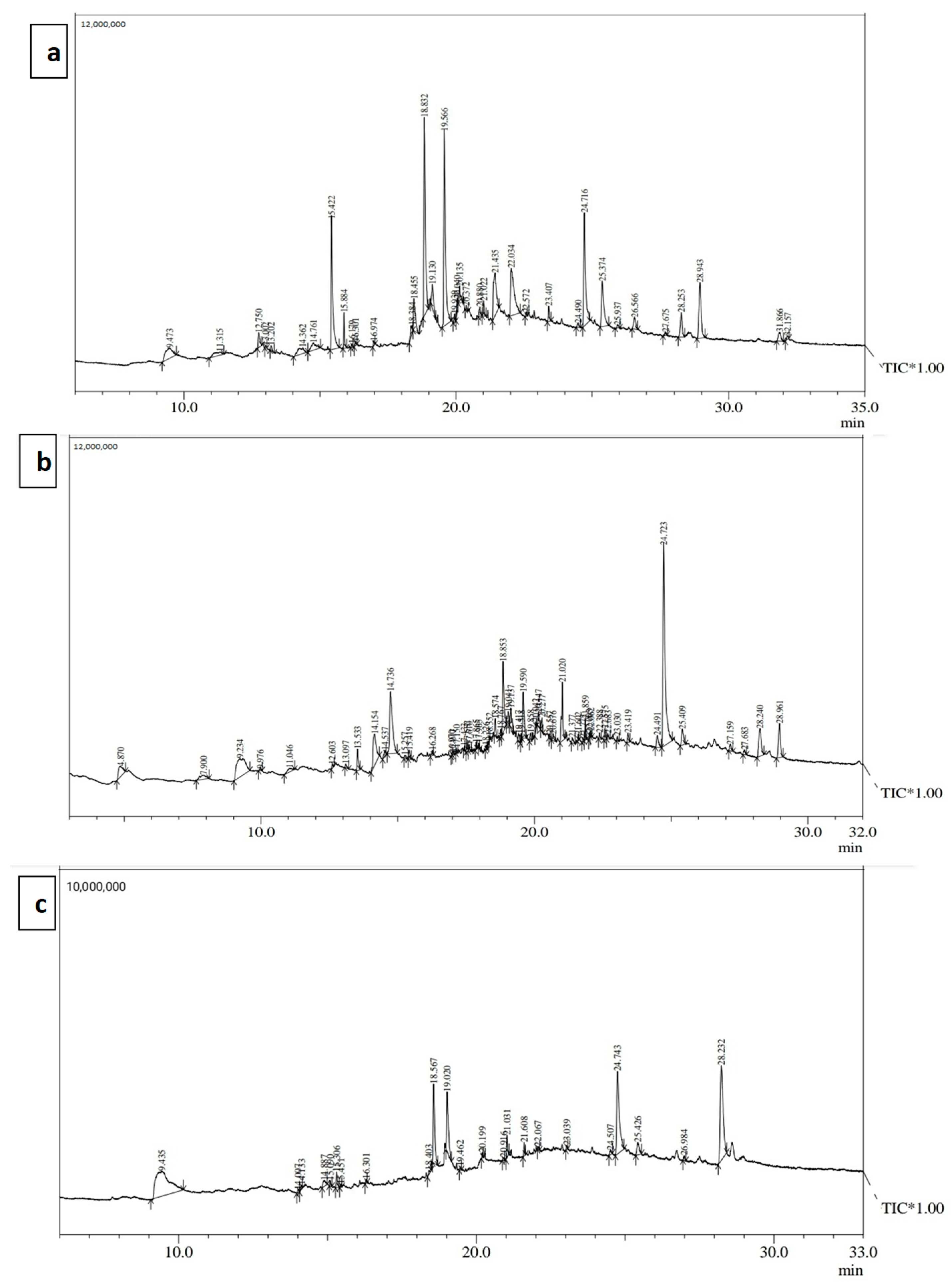
Regenerated C. punctatum shoots (adventitious and direct origin) were excised and transferred to media with various concentrations of IAA. The best responses were achieved in 1.0 mg/L IAA treatment (Figure 4a,b), which resulted in the 95.2% rooting percentage with 26.1 roots per shoot and an average root length of 6.2 cm (Table 4). The other tested PGR treatments had a low-to-moderate rooting influence on in vitro regenerated shoots. The rooted shoots were transplanted to a pot (Figure 4c,d) containing 3:1 soil and vermiculite. The plants with well-developed roots transferred to the greenhouse survived normally and showed a 70–80% survival frequency.

3.5. GC-MS Analysis

The GC-MS analysis of leaf methanolic extract of regenerated C. punctatum shoots revealed a total of 37 phytochemicals, presented in Figure 5a and Table 5. Some of the most important bioactive compounds are the Phytol (9.92%), 1,6-Octadien,3,5-Dimethyl-,Cis (13.56%), 4,8-Dimethylnona-3,8-dien-2-one (5.88%), 2,6-Octadiene,2,4-dimethyl- (8%), Stigmasterol (9.75%), Chondrillasterol (5%), and Lanosterylacetate (5.51%). Other detected compounds present in the leaf extract are Squalene, Neophytadiene, oxiranehexadecyl, and palmitic acid Trimethylsilyl (TMS) derivatives. In the callus, the methanolic extract shows a total of 57 phytocompounds (Figure 5b), among which Stigmasterol (24.85%), Guanosine (9.06%), Tridecanoicacid (5.72%), 1,6-Octadien,3,5-Dimethyl-,Cis(5.81%), and Squalene (4.40%) are the major ones (Table 6). Some compounds are present exclusively in the callus, like 4H-pyran-4-one, 2,3-dihydro-3,5-dihydroxy-6-methyl, and guanosine; in the literature, these were reported to display microbial inhibitory activity, inflammation suppression, growth restraining, neuro-inflammation, oxidative stress, and excitotoxicity activity. In the root methanolic extract, the GC-MS analysis revealed a low number of compounds, i.e., 23 phytocompounds (Figure 5c), the major compounds of which were Stigmasterol (17.42%), TMS derivatives (28.50%), Chrysantenyl2-methuylbutanoate (10.31%), 4-tert-Butoxybutan-1-ol, etc. (Table 7). Active compounds like chrysantenyl 2-methyl butanoate had previously shown antibacterial properties. Cedren-13-ol, 8- had lipid peroxidation; meanwhile, oleic acid and propyl ester, which showed antifungal properties and carbinoxamine present in the root extract, displayed antihistaminic potential. The order in terms of numbers of phytocompounds present in tissue sources is callus > leaf > root.

4. Discussion

In this present study, the whole plant was regenerated and multiplied via tissue culture by utilizing different PGRs. The in vitro-regenerated tissues including different plant parts (callus, leaf, and root) were analyzed using GC-MS for secondary metabolites. Firstly, the nodal explants with leaves were placed onto media with 2,4-D and NAA, which resulted in fast-growing calli. The exogeneous application of 2,4-D has long been known to be an important trigger in developing the callus by promoting cell division [23]. In this study, the callus induction frequency and biomass were high on the medium containing BAP and Kinetin combinations, and the callus developed on the nodal explants was compact. The present study thus indicates that the BAP and Kinetin combination showed fast cell division in the developing callus, similar to the effect of cytokinins exerted on other plant species. The callus-inducing ability of BAP and Kinetin was earlier noted in eggplant (Solanum melongena L.) [24], in pomelo (Citrus grandis (L.) Osbeck) [25], and in cycads (Encephalartos spp.) [26]. An increase in the concentration of BAP and Kinetin triggered direct shoot induction as well. Direct shoot regeneration from nodal ex plants was reported in many plants like water hyssop (Bacopa monnieri (L.) Pennell) [27], the winter jasmine (Jasminum nudiflorum Lindl.) [28], and Mansonia altissima [29]. Very similar reports, i.e., for cytokinin-induced shoots production, were observed in other plant species like plantain (Musa sp., “Oniaba” and “Apantu pa” cultivars) [30] and medicinal aloe (Aloe vera (L.) Burm. f.) [31]. It is known that cytokinins, functioning as plant growth regulators, regulate numerous processes of plant growth and development, such as the cell division, initiation, and development of shoots and cellular differentiation [32,33]. The combination of BAP and Kinetin was proved to be efficient in enhancing regeneration and shoot growth in a variety of plant species such as potato (Solanum tuberosum L.) [34], fever tea (Lippia javanica (Burm. f.) Spreng.) [35], white sandalwood (Santalum album L.) [36], and English oak (Quercus robur L.) [37]. In Aloe vera (L.) Burm. f.), the same BAP and Kinetin combinations demonstrated to be highly efficient in inducing callus development [38].
These in vitro-developed shoots were later rooted on IAA-containing medium. Similar responses, i.e., root induction with IAA, were observed in many other investigated plants such as tomato (Solanum lycopersicum L.) [39] and false chamomile (Tripleurospermum insularum Sch. Bip.) [40]. IAA, a naturally occurring auxin, has previously been considered to be crucial for in vitro rooting as well as in the plant regeneration process [41], as was demonstrated in Alocasia longiloba. The regenerated plants were finally transferred to the greenhouse, where they survived normally.
Similar to earlier work, the methanolic extracts of the in vitro-regenerated plant parts—leaf, callus, and root––were separately evaluated through GC-MS in order to detect the active compounds present in the extracts [17,42,43]. In the leaf, 37 phytochemical compounds were isolated, 57 were detected in the callus, and 23 bioactive compounds were found in the root extract. The extracts derived from C. punctatum leaves, callus, and roots contained several bioactive compounds exhibiting various medicinal properties. Neophytadiene, oxiranehexadecyl, and palmitic acid TMS derivative possess anti-inflammatory, antimicrobial, and antitumor properties [44,45,46,47]. Phytol, which is common in leaf and callus extracts, has anticancer, antioxidant, antitumor, antimicrobial, diuretic, and antimalarial properties [48]. 1-Docosanol acetate acts as a humectant, emulsifier, and thickener in cosmetics [49], whereas acetylcholine bromide acts as a neurotransmitter at cholinergic synapses [50]. Sivasubramanian and Brindha [2] reported that the ethanolic extract of C. punctatum’s aerial parts containing Eugenol, Spathulenol, Viridiflorol, Hexadecanoic acid, Phthalic acid, bis (7-methyloctyl) ester, Eicosane, and Squalene had antioxidant and cytotoxic effects with anticancerous potential.
Squalene, which is present in all three extracts, has antimicrobial, antioxidative, tumor-inhibitory actions, as well as cancer-preventing potential and immune system enhancing effects. In addition, disease-resistance activities like chemo-preventive, lipoxygenase suppressors ability, pesticidal characteristics, skin hydrating, and emollient activity were observed for squalene, present in various extracts of C. punctatum [45,51]. Vitamin E, present in this studied plant extract, is known for its multiple beneficial properties, including the ability to inhibit tumor growth, alleviate spasms, function as an antioxidant, dilate blood vessels, provide pain relief, mitigate diabetes, protect the liver, lower cholesterol levels, regulate blood sugar, and act against sterility [45,52]. 3.beta.- Ergost-5-en-3-ol and stigmasterol, present in all three extracts, and 3beta.-stigmast-5-en-3-ol, present in the callus, exhibit antioxidant, anti-inflammatory, hypo cholesterolemic, anticancerous, antipyretic, and diuretic properties [51]. 9,12-Octadecadienoic acid (Z,Z), an octyl ester, also has hypo-cholesterolemic properties [45].
Lanosteryl acetate, present in the C. punctatum leaf and callus, plays a role in cataract prevention and treatment [53]. Beta-amyrone, present in the leaf extract, is anti-inflammatory [54], whereas chondrillasterol, found in the leaf and root extract, is antimicrobial in nature [55]. Certain compounds limited to the callus, such as 4H-pyran-4-one and 2,3-dihydro-3,5-dihydroxy-6-methyl, may have antimicrobial, anti-inflammatory, and growth-inhibition properties [52]. Guanosine, also found only in the callus, is known to mitigate neuro-inflammation and impart an effect of oxidative stress and excitotoxicity. Furthermore, it has nourishing effects on neuronal and glial cells [56]. Isopropyl myristate is an emollient and is used in skin-lubricating lotions [57], whereas eremanthin is a compound with antidiabetic and antilipidemic effects [58,59]. Tetratetracontane, present in both the callus and root, has antioxidant as well as cytoprotective activities [60]). Delta-tocopherol, detected in the callus and root extract, exhibits antioxidant properties [61]. Some active compounds exhibiting medicinal properties are found in the root extract only, such as chrysantenyl 2-methyl butanoate; it is an essential oil possessing antibacterial properties [62]. Cedren-13-ol-8 has an affinity to inhibit lipid peroxidation [63]. Oleic acid propyl ester is antifungal, whereas carbinoxamine is antihistaminic [64]. Eicosane is a bronchodilator [65], and Hexatriacontane, present in the root extract, possesses antibacterial properties [66]. Thus, the plant C. punctatum is immensely valuable medicinally as the extracts of various tissues, especially the callus, synthesize a variety of phytocompounds, the yield of which may further be improved by adopting biotechnological strategies.

5. Conclusions

In vitro shoot regeneration was successfully induced on Centratherum punctatum explants using specific PGR combinations of BAP and Kn, added to MS medium. The GC-MS analysis revealed a diverse array of phytochemicals in the plant tissues, highlighting the medicinal significance of the species. The in vitro-regenerated plant or plant parts can provide a continuous source of raw materials for the utilization of secondary metabolites by mitigating overexploitation risks. This chemical profiling is pivotal for extending future research of therapeutic uses.

Author Contributions

Conceptualization: A.T. and A.M.; methodology, formal analysis: A.T., B.E. and Y.B.; investigation: A.T.; data curation: A.T., B.E. and Y.B.; writing—original draft preparation: A.T. and A.M.; writing—review and editing: A.T., A.M., Y.H.D. and K.M.-T.; validation: Y.H.D. and K.M.-T.; visualization: A.M., Y.H.D. and K.M.-T.; project administration: A.M. All authors have read and agreed to the published version of the manuscript.

Funding

The authors are highly thankful to Department of Botany, Jamia Hamdard for receiving financial assistance and other research facilitiesand Researchers Supporting Project number (RSP2023R375), King Saud University, Riyadh, Saudi Arabia.

Data Availability Statement

All data are presented in the article.

Acknowledgments

The authors acknowledge Researchers Supporting Project number (RSP2023R375), King Saud University, Riyadh, Saudi Arabia.

Conflicts of Interest

The authors declare no conflict of interest.

References

  1. Rahman, M.H.; Roy, B.; Chowdhury, G.M.; Hasan, A.; Saimun, M.S. Medicinal plant sources and traditional healthcare practices of forest-dependent communities in and around Chunati Wildlife Sanctuary in southeastern Bangladesh. Environ. Sustain. 2022, 5, 207–241. [Google Scholar] [CrossRef] [PubMed]
  2. Sivasubramanian, R.; Brindha, P. In vitro cytotoxic, antioxidant and GC-MS studies on Centratherum punctatum Cass. Int. J. Pharm. Pharm. Sci. 2013, 4, e8. [Google Scholar]
  3. Kirkman, L.K. Taxonomic revision of Centratherum and Phyllocephalum (Compositae: Vernonieae). Rhodora 1981, 83, 1–24. Available online: http://www.jstor.org/stable/23314048 (accessed on 26 October 2023).
  4. Gbolade, A.A.; Dzamic, A.M.; Ristic, M.S.; Marin, P.D. Essential oil composition of Centratherum punctatum from Nigeria. Chem. Nat. Compd. 2009, 45, 118–119. [Google Scholar] [CrossRef]
  5. Madhumitha, K.M.; Anbumalarmathi, J.; Sharmili, S.A.; Nandhini, G.; Priya, G.S. A comparative study of in vivo plant and in vitro callus extracts of Centratherum punctatum Cass. Annu. Res. Rev. Biol. 2020, 35, 1–13. [Google Scholar] [CrossRef]
  6. Smith, R.H. Plant Tissue Culture: Techniques and Experiments; Academic Press: Cambridge, MA, USA, 2012. [Google Scholar]
  7. Syeed, R.; Mujib, A.; Malik, M.Q.; Gulzar, B.; Zafar, N.; Mamgain, J.; Ejaz, B. Direct somatic embryogenesis and flow cytometric assessment of ploidy stability in regenerants of Caladium × hortulanum ‘Fancy’. J. Appl. Genet. 2022, 63, 199–211. [Google Scholar] [CrossRef]
  8. Oseni, O.M.; Pande, V.; Nailwal, T.K. A review on plant tissue culture, a technique for propagation and conservation of endangered plant species. Int. J. Curr. Microbiol. Appl. Sci. 2018, 7, 3778–3786. [Google Scholar] [CrossRef]
  9. Qiang, B.; Miao, J.; Phillips, N.; Wei, K.; Gao, Y. Recent advances in the tissue culture of American ginseng (Panax quinquefolius). Chem. Biodivers. 2020, 17, e2000366. [Google Scholar] [CrossRef]
  10. Amiri, S.; Fotovat, R.; Tarinejad, A.R.; Panahi, B.; Mohammadi, S.A. In vitro regeneration of periwinkle (Catharanthus roseus L.) and fidelity analysis of regenerated plants with ISSR markers. J. Plant Physiol. Breed. 2019, 9, 129–135. [Google Scholar]
  11. Abbasin, Z.; Zamani, S.; Movahed, S.; Khaksar, G.; Tabatabaei, B.S. In vitro micropropagation of Yew (Taxus baccata) and production of plantlets. Biotechnology 2010, 9, 48–54. [Google Scholar] [CrossRef]
  12. Maruyama, S.; Shibuya, N.; Kaku, H.; Desaki, Y. Arabidopsis cell culture for comparable physiological and genetic studies. Plant Signal. Behav. 2020, 15, 1781384. [Google Scholar] [CrossRef]
  13. Mohaddab, M.; El Goumi, Y.; Gallo, M.; Montesano, D.; Zengin, G.; Bouyahya, A.; Fakiri, M. Biotechnology and in vitro culture as an alternative system for secondary metabolite production. Molecules 2022, 27, 8093. [Google Scholar] [CrossRef] [PubMed]
  14. Aswathi, N.V.; Thomas, T.D. Direct and indirect shoot regeneration from leaf explants of Centratherum punctatum Cass., A wild ornamental plant. Sci. Hortic. 2023, 320, 112201. [Google Scholar] [CrossRef]
  15. Aswathi, N.V.; Thomas, T.D. Transverse thin cell layer (tTCL) technology: A promising tool for micropropagation of Centratherum punctatum Cass. In Vitro Cell. Dev. Biol.—Plant 2023, 59, 340–353. [Google Scholar] [CrossRef]
  16. Mamgain, J.; Mujib, A.; Syeed, R.; Ejaz, B.; Malik, M.Q.; Bansal, Y. Genome size and gas chromatography-mass spectrometry (GC–MS) analysis of field-grown and in vitro regenerated Pluchea lanceolate plants. J. Appl. Genet. 2023, 64, 1–21. [Google Scholar] [CrossRef]
  17. Bansal, Y.; Mujib, A.; Mamgain, J.; Dewir, Y.H.; Rihan, H.Z. Phytochemical composition and detection of novel bioactives in anther callus of Catharanthus roseus L. Plants 2023, 12, 2186. [Google Scholar] [CrossRef]
  18. Hübschmann, H.J. Handbook of GC-MS: Fundamentals and Applications; John Wiley and Sons: Hoboken, NJ, USA, 2015. [Google Scholar]
  19. Olivia, N.U.; Goodness, U.C.; Obinna, O.M. Phytochemical profiling and GC-MS analysis of aqueous methanol fraction of Hibiscus asper leaves. Future J. Pharm. Sci. 2021, 7, 59. [Google Scholar] [CrossRef]
  20. Arafa, N.M.; Girgis, N.; Ibrahem, M.; Mohamed, S.; El-Bahr, M.K. Phytochemical profiling by GC-MS analysis and antimicrobial activity potential of in vitro derived shoot cultures of some Egyptian herbal medicinal plants. Egypt. J. Chem. 2022, 65, 155–169. [Google Scholar] [CrossRef]
  21. Murashige, T.; Skoog, F. A revised medium for rapid growth and bioassays with tobacco tissue cultures. Phys. Plant 1962, 15, 473–497. [Google Scholar] [CrossRef]
  22. El-Ashmawy, I.M.; Aljohani, A.S.; Soliman, A.S. Studying the bioactive components and phytochemicals of the methanol extract of Rhanterium epapposum Oliv. Appl. Biochem. Biotechnol. 2023. [Google Scholar] [CrossRef]
  23. Fehér, A. Somatic embryogenesis—Stress-induced remodeling of plant cell fate. Biochim. Et Biophys. Acta (BBA)-Gene Regul. Mech. 2015, 1849, 385–402. [Google Scholar] [CrossRef]
  24. Foo, P.C.; Lee, Z.H.; Chin, C.K.; Subramaniam, S.; Chew, B.L. Shoot induction in white eggplant (Solanum melongena L. cv. BulatPutih) using 6-benzylaminopurine and kinetin. Trop. Life Sci. Res. 2018, 29, 119. [Google Scholar] [CrossRef]
  25. Zakaria, Z.B.; Zakaria, S.B.; Ishak, M.A.B.M. Induction of callus formation from different parts of Citrus grandis (Osbeck) flowers. Biotropia 2010, 17, 32. [Google Scholar] [CrossRef]
  26. Malwane, T.S.D. In Vitro Tissue Culture: Towards Conservation of Threatened Desiccation Sensitive Encephalartos cycads Seeds. Master’s Thesis, The University of Cape Town, Cape Town, South Africa, 2019. Available online: https://hdl.handle.net/11427/31805 (accessed on 26 October 2023).
  27. Sape, S.T.; Kandukuri, A.V.; Owk, A.K. Direct axillary shoot regeneration with nodal explants of Bacopa monnieri (L.) Pennell—A multi medicinal herb. J. Appl. Biol. Sci. 2020, 14, 190–197. Available online: https://www.jabsonline.org/index.php/jabs/article/view/726 (accessed on 26 October 2023).
  28. Bhat, M.S.; Rather, Z.A.; Nazki, I.T.; Banday, N.; Wani, T.; Rafiq, S.; Darwish, H. Standardization of in vitro micropropagation of Winter Jasmine (Jasminum nudiflorum) using nodal explants. Saudi J. Biol. Sci. 2022, 29, 3425–3431. [Google Scholar] [CrossRef] [PubMed]
  29. Oseni, O.M.; Nailwal, T.K.; Pande, V. Callus induction and multiple shoot proliferation from nodal explants of Mansonia altissima: Confirmation of genetic stability using ISSR and RAPD markers. In Vitro Cell. Dev. Biol.—Plant 2022, 58, 479–488. [Google Scholar] [CrossRef]
  30. Buah, J.N.; Danso, E.; Taah, E.A.; Abole, E.A.; Bediako, E.A.; Asiedu, J.; Baidoo, R. The Effects of Different Concentrations Cytokinins on the In Vitro Multiplication of Plantain (Musa sp.). 2010. Available online: http://hdl.handle.net/123456789/4934 (accessed on 26 October 2023).
  31. Singh, M.K.; Yadav, T.; Raman, R.K. A quick method for micro-propagation of Aloe vera L. from leaf explants via callus induction. J. Entomol. Zool. Stud. 2020, 8, 201–206. [Google Scholar]
  32. Hill, K.; Schaller, G.E. Enhancing plant regeneration in tissue culture: A molecular approach through manipulation of cytokinin sensitivity. Plant Signal. Behav. 2013, 8, 212–224. [Google Scholar] [CrossRef]
  33. Mujib, A.; Fatima, S.; Malik, M.Q. Gamma ray–induced tissue responses and improved secondary metabolites accumulation in Catharanthus roseus. Appl. Microbiol. Biotechnol. 2022, 106, 6109–6123. [Google Scholar] [CrossRef]
  34. Dilshad, E.; Asif, A.; Arooj, H.; Khan, S.H.; Bakhtiar, S.M. Impact of BAP on in vitro regeneration of potato (Solanum tuberosum L.). Curr. Trends OMICS 2021, 1, 67–79. [Google Scholar] [CrossRef]
  35. Mood, K.; Jogam, P.; Sirikonda, A.; Shekhawat, M.S.; Rohela, G.K.; Manokari, M.; Allini, V.R. Micropropagation, morpho-anatomical characterization, and genetic stability studies in Lippia javanica (Burm. f.) Spreng: A multipurpose medicinal plant. Plant Cell Tissue Organ. Cult. 2022, 150, 427–437. [Google Scholar] [CrossRef]
  36. Singh, C.K.; Raj, S.R.; Patil, V.R.; Jaiswal, P.S.; Subhash, N. Plant regeneration from leaf explants of mature sandalwood (Santalum album L.) trees under in vitro conditions. In Vitro Cell. Dev. Biol.—Plant 2013, 49, 216–222. [Google Scholar] [CrossRef]
  37. Martins, J.P.R.; Wawrzyniak, M.K.; Ley-López, J.M.; Kalemba, E.M.; Mendes, M.M.; Chmielarz, P. 6-Benzylaminopurine and kinetin modulations during in vitro propagation of Quercus robur (L.): An assessment of anatomical, biochemical, and physiological profiling of shoots. Plant Cell Tissue Organ. Cult. 2022, 151, 149–164. [Google Scholar] [CrossRef]
  38. Ahmad, S.; Jakhar, M.L.; Gothwal, D.K.; Ram, M.; Kumhar, B.L.; Kumawat, G.L. Effect of 6-Benzylaminopurine (BAP) and Kinetin (Kn) on callus induction under in vitro culture of Aloe vera. Biol. Forum 2022, 14, 890–894. [Google Scholar]
  39. Gerszberg, A.; Hnatuszko-Konka, K.; Kowalczyk, T.; Kononowicz, A.K. Efficient in vitro callus induction and plant regeneration protocol for different Polish tomato cultivars. Not. Bot. Horti Agrobot. 2016, 44, 452–458. [Google Scholar] [CrossRef]
  40. Inceer, H.; Cuce, M.; Imamoglu, K.V.; Ergin, T.; Ucler, A.O. In vitro propagation and cytogenetic stability of Tripleurospermum insularum (Asteraceae)—A critically endanger redinsular endemic species from Turkey. Plant Biosyst. 2022, 156, 1213–1221. [Google Scholar] [CrossRef]
  41. Abdulhafiz, F.; Mohammed, A.; Kayat, F.; Zakaria, S.; Hamzah, Z.; Reddy Pamuru, R.; Gundala, P.B.; Reduan, M.F. Micropropagation of Alocasia longiloba Miq and comparative antioxidant properties of ethanolic extracts of the field-grown plant, in vitro propagated and in vitro-derived callus. Plants 2020, 29, 816. [Google Scholar] [CrossRef]
  42. Padma, M.; Ganesan, S.; Jayaseelan, T.; Azhagumadhavan, S.; Sasikala, P.; Senthilkumar, S.; Mani, P. Phytochemical screening and GC–MS analysis of bioactive compounds present in ethanolic leaves extract of Silybum marianum (L). J. Drug Deliv. Ther. 2019, 15, 85–89. Available online: https://jddtonline.info/index.php/jddt/article/view/2174 (accessed on 26 October 2023). [CrossRef]
  43. Jahan, I.; Tona, M.R.; Sharmin, S.; Sayeed, M.A.; Tania, F.Z.; Paul, A.; Chy, M.N.; Rakib, A.; Emran, T.B.; Simal-Gandara, J. GC-MS phytochemical profiling, pharmacological properties, and in silico studies of Chukrasia velutina leaves: A novel source for bioactive agents. Molecules 2020, 25, 3536. [Google Scholar] [CrossRef]
  44. Musa, A.M.; Ibrahim, M.A.; Aliyu, A.B.; Abdullahi, M.S.; Tajuddeen, N.; Ibrahim, H.; Oyewale, A.O. Chemical composition and antimicrobial activity of hexane leaf extract of Anisopus mannii (Asclepiadaceae). J. Intercult. Ethnopharmacol. 2015, 4, 129. [Google Scholar] [CrossRef]
  45. Sujatha, P.; Evanjaline, M.; Muthukumarasamy, S.; Mohan, V.R. Determination of bioactive components of Barleria courtallicanees (Acanthaceae) by gas chromatography–mass spectrometry analysis. Asian J. Pharm. Clin. Res. 2017, 10, 273. [Google Scholar] [CrossRef]
  46. Bhardwaj, M.; Sali, V.K.; Mani, S.; Vasanthi, H.R. Neophytadiene from Turbinariaornata suppresses LPS-induced inflammatory response in RAW 264.7 macrophages and Sprague Dawley rats. Inflammation 2020, 43, 937–950. [Google Scholar] [CrossRef]
  47. Vandana; Deora, G.S. Preliminary phytochemical screening and GC-MS analysis of methanolic leaf extract of Tephrosia falciformis ramaswami from Indian Thar Desert. Int. J. Pharm. Sci. Res. 2020, 61, 3040–3046. [Google Scholar] [CrossRef]
  48. Tyagi, T.; Agarwal, M. Phytochemical screening and GC-MS analysis of bioactive constituents in the ethanolic extract of Pistia stratiotes L. and Eichhornia crassipes (Mart.) solms. J. Pharmacogn. Phytochem. 2017, 6, 195–206. [Google Scholar]
  49. Yamuna, P.; Abirami, P.; Vijayashalini, P.; Sharmila, M. GC-MS analysis of bioactive compounds in the entire plant parts of ethanolic extract of Gomphrena decumbens Jacq. J. Med. Plants Stud. 2017, 5, 31–37. [Google Scholar]
  50. Kokaz, S.F.; Deb, P.K.; Abed, S.N.; Al-Aboudi, A.; Das, N.; Younes, F.A.; Salou, R.A.; Bataineh, Y.A.; Venugopala, K.N.; Mailavaram, R.P. Pharmacology of acetylcholine and cholinergic receptors. In Frontiers in Pharmacology of Neurotransmitters; Springer: Berlin/Heidelberg, Germany, 2020; pp. 69–105. [Google Scholar] [CrossRef]
  51. Srinivasan, S.; Priya, V. Phytochemical screening and GC-MS analysis of Cyperus dubius, Rottb. (Cyperaceae). J. Med. Plants 2019, 7, 89–98. [Google Scholar]
  52. Mujeeb, F.; Bajpai, P.; Pathak, N. Phytochemical evaluation, antimicrobial activity, and determination of bioactive components from leaves of Aegle marmelos. BioMed Res. Int. 2014, 2014, 497606. [Google Scholar] [CrossRef]
  53. Zhang, K.; He, W.; Du, Y.; Zhou, Y.; Wu, X.; Zhu, J.; Lu, Y. Inhibitory effect of lanosterol on cataractous lens of cynomolgus monkeys using a subconjunctival drug release system. Precis. Clin. Med. 2022, 5, pbac021. [Google Scholar] [CrossRef]
  54. De Almeida, P.; Boleti Rüdiger, A.L.; Lourenço, G.A.; da Veiga Junior, V.F.; Lima, E.S. Anti-inflammatory activity of triterpenes isolated from Protium paniculatum oil-resins. Evid.-Based Complement. Altern. Med. 2015, 2015, 293768. [Google Scholar] [CrossRef]
  55. Mozirandi, W.; Tagwireyi, D.; Mukanganyama, S. Evaluation of antimicrobial activity of chondrillasterol isolated from Vernonia adoensis (Asteraceae). BMC Complement. Altern. Med. 2019, 19, 249. [Google Scholar] [CrossRef]
  56. Bettio, L.E.; Gil-Mohapel, J.; Rodrigues, A.L.S. Guanosine and its role in neuropathologies. Purinergic Signal. 2016, 12, 411–426. [Google Scholar] [CrossRef]
  57. Patel, M.R.; Patel, R.B.; Parikh, J.R.; Patel, B.G. Formulation consideration and skin retention study of microemulsion containing tazarotene for targeted therapy of acne. J. Pharm. Investig. 2016, 46, 55–66. [Google Scholar] [CrossRef]
  58. Maurya, A.; Mohan, S.; Verma, S.C. Antidiabetic potential of naturally occurring sesquiterpenes: A review. Curr. Top. Med. Chem. 2021, 21, 851–862. [Google Scholar] [CrossRef]
  59. Liu, T.; Zhao, X.; Song, D.; Liu, Y.; Kong, W. Anticancer activity of Eremanthin against the human cervical cancer cells is due to G2/M phase cell cycle arrest, ROS-mediated necrosis-like cell death and inhibition of PI3K/AKT signalling pathway. J. BUON 2020, 25, 1547–1553. [Google Scholar]
  60. Amudha, P.; Jayalakshmi, M.; Pushpabharathi, N.; Vanitha, V. Identification of bioactive components in Enhalusa coroides seagrass extract by gas chromatography-mass spectrometry. Asian J. Pharm. Clin. Res. 2018, 11, 313–315. [Google Scholar] [CrossRef]
  61. Rouvrais, C.; Bacqueville, D.; Bogdanowicz, P.; Haure, M.J.; Duprat, L.; Coutanceau, C.; Bessou-Touya, S. A new dermocosmetic containing retinaldehyde, delta- tocopherol glucoside and glycylglycineoleamide for managing naturally aged skin: Results from in vitro to clinical studies. In Clinical, Cosmetic and Investigational Dermatology; Taylor & Francis: Abingdon, UK, 2017; pp. 35–42. [Google Scholar] [CrossRef]
  62. Mugao, L.G.; Gichimu, B.M.; Muturi, P.W.; Mukono, S.T. Characterization of the volatile components of essential oils of selected plants in Kenya. Biochem. Res. Int. 2020, 2020, 8861798. [Google Scholar] [CrossRef] [PubMed]
  63. Nsofor, W.N.; Nwaoguikpe, R.N.; Ujowundu, F.N.; Keke, C.O.; Uba, M.T.U.; Edom, C.V. Phytochemical, GC-MS, FTIR and Amino acid profile of methanol extract of Tetrapleura tetraptera fruit. J. Drug Deliv. Ther. 2023, 13, 61–69. [Google Scholar] [CrossRef]
  64. Abubacker, M.N.; Devi, P.K. In vitro antifungal potentials of bioactive compound oleic acid, 3-(octadecyloxy) propyl ester isolated from Lepidagathis cristata Willd. (Acanthaceae) inflorescence. Asian Pac. J. Trop. Med. 2014, 7, S190–S193. [Google Scholar] [CrossRef]
  65. Subramanian, S.; Dowlath, M.J.H.; Karuppannan, S.K.; Saravanan, M.; Arunachalam, K.D. Effect of Solvent on the Phytochemical Extraction and GC-MS Analysis of Gymnema sylvestre. Pharmacogn. J. 2020, 12, 749–761. [Google Scholar] [CrossRef]
  66. Aodah, A.H.; Hashmi, S.; Akhtar, N.; Ullah, Z.; Zafar, A.; Zaki, R.M.; Ali, M.S. Formulation development, optimization by box–behnken design, and in vitro and ex vivo characterization of hexatriacontane-loaded transethosomal gel for antimicrobial treatment for skin infections. Gels 2023, 9, 322. [Google Scholar] [CrossRef]
Figure 1. Callus development on Centratherum punctatum leaf explants: (a) friable callus induced on medium with 2,4-D (2.5 mg/L) + NAA (2.0 mg/L), (b,c) compact nodal callus with occasional roots formation on medium containing BAP (4.0 mg/L) + Kn (3.5 mg/L) (scale bars: 0.5 cm).
Horticulturae 09 01189 g001
Figure 2. In vitro callus-mediated shoot regeneration in Centratherum punctatum on MS medium supplemented with BAP (4.5 mg/L) and Kn (4.0 mg/L): (a,b) adventitious shoot development from callus at different stages (scale bars: 1.0 cm).
Horticulturae 09 01189 g002
Figure 3. In vitro shoot development of Centratherum punctatum on medium supplemented with BAP (4.5 mg/L) and Kn (4 mg/L). (a): direct shoot induction from nodal explant; arrow indicates direct shoot regeneration, (b,c): Shoot proliferation after 4 and 6 weeks of culture (scale bars: 1.0 cm).
Horticulturae 09 01189 g003
Figure 4. Root induction and acclimatization of in vitro regenerated Centratherum punctatum. (a,b) In vitro root induction in 1.0 mg/L IAA-added medium and (c,d) successful transfer of tissue-cultured regenerated plants in outdoor (scale bars: 1 cm).
Horticulturae 09 01189 g004
Figure 5. GC-MS chromatogram of Centratherum punctatum methanolic extract of in vitro leaf (a), callus (b), and in vitro roots (c). TIC*1.00: total ion current (1.0 std).
Horticulturae 09 01189 g005
Table 1. Callus induction frequency, callus biomass from nodal explants on MS medium with various PGRs after 4 weeks of culture in Centratherum punctatum.
PGRs (mg/L)Callus Induction (%)Weight of Callus (g) Type of Callus Observed
2,4-DNAABAPKn
2.01.562.21 ± 1.28 c0.71 ± 0.08 cWhite-brownish
compact callus
2.52.076.40 ± 0.81 c0.85 ± 0.32 c
3.02.589.54 ± 0.31 b1.20 ± 0.09 b
4.0 *3.5 *98.32 ± 0.47 a*2.02 ± 0.04 a*
1.00.5 00Watery-friable callus
1.51.0 67.35 ± 0.49 c0.19 ± 0.02 c
2.01.5 78.13 ± 0.70 ab0.48 ± 0.01 ab
2.5 *2.0 * 82.27 ± 1.24 a*0.76 ± 0.21 a*
3.02.5 70.63 ± 0.89 c0.32 ± 0.03 c
* Represents highest % of response on corresponding medium. Values are mean ± SE of three replica with two experiments each. Mean values of two sets of separate experiments containing PGRs (BAP + Kn and 2,4-D + NAA) within a column followed by different letters are significantly different according to Duncan’s multiple range test (DMRT) at p ≤ 0.05.
Table 2. Shoot induction frequency, shoot numbers from callus of Centratherum punctatum in MS medium containing various concentrations of BAP and Kn.
PGRs (mg/L)Shoot Induction
(%)
No. of Shoots/Gram of CallusMean Shoot Length (cm)
BAPKn
2.01.00 c0 c0c
2.51.533.24 ± 0.13 b3.2 ± 1.27 b2.6 ± 0.35 b
3.02.540.67 ± 0.38 b4.1 ± 0.63 b3.3 ± 0.17 b
4.03.552.41 ± 0.24 a6.6 ± 0.35 a5.2 ± 0.69 a
4.5 *4.0 *66.33 ± 0.44 a*8.3 ± 0.75 a*6.1 ± 0.63 a*
For each PGR treatment, 24 explants were used. * Represents highest % of response on corresponding medium. Data were scored after 8 weeks of culture. Values are mean ± SE of three replica with two experiments each. Mean values within a column followed by different letters are significantly different according to Duncan’s multiple range test (DMRT) at p ≤ 0.05.
Table 3. Direct shoot induction frequency, shoot number, and growth of Centratherum punctatum in various BAP and Kn-containing MS medium after 4 weeks in culture.
PGRs (mg/L)Shoot Induction
(%)
No. of Shoots Mean Shoot Length (cm)
BAPKn
2.01.060.42 ± 0.87 d8.1 ± 0.57 d2.0 ± 0.12 d
2.51.568.14 ± 1.17 c12.3 ± 0.94 c4.0 ± 0.06 c
3.02.572.56 ± 1.44 c21.3 ± 1.0 c4.5 ± 0.29 c
4.03.588.28 ± 0.90 b26.4 ± 1.15 b6.2 ± 0.17 b
4.5 *4.0 *100.0 ± 2.89 a*30.2 ± 0.92 a*8.5 ± 0.28 a*
* Represents highest % of response on corresponding medium. Data were scored after 8 weeks of culture. Values are mean ± SE of three replica with two experiments each. Mean values within a column followed by different letters are significantly different according to Duncan’s multiple range test (DMRT) at p ≤ 0.05.
Table 4. Root induction in Centratherum punctatum on medium containing different IAA concentrations after 4 weeks in culture.
IAA
(mg/L)
Root
Induction (%)
No. of Roots/Shoot Mean Root Length (cm)
0.540.16 ± 1.18 c18.3 ± 0.81 c2.5 ± 0.33 c
1.0 *95.27 ± 0.60 a*26.1 ± 0.58 a*6.2 ± 0.21 a*
1.580.52 ± 0.52 b20.5 ± 1.25 b4.8 ± 0.11 b
For each PGR treatment, 24 explants were used. * Represents highest % of response on corresponding medium. Values are mean ± SE of three replica with two experiments each. Mean values within a column followed by different letters are significantly different according to Duncan’s multiple range test (DMRT) at p ≤ 0.05.
Table 5. Phytochemical compounds present in the methanolic leaf extract of C. punctatum in vitro culture using GC-MS analysis.
PeakR. TimeAreaArea%Name
19.47328141363.38Sucrose
211.31510879161.31Pentanoic acid, 2-(methoxymethyl)-4-oxo-
312.7509432001.13Neophytadiene
413.002891470.11Oxirane, hexadecyl-
513.2022865310.343,7,11,15-Tetramethyl-2-hexadecen-1-ol
614.36214941581.79Decane, 1-bromo-2-methyl-
714.76113763851.65Palmitic Acid, TMS derivative
815.42282619619.92Phytol
915.88410549281.27Phytol, TMS derivative
1016.1791238370.15Eicosane, 7-hexyl-
1116.3011957170.241-Docosanol, acetate
1216.9741859840.22N1-Isopropyl-2-methyl-1,2-propanediamine
1318.3847084580.85Acetylcholine bromide
1418.45524268752.91Crotonyl isothiocyanate
1518.8321128749013.561,6-Octadien, 3,5-Dimethyl-, Cis
1619.13017052252.05Bicyclo[3.1.1]heptan-3-one, 2,6,6-trimethyl-, (1.alpha.,2.alp
1719.5661248699315.002(5H)-Furanone, 5-(2-methyl-3-methylene-4-butyl)-
1819.9393081480.372-Butenoic acid, 2-methyl-, 1,1a,1b,4,4a,5,7a,7b,8,9-decahy
1920.0404425680.534,4′-((p-Phenylene)diisopropylidene)diphenol
2020.13510029991.202-Butenoic acid, 2-methyl-, 2-methyl-2-propenyl ester, (E)-
2120.3721729840.215A-Methyl-3,8-Dimethylene-2 Oxododecahydrooxireno[2′,3′:6,7]Naphtho[1,2B]
Furan-6-YL 2-Methyl-2-B-Utenoate
2220.8805023970.601-Azatricyclo[3.3.1.13,7]Decane-4,6,10-Trione,
2321.0226216150.75Squalene
2421.43548980595.884,8-Dimethylnona-3,8-dien-2-one
2522.03466583508.002,6-Octadiene, 2,4-dimethyl-
2622.5721520660.18Stigmasta-4,7,22-trien-3.alpha.-ol
2723.4076506750.78Vitamin E
2824.4902691700.325-Cholesten-3.beta.-acetoxy-24-thiol
2924.71681197069.75Stigmasta-5,22-Dien-3-Ol
3025.37441643145.00Chondrillasterol
3125.9371453520.17.beta.-Amyrone
3226.56610085501.21Noruns-12-Ene
3327.6751460230.18Acetic acid, 3-hydroxy-7-isopropenyl-1,4a-dimethyl-2,3,4,4
3428.25317642162.12Phytyl palmitate
3528.94345853715.51Acetic Acid 17-(1,5-Dimethyl-Hex-4-Enyl)-4,4,1
3631.8668932991.079,12-Octadecadienoic acid (Z,Z)-, octyl ester
3732.1572255090.279,10,12,13-Tetrabromooctadecanoic acid
83260312100.00
Table 6. Phytochemical compounds present in the methanolic callus extract of C. punctatum using GC-MS analysis.
PeakR. TimeAreaArea%Name
14.87017208172.564H-Pyran-4-one, 2,3-dihydro-3,5-dihydroxy-6-methyl-
27.9009911411.471,4-Diacetyl-3-acetoxymethyl-2,5-methylene-l-rhamnitol
39.23460987769.06Guanosine
49.976868950.13Butanoic Acid, 1-Methylhexyl Ester
511.0466300430.94Alpha-L-Galactopyranoside, methyl 6-deoxy-
612.6031543110.23Isopropyl myristate
713.0972003110.30Benzene, (1-ethylundecyl)-
813.53311197161.66Benzene, (1-Methyldodecyl)-
914.15438548405.72Tridecanoic acid
1014.5373687870.55Benzene, (1-methyltridecyl)-
1114.73665477949.72Azuleno[4,5-b]furan-2(3H)-one, 3a,4,6a,7,8,9,9a,9b-octahy
1215.2571482850.223-Methyl-2-Pent-2-Enyl-Cyclopent-2-Enone
1315.4194191020.62Phytol
1416.2682408700.36Tricosyl acetate
1516.970721070.112-Butanamine, 2-methyl-
1617.0171416870.215-Decanone
1717.1501528700.23Silane, Trichlorooctadecyl-
1817.433736770.11Tetracyclo[6.1.0.0(2,4).0(5,7)] Nonane, 3,3,6,6,9,9
1917.5322805210.423,3,6,6,9,9-Hexaethyltetracyclo[6.1.0.0~2,4~.0~
2017.6102334360.35Fumaric acid, 2-octyl 8-chlorooctyl ester
2117.8651703310.25Cyclopentanol, 3,3,4-Trimethyl-4-P-Tolyl-, (
2217.9651107570.16Triamylbenzenes
2318.2261687740.25Benzene, Hexadecylpentyl-
2418.3521092760.16Tetracyclo[6.1.0.0(2,4).0(5,7)]nonane, 3,3,6,6,9,9-hexaethy
2518.5747119131.06Valeric acid, 2-methyloct-5-yn-4-yl ester
2618.7672737380.41Triamylbenzenes
2718.85339140845.811,6-Octadien, 3,5-Dimethyl-, Cis
2819.0416981601.04Cyclooctanepropanal, 1-Nitro-2-Oxo-, (.+-.)-
2919.13711932801.772,5-Furandione, 3-(Dodecenyl)Dihydro-
3019.4173419150.513-(4-Isopropylphenyl)-2-Methylpropanal #
3119.5181834900.271-(4-Undecylphenyl)Ethanone
3219.59025650913.812(5H)-Furanone, 5-(2-methyl-3-methylene-4-butyl)-
3319.8583281230.493,3,6,6,9,9-Hexaethyltetracyclo[6.1.0.0~2,4~.0~
3420.0422813380.424,4’-((p-Phenylene)diisopropylidene)diphenol
3520.1477150851.06Tetracosatetraene, 2,6,10,15,19,23-Hexameth
3620.2775289920.791h-Purin-6-Amine, [(2-Fluorophenyl)Methyl]
3720.5571953660.29Heptacosane, 1-chloro-
3820.6764571020.683-(4-Isopropylphenyl)-2-Methylpropanal #
3921.02029655664.40Squalene
4021.3773173080.471-(4-Undecylphenyl)ethanone
4121.6021260370.19Tetratetracontane
4221.7721329760.20Tert-Butyl(Dimethyl)Silyl ([Tert-Butyl)Dim
4321.8595809650.86Oxirane, 2,2-dimethyl-3-(3,7,12,16,20-pentamethyl-3,7,11,
4422.0031546760.23Cyclododecasiloxane, Tetracosamethyl-
4522.0621490270.22.delta.-Tocopherol
4622.3882554730.382,2,4-Trimethyl-3-(3,8,12,16-Tetramethyl-Hept
4722.5753995520.59Stigmasta-4,7,22-trien-3.alpha.-ol
4822.6832795190.42(R)-6-Methoxy-2,8-dimethyl-2-((4R,8R)-4,8,12-trimethyltr
4923.0301733060.262-Methylhexacosane
5023.4194498270.67Vitamin E
5124.4919401731.40Ergost-5-en-3-ol, (3.beta.)-
5224.7231673292424.85Stigmasta-5,22-Dien-3-Ol
5325.40912336821.83Stigmast-5-En-3-Ol, (3.Beta.,24s)-
5427.1594826310.729,19-Cyclolanostan-3-ol, 24-methylene-, (3.beta.)-
5527.6833323630.4924-Norursa-3,12-diene
5628.24026323343.914,4a,6b,8a,11,11,12b,14a-Octamethyl-Docosah
5728.96125257553.75Acetic Acid 17-(1,5-Dimethyl-Hex-4-Enyl)-4,4,1
67346895100.00
Table 7. Phytochemical compounds present in the methanolic in vitro root extract of C. punctatum using GC-MS analysis.
PeakR. TimeAreaArea%Name
19.435908662128.504-tert-Butoxybutan-1-ol, TMS derivative
214.0071016380.32Chrysantenyl 2-methuylbutanoate
314.1331611450.511,2-Benzenedicarboxylic acid, bis(2-methylpropyl) ester
414.8873886091.22Azuleno[4,5-b]furan-2(3H)-one, 3a,4,6a,7,8,9,9a,9b-octahy
515.0901446610.456-Methyl-2-(4-methylcyclohex-3-en-1-yl)hepta-1,5-dien-4-
615.3064641061.46Cedren-13-ol, 8-
715.451769480.24Octyl tetradecyl ether
816.301968970.30Oleic Acid, Propyl Ester
918.4031387290.44Ethanamine, 2-[(4-Chlorophenyl)-2-Pyridiny
1018.567328814210.31Chrysantenyl 2-methuylbutanoate
1119.02027523018.638-Hydroxy-2,2-dimethyl-8-phenyl-oct-5-en-3-one
1219.462734190.23Tridecane, 7-hexyl-
1320.1991056080.33Eicosane
1420.916783200.25Tricosane, 2-Methyl-
1521.0316024901.89Squalene
1621.6082812070.88Hexatriacontane
1722.067850590.27.delta.-Tocopherol
1823.039812020.25Tetracontane
1924.5071732010.54Ergost-5-en-3-ol, (3.beta.)-
2024.743555460417.42Stigmasta-5,22-Dien-3-Ol
2125.4267581952.38Chondrillasterol
2226.9841690760.5324-Noroleana-3,12-diene
2328.232722308822.654,4a,6b,8a,11,11,12b,14a-Octamethyl-Docosah
31885266100.00
Disclaimer/Publisher’s Note: The statements, opinions and data contained in all publications are solely those of the individual author(s) and contributor(s) and not of MDPI and/or the editor(s). MDPI and/or the editor(s) disclaim responsibility for any injury to people or property resulting from any ideas, methods, instructions or products referred to in the content.

