Article

Nutritional Composition and Bioactivity of Salicornia europaea L. Plants Grown in Monoculture or Intercropped with Tomato Plants in Salt-Affected Soils

1
Department of Agriculture, Food and Environment, University of Pisa, Via del Borghetto 80, 56124 Pisa, Italy
2
Interdepartmental Research Center “Nutraceuticals and Food for Health”, University of Pisa, Via del Borghetto 80, 56124 Pisa, Italy
3
Centre for Agri-environmental Research E. Avanzi, University of Pisa, via Vecchia di Marina 6, Piero a Grado, 56122 Pisa, Italy
4
Institute of Biology and Agricultural Biotechnology (IBBA), CNR, Research Area of Pisa, Via Moruzzi 1, 56124 Pisa, Italy
5
Laboratoire des Plantes Extrêmophiles, Centre de Biotechnologie de Borj Cedria, BP 901, Hammam Lif 2050, Tunisia
6
Géoarchitecture Territoires, Urbanisation, Biodiversité, Environnement, Université de Brest, EA 7462, CS 93837, CEDEX 3, F-29238 Brest, France
*
Author to whom correspondence should be addressed.
Horticulturae 2022, 8(9), 828; https://doi.org/10.3390/horticulturae8090828
Submission received: 20 July 2022 / Revised: 30 August 2022 / Accepted: 6 September 2022 / Published: 8 September 2022
(This article belongs to the Collection Biosaline Agriculture)

Abstract

:
The increasing salinization of agricultural soils urges us to find alternative and sustainable farming systems in order to allow the exploitation of areas that are otherwise becoming less suitable for conventional crops. Thanks to their adaptation to extreme saline conditions, halophytes are promising plants for resilient farming systems, such as intercropping with glycophytes, to ameliorate their productivity in saline soils. This research aimed to evaluate whether the nutritional profile and the content of some health-promoting compounds of the edible portion of Salicornia europaea were influenced by its cultivation in consociation with tomato plants. Moreover, the antioxidant, antibacterial, and anti-inflammatory properties of S. europaea were studied to characterize its bioactivity. The farming system did not influence the concentration of nutrients and bioactive compounds, except for flavonoids. The antimicrobial and anti-inflammatory properties of Salicornia extract suggested the importance of this halophyte for animal and human health.

1. Introduction

The global population is expected to reach 9.7 billion by 2050, and there are concerns about the capacity of agriculture to produce enough food for the growing population. By some estimates, food production will need to go up by about 60 percent either through an increase in crop yields per unit area or expansion in the arable land by 2050 to meet the demand [1]. Furthermore, several regions already suffering from malnutrition, water scarcity, and soil degradation have been forecast to have a large population growth which raises serious concerns about whether traditional agricultural methods and crop species will have the capacity to sustain global food production targets [2,3].
Major cereal crops such as wheat, rice, barley, and corn fail to withstand increasing salinity and scarce water resources in marginal environments that are most vulnerable to climate change. Therefore, there is an urgent need to identify alternative solutions to sustaining and possibly increasing agricultural productivity in areas where growing traditional crops has become difficult and sometimes uneconomical. It becomes necessary to investigate the potential of plants that prefer to grow in salty soils or waters (i.e., halophytes) to produce food for human or livestock consumption [4].
Halophytes are unique plant species able to live in saline environments due to the development of adaptive mechanisms and responses that allow them to counteract the extreme adversity inherent to their biotope, such as the accumulation of non-enzymatic antioxidants to neutralize ROS and prevent oxidative damage [5,6,7]. These bioactive molecules involve vitamins, carotenoids, and polyphenols, which are in charge of the beneficial nutritional and medicinal properties of such plants. In fact, local communities living in coastal areas, such as those in the Mediterranean areas, have been using halophytes as food or in traditional medicine to relieve several human ailments and diseases, including hypertension and diabetes [8]. Due to the extreme conditions these halophytes must combat and face to survive, it is not surprising that these plants contain a higher level of phenolic compounds than glycophytes, which correlates to a higher antioxidant activity that, in some cases, is even more potent than that of synthetic antioxidants [9].
Recently, there has been increased attention to natural antimicrobial compounds since microorganisms are involved in the degradation of food or other derived products, and a wide range of diseases. Some plant species are a rich source of natural bioactive molecules with antimicrobial properties, which are able to fend off the growth of food infectious pathogens [5,10]. In addition, pathogens implicated in foodborne diseases or food-processing plant contamination are often capable to adhere and form biofilms. However, literature about plant constituents with antifouling capacity is scarce, particularly concerning halophytes.
Thanks to their ability to accumulate large amounts of salt in their shoots, halophytes are well known for their potential to remediate saline soil, thus allowing the subsequential cultivation of less tolerant crops. Such a feature can be also exploited to improve the productivity of salt-sensitive crops by cultivating them in intercrop systems with halophytes to enable more resilient agriculture systems and taking economic advantages from the simultaneous harvest of the two plant species [11].
Salicornia europaea L., or sea beans, is a halophyte that can be found along the coasts of America, Europe, South Africa, and the Mediterranean basin [12]. Previous studies with extracts of S. europaea from the Mediterranean areas divulged a high content of phenolics and flavonoids, which were suggested to contribute to considerable antioxidant activity [13,14]. Other interesting properties assigned to S. europaea extracts include its use to treat diverse diseases which is well known in oriental pharmacopeia [15], suggesting its potential use in several industrial and biotechnological fields. Although extracts of S. europaea have been previously reported to be rich in phenolic compounds and to display antioxidant properties, to our knowledge, there are no reports on its antimicrobial and anti-inflammatory activities.
The first aim of this study was to assess whether the cultivation of S. europaea in consociation with tomato plants modified the nutritional profile and the content of some bioactive compounds of the edible portion. The evaluation of the effectiveness of the halophyte–crop consociation is one of the main objectives of the HaloFarMs (Development and Optimization of Halophyte-based Farming systems in salt-affected Mediterranean Soils) project (https://mel.cgiar.org/projects/halofarms (accessed on 30 June 2022)), aiming to improve the crop yield in saline soils but also to determine the biochemical quality of the halophyte biomass and its bioactivity. Accordingly, the present research also aimed to evaluate the antioxidant, antibacterial, and anti-inflammatory properties of S. europaea, which could reinforce its potential application in the food industry and nutraceutical areas as a functional food.

2. Materials and Methods

2.1. Plant Material and Experimental Design

Salicornia (Salicornia europaea L. var. fruticosa) plants were cultivated in monoculture or in intercropping with tomato (Solanum lycopersicum L. cv. Optima) in a greenhouse located at the experimental station “Podere Rottaia” of the Department of Agriculture, Food and Environment of the University of Pisa (43°40′30.7″ N 10°18′38.6″ E), near to the seacoast. Both Salicornia and tomato plants were purchased from Italian nurseries (Azienda Bonato, Montagnana, Italy), which were later transplanted in the salt-affected soil of the experimental site. The soil where the experiment was carried out showed values of soil pH, electric conductivity (EC), and soluble Na content in the upper 15 cm of 7.4 ± 0.22, 1.66 ± 0.07 mS cm−1, and 814 ± 19 mg kg−1 soil, respectively.
The experimental design forecasted three different kinds of plots, namely Salicornia in monoculture (S), Salicornia consociated with tomato plants (S-T), and tomato in monoculture, with the latter condition not included in the present work. Each plot measured 5 × 4 m and was randomly assigned to one of the above-reported theses. Salicornia plots consisted of two double rows of twenty-five plants each (for a total of 100 plants per plot), while S-T plots consisted of two rows of thirteen tomato plants each, with twenty-five Salicornia plants planted at each side of the two tomato rows (Figure S1). Each plot was duplicated. Samples (the edible part of Salicornia plants, i.e., the apical ramets) were collected from each double row (giving a total of 4 replicates of 50 plants each for S and 4 replicates for S-T) and freeze-dried.

2.2. Proximate Composition

For proximate compositions, Salicornia plants were firstly pre-dried at 60 °C for 48 h and then milled to pass through a 1 mm screen. Dry matter, ether extract, crude protein, and ash were determined according to AOAC methods [16]. The dry matter content was determined by oven drying at 105 °C until a stable weight and total ash content by ashing at 550 °C for 3 h. The fat (ether extract) content was determined using high-temperature solvent extraction with an ANKOM model XT10 extractor (ANKOM Technology, NY, USA). The fiber fractions NDF (neutral detergent fiber), ADF (acid detergent fiber), and ADL (acid detergent lignin) were analyzed according to Goering and Van Soest [17] with some modifications using an ANKOM 200 fiber analyzer (ANKOM Tech. Corp., Fairport, NY, USA) [18]. Briefly, each milled sample was weighed into an ANKOM F57 filter bag (ANKOM Tech. Corp., Fairport, NY, USA), 0.5 ± 0.0025 g, and the bag was heat sealed. The bags were then suspended in 1900 mL of NDF detergent (Midland Scientific Inc., Omaha, NE) with 4 mL of heat stable α-amylase (ANKOM Tech. Corp., Fairport, NY, USA) and 20 g of Na2SO3. Bags were shaken for 60 min at 95 °C. As the NDF content is determined gravimetrically, the bags after the extraction process were rinsed with 25 °C distilled water, dried for 12 h in a 100 °C oven, and weighted. A similar procedure was followed to evaluate ADF content, with the samples undergoing the extraction process with the ADF solution (Midland Scientific Inc., Omaha, NE) for 60 min at 95 °C. ADL was performed by soaking the extracted ADL bags in 333 mL of 72% sulfuric acid for 3 h. Samples were stirred every 30 min to ensure uniform dispersion of the acid. Samples were then rinsed with 25 °C distilled water until the pH was neutral. The samples were then dried in a wire basket for 12 h in a 100 °C oven and weighted. The ADL content was expressed exclusive of residual ash. All samples were analyzed in 4 replicates.

2.3. Fatty Acid Composition

Fatty acid composition was evaluated by acid trans-methylation according to Christie [19] with some modification. Briefly, fatty acid methyl esters (FAME) were prepared by adding 4.5 mL of 10% HCl methanolic solution to 5 g of sample. Nonadecanoic acid (1 mg) was added as internal standard. After 8 h of incubation, 5 mL of n-hexane was used to extract the FAME after stirring for 1 min. The layers were allowed to separate, and the hexane fraction (1 μL) was injected into gas-chromatographic (GC) analysis by a GC2010 Shimadzu gas chromatograph (Shimadzu, Columbia, MD, USA) equipped with a flame-ionization detector and a high-polarity fused-silica capillary column (Chrompack CP-Sil88 Varian, 152 Middelburg, the Netherlands; 100 m, 0.25 mm i.d.; film thickness 0.20 μm). Hydrogen was used as carrier gas with 1 mL min−1 of flow. Split/splitless injector was fixed to a ratio of 1:40. The samples were analyzed with the following GC conditions: the initial temperature was 40 °C for 1 min; it was then increased to 163 °C at a rate of 2 °C/min and held at that level for 10 min; subsequently, the temperature was increased to 180 °C at 1.5 °C/min and held for 7 min and then to 187 °C at a rate of 2 °C/min; finally the temperature was increased to 220 °C with a rate of 3 °C/min and held for 25 min. The injector and detector temperature was set at 270 °C and 300 °C, respectively. FAME were identified by association with a standard mixture of 52 Component FAME Mix (Nu-Chek Prep Inc., Elysian, MN, USA).
The atherogenicity index (AI) and thrombogenicity index (TI) were calculated according to the equations reported by Ulbricht and Southgate [20].

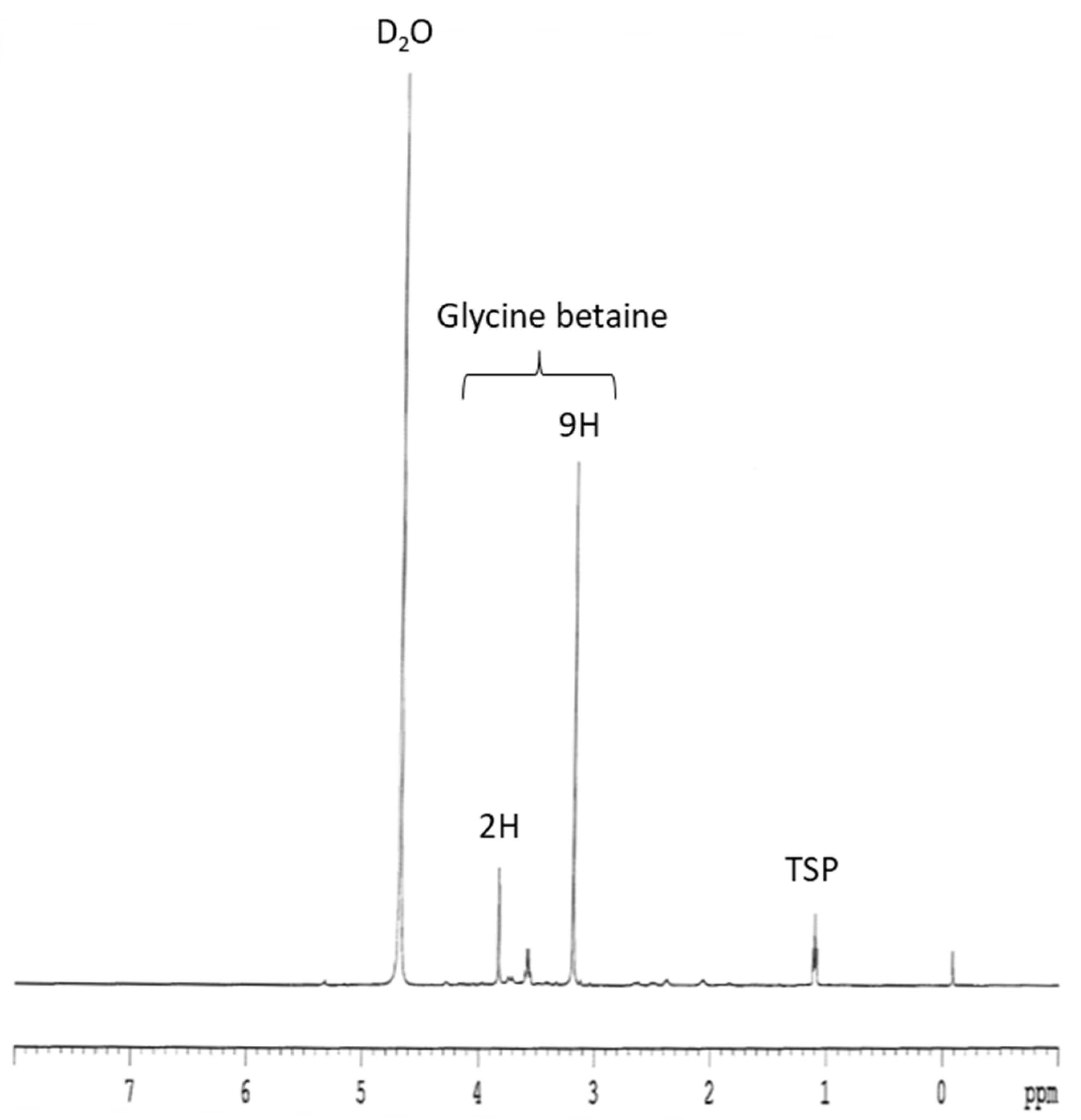
2.4. Glycine Betaine Extraction and Quantification

One hundred milligrams of each Salicornia powder were extracted with 1.5 mL of 30% ethanol, vortexed quickly, agitated for 15 min, and centrifuged at 4000× g for 5 min. These extraction steps were reproduced twice on the pellet. The supernatants collected after each extraction were then gathered and lyophilized.
Glycine betaine content in Salicornia aerial part was explored using 1H NMR spectroscopy. Dry extracts were resuspended in 800 µL of D2O. A BRUKER Avance 500 spectrometer equipped with a BBO probe was used. The spectra were performed with a 30° pulse using a 10 s delay at 298 °K. Calculation of glycine betaine concentration was based on integration of its NMR peaks. Areas of peaks corresponding to this metabolite were compared to those of trimethylsilyl propionate used as a standard with known concentration.

2.5. Extraction and Quantification of Total Phenolics, Flavonoids, and Tannins

Samples were extracted with 80% (v/v) aqueous methanol, as reported by Tavarini et al. [21]. The collected hydroalcoholic extracts were used to quantify total phenolics, flavonoids, and tannins.
Phenolic compounds were quantified by the Folin–Ciocalteu assay [22] by measuring the absorbance of the reaction mix at 750 nm and expressing their concentration as mg of gallic acid equivalents (GAE) g−1 FW.
Flavonoid concentration was determined by recording the absorbance at 510 nm of the reaction mix prepared following the method reported by Kim et al. [23]. Flavonoid concentration was expressed as mg of catechin equivalents (CAE) g−1 FW.
Tannin concentration was determined as the difference between phenolic concentration measured before and after tannin precipitation, according to the procedure reported by Makkar [24]. Thanks to their ability to form insoluble complexes with polyvinylpolypyrrolidone (PVPP), tannins can be removed from the hydroalcoholic extract by centrifugation, allowing the recovery of tannin-free supernatant.

2.6. Extraction and Quantification of Chlorophylls and Carotenoids

Pigments were extracted under dimmed room light in 100% HPLC-grade acetone with 1 mM sodium ascorbate according to the method reported by Castagna et al. [25]. Chlorophyll and carotenoid profile was achieved by HPLC separation (Spectra System P4000HPLC, UV 6000 LP photodiode array detector; Thermo Fisher Scientific, Waltham, MA, USA) using a Zorbax ODS column (SA, 5 μm particle size, 250 × 4.6 mm; Phenomenex, Castel Maggiore, Italy). The elution gradient is reported in Table 1. The detector was set at 445 nm, and pigments were quantified (mg g−1 FW) using calibration curves of commercial standards of chlorophyll a, chlorophyll b, lutein, and β-carotene (Sigma-Aldrich, Milan, Italy).

2.7. Evaluation of the In Vitro Antioxidant Activity

Antioxidant activity of the hydroalcoholic extract was determined by the ABTS (2,2-azinobis (3-ethylbenzothiazoline-6-sulphonic acid)) and the ferric-reducing antioxidant power (FRAP) assays.
ABTS test was carried out following the procedure reported by Re et al. [26]. based on the ability of antioxidants to reduce pre-formed ABTS●+ radicals to ABTS. The absorbance of the reaction mix was read at 734 nm after 4 min incubation, and the percentage inhibition was calculated against a blank control. The results were expressed as μmol Trolox equivalent (TE) g−1 FW.
The FRAP assay, based on the ability of antioxidant compounds to reduce the colorless ferric-tripyridyltriazine (Fe3+-TPTZ) complex to the blue ferrous-tripyridyltriazine (Fe2+-TPTZ) complex, was determined according to Benzie and Strain [27]. The absorbance was recorded at 593 nm, and the results were expressed as μmol of Fe (II) g−1 FW.

2.8. Evaluation of the In Vitro Antimicrobial Activity

A 30% ethanol extract was prepared from 1 g of Salicornia powders following the same procedure as for GB analysis. Dried extract was then resuspended in methanol, and its antimicrobial activity was screened against eight foodborne bacterial strains following the broth dilution method. The tested microorganisms included the Gram-positive Bacillus cereus (ATCC 6464), Bacillus subtilis (ATCC 6633), Listeria monocytogenes (ATCC 35152), Micrococcus luteus (ATCC 10240), and Staphylococcus aureus subsp. aureus (ATCC 33862) and the Gram-negative Escherichia coli (ATCC 4157), Pseudomonas aeruginosa (ATCC 27853), and Salmonella enterica (ATCC 13314). Bacterial strains were grown in liquid nutrient broth (Difco Surrey, England) at 37 °C for 24 h before being used.
Aliquots of Salicornia extract, corresponding to 2, 10, or 50 μg of dried extract, were dropped in sterile 96-well microplates (Nunc, Fisher Bioblok) in 6 replicates. After complete evaporation of the solvent, 100 μL of microorganism suspension (102 cells/mL) obtained by dilution from the culture tube was added to each well. Microbial suspension was used alone in some wells as positive control or in the presence of antibiotic mixture (5 mg mL−1 of streptomycin and 10 mg mL−1 of penicillin G) as negative control. Then, the microplates were aseptically sealed, agitated, and incubated for 24 h at 37 °C. Finally, microorganism growth was estimated by reading the absorbance in each well at 405 nm with a microplate spectrophotometer (Multiskan MCC/340, Titertek). Antimicrobial activity was expressed in percentage of growth inhibition using the following formula:
Growth inhibition (%) = [1 − (ASample − AC−)/(AC+ − AC−)] × 100,
where AC− and AC+ are the absorbances of the negative and positive controls, respectively.

2.9. Cytotoxic Effect and Evaluation of Inflammatory Mediators’ Expression in HT-29 Cells

The HT-29 cell line (DSMZ, Germany) was grown as previously described [28], and cell treatments were performed using a complete medium without FBS and phenol red. A toxicity curve using increasing concentrations of the hydroalcoholic extract (5, 10, 25, 50, 75, 100, 200, 500, and 1000 µg mL−1) of the edible portion of Salicornia plants cultivated in monoculture was performed, and cell viability was evaluated by the MTT assay according to Gabriele et al. [29].
Further experiments were carried out on HT-29 cells exposed for 24 h with or without 5 or 25 ng mL−1 of tumor necrosis factor α (TNFα) after 1 h pre-treatment with or without 50 and 200 µg mL−1 of Salicornia extract. Total RNA extraction, reverse transcription, and quantitative real-time PCR were performed as previously described [28]. Gene expression of IL-8 (CXC motif chemokine ligand 8) and COX-2 (prostaglandin-endoperoxide synthase 2) inflammatory markers was calculated by the 2−ΔΔCT relative quantification method using β-actin as gene housekeeping [30].

2.10. Statistical Analysis

Differences in the nutritional and nutraceutical parameters of Salicornia plants attributable to the cultivation method (monoculture vs. intercropping) were evaluated by Student’s t-test (n = 4, p ≤ 0.05).
One-way ANOVA followed by post hoc Tukey–Kramer test (p < 0.05) was applied to check the influence of the different concentrations of Salicornia extract on cell viability and markers of inflammation.
Statistical analysis was performed using the JMP software (SAS Institute, Inc., Cary, NC, USA). Data are expressed as mean ± standard error (SE).

3. Results

3.1. Nutrient Composition

3.1.1. Proximate Percentage Composition

The mean proximate composition of the edible portion of Salicornia plants grown in monoculture or in intercropping with tomato plants is reported in Table 2. No significant differences due to the cultivation method were observed for any parameter. The high percentage of ashes detected in these samples is related to the elevated Na concentration, which was 9.07 ± 0.58 and 7.71 ± 0.75 g 100 g−1 in the S and S-T samples, respectively.

3.1.2. Fatty Acid Composition

In accordance with the invariance of lipid content, the fatty acid composition did not undergo modification following the cultivation method. Interestingly, Salicornia plants, irrespective of being cultivated in monoculture or consociated with tomato, presented a high percentage of PUFAs, particularly those of the ω-3 series (PUFAn3), and a very low ω-3/ω-6 ratio (Table 3).

3.2. Glycine Betaine

The 1H-NMR spectrum of the hydroalcoholic extract of Salicornia aerial parts showed the predominance of glycine betaine among polar osmolytes since no other significant signals could be easily observed (Figure 1). Noteworthy, there were no significant differences in the NMR spectrum of Salicornia tissues cultivated alone or intercropped with tomato plants. From the spectrum, the GB level could be quantified, and its concentration was 5.99 and 6.26 g 100 g−1 for Salicornia cultivated in monoculture or the mixed system, respectively.

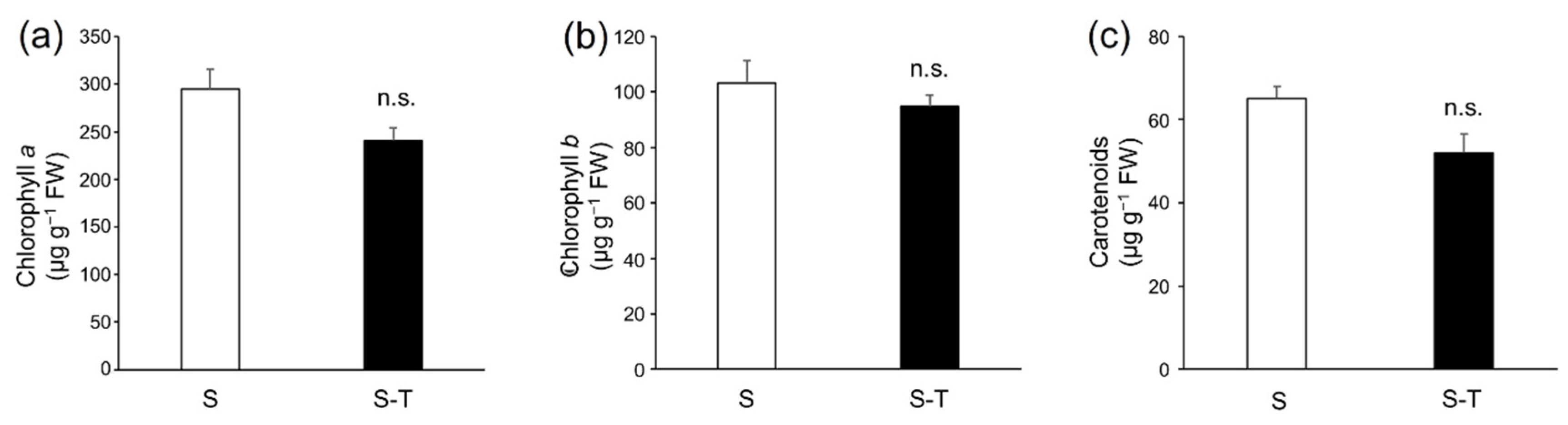
3.3. Chlorophylls and Carotenoids

As observed for most parameters, chlorophyll a and b were also present at similar concentrations in Salicornia grown in monoculture or consociated with tomato (Figure 2a,b). Moreover, carotenoid concentration underlined the absence of differences attributable to the cultivation method (Figure 2c). Irrespective of being cultivated in monoculture or the intercropping system, Salicornia contained β-carotene and the three xanthophylls lutein, neoxanthin, and violaxanthin which were similarly concentrated in the two samples. Lutein was the most concentrated carotenoid, accounting for about half of the total carotenoid concentration (50–52% for the S-T and S samples, respectively), followed by β-carotene, which ranged from 24% (S) to 31% (S-T).

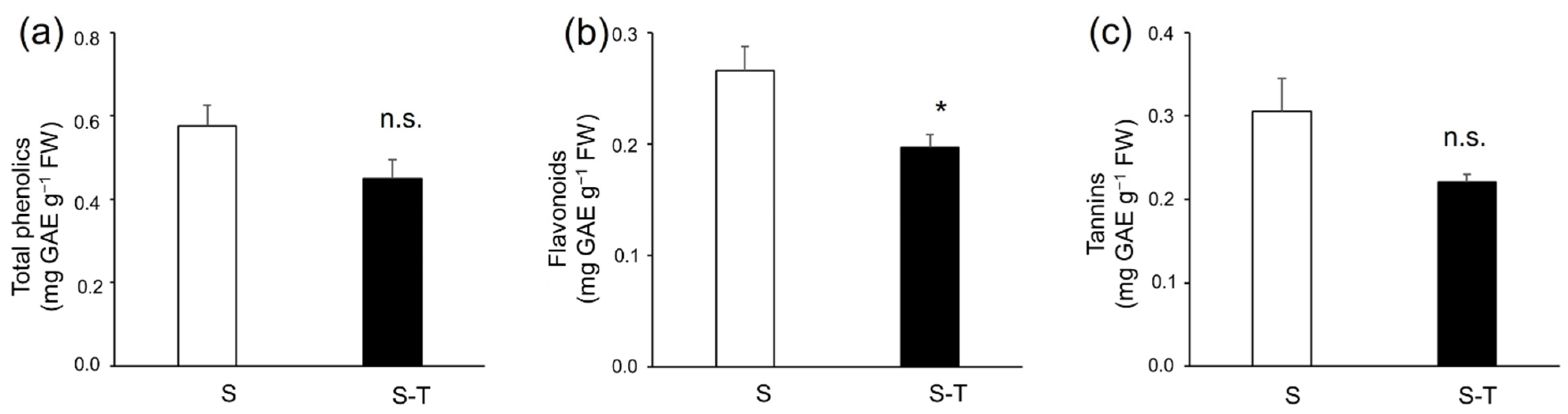
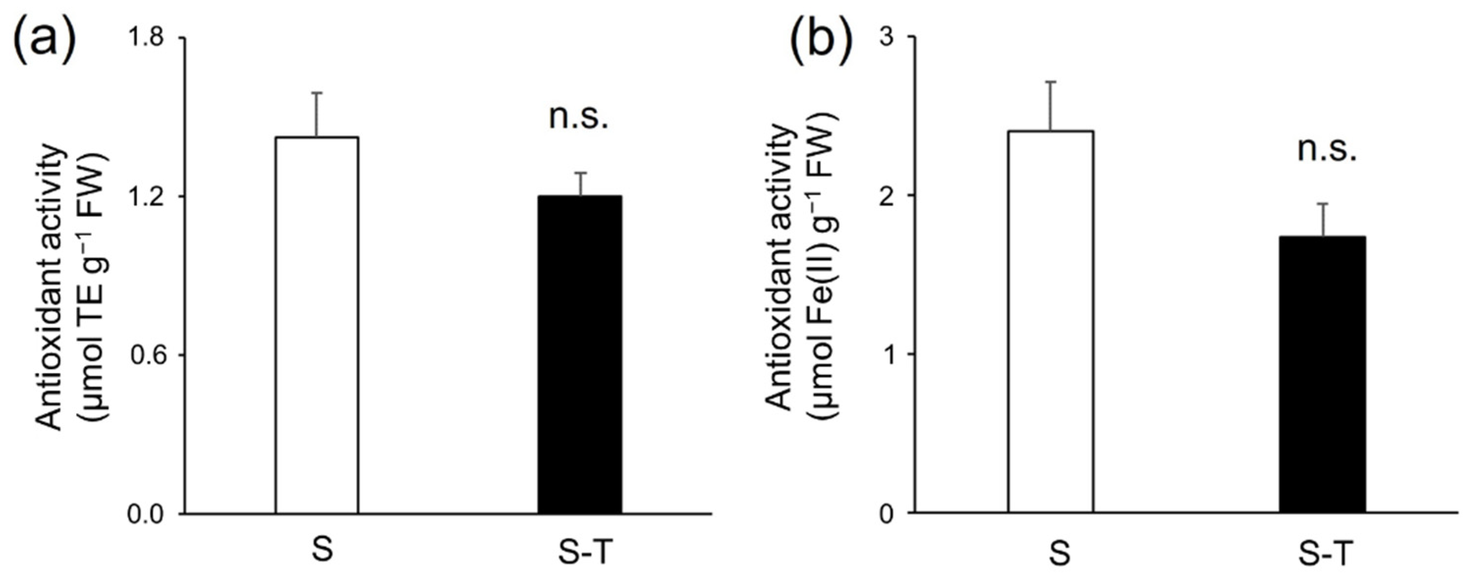
3.4. Total Phenolics, Flavonoids, Tannins, and Antioxidant Activity

The concentration of total phenolics, flavonoids, and tannins is reported in Figure 1. The cultivation method had no effect on the concentration of total phenols and tannins (Figure 3a,b), while it affected the flavonoid level. Specifically, the growth of Salicornia in consociation with tomato plants led to a significant decrease in flavonoid concentration (−26%, Figure 3c).
In accordance with the behavior shown by the total phenolic concentration, the antioxidant capacity of the hydroalcoholic extracts of the edible part of Salicornia, measured by both ABTS and FRAP assays, was similar in plants grown in monoculture or intercropped with tomato (Figure 4a,b).

3.5. Antimicrobial Activity

Antimicrobial screening of Salicornia extract was evaluated by means of a microdilution test of a number of Gram + and Gram − bacterial strains. On the whole, no significant growth inhibition was observed for seven out of the eight strains tested. Interestingly, however, 50 μg of Salicornia extract induced 80% inhibition of Bacillus subtilis growth so that the minimal inhibitory concentration (MIC) could be estimated at about 1 mg mL−1.

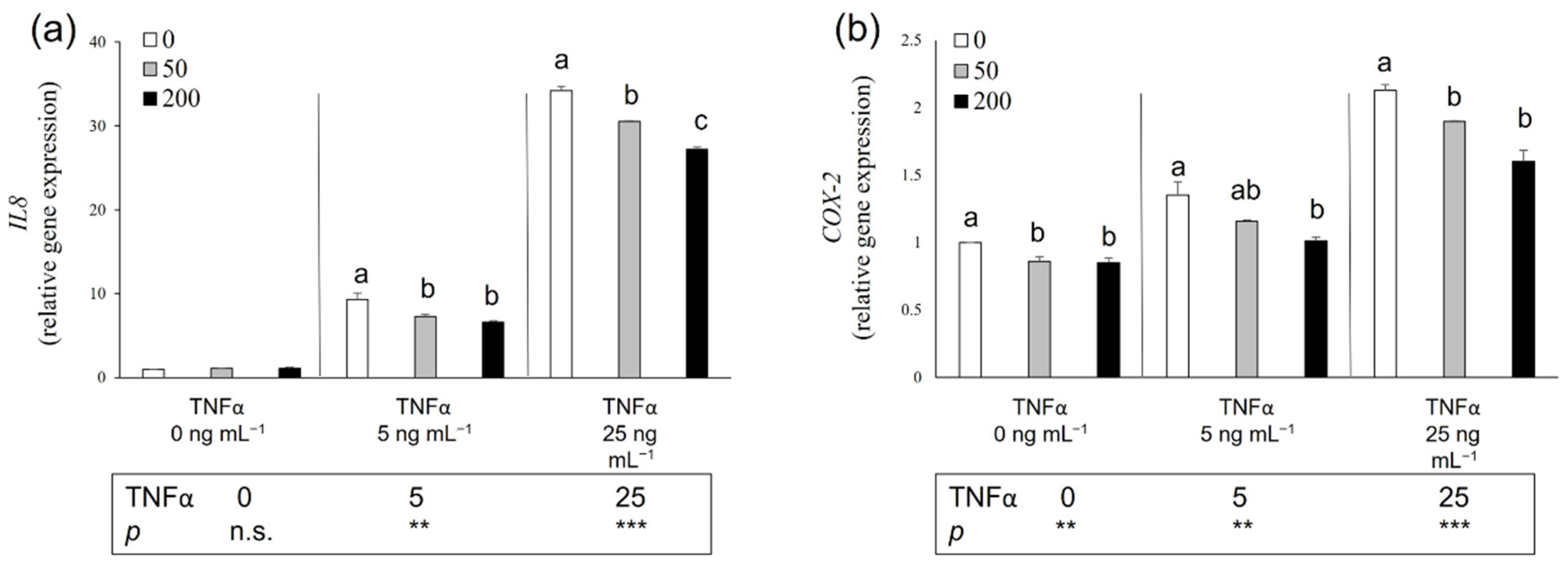
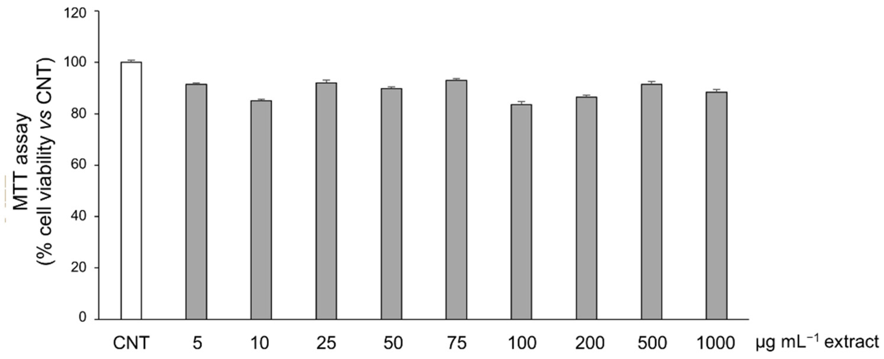
3.6. Potential Anti-Inflammatory Effect of Salicornia Extract on HT-29 Cells

Since the proximate composition and the nutraceutical content, except for flavonoids, were not influenced by the farming system, the assays were carried out using extracts from Salicornia grown in monoculture. To detect possible cytotoxic effects of the edible portion of Salicornia, we performed a toxicity curve using 5–1000 µg mL−1 as a range of extract concentrations. The treatment effect was evaluated in terms of cellular viability using the MTT assay. As shown in Figure 5, the HT-29 viability was not significantly affected following 24 h of exposure to each tested Salicornia concentration.
In this study, we assessed the potential protective effect of the edible Salicornia portion against TNFα-induced inflammatory alterations on the HT-29 human colonic adenocarcinoma cell line. Herein, the effects of 24 h exposure to 5 and 25 ng mL−1 of NFα, following 1 h pre-treatment with 50 and 200 µg mL−1 of Salicornia extract, were evaluated in terms of the altered gene expression of IL-8 and COX-2, both involved in the inflammation pathway. As shown in Figure 6a,b, Salicornia extracts did not affect the basal expression levels of the IL-8 gene while significantly decreasing the COX-2 ones compared to the control cells. The exposure of HT-29 to 5 and 25 ng mL−1 of TNFα resulted in a significant up-regulation of both IL-8 and COX-2 genes compared to the control cells. Conversely, at both tested doses, Salicornia pre-treatments induced a significant reduction in IL-8 expression. A similar inhibitory effect, depending on Salicornia concentration at a lower inflammatory grade, was observed for COX-2 levels following both Salicornia pre-treatments.

4. Discussion

This study was carried out in the framework of research aimed to evaluate the effectiveness of farming systems based on the cultivation of crops in consociation with halophytes in allowing crop cultivation in salt-affected soils. Besides the primary objective of improving the crop yield, the research also aimed to valorize the halophyte biomass in view of its possible use as food, feed, or a source of natural compounds with beneficial effects on animal and human health.

4.1. Nutrient Composition and Fatty Acid Profile

The proximate composition of S. europaea, irrespective of the cultivation method, highlighted a very high ash content, comparable with the values (29.2–31.6%) reported by Barreira et al. [31] for Arthrocnemum macrostachyum L., Sarcocornia perennis alpini L., and S. ramosissima L. collected in South Portugal (the soil salinity concentration was not specified) but lower than the nearly 50% ash content observed in S. bigelovii L. cultivated in California (27.5 dS m−1 EC; 8.4 pH; 210.6 mM Na+ concentration) [32] and S. ramosissima collected from a salt marsh in Central Portugal (the soil salinity concentration was not specified) [33]. The high ash content is a typical feature of halophytes and reflects the elevated amount of minerals that these plants accumulate in their tissues in relation to the saline environment where they grow.
The fiber content of S. europaea, cultivated both in monoculture and intercropped with tomato, was particularly elevated, in accordance with the values measured in other halophyte species [31,32]. NDF, besides being an index of forage quality, can be useful to estimate the amount of insoluble fiber in the human diet, important for its role in reducing the risk of many pathologies, among which cancer [34] and cardiovascular and coronary heart disease [35].
Our Salicornia samples seem to be a valuable source of proteins and lipids, as compared to the variable range of these nutrients reported in different halophyte species. Comparable levels of crude proteins were observed in S. bigelovii and Atriplex lentiformis (Torr.) [32] and in A. macrostachyum [31], while very lower percentages were present in wild Sarcocornia perennis perennis and S. ramosissima [31,33] and even less than 2% in S. ramosissima cultivated in soilless systems with different salt concentrations, ranging from 35 to 465 mM of NaCl [36].
Concerning the lipids, a high quantity of SFA was observed. High percentages of SFA are a common trait of halophyte plants, probably linked to their adaptation and resistance to a saline environment. It is, however, worth noting the high percentage of PUFAn3 detected in Salicornia ramets, accounting for about 44.5% of total lipids, that makes the lipid profile of these plants very interesting from a nutritional point of view. The extremely low n6/n3 ratio (about 2:1) is even below the recommended dietary n6/n3 ratio for health benefits (1:1–2:1). Considering that the n6/n3 ratio of Western eating habits ranges from 15:1 to 16.7:1 [37], consumption of Salicornia is advisable to achieve a more balanced FA intake. A positive role of PUFAn3 in preventing inflammation and reducing the onset of cancer, cardiovascular diseases, obesity, diabetes, and neurological disorders is recognized [37,38]. The interesting lipid profile of Salicornia is reflected in the extremely low AI and TI, which are used to estimate the potential benefits of a specific food related to the cardiovascular apparatus [39]. Barreira et al. [31] described the AI and TI ranging from 0.45 to 0.73 and from 0.21 to 0.37, respectively, in four halophyte species. According to these authors, the observed values were similar to those measured for tuna fish, which is commonly considered a valuable source of healthy unsaturated FAs. In our study, the AI and TI were even a little bit lower than those of A. macrostachyum, the best performing species described by Barreira et al. [31].

4.2. Glycine Betaine

The presence of the quaternary ammonium compound, glycine betaine, was reported in numerous halophytes from a large number of families [40] and in particular Salicornia species [41]. However, the osmolyte was particularly abundant in the plants cultivated here, and such GB concentration (6% DW) has never been reported hitherto.

4.3. Bioactive Compounds

Carotenoids have important nutritional and healthy properties, among which antioxidant and antitumoral activity, protection against cardiovascular and neurological diseases, provitamin A role (α-carotene, β-carotene, and β-cryptoxanthin), and prevention of age-related macular disease (lutein) [42]. Chlorophylls, despite usually being considered only an aesthetical and senescence-related trait that can guide consumer preference, may contribute to the antioxidant potential of green vegetables and fruits, as demonstrated by an EPR study in basil extracts [43]. Cárdenas-Pérez et al. [12] observed a reduction in the chlorophylls and carotenoids of S. europaea as the salinity of the nutrient solution increased, though their concentration remained stable at levels above 400 mM NaCl. Generally, cultivated halophytes were less rich in chlorophylls and carotenoids than field-collected ones [44]. Although our Salicornia was cultivated under greenhouse conditions, as in the research carried out by Cárdenas-Pérez et al. [12], chlorophyll levels were lower than the levels reported by these authors and more similar to the levels observed in other wild halophytes [44].
Phenolic compounds, and the subclass of flavonoids, have many recognized bioactive properties, and diets rich in these compounds have been associated with benefits for human health due to their antioxidant, anti-inflammatory, anti-ageing, and antimicrobial activities [45,46,47]. Tannins, besides sharing the same important health-protective functions as flavonoids, also possess interesting potential in animal feeding and veterinary [48,49].
The phenolic and flavonoid concentration of our Salicornia extract was lower than the values reported for other halophytes [50,51,52,53] among which also S. europaea [51].
However, Salicornia patua L. harvested in different Spanish locations (the soil salinity concentrations were not specified) presented values ranging from 2.989 to 4.209 mg g−1 dw [54], very similar to those measured in our samples (2.8–3.8 mg g−1).
Cultivation under greenhouse may negatively affect the synthesis of many compounds because of the lower light intensity as well as the different light spectral composition. Indeed, with light being an important factor that regulates the biosynthesis of flavonoids [55], the lower flavonoid content detected in intercropped Salicornia may be related to a shadow effect played by tomato plants. Moreover, the irrigation regimen and other environmental and climatic factors can interact with each other in an unpredictable way to determine the final phenolic (and flavonoid) concentration. Indeed, it is well known that the biosynthesis and accumulation of most secondary metabolites are influenced by a multitude of external variables, such as agricultural practices and pedoclimatic conditions. Castañeda-Loaiza et al. [44] reported that the phenolic and flavonoid content of Mesembryanthemum nodiflorum L. and Sarcocornia fruticosa L. was lower in cultivated (5.1 pH; 2.3 dS m−1 EC) than in wild plants collected in Spain (8.7 pH; 1.5 dS m−1 EC) and Portugal (8.4 pH; 3.7 dS m−1 EC). Interestingly, for Suaeda maritima L., the lowest content was instead detected in plants naturally grown in Spanish saltmarshes. Similarly, Kang et al. [56] observed that the cultivation of Salicornia herbacea L. with seawater (6.3% salinity) could result in high phenolic and flavonoid content as compared to the naturally grown one.
In accordance with the invariance of total phenolic content, the antioxidant activity of Salicornia extracts was unaffected by the cultivation method. The lower phenolic content detected in our samples, as compared to halophytes grown in open-air conditions, reflected in lower antioxidant activity. Costa et al. [57] reported about 10- to 20-fold phenolic content and antioxidant activity than ours in green, pink, and red Sarcocornia ambigua L. biotypes irrigated with saline effluent (water EC and pH of 46.0 dS m−1 and 8.56, respectively; 6.0 dS m−1 soil EC). These authors underlined the positive influence of open-field farming with respect to greenhouse cultivation. Indeed, the phenolic content of S. ambigua cultivated in a greenhouse (3.6 g L−1 water salinity; 8.0 pH) was 41 mg 100 g−1 F.W. [58], a value very similar to the content of our Salicornia plants.

4.4. Biological Activity

A significant antimicrobial activity of Salicornia extract was found against Bacillus subtilis. This finding is very interesting since plant antimicrobials are mainly apolar compounds (essential oils, terpenes, etc.). Here, the antimicrobial effect is due to the polar compound(s), which we have mentioned hereabove is mainly constituted of carbohydrates and glycine betaine. If the bacteriostatic or lethal activity has been already reported for betaine analogs [59], it is unlikely that such an antibacterial effect could be due to GB since it is generally considered an efficient osmoprotectant for most bacteria. Further experiments, including extract fractioning and bioguided study, are in progress to identify the potent antimicrobial agent in Salicornia aerial parts.
The potential cytotoxic effect of 24 h exposure of HT-29 cell line to increasing concentrations of S. europaea methanol extract (5–1000 µg mL−1) was estimated by MTT assay as cellular viability. Our findings demonstrate that the S. europaea extract was not toxic and did not alter the HT-29 viability by 24 h treatment, in line with Kang et al. [60] on the same cell line following 24 h exposure to 70% methanol Salicornia herbacea L. extract. Conversely, at higher concentration ranges and treatment conditions, Altay et al. [61] highlighted time- and dose-dependent cytotoxic effects on HT-29 cells following 48 and 72 h exposure to Salicornia freitagii L. extract.
To the best of our knowledge, this is the first study evaluating the potential protective effect of S. europaea methanol extract against TNFα-induced inflammatory alterations on the HT-29 human colonic adenocarcinoma cell line. The pre-treatment of HT-29 cells with 50 and 200 µg mL−1 of S. europaea extract was effective in significantly reducing the expression levels of the IL-8 and COX-2 genes, two markers of the inflammatory pathway, without affecting IL-8 basal levels while decreasing the COX-2 ones compared to the control cells. These findings are in line with the anti-inflammatory activity described by Sun et al. [62] on sepsis-induced acute lung injury and the anti-neuro-inflammatory effect observed by Kim et al. [63] in LPS-stimulated BV-2 microglial cells. Indeed, Salicornia plants contain several bioactive components, including β-cyanines, isoflavones, flavanones, etc., known for their antioxidant, anti-inflammatory, and neuroprotective properties [64].

5. Conclusions

Halophyte plants have great potential as cash crops due to their many uses (e.g., food and feed, source of bioactive compounds, etc.). Their consociation with salt-sensitive crops is a promising solution to overcome the progressive salinization of agricultural soils. Our results demonstrate that the cultivation of Salicornia europaea intercropped with tomato plants did not generally affect the content of nutrients and nutraceutical compounds. This finding contributes to valorizing the halophyte biomass produced in mixed crop systems. Salicornia exhibited antimicrobial activity against Bacillus subtilis and a good anti-inflammatory effect, without any significant cytotoxic consequences. Further studies on the content and profile of bioactive compounds and on the bioactivity of Salicornia extract are highly encouraged to confirm the potential pharmaceutical/veterinary use of this plant.

Supplementary Materials

The following supporting information can be downloaded at: https://www.mdpi.com/article/10.3390/horticulturae8090828/s1, Figure S1: Experimental design showing the two kinds of plots used in this research: Salicornia monoculture and Salicornia intercropped with tomato plants.

Author Contributions

Conceptualization, A.C., V.L., C.M. and A.R.; methodology A.C., M.G., V.L., C.M., G.C. and A.R.; formal analysis, A.C., M.G. and M.S.; investigation, G.M., M.G., A.S., X.D., G.F. and M.S.; resources, A.C., V.L., C.M., G.C. and A.R.; data curation, A.C., G.M., C.M., G.C. and M.S.; writing—original draft preparation, A.C., M.G., A.S. and C.M.; writing—review and editing, A.C., M.G., V.L., A.S., C.M., G.C., M.S. and A.R.; supervision, A.C., V.L., C.M., G.C. and A.R.; project administration, C.M. and A.R.; funding acquisition, C.M. and A.R. All authors have read and agreed to the published version of the manuscript.

Funding

This research was part of the project “HaloFarMs” by PRIMA, a program supported by the European Union, and by the following funding agencies: Italian Ministry of Education, University and Research (MIUR), Italy, and Agence Nationale de la Recherche (ANR), France.

Institutional Review Board Statement

Not applicable.

Informed Consent Statement

Not applicable.

Data Availability Statement

The data presented in this study are available within the article.

Acknowledgments

The authors would like to thank Marco Ginanni (Centre for Agri-environmental Research E. Avanzi, University of Pisa, via Vecchia di Marina 6, 56122 San Piero a Grado (Pisa), Italy) for his valuable support in the set-up of the greenhouse experimental conditions and plant cultivation.

Conflicts of Interest

The authors declare no conflict of interest.

References

  1. United Nations, Department of Economic and Social Affairs, Population Division. World Population Prospects 2019: Volume I: Comprehensive Tables; United Nations, Department of Economic and Social Affairs, Population Division: New York, NY, USA, 2019. [Google Scholar]
  2. Abdelly, C.; Barhoumi, Z.; Ghnaya, T.; Debez, A.; Ben Hamed, K.; Ksouri, R.; Talbi, O.; Zribi, F.; Ouerghi, Z.; Smaoui, A.; et al. Potential utilisation of halophytes for the rehabilitation and valorisation of salt-affected areas in Tunisia. In Biosaline Agriculture and Salinity Tolerance in Plants; Oztürk, M., Waisel, Y., Khan, M.A., Görk, G.G., Eds.; Birkhäuser Verlag: Basel, Switzerland, 2006; pp. 163–172. [Google Scholar]
  3. Daoud, S.; Elbrik, K.; Tachbibi, N.; Bouqbis, L.; Brakez, M.; Harrouni, M.C. The Potential Use of Halophytes for the Development of Marginal Dry Areas in Morocco. In Halophytes for Food Security in Dry Lands; Khan, M.A., Ozturk, M., Gul, B., Ahmed, M.Z., Eds.; Academic Press: San Diego, CA, USA, 2016; pp. 141–156. ISBN 978-0-12-801854-5. [Google Scholar]
  4. Duarte, B.; Feijão, E.; Pinto, M.V.; Matos, A.R.; Silva, A.; Figueiredo, A.; Fonseca, V.F.; Reis-Santos, P.; Caçador, I. Nutritional Valuation and Food Safety of Endemic Mediterranean Halophytes Species Cultivated in Abandoned Salt Pans under a Natural Irrigation Scheme. Estuar. Coast. Shelf Sci. 2022, 265, 107733. [Google Scholar] [CrossRef]
  5. Pereira, C.G.; Barreira, L.; da Rosa Neng, N.; Nogueira, J.M.F.; Marques, C.; Santos, T.F.; Varela, J.; Custódio, L. Searching for New Sources of Innovative Products for the Food Industry within Halophyte Aromatic Plants: In Vitro Antioxidant Activity and Phenolic and Mineral Contents of Infusions and Decoctions of Crithmum Maritimum L. Food Chem. Toxicol. 2017, 107, 581–589. [Google Scholar] [CrossRef] [PubMed]
  6. Souid, A.; Bellani, L.; Magné, C.; Zorrig, W.; Smaoui, A.; Abdelly, C.; Longo, V.; ben Hamed, K. Physiological and Antioxidant Responses of the Sabkha Biotope Halophyte Limonium Delicatulum to Seasonal Changes in Environmental Conditions. Plant Physiol. Biochem. 2018, 123, 180–191. [Google Scholar] [CrossRef]
  7. Souid, A.; Gabriele, M.; Longo, V.; Pucci, L.; Bellani, L.; Smaoui, A.; Abdelly, C.; ben Hamed, K. Salt Tolerance of the Halophyte Limonium Delicatulum Is More Associated with Antioxidant Enzyme Activities than Phenolic Compounds. Funct. Plant Biol. 2016, 43, 607–619. [Google Scholar] [CrossRef]
  8. Petropoulos, S.A.; Karkanis, A.; Martins, N.; Ferreira, I.C.F.R. Edible Halophytes of the Mediterranean Basin: Potential Candidates for Novel Food Products. Trends Food Sci. Technol. 2018, 74, 69–84. [Google Scholar] [CrossRef]
  9. Qasim, M.; Abideen, Z.; Adnan, M.Y.; Gulzar, S.; Gul, B.; Rasheed, M.; Khan, M.A. Antioxidant Properties, Phenolic Composition, Bioactive Compounds and Nutritive Value of Medicinal Halophytes Commonly Used as Herbal Teas. S. Afr. J. Bot. 2017, 110, 240–250. [Google Scholar] [CrossRef]
  10. Gyawali, R.; Hayek, S.A.; Ibrahim, S.A. Plant Extracts as Antimicrobials in Food Products: Mechanisms of Action, Extraction Methods, and Applications. In Handbook of Natural Antimicrobials for Food Safety and Quality; Elsevier Ltd.: Amsterdam, The Netherlands, 2015; pp. 49–68. ISBN 9781782420422. [Google Scholar]
  11. ben Hamed, K.; Castagna, A.; Ranieri, A.; García-Caparrós, P.; Santin, M.; Hernandez, J.A.; Espin, G.B. Halophyte Based Mediterranean Agriculture in the Contexts of Food Insecurity and Global Climate Change. Environ. Exp. Bot. 2021, 191, 104601. [Google Scholar] [CrossRef]
  12. Cárdenas-Pérez, S.; Rajabi Dehnavi, A.; Leszczyński, K.; Lubińska-Mielińska, S.; Ludwiczak, A.; Piernik, A. Salicornia europaea L. Functional Traits Indicate Its Optimum Growth. Plants 2022, 11, 1051. [Google Scholar] [CrossRef]
  13. Ventura, Y.; Wuddineh, W.A.; Shpigel, M.; Samocha, T.M.; Klim, B.C.; Cohen, S.; Shemer, Z.; Santos, R.; Sagi, M. Effects of Day Length on Flowering and Yield Production of Salicornia and Sarcocornia Species. Sci. Hortic. 2011, 130, 510–516. [Google Scholar] [CrossRef]
  14. Ameixa, O.M.C.C.; Rebelo, J.; Silva, H.; Pinto, D.C.G.A. Gall Midge Baldratia Salicorniae Kieffer (Diptera: Cecidomyiidae) Infestation on Salicornia Europaea L. Induces the Production of Specialized Metabolites with Biotechnological Potential. Phytochemistry 2022, 200, 113207. [Google Scholar] [CrossRef]
  15. Patel, S. Salicornia: Evaluating the Halophytic Extremophile as a Food and a Pharmaceutical Candidate. 3 Biotech 2016, 6, 104. [Google Scholar] [CrossRef] [PubMed]
  16. Helrich, K. Association of Official Analytical Chemist, Official Methods of Analysis of the AOAC, 15th ed.; AOAC: Arlington, VA, USA, 1990. [Google Scholar]
  17. Goering, H.K.; Van Soest, P.J. Forage Fibre Analysis (Apparatus, Reagents, Procedures and Some Applications); US Agricultural Research Service: Washington, DC, USA, 1970. [Google Scholar]
  18. Vogel, K.P.; Pedersen, J.F.; Masterson, S.D.; Toy, J.J. Evaluation of a Filter Bag System for NDF, ADF, and IVDMD Forage Analysis. Crop Sci. 1999, 39, 276–279. [Google Scholar] [CrossRef]
  19. Christie, W.W. Preparation of ester derivatives of fatty acids for chromatographic analysis. In Advances in Lipid Methodology—Two; Christie, W.W., Ed.; Oily Press: Dundee, Scotland, 1993; pp. 69–111. [Google Scholar]
  20. Ulbricht, T.L.v.; Southgate, D.A.T. Coronary Heart Disease: Seven Dietary Factors. Lancet 1991, 338, 985–992. [Google Scholar] [CrossRef]
  21. Tavarini, S.; Castagna, A.; Conte, G.; Foschi, L.; Sanmartin, C.; Incrocci, L.; Ranieri, A.; Serra, A.; Angelini, L.G. Evaluation of Chemical Composition of Two Linseed Varieties as Sources of Health-Beneficial Substances. Molecules 2019, 24, 3729. [Google Scholar] [CrossRef] [PubMed]
  22. Alonso Borbalán, Á.M.; Zorro, L.; Guillén, D.A.; García Barroso, C. Study of the Polyphenol Content of Red and White Grape Varieties by Liquid Chromatography-Mass Spectrometry and Its Relationship to Antioxidant Power. J. Chromatogr. A 2003, 1012, 31–38. [Google Scholar] [CrossRef]
  23. Kim, D.O.; Chun, O.K.; Kim, Y.J.; Moon, H.Y.; Lee, C.Y. Quantification of Polyphenolics and Their Antioxidant Capacity in Fresh Plums. J. Agric. Food Chem. 2003, 51, 6509–6515. [Google Scholar] [CrossRef]
  24. Makkar, H.P.S.; Blummel, M.; Borowy, N.K.; Becker, K. Gravimetric Determination of Tannins and Their Correlations with Chemical and Protein Precipitation Methods. J. Sci. Food Agric. 1993, 61, 161–165. [Google Scholar] [CrossRef]
  25. Castagna, A.; di Baccio, D.; Tognetti, R.; Ranieri, A.; Sebastiani, L. Differential Ozone Sensitivity Interferes with Cadmium Stress in Poplar Clones. Biol. Plant. 2013, 57, 313–324. [Google Scholar] [CrossRef]
  26. Re, R.; Pellegrini, N.; Proteggente, A.; Pannala, A.; Yang, M.; Rice-Evans, C. Antioxidant Activity Applying an Improved ABTS Radical Cation Decolorization Assay. Free. Radic. Biol. Med. 1999, 26, 1231–1237. [Google Scholar] [CrossRef]
  27. Benzie, I.F.F.; Strain, J.J. The Ferric Reducing Ability of Plasma (FRAP) as a Measure of “antioxidant Power”: The FRAP Assay. Anal. Biochem. 1996, 239, 70–76. [Google Scholar] [CrossRef] [Green Version]
  28. Caddeo, C.; Gabriele, M.; Nácher, A.; Fernàndez-Busquets, X.; Valenti, D.; Maria Fadda, A.; Pucci, L.; Manconi, M. Resveratrol and Artemisinin Eudragit-Coated Liposomes: A Strategy to Tackle Intestinal Tumors. Int. J. Pharm. 2021, 592, 120083. [Google Scholar] [CrossRef] [PubMed]
  29. Gabriele, M.; Pucci, L.; la Marca, M.; Lucchesi, D.; della Croce, C.M.; Longo, V.; Lubrano, V. A Fermented Bean Flour Extract Downregulates LOX-1, CHOP and ICAM-1 in HMEC-1 Stimulated by Ox-LDL. Cell. Mol. Biol. Lett. 2016, 21, 10. [Google Scholar] [CrossRef] [PubMed]
  30. Gabriele, M.; Pucci, L.; Árvay, J.; Longo, V. Anti-Inflammatory and Antioxidant Effect of Fermented Whole Wheat on TNFα-Stimulated HT-29 and NF-ΚB Signaling Pathway Activation. J. Funct. Foods 2018, 45, 392–400. [Google Scholar] [CrossRef]
  31. Barreira, L.; Resek, E.; Rodrigues, M.J.; Rocha, M.I.; Pereira, H.; Bandarra, N.; da Silva, M.M.; Varela, J.; Custódio, L. Halophytes: Gourmet Food with Nutritional Health Benefits? J. Food Compos. Anal. 2017, 59, 35–42. [Google Scholar] [CrossRef]
  32. Díaz, F.J.; Benes, S.E.; Grattan, S.R. Field Performance of Halophytic Species under Irrigation with Saline Drainage Water in the San Joaquin Valley of California. Agric. Water Manag. 2013, 118, 59–69. [Google Scholar] [CrossRef]
  33. Lopes, M.; Roque, M.J.; Cavaleiro, C.; Ramos, F. Nutrient Value of Salicornia Ramosissima—A Green Extraction Process for Mineral Analysis. J. Food Compos. Anal. 2021, 104, 104135. [Google Scholar] [CrossRef]
  34. Papandreou, D.; Noor, Z.T.; Rashed, M. The Role of Soluble, Insoluble Fibers and Their Bioactive Compounds in Cancer: A Mini Review. Food Nutr. Sci. 2015, 6, 53027. [Google Scholar] [CrossRef]
  35. Threapleton, D.E.; Greenwood, D.C.; Evans, C.E.L.; Cleghorn, C.L.; Nykjaer, C.; Woodhead, C.; Cade, J.E.; Gale, C.P.; Burley, V.J. Dietary Fibre Intake and Risk of Cardiovascular Disease: Systematic Review and Meta-Analysis. BMJ 2013, 347, f6879. [Google Scholar] [CrossRef]
  36. Lima, A.R.; Castañeda-Loaiza, V.; Salazar, M.; Nunes, C.; Quintas, C.; Gama, F.; Pestana, M.; Correia, P.J.; Santos, T.; Varela, J.; et al. Influence of Cultivation Salinity in the Nutritional Composition, Antioxidant Capacity and Microbial Quality of Salicornia Ramosissima Commercially Produced in Soilless Systems. Food Chem. 2020, 333, 127525. [Google Scholar] [CrossRef]
  37. Saini, R.K.; Keum, Y.S. Omega-3 and Omega-6 Polyunsaturated Fatty Acids: Dietary Sources, Metabolism, and Significance—A Review. Life Sci. 2018, 203, 255–267. [Google Scholar] [CrossRef]
  38. Shahidi, F.; Ambigaipalan, P. Omega-3 Polyunsaturated Fatty Acids and Their Health Benefits. Annu. Rev. Food Sci. Technol. 2018, 9, 345–381. [Google Scholar] [CrossRef] [PubMed]
  39. Garaffo, M.A.; Vassallo-Agius, R.; Nengas, Y.; Lembo, E.; Rando, R.; Maisano, R.; Dugo, G.; Giuffrida, D. Fatty Acids Profile, Atherogenic (IA) and Thrombogenic (IT) Health Lipid Indices, of Raw Roe of Blue Fin Tuna (Thunnus Thynnus L.) and Their Salted Product “Bottarga.”. Food Nutr. Sci. 2011, 2, 736–743. [Google Scholar] [CrossRef]
  40. Rodhes, D.; Hanson, A.D. Quaternary Ammonium and Tertiary Sulfonium Compounds in Higher Plants. Annu. Rev. Plant Physiol. Plant Mol. Biol. 2003, 44, 357–384. [Google Scholar]
  41. Moghaieb, R.E.A.; Saneoka, H.; Fujita, K. Effect of Salinity on Osmotic Adjustment, Glycinebetaine Accumulation and the Betaine Aldehyde Dehydrogenase Gene Expression in Two Halophytic Plants, Salicornia Europaea and Suaeda Maritima. Plant Sci. 2004, 166, 1345–1349. [Google Scholar] [CrossRef]
  42. Bohn, T.; Bonet, M.L.; Borel, P.; Keijer, J.; Landrier, J.F.; Milisav, I.; Ribot, J.; Riso, P.; Winklhofer-Roob, B.; Sharoni, Y.; et al. Mechanistic Aspects of Carotenoid Health Benefits—Where Are We Now? Nutr. Res. Rev. 2021, 34, 276–302. [Google Scholar] [CrossRef]
  43. Sgherri, C.; Pinzino, C.; Navari-Izzo, F.; Izzo, R. Contribution of Major Lipophilic Antioxidants to the Antioxidant Activity of Basil Extracts: An EPR Study. J. Sci. Food Agric. 2011, 91, 1128–1134. [Google Scholar] [CrossRef]
  44. Castañeda-Loaiza, V.; Oliveira, M.; Santos, T.; Schüler, L.; Lima, A.R.; Gama, F.; Salazar, M.; Neng, N.R.; Nogueira, J.M.F.; Varela, J.; et al. Wild vs Cultivated Halophytes: Nutritional and Functional Differences. Food Chem. 2020, 333, 127536. [Google Scholar] [CrossRef]
  45. Fan, X.; Fan, Z.; Yang, Z.; Huang, T.; Tong, Y.; Yang, D.; Mao, X.; Yang, M. Flavonoids—Natural Gifts to Promote Health and Longevity. Int. J. Mol. Sci. 2022, 23, 2176. [Google Scholar] [CrossRef]
  46. Maleki, S.J.; Crespo, J.F.; Cabanillas, B. Anti-Inflammatory Effects of Flavonoids. Food Chem. 2019, 299, 125124. [Google Scholar] [CrossRef]
  47. Barreca, D.; Trombetta, D.; Smeriglio, A.; Mandalari, G.; Romeo, O.; Felice, M.R.; Gattuso, G.; Nabavi, S.M. Food Flavonols: Nutraceuticals with Complex Health Benefits and Functionalities. Trends Food Sci. Technol. 2021, 117, 194–204. [Google Scholar] [CrossRef]
  48. Bonelli, F.; Turini, L.; Sarri, G.; Serra, A.; Buccioni, A.; Mele, M. Oral Administration of Chestnut Tannins to Reduce the Duration of Neonatal Calf Diarrhea. BMC Vet. Res. 2018, 14, 227. [Google Scholar] [CrossRef] [PubMed] [Green Version]
  49. Cappucci, A.; Mantino, A.; Buccioni, A.; Casarosa, L.; Conte, G.; Serra, A.; Mannelli, F.; Luciano, G.; Foggi, G.; Mele, M. Diets Supplemented with Condensed and Hydrolysable Tannins Affected Rumen Fatty Acid Profile and Plasmalogen Lipids, Ammonia and Methane Production in an in vitro Study. Ital. J. Anim. Sci. 2021, 20, 935–946. [Google Scholar] [CrossRef]
  50. Souid, A.; Della Croce, C.M.; Frassinetti, S.; Gabriele, M.; Pozzo, L.; Ciardi, M.; Abdelly, C.; Hamed, K.B.; Magné, C.; Longo, V. Nutraceutical Potential of Leaf Hydro-Ethanolic Extract of the Edible Halophyte Crithmum Maritimum L. Molecules 2021, 26, 5380. [Google Scholar] [CrossRef] [PubMed]
  51. Stanković, M.S.; Petrović, M.; Godjevac, D.; Stevanović, Z.D. Screening Inland Halophytes from the Central Balkan for Their Antioxidant Activity in Relation to Total Phenolic Compounds and Flavonoids: Are There Any Prospective Medicinal Plants? J. Arid. Environ. 2015, 120, 26–32. [Google Scholar] [CrossRef]
  52. Hulkko, L.S.S.; Chaturvedi, T.; Thomsen, M.H. Extraction and Quantification of Chlorophylls, Carotenoids, Phenolic Compounds, and Vitamins from Halophyte Biomasses. Appl. Sci. 2022, 12, 840. [Google Scholar] [CrossRef]
  53. Rodrigues, M.J.; Gangadhar, K.N.; Vizetto-Duarte, C.; Wubshet, S.G.; Nyberg, N.T.; Barreira, L.; Varela, J.; Custódio, L. Maritime Halophyte Species from Southern Portugal as Sources of Bioactive Molecules. Mar. Drugs 2014, 12, 2228–2244. [Google Scholar] [CrossRef] [PubMed]
  54. Sánchez-Gavilán, I.; Ramírez, E.; de la Fuente, V. Bioactive Compounds in Salicornia Patula Duval-Jouve: A Mediterranean Edible Euhalophyte. Foods 2021, 10, 410. [Google Scholar] [CrossRef]
  55. Santin, M.; Ranieri, A.; Castagna, A. Anything New under the Sun? An Update on Modulation of Bioactive Compounds by Different Wavelengths in Agricultural Plants. Plants 2021, 10, 1485. [Google Scholar] [CrossRef]
  56. Kang, S.; Kim, M.R.; Chiang, M.; Hong, J. Evaluation and Comparison of Functional Properties of Freshwater-Cultivated Glasswort (Salicornia Herbacea L.) with Naturally-Grown Glasswort. Food Sci. Biotechnol. 2015, 24, 2245–2250. [Google Scholar] [CrossRef]
  57. Costa, C.S.B.; Chaves, F.C.; Rombaldi, C.v.; Souza, C.R. Bioactive Compounds and Antioxidant Activity of Three Biotypes of the Sea Asparagus Sarcocornia Ambigua (Michx.) M.A.Alonso & M.B.Crespo: A Halophytic Crop for Cultivation with Shrimp Farm Effluent. S. Afr. J. Bot. 2018, 117, 95–100. [Google Scholar] [CrossRef]
  58. Pinheiro, I.; Arantes, R.; do Espírito Santo, C.M.; do Nascimento Vieira, F.; Lapa, K.R.; Gonzaga, L.V.; Fett, R.; Barcelos-Oliveira, J.L.; Seiffert, W.Q. Production of the Halophyte Sarcocornia Ambigua and Pacific White Shrimp in an Aquaponic System with Biofloc Technology. Ecol. Eng. 2017, 100, 261–267. [Google Scholar] [CrossRef]
  59. Cosquer, A.; Ficamos, M.; Jebbar, M.; Corbel, J.C.; Choquet, G.; Fontenelle, C.; Uriac, P.; Bernard, T. Antibacterial Activity of Glycine Betaine Analogues: Involvement of Osmoporters. Bioorganic Med. Chem. Lett. 2004, 14, 2061–2065. [Google Scholar] [CrossRef] [PubMed]
  60. Kang, S.; Kim, D.; Lee, B.H.; Kim, M.R.; Hong, J.; Chiang, M. Antioxidant Properties and Cytotoxic Effects of Fractions from Glasswort (Salicornia Herbacea) Seed Extracts on Human Intestinal Cells. Food Sci. Biotechnol. 2011, 20, 115–122. [Google Scholar] [CrossRef]
  61. Altay, A.; Celep, G.S.; Yaprak, A.E.; Baskose, I.; Bozoglu, F. Glassworts as Possible Anticancer Agents against Human Colorectal Adenocarcinoma Cells with Their Nutritive, Antioxidant and Phytochemical Profiles. Chem. Biodivers. 2017, 14, e1600290. [Google Scholar] [CrossRef] [PubMed]
  62. Sun, L.C.; Zhang, H.B.; Gu, C.D.; Guo, S.D.; Li, G.; Lian, R.; Yao, Y.; Zhang, G.Q. Protective Effect of Acacetin on Sepsis-Induced Acute Lung Injury via Its Anti-Inflammatory and Antioxidative Activity. Arch. Pharmacal Res. 2018, 41, 1199–1210. [Google Scholar] [CrossRef]
  63. Kim, J.; Karthivashan, G.; Kweon, M.H.; Kim, D.H.; Choi, D.K. The Ameliorative Effects of the Ethyl Acetate Extract of Salicornia Europaea L. and Its Bioactive Candidate, Irilin B, on LPS-Induced Microglial Inflammation and MPTP-Intoxicated PD-like Mouse Model. Oxidative Med. Cell. Longev. 2019, 2019, 6764756. [Google Scholar] [CrossRef]
  64. Giordano, R.; Saii, Z.; Fredsgaard, M.; Hulkko, L.S.S.; Poulsen, T.B.G.; Thomsen, M.E.; Henneberg, N.; Zucolotto, S.M.; Arendt-Nielsen, L.; Papenbrock, J.; et al. Pharmacological Insights into Halophyte Bioactive Extract Action on Anti-Inflammatory, Pain Relief and Antibiotics-Type Mechanisms. Molecules 2021, 26, 3140. [Google Scholar] [CrossRef]
Figure 1. 1H-NMR spectrum of Salicornia europaea raw extract dissolved in D2O. 2 H and 9 H signals show the two main types of proton in the GB molecule (CH2 and CH3, respectively). TSP, trimethylsilyl propionate.
Horticulturae 08 00828 g001
Figure 2. Concentration of (a) chlorophyll a, (b) chlorophyll b, and (c) carotenoids of the edible portion of Salicornia plants cultivated in monoculture (S) or in intercropping with tomato plants (S-T). Data represent the mean ± SE (n = 4). n.s. = not significant.
Horticulturae 08 00828 g002
Figure 3. Concentration of (a) total phenolics, (b) flavonoids, and (c) tannins of the edible portion of Salicornia plants cultivated in monoculture (S) or in intercropping with tomato plants (S-T). Data represent the mean ± SE (n = 4). Significance level: * p ≤ 0.05; n.s. = not significant.
Horticulturae 08 00828 g003
Figure 4. Antioxidant capacity measured through (a) ABTS and (b) FRAP assays of the hydroalcoholic extracts of the edible portion of Salicornia plants cultivated in monoculture (S) or in intercropping with tomato plants (S-T). Data represent the mean ± SE (n = 4). n.s. = not significant.
Horticulturae 08 00828 g004
Figure 5. Percentage of viability of HT-29 cells incubated for 24 h in the presence of increasing (5–1000 µg mL−1) of Salicornia extract. Data represent the mean ± SE (n = 4). No statistically significant differences were found according to one-way ANOVA followed by Tukey’s test (p ≤ 0.05).
Horticulturae 08 00828 g005
Figure 6. Relative gene expression of (a) IL-8 and (b) COX-2 in HT-29 cells 1h pre-treated with 0, 50, and 200 µg mL−1 of Salicornia extracts and exposed for 24 h to 0, 5, and 25 ng mL−1 of TNFα. Data represent the mean ± SE (n = 4). For each TNFα concentration, the presence of different letters indicates significant differences according to one-way ANOVA followed by Tukey’s test (p ≤ 0.05). Significance level: *** p ≤ 0.001; ** p ≤ 0.01; n.s. = not significant.
Horticulturae 08 00828 g006
Table 1. HPLC elution gradient used for chlorophyll and carotenoid analysis.
Time (min)Solvent A 1 (%)Solvent B 1 (%)
01000
81000
100100
260100
281000
321000
1 Solvent A (acetonitrile/methanol, 85/15, v/v); solvent B (methanol/ethyl acetate, 68/32, v/v). Flow rate 1 mL min−1.
Table 2. Proximate percentage composition (g 100 g−1) of the edible portion of Salicornia plants cultivated in monoculture (S) or in intercropping (S-T) with tomato plants. Data represent the mean of 4 replicates. SE, standard error. n.s. = not significant.
SS-TSEp
Dry matter18.2118.200.62n.s.
Crude protein13.8212.350.63n.s.
Ether extract1.371.280.06n.s.
Ashes31.2432.231.23n.s.
Carbohydrates 14.3916.410.88n.s.
NDF 139.1837.631.43n.s.
ADF 223.2923.081.00n.s.
ADL 35.234.190.34n.s.
1 NDF, neutral detergent fiber. 2 ADF, acid detergent fiber. 3 ADL, acid detergent lignin.
Table 3. Fatty acid profile of the edible portion of Salicornia plants cultivated in monoculture (S) or in intercropping (S-T) with tomato plants. Data are expressed as mg 100 g−1 and represent the mean of 4 replicates. The percentage of FAME content is also reported in brackets. SE, standard error. Significance level: n.s. = not significant.
SS-TSEp
C14:00.97 (0.20)0.90 (19)0.04n.s.
C16:0103.48 (21.79)102.23 (21.50)2.03n.s.
C16:1c72.24 (0.47)2.56 (0.54) 0.54n.s.
C16:1c90.69 (0.15)0.78 (0.16)0.03n.s.
C17:00.63 (0.13)0.54 (0.11)0.03n.s.
C18:07.54 (1.59)7.63 (1.60)1.03n.s.
C18:1c912.18 (2.57)12.27 (2.58)1.34n.s.
C18:1c111.77 (0.37)1.86(0.39)0.27n.s.
C18:2cc104.33 (21.97)104.52 (21.98)2.13n.s.
C20:014.00 (2.95)14.50 (3.05)1.56n.s.
C20:1c110.93 (0.20)0.88 (0.19)0.05n.s.
C18:3n3209.41 (44.10)210.0 (44.17)4.32n.s.
C21:00.42 (0.09)0.51 (0.11)0.04n.s.
C22:03.83 (0.81)3.74 (0.79)0.41n.s.
C20:3n62.23 (0.47)2.32 (0.49)0.51n.s.
C22:1t130.46 (0.10)0.57 (0.12)0.04n.s.
C20:3n30.41 (0.09)0.32 (0.07)0.05n.s.
C22:1c130.26 (0.05)0.35 (0.07)0.05n.s.
C23:01.48 (0.31)1.39 (0.29)0.33n.s.
C20:5n30.39 (0.08)0.41 (0.09)0.05n.s.
C24:05.63 (1.19)5.54 (1.17)0.66n.s.
C22:5n60.43 (0.09)0.54 (0.11)0.07n.s.
C22:6n31.10 (0.23)1.09 (0.23)0.12n.s.
SFA 1137.97 (29.06)136.98 (28.81)2.78n.s.
MUFA 218.52 (3.90)19.27 (4.05)1.67n.s.
PUFAn6 3106.99 (22.53)107.38 (22.58)2.34n.s.
PUFAn3 4211.30 (44.50)211.82 (44.55)4.56n.s.
n6/n3 ratio0.510.510.02n.s.
AI 50.320.310.04n.s.
TI 60.160.160.02n.s.
1 SFA, saturated fatty acids. 2 MUFA, monounsaturated fatty acids. 3 PUFAn6, ω-6 series of polyunsaturated fatty acids. 4 PUFAn3, ω-3 series of polyunsaturated fatty acids. 5 Atherogenicity index. 6 Thrombogenicity index.
Publisher’s Note: MDPI stays neutral with regard to jurisdictional claims in published maps and institutional affiliations.

