Article

In Search of Antifungals from the Plant World: The Potential of Saponins and Brassica Species against Verticillium dahliae Kleb.

1
Council for Agricultural Research and Economics, Research Centre for Genomics and Bioinformatics, Via San Protaso 302, I-29017 Fiorenzuola d’Arda, Italy
2
Council for Agricultural Research and Economics, Research Centre for Genomics and Bioinformatics, Via Ardeatina 546, I-00178 Roma, Italy
3
Council for Agricultural Research and Economics, Centre for Animal Production and Aquaculture, Viale Piacenza 29, I-26900 Lodi, Italy
*
Author to whom correspondence should be addressed.
Horticulturae 2022, 8(8), 729; https://doi.org/10.3390/horticulturae8080729
Submission received: 11 July 2022 / Revised: 5 August 2022 / Accepted: 11 August 2022 / Published: 14 August 2022
(This article belongs to the Special Issue Sustainable Control Strategies of Plant Pathogens in Horticulture)

Abstract

:
Control methods alternative to synthetic pesticides are among the priorities for both organic and conventional farming systems. Plants are potential sources of compounds with antimicrobial properties. In this study, the antifungal potentialities of saponins derived from Medicago species and oat grains and of brassica sprouts have been explored for the control of Verticillium dahliae, a widely distributed fungal pathogen that causes vascular wilt disease on over 200 plant species. All the tested plant extracts showed antifungal properties. Such compounds, able to reduce mycelium growth and conidia formation, deserve deeper in vivo evaluation, even in combination with a delivery system.

1. Introduction

The priority objectives that fall under the European Green Deal (2019) [1], more precisely in the From Farm to Fork Strategy (F2F; 2020) and the European Union (UN) Biodiversity Strategy for 2030 (2020), include the development of organic farming in the EU and the reduction of pesticides, antimicrobials, and synthetic fertilizers used in agriculture and animal husbandry. By 2030, member countries will undertake to halve the use of chemical pesticides, further reducing those most harmful to the environment and human health and to allocate 25% of UAA (Utilized Agricultural Area) to organic farming.
In recent years, biocontrol products and control measures alternative to synthetic pesticides, have attracted considerable interest by farmers. Biocontrol is generally defined as a method for insect, weed, and disease management, using natural enemies and natural products [2]. Biocontrol tools therefore include live organisms (generally microbes), chemicals of semisynthetic origin and natural substances extracted from plant, animal, or mineral sources. Among plant-natural products, various classes of secondary metabolites have been shown to possess potential as biocontrol agents, including saponins, polyphenols, and glucosinolates.
Saponins are a large family of secondary metabolites found in a wide range of plant species: their presence has been reported in more than 100 plant families and in some marine sources, such as starfish and sea cucumbers [3]. They are glycosidic substances consisting of a steroidal (C 27) or triterpenic backbone (C 30), known as aglycone or sapogenin, and a variable number of monosaccharide units, both pentose and hexose, joined by glycosidic bonds which give them an amphiphilic character [4,5,6,7] (see Figure 1 and Figure 2). They are synthesized from the cytosolic mevalonate pathway, and they derive from the triterpenoid or steroid cyclization products of 2,3-oxidosqualene [8]. Steroidal saponins, which are generally found in monocotyledonous angiosperms (but not exclusively), consist of a steroidal aglycone, a C 27 spirostane skeleton. In some cases, the hydroxyl group at position 26 is engaged in a glycosidic bond, and therefore the aglycone structure remains pentacyclic [9] (Figure 2A). Triterpene saponins, which are instead commonly found in dicotyledonous angiosperms, consist of a triterpenoid aglycone (a C 30 skeleton), comprising a pentacyclic structure (Figure 1 and Figure 2B).
Cereals are generally known to lack saponins, with the exception of oats, which accumulate both triterpenoid (avenacins) and steroidal (avenacosides) saponins. Their distribution is mutually exclusive, avenacosides have been reported to accumulate in the leaves while avenacins accumulate in the roots [8].
In several plant species, saponin production is induced in response to biotic (attack by herbivores and pathogens) and abiotic (humidity, nutrient deficiency, light, temperature) stresses.
Saponins showed a multitude of biochemical properties, such as being used as drugs and medicines, precursors for hormone synthesis, cholesterol-lowering agents, adjuvants, foaming agents, sweeteners, taste modifiers, and cosmetics.
Saponins have also been intensively studied as antimicrobial and biocontrol agents against human and plant pathogenic microorganisms and harmful insects [4]. Saponin extracts have also been tested against numerous Gram-positive and Gram-negative bacteria, yeasts, and molds [7]. Although the results reported so far are difficult to generalize, due to the high structural and biological diversity of plant saponins, antifungal activities were generally found to be stronger with respect to their antibacterial properties [6].
Another class of plant derived products with interesting potential as biocontrol agents are glucosinolates (GLSs). This class of secondary metabolites is produced almost exclusively by plants belonging to the Brassicaceae family and is composed of β-thioglucoside N-hydroxysulfates, consisting of a D-thioglucose group linked to a sulfonated aldoxime group and a variable side chain derived from amino acids [10]. Based on the structure of different amino acids precursors, GLSs have been divided into three classes: aliphatic (derived from methionine, isoleucine, leucine, or valine), aromatic (derived from phenylalanine or tyrosine) and indolic (derived from tryptophan). Out of more than 120 different GLSs identified, only some show a high abundance in Brassicaceae. Cultivar, developmental stage, organ, agronomic, and environmental conditions are known to significantly affect GLSs content and profile [11,12,13].
The involvement of GLSs in plant defense response mechanisms is well known as they are induced after wounding, pathogen infection, or insect and herbivore attack. The biological activity of GSLs depends on their enzymatic hydrolysis catalyzed by degradative enzymes known as myrosinases and by specific proteins acting as cofactors that release various toxic products (isothiocyanates, nitriles, epithioalkanes, and thiocyanates). As GLSs and myrosinase are stored in different cellular compartments, the hydrolysis products are released only after cells are mechanically damaged. Interestingly, the glucosinolate–myrosinase system, also known as the mustard-oil bomb, provides a plant defense response not only against herbivores and insect pests but also against soil borne pathogens and pests, such as nematodes, fungi, and some weeds. Due to the presence of such antimicrobial, antifungal, and biocidal compounds, Brassicaceae are used in agriculture for biofumigation through the preparation of commercial fumigants or growth as green manure or rotation crops.
Elicitation, i.e., application of biotic and abiotic stress factors during growth, such as extreme light or temperatures, saline or osmotic stress, elicitors or hormones, has been shown to further increase the content of bioactive molecules, including GLSs, in Brassicaceae [11,14,15]. Among others, treatment with sucrose has been reported to elicit the accumulation of GLSs and to induce the synthesis of anthocyanins in broccoli sprouts [15,16,17,18,19]. In previous works, Ferruzza et al. [20] obtained two aqueous juices from dark grown and sucrose-treated broccoli sprouts, showing different biological activities on a human intestinal cell line. Juices from broccoli sprouts were also shown to be protective in a cellular model of Alzheimer’s disease and in Spontaneously Hypertensive Stroke Prone rats [21,22,23]. The composition analyses of these juices revealed in the sucrose-treated sprouts a marked increase of anthocyanins and higher levels of 14 phenolic acids, including flavonoids [20].
Verticillium dahliae Kleb. is a widely distributed fungal pathogen that causes vascular wilt disease on over 200 plant species [24], including economically important crops and ornamental plants, native species, weeds, including both woody and herbaceous plants. The main economic hosts of V. dahliae include artichoke, eggplant, pepper, cotton, hops, lettuce, mint (Mentha spp.), rapeseed, olive, potato, strawberry, and tomato. It has also been isolated from root and crown tissues of cereals including barley, ryegrass, and winter wheat [25]. In fact, the fungus infects the roots and invades the xylem tissue, causing obstruction of the vascular tissue and the typical symptoms of vascular discoloration and wilting.
In addition, V. dahliae has the ability to survive for many years in the soil in the form of microsclerotia, small rigid survival structures capable of withstanding extreme temperatures and dehydration [25]. The wide host range of this pathogen and the lack of host resistance make this disease particularly difficult to manage [24].
In the present study, we tested the antifungal activity against V. dahliae of a panel of saponins extracts obtained from Medicago spp. leaves and roots and Avena sativa (common oat) seeds. Homogenates from Brassica oleracea sprouts, derived from either control or elicited conditions, were also tested to evaluate their potential activities to inhibit or delay the growth of V. dahliae. Both saponin-enriched extracts and Brassica homogenates were shown to inhibit, with different magnitudes, the mycelium growth in vitro. The potential practical applicability of the compounds for horticultural crop protection has been confirmed by the lack of phytotoxicity at the antifungal concentrations.

2. Materials and Methods

2.1. Plant Sources

We tested nine different extracts (including two homogenates from sprouts of Brassica oleracea) for their antifungal activity against V. dahliae. The plant sources from which we obtained the extracts are listed in Table 1.

2.2. Extraction, Purification, and Characterization of Saponins from Medicago spp.

Medicago plants used in this study were grown at the Research Centre for Animal Production and Aquaculture (CREA-ZA, Lodi, Italy). Tops from Medicago arborea L., M. polymorpha 25570, M. polymorpha 15504, and M. sativa L. were collected at plant anthesis, while M. sativa roots were collected at the end of the growing season. Saponins were extracted and purified following general procedures previously reported [26,27]. In addition, saponins from M. sativa were subjected to basic hydrolysis [28] to extract the related prosapogenins, which were also evaluated in this study.
The purified mixtures of saponins were obtained as whitish powders in high pure grade (85–90% purity) and characterized for their qualitative and quantitative aglycone composition by gas chromatography (GC) and gas chromatography-mass spectrometry (GC-MS) analyses of derivative sapogenins obtained after acid hydrolysis, as already reported [4,29]. To obtain information on saponin composition (e.g., chemical structure and monodesmoside/bidesmoside compounds), the saponin mixtures were then analyzed by HPLC-PDA and LC-MS and the results compared with available data [26,27,28].

2.3. Extraction and Characterization of Saponins from Avena Sativa Seeds

Oat grains from (Avena sativa cv. Novella Antonia) were milled with a Cyclotec Sample Mill (Foss Italia S.p.A., Padova, Italy) equipped with a 0.5 mm screen.
The flour was defatted in a Soxhlet apparatus with chloroform for two days. The extraction of the compounds of interest has been carried out in Soxhlet device using 100% methanol.
The methanol extract has been diluted to 20% with water, filtered, and ultracentrifuged with Beckman ultracentrifuge Model J2-21, set at temperature of 0 °C, for 25 min at 14,000 rpm. The supernatant was purified by open column chromatography filled with a C18 stationary phase, and four different fractions were collected by sequential elution with Methanol 20%, 50%, 70%, and 100%. All subsequent analyses were conducted on the fraction eluted with 70% Methanol (named Sap70 hereafter, see Table 1), given that most of the oat saponins were exclusively recovered in this fraction.
The saponin-enriched extract Sap70 was evaporated to dryness in a rotary evaporator set at 40 °C, yielding a crude saponin mixture powder. The presence of saponins in the extract was confirmed through a TLC.
The enriched oat saponin fraction Sap70 was subsequently characterized by LC–ESI-MS, with a C18 reversed-phase column (150 × 2.1 mm, 5 μm, KinetexR Core Shell, Phenomenex, Bologna, Italy). The mobile phase contained: (A) 5% acetonitrile and 0.1% formic acid solution and (B) acetonitrile 0.1% formic acid. The gradient elution program was as follows: 50% B (0–15 min); 50% B (15–24 min); 90% B (24–28 min); 90% B (28–30 min); 13% B (30–32 min); and 13% B (32–35 min). The flow rate was 0.2 mL/min and the injection volume was 10 μL. MS analysis was performed on the LTQ-XL ion trap (ThermoFisher, Monza, Italy) with an electrospray ionization (ESI) source in the negative ion mode. The mass scan range was set to 100–2000 m/z, with sheath gas 45 arb, auxiliary gas 20, capillary temperature set to 275 °C, and a spray voltage of 3.6 kV. MS2 data were obtained from a data-dependent approach, acquiring MS2 spectra on three most intense ions from the initial full scan event. Metabolites annotation was assigned on the basis of a combination of authentic standards, MS2 data and confirmation of the presence of the metabolites from phytochemical data already available in Avena spp.

2.4. Brassica Sprouts Juices Preparation

Homogenates from sprouts of Brassica oleracea convar. botrytis var. cimosa were obtained as described by Ferruzza et al. [20]. Briefly, after sterilization, seeds were transferred to the Vitaseed germinator (SUBA & UNICO, Longiano, Italy). Sprouts were grown for 5 days at 21 °C and 70% humidity in a dedicated climatic chamber (Weiss Gallenkamp, Loughborough, UK) in the dark (type A sprouts) or with 16 h of lighting and 8 h of darkness (type B sprouts). After the first 3 days of growth, type B sprouts were treated for 48 h with a 176 mM sucrose solution. After 5 days of growth, the seedlings were weighted and cold-pressed with an Angel Juicer 8500S (Living Juice Ltd., Lecco, Italy) for juice production. The juices obtained were centrifuged at 4000 g for 30 min at 4 °C. The supernatant was immediately frozen in liquid nitrogen and stored at −80 °C. At the time of use, the extracts were filtered sequentially first through 1.2 um, then 0.45, and finally 0.22 um filters in order to eliminate any bacterial and fungal loads that may be present.

2.5. Antifungal Activity In Vitro Test

The antifungal activity of saponin-enriched extracts and Brassica homogenates were evaluated against a V. dahliae strain isolated from tomato (S. lycopersicum) and stored at the Fungal Repository of the Università Cattolica del Sacro Cuore, Piacenza, Italy.
A 10% saponin stock was prepared with 100 mg of saponin extracts dissolved in 1 mL of sterile H2O + DMSO (900 µL of H2O + 100 µL of DMSO for saponin 1, 3, 4 and 6; 800 µL of H2O + 200 µL of DMSO for prosapogenin 5; 600 µL of H2O + 400 µL of DMSO for saponin 2) and different volumes were added to the Potato Dextrose Agar (PDA) medium before it solidified (at a temperature of about 60 °C) to obtain final concentrations of 0.25%, 0.5%, 1%, and 1.5%. For brassica sprouts juices, different volumes were added to the PDA medium before it solidified (at a temperature about 60 °C) to obtain final concentrations of 6%, 3%, and 1%. The medium thus prepared was distributed in 60 mm Petri dishes (about 5 mL of PDA / plate), which were subsequently inoculated with a rod of Verticillium dahliae from a holding plate. The plates were incubated at 20 °C with 12 h light and 12 h dark photoperiod. The diameter of the fungal mycelium was evaluated 3 to 12 days after inoculation. As controls, plates with PDA as such and added with DMSO (solution in which the saponins are dissolved) were used. The results were expressed as growth inhibition (I) calculated with the formula I = [(C−T)/C] × 100, where C = control mean and T = treatment mean. Control samples have 0% growth inhibition values. The experimental design consisted of six replicates per thesis. Moreover, mycelium morphology in control and treated samples was observed with the microscope Olympus DP50 (Olympus, Milan, Italy) and with the stereomicroscope Zeiss Discovery V8 at increasing magnifications.

2.6. Phytotoxicity Tests

The phytotoxicity of the best saponins in term of antifungal activity was evaluated in tomato seedlings. A panel of tomato cultivar (Cuore di Bue, Sailor, Mariner, Wilson and Rossoro) were used. A total of 20 seeds/variety were inserted into 2 mL centrifuge tubes containing 1mL of treatment solution. The tubes were kept at room temperature and gently stirring for 2 h. At the end of the incubation, the seeds were drained and dried on absorbent paper for 5 min. They were then transferred into Petri dishes (6 cm in diameter), containing 2 sterile paper filters wetted with 1ml of H2O. In each plate, 20 seeds were placed and the experiment was conducted in duplicate. As controls, seeds treated with DMSO or simply soaked with H2O were used.
After 10 days, the seedlings were measured (shoot and main radicle) and the percentage of germination was evaluated. The straightened roots and shoots were measured with a Vernier manual caliper, with a precision of 0.02 mm. Furthermore, for the different varieties and treatments, the vigor of the seed was calculated in accordance with Abdul-Baki and Anderson [30] by applying the formula:
vigor = % viability × (mm coleoptile length + mm main root length)
To analyze the potential phytotoxicity of the brassica sprouts juices, their effects on cereal seed germination was evaluated. Seeds of Zea mays (class 300) were washed for 6h in tap water, sterilized with NaClO 5% for 5′ and then drained and dried on absorbent paper for 5 min. They were then transferred into Petri dishes containing three layers of filter paper wetted with distilled water (control samples) or with the two types of brassica sprouts juices (treatment) at five different concentrations (0.1, 1, 10, 1000, and 5000 ppm). Each vessel contained 10 kernels and the experiment was repeated 3 times. The seeds were kept in the dark at 25 °C, 65% humidity, for 72 h to soak, and the percentage of seed germination was calculated after 72 h according to the UNICHIM 1651:2003 method.

2.7. Statistical Analysis

Fungal data and tomato vigor germination data were analyzed with t-test (R version 4.1.2 and R-studio 2021.09.01 build 372) with p < 0.05. Seed germination data from maize phytotoxicity tests were analyzed with a Kruskal–Wallis test, and post hoc comparison was done using the Mann–Whitney test. Statistical significance was established at p < 0.08.

3. Results

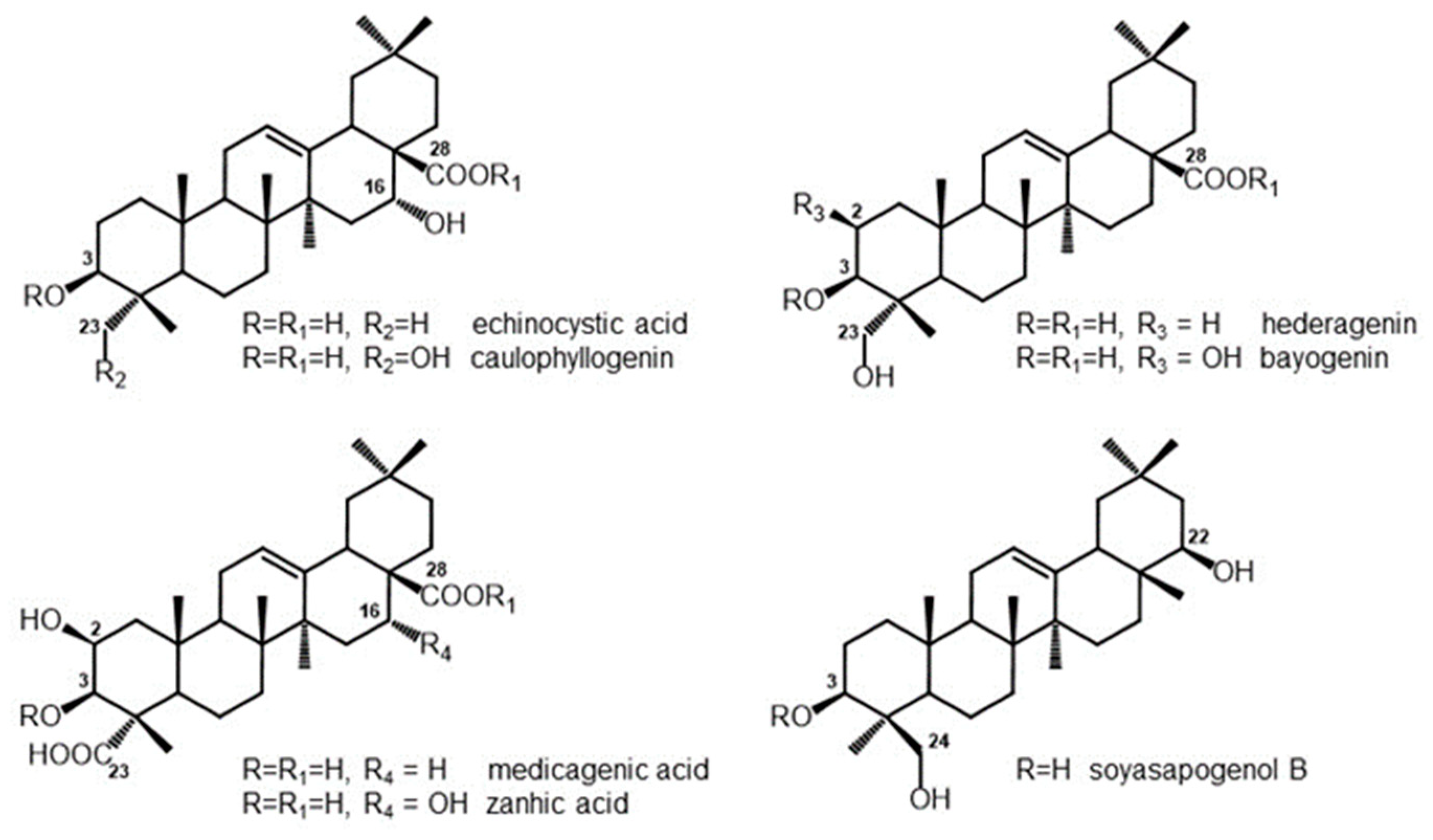
3.1. Composition of Saponins and Related Sapogenins from Medicago spp.

The chemical composition of Medicago saponin extracts differed according to the plant species. The composition of the most abundant sapogenins in the saponin mixtures is reported in Table 2. Based on the relative content of the dominant sapogenins after acid hydrolysis of the corresponding glycosides, saponins from M. arborea leaves (Sap1) were characterized by a higher amount of medicagenic and zanhic acids (27.5% and 45.8%, respectively, Figure 1). Echinocystic acid (Figure 1) was the dominant sapogenin in M. polymorpha 25570 leaves (Sap2), representing 76.5% of the total aglycones, while hederagenin (85.9%) (Figure 1) was the dominant sapogenin detected in M. polymorpha 15504 (Sap3). Medicagenic acid and zanhic acid were also the most abundant aglycones from saponins of M. sativa leaves (Sap4), accounting for 43.4% and 44.7%, respectively. M. sativa root saponins (Sap6) were instead characterized by a higher amount of medicagenic acid and hederagenin (Figure 1, 64.7% and 19.3%, respectively). Based on the HPLC-PDA/LC-MS comparison with authentic saponin standards [26,27,28], all the saponin mixtures here evaluated were found to be mainly constituted by bidesmosidic type saponins (70–80%). The M. sativa prosapogenins (Pros5), obtained after basic hydrolysis of the corresponding saponins, were instead entirely made up by monodesmosides (Figure 1).

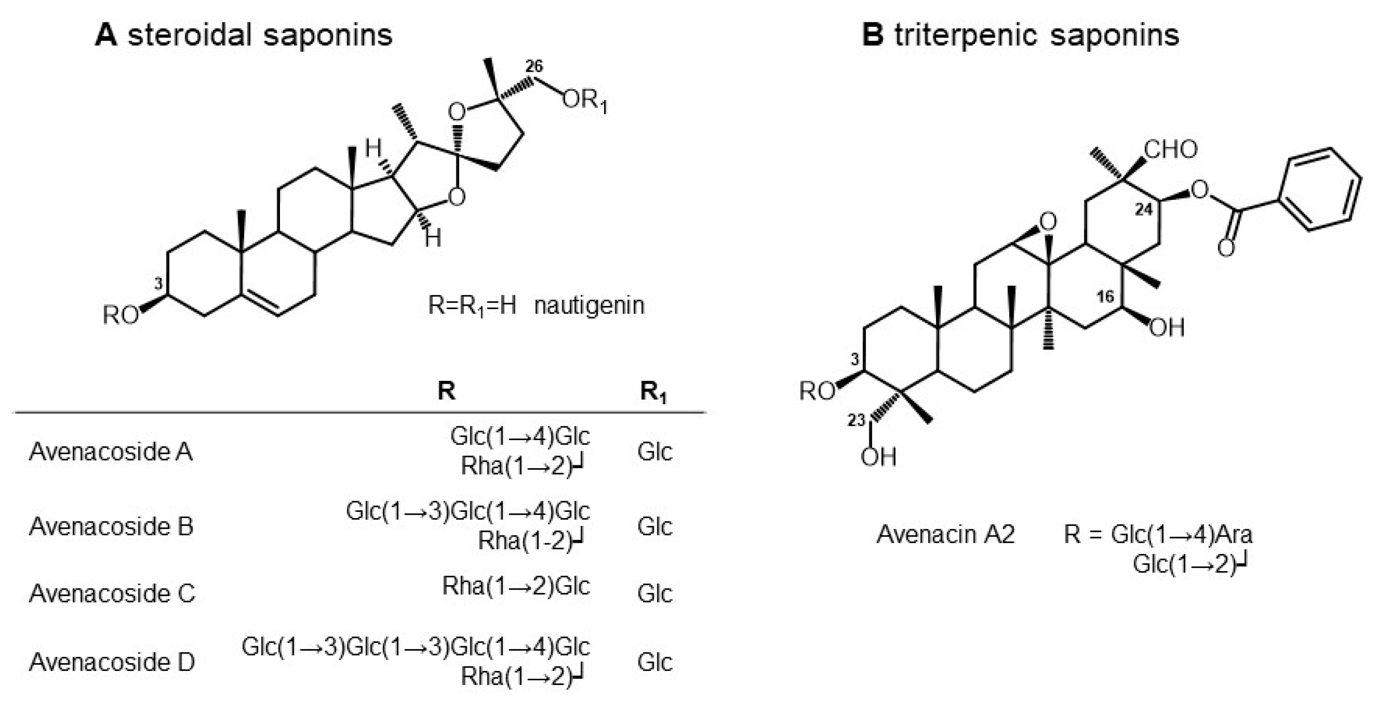
3.2. Composition of Saponins from Oat Seeds

In order to simplify the composition of the extract from oat grains and to obtain a saponin-enriched fraction, we fractionated the raw extract through RP-18 open column chromatography, with sequential elution steps with methanol washes at increasing concentrations. Of the 4 eluted fractions (obtained from sequential elution at 20, 50, 70, and 100% Methanol), TLC, HPLC, and LC-MS analysis showed that the 70% methanol fraction contained most of the saponins present in the initial raw extract. The 70% fraction was therefore analyzed by LC-ESI- MS2 analysis (Figure S1) to allow elucidation of the chemical structure of avenacins and avenacosides, which was achieved based on their chromatographic behavior, MS2 fragmentation spectra, and available literature data. When authentic standards were available, the annotation of putative saponin peaks was confirmed through co-elution of the corresponding pure compound. This enriched saponin fraction was shown to contain both avenacins and avenacosides (Table 3), in addition to over 70 authentic molecular ion signals for which a tentative annotation could not be provided.
Saponins were tentatively identified based on the molecular ion [M-H], on key fragment ions and other MS observations. In general, the loss of 146 m/z was indicative of deoxyhexose (e.g., rhamnose) and the loss of 162 m/z was indicative of hexose (e.g., glucose). The most abundant tentatively identified saponins are reported in Figure 2. Altogether, avenacins and avenacosides represent 35–40% of the total fraction, and their relative percentage composition was evaluated by LC-MS analyses and reported in Table 3.
The most abundant saponins in the oat 70% fraction were avenacoside B (compound 5, 35.3% of the total detected saponins) and avenacoside A (compound 6, 28.9%) followed by compound 1 (avenacin A2, 16.5%) and compound 4 (one of the isomer of avenacoside D, eluting at RT11.12, 7.1%). Avenacin C (both isomers 1 and 2, compounds 7 and 8) were also detected in lesser amount (see Table 3).

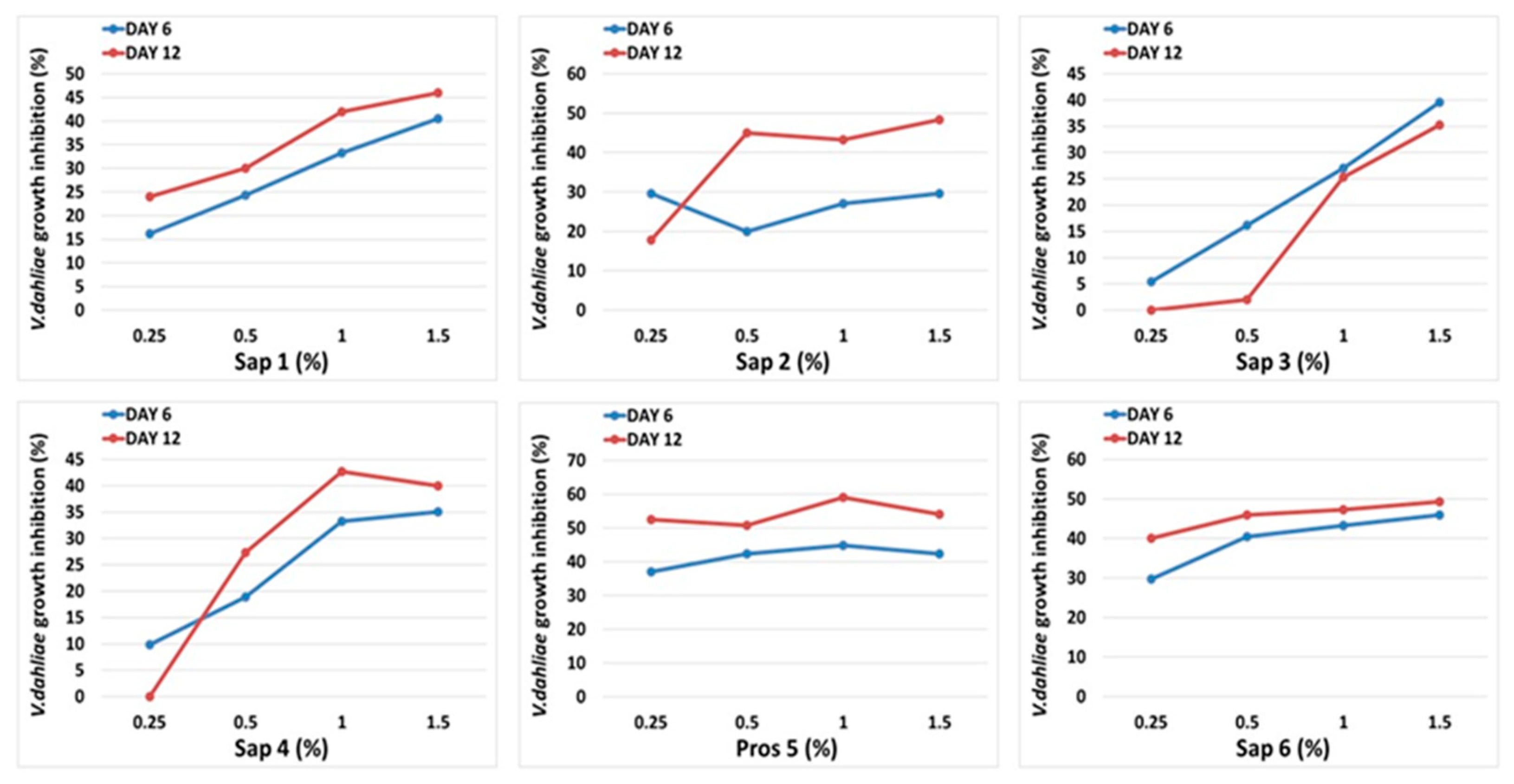
3.3. Medicago Saponins Antifungal Activity

All tested saponins had an inhibitory effect against Verticillium dahliae at concentrations greater than 0.5%. The most effective was the prosapogenin extract from Medicago sativa leaves, with a reduction in fungal growth of 50.8–52.5% at the lowest concentrations tested and up to an inhibition of 54.1–59% at the highest concentrations (12 days after inoculation). The saponin 6 extract from Medicago sativa roots reduced the growth of the pathogen by 40–46% at the lowest concentrations, up to an inhibition of 47.3–49.3% at the highest concentrations (12 days after inoculation). Saponins extracts 3 and 4 (from the leaves of Medicago polymorpha and Medicago sativa) were less effective in reducing the fungal growth, which at the highest doses reduced the development of the pathogen by 35.3–42.7%, respectively (at 12 days after inoculation) (Figure 3).

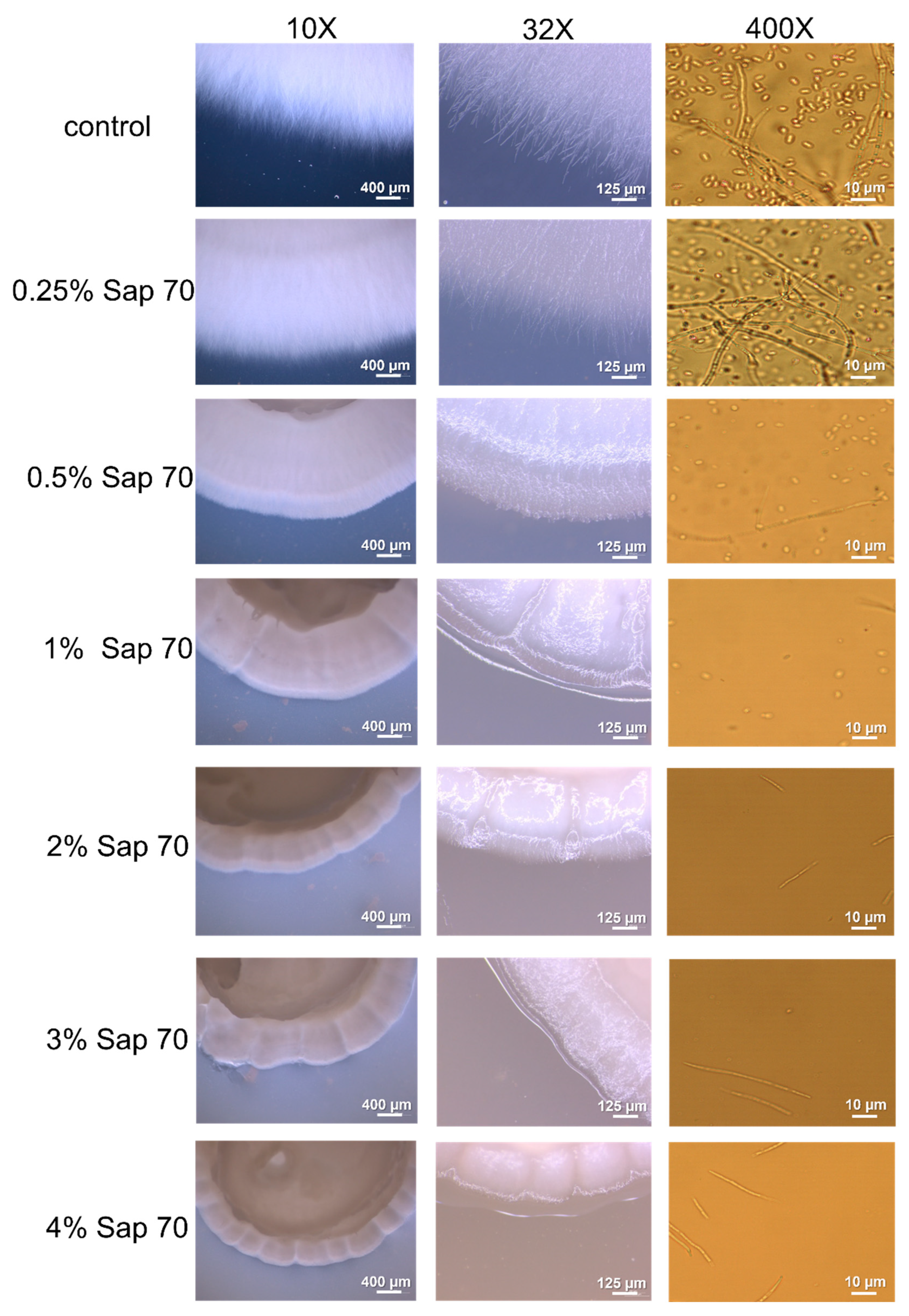
3.4. Oat Saponins Antifungal Activity

The saponin-enriched seed oat extract Sap70 significantly inhibited Verticillium dahliae growth at concentrations greater than 0.5% (Figure 4). Sap70 impact on mycelium growth is significant at 6 days after inoculation, with reductions ranging from 26% to 61% at increasing concentrations. The reduction in fungal growth ranged from 45% at 0.5% Sap70 concentration to 66% at the highest concentrations 12 days after inoculation.
In addition to seeing a strong effect on the growth of the fungus, the morphology of the mycelium is shaped by the saponins presence (Figure 5), with the transition from a cottony mycelium to a very compact one. This effect is particularly evident at the higher concentrations of the extract. It is noteworthy that Sap70 at the highest inhibited even the fungal conidia production.

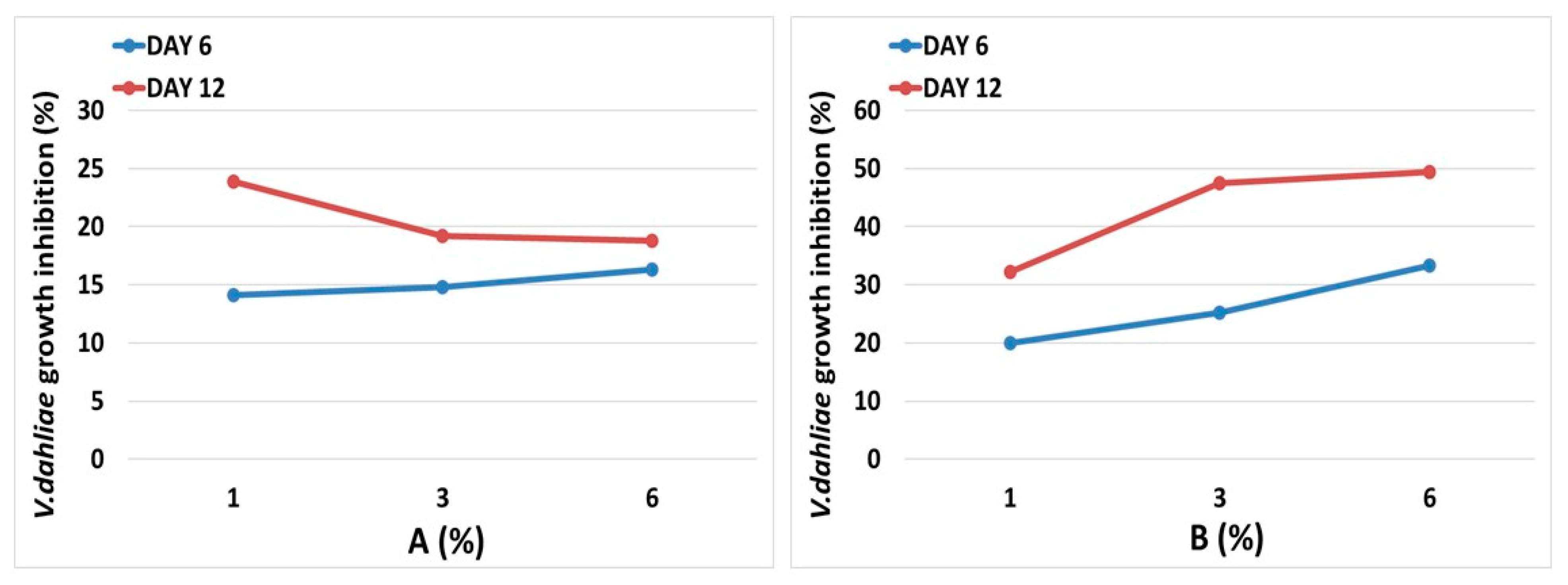
3.5. Brassica Sprouts Juices Antifungal Activity

The two brassica sprouts juices, A and B, were in vitro tested to evaluate the fungicidal-fungistatic effect on the pathogen Verticillium dahliae (Figure 6). The two juices significantly affect the mycelium growth at all the tested concentrations. The B juice was found more effective in limiting fungal growth, managing to reduce, after 12 days from inoculation, the diameter of the mycelium by 49.4%, when used at the highest concentration tested and by 32.2% at the lowest. The A juice reduced the radial growth of the pathogen by 18.8–23.9%, without significant differences between the three quantities tested.

3.6. Saponins Phytotoxicity

Phytoxicity evaluations were carried out on five tomato varieties using the test described in the Materials and Methods, which allows for the measuring of the germinative vigor of seeds treated with saponin extracts in comparison with untreated control seeds. The tests were conducted using the two saponin extracts that have shown the higher antifungal activity, i.e., Sap 6 and Sap 70, at the highest concentrations used to inhibit the mycelial growth of V. dahliae.
No significant differences in germination vigor were found between the control seeds and the treated seeds of the five varieties (Figure S2). In some varieties (i.e., Wilson, Mariner and Rossoro) a trend of greater vigor was observed in the treated seeds, although not statistically significant (Figure S2).

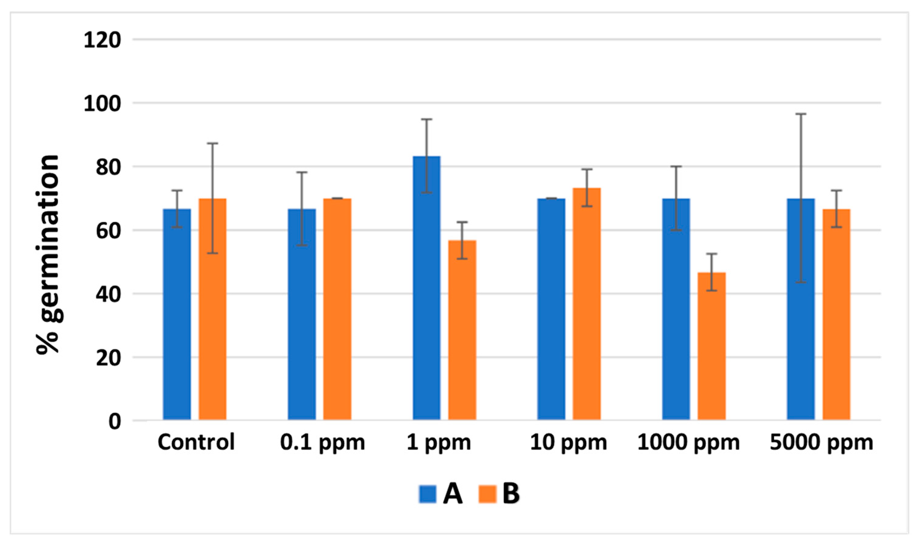
3.7. Brassica Sprouts Juices Phytotoxicity

There were no statistically significant differences in germination between control seeds and seeds treated with different concentrations of both types of brassica sprouts juices (Figure 7). Therefore the germination tests did not show any toxic effects of the analyzed extracts at concentrations up to 5000 ppm.

4. Discussion

The antimicrobial activity of plant saponins has been long known, having been reported since the initial purifications reported for this class of compounds [6]. Saponins are primarily implicated in the plant defense response to pathogen infections. Their presence in plant roots and leaves provides a chemical barrier to intruding soil-borne and phyllosphere microbes infecting plant tissue [36]. The pathogenic attack in some plants causes the hydrolysis of saponins to derivatives with strong antibiotic activity.
Saponins, as components of exogenous plant defense treatments, have been mainly proposed to control insects affecting crop production and bacterial and fungal pathogens of relevance for human and animal health. Less knowledge is available on the saponins potential as antifungal agents. This study contributed to the increase of such knowledge, finding that saponins extracted from different Medicago species and from oat seeds are effective antifungal agents against V. dahliae.
Regarding saponins present in Medicago spp., some studies have found that sapogenins (i.e., saponin aglycones) generally have higher antifungal activity with respect to intact saponins (which are extensively glycosylated), suggesting that the sugar moiety is not important for antimicrobial efficacy. In particular, the biological activities of M. sativa and M. arborea extracts were found to be related to the content of medicagenic acid [37]. Moreover, the antifungal properties of saponin mixtures from Medicago tops and roots, the corresponding mixtures of prosapogenins from tops, and purified saponins and sapogenins have been successfully evaluated against the fungus Pyricularia oryzae. The in vitro trials clearly demonstrated the antifungal effects of prosapogenin mixture from alfalfa tops [38].
Oats, the only saponin-accumulating cereal, store such molecules in leaves and roots (steroidal avenacosides and triterpene avenacins), where they have phytoprotectant activity, as demonstrated by the increased susceptibility to pathogens of avenacin-free mutant genotypes. Oat grains and husks have been reported as being rich in the inactive biological forms of the steroidal avenacosides A and B, which can then be converted to their active forms, 26-desglucoavenacoside A and B, by the action of glycosidases resulting from tissue damage or pathogen attack [39].
Despite the avenacins biosynthetic pathway elucidated in Avena strigosa root tissue [40,41], it is still unknown how the expression of genes involved in the pathway impacts on the seed avenacins content.
A pattern of molecular mechanisms has been proposed for saponins antimicrobial activities, including membrane lipid re-arrangement, pores formation, and cell lysis [42]. Recently, several studies suggested that many saponins do not solubilize the lipid layers of biological membranes but fluidize them, causing an increase of their permeability [43].
It is noteworthy that saponins extracted from Medicago and from oat seeds impact on fungus life cycle—inhibiting, at least at higher saponin concentrations, the production of fungal spores (Figure 5 and Figure 8). Such peculiarity of saponin treatments can be of particular interest to slow or to stop the vascular colonization of the plant, which occurs when conidia are sucked into the plant into the so-called trapping sites, where they germinate and invade adjacent vessel elements to continue the plant colonization.
Brassica juice from purple sprouts has been found significantly more efficient in inhibiting V. dahliae mycelium growth in comparison with brassica juice from etiolated sprouts. The juice from purple sprouts is enriched not only with GLSs but also with polyphenols and in particular with anthocyanins, all compounds which may also have antimicrobial and antifungal properties [44]. In common with other flavonoids, certain anthocyanins have demonstrable antiviral, antibacterial, and fungicidal activities. They have the potential, therefore, to protect plants from infections by pathogenic microorganisms. In general, however, the antimicrobial activities of anthocyanins are appreciably less effective than those of other phenolic compounds, such as key flavanols and hydroxycinnamic acids. On the other hand, anthocyanins have not been found to be toxic to any higher animal species. Aphid survival rates, for example, are unaffected by anthocyanins direct chemical defense is unlikely to be a major function of these pigments in plants [45].
Interestingly, the presence of increased levels of anthocyanins and other polyphenols in addition to GLSs in sucrose treated brassica sprouts juices can potentially be one of the reason for its higher protective effect.

5. Conclusions

The overall aim of this work has been to identify plant extracts and mixtures characterized by antifungal activity, low toxicity level and low preparation costs, as a potential application in fungal control in horticultural crops. Brassica sprouts, Medicago tissues, and oat seeds are all low-cost sources of natural antifungals. All tested extracts and mixtures were found effective in inhibiting the growth of the widespread pathogen V. dahliae at concentrations that are not toxic for the plant. To minimize the losses caused by the wilt, an integrated approach, i.e., a combination of cultural, chemical, biological, and genetic actions is now proposed [46]. Mazzotta et al. [47] recently proposed to deliver olive leaf extracts to tomato plants using chitosan nanoparticles as a carrier. In this frame, saponins able to reduce mycelium growth but first of all to block conidia formation deserve deeper in vivo evaluation, even in combination with delivery system, e.g., chitosan nanoparticles. Their applicability in open field and greenhouse environments can be proposed in the frame of organic productions.

Supplementary Materials

The following supporting information can be downloaded at: https://www.mdpi.com/article/10.3390/horticulturae8080729/s1, Figure S1. LC-MS analysis of the Avena sativa saponin-enriched extract. (a.) Base peak chromatogram of the saponin fraction eluted from the 70% MeOH wash; (b.) extracted ion chromatogram of m/z 1064.4, annotated as Avenacin A2; (c.) extracted ion chromatogram of m/z 1385.8, yielding two peaks annotated as the putative isomers of Avenacoside D; (d.) extracted ion chromatogram of m/z 1223.7, yielding two peaks annotated as the putative isomers of Avenacoside B; (e.) extracted ion chromatogram of m/z 1061.7, annotated as Avenacoside A by coelution of the corresponding authentic standard; (f.) extracted ion chromatogram of m/z 899.6, yielding two peaks annotated as the putative isomers of Avenacoside C. For details of MS identifications see Table 3; Figure S2. Effect of Sap70 and Sap 6 extracts at antifungal concentrations on tomato (five cultivars) seed germinative vigor. The germination vigor is reported on Y axis and was calculated as reported in Materials and Method section.

Author Contributions

Conceptualization, C.M., A.T., S.B., F.F. and V.T.; methodology, C.M., I.P., R.G., S.D., E.B. and B.F.; resources, V.T., A.T., S.B. and F.F.; data curation, C.M., S.D., S.B., F.S., A.T. and F.F.; writing—original draft preparation, V.T., C.M., S.B. and A.T.; writing—review and editing, F.S. and F.F.; funding acquisition, V.T. All authors have read and agreed to the published version of the manuscript.

Funding

This research was funded by the Italian Ministry of Agriculture and Forestry in the DiBio–BIOPRIME project (Prot. 76381, MiPAAF PQAI I).

Institutional Review Board Statement

Not applicable.

Informed Consent Statement

Not applicable.

Data Availability Statement

Not applicable.

Conflicts of Interest

The authors declare no conflict of interest.

References

  1. The EU Green Deal–A Roadmap to Sustainable Economies 2019. Available online: https://www.switchtogreen.eu/the-eu-green-deal-promoting-a-green-notable-circular-economy/ (accessed on 7 July 2022).
  2. Alabouvette, C.; Olivain, C.; Steinberg, C. Biological control of plant diseases: The European situation. Eur. J. Plant Pathol. 2006, 114, 329–341. [Google Scholar] [CrossRef]
  3. Kregiel, D.; Berlowska, J.; Witonska, I.; Antolak, H.; Proestos, C.; Babic, M.; Babic, L.; Zhang, B. Saponin-based, biological-active surfactants from plants. In Application and Characterization of Surfactants; Najjar, R., Ed.; InTech: Rijeka, Croatia, 2017; pp. 184–205. [Google Scholar]
  4. Tava, A.; Pecetti, L. Chemical Investigation of Saponins from Twelve Annual Medicago Species and their Bioassay with the Brine Shrimp Artemia salina. Nat. Prod. Commun. 2012, 7, 837–840. [Google Scholar] [CrossRef] [PubMed]
  5. Bonilla, H.; Carbajal, Y.; Gonzales, M.; Vásquez, V.; López, A. Determinación de la actividad insecticida de la saponina de la quinua (Chenopodium quinoa) en larvas de Drosophila melanogaster. Sci. Agropecu. 2019, 10, 39–45. [Google Scholar] [CrossRef]
  6. Zaynab, M.; Sharif, Y.; Abbas, S.; Afzal, M.Z.; Qasim, M.; Khalofah, A.; Ansari, M.J.; Khan, K.A.; Tao, L.; Li, S. Saponin toxicity as key player in plant defense against pathogens. Toxicon 2021, 193, 21–27. [Google Scholar] [CrossRef] [PubMed]
  7. Rai, S.; Acharya-Siwakoti, E.; Kafle, A.; Devkota, H.P.; Bhattarai, A. Plant-Derived Saponins: A Review of Their Surfactant Properties and Applications. Sci 2021, 3, 44. [Google Scholar] [CrossRef]
  8. Osbourn, A.E. Saponins in cereals. Phytochemistry 2003, 62, 1–4. [Google Scholar] [CrossRef]
  9. Sparg, S.G.; Luce, M.E.; Van Staden, J. Biological activities and distribution of plant saponins. J. Ethnopharmacol. 2004, 94, 219–243. [Google Scholar] [CrossRef]
  10. Blažević, I.; Montaut, S.; Burčul, F.; Olsen, C.E.; Burow, M.; Rollin, P.; Agerbirk, N. Glucosinolate structural diversity, identification, chemical synthesis and metabolism in plants. Phytochemistry 2020, 169, 112100. [Google Scholar] [CrossRef]
  11. Björkman, M.; Klingen, I.; Birch, A.N.E.; Bones, A.M.; Bruce, T.J.A.; Johansen, T.J.; Meadow, R.; Mølmann, J.; Seljåsen, R.; Smart, L.E.; et al. Phytochemicals of Brassicaceae in plant protection and human health—Influences of climate, environment and agronomic practice. Phytochemistry 2011, 72, 538–556. [Google Scholar] [CrossRef]
  12. Possenti, M.; Baima, S.; Raffo, A.; Durazzo, A.; Giusti, A.M.; Natella, F. Glucosinolates in food. In Glucosinolates, Reference Series in Phytochemistry; Merillon, J.M., Ramawat, K.G., Eds.; Springer International Publishing: Cham, Switzerland, 2017; pp. 87–132. [Google Scholar]
  13. Ilahy, R.; Tlili, I.; Pék, Z.; Montefusco, A.; Siddiqui, M.W.; Homa, F.; Hdider, C.; R’Him, T.; Lajos, H.; Lenucci, M.S. Pre- and post-harvest factors affecting glucosinolate content in broccoli. Front. Nutr. 2020, 7, 147. [Google Scholar] [CrossRef]
  14. Jahangir, M.; Abdel-Farid, I.B.; Kim, H.K.; Choi, Y.H.; Verpoorte, R. Healthy and unhealthy plants: The effect of stress on the metabolism of Brassicaceae. Environ. Exp. Bot. 2009, 67, 23–33. [Google Scholar] [CrossRef]
  15. Baenas, N.; Garcia-Viguera, C.; Moreno, D.A. Elicitation: A tool for enriching the bioactive composition of foods. Molecules 2014, 19, 13541–13563. [Google Scholar] [CrossRef] [PubMed]
  16. Fahey, J.W.; Zhang, Y.; Talalay, P. Broccoli sprouts: An exceptionally rich source of inducers of enzymes that protect against chemical carcinogens. Proc. Natl. Acad. Sci. USA 1997, 94, 10367–10372. [Google Scholar] [CrossRef]
  17. Guo, R.; Yuan, G.; Wang, Q. Sucrose enhances the accumulation of anthocyanins and glucosinolates in broccoli sprouts. Food Chem. 2011, 129, 1080–1087. [Google Scholar] [CrossRef]
  18. Guo, R.; Yuan, G.; Wang, Q. Effect of sucrose and mannitol on the accumulation of health-promoting compounds and the activity of metabolic enzymes in broccoli sprouts. Sci. Hortic. 2011, 128, 159–165. [Google Scholar] [CrossRef]
  19. Natella, F.; Maldini, M.; Nardini, M.; Azzini, E.; Foddai, M.S.; Giusti, A.M.; Baima, S.; Morelli, G.; Scaccini, C. Improvement of the nutraceutical quality of broccoli sprouts by elicitation. Food Chem. 2016, 201, 101–109. [Google Scholar] [CrossRef]
  20. Ferruzza, S.; Natella, F.; Ranaldi, G.; Murgia, C.; Rossi, C.; Trošt, K.; Mattivi, F.; Nardini, M.; Maldini, M.; Giusti, A.M.; et al. Nutraceutical improvement increases the protective activity of broccoli sprout juice in a human intestinal cell model of gut inflammation. Pharmaceuticals 2016, 9, 48. [Google Scholar] [CrossRef] [PubMed]
  21. Masci, A.; Mattioli, R.; Costantino, P.; Baima, S.; Morelli, G.; Punzi, P.; Giordano, C.; Pinto, A.; Donini, L.M.; d’Erme, M.; et al. Neuroprotective effect of Brassica oleracea sprouts crude juice in a cellular model of alzheimer’s disease. Oxid. Med. Cell. Longev. 2015, 2015, 781938. [Google Scholar] [CrossRef]
  22. Rubattu, S.; Di Castro, S.; Cotugno, M.; Bianchi, F.; Mattioli, R.; Baima, S.; Stanzione, R.; Madonna, M.; Bozzao, C.; Marchitti, S.; et al. Protective effects of Brassica oleracea sprouts extract toward renal damage in high-salt-fed SHRSP: Role of AMPK/PPARα/UCP2 axis. J. Hypertens. 2015, 33, 1465–1479. [Google Scholar] [CrossRef]
  23. Rubattu, S.; Stanzione, R.; Bianchi, F.; Cotugno, M.; Forte, M.; Della Ragione, F.; Fioriniello, S.; D’Esposito, M.; Marchitti, S.; Madonna, M.; et al. Reduced brain UCP2 expression mediated by microRNA-503 contributes to increased stroke susceptibility in the high-salt fed stroke-prone spontaneously hypertensive rat. Cell Death Dis. 2017, 8, 2891. [Google Scholar] [CrossRef]
  24. Hu, X.; Puri, K.D.; Gurung, S.; Klosterman, S.J.; Wallis, C.M.; Britton, M.; Durbin-Johnson, B.; Phinney, B.; Salemi, M.; Short, D.P.G.; et al. Proteome and metabolome analyses reveal differential responses in tomato-Verticillium dahliae-interactions. J. Proteom. 2019, 207, 103449. [Google Scholar] [CrossRef] [PubMed]
  25. Subbarao, K. Verticillium dahliae (Verticillium wilt). In Invasive Species Compendium; CABI: Wallingford, UK, 2020. [Google Scholar] [CrossRef]
  26. Tava, A.; Mella, M.; Avato, P.; Argentieri, M.P.; Bialy, Z.; Jurzysta, M. Triterpenoid glycosides from the leaves of Medicago arborea L. J. Agric. Food Chem. 2005, 53, 9954–9965. [Google Scholar] [CrossRef] [PubMed]
  27. Tava, A.; Pecetti, L.; Romani, M.; Mella, M.; Avato, P. Triterpenoid glycosides from the leaves of two cultivars of Medicago polymorpha L. J. Agric. Food Chem. 2011, 59, 6142–6149. [Google Scholar] [CrossRef]
  28. Tava, A.; Avato, P. Chemical and biological activity of triterpene saponins from Medicago species. Nat. Prod. Commun. 2006, 1, 1159–1180. [Google Scholar] [CrossRef]
  29. Tava, A.; Biazzi, E.; Mella, M.; Quadrelli, P.; Avato, P. Artefact formation during acid hydrolysis of saponins from Medicago spp. Phytochemistry 2017, 238, 116–127. [Google Scholar] [CrossRef]
  30. Abdul-Baki, A.A.; Anderson, J.D. Vigor Determination in Soybean Seed by Multiple Criteria. Crop Sci. 1973, 13, 630–633. [Google Scholar] [CrossRef]
  31. Crombie, W.M.L.; Crombie, L. Distribution of avenacins A-1, A-2, B-1 and B-2 in oat roots: Their fungicidal activity towards ‘‘take-all’’ fungus. Phytochemistry 1984, 25, 2069–2073. [Google Scholar]
  32. Hu, C.; Sang, S. Triterpenoid saponins in oat bran and their levels in commercial oat products. J. Agric. Food Chem. 2020, 68, 6381–6389. [Google Scholar] [CrossRef]
  33. Yang, J.; Wang, P.; Wu, W.; Zhao, Y.; Idehen, E.; Sang, S. Steroidal Saponins in Oat Bran. J. Agric. Food Chem. 2016, 64, 1549–1556. [Google Scholar] [CrossRef] [PubMed]
  34. Tschesche, R.; Tauscher, M.; Fehlhaber, H.W.; Wulff, G. Avenacosid A, ein bisdesmosidisches Steroidsaponin aus Avena sativa. Chem. Ber. 1969, 102, 2072–2082. [Google Scholar] [CrossRef]
  35. Pecio, Ł.; Wawrzyniak-Szołkowska, A.; Oleszek, W.; Stochmal, A. Rapid analysis of avenacosides in grain and husks of oats by UPLC-TQ-MS. Food Chem. 2013, 141, 2300–2304. [Google Scholar] [CrossRef] [PubMed]
  36. Osbourn, A.; Goss, R.J.M.; Field, R.A. The saponins: Polar isoprenoids with important and diverse biological activities. Nat. Prod. Rep. 2011, 28, 1261–1268. [Google Scholar] [CrossRef] [PubMed]
  37. Avato, P.; Bucci, R.; Tava, A.; Vitali, C.; Rosato, A.; Bialy, Z.; Jurzysta, M. Antimicrobial activity of saponins from Medicago sp.: Structure activity relationship. Phytother. Res. 2006, 20, 454–457. [Google Scholar] [CrossRef]
  38. Abbruscato, P.; Tosi, S.; Crispino, L.; Biazzi, E.; Menin, B.; Picco, A.M.; Pecetti, L.; Avato, P.; Tava, A. Triterpenoid glycosides from Medicago sativa as antifungal agents against Pyricularia oryzae. J. Agric. Food Chem. 2014, 62, 11030–11036. [Google Scholar] [CrossRef]
  39. Raguindin, P.F.; Itodo, O.A.; Stoyanov, J.; Dejanovic, G.M.; Gamba, M.; Asllanaj, E.; Minder, B.; Bussler, W.; Metzger, B.; Muka, T.; et al. A systematic review of phytochemicals in oat and buckwheat. Food Chem. 2021, 338, 127982. [Google Scholar] [CrossRef]
  40. Kemen, A.C.; Honkanen, S.; Melton, R.E.; Findlay, K.C.; Mugford, S.T.; Hayashi, K.; Haralampidis, K.; Rosser, S.J.; Osbourna, A. Investigation of triterpene synthesis and regulation in oats reveals a role for β-amyrin in determining root epidermal cell patterning. Proc. Natl. Acad. Sci. USA 2014, 111, 8679–8684. [Google Scholar] [CrossRef] [PubMed]
  41. Leveau, A.; Reed, J.; Qiao, X.; Stephenson, M.J.; Mugford, S.T.; Melton, R.E.; Rant, J.C.; Vickerstaff, R.; Langdon, T.; Osbourn, A. Towards take-all control: A C-21β oxidase required for acylation of triterpene defence compounds in oat. New Phytol. 2019, 221, 1544–1555. [Google Scholar] [CrossRef]
  42. Coleman, J.J.; Okoli, I.; Tegos, G.P.; Holson, E.B.; Wagner, F.F.; Hamblin, M.R.; Mylonakis, E. Characterization of plant-derived saponin natural products against Candida albicans. ACS Chem. Biol. 2010, 5, 321–332. [Google Scholar] [CrossRef]
  43. Wojciechowski, K.; Jurek, I.; Góral, I.; Campana, M.; Geue, T.; Gutberlet, T. Surface-active extracts from plants rich in saponins—Effect on lipid mono-and bilayers. Surf. Interfaces 2021, 27, 101486. [Google Scholar] [CrossRef]
  44. Singh, S.; Kaur, I.; Kariyat, R. The multifunctional roles of polyphenols in plant-herbivore interactions. Int. J. Mol. Sci. 2021, 22, 1442. [Google Scholar] [CrossRef]
  45. Schaefer, H.M.; Rentzsch, M.; Breuer, M. Anthocyanins reduce fungal growth in fruits. Nat. Prod. Commun. 2008, 3, 1267–1272. [Google Scholar] [CrossRef]
  46. Acharya, B.; Ingram, T.W.; Oh, Y.; Adhikari, T.B.; Dean, R.A.; Louws, F.J. Opportunities and challenges in studies of host-pathogen interactions and management of Verticillium dahliae in tomatoes. Plants 2020, 9, 1622. [Google Scholar] [CrossRef] [PubMed]
  47. Mazzotta, E.; Muzzalupo, R.; Chiappetta, A.; Muzzalupo, I. Control of the Verticillium Wilt on tomato plants by means of olive leaf extracts loaded on chitosan nanoparticles. Microorganisms 2022, 10, 136. [Google Scholar] [CrossRef] [PubMed]
Figure 1. Chemical structures of the most abundant sapogenins detected in the Medicago spp. plant extracts. Saponins: R = sugar or sugar chain, R1 = H: monodesmosides. R = R1 = sugar or sugar chain: bidesmosides.
Horticulturae 08 00729 g001
Figure 2. Chemical structures of the most abundant saponins identified in the Avena sativa seed extract, i.e., steroidal saponins (A) and triterpeninc saponins (B).
Horticulturae 08 00729 g002
Figure 3. Effect on the in vitro growth of Verticillium dahliae mycelium by saponins added to the growth medium after 6 and 12 days from inoculation. Sap1) Saponins extracted from leaves of M. arborea, Sap2) and Sap3) Saponins extracted from leaves of M. polymorpha, Sap4) Saponins extracted from leaves of M. sativa, Pros5) Prosapogenins extracted from leaves of M. sativa, Sap6) Saponins extracted from roots of M. sativa. Values are reported as inhibition percentages as compared to the control ± SD. The concentration percentages are intended as volume/volume. All the treatments with saponins and prosapogenins concentrations equal or higher than 0.5% are significant (t-test, p ≤ 0.05) in mycelium growth inhibition in comparison with not treated controls.
Horticulturae 08 00729 g003
Figure 4. Effect on the in vitro growth of Verticillium dahliae mycelium by the saponin-enriched seed oat extract Sap70 added to the growth medium after 6 and 12 days from inoculation. Values are reported as inhibition percentages as compared to the control ± SD. The concentration percentages are intended as volume/volume. The mycelium growth reduction is significant (t-test, p ≤ 0.05) in the presence of Sap70 concentrations greater than 0.5% after 6 days and equal or greater than 0.5% after 12 days of treatment.
Horticulturae 08 00729 g004
Figure 5. Verticillium dahliae mycelium images taken with the microscope and stereomicroscope using 3 magnifications (10×, 32×, 400×) after 10 days of growth in the absence (control) or in the presence of increasing Sap 70 concentrations.
Horticulturae 08 00729 g005
Figure 6. Effect of A and B brassica sprouts juices on the in vitro growth of Verticillium dahliae mycelium at 6 and 12 days after inoculation. Three concentrations (volume/volume) have been tested. Values are reported as inhibition percentages as compared to the control ± SD. All the treatments reduced significantly the mycelium growth in comparison with control (t-test, p ≤ 0.05).
Horticulturae 08 00729 g006
Figure 7. Effect of A and B brassica sprouts juices at the concentrations reported on x axis on the maize seed germination percentages (y axis). No statistically significant differences were detected between control and treatments at all the concentrations tested (Kruskal–Wallis test, p ≤ 0.08).
Horticulturae 08 00729 g007
Figure 8. Verticillium dahliae mycelium images taken at 400× magnification after 10 days of growth in absence (control) or in presence of increasing Sap6 concentrations.
Horticulturae 08 00729 g008
Table 1. The tested compounds and their sources are reported.
Plant SpeciesPlant TissueMain CompoundCode
Medicago arborealeavessaponinsSap1
Medicago polymorpha 22507leaves Sap2
Medicago polymorpha 155004leavesSap3
Medicago sativaleavesSap4
Medicago sativaleavesprosapogeninsPros5
Medicago sativarootssaponinsSap6
Avena sativaseedssaponinsSap70
Brassica oleraceaetiolated sproutsBrassica homogenatesA
Brassica oleraceapurple sprouts B
Table 2. Composition of the most abundant sapogenins in the saponin mixtures from Medicago spp., expressed as percentage (%) of the total sapogenins.
Echinocystic AcidCaulophyllogeninHederageninBayogeninMedicagenic AcidZanhic AcidSoyasapogenol B
M. arborea leavesSap1--0.13.527.545.810.1
M. polymorpha 25570 leavesSap276.54.69.71.5--2.0
M. polymorpha 15504 leavesSap30.30.185.91.2--2.3
M. sativa leavesSap4--0.21.443.430.213.1
M. sativa rootsSap6--19.32.364.72.93.1
Table 3. LC-ESI-MS2 analysis of the Avena sativa enriched saponin extract. “A” grade refers to a metabolite annotation confirmed by co-elution of the authentic standard; “B” grade refers to a metabolite annotation confirmed by MS2 analysis and previous reports from the literature; “C” grade refers to a metabolite annotation confirmed by MS2 analysis and previous reports from the literature.
ntRMolecular FormulaMonoisotopic Mass[M-H] (m/z)Metabolite AnnotationGradeMS2 (m/z)% of Total Detected SaponinsReference
110.09C54H80O211064.519181063.7Avenacin A2C 16.5Crombie et al., 1984 [31] (oat roots); Hu and Sang (2020) (sprouted oat bran) [32]
210.29C63H102O331386.63031385.8Avenacoside D (isomer 1)B1223.3 [M-H-Hexose]; 1061.0 [M-H-2Hexose]1.4Yang et al., 2016 [33] (oat bran)
310.52C57H92O281224.577481223.7Isomer to
Avenacoside B
B1077.4 [M-H-Rha]; 1061.5 [M-H-Hex]; 1043.3 [M-H-Hex-H2O]; 915.3 [M-H-Hex-Rha]; 899.4 [M-H-2Hex]2.7Yang et al., 2016 [33] (oat bran)
411.12C63H102O331386.63031385.8Avenacoside D (isomer 2)B1223.3 [M-H-Hex]; 1061.0 [M-H-2Hex]7.1Yang et al., 2016 [33] (oat bran)
511.34C57H92O281224.577481223.7Avenacoside B C 35.3Yang et al., 2016 [33] (oat bran)
611.57C51H82O231062.524661061.7Avenacoside AA915.3 [M-H-Rha]; 899.3 [M-H-Hex]; 753.3 [M-H-Rha-Hex]; 736.3 [M-H-2Hex]28.9Tschesche et al. Chem Ber 1969 [34] (seeds and leaves)
711.98C45H72O18900.47184899.6Avenacoside C B753.0 [M-H-Rha]; 737.3 [M-H-Hex]6.4Pecio et al., 2013 [35] (seeds)
812.52C45H72O18900.47184899.6Isomer to Avenacoside C C 1.9Pecio et al., 2013 [35] (seeds)
Publisher’s Note: MDPI stays neutral with regard to jurisdictional claims in published maps and institutional affiliations.

