Article

Jania adhaerens Primes Tomato Seed against Soil-Borne Pathogens

1
Department of Agricultural and Food Sciences, Alma Mater Studiorum, University of Bologna, Viale G. Fanin 40, 40127 Bologna, Italy
2
Department of Biomedical and Neuromotor Sciences, Alma Mater Studiorum, University of Bologna, Via Irnerio 48, 40126 Bologna, Italy
3
Banco Español de Algas, Instituto de Oceanografía y Cambio Global, IOCAG, Universidad de Las Palmas de Gran Canaria, 35214 Telde, Spain
4
Council for Agricultural Research and Economics, Research Centre for Agriculture and Environment, 40128 Bologna, Italy
*
Author to whom correspondence should be addressed.
Horticulturae 2022, 8(8), 746; https://doi.org/10.3390/horticulturae8080746
Submission received: 26 July 2022 / Revised: 11 August 2022 / Accepted: 15 August 2022 / Published: 18 August 2022
(This article belongs to the Special Issue Sustainable Control Strategies of Plant Pathogens in Horticulture)

Abstract

:
Managing soil-borne pathogens is complex due to the restriction of the most effective synthetic fungicides for soil treatment. In this study, we showed that seed priming with Jania adhaerens water-soluble polysaccharides (JA WSPs) was successful in protecting tomato plants from the soil-borne pathogens Rhizoctonia solani, Pythium ultimum, and Fusarium oxysporum under greenhouse conditions. WSPs were extracted from dry thallus by autoclave-assisted method, and the main functional groups were characterized by using FT-IR spectroscopy. WSPs were applied by seed treatment at 0.3, 0.6 and 1.2 mg/mL doses, and each pathogen was inoculated singly in a growing substrate before seeding/transplant. Overall, WSPs increased seedling emergence, reduced disease severity and increased plant development depending on the dose. Transcriptional expression of genes related to phenylpropanoid, chlorogenic acid, SAR and ISR pathways, and chitinase and β-1,3 glucanase activities were investigated. Among the studied genes, HQT, HCT, and PR1 were significantly upregulated depending on the dose, while all doses increased PAL and PR2 expression as well as β-1,3 glucanase activity. These results demonstrated that, besides their plant growth promotion activity, JA WSPs may play a protective role in triggering plant defense responses potentially correlated to disease control against soil-borne pathogens.

1. Introduction

Jania adhaerens (J.V. Lamour) (JA) is a red calcareous macroalga belonging to the Corallinaceae family of the Corallinales order, Planta kingdom. The genus Jania is widely present on the Mediterranean and Atlantic coasts [1], and in the Pacific, the Caribbean, and Gulf of Mexico coasts of North and Central America [2]. The typical red color is due to phycoerythrin, the major pigment in red algae [3]. The cell wall of JA is particularly rich in carbohydrates similar to other marine macroalgae [4]. Sulfated galactans are the main polysaccharides constituting the cell walls of most red seaweeds and are also produced by the Corallinales order including JA [1,5]. It has been reported that polysaccharides are rich in functional groups able to bind to some microelements and metal ions having an important role in plant growth [6]. Furthermore, polysaccharides are well-known potent elicitors of plant resistance to fungi, bacteria and viruses, and abiotic stresses [4,7]. The elicitor-induced host response involves the activation of complex signaling cascades followed by the synthesis of defense signaling molecules such as salicylic acid, jasmonic acid, and ethylene that activate the SAR and ISR metabolic pathways. Accordingly, several antimicrobial compounds such as phytoalexins, and pathogenesis-related proteins accumulate in elicited plants [8,9].
Soil-borne organisms are characterized by a subterranean part of their life cycle [10]. This definition encompasses many types of organisms such as bacteria, oomycetes, fungi and nematodes, amongst which some species are plant pathogens. Among the most important soil-borne pathogens that cause economic losses in horticulture, there are the oomycete Pythium ultimum and the fungi Rhizoctonia solani and Fusarium oxysporum. Pythium ultimum is a common inhabitant of fields, ponds, streams, and decomposing vegetation worldwide. This oomycete forms hyphae (without septa), oospores, and sporangia which are capable of long-term survival [11]. Mycelia and oospores in the soil can initiate infections of seeds or roots, leading to wilting, reduced yield, root rot, and mortality on >300 diverse hosts [12]. Rhizoctonia solani (multinucleate) is a collective species consisting of several unrelated strains, divided into anastomosis groups (AGs), differing in their host range and pathogenicity [13]. Species do not produce spores but are composed of hyphae and sclerotia, living in soil or on organic debris. Symptoms vary according to the host and the plant part affected: damping-off of seedlings is probably the most common disease, but also root and stem rots, stem cankers and fruit rot can occur [14]. Fusarium oxysporum is a species complex that comprises a multitude of saprophytic strains that grow and survive on organic matter in soil and the rhizosphere of many plant species. Some species are pathogenic to plants: spores germinate, and the hyphae penetrate the roots causing either root rot or vascular wilt diseases. Pathogenic species show a high level of host specificity, so they are classified into more than 120 formae speciales and races [15,16]. Symptoms include vein clearing and leaf epinasty, followed by stunting, yellowing of the lower leaves, progressive wilting, defoliation, and, finally, death of the plant. In addition, the vascular tissue turns brown due to fungal colonization [17].
Managing the control of soil-borne pathogens is problematic, because of the prohibition or restriction on the use of the most effective synthetic fungicides for soil treatment, according to Reg. (EC) No. 1107/2009. This regulation establishes the principles of integrated pest management and gives priority to eco-friendly alternatives wherever possible to ensure a high level of protection of human and animal health and the environment. Therefore, soil-borne disease management is now directed towards sustainable strategies such as the use of antagonistic microorganisms and opportune agronomic techniques. Other control means that have shown some efficacy against several plant pathogens include plant extracts and plant essential oils. Indeed, many plant secondary metabolites, such as phenols, are bioactive substances with a defensive role against biotic stresses [18]. Several plant extracts showed some activity against tomato soil-borne pathogens following different kinds of applications. For example, extracts from Calotropis procera Thymus vulgaris, Eugenia caryophyllata, Syzygium aromaticum, Stevia rebaudiana, and Allium tuncelianum applied by root dipping or in soil/growing substrate effectively reduced F. oxysporum wilt disease in tomato and enhanced plant growth parameters [19,20,21,22,23]. Still, in tomato, Rhizoctonia solani root rot was reduced by a clove extract and by extracts from Monsonia burkeana and Moringa oleifera applied by soil treatment [24,25].
Concerning seed treatment, the application of plant extracts might be an important tool for priming seeds against soil-borne pathogens, as well as triggering the plant defense and improving the overall performance of the plant [26]. Moreover, seed treatments require lower amounts of products than those required for soil irrigation or root soaking. Very few scientific papers report results with plant extracts against tomato soil-borne pathogens in in vivo experiments. An extract of Ocimum basilicum is an example of seed treatment effective against F. oxysporum in tomato [27].
As far as we know, studies regarding the use of JA extract for the control of soil-borne pathogens are very limited. This species has been primarily studied for its priming effects on tomato seeds against the soil-borne pathogen R. solani [26]. Among algal compounds, polysaccharides have been reported to be effective in stimulating biological activities which play an important role as inducers of plant resistance. Indeed, they increased the activity of various defense-related enzymes such as chitinase, β-1,3-glucanase, peroxidase, polyphenol oxidase, phenylalanine ammonia-lyase, and lipoxygenase [28,29,30,31]. Usually, the methods in use for polysaccharides extraction are hot water alone or combined with autoclaving, microwaving, and ultrasonication [32,33,34,35], alkaline or acidic solutions, and enzymatic treatments [36,37]. These methods appear to influence the characteristics of the extracted polysaccharides [38]. In a study conducted with an aqueous extract of JA at 50 °C under overnight agitation, a considerable reduction in disease severity was detected, as well as an increased seedling dry weight, stem caliber, plant chitinase activity, and deposition of lignin in root tissues [26].
To the best of our knowledge, there are no studies on water-soluble polysaccharides (WSPs) extracted from JA by the autoclave-assisted procedure. It was hypothesized that these polysaccharides possess enhanced bioactivity potentially useful in controlling the soil-borne pathogens R. solani, P. ultimum, and F. oxysporum. For this purpose, we used the tomato as a model plant, since it is an important economic crop. For the first time, the test was performed by seed treatment with JA WSPs for pathogen control and the elicitation of plant defense responses was also evaluated. The purpose of this paper meets the Sustainable Development Goals (SDGs) of the 2030 Agenda, which is based on positive and immediate benefits for both agriculture and the environment.

2. Materials and Methods

2.1. Alga and Preparation of the Water-Soluble Polysaccharides

The alga JA was provided by the Spanish Bank of Algae (BEA), University of Las Palmas de Gran Canaria, Spain. The alga was harvested at Bocabarranco beach, Las Palmas, East coast of Gran Canaria, washed in fresh water, and dried using an air heating drying system (B. Master, Tauro Essiccatori Srl, Camisano Vicentino, VI, Italy) at 65 °C for 24 h. The dry thallus was then ground to a fine powder with a mortar and pestle. A total of 10 mg of dry thallus was suspended in 10 mL of sterile distilled water, autoclaved for 20 min at 100 °C, 1 bar, and then centrifuged 3 times at 5000 rpm for 20 min (Beckman Coulter Allegra 21R centrifuge, Inc., Krefeld, Germany). The supernatant was filtered through a 0.45 μm syringe filter, frozen at −80 °C, and then lyophilized until experiments. The yield related to the WSPs was calculated according to [39] with modifications: (WSPs g/dry thallus g) × 100.
In all experiments, the JA WSPs were used at three concentrations, 0.3, 0.6, and 1.2 mg/mL, prepared by serial dilution (1:2) with sterile distilled water. A commercial product containing laminarin (45 g/L) was used at the recommended dose of 2 mL/L (0.09 mg laminarin/mL) as a reference treatment (RT).

2.2. FT-IR Analyses of the Extract

The FT-IR spectra of WSPs from JA and of the commercial product based on laminarin were performed by using a Bruker Tensor FT-IR instrument (Bruker Optics, Ettlingen, Germany) equipped with an accessory for analysis in micro-Attenuated Total Reflection (ATR). The sampling device contained a microdiamond crystal, a single reflection with an angle of incidence of 45° (Specac Quest ATR, Specac Ltd., Orpington, Kent, UK). Spectra were carried out from 4000 to 400 cm−1, with a spectral resolution of 4 cm−1 and 64 scans. Background spectra were also taken against air under the same conditions before each sample. Spectra were handled with the Grams/386 spectroscopic software (version 6.00, Galactic Industries Corporation, Salem, NH, USA).

2.3. Plant Material and Pathogens

The tomato cv. Marmande (L’Ortolano, Savini Vivai, Italy) was used as a model plant because it is among the crops that are most responsive to priming [40] and has already been tested for priming with a JA water extract [26].
The fungi R. solani DAFS3001 (RS) and an isolate belonging to F. oxysporum species complex DISTAL2019 (FO) of the collection of the Department of Agricultural and Food Sciences, University of Bologna, and the oomycete Pythium ultimum 22 (PU) belonging to the collection of the Council for Agricultural Research and Economics (CREA), Research Centre for Agriculture and Environment of Bologna, Italy, were used in this study. All the pathogen isolates were maintained on Potato Dextrose Agar (PDA, 3.9%) medium in tubes at 4 °C until use.

2.4. Seed Treatment and Growing Substrate

The seeds were treated by immersion in a 1000 μL aliquot of JA WSPs at 0.3, 0.6, and 1.2 mg/mL and of the RT (2 mL/L) overnight at room temperature in the dark [41] (with modifications). Sterile distilled water was used as a control. After the treatment, seeds were firstly washed with sterile distilled water to remove any treatment residues and then were left to dry on sterile filter paper in a laminar air flow hood for 10 min and then sown in a sterile peat/sand mix (7/3; weight/weight) used as a growth substrate. The different concentrations of JA WSPs were first tested for their effect on seed germination and seedling emergence by blotting paper and greenhouse assays, respectively. For the blotting paper assay, seeds were firstly surface sterilized with NaOCl water solution (2.5%) for 2 min, rinsed three times, and treated as above described with JA WSPs at 0.3, 0.6, and 1.2 mg/mL, RT at 2 mL/L, and water (control). Then, 10 seeds were sown on wet sterile paper inside sterile glass tubes (20 cm height, 2.7 cm diam.) and vertically incubated at 24–25 °C for 48 h in a growth chamber with a 12 h/12 h day/night photoperiod. Tubes were sealed with a cotton plug to avoid contamination and periodically irrigated with sterile distilled water. Three tubes were considered for each treatment and the control. After 15 days of incubation, the germinated seeds were assessed. The experiment was repeated three times (n = 3). For the greenhouse assay, seeds were treated as above described. A total of 50 seeds were sown in the growing substrate in a plastic pot (13.5 × 11.5 × 7.5 cm), with 3 pots per treatment and the control, and then incubated under greenhouse conditions at 24–26 °C (day), 20–22 °C (night), with a 12 h/12 h day/night photoperiod, 70% relative humidity. The seedling emergence was assessed every 3 days from the 6th day after sowing (DAS) until 15 DAS.

2.5. Systemic-Induced Resistance Bioassays in Greenhouse Pot Experiments

2.5.1. Rhizoctonia solani (RS) and Pythium ultimum (PU)

A bioassay was set up to test the effectiveness of the seed treatment with the JA WSPs in inducing defense response in the tomato plants against R. solani and P. ultimum. The substrate was inoculated with each pathogen singly before the seeding. For inoculation, 10-day-old colonies of RS or PU grew on a PDA medium were homogenized in sterile distilled water with a kitchen blender and then mixed with the substrate (2% weight/weight, pathogen/substrate). The inoculated substrate was covered with a black plastic film and incubated at 25 °C for 2 days in the greenhouse and then distributed in plastic pots (13.5 × 11.5 × 7.5 cm). A total of 50 seeds treated, as described above, with 0.3, 0.6, and 1.2 mg/mL of JA WSPs or with 2 mL/L of RT were sown in each pot inoculated with RS or PU. Seeds treated with water and sown in substrate infected with each pathogen were used as a positive control (C+), while seeds treated with water and sown in growing substrate not infected were used as a negative control (C-). Three pots per treatment and the controls were considered. Plants were grown under greenhouse conditions at 24–26 °C (day), 20–22 °C (night), with a 12 h/12 h day/night photoperiod, and 70% relative humidity.
The effect of treatment against RS and PU was assessed by recording seedling emergence over time from 6 days after sowing (DAS) until 10 DAS. To determine disease symptoms, and plant and root length, 20 DAS seedlings were carefully removed.
For RS, disease severity was visually evaluated as root necrosis symptoms by using a five-point scale modified from [26], where: 0, absence of necrosis (0% of symptoms); 1, very slight root necrosis (up to 5% of root with symptoms); 2, slight necrosis (6–20% of root with symptoms); 3, moderate root necrosis (20–50% of root with symptoms); 4, severe root necrosis (51–70% of root with symptoms); 5, severe root and crown necrosis (>70% of root with symptoms).
For PU, disease severity was measured on the whole plant using a visual disease assessment scale based on [42] with modifications, as follows: 0 = no visible disease symptoms; 1 ≤ 20% moderate level of general decay; 2 = extensive general decay, and with an obvious reduction in overall plant development (but <50% of root system missing); 3 = very severe levels of general decay associated with an extensive reduction in overall roots (>50% root system missing); and 4 = dead plant.
Each experiment was repeated three times (n = 3).

2.5.2. Fusarium oxysporum (FO)

The bioassay with JA WSPs against FO was carried out by transplanting tomato plantlets into a substrate previously inoculated with the pathogen. Firstly, JA-WSPs-treated seeds, the RT-treated seeds, and the water-treated control were sown in alveolate trays filled with non-inoculated substrate and grown under the same greenhouse conditions cited above until the second true leaf stage. After 20 days of incubation, plantlets were carefully removed and transplanted (3 plantlets/pot, 16 cm diameter) into the inoculated substrate (2% weight/weight, pathogen/substrate) prepared as reported for RS and PU. Plantlets from water-treated seeds transplanted in substrate infected with FO were used as a positive control (C+), while plantlets from water-treated seeds transplanted in the not infected substrate were used as a negative control (C-). Three pots per treatment and the controls were considered. Plants were grown under the same greenhouse conditions.
Disease severity was evaluated 20 days after the transplant (DAT) by using a 6-point scale based on [43], where: 0, plant without disease symptoms; 1, very slight wilt (mild chlorosis on lowest leaves only); 2, lower leaves dead and some upper leaves wilt slight chlorosis; 3, lower leaves dead and some upper leaves wilted; 4, lower leaves dead and severe wilt of upper leaves; 5, dead plant. The experiment was repeated three times (n = 3).

2.6. Expression of PR Protein and Polyphenol Pathway Genes and Enzymatic Activities

Seeds treated with 0.3, 0.6, and 1.2 mg/mL of JA WSPs, 2 mL/L of RT, or water (control) as above described were sowed in the not-infected substrate in plastic pots (13.5 × 11.5 × 7.5 cm), and incubated under greenhouse conditions at 24–26 °C (day), 20–22 °C (night), with a 12 h/12 h day/night photoperiod, and 70% relative humidity. A total of 50 seeds were sown in each pot, with 6 pots per treatment, and the control. The 6 pots per treatment and the control were randomly divided into two groups of 3 pots each: one group (G1) was used for gene expression analysis, and the other group (G2) was used for enzymatic activity assays; 20-day-old plantlets randomly chosen from each group pot were gently removed from the substrate, washed with tap water, and dried on filter paper. The plantlets from pots within each treatment were pooled together and snap-frozen in liquid nitrogen. Frozen tissues were then finely ground by using a pre-chilled mortar and pestle.

2.6.1. Expression of PR Protein and Polyphenol Pathway Genes

Cellular RNA was extracted from G1 ground frozen tissues with RNAeasy Plant Mini kit (Qiagen, Hilden, Germany), according to the manufacturer’s instructions. The RNA pellets were quantified by using a spectrophotometer (Nanovue, GE Healthcare Life Sciences, Buckinghamshire, UK), and the same amount of total RNA (250 ng) was reverse-transcribed by using PrimeScript™ RT Reagent Kit with gDNA Eraser (Takara, Kusatsu, Japan). The cDNA mixture (2 μL) was used in real-time PCR analysis in a LightCycler instrument (Roche Molecular Biochemicals, Basel, Swiss) using the TB Green Premix Ex Taq II (Takara). The following PCR conditions were used: activation of HotStart Taq DNA polymerase at 95 °C for 10 s, amplification (40 cycles: 95 °C for 5 s followed by appropriate annealing temperature for each target, as detailed below, for 20 s). Gene expression levels were calculated by the ΔΔ cycle threshold (Ct) method. The amount of mRNA was normalized to Actin-7 expression as a housekeeping gene in each sample. The sequences of primers (from Merck) are shown in Table 1. Finally, melting curves were evaluated to check amplicon specificity.

2.6.2. Chitinase and β-1,3-Glucanase Activities

Ground frozen tissues from the G2 group (§ 2.6) were used for protein extraction with 20 mM sodium acetate buffer pH 5.2 (1 mL/g of fresh weight) added with polyvinylpolypyrrolidone (1%) from Sigma-Aldrich (St Louis, MO, USA) under continuous gentle stirring at 4 °C for 90 min [44]. The protein crude extract was centrifuged twice at 12,000 rpm for 20 min at 4 °C, and then the supernatant was filtered using a GV Millex® Syringe Filter Unit (pore size 0.22 μm, Millipore Corporation, Burlington, MA, USA). Protein concentration was determined at the spectrophotometer (Tecan NanoQuant, Infinite M200PRO, Tecan Trading AG, Männedorf, Swiss) by the protein–dye-binding Bradford method [45] in a 96-well microplate (Greiner CELLSTAR®, Merck KGaA, Darmstadt, Germany), by using bovine serum albumin (Bio-Rad Laboratories, Inc., Segrate, Italy) as the standard.
For both chitinase and glucanase activities, the plate assay with modification was used [46]. For chitinase activity, each sample was assayed in triplicate in agarose gel (1.5%) containing 0.01% glycol chitin in a glass Petri plate (14 cm diam.). A total of 200 µL (20 μg of proteins) of each replicate along with chitinase standard (Streptomyces griseus (Sigma-Aldrich St Louis, MO, USA), were added to 7 mm-diameter wells cut in the agar. After incubation at 37 °C for 24 h, 50 mL of 500 mM Tris–HCl (pH 8.9) containing 0.01% fluorescent brightener was added to each plate for 2 h. The plates were rinsed three times with distilled water, flooded with distilled water overnight in the dark, and then observed under a 302 nm UV light source to visualize non-fluorescent lytic zones corresponding to the enzyme activity on a fluorescent background. Images of gels were taken with a digital camera, then the light intensity/mm2 chitinase activity lytic zone was calculated with the Quantity One 1-d analysis software v. 4.6.6 (Bio-Rad Laboratories Inc., Hercules, CA, USA). The specific chitinase activity was expressed as units. One unit corresponds to the mg of N-acetyl-d-glucosamine released/h/mg of protein in comparison to the standard.
For β-1,3-glucanase activity, 200 µL (20 μg of proteins) for each sample, in replicates of 3, was added to 7 mm-diameter wells in a 1.5% agarose gel containing 0.5 mg/mL laminarin (from Laminaria digitata, Sigma Aldrich St Louis, MO, USA) in a 14 cm-diameter glass Petri plate. As a standard, β-1,3-d-glucanase (from Helix pomatia, Sigma Aldrich) was used. After incubation at 37 °C for 24 h, 50 mL of 2,3,5-triphenyl tetrazolium chloride (0.15% in NaOH 1M) was added to each plate. The pink lytic zones on a white background corresponding to the glucanase activity were visible after further incubation at 37 °C for 30 min. Images of gels were taken and processed by the Quantity One 1-d analysis software as above described for chitinase activity. Glucanase activity was expressed as units. One unit corresponds to the release of 1 µmol of glucose from laminarin/min in comparison to the lytic zone of the standard.
Each assay was repeated three times (n = 3).

2.7. Statistical Analysis

All experiments were arranged according to a complete randomized design. All data were analyzed by ANOVA, and, if the p-value was less than 0.05, the means were separated by the LSD test (p < 0.05). All analyses were performed with GraphPad Prism software, San Diego, CA, USA, version 5.01.

3. Results

The content of crude WSPs extracted from JA was 6% of JA dry thallus.

3.1. FT-IR Analyses

The spectroscopic profiles of JA WSPs and RT samples are shown in Figure 1.
Both samples showed, in the range from 4000 cm−1 to 2000 cm−1, a broad intense band at 3309 cm−1 assigned to OH stretching, a shoulder at 2940 cm−1 in JA polysaccharide, and 2917 cm−1 in RT assigned to CH2 asymmetric stretching in methylene chain, respectively. Conversely, RT exhibited a strong band at 2885 cm−1 due to CH3 symmetric stretching vibration very close to the oxygen atom [47]. In the range from 1800 cm−1 to 1220 cm−1, both spectra differed in their functional groups: JA WSPs displayed two strong bands at 1594 cm−1 and 1410 cm−1 assigned to asymmetric and symmetric stretching motions in carboxylate of alduronic acids such as guluronic acid or mannuronic acid (Sterner et al. 2016); by contrast, in RT a small peak appeared at 1640 cm−1, attributed to the bending vibration of water. This was also observed in the laminarin standard FT-IR spectrum [48]. As for the CH deformation vibrations, these were in the region between 1465 and 1343 cm−1 of both spectra. Only in JA WSPs, however, did they seem like weak shoulders. The appearance of the bands in the region 1193–1141 cm−1 is typical of the glycosidic linkage formation in polysaccharides [49]. Additionally, the bands from 900 to 800 cm−1 region (anomeric region), are used to distinguish the anomeric carbon in α and β configurations [49]. However, in the RT sample, the band at 840 cm−1 may be related to the bands at 1200–1280 cm−1, and 1038 cm−1 assigned to S–O and C–O–S stretching in sulfonyl groups [49]. The bands below 800 cm−1 are related to the carbohydrate skeletal vibrations [50].

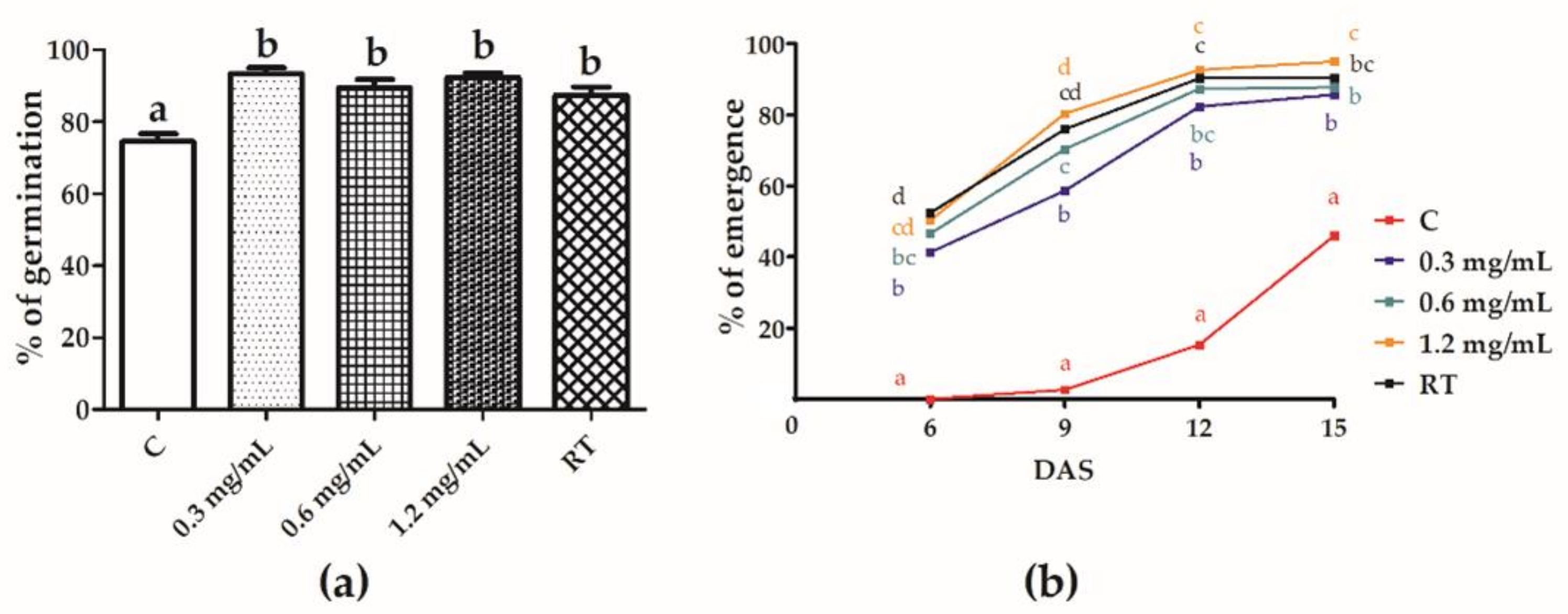
3.2. Effect of Seed Treatment on Seed Germination and Seedling Emergence

In the blotting paper assay (Figure 2a), the JA WSPs and RT have significantly increased the seed germination percentage as compared with the water-treated control. No differences among the JA WSPs concentrations were observed. In the greenhouse assay (Figure 2b), all JA concentrations and RT treatment significantly improved the seedling emergence over time with respect to the water treatment (control). At 15DAS, the JA WSPs concentration of 1.2 mg/mL showed the highest percentage emergence (95.0%).
As the treatments had no adverse effect on either seed germination or seedling emergence, they were further investigated in greenhouse experiments.

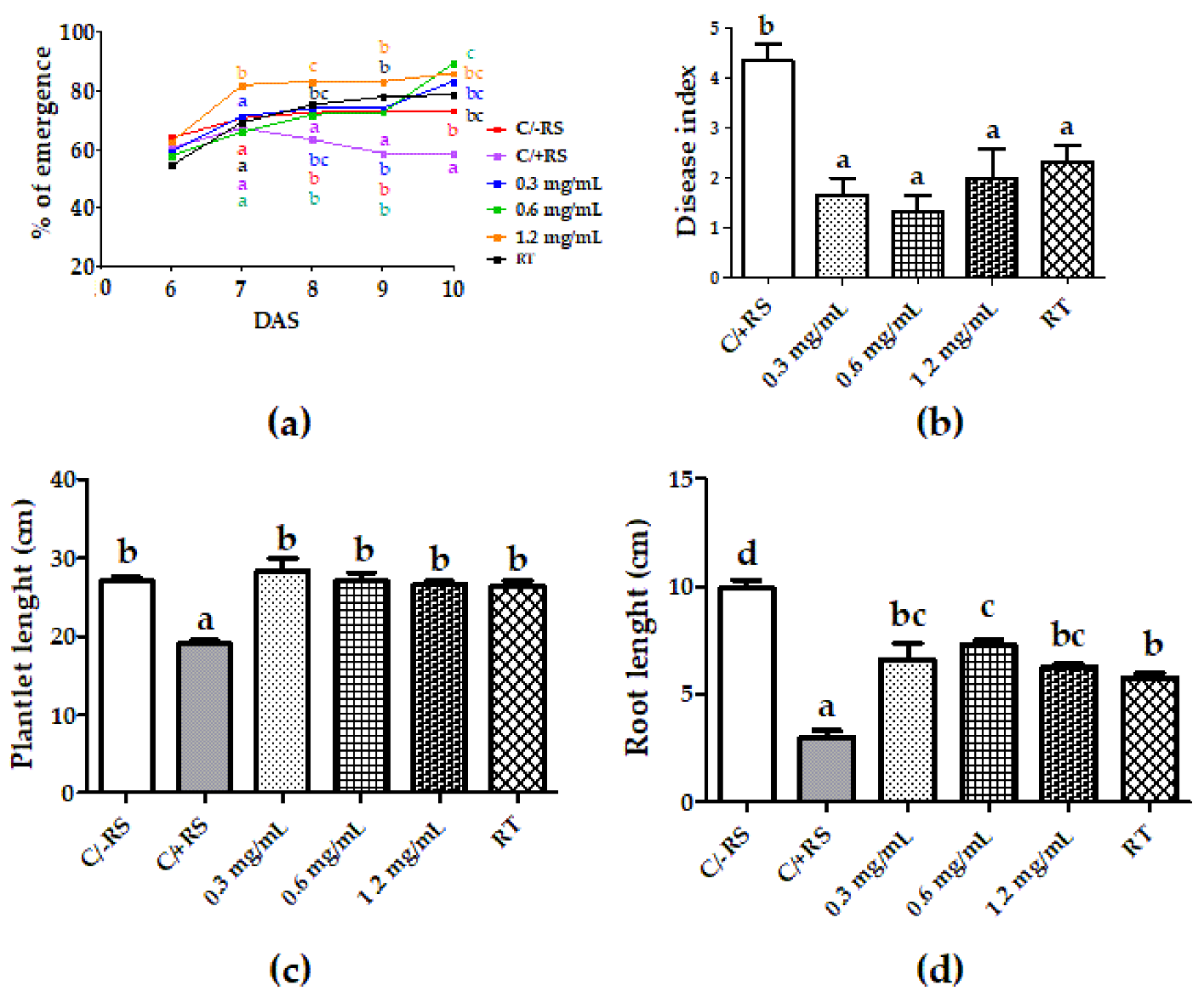
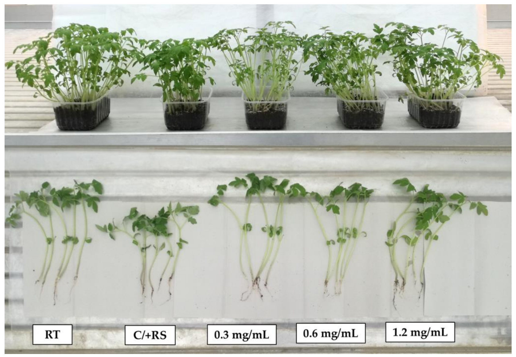
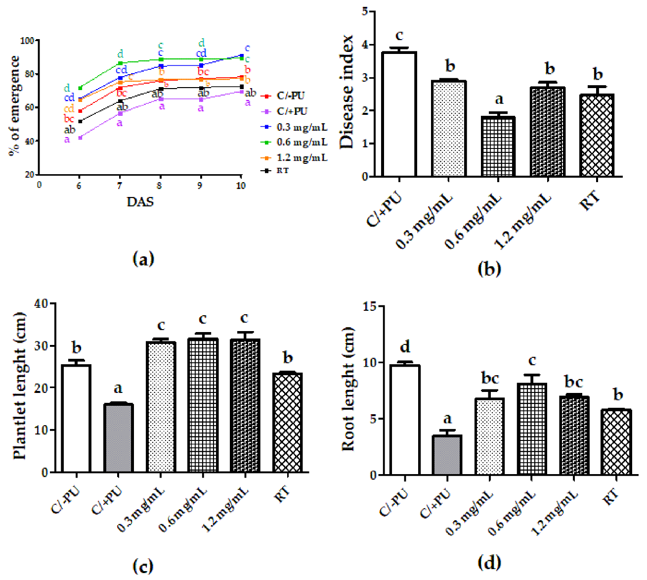
3.3. Systemic-Induced Resistance Bioassays in Greenhouse Pot Experiments

The treatment with JA WSPs at all doses increased the seedling emergence percentage (Figure 3a) in the growing substrate inoculated with RS with respect to the inoculated control (C/+RS). This was very noticeable and significant from 8DAS until 10DAS. In particular, at 10DAS 0.3, 0.6, and 1.2 mg/mL doses significantly increased the emergence percentage with respect to the inoculated control (C/+RS) by 41.9, 52.1, and 46.5%, respectively. No statistical difference was observed between the three doses of JA WSPs and RT, which increased the emergence by 34.0%.
All JA concentrations significantly reduced similarly the disease severity with respect to the C/+RS by 57.6% on average. The disease reduction obtained with JA WSPs was not statistically different from RT (Figure 3b and Figure 4). The WSPs significantly increased seedling length by 43.8% on average without differences among concentrations with respect to C/+RS and similarly to C/-RS and RT (increase by 38.4% vs. the C/+RS) (Figure 3c). The root lengths of seedlings from seeds treated with JA WSPs were significantly greater than that of C/+RS, even though they were smaller than C/-RS. The concentrations of 0.6 mg/mL significantly increased root length by 25.8% compared to RT treatment (Figure 3d).
As regards the effect of JA WSPs treatment against PU, Figure 5a shows that all concentrations significantly increased the seedling emergence at all DAS with respect to the inoculated control (C/+PU). Notably, RT treatment was not significantly different from C/+PU at all DAS. Overall, 0.3 and 0.6 mg/mL gave the highest emergence percentages from 8 to 10DAS (in average 86.7–90.3%). As regards the effect of the treatments against the disease, all JA WSPs concentrations and RT significantly reduced the disease index with respect to the C/+PU (Figure 5b). The highest disease reduction (52.6%) was obtained with 0.6 mg/mL. This concentration was 28% more effective than RT. Plant length was similar with the three concentrations (30.7, 31.7, and 31.3 cm for 0.3, 0.6, and 1.2 mg/mL, respectively) and higher than C/+PU (16.0 cm), C/-PU (25.3 cm), and RT (23.3 cm) (Figure 5c). The root length following seed treatment with all doses of JA WSPs was significantly higher than that of C/+PU and lower than C/-PU. The concentration of 0.6 mg/mL increased root length more effectively than RT by 40.6%, while the 0.3 and 1.2 mg/mL doses increased root length as well as RT treatment (Figure 5d).
Figure 6 shows the effect of seed treatment with different JA WSPs concentrations in the case of substrate inoculated with FO on disease severity and plant growth. The concentrations of 0.6 and 1.2 mg/mL displayed a significant disease index reduction with respect to C/+FO by 14.3 and 34.7%, respectively (Figure 6a). The highest statistical reduction was obtained with the 1.2 mg/mL concentration which was also 28.9% more effective than RT. Plant length was also increased significantly by 0.6 and 1.2 mg/mL (Figure 6b). The concentration of 1.2 mg/mL showed the highest plant length (54.7 cm) with regards to the C/+FO (25.7 cm). This value was even similar to that of C/-FO (63.0 cm). The three WSPs doses significantly increased root length with respect to the C/+FO (Figure 6c). The highest increase (228.3% vs. C/+FO) was obtained with the 1.2 mg/mL dose. Plant dry weight showed the highest significant values at 0.6 mg/mL (0.751 g) and 1.2 mg/mL (0.872 g) (Figure 6d). In particular, the plant dry weight with 1.2 mg/mL dose was significantly similar to that of the C/-FO control.

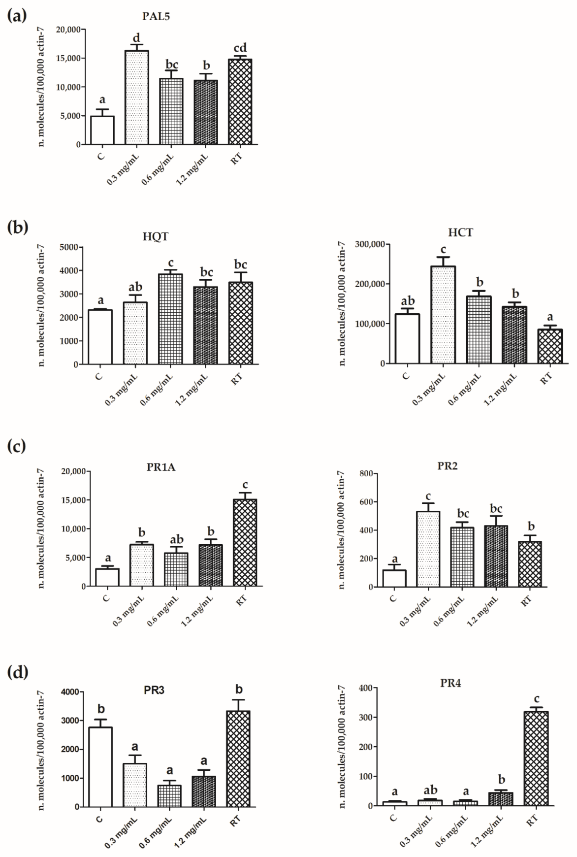
3.4. Expression of PR Protein and Polyphenol Pathway Genes

To shed some light on the molecular mechanisms associated with the induced resistance elicited by JA WSPs, the mRNA expression of pathogenesis-related (PR) proteins and key enzymes of polyphenol pathways was investigated in plantlets 20 days after seed treatment with JA WSPs or RT. Figure 7a shows that the transcript levels of phenylalanine ammonia-lyase (PAL5), a key enzyme in the phenylpropanoid biosynthetic pathway, were increased by JA WSPs at any concentration employed and similarly to RT. The expression pattern of two enzymes that are located downstream PAL, i.e., hydroxycinnamoyl CoA quinate hydroxycinnamoyl transferase (HQT) and hydroxycinnamoyl CoA shikimate hydroxycinnamoyl transferase (HCT) was also investigated and illustrated in Figure 7b. Seed treatment with JA WSPs or RT enhanced the expression level of HQT, while HCT was less affected. Even the expression of PR proteins involved in the systemic acquired resistance (SAR), i.e., PR1A and PR2, was enhanced by JA WSPs and RT (Figure 7c). On the contrary, we failed to observe an increase in the transcript level of the induced systemic resistance (ISR) protein, PR3 (Figure 7d), which actually resulted lower after JA WSPs compared to the control. However, the expression level of PR4, another protein involved in ISR, was significantly, but slightly increased by JA WSPs 1.2 mg/mL and, more markedly, by RT.

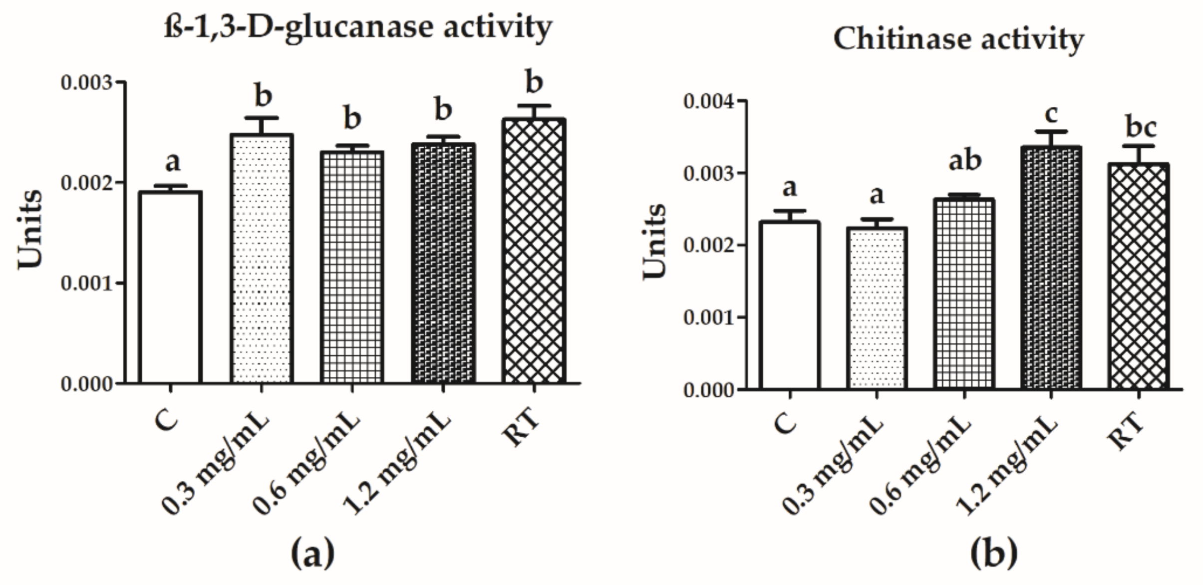
3.5. Chitinase and β-1,3-d-Glucanase Activities

Seed treatment with JA WSPs at different concentrations significantly enhanced the β-1,3-d-glucanase activity of seedlings with respect to the control by 25.3% on average (Figure 8a). No differences were observed among JA WSPs doses and RT. Chitinase activity was increased only at the higher dose, 1.2 mg/mL (44.6%), which produced a result similar to the RT treatment (Figure 8b).

4. Discussion

At present, plant pest control solutions for environmentally sustainable agriculture are highly recommended by European standards (Reg. EC 1107/2008; Dir. 128/2009). In particular, soil-borne disease management requires innovative strategies because the most effective pesticides for soil treatment underwent prohibition or restriction on their use.
Seaweed extracts are a source of several bioactive compounds that have a broad range of biological activity with a potential role in reducing the need for pesticides in agriculture [4,28,51,52,53,54,55]. Seaweeds are already widely used in agriculture due to their content of mineral substances, amino acids, vitamins, and plant growth regulators that ameliorate plant growth and enhance crop productivity [56,57]. Generally, the algal extracts are obtained with organic solvents such as methanol, ethanol, and acetone that on the one hand can optimize the extraction of specific components [55,58], but, on the other hand, represent a hazard to human health. On the contrary, our previous research showed that seaweed aqueous extracts that were obtained at 50 °C could be a sustainable and effective alternative to organic solvents to be used in plant disease management [26,59]. In particular, the study by [26] highlighted the potential of JA applied by seed priming technique for the control of R. solani on tomato plants and has led us to further research on this issue.
Seed priming is an alternative, low-cost, and feasible technique that involves seed imbibition with a low amount of water and various priming agents, such as osmoregulators, salts, plant growth regulators, beneficial microorganisms, magnetic waves, nanoparticles, and macro- and micronutrients, that allow the advancement of metabolic processes helping plants to overcome biotic and abiotic stress [60,61].
With the present study, we have demonstrated that JA WSPs could be a new potential tool for the control of the soil-borne pathogens R. solani, P. ultimum, and F. oxysporum in tomato by seed treatment. Moreover, we have highlighted that JA WSPs seed treatment may elicit plant defense responses likely involved in pathogen control. Although crude polysaccharide extracts from microalgae have previously been reported to induce immunity in plants [62], seed priming with JA WSPs is the first case study. FT-IR analysis demonstrated that JA WSPs differed in functional groups from laminarin used as reference treatment (RT). JA WSPs were characterized by typical carboxylate bands (1594 cm−1 and 1410 cm−1), most likely related to uronic acids. Our results are in agreement with those by Hentati et al. [1], who found uronic acids in the amount of about 6% in J. adhaerens. Moreover, ref. [62] found uronic acids in the red alga Porphyridium sp. Furthermore, based on their composition in functional groups, a structure–bioactivity relationship can be speculated. As a consequence, we can infer that carboxylate groups contributed to the anionic properties of JA WSPs, probably promoting interactions with other molecules capable of triggering several biochemical processes in treated tomato seeds. Indeed, polysaccharides are perceived in the plant as ‘danger’ molecules similar to what happens during the first interaction between a pathogen and the plant at the level of the cell wall and the plasma membrane [63]. To highlight the potential induced resistance effect by JA WSPs, treated seeds were water-washed after soaking, to remove any extract residues that could have a direct antifungal effect. Polysaccharides such as laminarin were reported to exert an antigerminative effect against the conidia of some fungi [64,65]. The polysaccharides extracted from JA might explain the elicitation of plant physiological responses against pathogens with the improvement of seedling emergence and plant growth, which may have concurred to the disease reduction. It is well known that the extracellular matrix of red seaweeds contains sulfated galactans as major components [66,67,68,69,70] which are elicitors of plant defense responses, as in the case of Phytophthora nicotianae on tobacco plants [7].
The seed treatment with JA WSPs showed a biostimulant-like effect in the preliminary assays since it increased seed germination and seedling emergence. Similarly, increases in germination were observed for tomato seeds treated with a different aqueous extract from the same red alga without pathogen challenge [26]. A biostimulant effect on plants was obtained with red alga extract applied by spraying and soil treatments as in the case of J. rubens on chickpea and maize [71,72] and by a foliar application with the red alga Kappaphycus alvarezii on wheat [73,74].
In the present study, the increase in the emergence rate was also obtained in response to the R. solani and P. ultimum challenge; indeed, JA WSPs at all doses increased the seedling emergence from 7DAS and 6DAS up to the last assessment, respectively. The plant growth promotion by the extract in the substrate inoculated with R. solani, P. ultimum and F. oxysporum was also observed in terms of plant and root length.
It is likely that the disease control is correlated with the plant growth promotion obtained with WSPs in substrates inoculated with each pathogen. All the concentrations reduced disease severity and increased both seedling and root length in a similar way, without showing a dose-dependent response. However, the 1.2 mg/mL concentration was the most effective in reducing F. oxysporum disease severity. We hypothesize that the disease control exerted by JA WSPs is due to the induction of plant defense responses, as shown by the increase in plant chitinase and glucanase activity observed and by the transcript levels of defense genes. This is in line with [75], who found a correlation between F. oxysporum disease control and the induction of plant defense responses by applying an extract of the brown alga Ascophyllum nodosum to cucumber plants. Chitinases and β-1,3-glucanase are enzymes constitutively produced by plants during their growth [76,77]. Some chitinases accumulate during their developmental program, in seeds of several species, while others, called defense enzymes, are produced as a response to a microbial attack or wounding [76]. During seed germination, both chitinases and β-1,3-glucanase are particularly important because seeds are exposed to soil-borne pathogens and the embryo becomes vulnerable to pathogen attack when the radicle crosses the endosperm tissue. Our findings showed that seed treatment with JA WSPs may have contributed to increasing the synthesis of chitinases and β-1,3-glucanase making the plant protected against pathogen attack. Ghule et al. [78] demonstrated that the activity of both these defense enzymes was increased in fenugreek plants derived from seeds treated with the marine polysaccharide chitosan. Following this treatment, plants were protected against root rot disease by Fusarium solani.
Two forms of induced resistance are described in response to stimuli that enhance plant defensive capacity versus pathogens: systemic acquired resistance (SAR), which involves the accumulation of PR1 and PR2 proteins and induced systemic resistance (ISR), which involves PR3 and PR4 proteins. According to the results shown in the present study (Figure 7), SAR response was elicited by JA polysaccharide seed treatment as judged by the increase in PR1A and PR2 expression. Instead, JA WSPs caused a decrease in PR3 gene expression, used as a marker of ISR response. It has been reported that crude polysaccharides extracted from Acanthophora spicifera (another red alga) enhanced rubber tree defenses against Phytophthora palmivora infection [79]. In accordance with our results, JA WSPs also induced PR1 and PR2 gene expression but suppressed ISR-related gene expression. Moreover, we have found an increase in the expression of PAL, known to be a key enzyme involved in plant development and defense responses to pathogens. PAL catalyzes the first and committed step in the biosynthesis of phenylpropanoids, which can give rise to a wide range of secondary metabolites, such as flavonoids and lignin. Among phenylpropanoid-derived phenolics and flavonoids, chlorogenic acid is known to activate the SAR pathway [80]. In this regard, we have also found increases in the expression of HQT and HCT, which are key enzymes of the biosynthetic pathway of chlorogenic acid.

5. Conclusions

Algae are considered an important resource of many biologically active compounds, and among them are polysaccharides. The chemical characteristics and potential biological properties make polysaccharides promising for ecological disease management in agriculture. In particular, seed priming with polysaccharides may be an attractive alternative to conventional fungal disease treatments. The present study actually shows that tomato seed priming with water-soluble polysaccharides extracted from the red alga Jania adhaerens increased seedling emergence and plant development, while reduced disease severity caused by three soil-borne pathogens, i.e., Rhizoctonia solani, Pythium ultimum and Fusarium oxysporum under greenhouse conditions. Furthermore, the protective effects were associated with the increased activity of defense enzymes and increased expression of genes involved in induced resistance and related pathways. In conclusion, we have described a red algal-based seed treatment that promotes plant growth and triggers plant defense responses against soil-borne pathogens.
This study may contribute to environmentally sustainable agriculture. However, there are still some aspects that need further investigation such as standardized protocols for extraction procedures and the isolation and purification of polysaccharides. These will help in determining the chemical structure and biological activity of polysaccharides. Currently, these procedures are limited to lab research and have not yet been applied on an industrial scale.

Author Contributions

Conceptualization, H.R., S.G., R.R. and S.C. (Silvia Cetrullo); methodology, S.C. (Stefano Cianchetta) and V.P.; formal analysis, H.R. and R.R.; investigation, H.R., S.G., R.R., O.F., S.C. (Silvia Cetrullo), V.P. and A.M.Q.; resources, R.R.; data curation, H.R., R.R., O.F., F.F. and S.C. (Silvia Cetrullo); writing—original draft preparation, H.R., R.R., S.G., S.C. (Silvia Cetrullo), O.F. and A.M.Q.; writing—review and editing, H.R., R.R., O.F. and F.F.; supervision, F.F.; funding acquisition, S.G. and A.M.Q. All authors have read and agreed to the published version of the manuscript.

Funding

This research was funded by the Italian Ministry of Agricultural, Food, Forestry and Tourism Policies (MiPAAFT) under the DIBIO-BIOPRIME project, (DM N. 3400, 20 December 2018) and by Interreg MAC Program 2014–2020, grant number MAC2/1.1b/269: REBECA-CCT.

Institutional Review Board Statement

Not applicable.

Informed Consent Statement

Not applicable.

Data Availability Statement

The data presented in this study are available upon request from the corresponding author.

Conflicts of Interest

The authors declare no conflict of interest. The funders had no role in the design of the study; in the collection, analyses, or interpretation of data; in the writing of the manuscript; or in the decision to publish the results.

References

  1. Hentati, F.; Barkallah, M.; Ben Atitallah, A.; Dammak, M.; Louati, I.; Pierre, G.; Fendri, I.; Attia, H.; Michaud, P.; Abdelkafi, S. Quality characteristics and functional and antioxidant capacities of algae-fortified fish burgers prepared from common barbel (Barbus barbus). Biomed. Res. Int. 2019, 2019, 2907542. [Google Scholar] [CrossRef] [PubMed]
  2. Kousha, M.; Daneshvar, E.; Esmaeli, A.R.; Jokar, M.; Khataee, A.R. Optimization of Acid Blue 25 removal from aqueous solutions by raw, esterified and protonated Jania adhaerens biomass. Int. Biodeter. Biodegr. 2012, 69, 97–105. [Google Scholar] [CrossRef]
  3. Ramu Ganesan, A.; Kannan, M.; Karthick Rajan, D.; Pillay, A.A.; Shanmugam, M.; Sathishkumar, P.; Johansen, J.; Tiwari, B.K. Phycoerythrin: A pink pigment from red sources (rhodophyta) for a greener biorefining approach to food applications. Crit. Rev. Food Sci. Nutr. 2022, 1–19. [Google Scholar] [CrossRef] [PubMed]
  4. Hamed, S.M.; Abd El-Rhman, A.A.; Abdel-Raouf, N.; Ibraheem, I.B. Role of marine macroalgae in plant protection & improvement for sustainable agriculture technology. Beni-Suef Univ. J. Basic. Appl. Sci. 2018, 7, 104–110. [Google Scholar]
  5. Navarro, D.A.; Stortz, C.A. The system of xylogalactans from the red seaweed Jania rubens (Corallinales, Rhodophyta). Carbohyd. Res. 2008, 343, 2613–2622. [Google Scholar] [CrossRef] [PubMed]
  6. Kaplan, D.; Christiaen, D.; Arad, S.M. Chelating properties of extracellular polysaccharides from Chlorella spp. Appl. Environ. Microbial. 1987, 53, 2953–2956. [Google Scholar] [CrossRef] [PubMed]
  7. Mercier, L.; Lafitte, C.; Borderies, G.; Briand, X.; Esquerré-Tugayé, M.T.; Fournier, J. The algal polysaccharide carrageenans can act as an elicitor of plant defence. New Phytol. 2001, 149, 43–51. [Google Scholar] [CrossRef]
  8. Lefevere, H.; Bauters, L.; Gheysen, G. Salicylic acid biosynthesis in plants. Front. Plant Sci. 2020, 11, 338. [Google Scholar] [CrossRef]
  9. Seyfferth, C.; Tsuda, K. Salicylic acid signal transduction: The initiation of biosynthesis, perception and transcriptional reprogramming. Front. Plant Sci. 2014, 5, 697. [Google Scholar] [CrossRef]
  10. Bruehl, G.W. Soilborne Plant Pathogens; Macmillan Publishing Co.: New York, NY, USA, 1987; ISBN 0029491304. [Google Scholar]
  11. Martin, F.N.; Loper, J.E. Soilborne plant diseases caused by Pythium spp.: Ecology, epidemiology, and prospects for biological control. Crit. Rev. Plant Sci. 1999, 18, 111–181. [Google Scholar] [CrossRef]
  12. Kamoun, S.; Furzer, O.; Jones, J.D.; Judelson, H.S.; Ali, G.S.; Dalio, R.J.; Roy, G.S.; Schena, L.; Zambounis, A.; Panabières, F.; et al. The Top 10 oomycete pathogens in molecular plant pathology. Mol. Plant. Pathol. 2015, 16, 413–434. [Google Scholar] [CrossRef]
  13. Lübeck, M. Molecular characterization of Rhizoctonia solani. In Applied Mycology and Biotechnology, 1st ed.; Khachatourians, G.G., Arora, D.K., Eds.; Volume 4: Fungal genomics; Elsevier Science B.V.: Amsterdam, The Netherlands, 2004; pp. 205–224. ISBN 0444516425. [Google Scholar]
  14. Marcou, S.; Wikström, M.; Ragnarsson, S.; Persson, L.; Höfte, M. Occurrence and anastomosis grouping of Rhizoctonia spp. inducing black scurf and greyish-white felt-like mycelium on carrot in Sweden. J. Fungi 2021, 7, 396. [Google Scholar] [CrossRef] [PubMed]
  15. Gordon, T.R. Fusarium oxysporum and the Fusarium wilt syndrome. Annu. Rev. Phytopathol. 2017, 55, 23–39. [Google Scholar] [CrossRef] [PubMed]
  16. Fravel, D.; Olivain, C.; Alabouvette, C. Fusarium oxysporum and its biocontrol. New Phytol. 2003, 157, 493–502. [Google Scholar] [CrossRef] [PubMed]
  17. Michielse, C.B.; Rep, M. Pathogen profile update: Fusarium oxysporum. Mol. Plant. Pathol. 2009, 10, 311. [Google Scholar] [CrossRef] [PubMed]
  18. Galletti, S.; Cianchetta, S.; Righini, H.; Roberti, R. A Lignin-rich extract of giant reed (Arundo donax L.) as a possible tool to manage soilborne pathogens in horticulture: A preliminary study on a model pathosystem. Horticulturae 2022, 8, 589. [Google Scholar] [CrossRef]
  19. Abo-Elyousr, K.A.; Ali, E.F.; Sallam, N.M. Alternative control of tomato wilt using the aqueous extract of Calotropis procera. Horticulturae 2022, 8, 197. [Google Scholar] [CrossRef]
  20. La Torre, A.; Caradonia, F.; Matere, A.; Battaglia, V. Using plant essential oils to control Fusarium wilt in tomato plants. Eur. J. Plant Pathol. 2016, 144, 487–496. [Google Scholar] [CrossRef]
  21. Sharma, A.; Rajendran, S.; Srivastava, A.; Sharma, S.; Kundu, B. Antifungal activities of selected essential oils against Fusarium oxysporum f. sp. lycopersici 1322, with emphasis on Syzygium aromaticum essential oil. J. Biosci. Bioeng. 2017, 123, 308–313. [Google Scholar]
  22. Ramírez, P.G.; Ramírez, D.G.; Mejía, E.Z.; Ocampo, S.A.; Díaz, C.N.; Martínez, R.I.R. Extracts of Stevia rebaudiana against Fusarium oxysporum associated with tomato cultivation. Sci. Hortic. 2020, 259, 108683. [Google Scholar] [CrossRef]
  23. Ozkaya, H.O.; Ergun, T. The effects of Allium tuncelianum extract on some important pathogens and total phenolic compounds in tomato and pepper. Pak. J. Bot. 2017, 49, 2483–2490. [Google Scholar]
  24. Sanaullah, N.A.R.; Atiq, M.; Rehman, A.; Khan, S.; Bashir, M.; Hameed, A.; Khan, B.; Shakir Baloch, M.; Kachelo, G.A. Antifungal potency of three plant extracts against Rhizoctonia solani damping-off disease in tomato. Int. J. Biosci. 2018, 13, 309–316. [Google Scholar]
  25. Hlokwe, M.T.P.; Kena, M.A.; Mamphiswana, D.N. Application of plant extracts and Trichoderma harzianum for the management of tomato seedling damping-off caused by Rhizoctonia solani. S. Afr. J. Sci. 2020, 116, 1–5. [Google Scholar] [CrossRef]
  26. Righini, H.; Francioso, O.; Di Foggia, M.; Prodi, A.; Quintana, A.M.; Roberti, R. Tomato seed biopriming with water extracts from Anabaena minutissima, Ecklonia maxima and Jania adhaerens as a new agro-ecological option against Rhizoctonia solani. Sci. Hortic. 2021, 281, 109921. [Google Scholar] [CrossRef]
  27. Akladious, S.A.; Isaac, G.S.; Abu-Tahon, M.A. Induction and resistance against Fusarium wilt disease of tomato by using sweet basil (Ocimum basilicum L.) extract. Can. J. Plant. Sci. 2015, 95, 689–701. [Google Scholar] [CrossRef]
  28. Righini, H.; Roberti, R.; Baraldi, E. Use of algae in strawberry management. J. Appl. Phycol. 2018, 30, 3551–3564. [Google Scholar] [CrossRef]
  29. Vera, J.; Castro, J.; Gonzalez, A.; Moenne, A. Seaweed polysaccharides and derived oligosaccharides stimulate defense responses and protection against pathogens in plants. Mar. Drugs 2011, 9, 2514–2525. [Google Scholar] [CrossRef]
  30. Vicente, T.F.L.; Lemos, M.F.L.; Félix, R.; Valentão, P.; Félix, C. Marine macroalgae, a source of natural inhibitors of fungal phytopathogens. J. Fungi 2021, 7, 1006. [Google Scholar] [CrossRef]
  31. Ben Salah, I.; Aghrouss, S.; Douira, A.; Aissam, S.; El Alaoui-Talibi, Z.; Filali-Maltouf, A.; El Modafar, C. Seaweed polysaccharides as bio-elicitors of natural defenses in olive trees against verticillium wilt of olive. J. Plant Interact. 2018, 13, 248–255. [Google Scholar] [CrossRef]
  32. Ansari, F.A.; Shriwastav, A.; Gupta, S.K.; Rawat, I.; Guldhe, A.; Bux, F. Lipid extracted algae as a source for protein and reduced sugar: A step closer to the biorefinery. Bioresour. Technol. 2015, 179, 559–564. [Google Scholar] [CrossRef]
  33. Chen, Y.Y.; Xue, Y.T. Optimization of microwave assisted extraction, chemical characterization and antitumor activities of polysaccharides from Porphyra haitanensis. Carbohydr. Polym. 2019, 206, 179–186. [Google Scholar] [CrossRef] [PubMed]
  34. Garcia-Vaquero, M.; O’Doherty, J.V.; Tiwari, B.K.; Sweeney, T.; Rajauria, G. Enhancing the extraction of polysaccharides and antioxidants from macroalgae using sequential hydrothermal-assisted extraction followed by ultrasound and thermal technologies. Mar. Drugs 2019, 17, 457. [Google Scholar] [CrossRef] [PubMed]
  35. Elarrouss, H.; Elmernissi, N.; Benhima, R.; El Kadmiri, I.M.; Bendaou, N.; Smouni, A.; Wahbya, I. Microalgae polysaccharides a promising plant growth biostimulant. J. Algal Biomass Util. 2016, 7, 55–63. [Google Scholar]
  36. Ozawa, T.; Yamamoto, J.; Yamagishi, T.; Yamazaki, N.; Nishizawa, M. Two fucoidans in the holdfast of cultivated Laminaria japonica. J. Nat. Med. 2006, 60, 236–239. [Google Scholar] [CrossRef] [PubMed]
  37. Sinurat, E.; Rosmawaty, P.; Saepudin, E. Characterization of fucoidan extracted from Binuangeun’s Brown Seaweeds. Int. J. Chem. Environ. Biol. Sci. 2015, 3, 329–332. [Google Scholar]
  38. Shen, S.; Cheng, H.; Li, X.; Li, T.; Yuan, M.; Zhou, Y.; Ding, C. Effects of extraction methods on antioxidant activities of polysaccharides from camellia seed cake. Eur. Food Res. Technol. 2014, 238, 1015–1021. [Google Scholar] [CrossRef]
  39. Álvarez-Gómez, F.; Korbee, N.; Figueroa, F.L. Analysis of antioxidant capacity and bioactive compounds in marine macroalgal and lichenic extracts using different solvents and evaluation methods. Cienc. Mar. 2016, 42, 271–288. [Google Scholar] [CrossRef]
  40. Nawaz, A.; Amjad, M.; Khan, S.M.; Afzal, I.; Ahmed, T.; Iqbal, Q.; Iqbal, J. Tomato seed invigoration with cytokinins. J. Anim. Plant. Sci. 2013, 23, 121–128. [Google Scholar]
  41. Burnett, F.J.; Boor, T.; Dussart, F.; Smith, J. Developing Sustainable Management Methods for Clubroot. Project Report No. 608. Agriculture and Horticulture Development Board. 2019. Available online: https://pure.sruc.ac.uk/ws/portalfiles/potal/18991123/pr608_final_project_report.pdf (accessed on 12 July 2022).
  42. Li, Q.; Yu, P.; Chen, X.; Li, G.; Zhou, D.; Zheng, W. Facilitative and inhibitory effect of litter on seedling emergence and early growth of six herbaceous species in an early successional old field ecosystem. Sci. World J. 2014, 2014, 101860. [Google Scholar] [CrossRef]
  43. De Cal, A.; Pascual, S.; Melgarejo, P. Infectivity of chlamydospores vs microconidia of Fusarium oxysporum f. sp. lycopersici on tomato. J. Phytopathol 1997, 145, 231–233. [Google Scholar] [CrossRef]
  44. Roberti, R.; Galletti, S.; Burzi, P.L.; Righini, H.; Cetrullo, S.; Perez, C. Induction of defence responses in zucchini (Cucurbita pepo) by Anabaena sp. water extract. Biol. Control 2015, 82, 61–68. [Google Scholar] [CrossRef]
  45. Bradford, M.M. A rapid and sensitive method for the quantitation of microgram quantities of protein utilizing the principle of protein-dye binding. Anal. Biochem. 1976, 72, 248–254. [Google Scholar] [CrossRef]
  46. Bargabus, R.L.; Zidack, N.K.; Sherwood, J.E.; Jacobsen, B.J. Screening for the identification of potential biological control agents that induce systemic acquired resistance in sugar beet. Biol. Control 2004, 30, 342–350. [Google Scholar] [CrossRef]
  47. Rao, C.N.R. Chemical Applications of Infrared Spectroscopy; Academic Press, Inc.: New York, NY, USA; London, UK, 1963; p. 681. [Google Scholar]
  48. Xu, R.B.; Yang, X.; Wang, J.; Zhao, H.T.; Lu, W.H.; Cui, J.; Cheng, C.L.; Zou, P.; Huang, W.W.; Wang, P.; et al. Chemical composition and antioxidant activities of three polysaccharide fractions from pine cones. Int. J. Mol. Sci. 2012, 13, 14262–14277. [Google Scholar] [CrossRef]
  49. Hong, T.; Yin, J.Y.; Nie, S.P.; Xie, M.Y. Applications of infrared spectroscopy in polysaccharide structural analysis: Progress, challenge and perspective. Food Chem. X 2021, 12, 100168. [Google Scholar] [CrossRef]
  50. Wiercigroch, E.; Szafraniec, E.; Czamara, K.; Pacia, M.Z.; Majzner, K.; Kochan, K.; Malek, K. Raman and infrared spectroscopy of carbohydrates: A review. Spectrochim. Acta A 2017, 185, 317–335. [Google Scholar] [CrossRef]
  51. Shukla, P.S.; Mantin, E.G.; Adil, M.; Bajpai, S.; Critchley, A.T.; Prithiviraj, B. Ascophyllum nodosum-based biostimulants: Sustainable applications in agriculture for the stimulation of plant growth, stress tolerance, and disease management. Front. Plant Sci. 2019, 10, 655. [Google Scholar] [CrossRef]
  52. Arioli, T.; Mattner, S.W.; Winberg, P.C. Applications of seaweed extracts in Australian agriculture: Past, present and future. J. Appl. Phycol. 2015, 27, 2007–2015. [Google Scholar] [CrossRef]
  53. Khan, W.; Rayirath, U.P.; Subramanian, S.; Jithesh, M.N.; Rayorath, P.; Hodges, D.M.; Critchley, A.T.; Craigie, J.S.; Norrie, J.; Prithiviraj, B. Seaweed extracts as biostimulants of plant growth and development. J. Plant Growth Regul. 2009, 28, 386–399. [Google Scholar] [CrossRef]
  54. O’ Keeffe, E.; Hughes, H.; McLoughlin, P.; Tan, S.P.; McCarthy, N. Methods of analysis for the in vitro and in vivo determination of the fungicidal activity of seaweeds: A mini review. J. Appl. Phycol. 2019, 31, 3759–3776. [Google Scholar] [CrossRef]
  55. Sharma, H.S.; Fleming, C.; Selby, C.; Rao, J.R.; Martin, T. Plant biostimulants: A review on the processing of macroalgae and use of extracts for crop management to reduce abiotic and biotic stresses. J. Appl. Phycol. 2014, 26, 465–490. [Google Scholar] [CrossRef]
  56. Abdel-Raouf, N.; Al-Homaidan, A.A.; Ibrahim, I.B.M. Agricultural importance of algae. Afr. J. Biotechnol. 2012, 1, 11648–11658. [Google Scholar] [CrossRef]
  57. Calvo, P.; Nelson, L.; Kloepper, J.W. Agricultural uses of plant biostimulants. Plant Soil 2014, 383, 3–41. [Google Scholar] [CrossRef]
  58. Arunkumar, K.; Sivakumar, S.R.; Rengasamy, R. Review on bioactive potential in seaweeds (marine macroalgae): A special emphasis on bioactivity of seaweeds against plant pathogens. Asian J. Plant Sci. 2010, 9, 227–240. [Google Scholar] [CrossRef]
  59. Righini, H.; Somma, A.; Cetrullo, S.; D’Adamo, S.; Flamigni, F.; Martel Quintana, A.; Roberti, R. Inhibitory activity of aqueous extracts from Anabaena minutissima, Ecklonia maxima and Jania adhaerens on the cucumber powdery mildew pathogen in vitro and in vivo. J. Appl. Phycol. 2020, 32, 3363–3375. [Google Scholar] [CrossRef]
  60. Arun, M.N.; Hebbar, S.S.; Senthivel, T.; Nair, A.K.; Padmavathi, G.; Pandey, P.; Singh, A. Seed priming: The way forward to mitigate abiotic stress in crops. In Plant Stress Physiology-Perspectives in Agriculture; Hasanuzzaman, M., Nahar, K., Eds.; IntechOpen: London, UK, 2022; Volume 11, p. 173. [Google Scholar]
  61. El-Mougy, N.S.; Abdel-Kader, M.M. Long term activity of bio-priming seed treatment for biological control of faba bean root rot pathogens. Aust. Plant Pathol. 2008, 37, 464–471. [Google Scholar] [CrossRef]
  62. Rachidi, F.; Benhima, R.; Kasmi, Y.; Sbabou, L.; Arroussi, H.E. Evaluation of microalgae polysaccharides as biostimulants of tomato plant defense using metabolomics and biochemical approaches. Sci. Rep. 2021, 11, 101860. [Google Scholar] [CrossRef]
  63. Chaliha, C.; Rugen, M.D.; Field, R.A.; Kalita, E. Glycans as modulators of plant defense against filamentous pathogens. Front. Plant Sci. 2018, 9, 928. [Google Scholar] [CrossRef]
  64. Lagogianni, C.S.; Tsitsigiannis, D.I. Effective biopesticides and biostimulants to reduce aflatoxins in maize fields. Front. Microbiol. 2019, 10, 2645. [Google Scholar] [CrossRef]
  65. de Borba, M.C.; Velho, A.C.; de Freitas, M.B.; Holvoet, M.; Maia-Grondard, A.; Baltenweck, R.; Magnin-Robert, M.; Randoux, B.; Hilbert, J.L.; Reignault, P.; et al. A laminarin-based formulation protects wheat against Zymoseptoria tritici via direct antifungal activity and elicitation of host defense-related genes. Plant Dis. 2022, 106, 1408–1418. [Google Scholar] [CrossRef]
  66. Aruna, P.; Mansuya, P.; Sridhar, S.; Kumar, J.S.; Babu, S. Pharmacognostical and antifungal activity of selected seaweeds from Gulf of Mannar region. Recent Res. Sci. Technol. 2010, 2, 115–119. [Google Scholar]
  67. Damonte, E.B.; Matulewicz, M.C.; Cerezo, A.S. Sulfated seaweed polysaccharides as antiviral agents. Curr. Med. Chem. 2004, 11, 2399–2419. [Google Scholar] [CrossRef] [PubMed]
  68. Matsuhiro, B.; Conte, A.F.; Damonte, E.B.; Kolender, A.A.; Matulewicz, M.C.; Mejías, E.G.; Pujol, C.A.; Zúñiga, E.A. Structural analysis and antiviral activity of a sulfated galactan from the red seaweed Schizymenia binderi (Gigartinales, Rhodophyta). Carbohyd. Res. 2005, 340, 2392–2402. [Google Scholar] [CrossRef] [PubMed]
  69. Pujol, C.A.; Scolaro, L.A.; Ciancia, M.; Matulewicz, M.C.; Cerezo, A.S.; Damonte, E.B. Antiviral activity of a carrageenan from Gigartina skottsbergii against intraperitoneal murine Herpes simplex virus infection. Planta Med. 2006, 72, 121–125. [Google Scholar] [CrossRef]
  70. Souza, B.W.S.; Cerqueira, M.A.; Bourbon, A.I.; Pinheiro, A.C.; Martins, J.T.; Teixeira, J.A.; Coimbra, M.A.; Vicente, A.A. Chemical characterization and antioxidant activity of sulfated polysaccharide from the red seaweed Gracilaria birdiae. Food Hydrocoll. 2012, 27, 287–292. [Google Scholar] [CrossRef]
  71. Abdel Latef, A.A.H.; Srivastava, A.K.; Saber, H.; Alwaleed, E.A.; Tran, L.S.P. Sargassum muticum and Jania rubens regulate amino acid metabolism to improve growth and alleviate salinity in chickpea. Sci. Rep. 2017, 7, 10537. [Google Scholar] [CrossRef]
  72. Safinaz, A.F.; Ragaa, A.H. Effect of some red marine algae as biofertilizers on growth of maize (Zea mays L.) plants. Int. Food Res. J. 2013, 20, 1629–1632. [Google Scholar]
  73. Zodape, S.T.; Mukherjee, S.; Reddy, M.P.; Chaudhary, D.R. Effect of Kappaphycus alvarezii (Doty) doty ex silva. extract on grain quality, yield and some yield components of wheat (Triticum aestivum L.). Int. J. Plant Prod. 2009, 3, 97–101. [Google Scholar]
  74. Zodape, S.T.; Gupta, A.; Bhandari, S.C.; Rawat, U.S.; Chaudhary, D.R.; Eswaran, K.; Chikara, J. Foliar application of seaweed sap as biostimulant for enhancement of yield and quality of tomato (Lycopersicon esculentum Mill.). J. Sci. Ind. Res. 2011, 70, 215–219. [Google Scholar]
  75. Jayaraman, J.; Norrie, J.; Punja, Z.K. Commercial extract from the brown seaweed Ascophyllum nodosum reduces fungal diseases in greenhouse cucumber. J. Appl. Phycol. 2011, 23, 353–361. [Google Scholar] [CrossRef]
  76. Witmer, X.; Nonogaki, H.; Beers, E.P.; Bradford, K.J.; Welbaum, G.E. Characterization of chitinase activity and gene expression in muskmelon seeds. Seed Sci. Res. 2003, 13, 167–178. [Google Scholar] [CrossRef]
  77. Morohashi, Y.; Matsushima, H. Development of β-1, 3-glucanase activity in germinated tomato seeds. J. Exp. Bot. 2000, 51, 1381–1387. [Google Scholar] [CrossRef]
  78. Ghule, M.R.; Ramteke, P.K.; Ramteke, S.D.; Kodre, P.S.; Langote, A.; Gaikwad, A.V.; Holkar, S.K.; Jambhekar, H. Impact of chitosan seed treatment of fenugreek for management of root rot disease caused by Fusarium solani under in vitro and in vivo conditions. 3 Biotech 2021, 11, 290. [Google Scholar] [CrossRef] [PubMed]
  79. Pettongkhao, S.; Bilanglod, A.; Khompatara, K.; Churngchow, N. Sulphated polysaccharide from Acanthophora spicifera induced Hevea brasiliensis defense responses against Phytophthora palmivora infection. Plants 2019, 8, 73. [Google Scholar] [CrossRef]
  80. Xu, X.; Chen, Y.; Li, B.; Zhang, Z.; Qin, G.; Chen, T.; Tian, S. Molecular mechanisms underlying multi-level defense responses of horticultural crops to fungal pathogens. Hortic. Res. 2022, 9, uhac066. [Google Scholar] [CrossRef] [PubMed]
Figure 1. FT-IR spectra of Jania adhaerens water-soluble polysaccharide (black line) and RT a commercial product based on laminarin (green line).
Horticulturae 08 00746 g001
Figure 2. Preliminary assays on the effect of tomato seed treatment with different concentrations (0.3, 0.6, and 1.2 mg/mL) of Jania adhaerens water-soluble polysaccharides on the seed germination percentage in blotting paper test (a) and on the percentage of seedling emergence in the experiment under greenhouse conditions (b). C = water control; RT = reference treatment (2 mL/L); DAS = days after sowing. In panel (a), columns are mean values of 3 independent experiments (n = 3) ± SD. Different letters indicate significant differences among treatments and control according to the LSD test (p < 0.05). In panel (b), each value is the mean of 3 independent experiments (n = 3); different letters indicate significant differences among treatments and the control within each DAS, according to the LSD test (p < 0.05).
Horticulturae 08 00746 g002
Figure 3. Effect of tomato seed treatment with different concentrations (0.3, 0.6, and 1.2 mg/mL) of Jania adhaerens water-soluble polysaccharides on the emergence percentage of seedlings (a), disease severity (b), plant length (c), and root length (d) in a growing substrate infected with Rhizoctonia solani (RS). C/-RS = non infected control; C/+RS = infected control; RT = reference treatment (2 mL/L); DAS = days after sowing. In panel (a), each value is the mean of 3 independent experiments (n = 3); different letters indicate significant differences among treatments and controls within each DAS, according to the LSD test (p < 0.05). In panels (bd), columns are mean values of 3 independent experiments (n = 3) ± SD. Different letters indicate significant differences among treatments and controls according to the LSD test (p < 0.05).
Horticulturae 08 00746 g003
Figure 4. Effect of tomato seed treatment with different concentrations (0.3, 0.6, and 1.2 mg/mL) of Jania adhaerens water-soluble polysaccharides on the disease severity caused by Rhizoctonia solani and plant growth. RT = reference treatment (2 mL/L); C/+RS = infected control.
Horticulturae 08 00746 g004
Figure 5. Effect of tomato seed treatment with different concentrations (0.3, 0.6, and 1.2 mg/mL) of Jania adhaerens water-soluble polysaccharides on the emergence of seedlings (a), plant length (b) and root length (c) in a growing substrate infected with Pythium ultimum (PU). C/-PU = non-infected control; C/+PU = infected control; RT = reference treatment (2 mL/L); DAS = days after sowing. In panel (a), each value is the mean of 3 independent experiments (n = 3); different letters indicate significant differences among treatments and controls within each DAS, according to the LSD test (p < 0.05). In panels (bd), columns are mean values of 3 independent experiments (n = 3) ± SD. Different letters indicate significant differences among treatments and controls according to the LSD test (p < 0.05).
Horticulturae 08 00746 g005
Figure 6. Effect of tomato seed treatment with different concentrations (0.3, 0.6, and 1.2 mg/mL) of Jania adhaerens water-soluble polysaccharides on the disease index (a), plant and root length (b,c) and plant dry weight (d) in a growth substrate infected with Fusarium oxysporum (FO). C/-FO = non infected control; C/+FO = infected control; RT = reference treatment (2 mL/L). Columns are mean values of 3 independent experiments (n = 3) ± SD. Different letters indicate significant differences among treatments and controls according to the LSD test (p < 0.05).
Horticulturae 08 00746 g006
Figure 7. Transcriptional expression levels of phenylpropanoid (a), chlorogenic acid (b), systemic acquired resistance (c), and induced systemic resistance (d) biosynthetic pathways genes in tomato plants as a response to seed treatment with different concentrations (0.3, 0.6, and 1.2 mg/mL) of Jania adhaerens water-soluble polysaccharides. C = control; RT = reference treatment (2 mL/L); PAL5, phenylalanine ammonia-lyase; HQT, hydroxycinnamoyl CoA quinate hydroxycinnamoyl transferase; HCT, hydroxycinnamoyl CoA shikimate hydroxycinnamoyl transferase; PR1A, PR2, PR3 and PR4, pathogenesis-related genes. Columns are mean values of 3 independent experiments (n = 3) ± SD. Different letters indicate significant differences among treatments and the control according to the LSD test (p < 0.05).
Horticulturae 08 00746 g007
Figure 8. β-1,3-d-glucanase (a) and chitinase (b) activities determined in protein extract of tomato seedlings following seed treatment with different concentrations (0.3, 0.6, and 1.2 mg/mL) of Jania adhaerens water-soluble polysaccharides. For β-1,3-d-glucanase activity, one unit corresponds to the release of 1 µmol of glucose from laminarin/min. in comparison to the standard; for chitinase activity, one unit corresponds to the mg of N-acetyl-d-glucosamine released/h/mg of protein in comparison to standard. C = control; RT = reference treatment (2 mL/L). Columns are mean values of 3 independent experiments (n = 3) ± SD. Different letters indicate significant differences among treatments and the control according to the LSD test (p < 0.05).
Horticulturae 08 00746 g008
Table 1. Primer sequences used in PCR amplification.
Gene NamesForwardReverseAnnealing T (°C)
Actin-7GGGATGGAGAAGTTTGGTGGTGGCTTCGACCAAGGGATGGTGTAGC61
PAL5CACTGTAAGCCAAGTAGCCAAACTGCAGGGGTCATCAGCATA59
HCTCGGACGTTACCATCACTGGAAAGGAGGACTCAGTAGCTTTG59
HQTGGTGTTTTGTTTGTTGAGGCTGGACTCCGCCACACTTGAAAC59
FLSGATTTGGCCTCCTCCTGCTATCCAAACCAAGCCCAAGTGA59
PR1aAGGATGCAACACTCTGGTGGGCACAACCAAGACGTACCGA60
PR2GGTGGATCCAATTCGCAAGCACCTGAGAACCCACCAGACT59
PR3AGAGTTCCAGGGTACGGTGTCCAATTCGACTTTCCGCTGC59
PR4GATGCTGACAAGCCTCTGGACCCTCAAGCATCTACCGCAT59
Publisher’s Note: MDPI stays neutral with regard to jurisdictional claims in published maps and institutional affiliations.

