Review

Recent Advancements in Enhancing Antimicrobial Activity of Plant-Derived Polyphenols by Biochemical Means

by
Likun Panda
1,2 and
Arturo Duarte-Sierra
1,2,3,*
1
Food Science Department, Laval University, Quebec, QC G1V 0A6, Canada
2
Institute on Nutrition and Functional Foods (INAF), Laval University, Quebec, QC G1V 0A6, Canada
3
Center for Research in Plant Innovation (CRIV), Laval University, Quebec, QC G1V 0A6, Canada
*
Author to whom correspondence should be addressed.
Horticulturae 2022, 8(5), 401; https://doi.org/10.3390/horticulturae8050401
Submission received: 30 March 2022 / Revised: 21 April 2022 / Accepted: 24 April 2022 / Published: 3 May 2022

Abstract

:
Plants are a reservoir of phytochemicals, which are known to possess several beneficial health properties. Along with all the secondary metabolites, polyphenols have emerged as potential replacements for synthetic additives due to their lower toxicity and fewer side effects. However, controlling microbial growth using these preservatives requires very high doses of plant-derived compounds, which limits their use to only specific conditions. Their use at high concentrations leads to unavoidable changes in the organoleptic properties of foods. Therefore, the biochemical modification of natural preservatives can be a promising alternative to enhance the antimicrobial efficacy of plant-derived compounds/polyphenols. Amongst these modifications, low concentration of ascorbic acid (AA)–Cu (II), degradation products of ascorbic acid (DPAA), Maillard reaction products (MRPs), laccase–mediator (Lac–Med) and horse radish peroxidase (HRP)–H2O2 systems standout. This review reveals the importance of plant polyphenols, their role as antimicrobial agents, the mechanism of the biochemical methods and the ways these methods may be used in enhancing the antimicrobial potency of the plant polyphenols. Ultimately, this study may act as a base for the development of potent antimicrobial agents that may find their use in food applications.

Graphical Abstract

1. Introduction

The quality and safety of food products are compromised by the loss of nutrients, sensory attributes, and microbial growth. The prior can be in terms of off odor, off flavor, discoloration, or production of toxic compounds as metabolic end products of the saccharolytic, proteolytic, pectinolytic, and lipolytic enzymes, which ultimately leads to food poisoning or intoxication. Ensuring food safety and meeting the demand for food without synthetic chemical preservatives has led to increased interest in natural alternatives to inactivate microorganisms and enzymes in food [1,2]. Although attempts have been made to produce additive-free foods, it is unlikely that the current marketing system could exist without the use of antimicrobials. In addition, requirements for toxicology safety have limited the ability of the industry to develop new chemical antimicrobials [3]. Therefore, it is essential for the food industry to find new and natural antimicrobial food alternatives.
Phytochemicals can be recovered from plant products and used as ingredients in food and cosmetics, as healthy antimicrobials, and as alternatives to chemical preservatives. A typical feature of plants is their ability to synthesize a wide range of phyto-compounds (i.e., secondary metabolites), which play essential roles in the interaction of the plant with its environment [4]. They can be structurally divided into five major groups: phenylpropanoids, flavonoids, polyketides, terpenoids, and alkaloids. Furthermore, it is increasingly clear that several phytocompounds in fruits and vegetables of different chemical classes are beneficial to human health [5,6]. Interestingly, several phytochemicals such as simple phenolic acids, polyphenols, terpenes, isothiocyanates, polyacetylenes, etc., also exhibit antimicrobial properties. There is sufficient evidence supporting the potential of plant-derived phytochemicals as natural antimicrobial agents [7,8,9].
However, only a few natural antimicrobials have found practical application in the food industry. Their use in foods as preservatives is often limited due to the need for high concentrations to achieve the desired activity, which may modify the sensory characteristics of food by making it unacceptable [10]. Another limitation is the interaction of natural antimicrobial with complex food matrices, mainly with hydrophobic compounds such as lipids [11]. Nonetheless, polyphenols can interact with proteins through hydrophobic or hydrophilic interactions, leading to the formation of soluble or insoluble complexes [12].
The “antimicrobial potency” of polyphenols can be altered and enhanced by biochemical means that could allow their application as antimicrobial agents. This means that the antimicrobial effect can be improved by reducing the effective concentration of the plant-derived compounds [13]. Biochemical and physiological studies have provided a large body of evidence to surmise that plant-derived polyphenols can be well adapted to achieve promising and potent antimicrobials for use in foods, ensuring microbial safety of foods without chemical additives [14,15]. Such biochemically modified natural ingredients would positively affect food preservation without compromising the sensory attributes and health of the consumers.

2. Plant Polyphenols as Antimicrobials

Recent studies have shown that plant compounds used as natural antimicrobials are safe alternatives to chemical additives [16,17]. Natural antimicrobials’ mechanism of action includes cell membrane rupture, defective nucleic acid mechanisms, decay of the proton motive force, and depletion of adenosine triphosphate (ATP). The antimicrobials from plants (polyphenols, essential oils) use the aforementioned mechanisms of action against foodborne bacteria [18]. Amongst all secondary metabolites in plants, polyphenols are the ones that play multiple essential roles in plant physiology, also in addition having potential health-benefiting properties such as having antioxidant, antiallergic, anti-inflammatory, anticancer, antihypertensive, and antimicrobial features [19,20]. Basically, they are divided into flavonoids and non-flavonoids, on the basis of their chemical structure.

2.1. Flavonoids

Flavonoids, such as catechins, flavones, and flavonols, have antifungal, antiviral, and antibacterial activities [21]. The antimicrobial activity of the flavonoid quercetin is attributed to the inhibition of the enzyme DNA gyrase and (-)-epigallocatechin gallate, which was reported to inhibit the energy metabolism [21]. Flavonoids, especially catechins and proanthocyanidins (due to antioxidant properties), have been proposed to neutralize bacterial toxic factors originating from Vibrio cholerae, Staphylococcus aureus, Vibrio vulnificus, Bacillus anthracis, Clostridium botulinum [22]. The citrus flavonoids, such as apigenin, kaempferol, quercetin, and naringenin, are effective antagonists of cell–cell signaling [23]. Recent reviews have provided lines of evidences on the antimicrobial activity of plant flavonoids along with their mechanism of actions [24,25].

2.2. Non-Flavonoids

Phenolic acids (benzoic, phenylacetic, and phenylpropionic acids) have been found to inhibit pathogenic and non-pathogenic bacteria and fungi such as Escherichia coli, Lactobacillus spp., S. aureus, Pseudomonas aeruginosa and Candida albicans [26]. Hydroxycinnamic acids (caffeic, coumaric, ferulic, and sinapic acids) have been found to inhibit Bacillus cereus and S. aureus; P. fluorescens [27]. In addition, the antibacterial activity of caffeic, ferulic, and p-coumaric acids against E. coli, S. aureus, and B. cereus, with p-coumaric acid being effective against E. coli, has been reported [27]. Hydroxycinnamic acids (i.e., nitrobenzoate, p-aminobenzoate, ethyl aminobenzoate, ethyl- and methyl-benzoate, salicylic acid, trans-cinnamic acid, trans-cinnamaldehyde, ferulic acid, o-acetoxy benzoic acid, and anthranilic acid) have been found to inhibit aflatoxins production from Aspergillus flavus and Aspergillus parasiticus [28]. Additionally, furocoumarins present in carrots, celery, citrus fruits, parsley, and parsnips have been reported for their antimicrobial activity against E. coli O157:H7, Erwinia carotovora, Listeria monocytogenes, and Micrococcus luteus [29].
The antibacterial properties of some common foods and beverages such as coffee against Legionella pneumophila and E. coli O157:H7 are attributed to its compounds such as caffeic acid, chlorogenic acid, and protocatechuic acid [30,31]. Furthermore, tea (Camellia sinensis) has also been found to display antimicrobial properties [32,33,34] through its predominant catechin, epigallocatechin gallate, against methicillin-resistant S. aureus (MRSA). The compound E-cinnamaldehyde has been found to significantly contribute to the antimicrobial properties of cinnamon stick extract (Ext) against B. cereus, E. coli, L. monocytogenes, S. aureus, and Salmonella [35].

2.3. Extraction of Polyphenols from Plant Products

Extraction methods have been developed recently using modern technology. These methods use fewer or no organic solvents, thereby minimizing environmental and health impacts and maximizing the yield of desired polyphenols by selective extraction [36]. Advanced methods such as microwave-assisted, ultrasound-assisted, pulsed -electric-field-assisted and enzyme-assisted extractions, as well as pressurized liquid and supercritical fluid extractions, are given prime importance these days to extract desired polyphenols from the plant products [37,38]. One of the recent studies has suggested extraction of non-extractable or bound polyphenols by pretreatment using the aforementioned methods, which are further cleaved using acid, alkaline, or enzyme treatments, followed by purification step using solid-phase extraction column chromatography and finally storage step using lyophilization [39]. Studies have illustrated that the bioavailability and yield of polyphenols are one of the most important factors of their antimicrobial activity [40,41]. However, along with these factors, their structure has also been found to play a critical role in their antimicrobial activity [42,43]. The relationship between the structure of polyphenols and their antimicrobial activity is elaborately illustrated in the proceeding section.

3. Antimicrobial Activity and Structural Relationship of Plant-Derived Polyphenols

The structural diversity of polyphenols is immense, and the impact of antimicrobial action they produce against microorganisms depends on their structural configuration [44]. For instance, Phenolic acids inhibit the activity of bacterial enzymes, disrupting their metabolism and depriving the substrates necessary for growth. The hydroxycinnamic acids (p-coumaric acid, caffeic, and ferulic acid) induced higher ion leakage and a more significant influx of protons into the cells, compared with hydroxybenzoic acids, gallic, vanillic, and syringic acid [45]. Additionally, these hydroxycinnamic acids have been found to meet Lipinski’s rules, proving their functional potential as drugs and antimicrobial agents. The relationship between chemical structure and biological activity has received considerable attention in recent years because it allows the prediction of chemical toxicity or bioactivity without an inordinate amount of time and effort.
The potency of an antimicrobial is attributed to its structural characteristics. The relationship of the antimicrobial activity of plant polyphenols is classified into four types: (1) position of functional groups (FNG), (2) number of FNG, (3) presence of C2=C3 double bond, and (4) type of FNG.

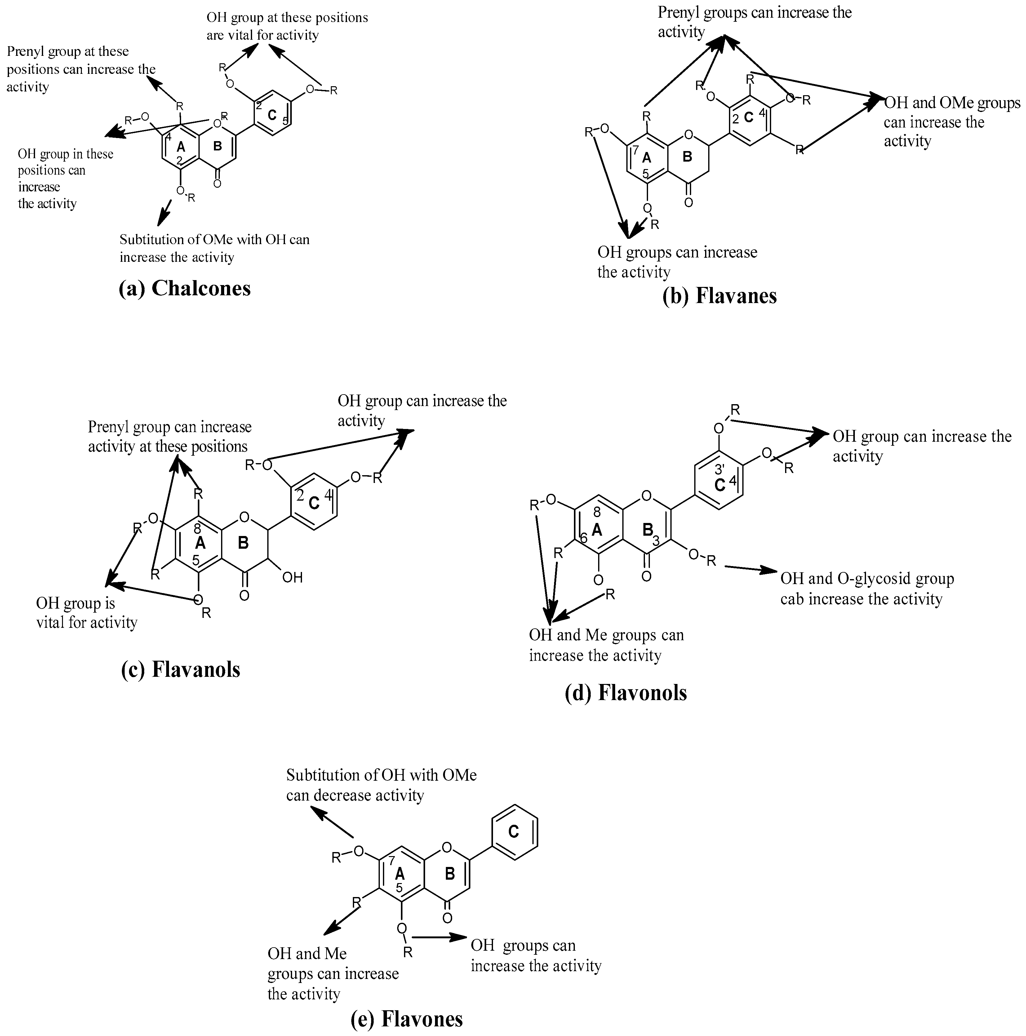
3.1. Position of Functional Group

The structural antimicrobial activity of the major plant polyphenols, i.e., flavonoids, is well documented [46]. The amphipathic features of flavonoids play an essential role as far as antibacterial properties are concerned [47]. The hydrophobic substituents such as prenyl groups, alkylamino chains, alkyl chains, and nitrogen or oxygen-containing heterocyclic moieties usually enhance the antibacterial activity of all flavonoids [48]. Different classes of flavonoids, mainly chalcones, flavanes, and flavan-3-ol exhibits better antimicrobial activity due to variation in the position of the functional group attached to the rings [46].

3.1.1. Chalcones

Several studies have suggested that chalcones with a lipophilic group such as isoprenoid and methoxy groups at positions 3′, 5′, and 2′ of ring A are the most potent inhibitors of MRSA strains [49]. Based on the activity of isobavachalcone (MIC: 30 μg/mL), (Figure 1A), the authors of [50] suggested the that A ring with a prenyl group displays adequate antimicrobial activity, but cyclization or addition of the prenyl group to B ring in addition to the A ring decreases this activity. Likewise, the hydroxy group at 4′, 4, and 6 of A and B rings increase the antimicrobial activity. For example, kuraridin and 7,9,2’,4’-tetrahydroxy-8-isopentenyl-5-methoxychalcone (THIPMC) compounds with the same structure, with only one difference in the OH of the B ring (2 and 4 instead of 4 and 6), showed high activity against the MRSA strain [51].

3.1.2. Flavanes and Flavanols

Flavanes with a prenyl group at the A ring have been found to be the most potent antibacterial compounds against S. aureus. It is established that the number and position of prenyl groups on this ring increase antimicrobial activity [52]. The presence of the hydroxy groups at different positions on A and B rings has also been reported to improve antibacterial activity (Figure 1B,C). The compound 3′-O-methydiplacol with OH at the 5, 3′, and 4′ positions of the A and B rings, respectively, as well as the geranyl group at C-6 and OMe at C-5′, showed satisfactory (i.e., MIC value of 4 μg/mL) activity against S. aureus [53]. additionally, sophoraflavanone with isogeranyl at C-8 and OH at 3, 2′, and 4′ at A and B rings were active (i.e., MIC value of 7.3 μg/mL) against S. aureus [51]. The position of prenyl, hydroxyl, and especially methoxy groups at positions 5 and 7 of the A ring, increased the antibacterial effect of flavanes and flavones [54]. Furthermore, different substitutions on position 3 of the C ring with a hydroxyl or an O-glycoside group could enhance the antimicrobial activity of certain flavones [46]. One of the earlier findings also suggested that the tetraflavonoids without OH on the C ring showed moderate activity against E. coli [55].

3.1.3. Flavonols

In the A ring, many studies have confirmed that hydroxylation at positions 5 and 7 together are critical for the antibacterial activity of flavonols against S. aureus strains [56]. In addition, hydroxylation on the B and C rings also increased the antimicrobial activity of these compounds. A comparison of compounds with the same structure showed that kaempferol with a hydroxy group at C-4′ had less activity than galangin (without OH at C-4′) against S. aureus (Figure 1D) [47]. The number of glycosyl groups instead of the hydroxy group at position 3 has been found to have a significant effect on antibacterial activity. For example, among the compounds extracted from Maytenus buchananii, quercetin-3-O-[α-L-rhamnopyranosyl-(1 → 6)-β-D glucopyranoside] with a disaccharide group at the same position was the better inhibitor of S. aureus growth than amentoflavone-7″,4‴-dimethyl-ether with monosaccharide group (quercetin-3-O-β-D-glucopyranoside) [57]. Substitution of the methoxy group at position 3 decreased the antimicrobial activity. For example, piliostigmol (with OMe and Me groups at positions 6 and 7 of the A ring and OH at position 3) was more active against S. aureus than 6-C-methylquercetin-3,3′,7-trimethyl ether (with OMe at the C-3 position) [58].

3.1.4. Flavones

Studies conducted on the antibacterial activity of flavones [59] suggested that at least one hydroxy group in the A ring (especially at C-7) is vital for antibacterial activity. Hydroxyl groups in other positions such as C-5 and C-6 can also increase the antibacterial action [60]. However, the substitution of OH with OMe at C-7 was seen to reduce the antibacterial activity. For instance, the compound 5,7-dihydroxy-flavone with two OH at positions 5 and 7 has been found to be more potent against Ralstonia solanacearum (i.e., MIC: 25 and 300 μg/mL) compared to 5-hydroxy-7-methoxy-flavone with OMe at position 7 and OH at position 5 (Figure 1E) [61]. The importance of the –OH group at position 5 of flavones for their antimicrobial activity against MRSA strains has also been reported [62]. One of the investigations on plant isoflavonones suggested that the hydroxyl group’s C-5, 6 and 7 position is crucial for antimicrobial action [63]. The presence of the prenyl (C5) group at position 6 without cyclization of this substituent with the A ring has also been reported to improve antibacterial activity [52].

3.2. Number of Functional Groups Attached

The number of functional groups attached has been found to have a significant influence on antimicrobial activity. The 2′, 4′- or 2′, 6′-dihydroxylation of the B ring and 5, 7-dihydroxylation of the A ring in the flavanone structure are essential for anti-MRSA activity [64]. Moreover, 5-hydroxyflavanones and 5-hydroxyisoflavanones with one, two or three additional hydroxyl groups at the 7′, 2′, and 4′ positions inhibited the growth of Streptococcus mutans and S. sobrinus [65]. Caffeic acid had higher antimicrobial activity than p-coumaric acid due to the additional –OH group on the phenolic ring of the former compound [66].

3.3. Presence of C2=C3 Double Bond

It has been observed that flavanones with C2=C3 are more active than the corresponding flavones. For example, naringenin showed antibacterial effects on all the tested bacteria, whereas apigenin showed almost no effect [67]. In addition, the C2=C3 double bond was found to be responsible for the antifungal activity of 5,7-dihydroxyflavonoids, while hydrogenation of the C2=C3 bond reduced the antifungal effect [68]. The flavonoids apigenin luteolin, dinatin, and daidzein, C2=C3 had better anti-influenza virus activities, compared with catechin and epicatechin belonging to the flavanols class of compounds that lack the C2=C3 bond [69].

3.4. Type of Functional Group

The hydrophobic substituents such as prenyl groups, alkylamino chains, alkyl chains, and nitrogen- or oxygen-containing heterocyclic moieties have been reported to enhance the activity of all the flavonoids [48]. Variation in the antimicrobial activity of polyphenols also depends on variation in the functional group they have [70]. The substitution of the phenyl moiety by a propyl or a methyl group has been found to be deleterious for the antibacterial effect against S. aureus and B. subtilis [71]. This negative antimicrobial effect was also observed against L. monocytogenes, when substituting the phenyl with the propyl moiety [71].
The naphthoquinone 5,8-dihydroxy-1,4-naphthoquinone without chlorine was very active against three Gram-positive (S. aureus, B. subtilis, and L. monocytogenes) and three Gram-negative (E. coli, P. aeruginosa, and S. Enteritidis) strains but in a lower extent against P. aeruginosa. The compound 2,3-dichloro-5,8-dihydroxy-1,4-naphthoquinone with chlorine was significantly less active against E. coli and P. aeruginosa [70]. The presence of gallic or galloyl moieties was found to promote the antibacterial activity of epigallocatechin gallate by inducing damage to the bacterial membrane [72]. The antibacterial action of caffeic acid and their alkyl esters against specific strains of S. aureus and E. coli showed that longer alkyl side chains were more effective against the Gram-positive bacterium, while caffeic acid esters with medium length alkyl side chain were more effective against the Gram-negative bacterium which was also far less susceptible to caffeic acid and its esters [73].

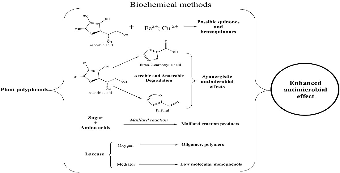
4. Enhancement of Antimicrobial Activity of Plant Derived Polyphenols by Biochemical Methods

Higher potency of the antimicrobial or their use in low concentration is preferred in food application to avoid any changes in the organoleptic properties and minimize interaction with the complex food matrices [14,74]. The following biochemical means may be used to obtain high potent antimicrobials.

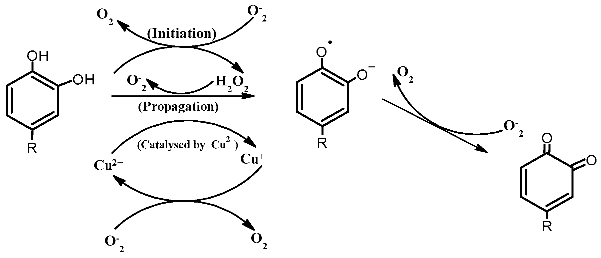
4.1. Enhancement Using Ascorbic Acid and Transition Metals

One of the promising approaches to enhance antimicrobial activity by non-enzymatic means can be mild oxidation of plant phytochemicals, particularly polyphenols by reactive oxygen species (ROS), using ascorbic acid (AA) and transition metals (Cu (II), Fe (II), Fe (III) systems. However, the reaction mechanism of AA oxidation in the presence of transition metals is still unclear, and different mechanisms have been proposed.
In one study, the AA and Cu2+ reaction were projected to yield one mole of hydrogen peroxide (H2O2) and one mole of dehydro-ascorbic acid in the pH range of 2.6–9.3 [75]. Additionally, it has been assumed that H2O2 is formed when AA reacts with Cu(II) in the presence of O2 within a pH range of 2.5–4.0 [76]. At physiological pH (7.0), H2O2 production increased when the copper (Cu2+) concentration was deficient, compared with AA [77]. It was further postulated that the ascorbate mono-anion would dominate when the pH is over 4.25. This form can further be oxidized with the concomitant reduction of copper II to copper I [78]. At pH higher than 4.25, rapid redox cycling of copper generates superoxide, peroxide, and hydroxyl radicals via a copper assisted Fenton reaction, and at a pH lower than 4.25, the level of superoxide in the solution decreases as superoxide anion reacts with hydrogen to form the hydroperoxyl radical (HO2) [78].
The reaction product of AA and Cu2+ leads to cleavage of viral and plasmid DNA, which could be withdrawn in the presence of metal chelators such as EDTA, stating that copper plays an essential role in the oxidation of AA [79]. The reaction was also withdrawn in the presence of the catalase enzyme, confirming the fact that H2O2 is mainly produced in the AA/Cu2+ reaction [79]. The hydroxyl radical generated by the AA/Cu2+ system is lesser than that generated by the ascorbate/H2O2 system [80]. It has also been reported that transition metal ions, such as Fe (III) and Cu (II), are reduced by ascorbate. Their lower oxidation states (e.g., Fe (II) and Cu (I), respectively) (Equations (1) and (2) below) may further give rise to Fenton reactions with H2O2, producing hydroxyl radicals [81].
  Cu + + H 2 O 2 Cu 2 + + OH + OH
Fe 2 + + H 2 O 2   Fe 3 + + OH + OH
In the presence of limited oxygen, polyphenols can be oxidized non-enzymatically [82]. The polyphenols containing a catechol ring are oxidized to semiquinone and benzoquinone radicals (Figure 2). At the same time, the oxygen is reduced to H2O2, and the whole process is mediated by the redox cycle of Fe3+/Fe2+ and Cu2+/Cu+ [83]. Recent reviews have illustrated the antimicrobial action of quinone and its derivatives [84,85,86]. It may be suggested that, in presence of ascorbic acid and transition metals, redox oxygen species may be generated, which may oxidize the polyphenols to corresponding quinones or benzoquinones, enhancing antimicrobial activity [87,88].
Phenolics from plants have been combined with other substances, sometimes referred to as adjuncts, such as transition metal ions or vitamin C to enhance phenolic efficacy [90,91]. Enhancement of antimicrobial activity was observed in the case of pomegranate rind Ext by Cu (II) alone or with both Cu (II) and AA combinations against many bacterial strains [92,93]. The addition of copper (II) sulfate and AA ascertained the enhancement of antimicrobial activities of whole and sub-fractionated white tea against S. aureus [94]. Enhanced antimicrobial activity against S. aureus and E. coli was also observed by the addition of the AA to the (+)-catechin–copper (II) mixture [95]. A recent study revealed the antibacterial effect of AA against S. enterica subsp. Enterica serovar Typhi and Vibrio fluvialis could be enhanced when applied in a combination with linalool and copper [96]. However, the mechanism behind the enhancement in antimicrobial activity due to the addition of transition metals and AA to the plant polyphenols has not been appropriately elucidated yet.

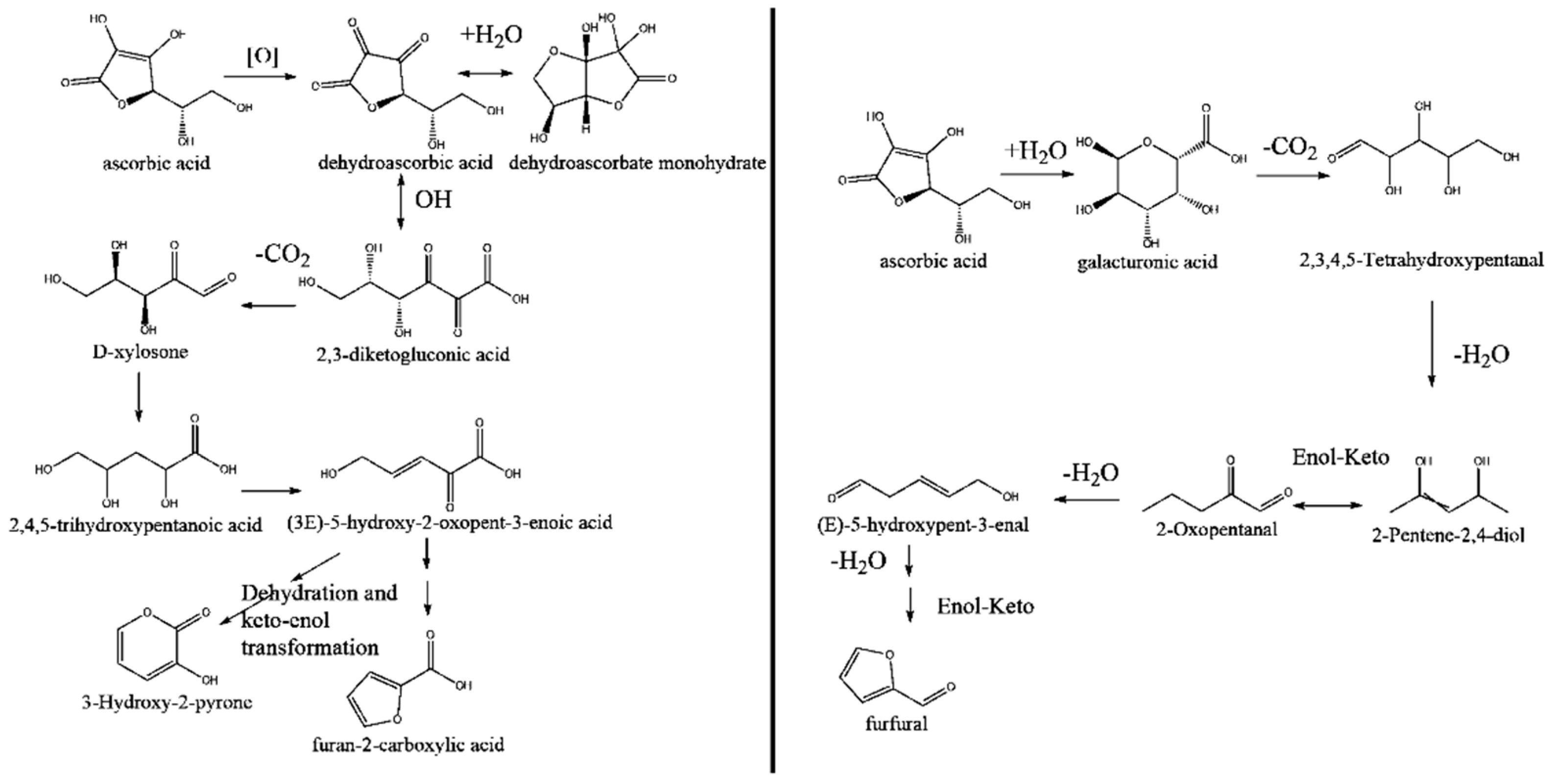
4.2. Enhancement Using Degradation Products of Ascorbic Acid in an Ethanolic Solution

In aqueous (AQ) systems, AA is very unstable and efficiently degraded both aerobic (AB) and anaerobically. The degradation process of AA is complex and involves many oxidation–reductions and intermolecular rearrangement reactions. The degradation of AA via AB and anaerobic (AAB) pathways depends upon oxygen, heat, light, storage temperature, and time [97,98]. However, degradation of AA mainly results in the formation of volatile and brown products via self-degradation and non-enzymatic browning. The most commonly reported terminal products resulting from the AB degradation of AA and dehydroascorbic acid in acidic AQ conditions (pH 1–3), were found to be 3-hydroxy-2-pyrone and 2-furoic acid [99]. These are also amongst the highest yielding products depending on the conditions utilized. Heat-induced (60–100 °C) AB degradation of different solutions of AA and dehydroascorbic acid demonstrated that both 3-hydroxy-2-pyrone and 2-furoic acid were the main degradation products of AA (Figure 3A). This included decarboxylation of 2,3-diketogulonic acid with the formation of xylosone, a mechanism already reported by the authors of [100], followed by a multi-step conversion of xylosone to the terminal products via oxidation, dehydration, and/or ketoenol tautomerism [100]. Xylosone is a gateway to numerous degradation products, and its presence as an AA or dehydroascorbic acid degradation product has been confirmed by several studies [101,102].
The term “AAB degradation” of AA refers to ascorbic acid degradation to some terminal product via a mechanistic pathway that does not require an oxidation step. The AAB degradation of AA does not generally require the removal of O2 from a reaction system; however, the lowering of oxygen concentration has the advantage of limiting the competing AB reactions [103]. The degradation of AA by reaction with oxygen occurs faster than its AAB degradation [104]. However, the AAB degradation rate can increase considerably with higher temperatures [104]. The pH level is also known to influence the rate of AAB degradation of AA, which increases as the pH is raised from 2.3 to 4.0 [105].
Regardless, the most commonly reported terminal product for the AAB degradation of AA is furfural [99,106]. The general mechanism to describe the formation of furfural is shown in (Figure 3B) and involves initial ring cleavage and hydration of AA rather than oxidation [107]. The subsequent steps require decarboxylation, acid-catalyzed dehydration, and cyclization. Several studies have shown that the formation of furfural is favored at lower pH values [108,109].
Another mechanism has been reported for the acid-catalyzed AAB degradation of AA in methanol, forming a bicyclic structure similar to dehydroascorbic acid. It then undergoes dehydration and decarboxylation via dihydrofuran-type intermediates to afford furfural [110]. The authors proposed that the mechanism would be equally valid in AQ systems but did not provide evidence.
Not all scholars agree that furfural is exclusive to the AAB degradation of AA. Instead, it has been suggested that it can be formed via an oxidatively generated dehydroascorbic acid degradation pathway [111,112]. Another product unique to the AAB pathway is, 4,5-dihydroxy-2-ketopentanal (otherwise known as 3-deoxypentosulose) generated upon storage of AA at pH 3.5, although the storage temperature was not so relevant to wine conditions (120 °C for 2 h) [113]. This compound has been proposed as an intermediate in the formation of furfural during the AAB degradation of AA. The intermediate furan compounds which were generated during AA degradation have been found to inhibit the proliferation of S. typhi and B. subtilis to different extents [114]. The MIC values of furfural and furoic acid (terminal products of AA degradation) against B. subtilis and S. typhi were 0.027, 0.015, and 0.029, 0.009 μM, respectively [114]. Recent studies have reported antimicrobial action of furoic acid and furan compounds [115,116] The reported end product of AB and AAB degradation might have potency in enhancing the antimicrobial activity of plant Ext. However, to date, there is no evidence of the enhancement of the antimicrobial activity of plant Ext using the degradation product of AA.

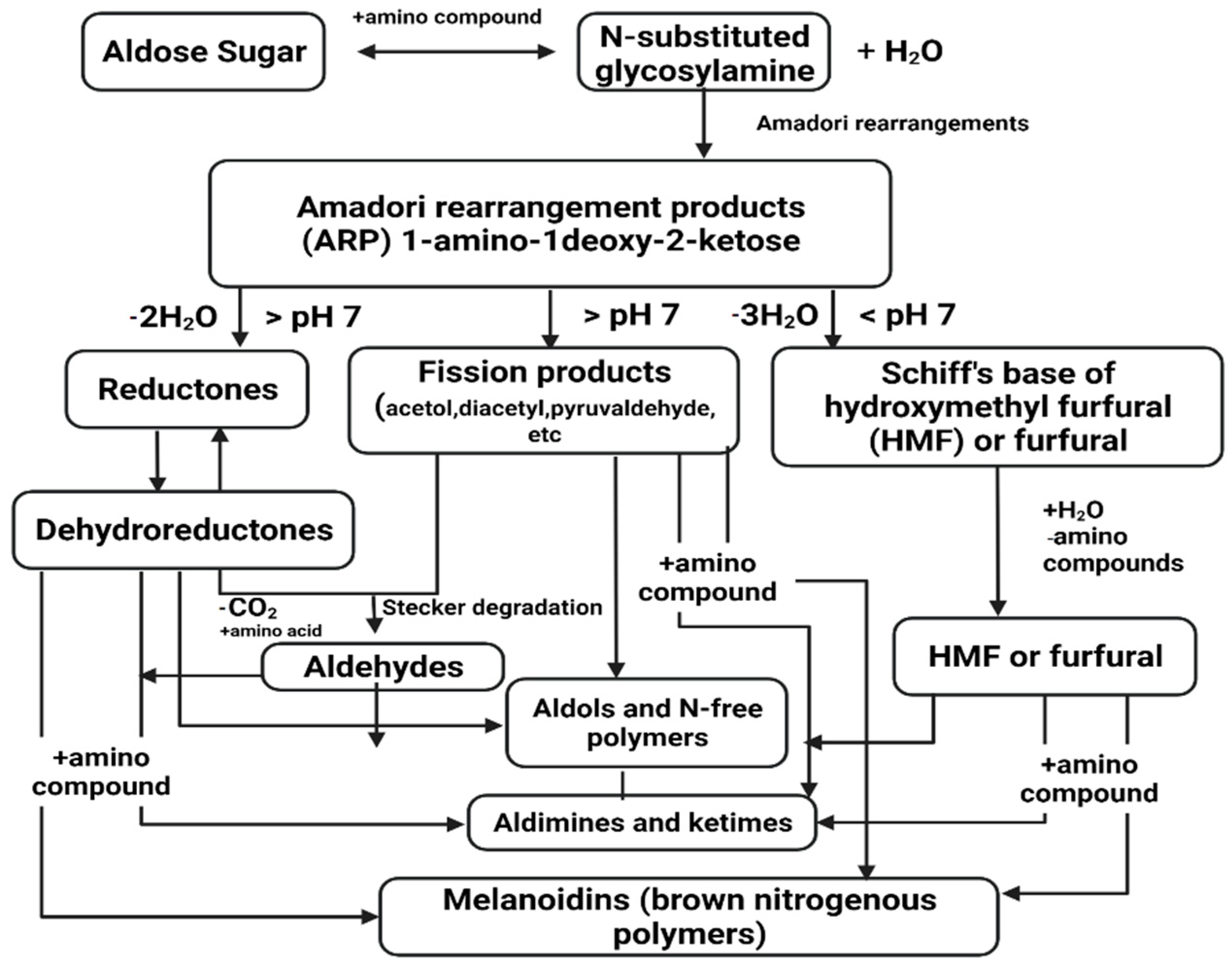
4.3. Enhancement Using Maillard Reaction Products

The Maillard reaction (MR) is a heat-induced browning reaction widely employed in various fields in the food industry that has been used as an effective method for protein modification and the production of remarkable changes in the structure and bioactivity of proteins [117,118]. In particular, MR products (MRPs) have been shown to have significant antibacterial activities against a wide range of bacteria, with lower toxicity than antibiotics [119,120]. The MRPs possess many intermediate products (Figure 4), such as aldehydes, ketones, and heterocyclic compounds, which can effectively inhibit the growth of some Gram-positive and Gram-negative bacteria [119].
MR products may have the potential to exhibit a synergistic antimicrobial effect in conjunction with phytochemicals from plant compounds, thus enhancing their potency ( i.e., lowering the MIC value of the plant-derived compounds) [121]. The plant-derived polyphenols have been found to have additive, synergistic antimicrobial effects with the intermediate products of the MR such as diacetyls, carbonyls, and furfural [122,123]. Methylglyoxal (one of the intermediate products of MR) and catechin have been reported to positively affect antibacterial activity [124]. However, the antimicrobial activity of plant Ext or plant phytochemicals using MRPs is not yet reported, and further research is needed in this area.

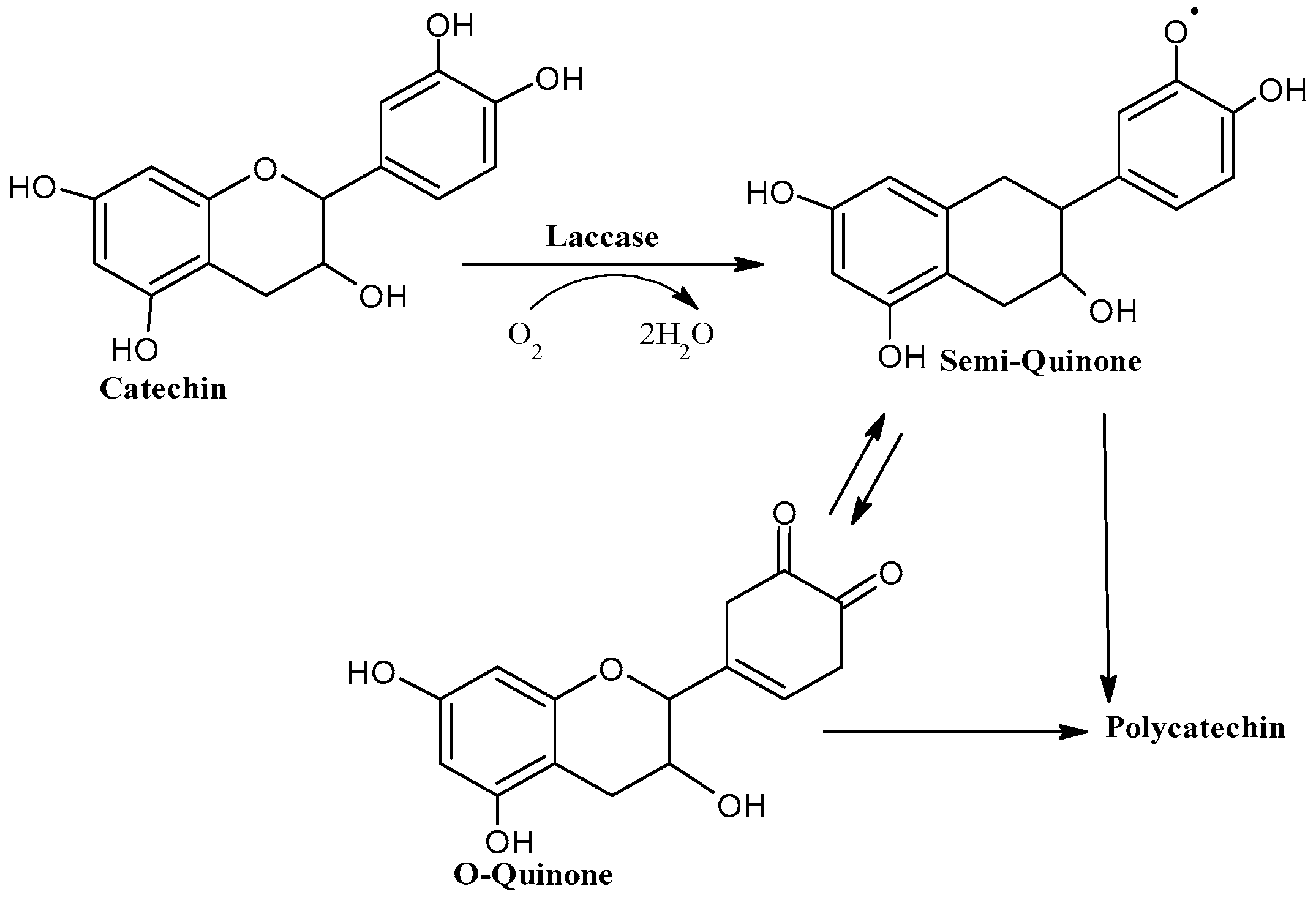
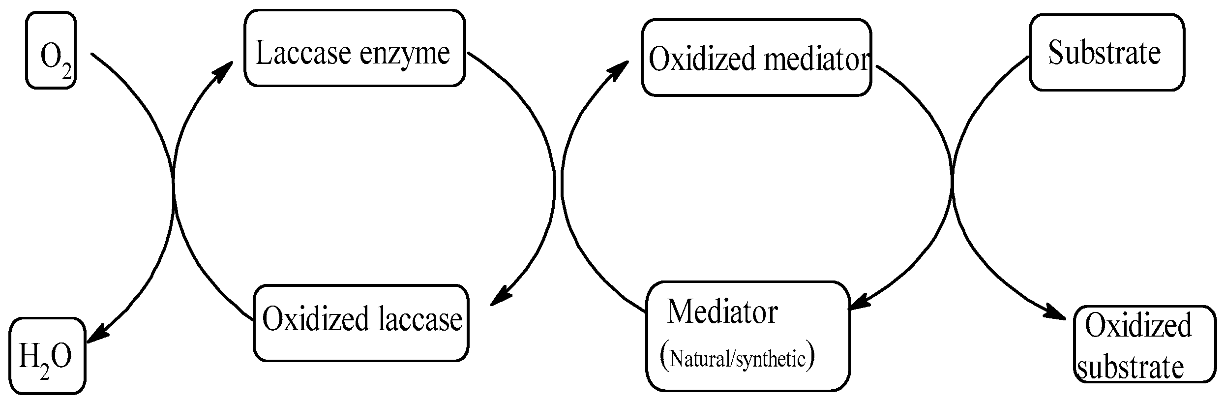
4.4. Enhancement Using Laccase–Mediator System

The oxidation of organic compounds to produce functionalized molecules is essential in organic synthesis [125]. Controlled enzymatic oxidation or hypo-oxidation can yet be another approach toward enhancing the antimicrobial activities of phytocompounds. Increased bioactivities have been observed by the biochemical transformation of triterpenes using oxidative enzymes [126]. Oxidoreductive enzymes such as laccase and peroxidise can transform phenols through oxidative coupling reactions with the production of polymeric products by self-coupling or cross-coupling with other molecules.
Laccase (EC 1.10.3.2), is a multi-copper oxidase that couples the four-electron reduction of oxygen with the oxidation of a broad range of organic substrates, including phenols, methoxy-substituted phenols, anilines, aryl diamines, hydroxyindoles, benzenethiols and inorganic/organic metal compounds by a one-electron transfer mechanism, making this green enzyme useful for carrying out several types of oxidative reactions [127,128,129,130,131,132]. Laccases use O2 as the electron acceptor to remove protons from the phenolic hydroxyl groups. This reaction gives rise to radicals that can spontaneously rearrange, which can either lead to the fission of C–C or C–O bonds of the alkyl side chains or the cleavage of aromatic rings [128]. The oxidation of a reducing substrate by laccase involves losing an electron and forming a free radical [129]. This radical is, in general, unstable and may undergo further laccase-catalyzed oxidation (e.g., quinone from phenol) or non-enzymatic reactions (e.g., hydration, disproportion, or polymerization) [132]. The electron transfer from the substrate to copper is controlled by the redox potential difference. The rate of substrate oxidation by laccase, which has high redox potential, is higher if it has a lower redox potential.
Enzymatic polymerization of phenolic compounds (catechol, resorcinol, and hydroquinone) has been carried out using laccase [133,134,135]. Intermediates (quinones) formation in the first stage of oxidation with further oxidation reaction, forming colored polymers, was observed while evaluating the polymerization and the structures of the polymers by UV–Vis and Fourier transform infrared spectroscopy [134]. Changes in the color of flavonoids due to oxidation by the laccase enzyme were due to the polymerization and linkage of the quinones (Figure 5) formed as an intermediate [133]. Laccase oxidation of caffeic acid and isoeugenol was shown to enhance their antimicrobial activity against S. aureus and E. coli in liquid media [135]. Some low molecular weight phenolic compounds are usually produced as a result of oxidative metabolism by C ring cleavage of catechin and epicatechins [136]. The antimicrobial properties of one of these low molecular weight polyphenols, 3,4 dihydroxy benzoic acid have been well established [137,138]. Moreover, dimers and polymers of flavonoids have also been found to have superior antimicrobial effects in comparison to the parent monomer [139,140,141]. It has been suggested that the toxicity of the laccase-treated olive Ext can be due to the presence of phenolic compounds such as ortho-benzoquinones, quinonoid, or oxidative coupling polymers, which results because of Lac treatment is more toxic than the parent compounds [142]. Many studies have suggested polymerization of the phenolics using the Lac enzyme [143,144]. Enhanced antimicrobial activity of the resulting oligomers and polymers has also been reported [145,146].
Although laccase can oxidize a wide range of substrates, some substrates of interest cannot be oxidized directly by laccases, either because they are too large to fit into the enzyme active site or because they have an exceptionally high redox potential. By mimicking nature, it is possible to overcome this limitation with the addition of so-called “chemical mediators”, which are suitable compounds that act as intermediate substrates for the laccase, whose oxidized radical forms can interact with the bulky or high redox-potential substrate targets [131]. Laccase–mediator system (Figure 6) has found its immense application in the degradation of lignin. Small redox molecules such as 3-hydroxyanthranilic acid (HAA) might act as “electron shuttles” between the enzyme and lignin and cause polymer de-branching and degradation [131]. Some examples of laccase mediators extensively used are 2,2′-azino-bis-(3-ethylbenzothiazoline-6-sulphonic acid) (ABTS), N-hydroxybenzo-triazole (HBT), N-hydroxyphtalimide (HPI), violuric acid (VLA), N-hydroxyacetanilide (NHA), methyl ester of 4-hydroxy-3,5-dimethoxy-benzoic acid (syringic acid), and 2,2,6,6-tetramethyl piperidine-1-yloxy (TEMPO) [131].
Laccase is capable of oxidizing unreactive iodide to reactive iodine [147]. Phenolic compounds such as vanillin, which resembles substructures of softwood lignin, can be directly iodinated by reacting with laccase and iodide, resulting in compounds with antifungal activity [147]. The addition of redox mediators “acetosyringone” in catalytic concentrations increased the rate of iodide oxidation by ten-fold and the yield of iodo-vanillin by 50% [147].
Functionalization is the process of adding new functions, features, capabilities, or properties to material by changing the surface chemistry of the material [148]. In one study, functionalization of chitosan with phenolic acids such as caffeic acid or gallic acid using laccase from Trametes versicolor formed a product with enhanced antimicrobial activity against E. coli and L. monocytogenes [149]. The authors proposed the functionalization of chitosan with the phenolic acids by laccase catalyzed oxidation of phenolic acids to electrophilic o-quinones, which undergo a new oligomer/polymer-forming structure originated by C–C coupling between the benzene rings and C–O–C coupling involved with the phenolic side chains.

4.5. Enhancement Using Peroxidase Enzyme

The peroxidases (EC 1.11.1.7) are heme proteins and contain iron (III) protoporphyrin IX (ferriprotoporphyrin IX) as the prosthetic group. They come under the class of oxidoreductases that catalyze the oxidation of a wide range of molecules, using peroxide as an electron acceptor [150]. The reduction of peroxides at the expense of electron-donating substrates makes peroxidases useful in several industrial and analytical applications. The common overall reaction of the peroxidases can be written as in the following Equation (3), where RH is a suitable peroxidase substrate and R is a free-radical product derived from it as follows:
2 RH + H 2 O 2 2 R + 2 H 2 O
Oxidation reactions carried out by peroxidase may be one-electron or two-electron oxidation. A classic example of one-electron oxidation is guaiacol assay through which guaiacol is oxidized to a free radical that undergoes a subsequent radical–radical combination to give a colored dimeric product. The dimerization can occur between two ring carbon atoms or by adding the oxygen of one phenoxy radical to the ring carbon of the other [151]. Two-electron oxidation is rare for most peroxidase enzymes. The example under this type is the oxidation of halide and pseudo-halide ions, specifically I, Br, Cl, and NCS. The oxidation of I and NCS is common for the peroxidases [152]. Many reactions are known in which oxidation of a substrate by peroxidases produces a two-electron oxidized product. These reactions can be rationalized by forming a free radical metabolite, followed by the second oxidation of the free radical to the final observed product.
Lactoperoxidase, together with thiocyanate ions and hydrogen peroxide generates, hypothiocyanite ions and the oxidized product which is known as the lactoperoxidase system. The oxidized effect possesses a broad spectrum of antimicrobial activity. Hence, much attention has been paid to the lactoperoxidase system [153], e.g., lactoperoxidase systems mediated by oxidized β-carotene/SCN cycling lead to enhanced antimicrobial effects [154].

4.6. Future Perspective

Structural changes in polyphenols by biochemical modifications can be elucidated through elemental analysis and spectral data (IR, 1H NMR). Furthermore, crystal, and molecular structures of the potential metal flavonoid or phenolic acid complexes can be identified by using single-crystal X-ray diffraction data. By electron paramagnetic resonance spectroscopy, transient oxidation species could also be identified. Morphological study of the bacterial and fungal cells triggered by biochemically modified polyphenols can reveal the possible antimicrobial effect on the cells. Membrane potential study may indicate the mechanism by which the antimicrobials could have affected the membrane permeability. Ultimately, antibacterial effects against food pathogens can be carried out to understand the scope of biochemically formulated antimicrobials in food applications.

5. Conclusions

Polyphenols are widely and easily available bioactive compounds that have health benefits. Despite their benefits, their use in food applications is limited, due to their low potency and high concentration needs. This review showed different biochemical means of improving the antimicrobial property of plant-based polyphenols. Considering the structural importance of the antimicrobial properties of polyphenols, this report also showed the significance of different classes of polyphenols as active antimicrobial agents. Although the mechanisms of the biochemical methods involved in enhancing the antimicrobial activity of plant polyphenols have been explained in this review, the possible structural and functional modifications that these biochemical methods may bring about in the modified polyphenols have not yet been established completely. Therefore, further validation of these methods through high-throughput techniques is crucial.

Author Contributions

Conceptualization: L.P., A.D.-S.; writing—original draft preparation: L.P., A.D.-S.; writing—review and editing: L.P., A.D.-S.; visualization: A.D.-S.; supervision: A.D.-S.; All authors have read and agreed to the published version of the manuscript.

Funding

This research received no external funding.

Institutional Review Board Statement

Not applicable.

Informed Consent Statement

Not applicable.

Data Availability Statement

Not applicable.

Conflicts of Interest

The authors declare no conflict of interest.

References

  1. Barbosa-Pereira, L.; Bilbao, A.; Vilches, P.; Angulo, I.; LLuis, J.; Fite, B.; Paseiro-Losada, P.; Cruz, J.M. Brewery waste as a potential source of phenolic compounds: Optimisation of the extraction process and evaluation of antioxidant and antimicrobial activities. Food Chem. 2014, 145, 191–197. [Google Scholar] [CrossRef] [PubMed]
  2. Schuenzel, K.M.; Harrison, M.A. Microbial antagonists of foodborne pathogens on fresh, minimally processed vegetables. J. Food Prot. 2002, 65, 1909–1915. [Google Scholar] [CrossRef] [PubMed]
  3. Del Nobile, M.A.; Lucera, A.; Costa, C.; Conte, A. Food applications of natural antimicrobial compounds. Front. Microbiol. 2012, 3, 103–115. [Google Scholar]
  4. Dixon, R.A. Natural products and plant disease resistance. Nature 2001, 411, 843–847. [Google Scholar] [CrossRef] [PubMed]
  5. Johnson, I.T. Phytochemicals and cancer. Proc. Nutr. Soc. 2007, 66, 207–215. [Google Scholar] [CrossRef] [PubMed]
  6. Bor, T.; Aljaloud, S.O.; Gyawali, R.; Ibrahim, S.A. Chapter 26—Antimicrobials from herbs, spices, and plants. In Fruits, Vegetables, and Herbs; Watson, R.R., Preedy, V.R., Eds.; CRC Press: Boca Raton, FL, USA, 2016. [Google Scholar]
  7. Bouarab Chibane, L.; Degraeve, P.; Ferhout, H.; Bouajila, J.; Oulahal, N. Plant antimicrobial polyphenols as potential natural food preservatives. J. Sci. Food Agric. 2019, 99, 1457–1474. [Google Scholar] [CrossRef] [Green Version]
  8. Mickymaray, S. Efficacy and Mechanism of Traditional Medicinal Plants and Bioactive Compounds against Clinically Important Pathogens. Antibiotics 2019, 8, 257. [Google Scholar] [CrossRef] [Green Version]
  9. Singh, P.A.; Desai, S.D.; Singh, J. A Review on Plant Antimicrobials of Past Decade. Curr. Top. Med. Chem. 2018, 18, 812–833. [Google Scholar] [CrossRef]
  10. Khaneghah, A.M.; Hashemi, S.M.B.; Es, I.; Fracassetti, D.; Limbo, S. Efficacy of Antimicrobial Agents for Food Contact Applications: Biological Activity, Incorporation into Packaging, and Assessment Methods: A Review. J. Food Prot. 2018, 81, 1142–1156. [Google Scholar] [CrossRef]
  11. Stojković, D.; Petrović, J.; Soković, M.; Glamočlija, J.; Kukić-Marković, J.; Petrović, S. In situ antioxidant and antimicrobial activities of naturally occurring caffeic acid, p-coumaric acid and rutin, using food systems. J. Sci. Food Agric. 2013, 93, 3205–3208. [Google Scholar] [CrossRef]
  12. Bandyopadhyay, P.; Ghosh, A.K.; Ghosh, C. Recent developments on polyphenol-protein interactions: Effects on tea and coffee taste, antioxidant properties and the digestive system. Food Funct. 2012, 3, 592–605. [Google Scholar] [CrossRef] [PubMed]
  13. Shao, S.; Zhou, T.; Tsao, R. Antimicrobials from Plants—Food Preservation and Shelf-Life Extension; Elsevier: Amsterdam, The Netherlands, 2011; Volume 4, pp. 645–658. [Google Scholar]
  14. Gutiérrez-del-Río, I.; Fernández, J.; Lombó, F. Plant nutraceuticals as antimicrobial agents in food preservation: Terpenoids, polyphenols and thiols. Int. J. Antimicrob. Agents 2018, 52, 309–315. [Google Scholar] [CrossRef] [PubMed]
  15. Aguilar-Veloz, L.M.; Calderón-Santoyo, M.; Vazquez Gonzalez, Y.; Ragazzo-Sánchez, J.A. Application of essential oils and polyphenols as natural antimicrobial agents in postharvest treatments: Advances and challenges. Food Sci. Nutr. 2020, 8, 2555–2568. [Google Scholar] [CrossRef] [PubMed]
  16. Hygreeva, D.; Pandey, M.C.; Radhakrishna, K. Potential applications of plant based derivatives as fat replacers, antioxidants and antimicrobials in fresh and processed meat products. Meat Sci. 2014, 98, 47–57. [Google Scholar] [CrossRef] [PubMed]
  17. Quinto, E.J.; Caro, I.; Villalobos-Delgado, L.H.; Mateo, J.; De-Mateo-Silleras, B.; Redondo-Del-Rio, M.P. Food Safety through Natural Antimicrobials. Antibiotics 2019, 8, 208. [Google Scholar] [CrossRef] [Green Version]
  18. Gonelimali, F.D.; Lin, J.; Miao, W.; Xuan, J.; Charles, F.; Chen, M.; Hatab, S.R. Antimicrobial Properties and Mechanism of Action of Some Plant Extracts Against Food Pathogens and Spoilage Microorganisms. Front. Microbiol. 2018, 9, 1639–1648. [Google Scholar] [CrossRef]
  19. Quideau, S.; Deffieux, D.; Douat-Casassus, C.; Pouységu, L. Plant polyphenols: Chemical properties, biological activities, and synthesis. Angew. Chem. Int. Ed. 2011, 50, 586–621. [Google Scholar] [CrossRef]
  20. Daglia, M. Polyphenols as antimicrobial agents. Curr. Opin. Biotechnol. 2012, 23, 174–181. [Google Scholar] [CrossRef]
  21. Cushnie, T.P.; Lamb, A.J. Antimicrobial activity of flavonoids. Int. J. Antimicrob. Agents 2005, 26, 343–356. [Google Scholar] [CrossRef]
  22. Ahmed, S.I.; Hayat, M.Q.; Tahir, M.; Mansoor, Q.; Ismail, M.; Keck, K.; Bates, R.B. Pharmacologically active flavonoids from the anticancer, antioxidant and antimicrobial extracts of Cassia angustifolia Vahl. BMC Complement. Altern. Med. 2016, 16, 1–9. [Google Scholar] [CrossRef] [Green Version]
  23. Bouyahya, A.; Chamkhi, I.; Balahbib, A.; Rebezov, M.; Shariati, M.A.; Wilairatana, P.; Mubarak, M.S.; Benali, T.; El Omari, N. Mechanisms, Anti-Quorum-Sensing Actions, and Clinical Trials of Medicinal Plant Bioactive Compounds against Bacteria: A Comprehensive Review. Molecules 2022, 27, 1484. [Google Scholar] [CrossRef] [PubMed]
  24. Górniak, I.; Bartoszewski, R.; Króliczewski, J. Comprehensive review of antimicrobial activities of plant flavonoids. Phytochem. Rev. 2019, 18, 241–272. [Google Scholar] [CrossRef] [Green Version]
  25. Biharee, A.; Sharma, A.; Kumar, A.; Jaitak, V. Antimicrobial flavonoids as a potential substitute for overcoming antimicrobial resistance. Fitoterapia 2020, 146, 104720–110442. [Google Scholar] [CrossRef] [PubMed]
  26. Cueva, C.; Moreno-Arribas, M.V.; Martín-Alvarez, P.J.; Bills, G.; Vicente, M.F.; Basilio, A.; Rivas, C.L.; Requena, T.; Rodríguez, J.M.; Bartolomé, B. Antimicrobial activity of phenolic acids against commensal, probiotic and pathogenic bacteria. Res. Microbiol. 2010, 161, 372–382. [Google Scholar] [CrossRef]
  27. Herald, P.J.; Davidson, P.M. Antibacterial Activity of Selected Hydroxycinnamic Acids. J. Food Sci. 1983, 48, 1378–1379. [Google Scholar] [CrossRef]
  28. Chipley, J.R.; Uraih, N. Inhibition of Aspergillus growth and aflatoxin release by derivatives of benzoic acid. Appl. Environ. Microbiol. 1980, 40, 352–357. [Google Scholar] [CrossRef] [Green Version]
  29. Ulate-Rodriguez, J.; Schafer, H.W.; Zottola, E.A.; Davidson, P.M. Inhibition of Listeria monocytogenes, Escherichia coli O157:H7, and Micrococcus luteus by Linear Furanocoumarins in Culture Media. J. Food Prot. 1997, 60, 1046–1049. [Google Scholar] [CrossRef]
  30. Dogasaki, C.; Shindo, T.; Furuhata, K.; Fukuyama, M. Identification of chemical structure of antibacterial components against Legionella pneumophila in a coffee beverage. Yakugaku Zasshi 2002, 122, 487–494. [Google Scholar] [CrossRef] [Green Version]
  31. Ibrahim, S.A.; Salameh, M.M.; Phetsomphou, S.; Yang, H.; Seo, C.W. Application of caffeine, 1,3,7-trimethylxanthine, to control Escherichia coli O157:H7. Food Chem. 2006, 99, 645–650. [Google Scholar] [CrossRef]
  32. Kim, S.; Fung, D.Y. Antibacterial effect of crude water-soluble arrowroot (Puerariae radix) tea extracts on food-borne pathogens in liquid medium. Lett. Appl. Microbiol. 2004, 39, 319–325. [Google Scholar] [CrossRef]
  33. Tadakatsu, S.; Wei-Hua, Z.; Zhi-Qing, H. Mechanism of Action and Potential for Use of Tea Catechin as an Antiinfective Agent. Anti-Infect. Agents Med. Chem. 2007, 6, 57–62. [Google Scholar]
  34. Lee, Y.L.; Cesario, T.; Wang, Y.; Shanbrom, E.; Thrupp, L. Antibacterial activity of vegetables and juices. Nutrition 2003, 19, 994–996. [Google Scholar] [CrossRef] [PubMed]
  35. Shan, B.; Cai, Y.-Z.; Brooks, J.D.; Corke, H. Antibacterial Properties and Major Bioactive Components of Cinnamon Stick (Cinnamomum burmannii): Activity against Foodborne Pathogenic Bacteria. J. Agric. Food Chem. 2007, 55, 5484–5490. [Google Scholar] [CrossRef] [PubMed]
  36. Dassoff, E.S.; Li, Y.O. Mechanisms and effects of ultrasound-assisted supercritical CO2 extraction. Trends Food Sci. Technol. 2019, 86, 492–501. [Google Scholar] [CrossRef]
  37. Panja, P. Green extraction methods of food polyphenols from vegetable materials. Curr. Opin. Food Sci. 2018, 23, 173–182. [Google Scholar] [CrossRef]
  38. Wu, N.-N.; Li, H.-H.; Tan, B.; Zhang, M.; Xiao, Z.-G.; Tian, X.-H.; Zhai, X.-T.; Liu, M.; Liu, Y.-X.; Wang, L.-P. Free and bound phenolic profiles of the bran from different rice varieties and their antioxidant activity and inhibitory effects on ɑ-amylose and ɑ-glucosidase. J. Cereal Sci. 2018, 82, 206–212. [Google Scholar] [CrossRef]
  39. Dzah, C.S.; Duan, Y.; Zhang, H.; Serwah Boateng, N.A.; Ma, H. Latest developments in polyphenol recovery and purification from plant by-products: A review. Trends Food Sci. Technol. 2020, 99, 375–388. [Google Scholar] [CrossRef]
  40. Dzah, C.S.; Duan, Y.; Zhang, H.; Wen, C.; Zhang, J.; Chen, G.; Ma, H. The effects of ultrasound assisted extraction on yield, antioxidant, anticancer and antimicrobial activity of polyphenol extracts: A review. Food Biosci. 2020, 35, 100547–100556. [Google Scholar] [CrossRef]
  41. Sangta, J.; Wongkaew, M.; Tangpao, T.; Withee, P.; Haituk, S.; Arjin, C.; Sringarm, K.; Hongsibsong, S.; Sutan, K.; Pusadee, T. Recovery of polyphenolic fraction from arabica coffee pulp and its antifungal applications. Plants 2021, 10, 1422. [Google Scholar] [CrossRef]
  42. Alizadeh, S.R.; Ebrahimzadeh, M.A. O-Glycoside quercetin derivatives: Biological activities, mechanisms of action, and structure–activity relationship for drug design, a review. Phytother. Res. 2022, 36, 778–807. [Google Scholar] [CrossRef] [PubMed]
  43. Renzetti, A.; Betts, J.W.; Fukumoto, K.; Rutherford, R.N. Antibacterial green tea catechins from a molecular perspective: Mechanisms of action and structure–activity relationships. Food Funct. 2020, 11, 9370–9396. [Google Scholar] [CrossRef] [PubMed]
  44. Gyawali, R.; Ibrahim, S.A. Natural products as antimicrobial agents. Food Control 2014, 46, 412–429. [Google Scholar] [CrossRef]
  45. Campos, F.M.; Couto, J.A.; Figueiredo, A.R.; Toth, I.V.; Rangel, A.O.; Hogg, T.A. Cell membrane damage induced by phenolic acids on wine lactic acid bacteria. Int. J. Food Microbiol. 2009, 135, 144–151. [Google Scholar] [CrossRef] [PubMed]
  46. Farhadi, F.; Khameneh, B.; Iranshahi, M.; Iranshahy, M. Antibacterial activity of flavonoids and their structure–activity relationship: An update review. Phytother. Res. 2019, 33, 13–40. [Google Scholar] [CrossRef] [PubMed] [Green Version]
  47. Echeverría, J.; Opazo, J.; Mendoza, L.; Urzúa, A.; Wilkens, M. Structure-Activity and Lipophilicity Relationships of Selected Antibacterial Natural Flavones and Flavanones of Chilean Flora. Molecules 2017, 22, 608. [Google Scholar] [CrossRef]
  48. Xie, Y.; Yang, W.; Tang, F.; Chen, X.; Ren, L. Antibacterial activities of flavonoids: Structure-activity relationship and mechanism. Curr. Med. Chem. 2015, 22, 132–149. [Google Scholar] [CrossRef]
  49. Omosa, L.K.; Midiwo, J.O.; Mbaveng, A.T.; Tankeo, S.B.; Seukep, J.A.; Voukeng, I.K.; Dzotam, J.K.; Isemeki, J.; Derese, S.; Omolle, R.A.; et al. Antibacterial activities and structure-activity relationships of a panel of 48 compounds from Kenyan plants against multidrug resistant phenotypes. Springerplus 2016, 5, 901–916. [Google Scholar] [CrossRef] [Green Version]
  50. Mbaveng, A.T.; Ngameni, B.; Kuete, V.; Simo, I.K.; Ambassa, P.; Roy, R.; Bezabih, M.; Etoa, F.X.; Ngadjui, B.T.; Abegaz, B.M.; et al. Antimicrobial activity of the crude extracts and five flavonoids from the twigs of Dorstenia barteri (Moraceae). J. Ethnopharmacol. 2008, 116, 483–489. [Google Scholar] [CrossRef] [Green Version]
  51. Oh, I.; Yang, W.Y.; Chung, S.C.; Kim, T.Y.; Oh, K.B.; Shin, J. In vitro sortase A inhibitory and antimicrobial activity of flavonoids isolated from the roots of Sophora flavescens. Arch. Pharm. Res. 2011, 34, 217–222. [Google Scholar] [CrossRef]
  52. Osorio, M.; Carvajal, M.; Vergara, A.; Butassi, E.; Zacchino, S.; Mascayano, C.; Montoya, M.; Mejías, S.; Martín, M.C.-S.; Vásquez-Martínez, Y. Prenylated Flavonoids with Potential Antimicrobial Activity: Synthesis, Biological Activity, and In Silico Study. Int. J. Mol. Sci. 2021, 22, 5472. [Google Scholar] [CrossRef]
  53. Smejkal, K.; Chudik, S.; Kloucek, P.; Marek, R.; Cvacka, J.; Urbanova, M.; Julinek, O.; Kokoska, L.; Slapetova, T.; Holubova, P.; et al. Antibacterial C-geranylflavonoids from Paulownia tomentosa Fruits. J. Nat. Prod. 2008, 71, 706–709. [Google Scholar] [CrossRef] [PubMed]
  54. Bitchagno, G.T.; Sama Fonkeng, L.; Kopa, T.K.; Tala, M.F.; Kamdem Wabo, H.; Tume, C.B.; Tane, P.; Kuiate, J.R. Antibacterial activity of ethanolic extract and compounds from fruits of Tectona grandis (Verbenaceae). BMC Complement. Altern. Med. 2015, 15, 265–271. [Google Scholar] [CrossRef] [Green Version]
  55. Bitchagno, G.T.M.; Tankeo, S.B.; Tsopmo, A.; Mpetga, J.D.S.; Tchinda, A.T.; Fobofou, S.A.T.; Wessjohann, L.A.; Kuete, V.; Tane, P. Lemairones A and B: Two new antibacterial tetraflavonoids from the leaves of Zanthoxylum lemairei (Rutaceae). Phytochem. Lett. 2015, 14, 1–7. [Google Scholar] [CrossRef]
  56. Shamsudin, N.F.; Ahmed, Q.U.; Mahmood, S.; Ali Shah, S.A.; Khatib, A.; Mukhtar, S.; Alsharif, M.A.; Parveen, H.; Zakaria, Z.A. Antibacterial Effects of Flavonoids and Their Structure-Activity Relationship Study: A Comparative Interpretation. Molecules 2022, 27, 1149. [Google Scholar] [CrossRef] [PubMed]
  57. Tebou, P.L.F.; Tamokou, J.D.D.; Ngnokam, D.; Voutquenne-Nazabadioko, L.; Kuiate, J.R.; Bag, P.K. Flavonoids from Maytenus buchananii as potential cholera chemotherapeutic agents. S. Afr. J. Bot. 2017, 109, 58–65. [Google Scholar] [CrossRef]
  58. Babajide, O.J.; Babajide, O.O.; Daramola, A.O.; Mabusela, W.T. Flavonols and an oxychromonol from Piliostigma reticulatum. Phytochemistry 2008, 69, 2245–2250. [Google Scholar] [CrossRef] [PubMed]
  59. Novak, Z.; Chlebek, J.; Opletal, L.; Jiros, P.; Macakova, K.; Kunes, J.; Cahlikova, L. Corylucinine, a new alkaloid from Corydalis cava (Fumariaceae), and its cholinesterase activity. Nat. Prod. Commun. 2012, 7, 859–860. [Google Scholar] [CrossRef] [Green Version]
  60. Wu, T.; He, M.; Zang, X.; Zhou, Y.; Qiu, T.; Pan, S.; Xu, X. A structure–activity relationship study of flavonoids as inhibitors of E. coli by membrane interaction effect. Biochim. Et Biophys. Acta (BBA)—Biomembr. 2013, 1828, 2751–2756. [Google Scholar] [CrossRef] [Green Version]
  61. Zhong, L.; Zhou, L.; Zhou, Y.; Chen, Y.; Sui, P.; Wang, J.; Wang, M. Antimicrobial flavonoids from the twigs of Populus nigra x Populus deltoides. Nat. Prod. Res. 2012, 26, 307–313. [Google Scholar] [CrossRef]
  62. Alcaraz, L.E.; Blanco, S.E.; Puig, O.N.; Tomas, F.; Ferretti, F.H. Antibacterial activity of flavonoids against methicillin-resistant Staphylococcus aureus strains. J. Biol. 2000, 205, 231–240. [Google Scholar] [CrossRef]
  63. Hummelova, J.; Rondevaldova, J.; Balastikova, A.; Lapcik, O.; Kokoska, L. The relationship between structure and in vitro antibacterial activity of selected isoflavones and their metabolites with special focus on antistaphylococcal effect of demethyltexasin. Lett. Appl. Microbiol. 2015, 60, 242–247. [Google Scholar] [CrossRef] [PubMed]
  64. Tsuchiya, H.; Sato, M.; Miyazaki, T.; Fujiwara, S.; Tanigaki, S.; Ohyama, M.; Tanaka, T.; Iinuma, M. Comparative study on the antibacterial activity of phytochemical flavanones against methicillin-resistant Staphylococcus aureus. J. Ethnopharmacol. 1996, 50, 27–34. [Google Scholar] [CrossRef]
  65. Osawa, K.; Yasuda, H.; Maruyama, T.; Morita, H.; Takeya, K.; Itokawa, H. Isoflavanones from the heartwood of Swartzia polyphylla and their antibacterial activity against cariogenic bacteria. Chem. Pharm. Bull. 1992, 40, 2970–2974. [Google Scholar] [CrossRef] [Green Version]
  66. Kępa, M.; Miklasińska-Majdanik, M.; Wojtyczka, R.D.; Idzik, D.; Korzeniowski, K.; Smoleń-Dzirba, J.; Wąsik, T.J. Antimicrobial potential of caffeic acid against Staphylococcus aureus clinical strains. BioMed Res. Int. 2018, 2018, 7413504–7413513. [Google Scholar] [CrossRef] [PubMed] [Green Version]
  67. Xie, Y.; Chen, J.; Xiao, A.; Liu, L. Antibacterial Activity of Polyphenols: Structure-Activity Relationship and Influence of Hyperglycemic Condition. Molecules 2017, 22, 1913. [Google Scholar] [CrossRef] [Green Version]
  68. Yang, S.; Zhou, J.; Li, D.; Shang, C.; Peng, L.; Pan, S. The structure-antifungal activity relationship of 5,7-dihydroxyflavonoids against Penicillium italicum. Food Chem. 2017, 224, 26–31. [Google Scholar] [CrossRef] [PubMed]
  69. Liu, A.-L.; Wang, H.-D.; Lee, S.M.; Wang, Y.-T.; Du, G.-H. Structure—Activity relationship of flavonoids as influenza virus neuraminidase inhibitors and their in vitro anti-viral activities. Bioorg. Med. Chem. 2008, 16, 7141–7147. [Google Scholar] [CrossRef]
  70. Bouarab-Chibane, L.; Forquet, V.; Lantéri, P.; Clément, Y.; Léonard-Akkari, L.; Oulahal, N.; Degraeve, P.; Bordes, C. Antibacterial Properties of Polyphenols: Characterization and QSAR (Quantitative Structure-Activity Relationship) Models. Front. Microbiol. 2019, 10, 829. [Google Scholar] [CrossRef]
  71. Zamuz, S.; Munekata, P.E.; Dzuvor, C.K.; Zhang, W.; Sant’Ana, A.S.; Lorenzo, J.M. The role of phenolic compounds against Listeria monocytogenes in food. A review. Trends Food Sci. Technol. 2021, 110, 385–392. [Google Scholar] [CrossRef]
  72. Kuete, V.; Alibert-Franco, S.; Eyong, K.O.; Ngameni, B.; Folefoc, G.N.; Nguemeving, J.R.; Tangmouo, J.G.; Fotso, G.W.; Komguem, J.; Ouahouo, B.M.; et al. Antibacterial activity of some natural products against bacteria expressing a multidrug-resistant phenotype. Int. J. Antimicrob. Agents 2011, 37, 156–161. [Google Scholar] [CrossRef] [Green Version]
  73. Andrade, M.; Benfeito, S.; Soares, P.; Magalhães e Silva, D.; Loureiro, J.; Borges, A.; Borges, F.; Simões, M. Fine-tuning of the hydrophobicity of caffeic acid: Studies on the antimicrobial activity against Staphylococcus aureus and Escherichia coli. RSC Adv. 2015, 5, 53915–53925. [Google Scholar] [CrossRef] [Green Version]
  74. Kardum, N.; Glibetic, M. Polyphenols and their interactions with other dietary compounds: Implications for human health. In Advances in Food and Nutrition Research; Elsevier: Amsterdam, The Netherlands, 2018; Volume 84, pp. 103–144. [Google Scholar]
  75. Weissberger, A.; LuValle, J.E. Oxidation Processes. XVII.1 The Autoxidation of Ascorbic Acid in the Presence of Copper. J. Am. Chem. Soc. 1944, 66, 700–705. [Google Scholar] [CrossRef]
  76. Shtamm, E.V.; Purmal, A.P.; Skurlatov, Y.I. The role of hydrogen peroxide in natural aquatic media. Int. J. Chem. Kin. 1991, 5, 1228–1248. [Google Scholar] [CrossRef]
  77. Baker, W.L.; Goode, J.; Cooper, L. Estimation of Hydrogen Peroxide Formed and Residual Ascorbate in the Copper Catalysed Oxidation Reaction of Ascorbate at pH 7. Mikrochim. Acta 1992, 106, 143–152. [Google Scholar] [CrossRef]
  78. Jansson, P.J.; Asplund, K.U.; Makela, J.C.; Lindqvist, C.; Nordstrom, T. Vitamin C (ascorbic acid) induced hydroxyl radical formation in copper contaminated household drinking water: Role of bicarbonate concentration. Free Radic Res. 2003, 37, 901–905. [Google Scholar] [CrossRef] [PubMed]
  79. Erdem, G.; Oner, C.; Onal, A.M.; Kisakurek, D.; Ogus, A. Free radical mediated interaction of ascorbic acid and ascorbate/Cu(II) with viral and plasmid DNAs. J. Biosci. 1994, 19, 9–17. [Google Scholar] [CrossRef]
  80. Nappi, A.J.; Emily, V. Hydroxyl radical production by ascorbate and hydrogen peroxide. Neurotox Res. 2012, 2, 343–355. [Google Scholar] [CrossRef]
  81. Akbıyık, T.; Sönmezoğlu, İ.; Güçlü, K.; Tor, İ.; Apak, R. Protection of Ascorbic Acid from Copper(II)–Catalyzed Oxidative Degradation in the Presence of Fruit Acids: Citric, Oxalic, Tartaric, Malic, Malonic, and Fumaric Acids. Int. J. Food Prop. 2012, 15, 398–411. [Google Scholar] [CrossRef]
  82. Oliveira, C.M.; Ferreira, A.C.S.; De Freitas, V.; Silva, A.M.S. Oxidation mechanisms occurring in wines. Food Res. Int. 2011, 44, 1115–1126. [Google Scholar] [CrossRef]
  83. Oliveira, V.B.; Yamada, L.T.; Fagg, C.W.; Brandão, M.G.L. Native foods from Brazilian biodiversity as a source of bioactive compounds. Food Res. Int. 2012, 44, 170–179. [Google Scholar] [CrossRef] [Green Version]
  84. Xu, M.; Zhou, J.; Heng, D.; Su, X.; Onakpa, M.M.; Bai, Y.; Duan, J.-A.; Che, C.-T.; Bi, H.; Zhao, M. Quinone Derivatives as Promising Anti-Helicobacter pylori Agents from Aerial Parts of Mitracarpus hirtus. J. Nat. Prod. 2022, 85, 1163–1173. [Google Scholar] [CrossRef] [PubMed]
  85. Alibi, S.; Crespo, D.; Navas, J. Plant-Derivatives Small Molecules with Antibacterial Activity. Antibiotics 2021, 10, 231. [Google Scholar] [CrossRef] [PubMed]
  86. Mone, N.S.; Bhagwat, S.A.; Sharma, D.; Chaskar, M.; Patil, R.H.; Zamboni, P.; Nawani, N.N.; Satpute, S.K. Naphthoquinones and Their Derivatives: Emerging Trends in Combating Microbial Pathogens. Coatings 2021, 11, 434. [Google Scholar] [CrossRef]
  87. Goel, S.; Parihar, P.S.; Meshram, V. Plant-derived quinones as a source of antibacterial and anticancer agents. In Bioactive Natural Products in Drug Discovery; Springer: Berlin/Heidelberg, Germany, 2020; pp. 245–279. [Google Scholar]
  88. Zhang, L.; Zhang, G.; Xu, S.; Song, Y. Recent advances of quinones as a privileged structure in drug discovery. Eur. J. Med. Chem. 2021, 223, 113632–113647. [Google Scholar] [CrossRef]
  89. Akagawa, M.; Shigemitsu, T.; Suyama, K. Production of hydrogen peroxide by polyphenols and polyphenol-rich beverages under quasi-physiological conditions. Biosci. Biotechnol. Biochem. 2003, 67, 2632–2640. [Google Scholar] [CrossRef]
  90. Iwasaki, Y.; Hirasawa, T.; Maruyama, Y.; Ishii, Y.; Ito, R.; Saito, K.; Umemara, T.; Nishikawa, A.; Nakazawa, H. Effect of interaction between phenolic compounds and copper ion onantioxidant and pro-oxidant activities. Toxicol. Vitr. 2011, 25, 1320–1327. [Google Scholar] [CrossRef]
  91. Hoshino, N.; Kimura, T.; Hayakawa, F.; Yamaji, A.; Ando, T. Bactericidal activity of catechin-copper (II) complexes against Staphylococcus aureus compared with Escherichia coli. Lett. Appl. Microbiol. 2000, 31, 213–217. [Google Scholar] [CrossRef] [Green Version]
  92. Gould, S.W.; Fielder, M.D.; Kelly, A.F.; El Sankary, W.; Naughton, D.P. Antimicrobial pomegranate rind extracts: Enhancement by Cu(II) and vitamin C combinations against clinical isolates of Pseudomonas aeruginosa. Br. J. Biomed. Sci. 2009, 66, 129–132. [Google Scholar] [CrossRef]
  93. McCarrell, E.M.; Gould, S.W.; Fielder, M.D.; Kelly, A.F.; El Sankary, W.; Naughton, D.P. Antimicrobial activities of pomegranate rind extracts: Enhancement by addition of metal salts and vitamin C. BMC Complement. Altern. Med. 2008, 8, 64–71. [Google Scholar] [CrossRef] [Green Version]
  94. Holloway, A.C.; Gould, S.W.; Fielder, M.D.; Naughton, D.P.; Kelly, A.F. Enhancement of antimicrobial activities of whole and sub-fractionated white tea by addition of copper (II) sulphate and vitamin C against Staphylococcus aureus; a mechanistic approach. BMC Complement. Altern. Med. 2011, 11, 115–124. [Google Scholar] [CrossRef] [Green Version]
  95. Holloway, A.C.; Mueller-Harvey, I.; Gould, S.W.; Fielder, M.D.; Naughton, D.P.; Kelly, A.F. The effect of copper(II), iron(II) sulphate, and vitamin C combinations on the weak antimicrobial activity of (+)-catechin against Staphylococcus aureus and other microbes. Met. Integr. Biometal Sci. 2012, 4, 1280–1286. [Google Scholar] [CrossRef] [PubMed]
  96. Ghosh, T.; Srivastava, S.K.; Gaurav, A.; Kumar, A.; Kumar, P.; Yadav, A.S.; Pathania, R.; Navani, N.K. A Combination of Linalool, Vitamin C, and Copper Synergistically Triggers Reactive Oxygen Species and DNA Damage and Inhibits Salmonella enterica subsp. enterica Serovar Typhi and Vibrio fluvialis. Appl. Environ. Microbiol. 2019, 85, 02487–02518. [Google Scholar] [CrossRef] [Green Version]
  97. Njus, D.; Wigle, M.; Kelley, P.M.; Kipp, B.H.; Schlegel, H.B. Mechanism of ascorbic acid oxidation by cytochrome b(561). Biochemistry 2001, 40, 11905–11911. [Google Scholar] [CrossRef] [PubMed]
  98. Sapei, L.; Hwa, L. Study on the Kinetics of Vitamin C Degradation in Fresh Strawberry Juices. Procedia Chem. 2014, 9, 62–68. [Google Scholar] [CrossRef] [Green Version]
  99. Shinoda, Y.; Komura, H.; Homma, S.; Murata, M. Browning of model orange juice solution: Factors affecting the formation of decomposition products. Biosci. Biotechnol. Biochem. 2005, 69, 2129–2137. [Google Scholar] [CrossRef] [PubMed] [Green Version]
  100. Kurata, T.; Fujimaki, M. Roles of l-xylosone in browning reaction of dehydro-l-ascorbic acid. Agric. Biol. Chem. 1976, 40, 1429–1430. [Google Scholar] [CrossRef]
  101. Whiting, G.C.; Coggins, R.A. Formation of l-xylosone from ascorbic acid. Nature 1960, 185, 843–844. [Google Scholar] [CrossRef]
  102. Kurata, T.; Sakurai, Y. Degradation of l-Ascorbic Acid and Mechanism of Nonenzymic Browning Reaction, Part III. Oxidative Degradation of l-Ascorbic Acid (Degradation of Dehydro-l-ascorbic Acid). Agric. Biol. Chem. 1967, 31, 177–184. [Google Scholar] [CrossRef]
  103. Chumakov, A.; Batalova, V.; Slizhov, Y. Electro-Fenton-like reactions of transition metal ions with electrogenerated hydrogen peroxide. AIP Conf. Proc. 2016, 1772, 040004. [Google Scholar] [CrossRef]
  104. Rodriguez, M.; Sadler, G.D.; Sims, C.A.; Braddock, R.J. Chemical Changes during Storage of an Alcoholic Orange Juice Beverage. J. Food Sci. 1991, 56, 475–479. [Google Scholar] [CrossRef]
  105. Finholt, P.; Paulssen, R.B.; Alsos, I.; Higuch, T. Rate Studies on the Anaerobic Degradation of Ascorbic Acid. Ii. Rate of Formation of Carbon Dioxide. J. Pharm. Sci. 1965, 54, 124–128. [Google Scholar] [CrossRef]
  106. Pastore, P.; Rizzetto, T.; Curcuruto, O.; Cin, M.D.; Zaramella, A.; Marton, D. Characterization of dehydroascorbic acid solutions by liquid chromatography/mass spectrometry. Rapid Commun. Mass Spectrom. 2001, 15, 2051–2057. [Google Scholar] [CrossRef]
  107. Sanchita, B.; Salima, V.; Mashenka, Z.; Gerhard, J.H. Inhibition of Streptococcus mutans and Other Oral Streptococci by Hop (Humulus lupulus L.) Constituents. Econ. Bot. 2003, 57, 118–125. [Google Scholar]
  108. Lschner, J.; Kroh, L.; Vogel, J. l-Ascorbic acid? A carbonyl component of non-enzymatic browning reactions. Z. Fr. Lebensm.-Unters. Und Forsch. 1990, 191, 302–305. [Google Scholar] [CrossRef]
  109. Yuan, J.-P.; Chen, F. Degradation of Ascorbic Acid in Aqueous Solution. J. Agric. Food Chem. 1998, 46, 5078–5082. [Google Scholar] [CrossRef]
  110. Goshima, K.; Maezono, N.; Tokuyama, K. A Novel Degradation Pathway of L-Ascorbic Acid under Non-oxidative Conditions. Bull. Chem. Soc. Jpn. 1973, 46, 902–904. [Google Scholar] [CrossRef] [Green Version]
  111. Veli’sek, J.; Davidek, J.; Kubelka, V.; Zelinkova, Z.; Pokorny, J. Volatile degradation products of l-dehydroascorbic acid. Z Lebensm Unters 1976, 162, 285–290. [Google Scholar] [CrossRef]
  112. Vernin, G.; Chakib, S.; Rogacheva, S.M.; Obretenov, T.D.; Párkányi, C. Thermal decomposition of ascorbic acid. Carbohydr. Res. 1997, 305, 1–15. [Google Scholar] [CrossRef]
  113. Schulz, A.; Trage, C.; Schwarz, H.; Kroh, L.W. Electrospray ionization mass spectrometric investigations of α-dicarbonyl compounds—Probing intermediates formed in the course of the nonenzymatic browning reaction of l-ascorbic acid. Int. J. Mass Spectrom. 2007, 262, 169–173. [Google Scholar] [CrossRef]
  114. Chai, W.M.; Liu, X.; Hu, Y.H.; Feng, H.L.; Jia, Y.L.; Guo, Y.J.; Zhou, H.T.; Chen, Q.X. Antityrosinase and antimicrobial activities of furfuryl alcohol, furfural and furoic acid. Int. J. Biol. Macromol. 2013, 57, 151–155. [Google Scholar] [CrossRef] [PubMed]
  115. Alizadeh, M.; Jalal, M.; Hamed, K.; Saber, A.; Kheirouri, S.; Pourteymour Fard Tabrizi, F.; Kamari, N. Recent Updates on Anti-Inflammatory and Antimicrobial Effects of Furan Natural Derivatives. J. Inflamm. Res. 2020, 13, 451–463. [Google Scholar] [CrossRef] [PubMed]
  116. Rama, R.; Meenakshi, S.; Manjunathan, J.; Abirami, G.; Karthikeyan, S. Furoate Based Functionalised Ionic Liquid: Antimicrobial and Antioxidant Studies. Aust. J. Chem. 2020, 74, 186–191. [Google Scholar] [CrossRef]
  117. Zha, F.; Dong, S.; Rao, J.; Chen, B. The structural modification of pea protein concentrate with gum Arabic by controlled Maillard reaction enhances its functional properties and flavor attributes. Food Hydrocoll. 2019, 92, 30–40. [Google Scholar] [CrossRef]
  118. Jiang, W.; Liu, Y.; Yang, X.; Hu, S. Antioxidant and antibacterial activities of modified crab shell bioactive peptides by Maillard reaction. Int. J. Food Prop. 2018, 21, 2730–2743. [Google Scholar] [CrossRef]
  119. Hauser, C.; Müller, U.; Sauer, T.; Augner, K.; Pischetsrieder, M. Maillard reaction products as antimicrobial components for packaging films. Food Chem. 2014, 145, 608–613. [Google Scholar] [CrossRef] [PubMed]
  120. Yu, M.; He, S.; Tang, M.; Zhang, Z.; Zhu, Y.; Sun, H. Antioxidant activity and sensory characteristics of Maillard reaction products derived from different peptide fractions of soybean meal hydrolysate. Food Chem. 2018, 243, 249–257. [Google Scholar] [CrossRef]
  121. Zhang, N.; Zhou, Q.; Fan, D.; Xiao, J.; Zhao, Y.; Cheng, K.-W.; Wang, M. Novel roles of hydrocolloids in foods: Inhibition of toxic maillard reaction products formation and attenuation of their harmful effects. Trends Food Sci. Technol. 2021, 111, 706–715. [Google Scholar] [CrossRef]
  122. Prince, A.E.; McDonald, D.J.; Roy, S. Exploration of the antimicrobial synergy between selected natural substances on Streptococcus mutans to identify candidates for the control of dental caries. Microbiol. Spectrum 2022, 0, e02357-21. [Google Scholar] [CrossRef]
  123. Zheng, J.; Guo, H.; Ou, J.; Liu, P.; Huang, C.; Wang, M.; Simal-Gandara, J.; Battino, M.; Jafari, S.M.; Zou, L. Benefits, deleterious effects and mitigation of methylglyoxal in foods: A critical review. Trends Food Sci. Technol. 2021, 107, 201–212. [Google Scholar] [CrossRef]
  124. Ndalane, R.J. The Effects of the Trapping of Methylglyoxal by Flavonoids on Antioxidant and Antibacterial Activity; University of Pretoria: Pretoria, South Africa, 2019. [Google Scholar]
  125. John, S.; Rafelt, J.H.C. Recent advances in the partial oxidation of organic molecules using heterogeneous catalysis. Catal. Today 2000, 57, 33–44. [Google Scholar]
  126. Muffler, K.; Leipold, D.; Scheller, M.-C.; Haas, C.; Steingroewer, J.; Bley, T.; Neuhaus, H.E.; Mirata, M.A.; Schrader, J.; Ulber, R. Biotransformation of triterpenes. Process. Biochem. 2011, 46, 1–15. [Google Scholar] [CrossRef]
  127. Burton, S.G. The search for the ideal biocatalyst. Trends Biotechnol. 2003, 20, 37–45. [Google Scholar] [CrossRef] [PubMed] [Green Version]
  128. Claus, H. Laccases: Structure, reactions, distribution. Micron 2004, 35, 93–96. [Google Scholar] [CrossRef]
  129. Morozova, O.V.; Shumakovich, G.P.; Shleev, S.V.; Yaropolov, Y.I. Laccase-mediator systems and their applications: A review. Appl. Biochem. Microbiol. 2007, 43, 523–535. [Google Scholar] [CrossRef]
  130. Riva, S. Laccases: Blue enzymes for green chemistry. Trends Biotechnol. 2006, 24, 219–226. [Google Scholar] [CrossRef]
  131. Kunamneni, A.; Plou, F.J.; Alcalde, M.; Ballesteros, A. Laccases and their applications: A patent review. Microb. Cell Factories 2008, 7, 10–24. [Google Scholar] [CrossRef] [PubMed] [Green Version]
  132. Flickinger, M.C.; Drew, S.W. Encyclopedia of Bioprocess Technology: Fermentation, Biocatalysis, and Bioseparation; John Wiley & Sons: Minneapolis, MN, USA, 2002. [Google Scholar]
  133. Kim, S.; Zille, A.; Murkovic, M.; Güebitz, G.; Cavaco-Paulo, A. Enzymatic polymerization on the surface of functionalized cellulose fibers. Enzym. Microb. Technol. 2007, 40, 1782–1787. [Google Scholar] [CrossRef] [Green Version]
  134. Sun, X.; Bai, R.; Zhang, Y.; Wang, Q.; Fan, X.; Yuan, J.; Cui, L.; Wang, P. Laccase-catalyzed oxidative polymerization of phenolic compounds. Appl. Biochem. Biotechnol. 2013, 171, 1673–1680. [Google Scholar] [CrossRef] [PubMed]
  135. Elegir, G.; Kindl, A.; Sadocco, P.; Orlandi, M. Development of antimicrobial cellulose packaging through laccase-mediated grafting of phenolic compounds. Enzym. Microb. Technol. 2008, 43, 84–92. [Google Scholar] [CrossRef]
  136. Chen, W.; Zhu, X.; Lu, Q.; Zhang, L.; Wang, X.; Liu, R. C-ring cleavage metabolites of catechin and epicatechin enhanced antioxidant activities through intestinal microbiota. Food Res. Int. 2020, 135, 109271–109281. [Google Scholar] [CrossRef]
  137. Syafni, N. 3,4-dihydroxybenzoic acid and 3,4-dihydroxybenzaldehyde from the fern Trichomanes chinense L.; isolation, antimicrobial and antioxidant properties. Indones. J. Chem. 2012, 12, 273–278. [Google Scholar] [CrossRef] [Green Version]
  138. Synowiec, A.; Żyła, K.; Gniewosz, M.; Kieliszek, M. An effect of positional isomerism of benzoic acid derivatives on antibacterial activity against Escherichia coli. Open Life Sci. 2021, 16, 594–601. [Google Scholar] [CrossRef] [PubMed]
  139. Latos-Brozio, M.; Masek, A.; Piotrowska, M. Thermally Stable and Antimicrobial Active Poly(Catechin) Obtained by Reaction with a Cross-Linking Agent. Biomolecules 2021, 11, 50. [Google Scholar] [CrossRef]
  140. Sahiner, N. One step poly(quercetin) particle preparation as biocolloid and its characterization. Colloids Surf. A Physicochem. Eng. Asp. 2014, 452, 173–180. [Google Scholar] [CrossRef]
  141. Aruwa, C.E.; Amoo, S.O.; Koorbanally, N.; Kudanga, T. Enzymatic dimerization of luteolin enhances antioxidant and antimicrobial activities. Biocatal. Agric. Biotechnol. 2021, 35, 102105–102115. [Google Scholar] [CrossRef]
  142. Awuchi, C.G. The Biochemistry, Toxicology, and Uses of the Pharmacologically Active Phytochemicals: Alkaloids, Terpenes, Polyphenols, and Glycosides. J. Food Pharm. Sci. 2019, 7, 1–21. [Google Scholar] [CrossRef]
  143. Wang, F.; Gong, J.; Zhang, X.; Ren, Y.; Zhang, J. Preparation of biocolorant and eco-dyeing derived from polyphenols based on laccase-catalyzed oxidative polymerization. Polymers 2018, 10, 196. [Google Scholar] [CrossRef] [PubMed] [Green Version]
  144. Su, J.; Noro, J.; Fu, J.; Wang, Q.; Silva, C.; Cavaco-Paulo, A. Coloured and low conductive fabrics by in situ laccase-catalysed polymerization. Process. Biochem. 2019, 77, 77–84. [Google Scholar] [CrossRef] [Green Version]
  145. Yu, C.; Liu, X.; Pei, J.; Wang, Y. Grafting of laccase-catalysed oxidation of butyl paraben and p-coumaric acid onto chitosan to improve its antioxidant and antibacterial activities. React. Funct. Polym. 2020, 149, 104511–104517. [Google Scholar] [CrossRef]
  146. Li, N.; Su, J.; Wang, H.; Cavaco-Paulo, A. Production of antimicrobial powders of guaiacol oligomers by a laccase-catalyzed synthesis reaction. Process. Biochem. 2021, 111, 213–220. [Google Scholar] [CrossRef]
  147. Ihssen, J.; Schubert, M.; Thony-Meyer, L.; Richter, M. Laccase catalyzed synthesis of iodinated phenolic compounds with antifungal activity. PLoS ONE 2014, 9, e89924–e89936. [Google Scholar] [CrossRef] [PubMed] [Green Version]
  148. Sprio, S.; Sandri, M.; Panseri, S.; Iafisco, M.; Ruffini, A.; Minardi, S.; Tampieri, A. biomineralization. In Bone Substitute Biomaterials; Mallick, K., Ed.; Woodhead Publishing: Sawston, UK, 2014; pp. 3–29. [Google Scholar]
  149. Božič, M.; Gorgieva, S.; Kokol, V. Laccase-mediated functionalization of chitosan by caffeic and gallic acids for modulating antioxidant and antimicrobial properties. Carbohydr. Polym. 2012, 87, 2388–2398. [Google Scholar] [CrossRef]
  150. Koua, D.; Cerutti, L.; Falquet, L.; Sigrist, C.J.; Theiler, G.; Hulo, N.; Dunand, C. PeroxiBase: A database with new tools for peroxidase family classification. Nucleic Acids Res. 2009, 37, D261–D266. [Google Scholar] [CrossRef] [PubMed] [Green Version]
  151. Booth., H.; Saunders, B.C. Studies in peroxidase action. Part, X. The oxidation of phenols. J. Chem. Soc. 1956, 940–948. [Google Scholar] [CrossRef]
  152. Harris, R.Z.; Newmyer, S.; De Montellano, P.O. Horseradish peroxidase-catalyzed two-electron oxidations. Oxidation of iodide, thioanisoles, and phenols at distinct sites. J. Biol. Chem. 1993, 268, 1637–1645. [Google Scholar] [CrossRef]
  153. Touch, V.; Hayakawa, S.; Yamada, S.; Kaneko, S. Effects of a lactoperoxidase-thiocyanate-hydrogen peroxide system on Salmonella enteritidis in animal or vegetable foods. Int. J. Food Microbiol. 2004, 93, 175–183. [Google Scholar] [CrossRef] [PubMed]
  154. Hayashi, M.; Naknukool, S.; Hayakawa, S.; Ogawa, M.; Ni’matulah, A.-B.A. Enhancement of antimicrobial activity of a lactoperoxidase system by carrot extract and β-carotene. Food Chem. 2012, 130, 541–546. [Google Scholar] [CrossRef]
Figure 1. Structure–activity (SAR) relationship of important flavonoids: (a) in chalcones, substitution of OMe, OH, and prenyl group in ring A, OH group in ring B, and OH groups in 2′ and 4′ position of ring C enhances antimicrobial activity; (b) on flavanes, substitution of prenyl and OH groups at 5′and 7′ positions in ring A and OH, OMe groups at 3′ and 5′ position and prenyl at 4′ position in ring C enhances antimicrobial activity; (c) in flavanols, substituting OH group at 5′ position and prenyl at 8′ position of ring A, and OH groups at 2′, 4′ position can enhance antimicrobial activity; (d) in flavonols, replacing OH and Me group in ring A, OH, O-glycoside group in ring B and OH groups in ring C can improve antimicrobial activity; (e) In flavones substitution of OH group at 5′, OH, Me groups at 6′, and OH, OMe group at 7′ of ring A can improve antimicrobial activity.
Horticulturae 08 00401 g001
Figure 2. The figure illustrates the oxidation of the catechol ring containing polyphenols, which is oxidized to benzoquinone radical with an intermediate formation of semiquinone in presence of Cu2+ catalyst. The reaction starts with the initiation step of O2 being converted to O2, followed by a propagation step during which O2 is converted to H2O2 [89].
Horticulturae 08 00401 g002
Figure 3. Mechanism of aerobic and anaerobic degradation of ascorbic acid. (a) aerobic degradation of ascorbic acid with the primary terminal product of degradation as 3-hydroxy-2-pyrone; (b) anaerobic degradation of ascorbic acid with the primary terminal product as furfural [103].
Horticulturae 08 00401 g003
Figure 4. Scheme of Maillard reaction in different pH levels consisting of early stage with the formation of the ARP products and Schiff base, followed by the advanced stage, consisting of fission reaction, Stecker degradation, resulting in formation of advanced glycated products with a final stage of oxidation, condensation, cyclization, and rearrangement resulting in melanoidin polymers.
Horticulturae 08 00401 g004
Figure 5. Mechanism of catechin oxidation by laccase where catechin is polymerized into polycatechin with the intermediate formation of semiquinone radicals and orthoquinone.
Horticulturae 08 00401 g005
Figure 6. Schematic representation of laccase-catalyzed redox cycles according to which laccase is oxydize in presence of oxygen, itself oxidizing the mediator, and the oxidized mediator further initiates the oxidation of the substrate, with high redox potential [131].
Horticulturae 08 00401 g006
Publisher’s Note: MDPI stays neutral with regard to jurisdictional claims in published maps and institutional affiliations.

