Article

Evaluation of Sacha Inchi (Plukenetia volubilis L.) By-Products as Valuable and Sustainable Sources of Health Benefits

by
Suwapat Kittibunchakul
1,
Chatrapa Hudthagosol
2,
Promluck Sanporkha
2,
Suwimol Sapwarobol
3,
Piya Temviriyanukul
1 and
Uthaiwan Suttisansanee
1,*
1
Food and Nutrition Academic and Research Cluster, Institute of Nutrition, Mahidol University, Salaya, Phuttamonthon, Nakhon Pathom 73170, Thailand
2
Faculty of Public Health, Mahidol University, Ratchathewi, Bangkok 10400, Thailand
3
Faculty of Allied Health Sciences, Chulalongkorn University, Pathumwan, Bangkok 10330, Thailand
*
Author to whom correspondence should be addressed.
Horticulturae 2022, 8(4), 344; https://doi.org/10.3390/horticulturae8040344
Submission received: 22 March 2022 / Revised: 12 April 2022 / Accepted: 14 April 2022 / Published: 17 April 2022

Abstract

:
By-products from sacha inchi (Plukenetia volubilis L.) oil extraction as the husk and shell are used as low value fertilizer or animal feed. The nutritive values, antioxidant activities, phenolics, and in vitro health-related activities of the sacha inchi husk and shell were investigated and compared to increase their economic potential as future food sources. Higher protein, carbohydrates, and total dietary fiber content were detected in the husk, while higher fat content and energy were found in the shell. Several phenolics were also detected in both the husk and shell, with p-coumaric acid being the most abundant phenolic in the shell and caffeic acid in the husk. Total phenolic content was 1.6-fold greater in the shell than in the husk, leading to 1.8–2.7-fold higher antioxidant activity and 1.2-fold higher anti-glycation activity. Various types and quantities of phenolics also led to diverse in vitro enzyme inhibitory activities in the husk and shell. Knowledge received from this research might be useful to maximize the utilization of by-products from sacha inchi oil extraction as future food sources with valuable nutritional compositions, phenolics, and potential health benefits. Further investigations on the health properties of the sacha inchi husk and shell should include toxicity, bioaccessibility, and in vivo experiments.

1. Introduction

Sacha inchi (Plukenetia volubilis L.) is an oleaginous plant in the Euphorbiaceae family, which is widely consumed by the Indigenous population. Sacha inchi production has made great progress in more than 20 different countries over the past decade, especially in Latin America, and the world’s largest producer is Peru, whose annual production has reached 1200 tons of seeds [1]. Sacha inchi seeds are composed of the seed covering layers (33–35%), including the husk and shell, and the oleaginous seed kernel (65–67%), which is the commercially important part, mainly industrialized into oil [2]. Sacha inchi oil extraction generates by-products, including the husk and shell that are used as animal feed or fertilizer. Limited information on the husk is available, while the shells have attracted attention with potential applications as bio-based adsorbents for heavy metals [3] and as biofuel [4]. Sacha inchi shells contain high crude fiber (77.84%), with lower protein (2.75%) and fat (0.39%) content than the seed [5,6]. Similar to the seed, the predominant fatty acid detected in the shell is linoleic acid with minor amounts of tocopherol [7]. The shell also possesses many bioactive compounds, such as tannins, phenolic acids, flavonoids, and lignans [2] as well as potential health benefits, including antioxidant activities, anti-cholesterol esterase activity, and anti-pigmentation [2,8,9]. Previous research on the husk also reported hypertension lowering effects [10]. It is possible that sacha inchi husk and shell might contain different nutrients and phytochemicals with various health-related activities other than what had been previously reported. However, more information to confirm this hypothesis is required.
This study investigated and compared the nutritional compositions, fatty acid, and phenolic profiles, non-enzymatic reactions (antioxidant and anti-glycation activities) and enzyme inhibitory activities of the husk and shell by-products from sacha inchi oil extraction. For enzyme inhibitory activities, the inhibitory assays were carried out on the key enzymes relevant to non-communicable diseases (NCDs), including hypertension (angiotensin-converting enzyme (ACE)), diabetes (α-glucosidase and α-amylase), obesity (lipase), Alzheimer’s disease (β-secretase (BACE-1), acetylcholinesterase (AChE), and butyrylcholinesterase (BChE)). Inhibition of these enzymes is one of the pathways to control NCDs as potential medicinal targets for the enzyme–ligand structural analysis.

2. Materials and Methods

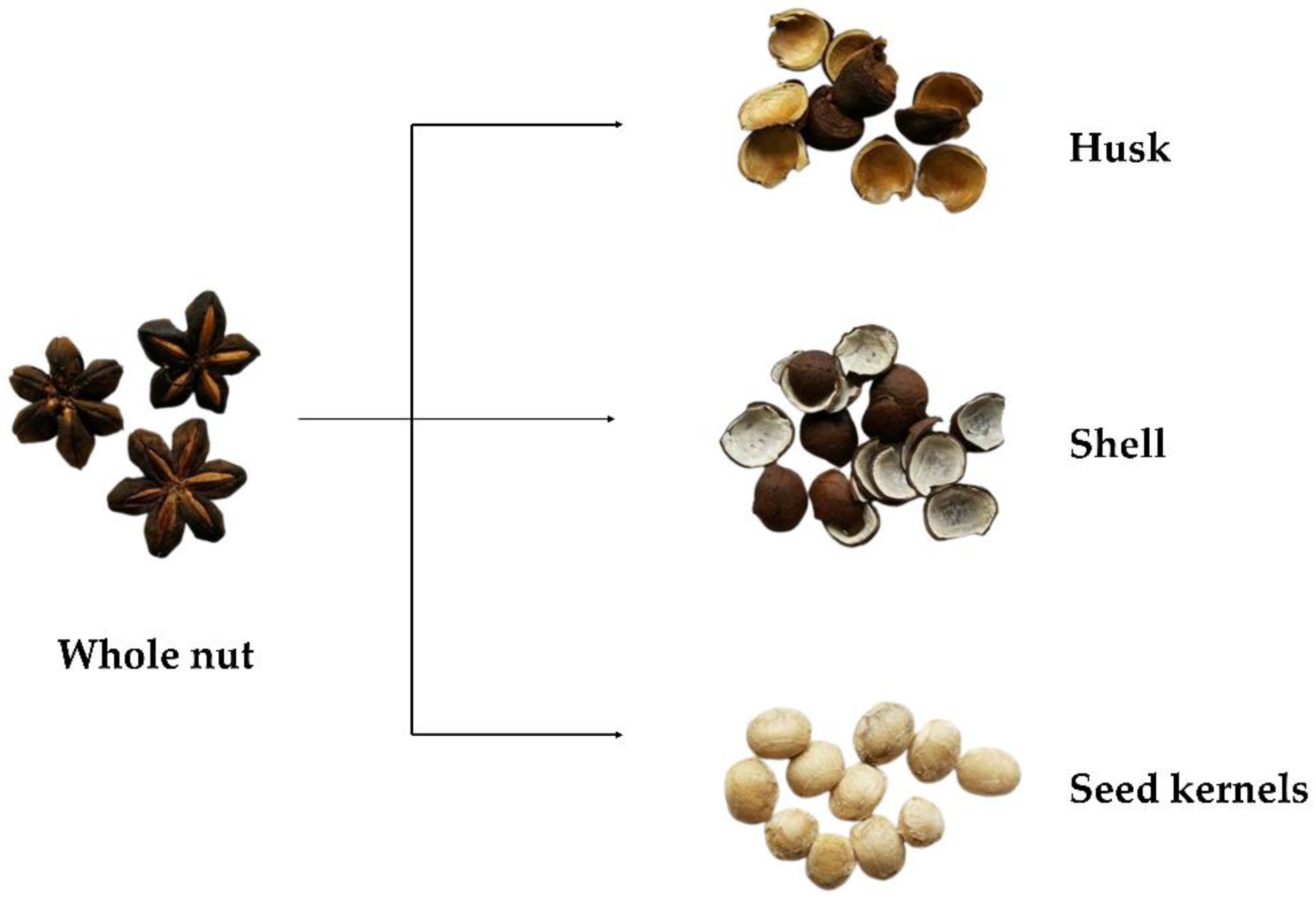
2.1. Sample Collection, Preparation, and Extraction

Husk and shell of sacha inchi were supplied by Thai Rubber Land and Plantation Co., Ltd., Muang district, Chiang Rai, Thailand, in March 2014 (all experiments were performed within a year after receiving the samples); the physical appearances are shown in Figure 1. The samples were divided into two groups. The first was ground using a Philips 600 W grinder (Philips Electronic Co., Ltd., Jakarta, Indonesia) and we analyzed their nutritional compositions (carbohydrate, protein, fat, energy, total dietary fiber, ash, and moisture content), while the second was dried using a Heto powerdry PL9000 freeze dryer (Heto Lab Equipment, Allerød, Denmark) under freeze-dried conditions of −50 °C, 0.086 mbar, and 72 h.
The extraction was performed using the powdery sample (0.2 g) in 20% (v/v) aqueous ethanol (100 mL). The mixture was incubated in a 70 °C WNE45 water bath shaker (Memmert GmBh, Eagle, WI, USA) for 15 min. After centrifugation at 3800× g for 10 min in a Hettich® ROTINA 38R centrifuge (Andreas Hettich GmbH, Tuttlingen, Germany), the supernatant was collected, and the clear extract was received by filtering the supernatants through a 0.45 µM polyether sulfone (PES) membrane syringe filter.

2.2. Determination of Nutritive Values

Analysis of the nutritional compositions followed the standard protocols of the Association of Official Analytical Chemists (AOAC) [11]. The investigation of protein (Kjeldahl method following AOAC 992.23), fat (acidic digestion and petroleum extraction following AOAC 948.15, 945.16), carbohydrates (by calculation of moisture, protein, fat, and ash), energy (by calculation from protein, fat, and carbohydrate), total dietary fiber (enzymatic gravimetric method following AOAC 985.29), ash (incineration following AOAC 930.30), and moisture content (weight consistency in hot-air oven following AOAC 930.04, 934.01) were carried out at the Institute of Nutrition, Mahidol University (Nakhon Pathom, Thailand) following the international standard for laboratory quality systems with ISO/IEC 17025:2005.

2.3. Determination of Fatty Acid Profile

To determine fatty acid profile, the samples were hydrolyzed using acidic methanol followed by chloroform extraction, while the identification and quantitation were analyzed using gas chromatography–mass spectrometry (CG–MS) following the in-house method based on AOAC 963.22, 969.33. The standards used in this experiment were caproic acid (C6:0, ≥98.0% FFC, FG), caprylic acid (C8:0, ≥98.0% FFC, FG), capric acid (C10:0, ≥99.5% FFC, FG), lauric acid (C12:0, ≥98.0% FFC, FG), myristic acid (C14:0, ≥98.0% GC), myristoleic acid (C14:1, ≥99.0% GC), palmitic acid (C16:0, ≥99.0% GC), palmitoleic acid (C16:1, ≥98.5% GC), stearic acid (C18:0, ≥98.5% GC), oleic acid (C18:1, ≥99.0% GC), linoleic acid (C18:2n-6, ≥98.0% GC), γ-linoleic acid (C18:3n-6, ≥98.5% GC), linolenic acid (C18:3n-3, ≥98.5% GC), arachidic acid (C20:0, ≥99.0% GC), eicosenoic acid (C20:1, ≥99.0% GC), eicosadienoic acid (C20:2, ≥98.0% liquid), eicosatrienoic acid (C20:3n-6, ≥98.5% GC), eicosatrienoic acid (C20:3n-3, ≥98.5% GC), arachidonic acid (C20:4n-6, >95.0% GC), eicosapentaenoic acid (C20:5n-3, ≥98.5% GC), behenic acid (C22:0, ≥99.0% GC), erucic acid (C22:1, ≥99.0% GC), docosadienoic acid (C22:2, ≥98% liquid), docosahexaenoic acid (C22:6n-3, ≥98.0% GC), lignoceric acid (C24:0, ≥99.0% GC), and nervonic acid (C24:1, ≥99.0% GC). All reagents and chemicals were obtained from Sigma-Aldrich (St. Louis, MO, USA).

2.4. Determination of Phenolic Profiles

To analyze phenolic profiles, the samples were extracted using acidic methanol containing tert-butylhydroquinone (TBHQ), while the identification and quantitation were determined using high-performance liquid chromatography (HPLC) with conditions and validations as previously described [12] without any modifications. The standards of phenolics, including apigenin (>98.0% HPLC), 4-hydroxybenzoic acid (>99.0% GC, T), caffeic acid (>98.0% HPLC, T), chlorogenic acid (>98.0% HPLC, T), ferulic acid (>98.0% GC, T), hesperidin (>90.0% HPLC, T), luteolin (>98.0% HPLC), kaempferol (>97.0% HPLC), naringenin (>93.0% HPLC, T), myricetin (>97.0% HPLC), p-coumaric acid (>98.0% GC, T), quercetin (>98.0% HPLC, E), syringic acid (>97.0% T), and sinapic acid (>99.0% GC, T) were received from the Tokyo Chemical Industry (Tokyo, Japan), isorhamnetin (≥99.0% HPLC) was purchased from Extrasynthese (Genay, France), and gallic acid (97.5–102.5% T) was obtained from Sigma-Aldrich (St. Louis, MO, USA). Detections at 280 and 325 nm were selected for phenolic acid identification, while detections at 338 and 368 nm were for flavonoids. The HPLC chromatograms were displayed in Supplementary Figure S1.
Total phenolic content (TPC) was evaluated using the Folin–Ciocalteu phenol (FCP) reagent on a SynergyTM HT 96-well UV-visible microplate reader (BioTek Instruments, Inc., Winooski, VT, USA) with a visualizing software, Gen 5 data analysis, as previously described [13], without any modifications. Briefly, the plant extract was mixed with 10% (v/v) FCP reagent and 7.5% (w/v) sodium carbonate for 2 h at 25 °C in prior to measuring absorbance at 765 nm. Gallic acid was used as a standard, while the results were expressed as mg gallic acid equivalent (GAE)/g dry weight (DW).

2.5. Determination of Antioxidant Activities

To determine antioxidant activities, ferric ion reducing antioxidant power (FRAP), oxygen radical absorbance capacity (ORAC), and 2,2-diphenyl-1-picrylhydrazyl (DPPH) radical scavenging assays were performed using the well-established protocols as previously described [14] without any modifications. Briefly, the FRAP assay was performed by incubating the plant extract with the FRAP reagent for 8 min at 25 °C in prior to measuring absorbance at 600 nm. The ORAC assay was performed using the plant extract, fluorescein, and 2,2′-azobis(2-amidinopropane) dihydrochloride, while its activity was determined as a fluorescein decay curve at an excitation wavelength (λex) of 485 nm and an emission wavelength (λem) of 528 nm. As for the DPPH radical scavenging assay, the plant extract was incubated with DPPH reagent for 30 min at 25 °C before measuring the absorbance at 520 nm. Trolox was used as a standard in all three assays, and the results were expressed as µmol Trolox equivalent (TE)/100 g DW.

2.6. Determination of Enzyme Inhibitory Activities

In vitro inhibitory activities of the key enzymes involving in obesity (lipase), Alzheimer’s disease (AChE, BChE, and BACE-1), diabetes (α-glucosidase and α-amylase), and anti-glycation reactions of bovine serum albumin (BSA) induced by either methylglyoxal (MG) or D-glucose were investigated following the well-established protocols as previously described [15] without any modifications. The inhibitory assay of ACE was performed according to the reported of Chupeerach et al., 2021 [16]. Briefly, the reactions consisted of the enzyme, substrate, and indicator, while their detections were visualized at particular wavelengths, as shown in Table 1. For the anti-glycation reaction, the plant extract was incubated with BSA and either MG or D-glucose for 3 weeks at 37 °C before measuring the fluorescence intensity at λex 330 nm and λem 410 nm. All reagents and chemicals were obtained from Sigma-Aldrich (St. Louis, MO, USA). The inhibitory assays were performed on the SynergyTM HT 96-well UV-visible microplate reader. The inhibitory activities were calculated as percentages of inhibition at particular extract concentrations.

2.7. Statistical Analysis

All experiments were carried out in triplicate (n = 3); all data are presented as mean ± standard deviation (SD). The statistical analysis was performed using the unpaired t-test to compare significant differences between values of two data sets at p < 0.05 using the statistical package for the social sciences (version 18 for Windows, SPSS Inc., Chicago, IL, USA).

3. Results

3.1. Nutritional Compositions

Nutritional compositions of the husk and shell of sacha inchi, including protein, fat, carbohydrates, total dietary fiber, and ash were analyzed and used to calculate their energy, as shown in Table 2. Protein and carbohydrate contents in the husk were 1.4- and 2.0-fold higher than the shell, while the shell exhibited higher fat content (3.4-fold) than the husk. Due to the higher carbohydrate content, the husk exhibited 2.5-fold higher total dietary fiber than the shell, while its ash content was also 1.3-fold higher. Fat provided the highest energy (9 kcal/g) [17], and the shell with higher fat content had 1.3-fold higher energy than the husk.

3.2. Fatty Acid Profiles

Fatty acid profiles of the husk and shell of sacha inchi were analyzed by HPLC, with results shown in Table 3. Out of 26 fatty acid standards, 10 were identified in the husk and 7 in the shell. Caprylic acid, capric acid, and γ-linoleic acid were only detected in the husk, while the most abundant fatty acid detected in both the husk and shell was linolenic acid, accounting for 38.80 and 43.96% of total fatty acid content, respectively. The second most abundant fatty acid was linoleic acid, accounting for 38.19 and 39.15% in the husk and shell, respectively. The other detected fatty acids were composed of less than 10% of total fatty acid content.

3.3. Phenolic Profiles

Phenolic profiles of the sacha inchi husk and shell were determined using an HPLC analysis, with results shown in Table 4 and Supplementary Figure S1. The husk contained three flavonoids—quercetin, kaempferol, and isorhamnetin—with the amount of quercetin being 6.4-fold higher than the others. The shell also contained three flavonoids—naringenin, hesperidin, and kaempferol—with the first two 2.3- and 1.9-fold higher than the last, respectively. The only flavonoid present in both the husk and shell was kaempferol, which was 48.6-fold higher in the shell than in the husk.
Higher contents and varieties of phenolic acids than flavonoids were detected in the husk, such as gallic acid, 4-hydroxybenzoic acid, chlorogenic acid, caffeic acid, ferulic acid, p-coumaric acid, and sinapic acid. Among these detected phenolic acids, caffeic acid was the most abundant at 1.8- to 7.9-fold higher than the others. The shell also contained similar phenolic acids except for chlorogenic acid, which was only presented in the husk, while vanillic acid and syringic acid were only presented in the shell. The most abundant phenolic acid in the shell was p-coumaric acid at 2.7- to 75.9-fold higher than the others. Comparisons between phenolic acids detected in both the husk and shell determined that the shell contained 3.7- to 59.3-fold higher content than the husk.
Higher flavonoid and phenolic acid content led to 1.6-fold higher TPCs in the shell compared to the husk.

3.4. Antioxidant Activities

Three assays, including FRAP, ORAC, and DPPH radical scavenging assays, were performed to determine the antioxidant activities of the husk and shell of sacha inchi. Results are shown in Table 5. The FRAP and DPPH radical scavenging assays were used to determine the antioxidant activities following a single electron transfer (SET) mechanism; however, the ORAC assay followed a hydrogen atom transfer (HAT) mechanism. Thus, to cover both mechanisms and precisely explain their functions, the three assays covering both mechanisms were chosen.
Results indicated that the shell exhibited higher antioxidant activities than the husk in all assays at 1.8-, 2.3-, and 2.7-fold higher in the FRAP, ORAC, and DPPH radical scavenging assays, respectively.

3.5. Enzyme Inhibitory Activities

The potential in vitro health properties of the husk and shell of sacha inchi were determined by measuring the inhibition of the key enzymes relevant to obesity (lipase), hypertension (ACE), diabetes (α-glucosidase and α-amylase), and Alzheimer’s disease (AChE, BChE and BACE-1). The inhibitory effects of the non-enzymatic glycation reaction induced by MG and D-glucose were also examined. The results were expressed as percentages of inhibition under the studied assay conditions with particular extract concentrations, as shown in Table 6.
Inhibition of lipase, a lipid degrading enzyme, leads to low lipid absorption into the body as a target for obesity prevention and treatment. Results indicated that inhibitory activity was only detected in the husk (23.80% inhibition), with no activity observed in the shell using extract concentration of 0.4 mg/mL.
Inhibition of ACE reduces angiotensin II, a peptidic factor that promotes increase in blood pressure. Inhibition of ACE in the husk was 2.8-fold higher than in the shell using extract concentration of 0.013 mg/mL.
Inhibition of the carbohydrate-degrading enzymes, α-glucosidase and α-amylase, also retards glucose absorption, resulting in a slow increase of serum glucose as one pathway for diabetic prevention. The husk displayed 1.3-fold higher α-glucosidase inhibition than the shell, while the latter exhibited 1.6-fold higher α-amylase inhibition than the former at the same extraction concentration of 0.025 mg/mL.
Decreasing the neurotransmitter, acetylcholine, by enzymatic degradation via AChE and BChE is a significant marker of Alzheimer’s disease pathology. Other than the cholinergic system, the deposition of amyloid beta (Aβ) plaques in the brain is another clinical marker. These plaques are formed by the aggregation of Aβ peptides produced by BACE-1 hydrolysis of amyloid precursor protein (APP). Thus, inhibition of these enzymes is considered as an alternative pathway for Alzheimer’s disease prevention. Results indicated that the shell exhibited 1.8-fold higher AChE inhibitory activity than the husk, while the latter exhibited 1.6-fold higher BChE inhibitory activity than the former using extract concentration of 0.017 mg/mL. However, insignificant differences in BACE-1 inhibitory activities was observed between these two samples using extract concentration of 0.2 mg/mL.
Glycation reactions of BSA induced by either MG or D-glucose and their inhibitory activities by sacha inchi extracts were also investigated. In the anti-glycation reaction induced by D-glucose, the shell exhibited 1.2-fold higher inhibitory activity than the husk using extract concentration of 0.125 mg/mL, while insignificant difference in inhibitory activities between the husk and shell was observed in the anti-glycation reaction induced by MG using the same extract concentration.

4. Discussion

Both the husk and shell are low value by-products of sacha inchi oil extraction that are used as fertilizer or animal feed. Limited previous research suggested that the shell was a nutritious source of fiber with some health-related bioactivities, while little information on the husk was available. Therefore, this research investigated and compared the nutritional compositions, fatty acid and phenolic profiles, antioxidant properties, anti-glycation activities, and inhibitory activities of the key enzymes relevant to some NCDs of the sacha inchi husk and shell. Results indicated that (i) husk exhibited higher protein, carbohydrates, and total dietary fiber than the shell, while the latter contained higher fat and energy; (ii) linolenic acid and linoleic acid were the major fatty acids found in both the husk and shell; (iii) TPC in the shell was higher than in the husk; (iv) shell exhibited higher antioxidant, anti-glycation, anti-α-amylase, and anti-AChE activities than the husk; and (v) husk exhibited higher inhibitory activities on lipase, α-glucosidase, ACE and BChE than the shell.
Only one report on the nutritive values of the sacha inchi shell was available in Spanish [5], while Goyal et al. (2022) summarized the available information and noted that the shell contained 77.84% crude fiber, 2.75% protein, 1.75% ash, 0.39% fat, and 17.27% non-nitrogenous components [6]. Our results indicated higher amounts of protein and total fat content than carbohydrates, while the husk contained higher protein and carbohydrates than fat. Our results also suggested that most of the carbohydrates were fiber (80.3% in the husk and 65.4% in the shell). De Souza et al. (2013) found 1.24% fat in the shell, with predominant fatty acids as linoleic acid (C18:2n-6, 325 mg/g total lipid) and linolenic acid (C18:3n-3, 330 mg/g total lipid), similar to seed (339 mg linoleic acid/g total lipid and 439 mg linolenic acid/g total lipid) [7]. Our results concurred with this report. However, shell with higher fat content also contained higher amounts of linoleic acid (C18:2n-6) and linolenic acid (C18:3n-3) than the husk.
Other than nutritive values, it was also previously reported that the shell exhibited TPCs ranging from 9.5 to 15.3 mg GAE/g depending on the extraction solvent [2]. A mixture of acetone:water:acetic acid in the ratio 80:19:1 and methanol:acetone:water:acetic acid in the ratio 40:40:10:1 at 60 °C using a two-step extraction of 60 and 30 min, respectively, were the most suitable solvents for the extraction of phenolics [2]. Presoaking the shell in 50% (v/v) aqueous ethanol for 24 h and then refluxing at 70 °C for 2 h gave higher TPC (129.9 mg GAE/g) than samples presoaked with absolute ethanol or water [9], while shell subjected to microwave-assisted extraction gave TPC of 41.97 mg GAE/g [18]. Hot water extraction of the husk (15 min boiling with further incubation for another 4 h) exhibited TPC at 74.8 mg GAE/g DW [10]. Interestingly, heating the shell at 50 °C in an oven overnight had little effect on TPC [10]. Compared to previous studies, our TPC from 20% (v/v) aqueous ethanolic extraction at 70 °C for 15 min were low at 3.24 and 5.04 mg GAE/g DW in the husk and shell, respectively, probably due to the short incubation time and lack of a presoaking step. Depending on plant samples, presoaking may reduce plant cell wall recalcitrance; thus, extraction using an appropriate solvent, temperature, and time releases high amounts of phenolics from plant cells [19].
Phenolics detected in the shell extracted using aqueous ethanolic extraction were reported as hydroxycinnamic acid, protocatechuic acid, cinnamic acid, and p-coumaric acid [2], while hydroxy-4-chromone, 3,4-dihydroxy-benzaldehyde, and their derivatives were reported as predominant phenolics in the shell undergone microwave-assisted extraction [18]. By contrast, we determined different results with caffeic acid, gallic acid, and p-coumaric acid as the predominant phenolics in the husk, and p-coumaric acid, 4-hydroxybenzoic acid, and gallic acid in the shell. Few phenolics were identified in previous research because limited phenolic standards were used in the analytical methods.
The biological functions of phenolics include effective antioxidants [20]. Depending on the extraction solvent, the shell exhibited antioxidant activities via the FRAP assay ranging from 45.0 to 114.0 µmol TE/g and an ORAC assay ranging from 92.5 to 192.6 µmol TE/g [2]. Shell subjected to microwave-assisted extraction gave FRAP activities of 374.39 ± 1.81 μmol Trolox/g [18], while hot water-extracted husk exhibited DPPH radical scavenging activity of 0.78 mg GAE/g DW [10]. These activities were higher than detected in our samples because of the different extraction conditions, as discussed above.
A strong correlation among phenolics, antioxidant activities, and anti-glycation properties was previously reported [15,21], and shell with higher TPC and antioxidant activity exhibited stronger anti-glycation activities than the husk. It was previously reported that caffeic acid (as the predominant phenolic detected in the husk in our study) inhibited 29.8% of advanced glycation end products (AGEs) formed in the BSA–glucose system using a concentration of 0.1 mM [22]. As the predominant phenolic detected in the shell in our study, p-coumaric acid at a concentration of 0.06 to 6.1 mM was previously reported to promote AGE formation in the BSA–glucose system, with no significant ability to scavenge glyoxal [23]. Thus, the second and third abundantly available phenolics in the shell as 4-hydroxybenzoic acid and gallic acid might be responsible for its higher anti-glycation activities than in the husk. It was found that 4-hydroxybenzoic acid (1 mM) inhibited glycation reaction in the BSA-glucose system at 24.3% [24], while gallic acid (0.6 μM) strongly reduced AGEs formed in a BSA–fructose system at 25.3% [25]. Thus, other phenolics than p-coumaric acid might be responsible for anti-glycation activity in the shell.
Interestingly, anti-obesity properties through cholesterol control were previously investigated in the husk. A hot water extract of husk was previously reported to exhibit 75% inhibition on 3-hydroxy-3-methylglutaryl-coenzyme A (HMG-CoA) reductase, the enzyme that hydrolyzes HMG-CoA to mevalonate, at a concentration of 0.125 mg/mL [10]. This extract also exhibited cholesterol esterase, a cholesterol ester hydrolyzing enzyme, with 38.1% inhibition using extract concentration of 0.125 mg/mL [10]. Inhibition of HMG-CoA reductase limited the synthesis of cholesterol because mevalonate is a starting compound for cholesterol synthesis. HMG-CoA reductase is also a drug target for the cholesterol lowering effect of statins, a synthetic drug used for controlling high cholesterol [26]. Likewise, inhibition of cholesterol esterase retards cholesterol ester absorption and transportation. Our research also concentrated on obesity, but through the fat lowering effect by targeting lipase inhibition. Only the husk exhibited lipase inhibition, while none was observed in the shell at extract concentration of 0.4 mg/mL. Most phenolics could act as lipase inhibitors [27]; therefore, the shell with higher TPC than the husk should also exhibit lipase inhibitory activity. Opposite results were observed in our study, suggesting that other than phenolics, anti-lipase agents in both the husk and shell might include non-phenolic bioactive compounds that were previously reported to exhibit effective lipase inhibition [28].
No anti-diabetic properties of the sacha inchi husk and shell were previously reported. Here, this property was first reported through α-amylase and α-glucosidase inhibition, with inhibitory activities detected in both the husk and shell. Many phenolics act as effective α-amylase and α-glucosidase inhibitors [29]. Caffeic acid, a predominant phenolic in the husk, exhibited half-maximal inhibitory concentration (IC50) of 20.4 µM against α-amylase and 27.6 µM against α-glucosidase [30]. Interestingly, p-coumaric acid, a predominant phenolic in the shell, exhibited stronger α-amylase inhibition with an IC50 value of 0.74 µM and α-glucosidase inhibition with IC50 value of 0.16 µM [31]. Most phenolics act as stronger inhibitors toward α-glucosidase than α-amylase [32], causing the higher inhibitory activities detected in both the husk and shell and making α-glucosidase a more captivating target for serum sugar control.
Likewise, no anti-Alzheimer’s disease property was previously reported in the sacha inchi husk and shell. This is the first paper to report husk and shell inhibitory activities against the key enzymes controlling cholinergic termination (AChE and BChE) and Aβ formation (BACE-1). The major phenolic in the husk as caffeic acid exhibited AChE and BChE inhibitions with IC50 values of 4.21 and 5.60 µg/mL, respectively [33], while p-coumaric acid, a predominant phenolic in the shell, improved learning and memory impairments in scopolamine-treated rats [34]. Two types of BACE-1 inhibitors were identified as peptidomimetic and nonpeptidomimetic compounds [35,36]. Caffeic acid exhibited an IC50 value of 16.67 µM against BACE-1 [37], while an IC50 value of >400 µM was reported in p-coumaric acid [38]. Higher phenolic content was detected in the shell, while the husk and shell exhibited insignificantly different BACE-1 inhibitory activities. Thus, other than phenolics (as nonpeptidomimetic BACE-1 inhibitors), peptidomimetic compounds might also be responsible for BACE-1 inhibition of the husk and shell.
For anti-hypertensive properties, one research paper reported that sacha inchi shell extract reduced systolic blood pressure and cardiac hypertrophy in spontaneously hypertensive rats [8]. Our results supported this finding, indicating that both the husk and shell exhibited inhibitory activities against ACE, a hypertensive promoting peptide enzyme that digests angiotensin I to angiotensin II. Predominant phenolics in the husk and shell, such as caffeic acid and p-coumaric acid, possessed ACE inhibition with IC50 values of 0.4 and 2.8 mM, respectively [39,40], suggesting their anti-ACE role in both the husk and shell. However, our results also suggested that husk exhibited higher ACE inhibitory activities than the shell, even though husk possessed lower phenolics than the shell. ACE inhibitors can be divided into two types—peptidic and non-peptidic inhibitors. Peptidic inhibitors such as food proteins/peptides have been identified as effective ACE inhibitors, while non-peptidic inhibitors include phenolics, xanthones, and secoiridoids [41,42]. Thus, anti-ACE activities detected in the husk and shell might be from the functions of bioactive compounds other than phenolics.

5. Conclusions

Sacha inchi by-products, as the husk and shell, contain nutrients and phenolics, with potential inhibitory effects against key enzymes relevant to some non-communicable diseases. The husk was a richer source of protein and carbohydrate, while the shell contained higher fat and phenolic content. Health properties through in vitro enzyme inhibitions indicated that phenolics might be responsible for some of these inhibitory activities; however, other bioactive compounds, such as active peptides/proteins or their synergistic effects, might also play roles in these enzyme inhibitions. Since only percentages of inhibition were reported using particular extract concentrations, the comparison of our results in enzyme inhibitions were limited and cannot be compared with other plant extracts. Thus, this is a limitation of our work. Nevertheless, information gained from this research could pique the interests of other researchers to pursue further in-depth studies on the health benefits of sacha inchi by-products to increase their value and utilization. Moreover, this information could help to open-up future opportunities for the valorization of the husk and shell of sacha inchi, for producing health-promoting products, thus facilitating the sustainable circular economy following the circular food supply chain concept. Functional foods or supplements from sacha inchi husk and shell can be developed as potential nutrient-rich sources, with high phenolics for health benefits after assessing their toxicology and bioaccessibility.

Supplementary Materials

The following supporting information can be downloaded at: https://www.mdpi.com/article/10.3390/horticulturae8040344/s1, Figure S1: High-performance liquid chromatograms of phenolics in (A) husk and (B) shell of sacha inchi.

Author Contributions

Conceptualization, C.H., P.S., S.S. and U.S.; methodology, U.S.; software, S.K. and U.S.; validation, S.K. and U.S.; formal analysis, S.K.; investigation, S.K.; resources, U.S.; data curation, S.K. and U.S.; writing—original draft preparation, S.K., C.H., P.S., S.S., P.T. and U.S.; writing—review and editing, S.K., P.T. and U.S.; visualization, S.K. and U.S.; supervision, U.S.; project administration, U.S.; funding acquisition, C.H. All authors have read and agreed to the published version of the manuscript.

Funding

This research was funded by Thai Rubber Land and Plantation Co., Ltd., Thailand.

Institutional Review Board Statement

Not applicable.

Informed Consent Statement

Not applicable.

Data Availability Statement

Data are contained within this article and Supplementary Materials.

Conflicts of Interest

The authors declare no conflict of interest.

References

  1. Kodahl, N.; Sørensen, M. Sacha inchi (Plukenetia volubilis L.) is an underutilized crop with a great potential. Agronomy 2021, 11, 1066. [Google Scholar] [CrossRef]
  2. Chirinos, R.; Necochea, O.; Pedreschi, R.; Campos, D. Sacha inchi (Plukenetia volubilis L.) shell: An alternative source of phenolic compounds and antioxidants. Int. J. Food Sci. Technol. 2016, 51, 986–993. [Google Scholar] [CrossRef]
  3. Kumar, B.; Smita, K.; Sánchez, E.; Stael, C.; Cumbal, L. Andean Sacha inchi (Plukenetia volubilis L.) shell biomass as new biosorbents for Pb2+ and Cu2+ ions. Ecol. Eng. 2016, 93, 152–158. [Google Scholar] [CrossRef]
  4. Rueda-Ordóñez, Y.J.; Mariño-Bohórquez, M.A.; Rueda-Ordóñez, D.A. Thermal upgrading of sacha inchi shell: Kinetics and combustion characteristics. Bioresour. Technol. Rep. 2021, 15, 100807. [Google Scholar] [CrossRef]
  5. Benítez, R.; Coronell, C.; Martin, J. Chemical characterizaction sacha inchi (Plukenetia volubilis) seed: Oleaginosa promising from the Colombian Amazon. Int. J. Curr. Sci. Res. Rev. 2018, 1, 11–22. [Google Scholar]
  6. Goyal, A.; Tanwar, B.; Sihag, M.K.; Sharma, V. Sacha inchi (Plukenetia volubilis L.): An emerging source of nutrients, omega-3 fatty acid and phytochemicals. Food Chem. 2022, 373, 131459. [Google Scholar] [CrossRef]
  7. de Souza, A.H.P.; Gohara, A.K.; Rodrigues, A.C.; de Souza, N.E.; Visentainer, J.V.; Matsushita, M. Sacha inchi as potential source of essential fatty acids and tocopherols: Multivariate study of nut and shell. Acta Sci. Technol. 2013, 35, 757–763. [Google Scholar] [CrossRef] [Green Version]
  8. Li, P.; Cai, X.; Xiao, N.; Ma, X.; Zeng, L.; Zhang, L.H.; Xie, L.; Du, B. Sacha inchi (Plukenetia volubilis L.) shell extract alleviates hypertension in association with regulation of gut microbiota. Food Funct. 2020, 11, 8051–8067. [Google Scholar] [CrossRef]
  9. Sainakham, M.; Mungmai, L. In vitro anti-oxidative activity and tyrosinase inhibition of Inca peanut (Plukenetia volubilis L.) shell extracts from different preparation methods. TJST 2020, 9, 407–417. [Google Scholar]
  10. Prasongsub, W.; Pimsan, N.; Buranapattarachote, C.; Punturee, K. Anti-HMG-CoA reductase and antioxidant activities of Sacha inchi (Plukenetia volubilis L.) nutshell extract. J. Assoc. Med. Sci. 2021, 54, 18–26. [Google Scholar]
  11. Latimer, G.W. Official Method of Analysis of AOAC International, 21st ed.; AOAC International: Rockville, ML, USA, 2019. [Google Scholar]
  12. Temviriyanukul, P.; Sritalahareuthai, V.; Promyos, N.; Thangsiri, S.; Pruesapan, K.; Srinuanchai, W.; Nuchuchua, O.; Siriwan, D.; On-nom, N.; Suttisansanee, U. The effect of sacred lotus (Nelumbo nucifera) and its mixtures on phenolic profiles, antioxidant activities, and inhibitions of the key enzymes relevant to Alzheimer’s disease. Molecules 2020, 25, 3713. [Google Scholar] [CrossRef] [PubMed]
  13. Hinkaew, J.; Sahasakul, Y.; Tangsuphoom, N.; Suttisansanee, U. The effect of cultivar variation on total phenolic contents and antioxidant activities of date palm fruit (Phoenix dactylifera L.). Curr. Res. Nutr. Food Sci. 2020, 8, 155–163. [Google Scholar] [CrossRef]
  14. Sripum, C.; Kukreja, R.K.; Charoenkiatkul, S.; Kriengsinyos, W.; Suttisansanee, U. The effect of extraction conditions on antioxidant activities and total phenolic contents of different processed Thai Jasmine rice. Int. Food Res. J. 2017, 24, 1644–1650. [Google Scholar]
  15. Sirichai, P.; Kittibunchakul, S.; Thangsiri, S.; On-Nom, N.; Chupeerach, C.; Temviriyanukul, P.; Inthachat, W.; Nuchuchua, O.; Aursalung, A.; Sahasakul, Y.; et al. Impact of drying processes on phenolics and in vitro health-related activities of indigenous plants in Thailand. Plants 2022, 11, 294. [Google Scholar] [CrossRef]
  16. Chupeerach, C.; Aursalung, A.; Watcharachaisoponsiri, T.; Whanmek, K.; Thiyajai, P.; Yosphan, K.; Sritalahareuthai, V.; Sahasakul, Y.; Santivarangkna, C.; Suttisansanee, U. The effect of steaming and fermentation on nutritive values, antioxidant activities, and inhibitory properties of tea leaves. Foods 2021, 10, 117. [Google Scholar] [CrossRef]
  17. FAO. Food energy: Methods of analysis and conversion factors. In Report of a Technical Workshop, Food and Nutrition; Agricultural Research Service, US Department of Agriculture: Beltsville, MD, USA, 2003; p. 77. [Google Scholar]
  18. Sanchez-Reinoso, Z.; Mora-Adames, W.I.; Fuenmayor, C.A.; Darghan-Contreras, A.E.; Gardana, C.; Gutiérrez, L.F. Microwave-assisted extraction of phenolic compounds from Sacha Inchi shell: Optimization, physicochemical properties and evaluation of their antioxidant activity. Chem. Eng. Process. Process Intensif. 2020, 153, 107922. [Google Scholar] [CrossRef]
  19. Boateng, J.; Verghese, M.; Walker, L.T.; Ogutu, S. Effect of processing on antioxidant contents in selected dry beans (Phaseolus spp. L.). LWT-Food Sci. Technol. 2008, 41, 1541–1547. [Google Scholar] [CrossRef]
  20. Spiegel, M.; Kapusta, K.; Kołodziejczyk, W.; Saloni, J.; Żbikowska, B.; Hill, G.A.; Sroka, Z. Antioxidant activity of selected phenolic acids-ferric reducing antioxidant power assay and QSAR analysis of the structural features. Molecules 2020, 25, 3088. [Google Scholar] [CrossRef]
  21. Suttisansanee, U.; Thiyajai, P.; Chalermchaiwat, P.; Wongwathanarat, K.; Pruesapan, K.; Charoenkiatkul, S.; Temviriyanukul, P. Phytochemicals and in vitro bioactivities of aqueous ethanolic extracts from common vegetables in Thai food. Plants 2021, 10, 1563. [Google Scholar] [CrossRef]
  22. Cao, X.; Xia, Y.; Zeng, M.; Wang, W.; He, Y.; Liu, J. Caffeic acid inhibits the formation of advanced glycation end products (AGEs) and mitigates the AGEs-induced oxidative stress and inflammation reaction in human umbilical vein endothelial cells (HUVECs). Chem. Biodivers. 2019, 16, e1900174. [Google Scholar] [CrossRef]
  23. Chen, H.; Virk, M.S.; Chen, F. Phenolic acids inhibit the formation of advanced glycation end products in food simulation systems depending on their reducing powers and structures. Int. J. Food. Sci. Nutr. 2016, 67, 400–411. [Google Scholar] [CrossRef] [PubMed]
  24. Rahbar, S.; Yerneni, K.K.; Scott, S.; Gonzales, N.; Lalezari, I. Novel inhibitors of advanced glycation endproducts (part II). Mol. Cell Biol. Res. Commun. 2000, 3, 360–366. [Google Scholar] [CrossRef] [PubMed]
  25. Adisakwattana, S.; Thilavech, T.; Sompong, W.; Pasukamonset, P. Interaction between ascorbic acid and gallic acid in a model of fructose-mediated protein glycation and oxidation. Electron. J. Biotechnol. 2017, 27, 32–36. [Google Scholar] [CrossRef]
  26. Istvan, E.S. Structural mechanism for statin inhibition of 3-hydroxy-3-methylglutaryl coenzyme A reductase. Am. Heart J. 2002, 144, S27–S32. [Google Scholar] [CrossRef]
  27. Buchholz, T.; Melzig, M.F. Polyphenolic compounds as pancreatic lipase inhibitors. Planta Med. 2015, 81, 771–783. [Google Scholar] [CrossRef] [Green Version]
  28. Kumar, A.; Chauhan, S. Pancreatic lipase inhibitors: The road voyaged and successes. Life Sci. 2021, 271, 119115. [Google Scholar] [CrossRef]
  29. Giuberti, G.; Rocchetti, G.; Lucini, L. Interactions between phenolic compounds, amylolytic enzymes and starch: An updated overview. Curr. Opin. Food Sci. 2020, 31, 102–113. [Google Scholar] [CrossRef]
  30. Oboh, G.; Agunloye, O.M.; Adefegha, S.A.; Akinyemi, A.J.; Ademiluyi, A.O. Caffeic and chlorogenic acids inhibit key enzymes linked to type 2 diabetes (in vitro): A comparative study. J. Basic. Clin. Physiol. Pharmacol. 2015, 26, 165–170. [Google Scholar] [CrossRef]
  31. Taslimi, P.; Gulçin, İ. Antidiabetic potential: In vitro inhibition effects of some natural phenolic compounds on α-glycosidase and α-amylase enzymes. J. Biochem. Mol. Toxicol. 2017, 31, e21956. [Google Scholar] [CrossRef]
  32. Tadera, K.; Minami, Y.; Takamatsu, K.; Matsuoka, T. Inhibition of α-glucosidase and α-amylase by flavonoids. J. Nutr. Sci. Vitaminol. 2006, 52, 149–153. [Google Scholar] [CrossRef] [Green Version]
  33. Oboh, G.; Agunloye, O.M.; Akinyemi, A.J.; Ademiluyi, A.O.; Adefegha, S.A. Comparative study on the inhibitory effect of caffeic and chlorogenic acids on key enzymes linked to Alzheimer’s disease and some pro-oxidant induced oxidative stress in rats’ brain-in vitro. Neurochem. Res. 2013, 38, 413–419. [Google Scholar] [CrossRef] [PubMed]
  34. Kim, H.B.; Lee, S.; Hwang, E.S.; Maeng, S.; Park, J.H. p-Coumaric acid enhances long-term potentiation and recovers scopolamine-induced learning and memory impairments. Biochem. Biophys. Res. Commun. 2017, 492, 493–499. [Google Scholar] [CrossRef] [PubMed]
  35. Moussa-Pacha, N.M.; Abdin, S.M.; Omar, H.A.; Alniss, H.; Al-Tel, T.H. BACE1 inhibitors: Current status and future directions in treating Alzheimer’s disease. Med. Res. Rev. 2020, 40, 339–384. [Google Scholar] [CrossRef] [PubMed]
  36. Huang, W.H.; Sheng, R.; Hu, Y.Z. Progress in the development of nonpeptidomimetic BACE 1 inhibitors for Alzheimer’s disease. Curr. Med. Chem. 2009, 16, 1806–1820. [Google Scholar] [CrossRef]
  37. Omar, S.H.; Scott, C.J.; Hamlin, A.S.; Obied, H.K. Biophenols: Enzymes (β-secretase, Cholinesterases, histone deacetylase and tyrosinase) inhibitors from olive (Olea europaea L.). Fitoterapia 2018, 128, 118–129. [Google Scholar] [CrossRef]
  38. Takahashi, T.; Miyazawa, M. Serotonin derivatives as inhibitors of β-secretase (BACE 1). Pharmazie 2011, 66, 301–305. [Google Scholar]
  39. Bhullar, K.S.; Lassalle-Claux, G.; Touaibia, M.; Rupasinghe, H.P.V. Antihypertensive effect of caffeic acid and its analogs through dual renin–angiotensin–aldosterone system inhibition. Eur. J. Pharmacol. 2014, 5, 125–132. [Google Scholar] [CrossRef]
  40. Al Shukor, N.; Van Camp, J.; Gonzales, G.B.; Staljanssens, D.; Struijs, K.; Zotti, M.J.; Raes, K.; Smagghe, G. Angiotensin-converting enzyme inhibitory effects by plant phenolic compounds: A study of structure activity relationships. J. Agric. Food Chem. 2013, 61, 11832–11839. [Google Scholar] [CrossRef]
  41. Murray, B.A.; FitzGerald, R.J. Angiotensin converting enzyme inhibitory peptides derived from food proteins: Biochemistry, bioactivity and production. Curr. Pharm. Des. 2007, 13, 773–791. [Google Scholar] [CrossRef]
  42. Barbosa-Filho, J.M.; Martins, V.K.M.; Rabelo, L.A.; Moura, M.D.; Silva, M.S.; Cunha, E.V.L.; Souza, M.F.V.; Almeida, R.N.; Medeiros, I.A. Natural products inhibitors of the angiotensin converting enzyme (ACE). A review between 1980–2000. Rev. Bras. Farmacogn. 2006, 16, 421–446. [Google Scholar] [CrossRef]
Figure 1. The physical appearances of the husk and shell of sacha inchi.
Horticulturae 08 00344 g001
Table 1. The components of the inhibitory assays.
AssayEnzyme (Effectiveness, Source)SubstrateIndicatorDetecting Wavelength
Lipase≥700 unit/mg (type VII), Candida rugosaDNPDBDTNB412 nm
AChE1000 units/mg, Electrophorus electricus acetylthiocholine
BChE≥10 units/mg, equine serumbutyrylthiocholine
BACE-1a BACE-1 FRET assay kitλex = 320 nm, λem = 405 nm
ACE≥2 unit/mg, rabbit lunghippuryl-histidyl-leucinePDAλex = 360 nm, λem = 485 nm
α-Glucosidase≥10 U/mg protein (type I), Saccharomyces cerevisiaep-nitrophenyl-α-d-glucopyranoside405 nm
α-Amylase≥10 unit/mg (type VII), porcine pancreaticp-nitrophenyl-α-d-maltopentaoside
DNPDB: 5–5′-dithiobis(2-nitrobenzoic-N-phenacyl-4,5-dimethyyhiazolium bromide; DTNB: 5,5′-dithiobis(2-nitrobenzoic acid); AChE: acetylcholinesterase; BChE: butyrylcholinesterase; BACE-1: β-secretase; FRET: fluorescence resonance energy transfer; ACE: angiotensin-converting enzyme; PDA: o-phthaldialdehyde; λex: excitation wavelength; λem: emission wavelength.
Table 2. Nutritive values in the husk and shell of sacha inchi.
Nutritive Values
(Per 100 g Fresh Weight)
Parts of Sacha Inchi
HuskShell
Energy (kcal)421.47 ± 0.42 *552.89 ± 0.98
Moisture (g)3.01 ± 0.01 *4.66 ± 0.04
Protein (g)59.97 ± 0.06 *43.12 ± 0.36
Total fat (g)11.25 ± 0.05 *37.87 ± 0.16
Total carbohydrates (g)20.10 ± 0.07 *9.90 ± 0.47
Dietary fiber (g)16.14 ± 0.16 *6.47 ± 0.27
Ash (g)5.68 ± 0.03 *4.46 ± 0.01
All experiments were carried out in triplicate (n = 3); all data are presented as mean ± standard deviation (SD); * shows significant difference of the same nutritional composition in the husk and shell at p < 0.05 using unpaired t-test.
Table 3. Fatty acid profile in the husk and shell of sacha inchi.
Fatty Acids
(Per 100 g Fresh Weight)
Parts of Sacha Inchi
HuskShell
Amt (g)% of TFCAmt (g)% of TFC
Caprylic acid (C8:0) 0.020.16NDND
Capric acid (C10:0)0.020.18NDND
Lauric acid (C12:0)0.151.430.070.20
Myristic acid (C14:0)0.100.900.050.14
Palmitic acid (C16:0) 0.716.581.744.79
Stearic acid (C18:0) 0.454.141.183.26
Oleic acid (C18:1) 1.039.583.088.50
Linoleic acid (C18:2n-6) 4.1138.1914.1739.15
γ-Linoleic acid (C18:3n-6)0.0020.02NDND
Linolenic acid (C18:3n-3) 4.1738.8015.9143.96
ND: not detected; Amt: amount; TFC: total fatty acid content; ND: not detected.
Table 4. Phenolic profiles and total phenolic content in the husk and shell of sacha inchi.
Phenolics
(mg/100 g DW)
Parts of Sacha Inchi
HuskShell
Flavonoids
Quercetin1.72 ± 0.06ND
NaringeninND29.21 ± 0.17
HesperidinND23.92 ± 1.55
Kaempferol0.27 ± 0.01 *12.63 ± 0.45
Isorhamnetin0.27 ± 0.01 ND
Phenolic acids
Gallic acid2.51 ± 0.09 *49.13 ± 2.67
4-Hydroxybenzoic acid2.28 ± 0.13 *54.61 ± 3.04
Chlorogenic acid2.16 ± 0.12 ND
Vanillic acidND5.20 ± 0.15
Caffeic acid4.44 ± 0.04 *16.21 ± 0.10
Syringic acidND1.96 ± 0.08
p-Coumaric acid2.51 ± 0.15 *148.74 ± 2.46
Ferulic acid0.56 ± 0.04 *5.71 ± 0.09
Sinapic acid0.92 ± 0.03 *17.41 ± 0.40
Total phenolic content
(mg GAE/100 g DW)
323.74 ± 2.25 *503.96 ± 5.16
All experiments were carried out in triplicate (n = 3); all data are presented as mean ± standard deviation (SD). GAE: gallic acid equivalent; DW: dry weight; ND: not detected; * shows significantly different contents of the same phenolics detected in both the husk and shell at p < 0.05 using unpaired t-test.
Table 5. Antioxidant activities in the husk and shell of sacha inchi.
Antioxidant Activities
(µmol TE/100 g DW)
Parts of Sacha Inchi
HuskShell
FRAP activity100.28 ± 4.75 *180.17 ± 6.93
ORAC activity4238.67 ± 89.15 *9751.06 ± 116.58
DPPH radical scavenging activity0.03 ± 0.00 *0.08 ± 0.00
All experiments were carried out in triplicate (n = 3); all data are presented as mean ± standard deviation (SD). DW: dry weight; TE: Trolox equivalent; FRAP: ferric reducing antioxidant power; ORAC: oxygen radical absorbance capacity; DPPH: 2,2-diphenyl-1-picrylhydrazyl; * shows significantly different antioxidant activities of the same assay detected in the husk and shell at p < 0.05 using unpaired t-test.
Table 6. Inhibitory activities of enzymatic and non-enzymatic (chemical) reactions in the husk and shell of sacha inchi.
Reaction TypesInhibitory Activities
(% Inhibition)
Parts of Sacha Inchi
HuskShell
Enzymes1 Lipase23.80 ± 2.16ND
2 ACE90.85 ± 3.42 *32.22 ± 2.59
3 α-Amylase8.97 ± 1.36 *14.72 ± 1.47
3 α-Glucosidase61.39 ± 3.04 *46.46 ± 2.54
4 AChE28.84 ± 0.58 *51.88 ± 4.67
4 BChE63.61 ± 2.96 *40.90 ± 3.13
5 BACE-191.17 ± 2.5094.83 ± 0.89
Chemicals6 MG-induced glycation29.43 ± 0.6331.53 ± 3.40
6 Glucose-induced glycation47.67 ± 2.85 *55.81 ± 1.13
All experiments were carried out in triplicate (n = 3); all data are presented as mean ± standard deviation (SD). ACE: angiotensin-converting enzyme; AChE: acetylcholinesterase; BChE: butyrylcholinesterase; BACE-1: β-secretase; MG: methylglyoxal; * shows significantly different inhibitions of the same assay detected in the husk and shell at p < 0.05 using unpaired t-test. 1 Final concentration of the extracts was 0.4 mg/mL; 2 final concentration of the extracts was 0.013 mg/mL; 3 final concentration of the extracts was 0.025 mg/mL; 4 final concentration of the extracts was 0.017 mg/mL; 5 final concentration of the extracts was 0.2 mg/mL; 6 final concentration of the extracts was 0.125 mg/mL.
Publisher’s Note: MDPI stays neutral with regard to jurisdictional claims in published maps and institutional affiliations.

