Article

Comparison of Different Physical Methods and Preservatives for Control of Fusarium proliferatum Rot in Garlic

1
Department of Sustainable Crop Production, Università Cattolica del Sacro Cuore, Via Emilia Parmense 84, 29122 Piacenza, Italy
2
Applied Mycology Group, Institute of BioScience and Technology, Cranfield University, Silsoe, Bedford MK45 4DT, UK
*
Author to whom correspondence should be addressed.
Horticulturae 2022, 8(12), 1203; https://doi.org/10.3390/horticulturae8121203
Submission received: 17 November 2022 / Revised: 7 December 2022 / Accepted: 13 December 2022 / Published: 15 December 2022
(This article belongs to the Special Issue Horticultural Plants Pathology and Advances in Disease Management)

Abstract

:
Dry rot is an emerging issue for garlic production worldwide and Fusarium proliferatum is its major causal agent. Since the disease is seed-transmitted, sowing healthy cloves is crucial. In this study, some disinfection strategies were tested on garlic seeds, including steam, dry heat, chemical disinfectants and gaseous ozone (O3). Steam reduced the Colony Forming Units·g−1 (CFUs·g−1) by up to 92% in garlic seeds, but, at the same time, it affected their germination (−36%). Similarly, hydrogen peroxide (H2O2) and peracetic acid (C2H4O3) reduced the CFUs·g−1 by up to 83%; however, these methods also severely impaired germination (−40%). Dry heat did not negatively impact germination, but fungal contamination was not significantly reduced. The most promising strategy was gaseous O3 treatment; it decreased CFUs·g−1 by up to 96%, without causing any reduction of germination. The treatments applied were partially effective because the fungus is predominantly located in the outer layer of the seed, although it is also found in the inner portions. Some of these treatments can contribute to garlic protection from seed-borne pathogens and possibly reduce the occurrence of garlic dry rot.

1. Introduction

Garlic (Allium sativum L.) is amongst the oldest horticultural crops cultivated in the world, especially in temperate regions [1]. In addition to its culinary utility, a lot of beneficial properties have been reported from its consumption, including antioxidant, anti-microbial, anti-diabetic, anti-coagulant, anti-carcinogenic and immunomodulation effects [2,3]. The world’s annual garlic production is about 28.49 million tonnes, cultivated on approx. 1.54 million ha. The most important garlic-producing countries include China, India, Bangladesh, Egypt, South Korea and Spain. In Europe, Italy is the 4th largest producer with approx. 29,270 tonnes, and the 7th in terms of cultivation areas (3410 ha) [4]. While the Italian growing areas are limited, the focus is on high quality garlic and includes products with Denomination of Origin Protected (DOP), controlled quality (QC) characteristics, which are appreciated for their special flavors and aromas. Amongst them, Aglio di Voghiera DOP, from the province of Ferrara, Emilia-Romagna region, and Aglio Bianco Polesano DOP, from Polesine, in Veneto, are particularly noteworthy. In addition, there are garlic varieties from Caraglio (Slow Food Presidium), in the province of Cuneo, Piedmont, and the Aglio Bianco Piacentino, in the Piacenza area, in Emilia-Romagna, which are famous for their aromatic richness and high concentrations of allicin. In Northern Italy, garlic is commonly sown from mid-October to early November and harvested in July. Garlic cloves harvested in the previous cropping season are used as seeds and sown for the following growing season [5].
Garlic dry rot is a major post-harvest issue for garlic production worldwide. Since 2002, it has been reported in several countries [6,7,8,9,10,11,12,13,14,15], including Italy [16]. Bulbs, even if apparently firm at harvest, become emptied post-harvest, develop necrotic spots, and can become centrally depressed when observing the bulb sheaths. Sometimes, white mycelium even becomes visible.
Fusarium proliferatum has been identified as the main causal agent of garlic dry rot and it is predominantly isolated from the cloves; F. oxysporum can co-occur with F. proliferatum, but it mainly affects the basal plate/roots [5]. F. proliferatum is a cosmopolitan saprophytic fungal pathogen which can attack different plant species, such as maize [17], rice [18], date palm [19] and ornamental palms [8,20]. It is known to produce fumonisins (FBs), mainly fumonsin B1 and B2 (FB1, FB2), in addition to other toxic metabolites, including moniliformin [21], beauvericin [22,23], fusaric acid (FA) [24] and fusaproliferin [25]. FBs are a group of mycotoxins with a similar structure and, among these, FB1 is the most toxic. It is not only involved in many animal diseases, such as equine leukoencephalomalacia [26] and pulmonary edema in swine [27], but it is also possibly carcinogenic to humans [28]. Therefore, as fresh garlic is consumed worldwide, the production of FBs in infected cloves requires serious consideration, and reducing F. proliferatum infection and dissemination in garlic bulbs is crucial to reduce losses due to waste and to conserve yield, quality and safety.
Although the symptoms of the disease are mostly recorded post-harvest, seeds play an important role in garlic dry rot. Indeed, Dugan, et al. [29] observed that up to 77% of visibly healthy bulbs at harvest developed symptoms after 9–16 months, indicating that the cloves used as seeds, even if apparently healthy, can be systemically infected with Fusarium spp. Similarly, Mondani, et al. [5,30] noted that apparently healthy cloves could develop symptoms during the early stages of garlic growth. Thus, with this evidence that garlic dry rot is seed-transmitted, sowing healthy cloves is of critical importance. In the past, seed treatments were carried out mainly by applying fungicides. Today, alternative non-chemical methods are more commonly applied to minimize environmental impacts and the development of pathogen resistance. These include physical techniques such as thermotherapy and ozone (O3) application, and seed coating using biocontrol agents (BCAs) or plant extracts that contain natural antimicrobial compounds [31].
Temperature is a crucial factor that can influence pathogen occurrence, including Fusarium spp., as revealed by studies conducted on chickpeas, oat and corn [32,33,34]. Nevertheless, heat stress can affect seed germination, as shown for Arabidopsis, wheat and rice [35,36,37]. Therefore, it is necessary to find the right combination of temperature and exposure time to reduce fungal growth without damaging seed germination. Gaseous O3 application leaves no harmful residue because of its rapid conversion into oxygen (O2); its efficacy against fungi has already been demonstrated in wheat, barley, maize and pea seeds [38,39].
Thus, the objective of this work was to examine different physical approaches, including (a) gaseous O3, (b) heat treatment and (c) chemical disinfectants, to reduce F. proliferatum incidence in garlic cloves while conserving high seed germinability. Furthermore, microscopy techniques were used to identify the location of the fungal mycelium inside garlic cloves.

2. Materials and Methods

2.1. Garlic Samples

Garlic cloves from commercial bulbs of the Ottolini variety (local variety of white garlic grown in Piacenza, north Italy) harvested in the province of Piacenza (Northern Italy) in 2021 and stored at −2 °C were used for all of the in vivo trials. All cloves were symptomless, selected for use as seeds for the following growing season. Cloves were classified into two quality categories according to their size: first category, clove length >15 mm; second category, clove length 10–15 mm (Figure 1a).

2.2. Treatments

2.2.1. Gaseous Ozone

(a)
In vitro trial
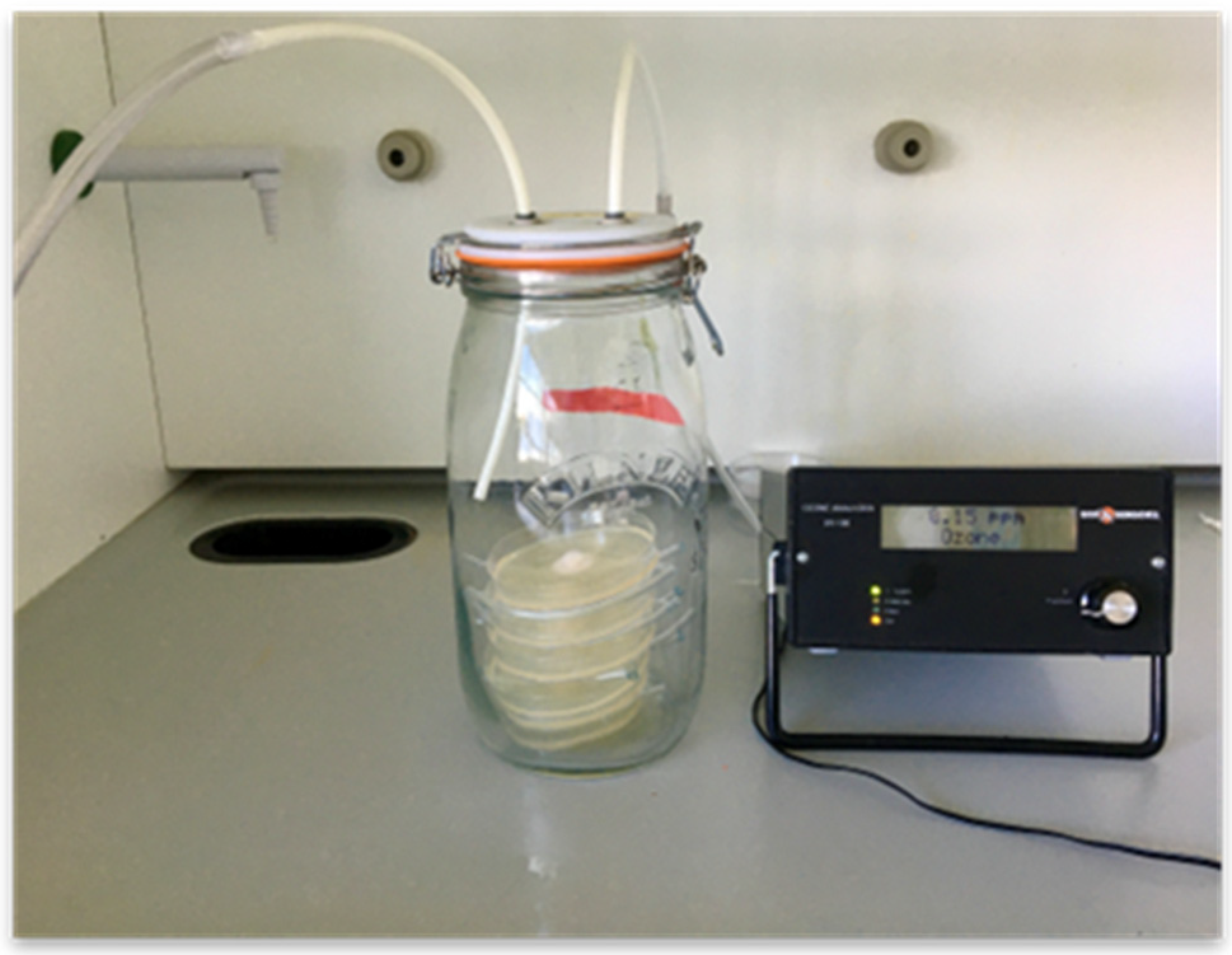
O3 was generated in the laboratory using a C-Lasky series O3 generator purchased from AirTree Ozone Technology Co. (model CL010DS, Sijhih, Taiwan; Figure 2). This equipment generates O3 by corona discharge between two tubs, with no metals involved for efficiency improvement, generation stability and lower energy consumption. The generated O3 was directed into the exposure chamber using a Teflon tube, which was properly connected to the generator. For safety reasons, the experiment was carried out in a fume cupboard to prevent O3 from spreading into the laboratory atmosphere. The O3 concentration was measured using an O3 analyzer (Model UV-100, Eco Sensor, Santa Fe, NM, USA), which was connected to the chamber to measure the exit gas accurately. It should be noted that 1 mg·kg−1 of O3 generated is equivalent to 2.14 mg·L−1 of O3 in the air. This allows for comparison with some other studies.
The in vitro exposure system of O3 for germination and mycelial growth assays was a 5-L airtight glass jar. The O3 inlet of the system was connected from the generator to the lid of the jar using a Teflon tube, which was inserted into the bottom of the jar. The outlet of the system was also located in the lid of the jar and connected to the O3 analyzer using another Teflon tube. This ensured accurate measurement of the O3 concentrations in the glass container. The flow rate of the generated gaseous O3 used was 6 L·min−1 and the treatments used were 0, 50, 100 and 200 mg·kg−1 for 15, 30 and 60 min [40].
Garlic Medium (GM; 15 g agar (2%; Oxoid®), 2% of garlic powder, 92 mL of glycerol, 1 L of bi-distilled water) in 9 cm Petri plates was prepared for both spore germination and mycelial growth assays. For spore germination assays, the GM media were inoculated with 0.1 mL of conidial suspensions of either F. proliferatum MPVPG29 (FP; ITEM18687) (8.59 × 10⁶ conidia·mL−1) or F. oxysporum MPVPG152 (FO; ITEM 18686) (7.08 × 10⁶ conidia·mL−1). These two fungal strains were isolated by garlic cloves; their identification was confirmed by molecular identification [5]. They are stored in the fungal collection of DIPROVES (Department of Sustainable Crop Production, Università Cattolica del Sacro Cuore, code MPVP) and in the fungal collection of ISPA-CNR (Institute of Science of Food Production, National Research Centre, code ITEM; http://server.ispa.cnr.it/ITEM/Collection/ accessed on 28 October 2022).
The conidia were spread-plated over the surface with a surface-sterilized glass spreader. These were inserted and stacked into the glass chamber without lids and exposed to the gaseous O3 treatments at the concentrations and for the time periods detailed; the trial was managed in triplicate.
The Petri plates were incubated at 25 °C, and after 24 h and 48 h incubation, two 1 cm diameter discs from each plate were transferred with a surface-sterilized cork borer to a microscope slide, then stained with a drop of lactophenol/cotton blue. A coverslip was placed over the agar disc and 25 spores were randomly counted from each disc and replicate. The percentage of spore germination was computed according to the following formula:
Spore germination (%) = n. germinated spores/25 × 100
For mycelial growth, the GM media were centrally inoculated with a 5 mm mycelial disc from the margin of a colony of either F. proliferatum MPVPG29 (FP) or F. oxysporum MPVPG152 (FO). The fungal species were exposed to different treatments and the radial growth of the pathogen was measured after incubation at 25 °C for 24 and 48 h. In all cases, comparisons were made with untreated controls.
  • (b) In vivo trial
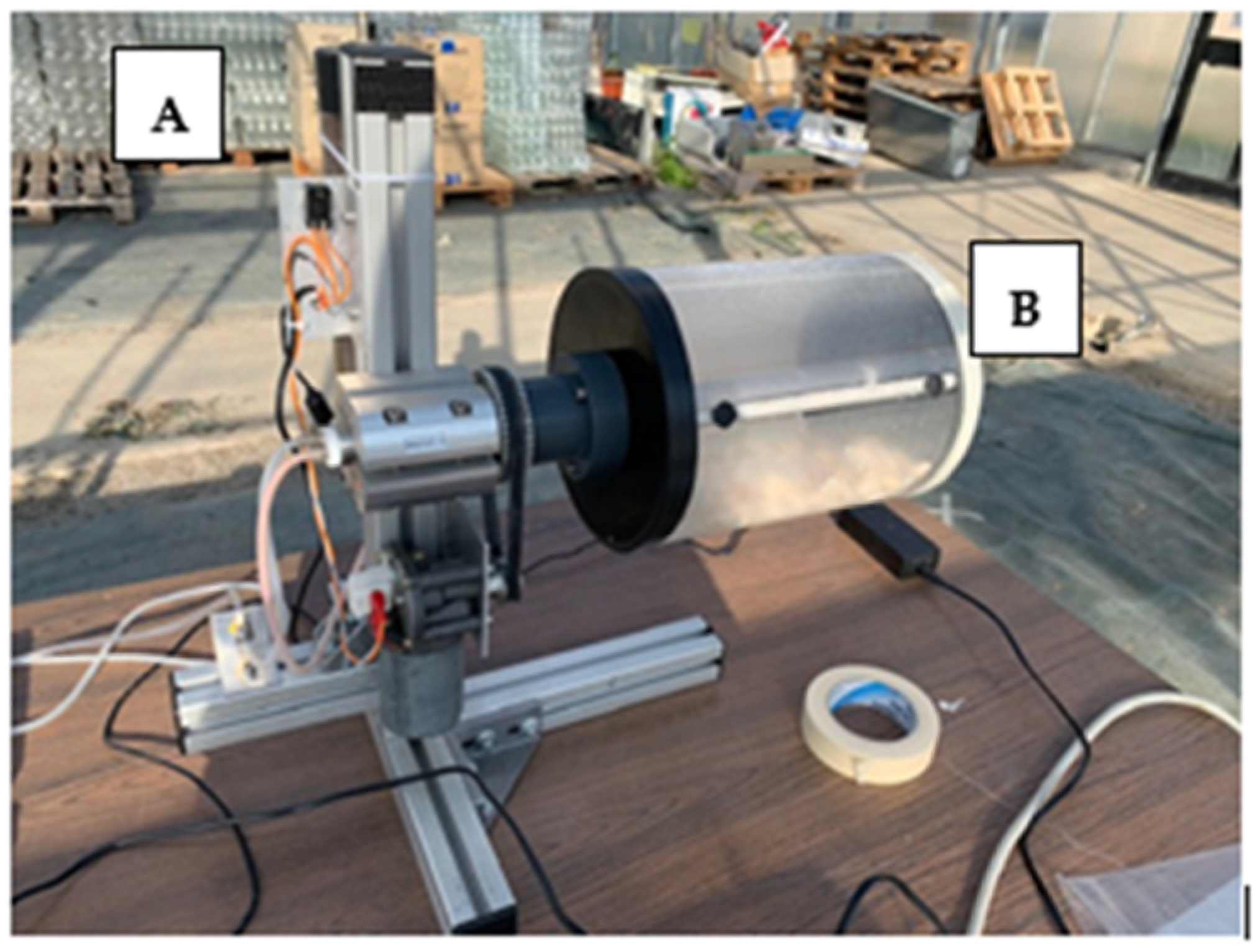
The system consisted of a prototype O3 generation machine that delivered different concentrations of the gas in a cylinder in which the garlic bulbs were placed (Figure 3). The cylinder rotated slowly to ensure a uniform exposure of the product to O3. The gas concentration used in the cylinder was 500 mg·kg−1, which stayed constant while the O3 generator was active. This decreased rapidly when switched off.
Garlic cloves of both defined categories (first and second) were placed in the rotary cylindrical chamber of the machine, subjected to the O3 exposure treatment and left in the machine for different time periods up to 60 min. Untreated cloves were used as the control. Trials were carried out with three replicates per treatment. Trials were done on two different dates; thus, two untreated control cloves were included.

Fungal Populations of The Garlic Cloves Treated with Gaseous O3

A total of 10 cloves per treatment were used for evaluating the effect of the gaseous O3 treatments on the fungal populations (CFUs·g−1) in garlic bulbs. The garlic bulbs were weighed and cut into small pieces with a surface-sterilized knife. Serial dilutions (10−2–10−5) were made using the spread plate method with 0.1 mL of the diluent onto Modified Nash and Snyder’s Medium (MNSM; 20 g of agar (2%; Oxoid®), 1 g of KH2PO4, 0.5 g·L−1 of MgSO4 × 7 H2O, 15 g of peptone, 1 g of pentachloronitrobenzene (PCNB), 0.3 g of streptomycin sulfate, 0.12 g of neomycin sulfate, 1 L of bi-distilled water [41]). The Petri plates were incubated at 25 °C for 7 days with a 12 h photoperiod. At the end of incubation, colonies were counted with a colony counter (Sibata, CL-570, Saitama, Japan). Both the total fungal CFUs·g−1, and the Fusarium spp. CFUs·g−1 were quantified.

Garlic Clove Germinative Capacity

Seed germination tests were done in germination chambers consisting of an aluminum tray with a sterile paper layer on the bottom to which 30 mL of bi-distilled water was added. 10 treated cloves were included in each replicate. These were incubated at 15 °C for 20 days in the dark before counting the number of germinated cloves.

2.2.2. Heat Treatment

The use of dry heat in an oven (F.lli Galli, F-CELL 035, Fizzonasco, Italy) or steam were tested on the 2 categories of garlic cloves. The samples were exposed to temperatures ranging from 43 to 70 °C for different time periods of 10 to 180 min; untreated cloves were used as the controls. Two experiments were carried out; in the first one, both steam and dry heat were tested on garlic seeds, whereas in the second experiment, only dry heat was used. The total number of fungal populations remaining after treatment (CFUs·g−1) and the effect on seed germination were quantified as described previously.

2.2.3. Chemical Disinfectants

Three chemical disinfectants were examined: (a) hydrogen peroxide (H2O2), (b) peracetic acid (C2H4O3) and (c) D50 (a mixture of C2H4O3 5% and H2O2 20%). They were tested in vitro at three different concentrations of 0.1%, 0.3% and 0.5%. The experiments were carried out with 4 replicates per treatment.
Based on in vitro results, 0.3% of H2O2 and C2H4O3 were subsequently tested in in vivo trials. Two different application systems were used. These were either a spray treatment or direct immersion of the cloves for different time periods (0, 5, 10 min) in the chemical disinfectants. Untreated controls were included in the study; water was applied in the same proportion as for disinfectants.
In the in vivo trials, the fungal populations (CFUs·g−1) with dilutions from 10−1 to 10−4, and the germination tests were conducted as described previously.

2.3. Fusarium Occurrence and Location in Garlic Cloves

10 pieces of the outer tunics and 10 cloves were randomly sampled from 4 garlic bulbs. The cloves, washed for 15 min in running tap water, were surface disinfected for one minute in 1% NaOCl solution, rinsed three times in sterile distilled water, and dried in sterile conditions. Each clove was cut into 4 pieces (base, germ, upper and central portion) with a surface-sterilized scalpel (Figure 1b). Clove pieces and tunics were plated on Petri dishes filled with Water Agar (WA; 20 g of agar (2%; Oxoid®, Basingstoke, UK), 1 L of bi-distilled water) and incubated at 25 °C for 7 days with a 12 h photoperiod. At the end of incubation, growing colonies were transferred to dishes filled with Potato Dextrose Agar (PDA; 15 g of agar (2%; Oxoid®), 10 g of dextrose, 1 L of potato broth (200 g of potato·L−1 of water) to obtain pure cultures and observed, with the support of a light microscope (Nikon, Eclipse 50, Tokyo, Japan; magnification 500×) for their identification at the genus level, or at species level for Fusarium spp. [42]. Some isolates morphologically identified as F. proliferatum and F. oxysporum were confirmed by molecular analysis [5]. Fungal incidence (%), was assessed as the number of infected pieces of the total number of those plated, both as total fungi and Fusarium spp.
In a second study, 4 cloves were randomly sampled from 3 garlic bulbs (12 cloves in total). The cloves were sterilized, as previously described, and cut in half with a surface-sterilized scalpel. One half was observed through the stereo microscope (Leica Biosystems, Wild M10, Germany; magnification 80×), focusing on the germ. The other half was cut into 3 pieces (base, germ, and the remaining part of the clove) and examined as previously described.

2.4. Statistical Analysis

Spore germination (%), seed germination (%), PGI (%), fungi and Fusarium ssp. incidence (%) were arcsen transformed to homogenize means [43]. The analysis of variance (ANOVA) was applied to all data collected and Tukey’s test was used to compare means. The statistical package SPSS statistics was used for data analysis (ver. 27, SPSS Inc., Chicago, IL, USA, 2021).

3. Results

3.1. Gaseous Ozone

(a)
In vitro studies
O3 concentration, exposure and incubation time significantly affected spore germination (p < 0.01), whereas no statistical differences were detected between the fungal species (Table 1).
A significant decrease of spore germination was observed, from 97.0% to 32.3%, when comparing the untreated samples with the highest gas concentration (200 mg·kg−1). The 30- and 60-min exposure periods had a greater effect on spore germination than shorter (15 min) exposure where a decrease of only 26% was observed. There was an increase of the germination observed after 48 h incubation suggesting that the spores had recovered some germinative capacity between 24 and 48 h.
Regarding fungal growth, F. proliferatum reached a significantly larger diameter compared to F. oxysporum (3.4 cm vs. 3.1 cm; p < 0.01) (Table 1). This was probably related to a better adaptability to the GM medium. However, both 100 and 200 mg·kg−1 O3 significantly reduced the mycelial growth of the two species when compared to 0 and 50 mg·kg−1 (p < 0.01).
(b)
In vivo studies
Gaseous O3 treatment of garlic cloves did not significantly affect the CFUs·g−1 depending on clove categories. However, there were differences amongst the O3 treatments (p < 0.01; Table 2).
Treatments in which the garlic cloves were exposed to the maximum gas concentration for a longer period were more effective. In particular, the samples exposed for 20 min or those with 30/40 min exposure showed reductions of 91.9%, 92.6% and 96.1%, respectively. The interaction between treatments and clove quality categories were also significant (p < 0.05).
However, there was little effect on seed germination. Indeed, no statistical differences were detected between treatments and their untreated controls. Furthermore, more of the first-category seeds germinated than did second-category seeds (85.9% vs. 73.1%) (p < 0.05).

3.2. High Temperatures

In the first experiment, steam was found to be more effective than dry heat in reducing the fungal CFUs·g−1 (mean reduction of 77.2% vs. 65.8%, compared to the untreated control) (Table 3). The most effective treatments were those with steam at 49 °C for 30 min, 49 °C for 20 min and 46 °C for 60 min. They caused reductions of 92.0%, 86.5% and 76.7%, in CFUs·g−1, respectively. However, steam also significantly reduced seed germination (average reduction of 36.9%; p < 0.01).
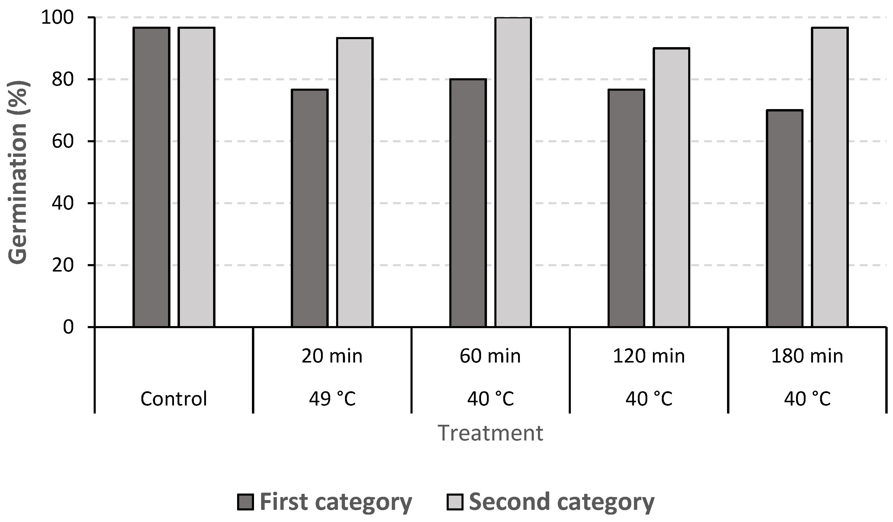
In the second experiment done with dry heat, the fungal CFUs·g−1 were not reduced compared to the untreated control. Again, treatments significantly affected seed germination (average reduction of 11.6%; p < 0.05) (Table 4). Regarding the two qualities of garlic cloves used, the first category germinated better than the second one (75.6% vs. 65.2%, respectively) (p < 0.01) (Table 4), whereas in the second experiment, the second category of garlic cloves germinated better than the first category (95.3% vs. 80.0%, respectively; p < 0.01) (Table 4).
In the second experiment, the interaction between treatments and clove quality categories was significant (p < 0.01), confirming that, in almost all the treatments, second-category cloves germinated better than first-category ones (Figure 4).

3.3. Chemical Disinfectants

(a)
In vitro trial
In this study, C2H4O3 and H2O2 were the most effective compounds in reducing pathogen growth (PGI = 70.4, 73.8%, respectively; Table 5).
Regarding concentrations, a significant increase of reducing pathogen development was observed ranging from 0.1% (PGI = 14.2%) to 0.3% (PGI = 73.4%) and 0.5% (PGI = 77.2%; p < 0.01).
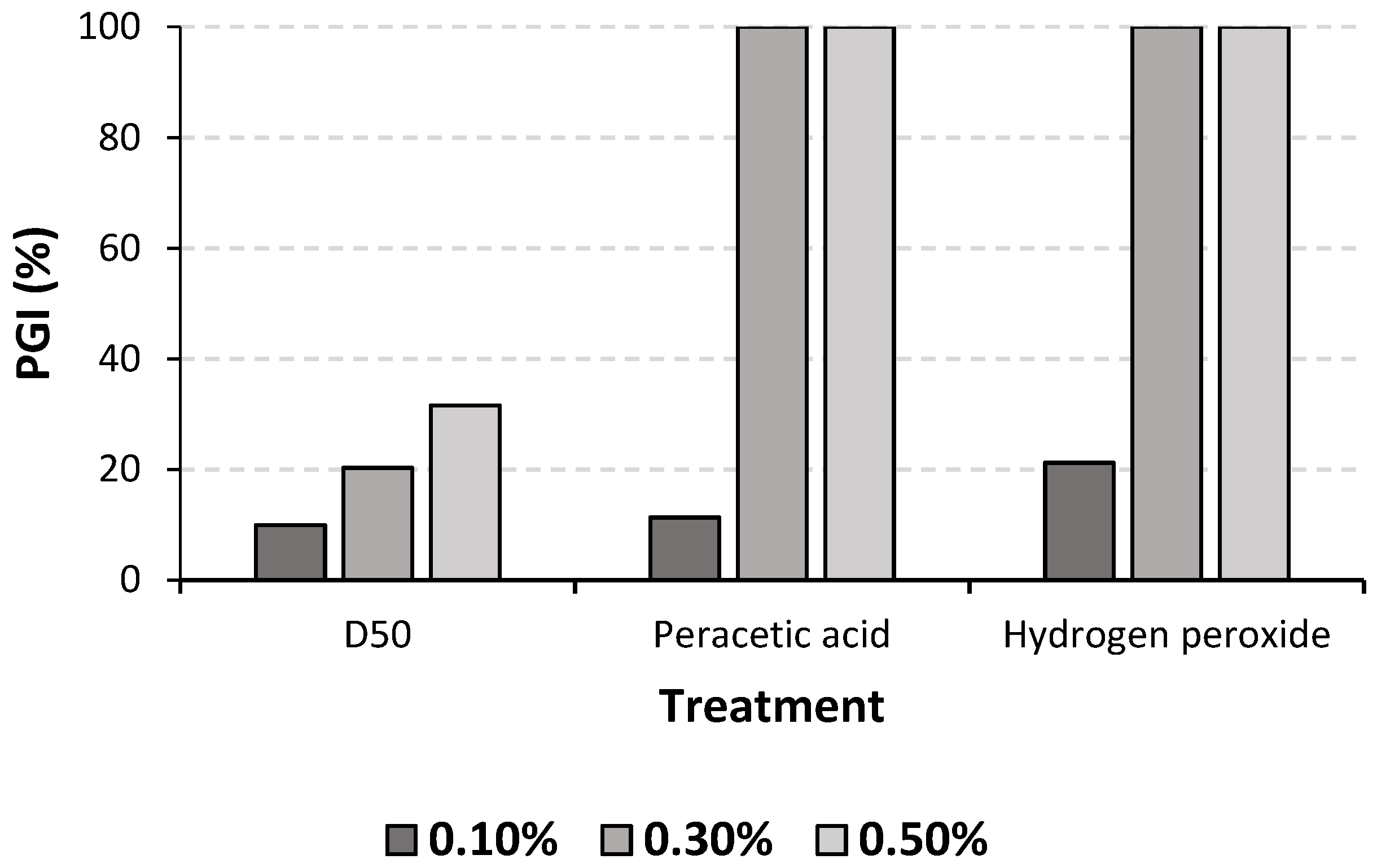
Figure 5 shows the interaction between treatments and concentrations. 0.3% of C2H4O3 and 0.3% of H2O2 completely inhibited the pathogen growth.
(b)
In vivo trial
A statistical difference was detected among treatments in terms of fungal CFUs·g−1 (p < 0.01) (Table 6). Immersion in 0.3% of C2H4O3 for 10 min resulted in the best performance with a decrease of 83.3% of fungal CFUs·g−1, when compared to the untreated control. However, it significantly affected seed germination (reduction of approx. 70%). Generally, all the treatments reduced seed germination compared to the untreated control; the average germination reduction caused by H2O2 was 34.0%, whereas that caused by C2H4O3 was 46.3% (p < 0.01).

3.4. Fusarium Occurrence in Garlic Cloves

The study aimed to identify the areas where fungi were present in the cloves. This showed that the outer tunics were always infected (100% incidence), and other inner parts of the cloves were also colonized, although with a minor incidence (Table 7). Sometimes, fungal mycelium was found in the inner germ (incidence 30%). F. proliferatum was the main detected species; however, F. oxysporum, Penicillium spp. and Alternaria spp. were also detected.
In the second trial, observation through the stereo microscope did not show traces of fungal contamination in the inner portion of the cloves. Nevertheless, the analysis carried out with the other half of the cloves showed that the germ was also infected, although with a lower incidence compared to the base and the remaining part of the cloves (17% vs. 67% and 83%, respectively), thus confirming the results obtained from the first trial. Additionally in this trial, F. proliferatum was the main detected species.

4. Discussion

Garlic dry rot is an emerging disease particularly noticeable during the post-harvest phase in garlic cloves, mainly attributed to F. proliferatum, which has caused significant economic losses. The role of garlic seeds as a source of inoculum was confirmed and sowing healthy seeds is considered fundamental in ensuring that the product has both a high yield and quality as well as good preservability [44]. Few fungicides are allowed for garlic seed treatments, but they are not very effective. It was recently confirmed the poor efficacy of chemical and biological fungicides applied at sowing as spray seed treatments [45]. Therefore, this study was carried out to reduce F. proliferatum incidence in garlic cloves with low impact interventions, while facilitating the maintenance of high levels of seed germination. To achieve this goal, three strategies were tested: heat, O3 and chemical disinfectants.
The growth and multiplication rate of Fusarium spp., as well as the development of diseases, are affected by temperature. Several studies have shown that 25 °C is a suitable temperature for Fusarium mycelial growth [46], while exposure to both low (15–20 °C) and high (30–35 °C) temperature ranges were found to inhibit mycelial development [47]. Mogensen, et al. [48] reported that no mycelial growth was observable at temperatures ≥40 °C for five strains of Fusarium species, including F. proliferatum, F. verticillioides and F. oxysporum. Thus, heat, applied mainly via water, air or vapor, can be an interesting zero-impact technique to control fungal diseases of garlic cloves [32,49,50]. However, temperature might influence the percentage and rate of seed germination. Seed germination is, in fact, a complex process involving many individual reactions and phases, each of which is affected by this parameter. Once seeds start to germinate, high temperatures stimulate faster germination up to an optimal point, after which, the speed of germination declines rapidly [51]. Indeed, in oilseed rape, Arabidopsis, wheat and rice, heat stress was found to affect seed performance, dormancy and seedling growth [35,36,37,52]. Similar results were also obtained by studies conducted on Lolium rigidum and sunflower seeds [53,54]. Therefore, for successful application of heat treatments, pre-tests using germination assays are needed to determine the optimal temperature-time combination for a given batch of seeds that does not affect its germination. The present study has thus investigated both steam and dry heat on garlic Fusarium strains and on seed germination. Steam provided the best control of the pathogen, but it also affected seed germination. In contrast, dry heat did not impact negatively on germination, but it had no significant effect on the reduction of Fusarium incidence. Thus, none of the treatments tested fully achieved the goal of the study. Nevertheless, to have a more complete overview of the effect of high temperatures on garlic seeds, further combinations of temperature and time should be considered.
Similar results were obtained with H2O2 and C2H4O3. H2O2 produces free radicals, which cause oxidative damage to proteins and membrane lipids of pathogens. Similarly, C2H4O3 also denatures proteins and disrupt cell wall permeability. Thus, these two chemical disinfectants may be effective against a wide range of microorganisms [55,56,57,58]. In the present study, both H2O2 and C2H4O3 were applied to Fusarium-infected garlic seeds at a concentration of 0.3% by two different application systems, spraying and immersion. This choice was based on the results of preliminary in vitro assays. Some treatments were successful in reducing Fusarium growth immersion in C2H4O3 for 10 min. On the other hand, they strongly affected seed germination. Therefore, these products are not fully suitable for garlic seed treatments.
Amongst all the physical treatments examined, the most promising approach was found to be the use of gaseous O3, a powerful antimicrobial agent, which is currently used as a disinfectant for microorganisms. Its antimicrobial activity is based on its oxidizing effect, which causes damage to the fatty acids in the cell membrane and to proteins and DNA [59]. The application of gaseous O3 to Fusarium-infected garlic cloves in the present study reduced the pathogen growth and conserved high levels of seed germination, similar to the untreated samples. Treatments in which cloves were exposed to the maximum gas concentration for a longer period performed the best. Therefore, gaseous O3 is a promising approach for the disinfection of garlic seeds, which can be used as a safe and green technology for the control of the disease. O3 was previously effectively used to control Fusarium infections in germinated barley and whole wheat grains [60,61,62]. Furthermore, it reduced the growth of many other fungal species in wheat, barley and pea seeds [38], and mycotoxin contamination in different crops, such as peanuts, figs and Brazil nuts [63,64,65,66,67,68]. One previous study on the efficacy of O3 on garlic post-harvest found a notable reduction of garlic decay caused by F. proliferatum, while at the same time, conserving the sensory profile [69]. Its potential and the possible combination with other actions should be explored in more depth.

5. Conclusions

In this study, several methods to reduce F. proliferatum incidence in garlic cloves as preventive action for preserving garlic from dry rot were tested, favoring low-impact strategies. H2O2 and C2H4O3 showed a marginal effect in garlic dry rot resolution, whereas other disinfection actions, particularly the use of gaseous O3, gave encouraging results for the control of Fusarium infection and germinative capacity. This represents a good strategy for garlic protection from seed-borne pathogens, which can improve both stand quality and yields. However, the treatments used were not able to eradicate the fungus completely, probably because of the internal location, which made it more difficult for treatments to be effective. Further studies should be conducted to refine these techniques and to establish their practicality at the industrial level. The approaches suggested could also be useful for garlic destined for consumption, where the effects on seed germination are less important.

Author Contributions

Conceptualization, P.B. and N.M.; methodology, P.B., L.L.P. and P.G.; formal analysis, L.L.P., L.M. and P.G.; data curation, L.L.P.; writing—original draft preparation, P.B. and L.L.P.; writing—review and editing, P.B., P.G. and N.M.; funding acquisition, P.B. All authors have read and agreed to the published version of the manuscript.

Funding

The research was funded by the PSR program 16.1.01 “Gruppi operativi del PEI per la produttività e le sostenibilità dell’agricoltura” Sottomisura 16.1 of Emilia Romagna region, Focus Area 3A. Project N. 5116572 “Organisation and sustainable process innovation for the healthy seed production chain of white garlic from Piacenza”.

Data Availability Statement

Data are available on request to the authors.

Conflicts of Interest

The authors declare no conflict of interest.

References

  1. Elshahawy, I.E.; Saied, N.M.; Morsy, A.A. Fusarium proliferatum, the main cause of clove rot during storage, reduces clove germination and causes wilt of established garlic plants. J. Plant. Pathol. 2017, 99, 85–93. [Google Scholar] [CrossRef]
  2. Horáková, M.K.; Tancik, J.; Barta, M. Fusarium proliferatum causing dry rot of stored garlic in Slovakia. J. Plant. Pathol. 2021, 103, 997–1002. [Google Scholar] [CrossRef]
  3. Patón, L.G.; Marrero, M.; Llamas, D.P. In vitro and field efficacy of three fungicides against Fusarium bulb rot of garlic. Eur. J. Plant Pathol. 2016, 148, 321–328. [Google Scholar] [CrossRef]
  4. FAO-STAT. Available online: https://www.fao.org/faostat/en/#data/QCL (accessed on 28 October 2022).
  5. Mondani, L.; Chiusa, G.; Battilani, P. Fungi associated with garlic during the cropping season, with focus on Fusarium proliferatum and F. oxysporum. Plant Health Prog. 2021, 22, 37–46. [Google Scholar] [CrossRef]
  6. Seefelder, W.; Gossmann, M.; Humpf, H.U. Analysis of fumonisin B(1) in Fusarium proliferatum-infected asparagus spears and garlic bulbs from Germany by liquid chromatography-electrospray ionization mass spectrometry. J. Agric. Food Chem. 2002, 50, 2778–2781. [Google Scholar] [CrossRef] [PubMed]
  7. Dugan, F.M.; Hellier, B.C.; Lupien, S.L. First report of Fusarium proliferatum causing rot of garlic bulbs in North America. Plant Pathol. 2003, 52, 426. [Google Scholar] [CrossRef]
  8. Stankovic, S.; Lević, J.; Petrović, T.; Logrieco, A.F.; Moretti, A. Pathogenicity and mycotoxin production by Fusarium proliferatum isolated from onion and garlic in Serbia. Eur. J. Plant Pathol. 2007, 118, 165–172. [Google Scholar] [CrossRef]
  9. Palmero, D.; De Cara, M.; Iglesias, C.; Moreno, M.M.; González, N.; Tello, J.C. First report of Fusarium proliferatum causing rot of garlic bulbs in Spain. Plant Dis. 2010, 94, 277. [Google Scholar] [CrossRef] [PubMed]
  10. Sankar, N.R.; Babu, G.P. First report of Fusarium proliferatum causing rot of garlic bulbs (Allium sativum) in India. Plant Dis. 2012, 96, 290. [Google Scholar] [CrossRef] [PubMed]
  11. Fuentes, Y.M.O.; Ortiz, J.C.D.; Chávez, E.C.; Castillo, F.D.H.; Olivas, A.F.; Morales, G.G.; Martinez, O.; Guerra, R.R. The first report of Fusarium proliferatum causing garlic bulb rots in Mexico. Afr. J. Agric. Res. 2013, 8, 570–573. [Google Scholar] [CrossRef]
  12. Salvalaggio, A.E.; Ridao, A.D.C. First report of Fusarium proliferatum causing rot on garlic and onion in Argentina. Plant Dis. 2013, 97, 556. [Google Scholar] [CrossRef]
  13. Moharam, M.H.A.; Farrag, E.S.H.; Mohamed, M.D.A. Pathogenic fungi in garlic seed cloves and first report of Fusarium proliferatum causing cloves rot of stored bulbs in upper Egypt. Arch. Phytopathol. 2013, 46, 2096–2103. [Google Scholar] [CrossRef]
  14. Leyronas, C.; Chretien, P.L.; Troulet, C.; Duffaud, M.; Villeneuve, F.; Morris, C.E.; Hunyadi, H. First report of Fusarium proliferatum causing garlic clove rot in France. Plant Dis. 2018, 102, 2658. [Google Scholar] [CrossRef]
  15. Tonti, S.; Mandrioli, M.; Nipoti, P.; Pisi, A.; Toschi, T.G.; Prodi, A. Detection of fumonisins in fresh and dehydrated commercial garlic. J. Agric. Food Chem. 2017, 65, 7000–7005. [Google Scholar] [CrossRef] [PubMed]
  16. Tonti, S.; Prà, M.D.; Nipoti, P.; Prodi, A.; Alberti, I. First report of Fusarium proliferatum causing rot of stored garlic bulbs (Allium sativum L.) in Italy. Phytopathology 2012, 160, 761–763. [Google Scholar] [CrossRef]
  17. Logrieco, A.F.; Moretti, A.; Ritieni, A.; Bottalico, A.; Corda, P. Occurrence and toxigenicity of Fusarium proliferatum from preharvest maize ear rot, and associated mycotoxins, in Italy. Plant Dis. 1995, 79, 727–731. [Google Scholar] [CrossRef]
  18. Desjardins, A.E.; Plattner, R.D.; Nelson, P.E. Production of fumonisin B(inf1) and moniliformin by Gibberella fujikuroi from rice from various geographic areas. Appl. Environ. Microbiol. 1997, 63, 1838–1842. [Google Scholar] [CrossRef] [PubMed] [Green Version]
  19. Abdalla, M.Y.; Al-Rokibah, A.; Moretti, A.; Mulè, G. Pathogenicity of toxigenic Fusarium proliferatum from date palm in Saudi Arabia. Plant Dis. 2000, 84, 321–324. [Google Scholar] [CrossRef] [Green Version]
  20. Armengol, J.; Moretti, A.; Perrone, G.; Vicent, A.; Bengoechea, J.; García-Jiménez, J. Identification, incidence and characterization of Fusarium proliferatum on ornamental palms in Spain. Eur. J. Plant Pathol. 2005, 112, 123–131. [Google Scholar] [CrossRef]
  21. Marasas, W.F.O.; Thiel, P.G.; Rabie, C.J.; Nelson, P.E.; Toussoun, T.A. Moniliformin production in Fusarium section Liseola. Mycologia 1986, 78, 242–247. [Google Scholar] [CrossRef]
  22. Logrieco, A.; Moretti, A.; Castella, G.; Kostecki, M.; Golinski, P.; Ritieni, A.; Chelkowski, J. Beauvericin production by Fusarium species. Appl. Environ. Microbiol. 1998, 64, 3084–3088. [Google Scholar] [CrossRef] [PubMed] [Green Version]
  23. Plattner, R.D.; Nelson, P.E. Production of beauvericin by a strain of Fusarium proliferatum isolated from corn fodder for swine. Appl. Environ. Microbiol. 1994, 60, 3894–3896. [Google Scholar] [CrossRef] [PubMed] [Green Version]
  24. Bacon, C.W.; Porter, J.K.; Norred, W.P.; Leslie, J.F. Production of fusaric acid by Fusarium species. Appl. Environ. Microbiol. 1996, 62, 4039–4043. [Google Scholar] [CrossRef] [PubMed] [Green Version]
  25. Ritieni, A.; Fogliano, V.; Randazzo, G.; Scarallo, A.; Logrieco, A.; Moretti, A.; Mannina, L.; Bottalico, A. Isolation and characterization of fusaproliferin, a new toxic metabolite from Fusarium proliferatum. Nat. Toxins 1995, 3, 17–20. [Google Scholar] [CrossRef] [PubMed]
  26. Kellerman, T.S.; Marasas, W.F.O.; Thiel, P.G.; Gelderblom, W.C.A.; Cawood, M.; Coetzer, J.A.W. Leukoencephalomalacia in two horses induced by oral dosing of fumonisin B1. Onderstepoort J. Vet. Res. 1990, 57, 269–275. [Google Scholar] [PubMed]
  27. Harrison, L.R.; Colvin, B.M.; Greene, J.T.; Newman, L.E.; Cole, J.R., Jr. Pulmonary edema and hydrothorax in swine produced by fumonisin B1, a toxic metabolite of Fusarium moniliforme. J. Vet. Diagn. Invest. 1990, 2, 217–221. [Google Scholar] [CrossRef] [Green Version]
  28. Pfohl-Leszkowicz, A.; Manderville, R.A. Ochratoxin A: An overview on toxicity and carcinogenicity in animals and humans. Mol. Nutr. Food Res. 2007, 51, 61–99. [Google Scholar] [CrossRef]
  29. Dugan, F.M.; Lupien, S.L.; Hellier, B.C. Infection by Fusarium proliferatum in aerial garlic bulbils is strongly reduced compared to rates in seed cloves when both originate from infected bulbs. Crop Prot. 2019, 116, 43–48. [Google Scholar] [CrossRef]
  30. Mondani, L.; Chiusa, G.; Battilani, P. Chemical and biological control of Fusarium species involved in garlic dry rot at early crop stages. Eur. J. Plant Pathol. 2021, 160, 575–587. [Google Scholar] [CrossRef]
  31. Mancini, V.; Romanazzi, G. Seed treatments to control seedborne fungal pathogens of vegetable crops. Pest Manag. 2014, 70, 860–868. [Google Scholar] [CrossRef]
  32. Landa, B.B.; Navas-Cortés, J.A.; Del Mar Jiménez-Gasco, M.; Katan, J.; Retig, B.; Jiménez-Díaz, R.M. Temperature response of chickpea cultivars to races of Fusarium oxysporum f. sp. ciceris, causal agent of Fusarium wilt. Plant Dis. 2006, 90, 365–374. [Google Scholar] [CrossRef] [Green Version]
  33. Schöneberg, T.; Kibler, K.; Wettstein, F.E.; Bucheli, T.D.; Forrer, H.R.; Musa, T.; Mascher, F.; Bertossa, M.; Keller, B.; Vogelgsang, S. Influence of temperature, humidity duration and growth stage on the infection and mycotoxin production by Fusarium langsethiae and Fusarium poae in oats. Plant Pathol. 2019, 68, 173–184. [Google Scholar] [CrossRef] [Green Version]
  34. Samapundo, S.; Devliehgere, F.; De Meulenaer, B.; Debevere, J. Effect of water activity and temperature on growth and the relationship between fumonisin production and the radial growth of Fusarium verticillioides and Fusarium proliferatum on corn. J. Food Prot. 2005, 68, 1054–1059. [Google Scholar] [CrossRef] [PubMed]
  35. He, H.; de Souza Vidigal, D.; Snoek, L.B.; Schnabel, S.; Nijveen, H.; Hilhorst, H.; Bentsink, L. Interaction between parental environment and genotype affects plant and seed performance in Arabidopsis. J. Exp. Bot. 2014, 65, 6603–6615. [Google Scholar] [CrossRef] [PubMed] [Green Version]
  36. Walker-Simmons, M.K.; Sesing, J. Temperature effects on embryonic abscisic acid levels during development of wheat grain dormancy. J. Plant Growth Regul. 2005, 9, 51–56. [Google Scholar] [CrossRef]
  37. Begcy, K.; Sandhu, J.; Walia, H. Transient heat stress during early seed development primes germination and seedling establishment in rice. Front. Plant Sci. 2018, 9, 1768. [Google Scholar] [CrossRef]
  38. Ciccarese, F.; Sasanelli, N.; Ciccarese, A.; Ziadi, T.; Ambrico, A.; Mancini, L. Seed disinfestation by ozone treatments. In Proceedings of the IOA Conference and Exhibition, Valencia, Spain, 29–31 October 2007; pp. 1–8. [Google Scholar]
  39. Mylona, K.; Kogkaki, E.A.; Sulyok, M.; Magan, N. Efficacy of gaseous ozone treatment on spore germination, growth and fumonisin production by Fusarium verticillioides in vitro and in situ in maize. J. Stored Prod. Res. 2014, 59, 178–184. [Google Scholar] [CrossRef]
  40. Baazeem, A.; Medina, A.; Magan, N. Impacts of gaseous ozone (O3) on germination, mycelial growth, and aflatoxin B1 production in vitro and in situ contamination of stored pistachio nuts. Toxins 2022, 14, 416. [Google Scholar] [CrossRef]
  41. Nash, S.M.; Snyder, W.C. Quantitative estimations by plate counts of propagules of the bean root rot Fusarium in field soils. Phytopathology 1962, 52, 567–572. [Google Scholar]
  42. Leslie, J.F.; Summerell, B.A. The Fusarium Laboratory Manual; Blackwell Publishing: Hoboken, NJ, USA, 2006; p. 416. [Google Scholar]
  43. Clewer, A.G.; Scarisbrick, D.H. Practical Statistics and Experimental Design for Plant and Crop Science; Huxley, T.H., Ed.; John Wiley & Sons: Chichester, UK, 2001. [Google Scholar]
  44. Mondani, M.; Chiusa, G.; Pietri, A.; Battilani, P. Monitoring the incidence of dry rot caused by Fusarium proliferatum in garlic at harvest and during storage. Postharvest Biol. Technol. 2021, 173, 111407. [Google Scholar] [CrossRef]
  45. Mondani, L.; Chiusa, G.; Battilani, P. Efficacy of chemical and biological spray seed treatments in preventing garlic dry rot. Phytopathol. Mediterr. 2022, 61, 27–37. [Google Scholar] [CrossRef]
  46. Cendoya, E.; Pinson-Gadais, L.; Farnochi, M.C.; Ramírez, M.L.; Chéreau, S.; Marcheguay, G.; Ducos, C.; Barreau, C.; Richard-Forget, F. Abiotic conditions leading to FUM gene expression and fumonisin accumulation by Fusarium proliferatum strains grown on a wheat-based substrate. Int. J. Food Microbiol. 2017, 253, 12–19. [Google Scholar] [CrossRef] [PubMed]
  47. Saleh, A.A.; Sharafaddin, A.H.; El Komy, M.H.; Ibrahim, Y.E.; Hamad, Y.K. Molecular and physiological characterization of Fusarium strains associated with different diseases in date palm. PLoS ONE 2021, 16, e0254170. [Google Scholar] [CrossRef] [PubMed]
  48. Mogensen, J.M.; Nielsen, K.F.; Samson, R.A.; Frisvad, J.C.; Thrane, U. Effect of temperature and water activity on the production of fumonisins by Aspergillus niger and different Fusarium species. BMC Microbiol. 2009, 9, 281. [Google Scholar] [CrossRef] [PubMed] [Green Version]
  49. Suárez-Estrella, F.; Vargas-García, M.C.; Elorrieta, M.A.; López, M.J.; Moreno, J. Temperature effect on Fusarium oxysporum f.sp. melonis survival during horticultural waste composting. J. Appl. Microbiol. 2003, 94, 475–482. [Google Scholar] [CrossRef]
  50. Marín, S.; Sanchis, V.; Magan, N. Water activity, temperature, and pH effects on growth of Fusarium moniliforme and Fusarium proliferatum isolates from maize. Can. J. Microbiol. 1995, 41, 1063–1070. [Google Scholar] [CrossRef]
  51. Roberts, E.H. Temperature and seed germination. Symp. Soc. Exp. Biol. 1988, 42, 109–132. [Google Scholar]
  52. Brunel-Muguet, S.; D’Hooghe, P.; Bataillé, M.P.; Larré, C.; Kim, T.H.; Trouverie, J.; Avice, J.C.; Etienne, P.; Dürr, C. Heat stress during seed filling interferes with sulfur restriction on grain composition and seed germination in oilseed rape (Brassica napus L.). Front. Plant Sci. 2015, 6, 213. [Google Scholar] [CrossRef] [Green Version]
  53. Steadman, K.J. Dormancy release during hydrated storage in Lolium rigidum seeds is dependent on temperature, light quality, and hydration status. J. Exp. Bot. 2004, 55, 929–937. [Google Scholar] [CrossRef]
  54. Bazin, J.; Batlla, D.; Dussert, S.; El-Maarouf-Bouteau, H.; Bailly, C. Role of relative humidity, temperature, and water status in dormancy alleviation of sunflower seeds during dry after-ripening. J. Exp. Bot. 2011, 62, 627–640. [Google Scholar] [CrossRef]
  55. Baldry, M.G. The bactericidal, fungicidal and sporicidal properties of hydrogen peroxide and peracetic acid. J. Appl. Microbiol. 1983, 54, 417–423. [Google Scholar] [CrossRef] [PubMed]
  56. Kitis, M. Disinfection of wastewater with peracetic acid: A review. Environ. Int. 2004, 30, 47–55. [Google Scholar] [CrossRef] [PubMed]
  57. Ayoub, F.; Ben Oujji, N.; Chebli, B.; Ayoub, M.; Hafidi, A.; Salghi, R.; Jodeh, S. Antifungal effectiveness of fungicide and peroxyacetic acid mixture on the growth of Botrytis cinerea. Microb. Pathog. 2017, 105, 74–80. [Google Scholar] [CrossRef] [PubMed]
  58. Soares, C.E.S.; Cartabiano-Leite, C.E.; Ferreira, W.X.; Maiorka, A.; Dahlke, F.; De Dea Lindner, J. Peracetic acid: Effect on the chicken eggshell cuticle and decontaminating action on filamentous fungi. Jokull J. 2021, 71, 82–96. [Google Scholar]
  59. Prabha, V.M.D.; Barma, R.D.; Singh, R.; Madan, A. Ozone technology in food processing: A review. Trends Biosci. 2015, 8, 4031–4047. [Google Scholar]
  60. Dodd, J.G.; Vegi, A.; Vashisht, A.; Tobias, D.; Schwarz, P.; Wolf-Hall, C.E. Effect of ozone treatment on the safety and quality of malting barley. J. Food Prot. 2011, 74, 2134–2141. [Google Scholar] [CrossRef] [PubMed]
  61. Piacentini, K.C.; Savi, G.D.; Scussel, V.M. The effect of ozone treatment on species of Fusarium growth in malting barley (Hordeum vulgare L.) grains. Qual. Assur. 2017, 9, 383–389. [Google Scholar] [CrossRef]
  62. Savi, G.D.; Piacentini, K.C.; Bittencourt, K.O.; Scussel, V.M. Ozone treatment efficiency on Fusarium graminearum and deoxynivalenol degradation and its effects on whole wheat grains (Triticum aestivum L.) quality and germination. J. Stored Prod. Res. 2014, 59, 245–253. [Google Scholar] [CrossRef]
  63. Dwarakanath, C.T.; Rayner, E.T.; Mann, G.E.; Dollear, F.G. Reduction of aflatoxin levels in cottonseed and peanut meals by ozonization. J. Am. Oil Chem. Soc. 1968, 45, 93–95. [Google Scholar] [CrossRef] [PubMed]
  64. Zorlugenç, B.; Kiroğlu Zorlugenç, F.; Oztekin, S.; Evliya, I.B. The influence of gaseous ozone and ozonated water on microbial flora and degradation of aflatoxin B(1) in dried figs. Food Chem. Toxicol. 2008, 46, 3593–3597. [Google Scholar] [CrossRef]
  65. Scussel, V.M.; Giordano, B.N.; Simao, V.; Manfio, D.; Galvao, S.; Rodrigues, M.N. Effect of oxygen-reducing atmospheres on the safety of packaged shelled brazil nuts during storage. Int. J. Anal. Chem. 2011, 2011, 813591. [Google Scholar] [CrossRef] [PubMed] [Green Version]
  66. Cataldo, F. Ozone decomposition of patulin—A micotoxin and food contaminant. Ozone Sci. Eng. 2008, 30, 197–201. [Google Scholar] [CrossRef]
  67. Karaca, H.; Velioglu, Y.S.; Nas, S. Mycotoxins: Contamination of dried fruits and degradation by ozone. Toxin Rev. 2010, 29, 51–59. [Google Scholar] [CrossRef]
  68. Karaca, H.; Sedat Velioglu, Y. Effects of some metals and chelating agents on patulin degradation by ozone. Ozone Sci. Eng. 2009, 31, 224–231. [Google Scholar] [CrossRef]
  69. De Santis, D.; Garzoli, S.; Vettraino, A.M. Effect of gaseous ozone treatment on the aroma and clove rot by Fusarium proliferatum during garlic postharvest storage. Heliyon 2021, 7, e06634. [Google Scholar] [CrossRef] [PubMed]
Figure 1. (A) Clove quality categories: first category cloves length >15 mm; second category clove lenght 10–15 mm; (B) garlic cloves sections considered in the different in vitro trials: A, base; B, central portion; C, upper portion; D, germ; E, outer tunics.
Horticulturae 08 01203 g001
Figure 2. Ozone meter and a hermetic pot in which Petri dishes were put during ozone treatments in in vitro trials.
Horticulturae 08 01203 g002
Figure 3. Prototype machine used in in vivo trials with ozone. (A) Ozone generator; (B) Treatment chamber, a cylinder where the ozone is delivered.
Horticulturae 08 01203 g003
Figure 4. Seed Germination (%) of first or second clove category (first category cloves length >15 mm; second category 10–15 mm clove length), tested in response to dry heat, during the second experiment with high temperatures.
Horticulturae 08 01203 g004
Figure 5. PGI (%) of F. proliferatum in PDA plates with three chemical disinfectants (D50, peracetic acid and hydrogen peroxide) at three concentrations (0.10%, 0.30% and 0.50%).
Horticulturae 08 01203 g005
Table 1. Analysis of variance of spore germination (%) and radial growth for the two Fusarium species tested on GM plates at the end of the in vitro trial with ozone. Plates were exposed to different ozone concentrations (0, 50, 100 and 200 mg·kg−1) for 15, 30 or 60 min and then incubated for 0, 24 or 48 h. Different letters indicate significant differences according to Tukey’s test (p < 0.01).
FactorsSpore Germination (%)Radial Growth (cm)
1.
Fungal species
n.s.**
F. oxysporum MPVPG152 (FO)56.53.1 a
F. proliferatum MPVPG29 (FP)51.73.4 b
2.
Ozone concentration (mg·kg−1)
****
097.0 c3.3 b
5046.1 b3.3 b
10040.9 b3.2 a
20032.3 a3.2 a
3.
Exposure time (min)
****
1565.6 b3.3 c
3051.7 a3.2 b
6045.0 a3.1 a
4.
Incubation time (h)
****
0 2.4 a
2433.8 a3.3 b
4874.4 b4.1 c
Interaction
A × B**
A × Cn.s.*
A × Dn.s.n.s.
B × C**n.s.
B × D****
C × Dn.s.**
A × B × Cn.s.**
A × B × Dn.s.**
A × C × Dn.s.n.s.
B × C × Dn.s.*
A × B × C × Dn.s.n.s.
** p < 0.01, * p < 0.05, n.s. = not significant.
Table 2. Analysis of variance of CFU·g−1 and seed germination (%) at the end of the in vivo trial with ozone. Ozone treatments were applied to garlic cloves divided in the two quality categories (first category cloves length > 15 mm, second category 10–15 mm clove length). Different letters indicate significant differences according to Tukey’s test (p < 0.01).
FactorsOzone Production Time (min)Exposure Time without Ozone Production (min)CFU·g−1Seed Germination (%)Seed Germination Variation (%)
A.
Treatment
***
1 (Control 1) 1.8 × 10³ ab88.3 ab
22.5105.5 × 102 abc88.3 ab0.0
32.5301.4 × 102 bc90.0 ab+1.9
42.5606.9 × 102 abc90.0 ab+1.9
55102.7 × 102 abc93.3 a+5.7
65302.0 × 102 cd93.3 a+5.7
71057.9 × 102 bc96.7 a+9.4
810102.3 × 102 cd88.3 ab0.0
910301.4 × 100 e73.3 abc−17.0
1010403.2 × 101 de76.7 abc−13.2
112002.3 × 101 de80.0 abc−9.4
12 (Control 2) 7.5 × 10³ a58.3 bc
133001.3 × 100 e50.0 c−14.3
144005.1 × 10−1 e46.7 c−20.0
B.
Seed category
n.s.*
First 4.0 × 10285.9 a
Second 1.3 × 10373.1 b
Interaction
A × B *n.s.
** p < 0.01, * p < 0.05, n.s. = not significant.
Table 3. Analysis of variance of CFU·g−1 and seed germination (%) at the end of the first experiment with high temperatures. Dry heat and steam were applied to garlic cloves divided in the two quality categories (first category cloves length > 15 mm, second category 10–15 mm clove length). Different letters indicate significant differences according to Tukey’s test (p < 0.01).
FactorsTemperature (°C)Time (min)CFU·g−1Seed Germination (%)Seed Germination Variation (%)
A.
Treatment
****
Control2504.2 × 103 c95.0 a
Dry heat49201.4 × 103 bc81.7 ab−14.0
Dry heat49301.5 × 103 bc65.0 b−31.6
Dry heat60151.4 × 103 bc70.0 b−26.3
Dry heat70101.4 × 103 bc78.3 ab−17.5
Steam43901.9 × 103 bc60.0 b−36.8
Steam46609.8 × 102 ab60.0 b−36.8
Steam49205.7 × 102 ab58.3 b−38.6
Steam49303.4 × 102 a65.0 b−31.6
B.
Seed category
n.s.**
First 1.2 × 10375.6 a
Second 1.8 × 10365.2 b
Interaction
A × B n.s.n.s.
** p < 0.01, n.s. = not significant.
Table 4. Analysis of variance of CFU·g−1 and seed germination (%) at the end of the second experiment with high temperatures. Dry heat was applied to garlic cloves divided in the two quality categories (first category cloves length > 15 mm, second category 10–15 mm clove length). Different letters indicate significant differences according to Tukey’s test (p < 0.01).
FactorsTemperature (°C)Time (min)CFU·g−1Seed Germination (%)Seed Germination Variation (%)
A.
Treatment
n.s.*
Control2501.2 × 10496.7 a
Dry heat49206.7 × 10485.0 b−12.1
Dry heat40604.7 × 10390.0 ab−6.9
Dry heat401208.2 × 10383.3 b−13.8
Dry heat401806.3 × 10483.3 b−13.8
B.
Seed category
n.s.**
First 5.3 × 10480.0 b
Second 9.4 × 10395.3 a
Interaction
A × B n.s.*
** p < 0.01, * p < 0.05, n.s. = not significant.
Table 5. Analysis of variance of PGI (%) at the end of the in vitro trial with chemical disinfectants. Three products (D50, peracetic acid and hydrogen peroxide) at three different concentrations (0.1%, 0.3% and 0.5%) were tested on inoculated PDA plates. Different letters indicate significant differences according to Tukey’s test (p < 0.01).
FactorsPGI (%)
A.
Treatment
**
D5020.6 b
Peracetic acid70.4 a
Hydrogen peroxide73.8 a
B.
Concentration (%)
**
0.114.2 b
0.373.4 a
0.577.2 a
Interaction
A × B**
** p < 0.01.
Table 6. Analysis of variance of CFU·g−1 and seed germination (%) at the end of the in vivo trial with chemical disinfectants. 0.3% of hydrogen peroxide or 0.3% of peracetic acid were applied to garlic cloves divided in the two quality categories (first category cloves length > 15 mm, second category 10–15 mm clove length). Two different application systems were tested (spray and immersion). Different letters indicate significant differences according to Tukey’s test (p < 0.01).
FactorsApplication SystemTime (min)CFU·g−1Seed Germination (%)Seed Germination Variation (%)
A.
Treatment
****
Control 1.2 × 10⁴ c90.0 a
Hydrogen peroxideSpray06.8 × 103 bc60.0 b−33.3
Hydrogen peroxideImmersion52.1 × 103 ab70.0 b−22.2
Hydrogen peroxideImmersion104.6 × 103 abc48.3 bc−46.3
Peracetic acidSpray05.0 × 103 abc70.0 b−22.2
Peracetic acidImmersion52.6 × 103 ab48.3 bc−46.3
Peracetic acidImmersion102.0 × 103 a26.7 c−70.4
B.
Seed category
n.s.n.s.
First 4.8 × 10356.7
Second 5.1 × 10361.4
Interaction
A × B n.s.n.s.
** p < 0.01, n.s. = not significant.
Table 7. Analysis of variance of total fungal incidence (%) and Fusarium spp. incidence (%) in the 5 portions of the clove analyzed (outer tunics, base, germ, upper and central portion of the clove) at the end of the first trial with microscopy techniques. Different letters indicate significant differences according to Tukey’s test (p < 0.01).
FactorsTotal Fungi Incidence (%)Fusarium spp. Incidence (%)
Clove portion***
Outer tunics100.0 a85.0 a
Upper portion40.0 bc37.5 b
Central portion40.0 bc40.0 b
Base75.0 ab65.0 ab
Germ30.0 c25.0 b
**p < 0.01, * p < 0.05.
Publisher’s Note: MDPI stays neutral with regard to jurisdictional claims in published maps and institutional affiliations.