Article

Micropropagation of Feverfew (Tanacetum parthenium) and Quantification of Parthenolide Content in Its Micropropagated and Conventionally Grown Plants

by
Huda E. Mahood
1,
Majeed Kadhem Abbas
1 and
Nisar Ahmad Zahid
2,*
1
Department of Horticulture, College of Agriculture, University of Al-Qadisiyah, Al Diwaniyah 58002, Iraq
2
Department of Biotechnology, Faculty of Plant Sciences, Afghanistan National Agricultural Sciences and Technology University, Kandahar 3801, Afghanistan
*
Author to whom correspondence should be addressed.
Horticulturae 2022, 8(1), 50; https://doi.org/10.3390/horticulturae8010050
Submission received: 8 November 2021 / Revised: 29 December 2021 / Accepted: 3 January 2022 / Published: 5 January 2022
(This article belongs to the Collection Application of Tissue Culture to Horticulture)

Abstract

:
Feverfew (Tanacetum parthenium) is a well-known multi-functional plant with anti-inflammatory, cardiotonic, antiangiogenic, and anticancer effects. The therapeutic value of this plant is due to its phytochemical constitutes, especially parthenolide. Tissue culture techniques have been applied to improve the bioactive components of many herbal plants. Hence, this study, was carried out to establish a protocol for micropropagation of the feverfew plant and to quantify parthenolide content in its micropropagated and conventionally grown plants. To establish an aseptic culture, different concentrations of sodium hypochlorite (NaOCl) were investigated for seed surface sterilization. Besides, the effects of plant growth regulators (PGRs) on the callus induction, shoot organogenesis from callus and in vitro rooting were evaluated. Additionally, the parthenolide yield of the micropropagated and conventionally grown plants was determined by using high-performance liquid chromatography (HPLC). The results showed that surface sterilization of feverfew seeds with 6% NaOCl for 15 min obtained 65.00 ± 2.69% aseptic seeds. Murashige and Skoog (MS) medium supplemented with 0.4 mg/L thidiazuron (TDZ) and 2 mg/L 2,4-dichlorophenoxy acetic acid (2,4-D) resulted in 86.00 ± 1.72% callus induction. The highest number of shoots (5.00 ± 0.15) per explant was obtained in the treatment of MS medium supplemented with 5 mg/L zeatin. MS medium fortified with 3 mg/L indole-3-butyric acid (IBA) produced the maximum number of roots per plantlet (8.90 ± 0.35). A total of 90% of the micropropagated plantlets survived when planted in perlite + peat moss (1:1 v/v); the micropropagated plantlets were successfully established in the ex vitro conditions. According to parthenolide analysis, its level was significantly higher in the micropropagated plants than conventionally grown plants. Among different solvents, ethanolic extraction obtained the highest parthenolide content of the feverfew plant. Hence, it can be concluded that micropropagation of feverfew could be applied to produce disease-free planting materials and to improve the parthenolide content of the feverfew plant.

1. Introduction

Feverfew (Tanacetum parthenium) is an important herbal plant from the Asteraceae family [1]. It is native to Kazakhstan, Central Asia, and the Mediterranean zone; its cultivation is common around the world [2]. This plant has been used in the treatment of several problems, such as insect bites, menstrual issues, infertility, fever, toothache, stomachache, migraine, arthritis, helminthiasis, and asthma [1,3,4]. The therapeutic value of the feverfew plant is due to its richness of the bioactive compounds; parthenolide is a major bioactive compound of this plant [2]. Parthenolide is a sesquiterpene lactone that has a methylene-lactone ring and an epoxide unit that can be bonded with the nucleophilic sites of biological molecules [5]. Parthenolide has been extensively utilized as an herbal remedy for the treatment of migraine and inflammatory diseases [6]. The anticancer potential of parthenolide was also reported by Penthala et al. [7]. Parthenolide can cause cancer cell death without affecting the normal cells [8,9]. Furthermore, parthenolide affects the CD34+ cells’ death due to acute myelogenous leukemia [10]. Parthenolide also decreased ABCB5+ melanoma cell frequency in melanospheres [11]. Due to all the therapeutic values of parthenolide, its production has gained interest to be studied in several plant sources. Feverfew plant is one of the rich sources of this particular bioactive compound.
The effects of different factors such as environmental factors, plant development stages, plant parts, plant growth regulators (PGRs) and elicitors on the parthenolide production of the feverfew plant have been studied [12,13,14]. Besides, plant tissue culture has become a viable biotechnological strategy that has been used for the precise regulation of secondary metabolites production in several plant species [15,16]. The type of cultivated tissue, carbon supply source and plant growth regulators have been linked to the production of parthenolide in the in vitro cultures of the feverfew plant [14,17]. Nieto-Trujillo et al. [14] found that PGRs (kinetin and naphthalene acetic acid (NAA)) application significantly enhanced the production of phenolic acids and parthenolide in the in vitro cultures of the feverfew plant. The authors also indicated higher contents of parthenolide and phenolic acids in the plantlets’ shoots compared to the root culture of the feverfew plant. Besides, the application of some elicitors has also improved the accumulation of parthenolide content in the in vitro culture of the feverfew plant [17,18]. Hence, applying plant tissue culture techniques could be a useful approach for improving the therapeutic value of this plant.
The previous studies about in vitro culture and parthenolide evaluation of the feverfew plant have focused on the parthenolide production only during the in vitro culture stages. However, there is a lack of information about the micropropagation and parthenolide of the micropropagated feverfew plant after establishment in the ex vitro conditions. Therefore, this study was carried out to establish an efficient protocol for micropropagation of the feverfew plant. Besides, the study also aimed to quantify the total parthenolide content of the micropropagated and conventionally grown feverfew plants after seven months of their cultivation inside a naturally ventilated greenhouse.

2. Materials and Methods

2.1. T. parthenium Micropropagation

2.1.1. Plant Materials and Reagents

Feverfew’s seeds were obtained from Pan Seed American Company, Baghdad, Iraq. Murashige and Skoog (MS) [19] medium, zeatin, thidiazuron (TDZ), 6-benzyladenine (BA), indole-3-acetic acid (IAA), indole-3-butyric acid (IBA), NAA, 2,4-dichlorophenoxy acetic acid (2,4-D), parthenolide standard and solvents were obtained from Sigma-Aldrich (St. Louis, MO, USA).

2.1.2. Seed Surface Sterilization and Culture Initiation

Feverfew’s seeds were surface sterilized with different concentrations (2, 3, 4, 5, and 6% w/v) of NaOCl for 15 min. Subsequently, the seeds were washed three times with sterilized distilled water and cultured in 40 mL culture vails containing MS medium. The pH of the medium was adjusted to 5.8 and solidified with the addition of 8 g/L agar. The culture vails were closed with aluminum foil and autoclaved at 121 °C for 20 min under 104 kPa pressure. Ten replications were used per treatment; each replicate contained ten seeds cultured in an individual vail. The culture vails were incubated on the rocks at 25 °C and 16 h photoperiod. The percentage of the aseptic seeds and seed germination was recorded after two weeks of inoculation.

2.1.3. Callus Induction

Four-week old in vitro-raised seedling leaves of the feverfew plant were used for callus induction. The leaves’ segments were about 1 cm and were cultured on MS medium fortified with different concentrations of TDZ (0.0, 0.2, 0.3, and 0.4 mg/L) and 2,4-D (0, 1, 2 and 3 mg/L). Each treatment was replicated ten times with ten explants per replicate. The callus induction (%) was determined after four weeks of inoculation using the following equation:
Callus induction percentage = (Number of explants induced callus/total number of explants) × 100.

2.1.4. Shoot Induction

About 200 mg fragments of callus from the best treatment of callus induction (MS medium containing 0.4 mg/L TDZ and 2 mg/L 2,4-D) were used as explants for shoot induction. The addition of different types of cytokinins (zeatin, kinetin and BA at 5 mg/L and TDZ at 2 mg/L) in MS medium was evaluated for shoot induction. MS cytokinin-free medium was considered as a control treatment. Ten replications were used per treatment and each replicate contained four explants. The time (number of days) to the start of shoot initiation was recorded when the shoot primordia appeared. The shoot induction percentage, number of shoots per explant, shoot length (cm), and number of leaves per shoot were recorded after six weeks of culture.

2.1.5. Rooting

About 3 cm in vitro-raised shoots of the feverfew plant were used as explants for the root induction experiment. All roots were removed from the shoots before culturing onto the rooting medium. Three different types of auxins, namely IAA, IBA and NAA at 3 mg/L and MS auxin-free medium (control) were examined for the in vitro rooting of the feverfew plant. Each treatment was replicated ten times with four explants per replicate. The time (number of days) to the start of root induction was recorded when the root primordia emerged from the explant. The number and length of roots (cm) were recorded after six weeks of inoculation.

2.1.6. Acclimatization

The six-week old feverfew in vitro-rooted plantlets at the 3–4 leaves stage were taken from the rooting medium. The gelled medium was washed from their roots under running tap water. A total of 100 rooted plantlets were transplanted into 200 mL pots filled with perlite + peat moss (1:1 v/v). The pots containing in vitro-raised plantlets were wrapped with a transparent plastic film for 20 days and kept inside a greenhouse at 25 °C and 60–70% relative humidity. The plastic film was removed after 20 days; the survival percentage of the acclimatized plantlets was recorded after four weeks of acclimatization.

2.2. Parthenolide Quantification of the Feverfew Plant

2.2.1. Preparation of Sample and Standard

Leaves from three biological replicates of the seven-month old micropropagated and conventionally grown feverfew plants, which were grown inside a naturally ventilated greenhouse, were collected. After washing, the leaves were dried for 30 min at room temperature. These leaves were dried in an oven at 40 °C for 72 h. The oven-dried leaves were powdered using a food mixer. The extraction was carried out according to the method applied by Hakiman and Maziah [20]. Briefly, 0.5 g of the sample powder was placed in 150 mL conical flasks, which were covered with aluminum foil. Ethanol, acetone, hexane and distilled water were used as solvents; 25 mL of each solvent was added into the flask to dissolve the sample. The flasks containing the solutions were shaken for 1 h in dark at 25 °C using an orbital shaker adjusted at 300 rpm. The solutions were filtered through No. 1 Whatman paper [21]. The filtered extracts were used for parthenolide quantification. The standard stock solution of parthenolide at 10 ppm concentration was prepared by dissolving 0.248 mg of the parthenolide (Sigma-Aldrich, 98% pure) with 5 mL HPLC grade methanol (99.9%) in a 25 mL volumetric flask; the volume was then completed to the mark by adding more methanol.

2.2.2. Quantification of Parthenolide Content

The standard and samples were analyzed using HPLC equipped with a vacuum degasser, S2100 quaternary pump, ODS C18 column (250 mm × 4.6 mm, 5 μm particle size) and a S3210 UV/Vis detector, all from Sykam GmbH (Gewerbering, Eresing, Germany) set at 210 nm. The mobile phase was solvent A: 50 mM NaH2PO4 and solvent B: CH3CN–CH3-OH (90:10, v/v). Elution was run at a 1 mL/min flow rate with a linear gradient of 50 to 15% A in B over 20 min [22]. The volume of 20 µL of the standard and samples were injected in the column at 0.7 mL/min flow rate and 30 °C temperature that was maintained through the complete process [18]. The samples and standard were injected thrice, and the amount of parthenolide in the samples was determined by comparing the retention times of their peaks with the standard.

2.3. Statistical Analysis

A completely randomized design (CRD) was applied for all experiments. Ten replications per treatment were used, except for the HPLC samples, which were replicated three times. The data relating to the percentage-based parameters (aseptic seeds, seed germination, shoot induction and root induction) were statistically analyzed by logistic regression [23] using the statistical analysis software (SAS) 9.4 version. The significant difference of the main and interaction effects of TDZ and 2,4-D concentrations on callus induction percentage was also analyzed by using logistic regression. The count data (number of days to the start of shoot initiation, number of shoots per explant, number of leaves per shoot, number of days to the start of root induction and number of roots per plantet) were analyzed by using Poisson regression. The data relating to the length of shoot and root were analyzed by the analysis of variance (ANOVA). The significant differences of the main and interaction effects of propagation methods and solvent types on the parthenolide content of the feverfew plant were detected by using a factorial ANOVA. The mean differences were calculated by using Duncan’s multiple range test (DMRT) at p ≤ 0.05. The percentage-based or binomial data and count-based data were arcsine and square-root transformed, respectively, before the mean comparison test.

3. Results and Discussion

3.1. Micropropagation of the Feverfew Plant

3.1.1. Seed Surface Sterilization

Feverfew aseptic seeds percentage was significantly affected by using different concentrations of NaOCl for their surface sterilization (Table 1). Using 2 and 3% NaOCl resulted in 31.00 ± 1.00 and 33.00 ± 1.53% aseptic seeds, respectively, which were not significantly different from each other. Increasing NaOCl concentration of more than 3%, significantly increased aseptic seeds percentage. Using 4, 5 and 6% NaOCl resulted in 41.00 ± 1.80, 47.00 ± 2.13 and 65.00 ± 2.69% contamination-free seeds, respectively.
The seed germination was also significantly affected by seed surface sterilization with different concentrations of NaOCl. Using 2–4% NaOCl resulted in the maximum percentage of seed germination (90%). The seed germination percentage was significantly reduced by increasing NaOCl concentration of more than 4%. Seed surface sterilization with 6% NaOCl caused the lowest percentage of seed germination (73.0 ± 2.6%); however, it was not significantly different from the treatment of 5% NaOCl, which resulted in 81.0 ± 1.8% seed germination. Similarly, the seed germination percentage of Lathyrus chrysanthus was also significantly reduced by surface sterilization with 5% NaOCl compared to 3.75% NaOCl for 15 min [24]. These results show that using NaOCl for surface sterilization of the feverfew seeds at higher concentration than 4% might harm the embryo of the feverfew seed, which may reduce the seed germination by lowering the seed viability. Thus, it is suggested that less than 4% NaOCl should be used for surface sterilization of the feverfew seeds.

3.1.2. Callus Induction

The logistic regression analysis showed that the main and interaction effects of 2,4-D and TDZ concentrations were significant on callus induction of the feverfew plant in vitro-derived leaf explant. Callus induction did not occur in the TDZ-free medium (Table 2). The combination of TDZ and 2,4-D significantly improved callus induction. In the 2,4-D-free MS medium, increasing TDZ concentration from 0.0 to 0.4 mg/L significantly increased callus induction. However, when 1–3 mg/L 2,4-D was added in the culture medium, the callus induction was significantly increased by increasing TDZ concentration only up to 0.3 mg/L. The treatment of 2 mg/L 2,4-D + 0.4 mg/L TDZ resulted in the maximum callus induction (86.00 ± 1.72%); however, it was not significantly different from the treatment of 2 mg/L 2,4-D + 0.3 mg/L TDZ, which resulted in 84.00 ± 1.65% of callus induction. It is worthwhile to note that TDZ was more effective compared to 2,4-D for callus induction of the feverfew’ leaf explant.
The results indicated that the supplementation of TDZ alone or in combination with 2,4-D effectively induced callus induction from the feverfew plant leaves’ explants. The effectiveness of TDZ and 2,4-D combination was also reported for callus induction of Gynura aurantiaca (Asteraceae) [25]. Sié et al. [26] also reported the significant effects of TDZ and 2,4-D combination on callus induction of Hibiscus sabdariffa L. This promoting effect of TDZ may be due to its involvement in the DNA and RNA synthesis, which lead to protein synthesis, and which may eventually increase cell division. TDZ has both auxin and cytokinin-like activity [27,28], hence it improves cell division and elongation. Erland et al. [29] reported that TDZ increases the endogenous auxin, which typically stimulates callus induction and improves the physiological and biochemical activities in cells/tissues needed for their division and restoration. In addition, several biological processes, such as nutrient absorption and transport, assimilation, alteration of cell membranes and energy utilization, could be activated by TDZ, which contributes to developing and sustaining the growth of the plants [30].

3.1.3. Shoot Induction

The results showed that cytokinins types significantly affected shoot organogenesis from the callus of the feverfew plant (Table 3). In the treatments of zeatin (5 mg/L) and TDZ (2 mg/L), 100% of the explants responded to shoot induction. The shoot induction percentage was substantially reduced to 80.00 ± 3.33, 72.50 ± 2.50 and 60.00 ± 4.08% in the treatments of BA, kinetin (5 mg/L) and control (MS medium without cytokinin addition), respectively. The start of shoot initiation from callus explant simultaneously occurred after 7 days of the culture in the presence of kinetin and zeatin in the culture medium. In the treatments of BA and TDZ, it took 9 days to the start of shoot initiation. The latest response (after 12 days) of callus explant to start shoot initiation occurred in the control treatment.
The treatment of zeatin also produced the maximum shoots per explant (5.00 ± 0.15). After zeatin, TDZ treatment produced significantly more shoots per explant (4.00 ± 0.26) than BA and kinetin treatments, in which 3.00 ± 0.26 shoots per explant were produced in both treatments. The lowest number of shoots per explant (2.10 ± 0.23) was recorded in the control treatment. For shoot length, TDZ treatment resulted in the longest shoots (2.90 ± 0.02 cm) followed by zeatin treatment (2.50 ± 0.05 cm), which were statistically not significantly different. The shoot length in BA, kinetin and control treatments was not significantly different from each other. The data of the number of leaves per shoot showed that the maximum leaves per shoot (3.00 ± 0.15) were recorded in the kinetin treatment. The number of leaves per shoot in the treatments of zeatin, BA and TDZ, were not significantly different from each other. The lowest number of leaves per shoot (1.25 ± 0.16) was recorded in the control treatment.
Among all types of cytokinins, zeatin was found more effective for shoot organogenesis from the callus explant of the feverfew plant. This result confirms the findings of Zahid et al. [31], who found that significantly more shoots per explant of Zingiber officinale were produced with the treatment of zeatin than the treatments of BAP, TDZ and kinetin. Moreover, several other plant species have also responded better to zeatin and TDZ compared to BAP and kinetin for their shoot induction [32,33]. This could be owing to the higher biological activity of zeatin [34]. The variation in biological activities of cytokinins might be due to the difference of cytokinin response according to the structural fluctuations in the cytokinin side chain or alterations in the adenine ring at the N7 and N9 positions. The strong biological activity of zeatin could be due to its unique structure, in which C2 and C3 of its side chain are linked with a double bond and C4 is bonded with an OH group [31].

3.1.4. Root Induction

The results of root induction, as presented in Table 4, show that root initiation of the feverfew plant in all the treatments of auxins and control occurred at the same time after 7 days of culture; 90% of the explants responded to root induction. However, the number and length of roots were significantly different among different treatments of auxins. The treatments of IBA and NAA were not significantly different regarding the number and length of roots; they produced considerably more and longer roots compared to IAA and control treatments. The treatment of IAA by producing 5.00 ± 0.61 roots per plantlet with 2.20 ± 0.14 cm root length was not significantly different from the control treatment, which produced 4.10 ± 0.28 roots per plantlet with 2.00 ± 0.08 cm root length.
The results of the present study showed that IBA and NAA were more effective than IAA for root induction of the feverfew plant. Similarly, Jogam et al. [35] found that IBA and NAA were more effective than IAA for the in vitro rooting of Artemisia vulgaris L. (Asteraceae). In the other Asteraceae species, such as Stevia rebaudiana [36] and Vernonia amygdalina [37], IBA treatment also produced more roots per plantlet than the IAA treatment. It could be concluded that IBA and NAA are more effective than IAA for the in vitro rooting of the feverfew plant.

3.1.5. Acclimatization of the Micropropagated Feverfew Plantlets

Feverfew micropropagated plantlets were acclimatized with a 90% survival when transplanted in a mixture of peat moss and perlite (1:1 v/v). Acclimatization is the last stage of micropropagation; the success of the in vitro regeneration protocol is more dependent on this stage. As the acclimatized feverfew plantlets were able be established in the ex vitro conditions, micropropagation could be successfully applied for the production of the feverfew plants.

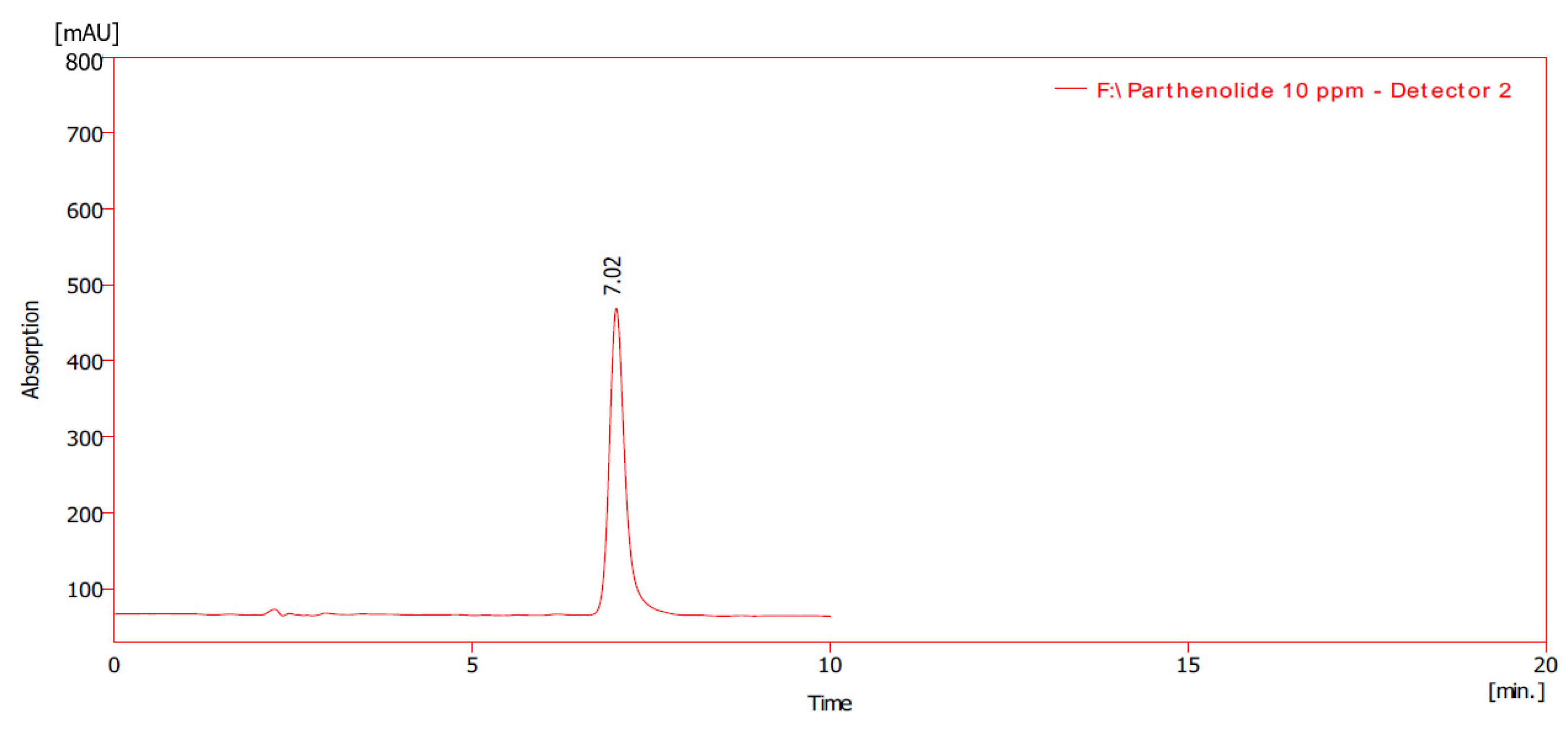
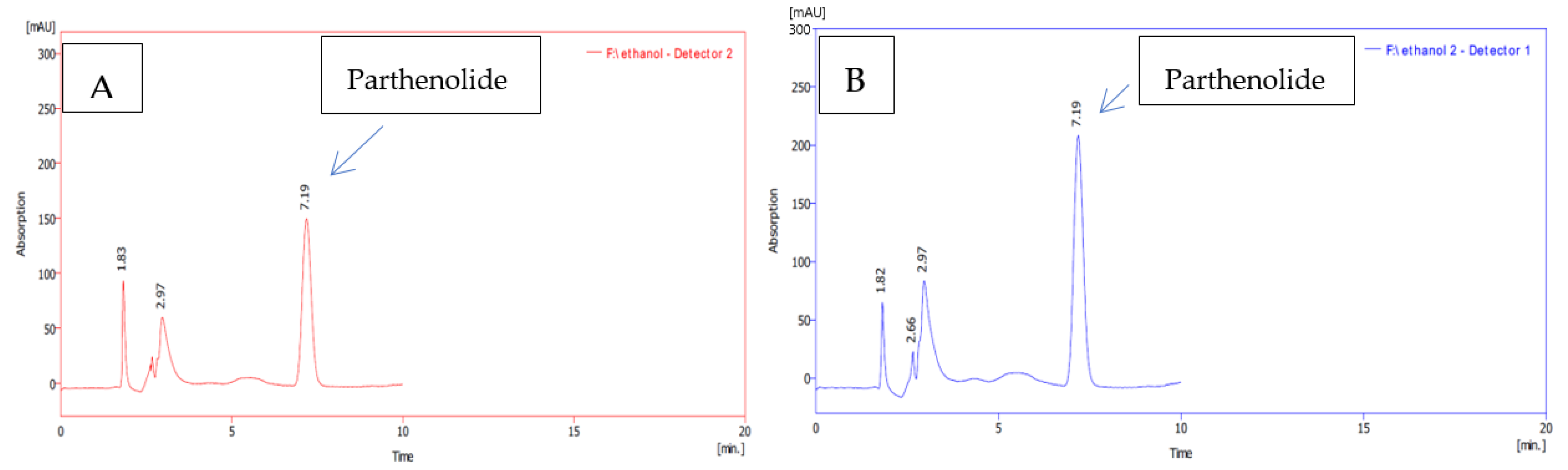
3.2. Quantification of Parthenolide Content of the Feverfew Plants

The results showed that the parthenolide content of the micropropagated feverfew plants leaves’ extract was significantly higher than the conventionally grown feverfew plant leaves’ extract (Table 5, Figure 1 and Figure 2). The extracted amount of parthenolide contents in the micropropagated and conventionally grown feverfew plants’ leaves were also significantly influenced by different types of solvents. The highest parthenolide content (30.60 ± 0.12 mg/g DW) was obtained from the ethanolic extract of the micropropagated plants, followed by the ethanolic extract of the conventionally grown plants (28.40 ± 0.06 mg/g DW). After ethanolic extract, acetone extract obtained significantly higher parthenolide content in both micropropagated (27.90 ± 0.06) and conventionally grown plants (25.60 ± 0.10) compared to the aqueous and hexane extracts. Hexane produced the lowest parthenolide content in both micropropagated (17.50 ± 0.06 mg/g DW) and conventionally grown plants (17.30 ± 0.06 mg/g DW), which were not significantly different from each other.
The higher parthenolide content of the micropropagated feverfew plants compared to the conventionally grown plants could be due to the PGRs application during the micropropagation stage [14,17]. The production of secondary metabolites also can be enhanced under abiotic stress conditions [13]. Hence, the higher parthenolide content in the micropropagated feverfew plants could be due to experiencing the stressful conditions during their acclimatization stage. The amount of the extracted parthenolide content of the feverfew plants’ leaves was considerably affected by different polarities of the solvents. Ethanol, a relatively polar solvent, maximized the amount of the parthenolide content in both micropropagated and conventionally grown plants. The results of the present study confirm the findings of Prashanth et al. [38], who also obtained the maximum parthenolide content from the ethanolic extract of the feverfew plants’ leaves. Tadic et al. [39] also reported that the maximum parthenolide contents of the feverfew plant’s leaves were extracted by ethanol. Based on these findings, it can be concluded that ethanol is an appropriate solvent for extracting the parthenolide content of the feverfew plant. Ethanol was also found to be a suitable solvent for extracting the total flavonoid content of Zingiber officinale rhizome [31]. Moreover, the toxicity of ethanol is less than acetone and hexane; and using it as a solvent is suitable for human utilization [40]. Thus, ethanol could be a suitable solvent for parthenolide extraction from the feverfew and other herbal plants.

4. Conclusions

The study’s findings establish a successful protocol for micropropagation of the feverfew plant. Using 4% NaOCl for surface sterilization of feverfew plant seeds was found to be suitable, resulting in 41% aseptic seeds and 90% germination of the seeds cultured in the in vitro conditions. Feverfew plant leaves’ explants were capable of inducing callus in the presence of TDZ in the culture medium. The addition of 2,4-D further improved callus induction; MS medium supplemented with 0.4 mg/L TDZ and 2 mg/L 2,4-D was found the most effective medium for callus induction of the feverfew plant leaves’ explant. The callus induced multiple shoots with the addition of cytokinins in the culture medium; zeatin was found to be the most effective type of cytokinin for shoot induction from the callus of the feverfew plant. The in vitro-raised shoots were capable of producing roots in the rooting medium; IBA was found to be the best type of auxin for in vitro rooting of the feverfew plant. Feverfew in vitro-raised plantlets were effectively established in the ex vitro conditions. These results show that micropropagation could be successfully applied to produce disease-free planting materials of the feverfew plant for its wide-scale production. Micropropagation also enhanced the parthenolide content of the feverfew plants compared to its conventionally grown plants. However, it is not clear whether the improvement in the parthenolide content in the micropropagated plants could be originated from their genetic variation or from the effects of PGRs used in the culture medium. Therefore, it is necessary to study the genetic fidelity of micropropagated feverfew plants in comparison with their mother plants.

Author Contributions

Conceptualization, H.E.M. and N.A.Z.; methodology, H.E.M. and N.A.Z.; resources, H.E.M. and M.K.A.; curation, N.A.Z.; writing—original draft preparation, H.E.M. and N.A.Z.; writing—review and editing, H.E.M., N.A.Z. and M.K.A.; funding acquisition, H.E.M. and M.K.A. All authors have read and agreed to the published version of the manuscript.

Funding

This research received no external funding.

Institutional Review Board Statement

Not applicable.

Informed Consent Statement

Not applicable.

Data Availability Statement

The data presented in this study are available in the article.

Acknowledgments

As this study was carried out in the Ministry of Sciences and Technology of Iraq, so the authors would like to thank all the staff members for their support during this study.

Conflicts of Interest

The authors declare no conflict of interest.

References

  1. Pareek, A.; Suthar, M.; Rathore, G.; Bansal, V. Feverfew (Tanacetum parthenium L.): A systematic review. Pharmacogn. Rev. 2011, 5, 103. [Google Scholar] [CrossRef] [Green Version]
  2. Pourianezhad, F.; Tahmasebi, S.; Abdusi, V.; Nikfar, S.; Mirhoseini, M. Review on feverfew, a valuable medicinal plant. J. HerbMed. Pharmacol. 2016, 5, 45–49. [Google Scholar]
  3. Sadat-Hosseini, M.; Farajpour, M.; Boroomand, N.; Solaimani-Sardou, F. Ethnopharmacological studies of indigenous medicinal plants in the south of Kerman, Iran. J. Ethnopharmacol. 2017, 199, 194–204. [Google Scholar] [CrossRef]
  4. Mahmoodzadeh, Y.; Mazani, M.; Rezagholizadeh, L. Hepatoprotective effect of methanolic Tanacetum parthenium extract on CCl4-induced liver damage in rats. Toxicol. Rep. 2017, 4, 455–462. [Google Scholar] [CrossRef]
  5. Sztiller-Sikorska, M.; Czyz, M. Parthenolide as cooperating agent for anti-cancer treatment of various malignancies. Pharmaceuticals 2020, 13, 194. [Google Scholar] [CrossRef] [PubMed]
  6. Di Cesare Mannelli, L.; Tenci, B.; Zanardelli, M.; Maidecchi, A.; Lugli, A.; Mattoli, L.; Ghelardini, C. Widespread pain reliever profile of a flower extract of Tanacetum parthenium. Phytomedicine 2015, 22, 752–758. [Google Scholar] [CrossRef]
  7. Penthala, N.R.; Janganati, V.; Alpe, T.L.; Apana, S.M.; Berridge, M.S.; Crooks, P.A.; Borrelli, M.J. N-[11CH3]Dimethylaminoparthenolide (DMAPT) uptake into orthotopic 9LSF glioblastoma tumors in the rat. Bioorganic. Med. Chem. Lett. 2016, 26, 5883–5886. [Google Scholar] [CrossRef] [PubMed] [Green Version]
  8. Czyz, M.; Lesiak-Mieczkowska, K.; Koprowska, K.; Szulawska-Mroczek, A.; Wozniak, M. Cell context-dependent activities of parthenolide in primary and metastatic melanoma cells. Br. J. Pharmacol. 2010, 160, 1144–1157. [Google Scholar] [CrossRef] [PubMed] [Green Version]
  9. Baranello, M.P.; Bauer, L.; Jordan, C.T.; Benoit, D.S.W. Micelle delivery of parthenolide to acute myeloid leukemia cells. Cell. Mol. Bioeng. 2015, 8, 455–470. [Google Scholar] [CrossRef] [Green Version]
  10. Flores-Lopez, G.; Moreno-Lorenzana, D.; Ayala-Sanchez, M.; Aviles-Vazquez, S.; Torres-Martinez, H.; Crooks, P.A.; Guzman, M.L.; Mayani, H.; Chávez-González, A. Parthenolide and DMAPT induce cell death in primitive CML cells through reactive oxygen species. J. Cell. Mol. Med. 2018, 22, 4899–4912. [Google Scholar] [CrossRef] [PubMed]
  11. Czyz, M.; Koprowska, K.; Sztiller-Sikorska, M. Parthenolide reduces the frequency of ABCB5-positive cells and clonogenic capacity of melanoma cells from anchorage independent melanospheres. Cancer Biol. Ther. 2013, 14, 135–145. [Google Scholar] [CrossRef] [PubMed] [Green Version]
  12. Majdi, M.; Abdollahi, M.R.; Maroufi, A. Parthenolide accumulation and expression of genes related to parthenolide biosynthesis affected by exogenous application of methyl jasmonate and salicylic acid in Tanacetum parthenium. Plant Cell Rep. 2015, 34, 1909–1918. [Google Scholar] [CrossRef] [PubMed]
  13. Khajavi, M.; Rahaie, M.; Ebrahimi, A. The effect of TiO2 and SiO2 nanoparticles and salinity stress on expression of genes involved in parthenolide biosynthesis in feverfew (Tanacetum parthenium L.). Caryologia 2019, 72, 3–14. [Google Scholar]
  14. Nieto-Trujillo, A.; Buendía-González, L.; García-Morales, C.; Román-Guerrero, A.; Cruz-Sosa, F.; Estrada-Zúñiga, M.E. Phenolic compounds and parthenolide production from in vitro cultures of Tanacetum parthenium. Rev. Mex. Ing. Quim. 2017, 16, 371–383. [Google Scholar]
  15. Mahood, H.E. Effect of plant growth regulators and explant source on the induction of callus of Dianthus caryophyllus L. Basrah J. Agric. Sci. 2021, 34, 100–106. [Google Scholar] [CrossRef]
  16. Chandran, H.; Meena, M.; Barupal, T.; Sharma, K. Plant tissue culture as a perpetual source for production of industrially important bioactive compounds. Biotechnol. Rep. 2020, 26, e00450. [Google Scholar] [CrossRef]
  17. Pourianezhad, F.; Rahnama, H.; Mousavi, A.; Khosrowshahli, M.; Mafakheri, S. Parthenolide production in cell suspension culture of feverfew. Bioresour. Bioprocess. 2019, 6, 23. [Google Scholar] [CrossRef] [Green Version]
  18. Pourianezhad, F.; Rahnama, H.; Mousavi, A.; Khosrowshahli, M.; Mafakheri, S. Effects of combined elicitors on parthenolide production and expression of parthenolide synthase (TpPTS) in Tanacetum parthenium hairy root culture. Plant Biotechnol. Rep. 2019, 13, 211–218. [Google Scholar] [CrossRef]
  19. Murashige, T.; Skoog, F. A revised medium for rapid growth and bio assays with tobacco tissue cultures. Physiol. Plant. 1962, 15, 473–497. [Google Scholar] [CrossRef]
  20. Hakiman, M.; Maziah, M. Non enzymatic and enzymatic antioxidant activities in aqueous extract of different Ficus deltoidea accessions. J. Med. Plants Res. 2009, 3, 120–131. [Google Scholar]
  21. Haida, Z.; Syahida, A.; Ariff, S.M.; Maziah, M.; Hakiman, M. Factors affecting cell biomass and flavonoid production of Ficus deltoidea var. kunstleri in cell suspension culture system. Sci. Rep. 2019, 9, 9533. [Google Scholar] [CrossRef]
  22. Aburashed, E.A.; Khan, I.A. Determination of parthenolide in selected feverfew products by liquid chromatography. J. AOAC Int. 2000, 83, 789–801. [Google Scholar] [CrossRef] [Green Version]
  23. García-Pérez, P.; Lozano-Milo, E.; Landín, M.; Gallego, P.P. Machine learning technology reveals the concealed interactions of phytohormones on medicinal plant in vitro organogenesis. Biomolecules 2020, 10, 746. [Google Scholar] [CrossRef]
  24. Telci, C.; Yildiz, M.; Pelit, S.; Onol, B.; Erkilic, E.G.; Kendir, H. The effect of surface-disinfection process on dormancy-breaking, seed germination, and seedling growth of Lathyrus chrysanthus bois. under in vitro conditions. Propag. Ornam. Plants 2011, 11, 10–16. [Google Scholar]
  25. Cui, J.; Wei, X.; Deng, M.; Chen, J. Regeneration of Gynura aurantiaca ‘Purple Passion’ via indirect shoot organogenesis. Sci. Hortic. 2019, 246, 176–181. [Google Scholar] [CrossRef]
  26. Sié, R.S.; Charles, G.; Sakhanokho, H.F.; Toueix, Y.; Djè, Y.; Sangaré, A.; Branchard, M. Protocols for callus and somatic embryo initiation for Hibiscus sabdariffa L. (Malvaceae): Influence of explant type, sugar, and plant growth regulators. Aust. J. Crop Sci. 2010, 4, 98–106. [Google Scholar]
  27. Dewir, Y.H.; Nurmansyah; Naidoo, Y.; da Silva, J.A.T. Thidiazuron-induced abnormalities in plant tissue cultures. Plant Cell Rep. 2018, 37, 1451–1470. [Google Scholar] [CrossRef]
  28. Guo, B.; Abbasi, B.H.; Zeb, A.; Xu, L.L.; Wei, Y.H. Thidiazuron: A multi-dimensional plant growth regulator. African J. Biotechnol. 2011, 10, 8984–9000. [Google Scholar]
  29. Erland, L.A.E.; Giebelhaus, R.T.; Victor, J.M.R.; Murch, S.J.; Saxena, P.K. The morphoregulatory role of thidiazuron: Metabolomics-guided hypothesis generation for mechanisms of activity. Biomolecules 2020, 10, 1253. [Google Scholar] [CrossRef]
  30. Xu, J.; Chen, L.; Sun, H.; Wusiman, N.; Sun, W.; Li, B.; Gao, Y.; Kong, J.; Zhang, D.; Zhang, X.; et al. Crosstalk between cytokinin and ethylene signaling pathways regulates leaf abscission in cotton in response to chemical defoliants. J. Exp. Bot. 2019, 70, 1539–1551. [Google Scholar] [CrossRef] [Green Version]
  31. Zahid, N.A.; Jaafar, H.Z.E.; Hakiman, M. Micropropagation of ginger (Zingiber officinale Roscoe) ‘Bentong’ and evaluation of its secondary metabolites and antioxidant activities compared with the conventionally propagated plant. Plants 2021, 10, 630. [Google Scholar] [CrossRef]
  32. Bakhshipour, M.; Mafakheri, M.; Kordrostami, M.; Zakir, A.; Rahimi, N.; Feizi, F.; Mohseni, M. In vitro multiplication, genetic fidelity and phytochemical potentials of Vaccinium arctostaphylos L.: An endangered medicinal plant. Ind. Crops Prod. 2019, 141, 111812. [Google Scholar] [CrossRef]
  33. Najhah, M.Y.; Jaafar, H.Z.E.; Nakasha, J.J.; Hakiman, M. Shoot multiplication and callus induction of labisia pumila var. Alata as influenced by different plant growth regulators treatments and its polyphenolic activities compared with the wild plant. Molecules 2021, 26, 3229. [Google Scholar] [CrossRef]
  34. Zahid, N.A.; Jaafar, H.Z.E.; Hakiman, M. Alterations in microrhizome induction, shoot multiplication and rooting of ginger (Zingiber officinale Roscoe) var. Bentong with regards to sucrose and plant growth regulators application. Agronomy 2021, 11, 320. [Google Scholar] [CrossRef]
  35. Jogam, P.; Sandhya, D.; Shekhawat, M.S.; Alok, A.; Manokari, M.; Abbagani, S.; Allini, V.R. Genetic stability analysis using DNA barcoding and molecular markers and foliar micro-morphological analysis of in vitro regenerated and in vivo grown plants of Artemisia vulgaris L. Ind. Crops Prod. 2020, 151, 112476. [Google Scholar] [CrossRef]
  36. Pradhan, N.; Dwivedi, P. In vitro shoot multiplication of Stevia rebaudiana, an important plant with high economic and medicinal values. Vegetos 2016, 29, 1000180. [Google Scholar] [CrossRef]
  37. Joshi, B.; Bhandari, A.; Panwar, G.S. An efficient micropropagation protocol for Vernonia amygdalina Delile—An economically valuable shrub. J. Herbs Spices Med. Plants 2020, 26, 267–274. [Google Scholar] [CrossRef]
  38. Prashanth, S.; Pooja, S.; Suchetha, K.N.; Vidya, V.K. Radical scavenging and antioxidant activities of ethanolic and aqueous extract from the leaves of feverfew (Tanacetum parthenium L.) and a synthetic compound parthenolide. J. Pharmacogn. Phytochem. 2015, 4, 223–227. [Google Scholar]
  39. Tadić, V.; Živković, J.; Bigović, D.; Žugić, A. Variation of parthenolide and phenolic compounds content in different parts of Tanacetum parthenium (L.) Schulz Bip., Asteraceae during 18 months storage. Lek. Sirovine 2019, 39, 35–39. [Google Scholar] [CrossRef] [Green Version]
  40. Tomsone, L.; Kruma, Z. Comparison of different solvents for isolation of phenolic compounds from horseradish (Armoracia rusticana L.) leaves. Res. Rural Dev. 2013, 1, 104–110. [Google Scholar]
Figure 1. HPLC analysis of parthenolide standard for 10 ppm at 210 nm, flow rate 1 mL/min.
Horticulturae 08 00050 g001
Figure 2. HPLC analysis of ethanolic extracts of (A) micropropagated (B) conventionally propagated feverfew plants’ leaves.
Horticulturae 08 00050 g002
Table 1. Effects of NaOCl concentrations on aseptic seeds and seed germination of the feverfew plant.
NaOCl (%)Aseptic Seeds (%)Seed Germination (%)
231.00 ± 1.00 d90.00 ± 1.49 a
333.00 ± 1.53 d90.00 ± 2.11 a
441.00 ± 1.80 c90.00 ± 2.58 a
547.00 ± 2.13 b81.00 ± 1.80 b
665.00 ± 2.69 a73.00 ± 2.60 b
Values represent means ± standard error. Means assigned with different letters in the column indicate significant difference among the treatments according to Duncan’s multiple range test (DMRT) (p < 0.05).
Table 2. Interaction effects of 2,4-D and TDZ concentrations on callus induction (%) of the feverfew plant.
2,4-D (mg/L)TDZ (mg/L)Callus Induction%
00.00.00 ± 0.00 i
00.233.00 ± 3.19 g
00.347.00 ± 2.53 ef
00.462.00 ± 3.82 c
10.00.00 ± 0.00 i
10.243.00 ± 2.34 f
10.372.00 ± 4.71 b
10.475.00 ± 4.34 b
20.00.00 ± 0.00 i
20.223.00 ± 1.17 h
20.384.00 ± 1.65 a
20.486.00 ± 1.72 a
30.00.00 ± 0.00 i
30.253.00 ± 2.78 de
30.361.00 ± 3.73 cd
30.462.00 ± 2.36 c
Data presented as means ± standard error. Means assigned with different letters indicate significant difference among the combination according to Duncan’s multiple range test (DMRT) (p < 0.05).
Table 3. Effects of different types of cytokinins on shoot organogenesis from the callus explant of the feverfew plant.
Cytokinin
(mg/L)
Shoot Induction (%)Number of days to Shoot initiationNumber of Shoots/ExplantShoot Length (cm)Number of Leaves/Shoot
Control (0)60.00 ± 4.08 c12.00 ± 0.47 a2.10 ± 0.23 d2.00 ± 0.33 c1.25 ± 0. 16 c
Kinetin (5)72.50 ± 2.50 b7.00 ± 0.21 c3.00 ± 0.26 c2.10 ± 0.02 bc3.00 ± 0.15 a
BA (5)80.00 ± 3.33 b9.00 ± 0.52 b3.00 ± 0.26 c2.40 ± 0.07 bc2.00 ± 0.21 b
Zeatin (5)100.00 ± 0.00 a7.00 ± 0.26 c5.00 ± 0.15 a2.50 ± 0.05 ab2.10 ± 0.18 b
TDZ (2)100.00 ± 0.00 a9.00 ± 0.21 b4.00 ± 0.26 b2.90 ± 0.02 a2.20 ± 0.13 ab
Values represent means ± standard error. Means assigned with different letters in the column indicate significant difference among the treatments according to Duncan’s multiple range test (DMRT) (p < 0.05).
Table 4. Effects of different types of auxins on the in vitro rooting of the feverfew plant.
TreatmentRoot Induction (%)Time to Root Induction (Days)Number of Roots/PlantletRoot Length (cm)
Control90.00 ± 4.08 a7.00 ± 0.42 a4.10 ± 0.28 b2.00 ± 0.08 b
IAA90.00 ± 5.53 a7.00 ± 0.54 a5.00 ± 0.61 b2.20 ± 0.14 b
IBA90.00 ± 6.67 a7.00 ± 0.47 a8.90 ± 0.35 a3.80 ± 0.19 a
NAA90.00 ± 7.64 a7.00 ± 0.56 a8.10 ± 0.57 a3.40 ± 0.15 a
Values represent means ± standard error. Means assigned with the same letters in the column indicate no significant difference among the treatments according to Duncan’s multiple range test (DMRT) (p < 0.05).
Table 5. Parthenolide content (mg/g DW) of micropropagated and conventionally grown feverfew plants’ leaves analyzed by HPLC using different solvent extracts.
Propagation MethodSolventParthenolide (mg/g DW)
ConventionalAqueous23.23 ± 0.13 e
Ethanol28.40 ± 0.06 b
Acetone25.60 ± 0.10 d
Hexane17.30 ± 0.06 f
MicropropagationAqueous25.50 ± 0.29 d
Ethanol30.60 ± 0.12 a
Acetone27.90 ± 0.06 c
Hexane17.50 ± 0.06 f
Values represent means ± standard error. Means assigned with different letters indicate significant difference according to Duncan’s multiple range test (DMRT) (p < 0.05).
Publisher’s Note: MDPI stays neutral with regard to jurisdictional claims in published maps and institutional affiliations.

