Article

Mechanism of the Alternaria alternata Pathogenicity in ‘Fortune’ Mandarin

Departamento de Biología Vegetal (Fisiología Vegetal), Facultad de Biología, Universidad de Murcia, Campus de Espinardo, 30110 Murcia, Spain
*
Author to whom correspondence should be addressed.
Horticulturae 2018, 4(4), 54; https://doi.org/10.3390/horticulturae4040054
Submission received: 23 October 2018 / Revised: 27 November 2018 / Accepted: 5 December 2018 / Published: 7 December 2018
(This article belongs to the Special Issue Horticultural Plant Pathology and Prevention)

Abstract

:
Alternaria brown spot, caused by Alternaria alternata (Fr.) Keissl, is an important disease in tangerines and their hybrids, affecting leaves, twigs, and immature fruit. Differences in susceptibility to this pathogenic fungus have been described for different Citrus species. In this paper, the expression of the mycotoxins alternariol and alternariol monomethyl ether in different A. alternata isolates was analyzed by HPLC-MS. A correlation was observed between the mycotoxins content and the pathogenicity of each isolated of A. alternata used, suggesting that the mycotoxins may be involved in the evolution of brown spot in ‘Fortune’ fruits caused by this fungus. The increased expression of the above mycotoxins was associated with the end of mycelia growth, high sporulation, and an increase in hyphal melanization in the fungus. On the other hand, the presence of laccase activity in the xylem of ‘Fortune’ fruits inoculated with A. alternata suggests that this is the way the fungus propagates in the plant. These results add to our knowledge of the pathogenesis of A. alternata in Citrus.

1. Introduction

Fungi of the genus Alternaria cause several diseases in Citrus species, mainly in preharvest fruits. The degree of susceptibility to A. alternata depends on the Citrus species in question, having been described that the cultivars of Citrus reticulata and its hybrids, including tangelos ‘Minneola’ and ‘Orlando’; the tangor ‘Murcott’; and the hybrids ‘Fortune’, ‘Nova’, ‘Fairchild’, ‘Lee’, and ‘Sunburst’ are susceptible to the fungus. Other species, such as Citrus sinensis, Citrus limon (L.) Burm., and Citrus margarita (Lour.) Swing. are resistant, while Satsumas (Citrus unshiu Mark, Marc.) and Clementines (Citrus clementina Hort. ex Tan.), show a certain degree of resistance to this pathogen [1,2,3,4,5].
Some studies reveal that the differences in the susceptibility of Citrus fruits to A. alternata are due to complex endogenous signaling processes, modulated by several factors, such as ethylene produced during infection, the fruit growth stage, the phytoanticipins and/or phytolaexins present or induced in the fruits, and changes in laccase activity—a virulence factor of the fungus produced during infection [5,6,7,8].
On the other hand, it has been described that the system of pathogenesis of this fungus is related to the production of toxins [9], which are released during the germination of the spores and cause necrosis in leaves and fruits. In Alternaria species, more than seventy different molecules have been identified, including dibenzopyrones, tetrahydric acids, quinones, peptides, and lactone derivatives. Many of these compounds are mycotoxins, some of which are phytotoxic for the plant, while showing specific or nonspecific phytotoxicity towards the host. Likewise, it has been described that these mycotoxins can have adverse effects on human health, and are related to or involved in the appearance of infections, allergies, chromosomal aberrations, and the induction of liver cancer [10].
At present, little information is available on the mycotoxins produced by the different A. alternata pathotypes that affect Citrus, as well as on the phytotoxicity of these compounds and their mechanism of action in the physiology of the plant [11,12,13,14,15,16].
The objective of this work was to (i) characterize the mycotoxins produced by some selected isolates of A. alternata, (ii) establish the correlation between the degree of pathogenicity of these isolates in unwounded fruits and the levels of mycotoxins produced by them, analyzing whether these mycotoxins had phytotoxic activity, and (iii) histologically study the route of propagation of the fungus in the fruit revealing the laccase activity with 2,2-azinobis (3-ethylbenzothiazoline-6-sulfonic acid) (ABTS).

2. Materials and Methods

2.1. Selection of Alternaria alternata Isolates, Culture of the Fungus, and Pathogenicity Studies

Different isolates of A. alternata were obtained from fruits affected by “brown spot” in a commercial plantation of mandarin ‘Fortune’ (Citrus clementina × Citrus tangerina), located in Campo de Cartagena (Murcia, Spain). For this study, the Alt3 and Alt 14 isolates were selected because they showed low and high degrees of aggressiveness, respectively, in unwounded ‘Fortune’ fruits [5].
These A. alternata isolates were grown in PDA medium at 25 °C to serve as inoculum in the different studies. Fungal pathogenicity was evaluated by measuring the growth of the lesion produced after inoculation of unwounded fruits with each one of the isolates. Prior to inoculation, fruits were disinfected with a solution of NaClO (0.5%) for 5 min and then washed with sterile water and dried with cellulose paper previously autoclaved. Ten similar fruits were used in each of the inoculation assays described below, in which a 5 mm diameter disk of 15-day-old PDA culture medium with mycelium of A. alternata (Alt3 or Alt14) was deposited on unwounded fruit and sealed with adhesive tape. The inoculations were performed under sterile conditions in a laminar flow cabinet. After inoculation, they were kept in a chamber at 20 °C and 85% relative humidity. At different times post-inoculation, the growth of the fungus was determined by measuring the diameter of lesion produced in mm.
For laccase enzyme extracts, the A. alternata was cultivated in Saboreaud liquid medium (SIGMA) (peptone 10 g/L, d-glucose 40 g/L, pH 5.6 +/− 0.2) supplemented with 5 mM Cu2SO4 [17] in 250 mL flasks and the culture was kept in a growth chamber at 25 °C, in darkness and stirring at 170 rpm [8].

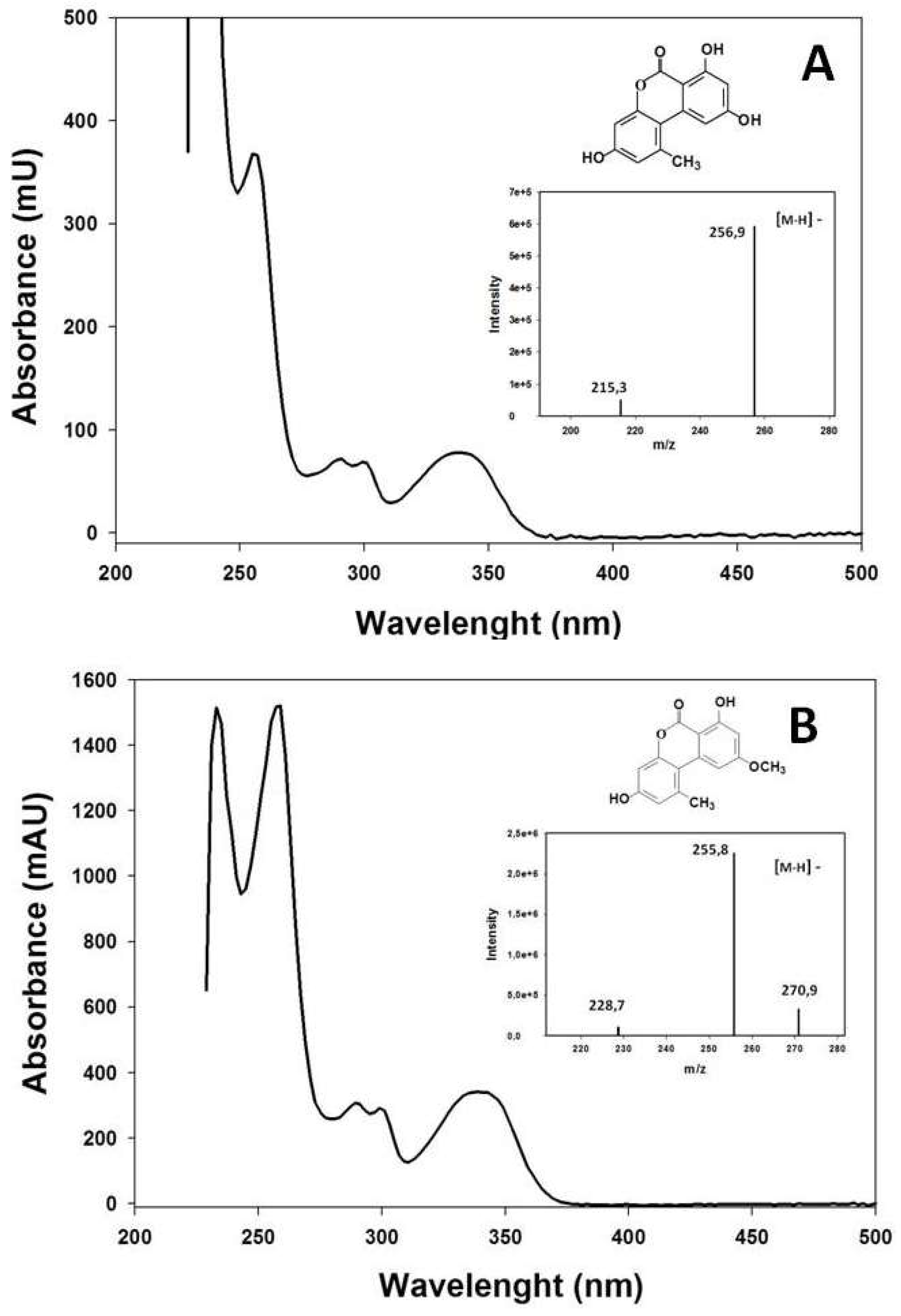
2.2. Extraction and Measurement of A. alternata Mycotoxins

Cultures of Alt3 and Alt14 isolates (15 days-old) were used to extract the mycotoxins produced in each isolate. The contents of the plates were dried in an oven at 60 °C and ground before being extracted with acetonitrile (1 g/10 mL) while stirring for 2 h, and subsequently filtered through to 0.45 μm nylon membrane before proceeding to analysis of these compounds. Occasionally, the mycelium was separated into different zones in the petri dish (internal, middle, and external) to carry out these extractions.
The analysis of mycotoxins present in these extracts was carried out in a Jasco Liquid Chromatography system equipped with a pump (Model PU-2089 Plus, JASCO, Easton, MD, USA), a photodiode array detector (Model MD-2010 Plus, JASCO, Easton, MD, USA), and an autosampler (Model AS-2055 Plus, JASCO, Easton, MD, USA). For separation of the different mycotoxins, a LiChroCARTR C18 (Agilent, Santa Clara, CA, USA) analytical column (250 × 4 mm i.d.) with an average particle size of 5 μm as stationary phase was used at 30 °C. As a mobile phase, a binary water gradient was used as solvent (A) and acetonitrile as solvent (B): initially, 100% (A) was held for 20 min; then, the composition of the solvent changed in a linear gradient to 20% (A) by 70 min, which was maintained for 20 min. The eluent flow was 1 mL/min. Changes in absorbance were recorded at 280 nm. The quantities of these compounds were determined using the response factor of the corresponding standards.
Identification of these compounds was carried out by HPLC-MS with an Agilent model VL ion trap mass spectrometer equipped with ESI interface coupled to an HPLC (Agilent 1100, Santa Clara, CA, USA). A 5 μm (250 × 4 mm i.d.) C18 Kromasil 100 (Tecnokroma) column was used for the separation, which was performed by means of an elution gradient similar to that described above. The column was maintained at 30 °C. ESI mass spectra were acquired in both positive and negative ion modes by scanning over the 50 to 1000 mass range. The ESI parameters were: source voltage 3.5 kV, dry temperature 350 °C, nebulizer 60 psi, and dry gas 9 L/min.
To carry out the corresponding biological assays into the possible participation of these mycotoxins in the mechanism of A. alternate pathogenesis, the fractions corresponding to alternariol (AOH) and alternariol monomethyl ether (AME) were collected with a fraction collector (Gilson FC 203B) at the exit of the HPLC column.

2.3. Extraction and Measurement of Laccase Activity in the Mycelium of A. alternata

Fifteen-day-old mycelia of Alt3 and Alt14 were separated from the liquid medium by filtration and the enzyme in each case was partially purified with acetone 15% (v/v). Laccase activity was measured at 25 °C using 2,2-azinobis (3-ethylbenzothiazoline-6-sulfonic acid) (ABTS) (20 mM) as substrate [18]. The reaction was followed by measuring the increase in absorbance at 436 nm (ε = 29300 M−1 cm−1) in a UNICAM UV 500 spectrophotometer (Thermo Electronics, Cambridge, UK) according to a previously described procedure [8]. One unit of laccase activity was defined as the amount of enzyme required to oxidize 1 µmol of ABTS per minute at 25 °C in the standard reaction medium.

2.4. Histological Localization of the Laccase Acitvity

ABTS was used as reagent to locate the presence of laccase activity in the tissues of the fruits infected with A. alternata. Ten days after inoculating the fruits of ‘Fortune’ with A. altenata (Alt14 isolate), peel sections were obtained and treated with 5 mM ABTS in citrate buffer pH 3.5. Similarly, after removing the peel, the surface of the fruits was impregnated with the same ABTS solution. The presence of laccase activity was revealed by the formation of the oxidized form of ABTS, which has a blue coloration.

2.5. Statistical Analysis

The values are given as means ± standard error. Data were analyzed by a General Linear Model (GLM). Duncan’s Multiple Range Test (DMRT) was used for the analysis of the variance in the data. All statistical analyses were performed using Statgraphics 5.0 software.

3. Results

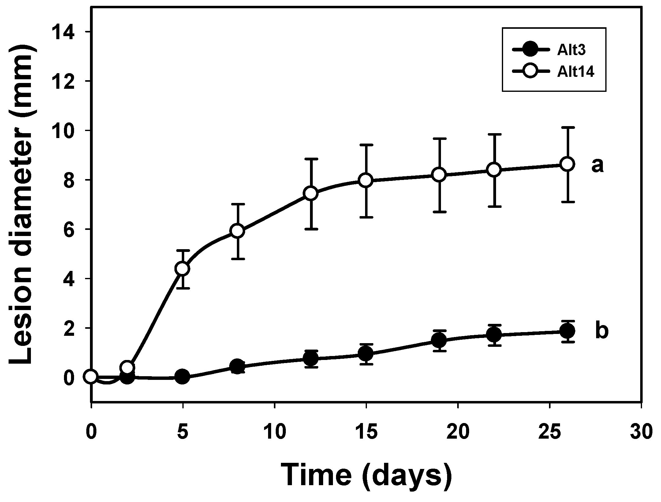
3.1. Study of the Pathogenicity and the Mycotoxins Content of the A. alternata Isolates

The differences observed in the lesion diameters after inoculations with the Alt3 and Alt14 isolates in the corresponding unwounded ‘Fortune’ fruits are shown in Figure 1. It can be seen that these isolates differed in aggressiveness: Alt3 was not very aggressive and only produced slight lesion growth, while Alt14 was highly aggressive.
The chromatographic profile corresponding to the extracts of mycelia of Alt3 and Alt14 isolates revealed the presence of two mycotoxins for both isolates. The minor compound had a retention time (tR = 48.9 min, compound 1) coincident with that of alternariol (AOH) while the major compound had a retention time (tR = 55.8 min, compound 2) coincident with that of alternariol monomethyl ether (AME) (Figure 2).
The UV/Vis absorption spectra obtained by the diode array detector and those obtained by mass spectrometry were identical to those obtained for the standards of 3,7,9-trihydroxy-1-methyl-6H-dibenzo [b,d] pyran-6-one (AOH) (Figure 3A) and 3,7-dihydroxy-9-methoxy-1-methyl-6H-dibenzo [b,d] pyran-6-one (AME) (Figure 3B).
Higher levels of AME and AOH were detected in the Alt14 isolate (58 and 28 μmol/g DW, respectively) than in the Alt3 isolate (32 and 3 μmols/g DW, respectively) (Table 1). When mycotoxin expression was analyzed in different zones of the Alt14 mycelium (15 day-old), the highest levels were detected in the internal zone, where it was associated with strong melanization of the hyphae and a high degree of sporulation (80 and 36 µmol/g DW of AME and AOH, respectively), followed by the middle zone, with intermediate levels of melanization and sporulation (25 and 11 µmol/g DW of AME and AOH, respectively). In the external zone, which showed intense mycelial growth and scant melanization and sporulation, neither of these mycotoxins were detected. A similar distribution of mycotoxins was observed in the Alt3 mycelium, although their concentrations were lower.
To ascertain whether these mycotoxins may participate in the pathogenesis of A. alternata in ‘Fortune’ mandarin, biological assays were carried out in which AME and AOH were applied to fruits of ‘Fortune’. The results revealed that both AOH and AME were capable of inducing necrosis in this organ (data not shown), suggesting that A. alternata mycotoxins are involved in the development of “brown spot” in ‘Fortune’ fruits.

3.2. Correlation between the Laccase Activity of A. alternata Isolates and Their Pathogenicity

Table 1 shows the extracellular laccase activity released by the two A. alternata isolates tested (Alt3 and Alt14) and cultured in liquid medium. After 15 days of culture, the activity of the Alt14 isolate was much higher (215 mU/mL) than that detected for the Alt3 isolate (105 mU/mL). The differences in the expression of this enzyme suggests that laccase could be involved in the mechanism of pathogenesis of A. alternata in ‘Fortune’ mandarin.

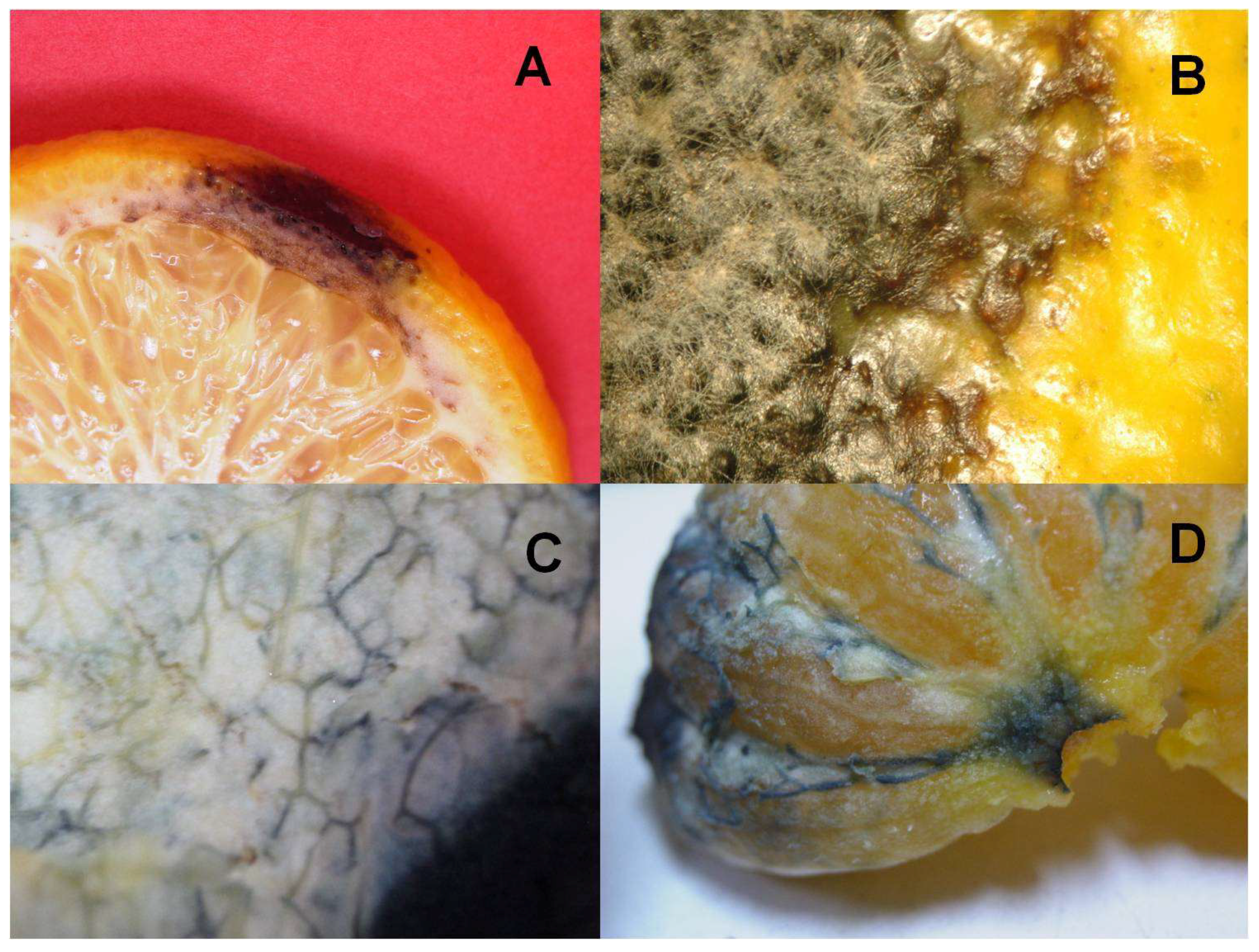
3.3. Location of Laccase Activity in the Tissues of ‘Fortune’ Fruits Inoculated with A. alternata

Figure 4A shows peel necrosis of ‘Fortune’ fruits after inoculation with A. alternata (Alt14 isolate). The browning of both the flavedo and the albedo suggests the involvement of oxidizing enzyme systems. The advance of the lesion was accompanied by fungal growth both on the surface of the fruit (flavedo) (Figure 4B) and inside (data not shown). A blue coloration was observed when the peel or the pulp of infected fruits was treated with a solution of 5 mM ABTS, the fibers (xylem) being particularly stained (Figure 4C,D). In uninfected fruits or in areas far from the inoculum, no such bluish coloration was observed. These results indicated the presence of extracellular laccase activity and therefore suggest the presence of A. alternata mycelium in this conductive tissue.

4. Discussion

The results concerning the presence of AOH and AME mycotoxins in A. alternata isolates which affect ‘Fortune’ fruits have also been described by other authors using oranges and tangerines infected with A. alternata [9,19,20]. Of particular note was the positive correlation between the degree of aggressiveness of the isolate of A. alternata and the level of expression of both mycotoxins (Figure 1, Table 1). Thus, the Alt14 isolate, which expressed higher levels of AOH and AME than the Alt3 isolates, also showed greater aggressiveness against ‘Fortune’ compared with that observed for Alt3 [5]. The strong expression of these mycotoxins associated with high sporulation and increases in the hyphal melanization of the fungus showed that endogenous factors, such as the development phase of the fungus, are related to the degree of pathogenicity of the same in ‘Fortune’ fruits.
The strong pathogenicity of the Alt14 isolate was also associated with a higher expression of the laccase enzyme, considered as a virulence factor of this fungus [8]. This enzyme could be responsible for the browning of the fruit peel (albedo and flavedo) observed when ‘Fortune’ fruits are inoculated with this fungus, forming part of an enzymatic complex capable of oxidizing the phenolic compounds. In this sense, this enzyme may be responsible for the degradative metabolism of flavanones and flavones in Citrus fruits infected with A. alternata [8]. The oxidation of these phenolic compounds could be a defense mechanism by the fungus against these secondary metabolites, many with antifungal activity, without ruling out their involvement in the metabolism of lignins given their lignolytic activity.
The histological study of the localization of laccase activity revealed that this enzyme, and possibly the fungus, invade the internal tissues of the fruit, where their presence can be detected in the albedo and, specifically, the fibers, possibly in the xylem, suggesting that this is the pathway in which the fungus propagates in these fruits.

5. Conclusions

The involvement of the mycotoxins AOH and AME, and of laccase activity, in the mechanism of Alternaria alternata pathogenicity in ‘Fortune’ mandarin should be taken into account when developing new treatments to control this disease.

Author Contributions

All authors conceived, designed and performed the experiments, analyzed the data and wrote the paper.

Funding

This research received no external funding.

Conflicts of Interest

The authors declare no conflicts of interest.

References

  1. Pegg, K.G. Studies of a strain of Alternaria citri Pierce, the casual organism of brown spot of Emperor mandarin. Queensland J. Agric. Anim. Sci. 1966, 23, 15–18. [Google Scholar]
  2. Gardner, J.M.; Kono, Y.; Chandler, J.L. Bioassay and host-selectivity of Alternaria citri toxins affecting rouge lemon and mandarins. Physiol. Mol. Plant Pathol. 1986, 29, 293–304. [Google Scholar] [CrossRef]
  3. Kohmoto, K.; Akimitsu, K.; Otani, H. Correlation of resistance and susceptibility of citrus to Alternaria alternata with sensitivity to host-specific toxins. Phytopathology 1991, 81, 719–722. [Google Scholar] [CrossRef]
  4. Reis, R.F.; De Almedia, T.F.; Stuchi, E.S.; De Goes, A. Susceptibility of citrus species to Alternaria alternata, the causal agent to the Alternaria brown spot. Sci. Hortic. 2007, 113, 336–342. [Google Scholar] [CrossRef]
  5. Nemsa, I.; Hernández, M.A.; Lacasa, A.; Porras, I.; García-Lidón, A.; Cifuentes, D.; Bouzid, S.; Ortuño, A.; Del Río, J.A. Pathogenicity of Alternaria alternata on fruits and leaves of ‘Fortune’ mandarin (Citrus clementina × Citrus tangerina). Can. J. Plant Pathol. 2012, 34, 195–202. [Google Scholar] [CrossRef]
  6. Ortuño, A.; Nemsa, I.; Álvarez, N.; Lacasa, A.; Porras, I.; García-Lidón, A.; Del Río, J.A. Correlation of ethylene synthesis in Citrus fruits and their susceptibility to Alternaria alternata pv. citri. Physiol. Mol. Plant Pathol. 2008, 72, 162–166. [Google Scholar] [CrossRef]
  7. Ortuño, A.; Del Río, J.A. Role of Citrus phenolic compounds in the resistance mechanism against pathogenic fungi. In Tree and Forestry Science and Biotechnology; Tennant, P., Benkeblia, N., Eds.; Global Science Books, Ltd.: London, UK, 2009; Volume 3, pp. 49–53. ISBN 978-4-903313-25-2. [Google Scholar]
  8. Díaz, L.; Del Río, J.A.; Pérez-Gilabert, M.; Ortuño, A. Involvement of an extracelular fungus laccase in the flavonoid metabolism in Citrus fruits inoculated with Alternaria alternata. Plant Physiol. Biochem. 2015, 89, 11–17. [Google Scholar] [CrossRef] [PubMed]
  9. Logrieco, A.; Visconti, A. Mandarin fruit rot caused by Alternaria alternata and associated mycotoxins. Plant Dis. 1990, 74, 415–417. [Google Scholar] [CrossRef]
  10. Ostry, V. Alternaria mycotoxins: An overview of chemical characterization, producers, toxicity, analysis and occurrence in foodstuffs. World Mycotoxin J. 2008, 1, 175–188. [Google Scholar] [CrossRef]
  11. Kohmoto, K.; Scheffer, R.P.; Whiteside, J.O. Host-selective toxins from Alternaria citri. Phytopathology 1979, 69, 667–671. [Google Scholar] [CrossRef]
  12. Gardner, J.M.; Kono, Y.; Tatum, J.H.; Suzuki, Y.; Takeuchi, S. Structure of major component of ACRL toxins, host-specific phytotoxic compound produced by Alternaria citri. Agric. Biol. Chem. 1985, 49, 1235–1238. [Google Scholar]
  13. Kono, Y.; Gardner, J.M.; Takeuchi, S. Structure of the host-selective toxins produced by a pathotype of Alternaria citri causing brown spot disease of mandarins. Agric. Biol. Chem. 1986, 50, 801–804. [Google Scholar] [CrossRef]
  14. Nakatsuka, S.; Goto, T.; Kohmoto, K.; Nishimura, S. Natural Products and Biological Activities; Imura, H., Ed.; University of Tokyo Press: Tokyo, Japan, 1986; pp. 11–18. ISBN 978-4-13-068118-6. [Google Scholar]
  15. Visconti, A.; Bottalico, A.; Solfrizzo, M. Activity of Alternaria alternata metabolites on tomato leaves and Geotrichum candidum. In Phytotoxins and Plant Patogénesis; Graniti, A., Durban, R., Ballio, A., Eds.; Springer: Berlin, Germany, 1989; p. 457. ISBN 978-3-642-73178-5. [Google Scholar]
  16. Akimitsu, K.; Peever, T.L.; Timmer, L.W. Molecular, ecological and evolutionary approaches to understanding Alternaria diseases of citrus. Mol. Plant Pathol. 2003, 4, 435–446. [Google Scholar] [CrossRef] [PubMed]
  17. Palmieri, G.; Giardina, P.; Bianco, C.; Fontanella, B.; Sannia, G. Cooper Induction of laccase isoenzymes in the ligninolytic fungus Pleurotus ostreatus. Appl. Environ. Microb. 2000, 66, 920–924. [Google Scholar] [CrossRef]
  18. Niku-Paavola, M.L.; Karhunen, E.; Salola, P.; Raunio, V. Ligninolytic enzymes of the White-rot fungus Phlebiaradiata. Biochem. J. 1988, 254, 877–884. [Google Scholar] [CrossRef] [PubMed]
  19. Stinson, E.E.; Osman, S.F.; Heisler, E.G.; Siciliano, J.; Bills, D.D. Mycotoxin production in whole tomatoes, apples, oranges, and lemons. J. Agric. Food Chem. 1981, 29, 790–792. [Google Scholar] [CrossRef] [PubMed]
  20. Magnani, R.F.; De Souza, G.D.; Rodrigues-Filho, E. Analysis of Alternariol and Alternariol Monomethyl Ether on Flavedo and Albedo Tissues of Tangerines (Citrus reticulata) with Symptoms of Alternatia Brown Spot. J. Agric. Food Chem. 2007, 55, 4980–4986. [Google Scholar] [CrossRef] [PubMed]
Figure 1. Development of lesions caused by two A. alternata isolates in unwounded fruits of ‘Fortune’ mandarin: Alt3 (●) and Alt14 (○). The data represent mean values of lesion diameter (mm) at different days post-inoculation. Vertical bars denote ± SE (n = 3) when larger than symbols. Values not sharing a common superscript letter are significantly different (P < 0.05).
Horticulturae 04 00054 g001
Figure 2. HPLC elution profile of an extract of 15 day-old A. alternata mycelia (Alt14 isolate). 1, alternariol; 2, alternariol monomethyl ether.
Horticulturae 04 00054 g002
Figure 3. UV/Vis absorbance and mass spectra of alternariol (A) and alternariol monomethyl ether (B) mycotoxins isolated from 15 day-old A. alternata mycelia (Alt14 isolate).
Horticulturae 04 00054 g003
Figure 4. Image of the injury caused by Alt14 on the peel (A) and the flavedo (B) of ‘Fortune’ mandarin. Location of the laccase activity revealed with ABTS (5 mM) in the albedo (C) and in the xylem (D) of mandarin ‘Fortune’ infected with Alt14.
Horticulturae 04 00054 g004
Table 1. Alternariol (AOH) and alternariol monomethyl ether (AME) and laccase activity levels in Alt3 and Alt14 isolates of A. alternate mycelia (15-days-old). The data represent mean values ± SE (n = 3) of these mycotoxins (µmol/g DW). Values not sharing a common superscript letter are significantly different (P < 0.05).
A. alternata IsolateMycotoxin (µmol/g DW)Laccase Activity (mU/mL)
AMEAOH
Alt332 ± 2 b3 ± 0.2 b105 ± 21 b
Alt1458 ± 3 a28 ± 4 a215 ± 25 a

