Review

Mealybug Wilt of Pineapple and Associated Viruses

1
Florida Department of Agriculture and Consumer Services, Gainesville, FL 32608, USA
2
Department of Plant and Environmental Protection Sciences, University of Hawaii, Honolulu, HI 96822, USA
*
Author to whom correspondence should be addressed.
These authors contributed equally to this work.
Horticulturae 2018, 4(4), 52; https://doi.org/10.3390/horticulturae4040052
Submission received: 9 October 2018 / Revised: 16 November 2018 / Accepted: 20 November 2018 / Published: 30 November 2018
(This article belongs to the Special Issue Horticultural Plant Pathology and Prevention)

Abstract

:
Mealybug wilt of pineapple (MWP) is a disease of pineapple that has a long history in Hawaii, but is present throughout the world where pineapples are grown in tropical regions. The disease has an interesting etiology that is poorly understood but involves an association with virus particles, mealybug vectors, and ants which spread the mealybug vectors. Several distinct pineapple mealybug wilt-associated virus (PMWaV) species have been identified thus far with potential further member species yet to be characterized. Pineapple mealybug wilt-associated viruses are member species of the Ampelovirus genus of the Closteroviridae family. Ampeloviruses are split into two subgroups, subgroup I and subgroup II. PMWaV-2 is a subgroup II member, and these have a longer and more complex genome with additional genes on the 3’ terminus of the RNA genome compared to subgroup I ampeloviruses. PMWaV-2, along with the presence of mealybug vectors, have been shown to be necessary factors in symptom development in Hawaii. Some of these extra genes in the 3’ of PMWaV-2 have recently been shown to function as silencing suppressors, and may play a role in the virulence of PMWaV-2 and symptom development. In other regions of the world, reports of symptomatic plants without PMWaV-2 infection, but with PMWaV-1, -3 or some combination, contradict the requirement of PMWaV-2 for symptom development in MWP. It is possible that further, uncharacterized PMWaVs may be present in symptomatic pineapple plants that test negative for PMWaV-2, explaining the inconsistency in symptom development. More research is necessary to explore the confusing etiology of the MWP disease, and to perhaps shed light upon the symptom development.

1. Mealybug Wilt of Pineapple

1.1. A History of Pineapple Production in Hawaii

Pineapples, Ananas comosus (L.) Merr. of the Bromeliaceae family, are xerophytic tropical monocotyledonous plants which are perennial in nature [1]. Pineapple cultivars are classified into five morphological groups, namely, Cayenne, Queen, Spanish, Brazilian, and Maipure [2]. Many varieties of pineapple are used for human consumption; the most common varieties are “Smooth Cayenne”, “Red Spanish”, “Perolera”, “Pernambuco”, and “Primavera”. The Pineapple Research Institute clones of “Smooth Cayenne” variety, notably “Champaka”, had been the most important variety commercially that were used to produce fresh pineapple, canned pineapple, and various other processed pineapple products.
Pineapple originated in South America and was domesticated from a A. comosus variant, “Ananasoides”. It was documented that pineapple was widely distributed in the ancient world, thousands of years before Christopher Columbus and his sailors discovered it on an island in the West Indies [3] and spread its cultivation to the world. Over the next 400 years, A. comosus and its many varieties were grown in various tropical and sub-tropical locations. There is no specific record of when pineapples were introduced by the Spaniards into Hawaii, but the first record of pineapple in Hawaii was in 1813 [3]. One of Hawaii’s chief contributions to pineapple production can be attributed to the invention of the canning process, allowing for the long-term storage of pineapple for overseas trade [3]. The history of the rise and fall of the Hawaiian pineapple industry is richly documented in literature. For a comprehensive review, readers are referred to a recent book by Larsen and Marks (2010) [4]. After reaching the pinnacle of production by the late 1960s, the pineapple industry in Hawaii began declining, primarily due to competition for cheaper labor and land prices from other countries. Moreover, there was a gradual shift in consumer preference from canned pineapple to fresh pineapples, due to the sudden development of refrigerated shipping. Besides, “Smooth Cayenne”, which was used exclusively for canning, became more acidic in winter months, rendering it less desirable as fresh fruit [5]. To offset the losses, Dole and Del Monte, the two major pineapple corporations in Hawaii, began exploring relocation to countries where labor and land costs were low. Eventually, in the 1980s, both companies moved their primary production from Hawaii to Thailand and the Philippines. The demand for year-round production of fresh fruit led the Hawaiian pineapple industry to undergo considerable transformation. In the mid-1980s, “MD-2”, a complex hybrid trademarked as Del Monte “Gold Extra Sweet”, was bred at the pineapple Research Institute of Hawaii (since closed). Del Monte planted huge acreages of this variety in Costa Rica. “MD-2” soon became very popular worldwide. The vigorous and rapid growth of this cultivar reduced the production cycle time, which resulted in larger yields per acre per year. This fruit is large, attractive, aromatic, extremely sweet, and has high sugar content. Moreover, it has exceptionally long storage life, and importantly, the fruit produced in the winter months has low acidity, which makes it ideal as a fresh fruit cultivar in global markets. By early 2000, “MD-2” had almost completely replaced “Smooth Cayenne” in the rapidly-growing fresh pineapple markets in the U.S. and Europe [6]. Another hybrid that gained considerable commercial success was “MD-1”, which is commercially grown in Australia.
According to Food and Agriculture organization (FAO) statistics, the pineapple is the eleventh most cultivated fruit, with just over 24.8 million tons produced in 2013. The world production has risen by more than 8 million tons from 2000 to 2013. In 2014, global production was dominated by Thailand, followed by Costa Rica, Brazil, and the Philippines. The export of fresh pineapple increased by 179%, from 901,694 tons in 1997 to 3 million tons in 2014. The fresh pineapple export market is dominated by Costa Rica, the Philippines, and Brazil.

1.2. Disease and Symptoms of Mealybug Wilt of Pineapple

The history of Mealybug Wilt of Pineapple (MWP) in Hawaii is as rich as the industry itself. It has been a limiting factor contributing to the reduction in yields in all the pineapple-growing countries of the world. MWP was first described in Hawaii in 1910 [7,8,9].
Although scientific literature refers to mealybug wilt as a “quick” wilt, MWP has been historically described as either a “slow” or a “quick” wilt [10]. In quick wilt, the drying and wilting symptoms begin at the leaf tips which turn a reddish-yellow or pinkish color, particularly in the “Smooth Cayenne” variety [11]. This symptom does not develop in plants afflicted by “slow” wilt. The wilting of leaves is divided into four stages: (1) initial reddish coloration development in the inner leaves; (2) inward curling of leaf margins, with a change from red to pink coloration of the wilted leaves; (3) the leaves’ development of a glossy appearance; and (4) leaf tips becoming necrotic, and desiccating [12] (Figure 1). However, in new hybrid varieties, symptoms are less severe and result in more yellow chlorotic leaves than the red to pink discoloration seen in older varieties (Figure 1). Below ground, the root system collapses and results in plant death [13]. In both quick and slow wilt, the plant may die prematurely and may not produce any fruit. It was observed that plants showed a recovery phenomenon later on, which was a quicker process in slow wilt than quick wilt, but the fruits produced were small [14].
The wilt problem became so serious in Hawaii that by the 1930s, the industry undertook major efforts to grow pineapple varieties, especially “Smooth Cayenne”, in other countries [3]. Efforts to breed cultivars of “Smooth Cayenne” resistant to mealybug wilt were not successful [15], and resistant cultivars have never been developed [16].

2. Etiology of MWP

2.1. Association of Mealybug Vectors to MWP

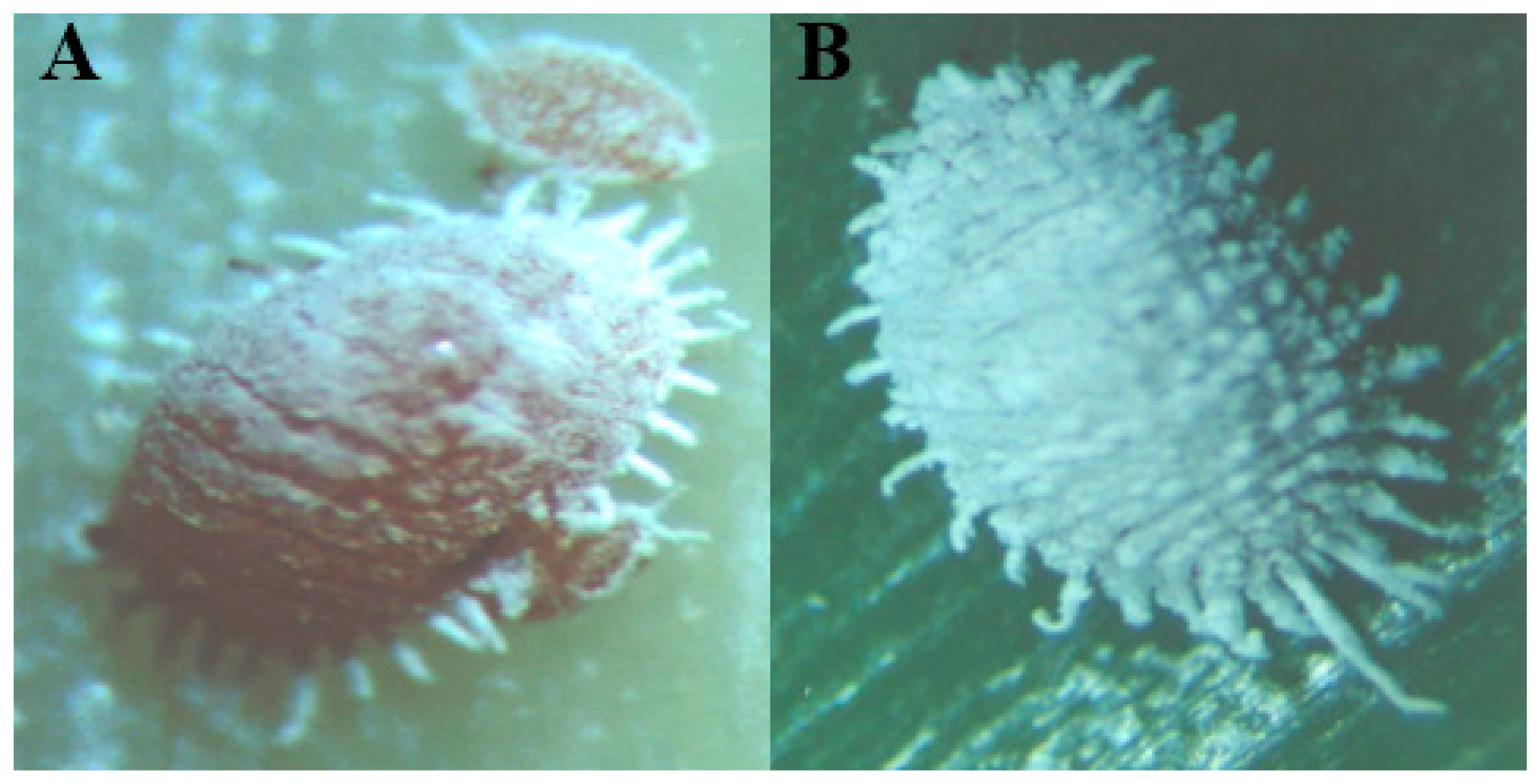
Originally, all members of the pineapple mealybug complex were considered a single-species Dysmicoccus brevipes [17], formerly named Pseudococcus brevipes [10,18]. D. brevipes was first documented from Hawaii in 1910 where it was a pest in pineapple cultivation. MWP has been only reported from areas of the world where members of the D. brevipes species complex occur [19,20]. Historically, D. brevipes was differentiated into those that caused green spots because of mealybug feeding, and non-green-spotting forms [10,13,21,22]. Carter [21] demonstrated that the green-spotting form could exhibit this characteristic, whereas the non-spotting form failed to do so even if they had fed on green spots. These two forms were eventually separated into two species: D. brevipes, the non-green spotting ones known as the pink pineapple mealybug; and D. neobrevipes, those that produce green spots, known as the gray pineapple mealybug [19] (Figure 2).
In Hawaii, green spotting was only associated with the bi-parental nature (both males and females present) of D. neobrevipes, but not seen during feeding by D. brevipes that only occur as females in Hawaii. However, in Brazil, and elsewhere in the tropics of the world where bi-parental forms are present, green spotting was also found to be associated with D. brevipes [19,23,24]. This possibly suggests that the bi-parental form of the pink mealybug may be capable of producing green spots, whereas the uni-parental form present in Hawaii does not.
Although D. brevipes and D. neobrevipes are present in all pineapple-growing regions of the world, D. brevipes are more widely distributed than D. neobrevipes. It is believed that D. neobrevipes originated in South America. D. brevipes is distributed on almost all South Pacific islands, Micronesia, and southern Asia, as well as Central and South America.
There are many differences between D. neobrevipesis and D. brevipes, in terms of their biology, host preferences, feeding behaviors, and their associated internal symbionts [14,18,19,25]. D. neobrevipesis is generally found on the aerial portions of pineapple, including fruits and flowers, and occasionally inside blossoms [19,24], whereas D. brevipes generally feeds at the base of the leaves, stems, roots, and crowns, sometimes underground [19,26]. Interestingly, in the absence of D. neobrevipes, D. brevipes may occur on aerial parts of the plant [27]. The developmental stages of the two pineapple mealybugs are similar [18], with females having three larval instars prior to the adult stage. The lifespan of D. neobrevipes ranges from 78 to 111 days, and adults produce an average of 346 first instar crawlers [18]. Males live from 31 to 47 days, and have four larval instars. Sexual differentiation takes place in the second instar. The first two instars feed actively, and by the end of the second instar phase, the insect spins a cocoon around itself in which the second, third, and fourth molting occurs. The fully-differentiated emerging males survive for only a few days [18]. In comparison, females of the pink mealybug (D. brevipes) have a life span of 59–117 days [18] and can bear up to 234 crawlers.
D. neobrevipes has a limited host range and can survive on agave (Agave sisalana), red ginger (Alpinia purpurata), koa (Acacia koa), klu (Acacia farnesiana), screw pine (Pandanas spp.), milo (Thespesia populnea), prickly pear cactus (Opuntia megacantha), and monkeypod (Samanea saman), in addition to pineapple. In contrast, D. brevipes has a very wide host range and can survive on plants from many families, including the Bromeliaceae, Umbelliferaceae, Cannaceae, Malvaceae, Euphorbiaceae, Fabaceae, Musaceae, Orchidaceae, Poaceae, and Portulacaceae.
Another less common mealybug species, the long-tailed Pseudococcus longispinus, is occasionally seen in pineapple fields, and has also been found to be associated with mealybug wilt [9]. However, in earlier experiments by Carter, no clear association with MWP was found in the field, but this species was able to induce MWP symptoms in the laboratory [14,28]. Pseudococcus longispinus is common in greenhouses and can survive well in the absence of ants [8]. In contrast to D. neobrevipes and D. brevipes, P. longispinus is oviparous. Females have four instars, and males have five [26]. Other mealybug species, such as the pink sugarcane mealybug Saccharicoccus sacchari, are also present in pineapple fields, but not considered important because they only occur sporadically and in low numbers [14].
All species of mealybugs prefer young succulent plants, and in fields where both young and old pineapple are present. Most mealybug colonies congregate on young tender plants, although populations can still be found on older plants. Before the pineapple inflorescence forms, mealybugs feed on the tender central leaves, but after the formation of fruit, they colonize the fruit and crown areas [21]. Once established on fruit and crowns, their populations expand rapidly because of the greater shelter and protection they receive from predators. Mealybugs do not survive well when infested propagation material is planted in new fields, either because of the failure of ants to establish in these new areas, or because of increased predation or parasitization [12,21]. In the advanced stages of MWP, mealybug colonies abandon the pineapple plants [21]. This may be due to fewer ants tending the mealybug colonies in response to the reducing secretion of honeydew by mealybugs on the dying plants.

2.2. Association of Ants to Mealybugs and MWP

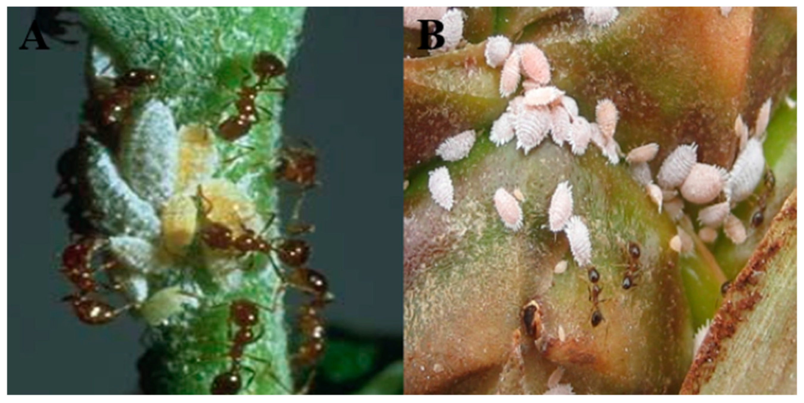
Carter first observed the correlation between ants, mealybugs, and MWP even before the virus was implicated in the disease etiology [29]. In the field, ants play a key role in dispersing mealybugs from alternate hosts or older pineapples to newer plantings of pineapple. The mutualistic relationship (Figure 3) of ants offering protection to mealybugs against their natural enemies in return for honeydew rich in amino acids and sugars secreted by mealybugs is common in many different species of ants [30]. The ants most commonly associated with pineapple mealybugs throughout the world are the Pheidole and Solenopsis species. In Hawaii, the big-headed ant (Pheidole megacephala) is the dominant ant species associated with pink and gray pineapple mealybugs [8,31,32,33,34]. P. megacephala is present throughout the tropical regions of the world. A direct relationship between high incidences of MWP and high populations of mealybugs capable of transmitting PMWaVs was shown by Sether [35,36].
Control of the ants protecting the mealybug colonies is the predominant means for checking the wilt [37]. In Hawaii, it was shown that if ants are controlled, predators keep mealybugs under control [38]. The association of wilt, mealybugs, and ants was first proposed in 1925 [8] when a relationship was noticed between controlling ants, which protect the mealybugs, and the reduction of severity of MWP disease incidence. Illingworth later showed the direct relationship between MWP and mealybugs [39].
Field studies later supported the association between mealybugs and wilt in major pineapple-growing regions of the world. Carter [10,25] proposed a hypothesis that mealybug wilt was a toxemia reaction (i.e., the saliva of mealybugs is toxic to the plant) based on his observations of the correlation between large populations of actively feeding mealybugs on pineapple plants, the length of time spent feeding, and the subsequent onset of wilting symptoms. Later, this toxin hypothesis was modified, considering new experimental evidence that was inconsistent with the toxin hypothesis.

2.3. Association of Viral Particles to MWP

The observation that exposure to large numbers of mealybugs did not always result in wilt symptoms, and that healthy plants became infected when mealybugs were transferred to them from symptomatic plants, strongly suggested the presence of a “transmissible factor” in MWP etiology [40,41]. Based on those new findings, a working hypothesis was put forward, implicating a latent virus that could be transmitted to and which could multiply in vegetatively propagated pineapple plants [42]. It was Ito [43] who drew a clear conclusion of the virus’ involvement from field experiments. He also noticed that plants that had recovered from MWP exhibited immunity to future disease. In cases where plants were incompletely protected, he presumed the involvement of different strains of the virus [43]. Unfortunately, serological techniques were not available at that time to confirm his hypothesis.
It took another two decades before various researchers were able to isolate long, flexuous, rod-shaped virus particles from MWP-symptomatic pineapple plants in Hawaii, Australia, and Cuba [44,45,46,47,48,49]. Based on particle morphology and the presence of multiple, high molecular weight, double-stranded RNAs (dsRNAs) in MWP-symptomatic plants [46,47,50], viruses were confirmed to be present in plants afflicted with MWP. The virus found was placed in the Closteroviridae family [44,51,52]. The virus, once named “pineapple closterovirus” (PC or PCV), was renamed as the “pineapple mealybug wilt-associated virus” (PMWaV) [47,49,50]. PMWaV is a currently recognized as a complex of viruses belonging to three recognized species, designated as Pineapple mealybug-wilt associated virus 1 (PMWaV-1), PMWaV-2, PMWaV-3, and the putative PMWaV-4 and PMWaV-5. Following the convention used for the Grapevine leaf roll-associated viruses [51,53,54,55,56,57] the PMWaVs were placed in the genus Ampelovirus [58]. The other genera in the family Closteroviridae are Closterovirus, Crinivirus, and Velarivirus [59]. These genera are differentiated based on virion morphologies, genome organization, and vector-transmission properties. It was also suggested that the genus Ampelovirus be further divided into two clades, based on the difference in phylogeny and genome organization between GLRaV-3 (type member of genus Ampelovirus), to which PMWaV-2 is closely related, and other Ampeloviruses, including PMWaV-1 and PMWaV-3 [55,57]. The identification of more than four related but genetically distinct viruses in pineapple is similar to Grapevine leaf roll-associated viruses identified in grape vines.

2.4. Transmission and Interaction of PMWaVs

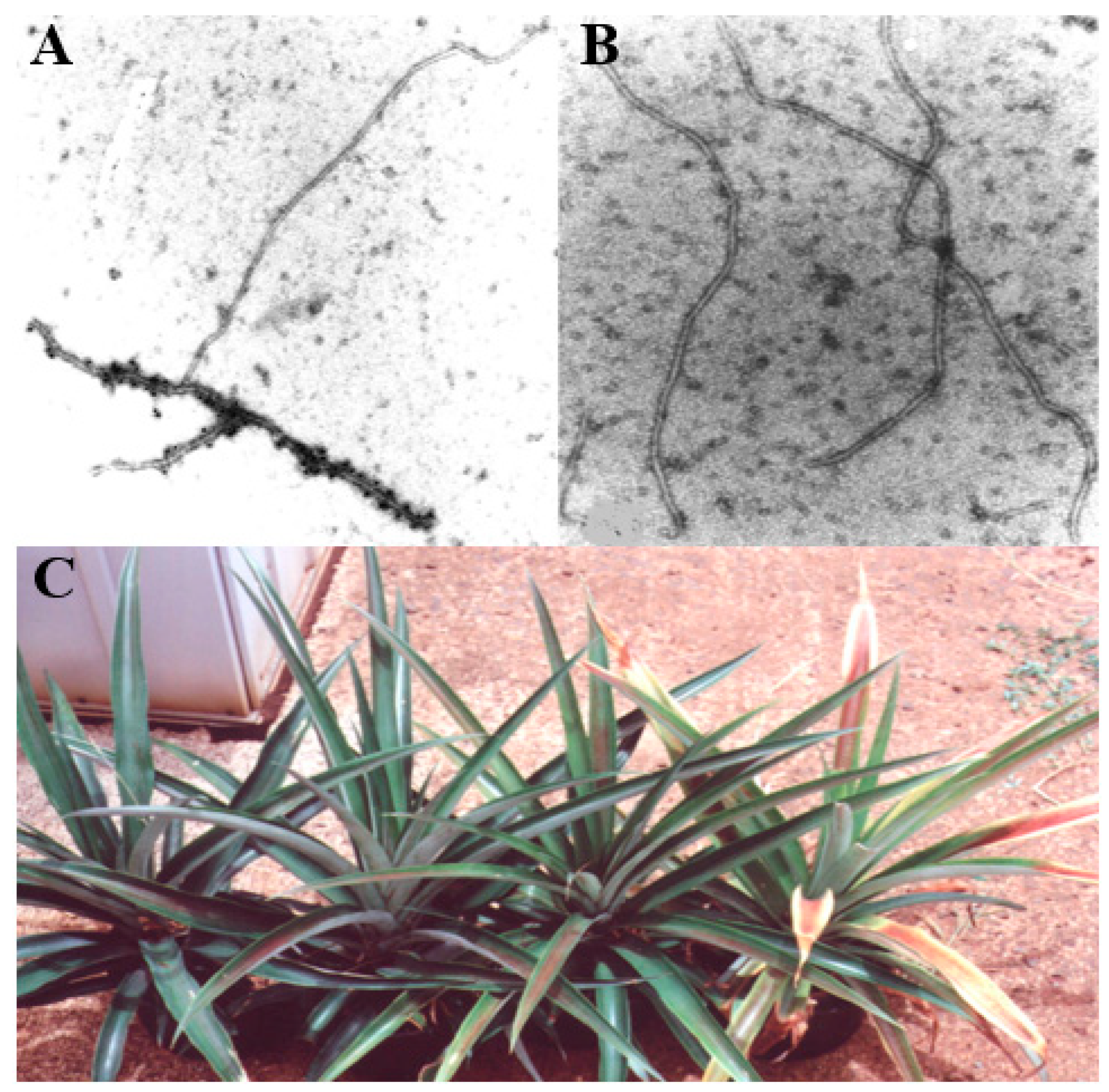
The development of monoclonal antibodies (MAbs) for PMWaV-1 [50] and PMWaV-2 [56] and the use of these antibodies in tissue-blot immunoassays (TBIAs) and immunosorbent electron microscopy (ISEM) made the accurate assessment of PMWaV infection in plants possible (Figure 4) [49]. The later development of reliable and sensitive reverse-transcription PCR (RT-PCR) assays allowed for further evaluation of the ability of mealybugs to acquire and transmit PMWaV from diseased to healthy plants. Using these assays, PMWaVs were readily detected in mealybugs collected from MWP-afflicted pineapple plants, but were absent in mealybugs reared on squash [50]. Moreover, MWP-symptomatic pineapple plants were shown to have consistently higher PMWaV infection rates than asymptomatic plants [49]. This consistent association of PMWaVs with wilting symptoms reinforced the earlier latent factor hypothesis and pointed to a virus as being the cause of MWP.
However, the etiology was complicated by several other factors. First, the expression of wilting symptoms was found to be variable and linked to many factors, including environmental conditions, mealybug populations, and pineapple genotype [12,22]. Secondly, some plants could remain asymptomatic even though they were infected with PMWaVs [49,50,60].
Findings by Sether et al. [35,61,62] suggest the involvement of only one member of the PMWaV family (PMWaV-2), together with active mealybug feeding, for MWP to develop. Neither factor alone without the other could cause the wilting symptoms to develop (Figure 4). These authors also showed that all pineapple plants with MWP symptoms had PMWaV-2 infections, but infections with PMWaV-1 or PMWaV-3 were not correlated with MWP symptom development [49,62]. In the same study, it was shown that PMWaV-1, PMWaV-2, and PMWaV-3 could all be transmitted by two pineapple mealybug species, D. brevipes and D. neobrevipes [35,36,54,63,64,65]. The correlation between infection with PMWaV-2, active mealybug feeding, and MWP symptoms suggests that some component of insect origin or possibly from an endogenous microbe present within the insect, is introduced into the plant together with PMWaV-2 during mealybug feeding.
The occurrence and levels of all PMWaVs in infected pineapple were also found to differ depending on the hybrid, the origin of planting materials, and growing locations. Similar studies by Australian workers did not show any clear association of PMWaV-1, PMWaV-2, or PMWaV-3 with MWP disease in Australia. Furthermore, PMWaV-2 was not found to be common in Australian plantings, and dual infections with PMWaV-1 and -3 or single infections with PMWaV-3 were found to be correlated with MWP symptom development. A new species (PMWaV-5) that is most closely related to PMWaV-1 has also been found in Australia, but it has not been shown to induce MWP symptoms [53].

2.5. Detection of PMWaVs

In the field of plant pathology, it cannot be over-emphasized how our growing knowledge of diseases is parallel to the advancement of technologies. MWP provides a case study in which our current understanding of the disease has advanced with the technologies available at any given time. In the early 60s, circumstantial evidence implicated a virus in MWP etiology, but this could not be proven until the advent of electron microscopy. In the 1990s, it was the development of serological techniques that greatly advanced our knowledge of the virus association and distributions in the field. The robustness of the specific monoclonal antibodies developed against PMWaV-1 and -2 enabled rapid screening of thousands of pineapple plants growing worldwide. The low titers of PMWaVs in plants and its presence in asymptomatic plants warranted the development of very specific Reverse Transcription-PCR (RT-PCR) detection techniques in the 1980s. RT-PCR, together with serology-based techniques, enabled the development of robust detection systems that allowed for screening of large numbers of pineapple samples. Furthermore, samples which gave equivocal results using ELISA (Enzyme Linked Immunosorbent Assay) or TBIA (Tissue Blot Immunoassay) assays could be verified with RT-PCR. More recently, the development of quantitative (real-time) PCR assays allowed for accurate quantification of PMWaV-2 titers in different parts of plants.
TBIA has been shown to be very practical for large-scale screening of PMWaVs, allowing the processing of hundreds of samples directly in the field with minimal preparation. TIBA membranes can be prepared and blotted in the field, and can then be shipped to a laboratory for testing instead of transporting infected planting material itself for testing, thereby avoiding quarantine-related problems. A potential shortcoming of TBIA is its inability to reliably detect infection when virus titers are low.
Techniques such as loop-mediated isothermal amplification (LAMP) could also be useful [66]. In addition to its ability to directly detect PMWaVs in the field, it also has much higher sensitivity than TBIA. Recently, a complete “lab on a chip” has been developed for the rapid detection of PMWaVs. This chip completes RNA extraction, reverse-transcription, and fluorescence detection by PCR at isothermal temperatures in under 40 min, with a sensitivity threshold of 50 DNA copies/reaction. However, the cost of applying this technique to large-scale screenings may be a prohibitive [67]. A simple colorimetric technique using gold nanoparticles in LAMP reactions to detect PMWaV-1 and PMWaV-2 has also been developed, which takes 40 min to complete and has a detection limit of 10 copies of viral DNA. Another novel technique, single-tube dual primer-PCR (STDP-PCR) using nested PCR primers has been developed that can detect very low titers of PMWaV-2 that are below the levels detected with RT-PCR [68].
In recent surveys for PMWaVs in Hawaii and Australia, two new strains of PMWaVs, namely PMWaV-3 and PMWaV-5, have been identified, suggesting the possibility of more diverse PMWaVs in pineapple that the degenerate PCR primers currently available cannot detect. Double-stranded RNA (dsRNA) analysis is more likely to identify other positive-sense RNA viruses present in pineapple tissues, but very low titer viruses may not be identified by this approach. The fact that pineapple plants have been identified that are infected with badnaviruses and other pararetoviruses, and may be infected with other unknown viruses, calls for a metagenomic analysis of pineapple. This approach has been shown to be able to identify the whole viral complement present in plants [69,70]. Next-generation sequencing (NGS) is finding greater use for identifying diseases of unknown etiology. Coetzee [71] used NGS to create a whole viral profile in diseased vineyards, and identified a new GLRaV-3 variant not previously known. Although the cost of NGS is dropping, it has not yet become useful for routine diagnoses. However, it is a very powerful tool that can reveal the identities of the virus(s) present and may help in the process of elucidating the etiology of diseases where a complex of viruses, such as GLRaVs and PMWaVs, may be involved.

3. Genome Organization and Diversity of PMWaVs

3.1. PMWaV-2

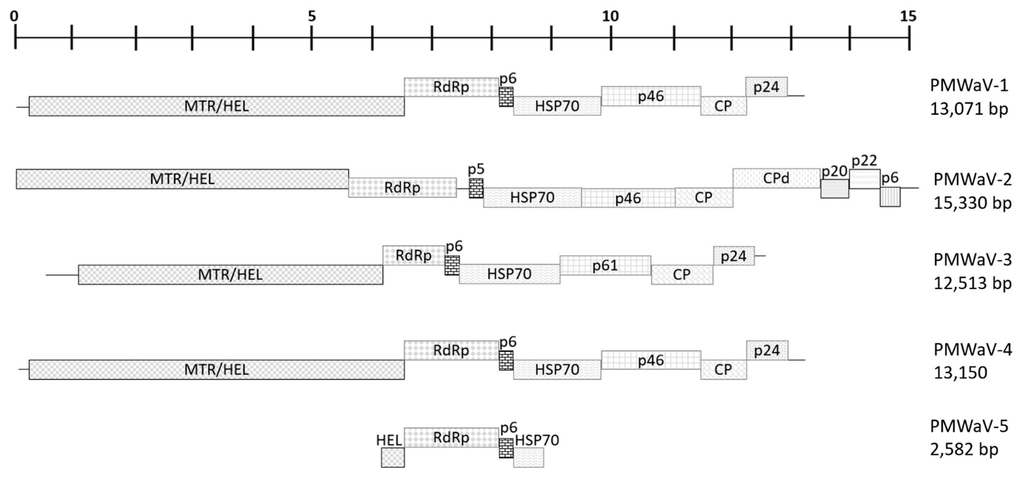
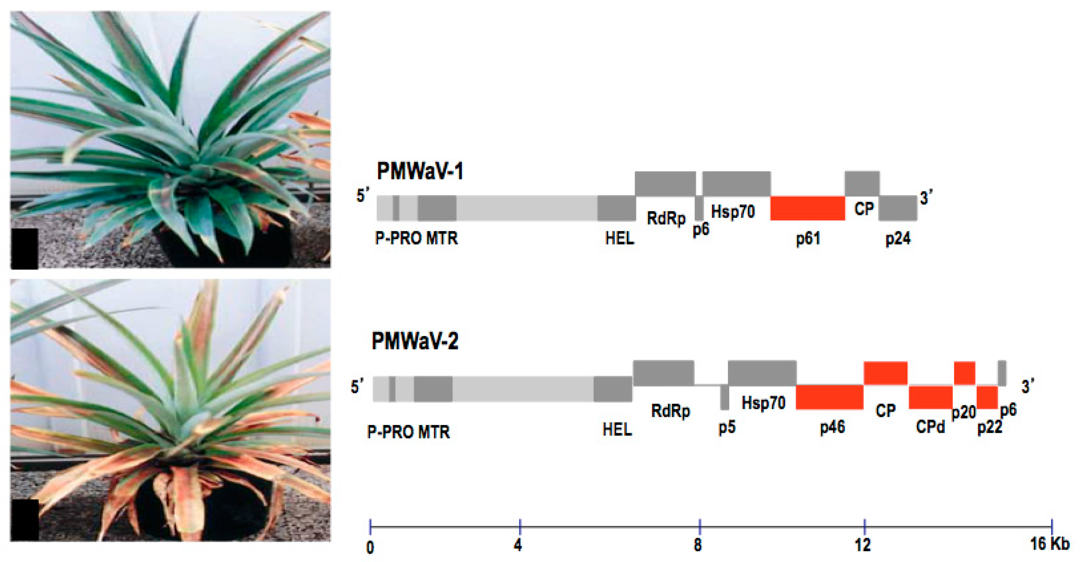
PMWaV virions are flexuous rods, with lengths of about 1200 nm and diameters of 10 to 12 nm. The linear, positive-sense, single-stranded RNA genomes of these viruses are variable in size, ranging between 13 and 15.5 kilobases. PMWaV-2 (AF283103) [56], which had been found to have a role in MWP in Hawaii, has most of its genome sequenced. The 14,851 nt of the genome contains 10 open reading frames (ORFs) which, from 5’ to 3’, encode: a > 204 kDa polyprotein containing papain-like protease, methyltransferase, and helicase domains (ORFla); a 65 kDa RNA-dependent RNA polymerase (ORFlb); a 5 kDa hydrophobic protein (ORF2); a 59 kDa heat-shock protein 70 homologue (ORF3); a 46 kDa protein with unknown function (ORF4); a 34 kDa coat protein (ORF5); a 56 kDa diverged coat protein (ORF6); a 20 kDa protein with unknown function (ORF7); a 22 kDa protein with unknown function (ORF8); and a 6 kDa protein with unknown function (ORF9). A 132 nucleotide-untranslated region is present at the 3’-terminus of the genome. A genomic frame shift presumably allows expression of ORFlb (Figure 5).

3.2. PMWaV-1 and -3

The complete genome of PMWaV-1 and partial genome of PMWaV-3 have been sequenced (PMWaV-1, AF414119; PMWaV-3, DQ399259). PMWaV-1 and -3 both have seven open reading frames, but PMWaV-1 lacks the intergenic region that occurs between RdRp and p6 ORFs in PMWaV-3, and the CPd ORF [57]. The PMWaV-3 genome sequence encompasses 11891 nt, and encodes seven ORFs and the untranslatable region at the 3’-end [55,68]. PMWaV-3 lacks an intergenic region between ORFlb and ORF2 that is present in PMWaV-2, and encodes a 28.8 kDa coat protein but lacks the coat protein duplicate (CPd) found in PMWaV-2. (Figure 5) PMWaV-3 shares amino-acid identity with PMWaV-1 of 63.9%, 72.5%, and 79.2% for the RNA-dependent RNA polymerase, small hydrophobic protein, and HSP-70-homolog ORFs, respectively.

3.3. Putative PMWaV-4

In the past, a TBIA [50,62] using monoclonal antibodies (MAb) specific to PMWaV-1 or PMWaV-2 was used for virus detection when screening pineapple; if the TBIA was negative for PMWaV-1 or 2, then Ampelovirus degenerate HSP70 primers were used to investigate the potential of novel PMWaVs other than PMWaV-1 or PMWaV-2 underlying the discovery of PMWaV-3 and the putative PMWaV-4.
The sequenced HSP70 gene of the putative PMWaV-4 has a 74% nt and 87% aa similarity to PMWaV-1 [55]. However, at the time, the criteria for species demarcation for inclusion in the Ampelovirus genus was 10% similarity of the amino acid sequence (RdRp, HSP70 and CP) indicating a possible new fourth species of PMWaV. The amino acid sequence similarity from recent NGS data of PMWaV-4 compared to PMWaV-1 (AF414119) is 89% for the RdRp, 87% for HSP70, and 85% for the CP, and based on the additional data generated from this study, we can now confirm that the putative PMWaV-4 is a strain of PMWaV-1 and not a separate species [72]. Additionally, it was reported in China that the PMWaV-1 isolate Hainan (PMWaV-1 HN) has an additional 72 bp in the 3’ of the HSP70 gene compared to other PMWaV-1 isolates. The additional sequence on PMWaV-1 HN encodes an aa residue of “ETGLLTLGRQQREIIYKRHGFESN”, and interestingly has a 65% similarity to that of the 3’ end of the PMWaV-4 HSP70 gene, which has the same increased length of coding region [73].
Further, the antibodies developed for a TBIA [50] to detect PMWaV-1 and PMWaV-2 were used to test if PMWaV-4 (now known to be a strain of PMWaV-1—not a distinct species) could be detected; the MAb developed to detect PMWaV-1 were unable to detect the PMWaV-4 strain of PMWaV-1.

3.4. Putative PMWaV-5

In Australia, a putative fifth PMWaV, PMWaV-5, was reported in 2008 by Gambley [53], prior to the revised Ampelovirus species demarcation criteria by Martelli [74] in 2012. This fifth PMWaV was detected in 42 separate plants from 4 separate locations in symptomless and symptomatic plants. Only four sequences of the putative PMWaV-5 were reported, comprising of partial coding sequences (CDS) for the HEL, RdRp, p6, and HSP70 genes (Figure 5). PMWaV-5 is grouped into subgroup II of Ampeloviruses, as it shows a higher level of sequence homology to PMWaV-1, PMWaV-3, and other members of subgroup II than the subgroup I members. A 546 bp partial CDS of the HSP70 gene of PMWaV-5 (EF488753.1) shares 69% nt and 72% aa similarity to PMWaV-1 (NC_010178.1), and 66% nt and 67% aa similarity to PMWaV-3 (DQ399259.2). A 1546 bp partial CDS of the HEL and RdRp genes of PMWaV-5 (EF467922.1) shares 68% nt and 72% aa (RdRp), 64% aa (HEL) similarity to PMWaV-1 (NC_010178.1), and 67% nt and 67% aa (RdRp), 71% aa (HEL) similarity to PMWaV-3 (DQ399259.2). A 551 bp partial CDS of the HEL gene of PMWaV-5 (EF467921.1) shares 79% nt and 55% aa similarity to PMWaV-1 (NC_010178.1), and 79% nt and 53% aa similarity to PMWaV-3 (DQ399259.2). A 1751 bp partial CDS of the RdRp and HSP70 and a complete CDS of the p6genes of PMWaV-5 (EF467920.1) shares a 66% nt and 69% aa (RdRp), 69% aa (HSP70), 57% aa (p6) similarity to PMWaV-1 (NC_010178.1), and 66% nt and 65% aa (RdRp), 63% (HSP70), 57% (p6) similarity to PMWaV-3 (DQ399259.2). Based on the revised species demarcation criteria for Ampeloviruses, and due to the limited sequence information available, we cannot confidently conclude that PMWaV-5 is a distinct species from the other recognized PMWaV-type members.

3.5. Phylogenetic Analysis of PMWaV Members

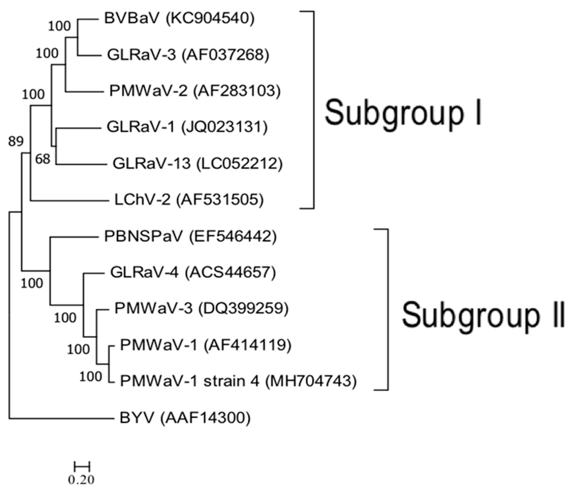
Based on phylogenetic analysis, PMWaV-1, PMWaV-3, PMWaV-4, Plum bark necrosis stem pitting-associated virus (PBNSPaV), and some GLRaVs belong to a distinct clade (Subgroup II) within the genus Ampelovirus (Figure 6) [55,57,69,70]. Phylogenetic analysis establishes that the PMWaVs are more closely related to other mealybug-transmitted viruses than to either aphid- or whitefly-transmitted closteroviruses [56]. PMWaV-2 shares the greatest sequence identity with the mealybug-transmitted Grapevine leafroll-associated virus-3 (GLRaV-3); amino acid identities between PMWaV-2 and GLRaV-3 are 47.5, 51.2, 47.0, 30.1, 42.2, and 30.5% for the helicase, RdRp, HSP70, p46, CP, and CPd, respectively [56]. Sequence homology between PMWaV-3, PMWaV-1, and PMWaV-2 decreases across the HSP-70 homolog, small hydrophobic protein, and RNA-dependent RNA polymerase open reading frames (ORF). PMWaV-3 cannot be detected using monoclonal antibodies specific for PMWaV-1 or PMWaV-2. PMWaV-1, -2, and -3 can all be transmitted by the pink pineapple mealybug, D. brevipes, and the grey pineapple mealybug, D. neobrevipes [35,36,54,69]. Primarily based on their mealybug transmissibility, PMWaV-1, PMWaV-2, and tentatively, PMWaV-3 have been placed in the Ampelovirus genus [58].
Multi-loci analysis was done using a concatenated amino acid sequence of the RNA-dependent RNA polymerase (RdRp), heat-shock protein 70 homolog (HSP70h), and coat protein (CP) genes of the putative pineapple mealybug wilt-associated virus-4 (PMWaV-4) in relation to ampelovirus member species. The multi-loci analysis resulted in a phylogenetic tree constructed using the Maximum Likelihood method based on the Le Gascuel model. The tree was drawn to scale, branch lengths were measured in the number of substitutions per site, and there was a total of 1159 positions in the final dataset. The sequences of the following viruses were retrieved from GenBank and included in the analysis: Blackberry vein banding-associated virus (KC904540), Grapevine leafroll-associated virus-1 (GLRaV-1, JQ023131), GLRaV-3 (AF037268), GLRaV-4 (ACS44657), GLRaV-13 (LC052212), Little cherry virus 2 (LChV-2, AF531505), Plum bark necrosis stem pitting-associated virus (PBNSPaV, EF546442), PMWaV-1 (AF414119), PMWaV-2 (AF283103), PMWaV-3 (DQ399259), and the closterovirus type member Beet yellow virus (AAF14300).

4. Role of RNA-Silencing Suppressors in the Etiology of MWP

RNA silencing is generally recognized as the major anti-viral-defense response in plants [75]. It is generally recognized that almost all viruses encode proteins, which can function as a suppressor of gene silencing. In the case of viruses with long and complex genomes, as within the family Closteroviridae, the role of suppressor proteins was found to be even more critical in its life cycle. It was hypothesized that to have such a slow replication cycle in their host, the majority of such viruses employ some multi-component and/or multi-level counter-defense mechanisms to protect their degradation from attack by host RNA-silencing machinery [76], as opposed to viruses with smaller genomes which can replicate faster and evade RNA silencing. The discovery that Citrus tristeza virus (CTV) encodes three suppressors, which operates both locally and systemically, explains how it switches from abundant expression of the local suppressor protein as it enters the host, trying to establish itself, to the expression of systemic suppressors, further enabling it to systemically move to distant tissues [77]. Similar multi-component silencing suppression has also been discovered in the Tomato chlorosis virus [78] and Sweet potato chlorotic stunt virus (SPCSV) [79], all members of the family Closteroviridae. Screening the genomes of viruses in Closteroviridae have revealed that the majority of such proteins are located towards the 3’ end of their genomes, all expressed via the nested set of the 3’ coterminal subgenomic (sg) RNAs, another strategy for the abundant production of proteins.
It was therefore not surprising when multiple suppressors (two local suppressors, CP and p20, and three systemic suppressors, CPd, CP, p22, and p20) were identified in the genome of PMWaV-2 (Figure 7), suggesting that a similar multi-component silencing suppression strategy might be employed by this virus in this pathosystem [80]. Further analyses of the local suppressors have identified p20 and p22 of PMWaV-2 to be determinants of pathogenicity factors or virulence factors. Many viral encoded proteins that were initially identified as determinants of pathogenicity or virulence factors were later identified as suppressors of RNA silencing [81,82]. In comparison, screening the PMWaV-1 genome showed only one protein, p61, with systemic silencing-suppressor activity [80] (Figure 7).
The fact that PMWaV-2 is involved in MWP symptom induction, but not PMWaV-1 in the etiology of MWP [36] might be due to the local suppressor and pathogenicity factors identified in PMWaV-2 that are absent in PMWaV-1. The different silencing suppressors present in the genomes of PMWaV-2 and PMWaV-1 might also explain how viruses of perennial crops such as pineapple are susceptible to persistent viral infections that are not lethal to the host, whereas many viruses that encode strong suppressors and infect herbaceous hosts are often lethal. The absence of any local suppressor activity in PMWaV-1, which only encodes a single protein with systemic suppressor activity, further supports our hypothesis that viruses encoding only weak systemic silencing suppressors might be favored in persistent infections that do not lead to plant death.
The identification of suppressors of RNA silencing in the genomes of PMWaV-1 and PMWaV-2 could be further explored to gain deeper insights into the role of viral gene products in mealybug wilt of pineapple. One promising technique which enables direct testing of the role of viral gene products in the host, is by creating an artificial clone of the virus, also known as an infectious clone. By systematically knocking out targeted viral genes, it would make it possible to reveal their functions in the host plant. To confirm the involvement of the identified suppressors and pathogenicity factors in MWP, development of an infectious clone of PMWaV-2 has been initiated [80,83]. This research might eventually pin-point the role of PMWaV in the complex etiology of MWP.

5. Badnaviruses Infecting Pineapple

5.1. Badnaviruses

Badnavirus-like particles have often been observed in purified preparations of pineapple in Hawaii [35] and Australia [64], and more recently in Cuba [84]. Badnaviruses are reverse-transcribing viruses belonging to the family of Caulimoviridae. They are non-enveloped, bacilliform-shaped virions approximately 30 nm wide and 120 to 150 nm long [85]. Their genomes are approximately 7 to 9 kilobases [86] circular, noncovalently closed, double-stranded DNAs that encode at least three ORFs. Of the eight genera (Badnavirus, Caulimovirus, Cavemovirus, Petuvirus, Rosadnavirus, Solendovirus, Soymovirus, and Tungrovirus) [87], the badnavirus is the most important genera due to many cultivated plants being infected by it. Most of such plants are perennial in nature and propagated vegetatively. This might explain the large number of such badnaviruses being discovered in those plants. There are 40 badnavirus species currently being recognized by the International Committee on Taxomomy of Viruses (https://talk.ictvonline.org/taxonomy/). One particular importance is their ability to integrate in their host genomes [88,89] for which they are referred to as endogeneous pararetrovirus (EPRVs). The integration is believed to be due to illegitimate recombination with the host genome. Some of them also have the ability to “excise” from their host genome and exist as an independent entity, thereby being capable of systemic infection (episomal forms); [90]. In some cases, abiotic stress, such as temperature fluctuations, nutrient scarcity, or tissue culture manipulations, were known to trigger the virus to cause infection [91]. Many integrated sequences have been shown to have a relatively high degree of sequence identity to several non-integrative viruses in the Caulimoviridae family [85,92,93]. These include the Banana streak virus [92,94,95], and Rice tungro virus. Badnaviruses and Ampeloviruses are transmitted by insects. The Banana streak virus (BSV), Cocoa swollen shoot virus (CSSV), Kalanchoë top-spotting virus (KTSV), Schefflera ringspot virus (SRV), Canna yellow mottle virus (CaYMV), Piper yellow mottle virus (PYMV), and Sugarcane bacilliform virus (SCBV) are transmitted by mealybugs. Aside from insect vector transmission, BSV is also transmitted through seeds [96], but because endogenous BSV sequences occur in the nuclear genome of most cultivars [91], seed transmission is difficult to show. Some of the economically important badaviruses are the Banana streak virus (BSV) [97,98], Cacao swollen shoot virus (CSSV) [99], Canna yellow mottle virus (CaYMV) [100], Dioscorea bacilliform AL virus (DBV) [101,102], Kalanchoe top-spotting virus (KTSV) [103], Piper yellow mottle virus (PYMV) [104], Schefflera ringspot virus (SRV) [105], Sugarcane bacilliform virus (SCBV) [100], and Citrus yellow mosaic virus (CYMV) [106].

5.2. Synergism of Badnaviruses

Several reports have demonstrated a synergism between badnaviruses and other unrelated viruses in co-infections in plants, which may result in severe symptom development. For example, rice tungro disease results from synergism between the badnavirus-like Rice tungro bacilliform virus and an unrelated spherical virus [107]. Although no clear association between badnavirus infection and symptoms in pineapple have been observed [108], the synergism displayed by badnaviruses with other unrelated viruses in various crops raises concerns that the badnavirus in pineapple may have a synergistic effect with the PMWaVs in MWP symptom development.

5.3. Pineapple Bacilliform Viruses

Virions of Pineapple bacilliform virus (PBV), a tentative badnavirus species, were first detected in pineapples by Walkman et al. in 1995 [60] by viewing them directly under a transmission electron microscope (TEM). In 1996, Thomson et al. [109] described a badnavirus-like sequence (PBV) in pineapples. This sequence was later also detected by PCR in a large-scale field survey. Based upon sequencing of this virus, a degenerate PCR assay was developed for use together with immune-capture PCR in other large-scale surveys studying the diversity of badnaviruses in pineapples grown in Australia [53]. In that study, two new badnaviruses, the Pineapple bacilliform comosus virus (PBCoV) and Pineapple bacilliform erectifolius virus (PBErV), and an endogenous badnavirus, Endogenous pineapple pararetrovirus-1 (ePPPV-1), and a retrotransposon, Ananas metavirus (AMtV), which was previously identified as pineapple badnavirus (PBV) [64] were identified. In a more recent survey done on Hawaiian pineapples by Sether [110] in 2012, similar diversity of pineapple badnaviruses was found. Pineapple badnavirus, designated as Pineapple bacilliform comosus virus –HI1 (PBCoV-HI1) along with nine genomic variants of this virus (A through H) were identified. Sequence comparisons for the RT and RNaseH regions of PBCoV-HI1 showed high nucleotide identity (98%) with PBCoV from China [111] and a partial sequence of pineapple badnavirus characterized from Australia. This suggests that these viruses may be strains or variants of a single species. However, at the amino acid level, the identities for ORF1, ORF2, and ORF3 were only 47% to 80%. Please refer to https://talk.ictvonline.org/taxonomy/ for more updated and complete information about the recent classification of badnaviruses.
These studies also looked at the mealybug transmissibility of badnaviruses and their possible role in MWP. Gambley [108] examined the transmissibility of Australian PBCoV and PBErV by pink pineapple mealybugs (D. brevipes) and the citrus mealybug (Planococcus citri) and found a 20% transmission rate of PBCoV (by D. brevipes) and a 10% transmission rate by P. citri. No transmission of PBErV by either of these mealybug species was detected. All the test plants remained symptomless throughout the study period. In work carried out in Hawaii, Sether [110] found the transmission of PMCoV-HI1 and PMCoV-HI1 variant A by grey pineapple mealybugs (D. neobrevipes) both to be 80%.
In a more recent survey for PMWaVs and badnaviruses in commercially-grown pineapple hybrids which were recently imported into Hawaii, and 131 pineapple accessions maintained at the USDA-ARS germplasm repository in Hilo, Hi, Subere [112] found four badnavirus-like sequences that were tentatively grouped into four clades (A, B, C, and D), occurring in mixed infections with PMWaV-1, -2, -3, or 4, using reverse-transcription PCR assays with degenerate and specific primers [112]. Badnavirus-like particles were also confirmed by electron microscopy in the same study. Based on the widespread occurrence seen in these surveys, and no clear correlation of their presence with MWP symptoms, it is now believed that these badnaviruses are not the primary cause of MWD [35,112]. However, considering the synergistic relationships that many badnaviruses have with various unrelated viruses, and the finding that PMWaV-2 alone does not have an unequivocal role in the etiology of MWP, further research into this relationship needs to be conducted.

6. Control of MWP

The realization that ants play a key role in protecting mealybugs led to the aim of reducing ant populations to control the spread of mealybugs early on in the history of MWP. Various cultural practices, such as the removal of old pineapple stumps, destroying wilt-infected plants and pineapples after the first ratoon harvest, and initiating fallow periods, have all been found to be effective for the integrated management of MWP. The quickest control of ant and mealybug populations is by chemical means; treatment of planting materials by insecticide dips was found to be effective for the pre-planting control of mealybugs. Spray applications of systemic insecticides, such as Diazinon® and other contact insecticides are very effective in killing mealybugs, but because mealybugs live within the crowns and blossom cups of plants, they are often missed by such sprays [24]. Moreover, spray applications need to ensure that the entire field is sprayed, leaving no refuge available for the mealybugs. Insecticidal baits are the most common and effective way to control ants in pineapple fields [113]. One of the most promising baits based upon extensive field trials are Amdro® and other insect-growth regulators [8,32,33].
Because of environmental concerns, insecticidal formulations are coming under heavy regulation, and more emphasis is being placed on natural solutions, such as biological control for mealybugs. Biological controls for the pink and grey pineapple mealybug were sought in Central America, the center of the origin of pineapple [25]. Several potential natural enemies have been introduced into Hawaii [14], including Lobodiplosis pseudococci Felt (Diptera: Cecidomyiidae), Nephus bilucernarius Mulsant (Coleoptera: Coccinellidae), and Anagyrus ananatis Gahan (Hymenoptera: Encyridae), but none have provided sufficient control of mealybugs when ants are present [8,14]. The search for natural enemies of ants has primarily focused on the control of Solenopsis spp. [114]. Fungal and bacterial pathogens of ants are rare because of the antibiotic exocrine secretions that ants produce.
The direct introduction of transgenes into plants is an effective tool that can be used to modify undesirable traits or introduce new traits into plants, especially in cases where conventional breeding is not possible for several reasons. For example, the pineapple cultivar “Smooth Cayenne”, which is very susceptible to mealybug wilt [13], is not amenable to conventional breeding due to sterility problems [115]. Genetic transformation has been used to produce transgenic pineapple plants with putative resistance to MWP, using various gene constructs which employ RNA-mediated resistance technology [116]. The coat protein (CP) gene of PMWaV-2 was constructed as an inverted-repeat in the pCAMBIA 1300 vector, and used to produce transgenic pineapple plants using particle bombardment and Agrobacterium-mediated transformation approaches. Several lines of putatively transgenic pineapple plants resistant to PMWaV-2 were produced. These plants remained resistant to PMWaV-2 infection after multiple challenges with viruliferous mealybugs, but the transformed lines eventually became infected with PMWaV [65]. Although no further attempts have been undertaken to make transgenic pineapple plants with resistance to MWP, the recent development of a transgenic “Smooth Cayenne” pineapple engineered for resistance to Black Heart using an RNA-silencing approach [116] could encourage researchers to revisit transgenic work designed to produce plants with MWP resistance.

7. Conclusions and Future Perspectives

Previous studies have shown a positive correlation between the induction of MWP symptoms with mealybug feeding and PMWaV-2 infection in “Smooth Cayenne” cultivars in Hawaii. Similar correlations have also been shown for some hybrid plants with either single PMWaV-2 infections or combinations of PMWaV-1 or PMWaV-3 with PMWaV-2. However, this trend was not observed in surveys carried out in Australia where MWP was not consistently associated with PMWaV-1, PMWaV-2, PMWaV-3, or PMWaV-5 infections. MWP symptoms were found to vary between hybrids and “Smooth Cayenne” cultivars, whose reddening of leaves could be due to their higher anthocyanin content.
Observations that MWP was present on plants without a PMWaV-2 infection but also in association with other PMWaV variants raises the possibility that other PMWaV-2-like viruses may also be present. Moreover, the detection of badnaviruses in mixed infections with PMWaVs calls for a deeper investigation of the roles of such viruses. With the large-scale planting of hybrid varieties throughout the world replacing the traditional “Smooth Cayenne” variety, there is a shift in the dynamics of the host pathogen interactions. Things like how the nature of the badnaviruses are known to infect and cause damage in important tropical crops, the integration of badnavirus sequences into some host plant genomes, the synergistic relationships of many badnaviruses with other viruses, and badnavirus transmissibility by mealybugs, indicate and justify the need to further investigate this plant virus group.
The complexity of the disease etiology of MWP suggests commonalities with grapevine viruses, and novel techniques used in these studies may be applicable in the study of MWP. For example, in the decline of Syrah, grapevines can remain asymptomatic or only show mild symptoms. Results from high-throughput sequencing associated with Syrah decline [70] revealed mixed infections of grapevine with three RNA viruses already known to be associated with Syrah decline, but also other species at very low titers, including a viroid identified as an Australian grapevine viroid. Moreover, it has been reported that diseases of grapevine could be due to the modified interactions of multiple viruses that, individually, may be benign. In such mixed infections, the Rupestris stem pitting-associated virus (RSPaV) [117] has been linked with declining symptoms of Syrah vines from California and France. Interestingly, RSPaV may be asymptomatic when present singly in grapevines. This situation is analogous to MWP with an association of PMWaV-2 with mealybug in the disease etiology in Hawaii, while in Australia, symptom induction was rarely found to be associated with PMWaV-2, but more often associated with PMWaV-1 and PMWaV-3. The presence of other undetected Ampeloviruses still yet to be found cannot be dismissed, even though the present serological and molecular detection techniques have been unsuccessful.
The association of badnaviruses with pineapple and the possibility of other unidentified agents associated with MWP still yet to be found can be addressed by looking at signature molecules called short interfering RNAs (siRNA). Plants generate virus-specific siRNAs as a defense against RNA viruses and some DNA viruses [118]. Although they are only 20 to 24 nucleotides in length, these siRNAs have been used to reconstruct entire genomes of viruses from infected plants [119]. In the case of MWP, deep-sequencing of siRNAs from plants which do not show a strong correlation with PMWaV-2 with wilting symptoms might help to pinpoint the agents responsible for disease, since siRNAs will be produced by the host pineapple plants in response to any viruses, identified or unidentified. Another advantage of this approach would be the deep-sequencing of the vector mealybug, and allowing us to look for any insect virus responsible for MWP together with PMWaVs in mealybugs. Similar research has also been done with whiteflies (Bemesia tabaci) which vector many plant viruses [119].

Author Contributions

K.K.D., J.C.G. († next to name) share primary authorship and M.M., W.B., J.S.H. contributed to editing and revision of the manuscript.

Funding

The research was funded in part by the United State Department of Agriculture National Institute of Food and Agriculture, Hatch HAW09025-H (1001478), and the United State Department of Agriculture-Agricultural Research Service (58-5320-4-012).

Conflicts of Interest

The authors declare no conflict of interest.

References

  1. Krauss, B.H. Anatomy of the vegetative organs of the pineapple, Ananas comosus (L.) Merr Merr. I. Introduction, organography, the stem, and the lateral branch or axillary buds. Bot. Gazette 1948, 110, 159–217. [Google Scholar] [CrossRef]
  2. Samson, J.A. Pineapple. In Tropical Fruits. Gordon Wrigley; Logman Scientic and Technical; Longman Inc.: New York, NY, USA, 1986; pp. 190–215. [Google Scholar]
  3. Collins, J.L. The Pineapple; Leonard Hill Books Ltd.: London, UK, 1960. [Google Scholar]
  4. Larsen, J.L.; Marks, T.A. Hawaiian Pineapple Entrepreneurs 1894–2010; Creative Co.: McMinnville, OR, USA, 2010. [Google Scholar]
  5. Gortner, W.A. Field-Fresh Pineapple for Export; Pineapple Research Institute of Hawaii: Honolulu, HI, USA, 1963; PRI no.99. [Google Scholar]
  6. Bartholomew, D. MD-2’pineapple transforms the world’s pineapple fresh fruit export industry. Pineapple News 2009, 16, 2–5. [Google Scholar]
  7. Larsen, L.D. Diseases of pineapple. Hawaii Sugar Planters Association Pathol. Physiol. Ser. Exp. Stn. Bull. 1910, 10, 1–72. [Google Scholar]
  8. Rohrbach, K.G.; Beardsley, J.; German, T.; Reimer, N.; Sanford, W. Mealybug wilt, mealybugs, and ants of pineapple. Plant Dis. 1988, 72, 558–565. [Google Scholar] [CrossRef]
  9. German, T.L.; Ullman, D.E.; Gunashinghe, U. Advances in Disease Vector Research; Springer: Berlin, Germany, 1992; pp. 241–259. [Google Scholar]
  10. Carter, W. The pineapple mealy bug, Pseudococcus brevipes, and wilt of pineapples. Phytopathology 1933, 23, 207–242. [Google Scholar]
  11. Singh, S.; Sastry, K. Wilt of pineapple-a new virus disease in India. Indian Phytopathol. 1974, 27, 298–303. [Google Scholar]
  12. Carter, W. Some etiological aspects of mealybug wilt. Phytopathology 1944, 35, 305–315. [Google Scholar]
  13. Carter, W.; Collins, J. Resistance to mealybug wilt of pineapple with special reference to a Cayenne-Queen hybrid. Phytopathology 1947, 37, 332–348. [Google Scholar]
  14. Carter, W. Insects and Related Pests of Pineapple in Hawaii; A Manual for Fieldman; Pineapple Research Institute of Hawaii: Honolulu, HI, USA, 1967; pp. 36–53. [Google Scholar]
  15. Lyon, H. A survey of the pineapple problems. Hawaii Plant Rec. 1915, 13, 125–139. [Google Scholar]
  16. Williams, D.; Fleisch, H. Historical review of pineapple breeding in Hawaii. In Proceedings of the 1st International Pineapple Symposium, Honolulu, HI, USA, 2–6 November 1992; pp. 67–76. [Google Scholar]
  17. Ferris, G.F. Atlas of the Scale Insects of North America, V, The Pseudococcidae (Part I); Stanford University Press: Redwood City, CA, USA, 1950. [Google Scholar]
  18. Ito, K. Studies on the Life History of the Pineapple Mealybug, Pseudococcus brevipes (Ckll.). J. Econ. Entomol. 1983, 31, 291–298. [Google Scholar] [CrossRef]
  19. Beardsley, J.W. On the taxonomy of pineapple mealybugs in Hawaii, with a description of a previously unnamed species (Homoptera: Pseudococcidae). Proc. Hawaiian Entomol. Soc. 1959, 17, 29–37. [Google Scholar]
  20. Carter, W. Insects in Relation to Plant Disease; Interscience Publishers (John Wiley & Sons): New York, London, UK, 1962. [Google Scholar]
  21. Carter, W. Studies of populations of Pseudococcus brevipes (Ckl.) occurring on pineapple plants. Ecology 1932, 13, 296–304. [Google Scholar] [CrossRef]
  22. Collins, J.; Carter, W. Wilt resistant mutations in the Cayenne variety of pineapple. Phytopathology 1954, 44, 662–666. [Google Scholar]
  23. Beardsley, J.W. Notes on the pineapple mealybug complex, with descriptions of two new species (Homoptera: Pseudocoecidae). Proc. Hawaiian Entomol. Soc. 1965, 19, 55–68. [Google Scholar]
  24. Jahn, G.C. Gray Pineapple Mealybugs, Dysmicoccus neobrevipes Beardsley (Homoptera: Pseudococcidae), Inside Closed Pineapple Blossom Cups; University of Hawaii at Manoa: Honolulu, HI, USA, 1993. [Google Scholar]
  25. Carter, W. Studies on the biological control of Pseudococcus brevipes and wilt of pineapple. Phytopathology 1935, 28, 1037–1041. [Google Scholar]
  26. McKenzie, H.L. Mealybugs of California: With Taxonomy, Biology, and Control of North American Species (Homoptera, Coccoidea, Pseudococcidae); University of California Press: Berkeley, CA, USA, 1967. [Google Scholar]
  27. Gonzalez-Hernandez, H. The Status of Biological Control of Pineapple Mealybugs in Hawaii. Ph.D. Thesis, University of Hawaii at Manoa, Honolulu, HI, USA, 1995. [Google Scholar]
  28. Petty, G. The Pineapple Mealybug; Pineapple H.15; Farming in South Africa, Department of Agricultural Technical Services: South Africa, 1978.
  29. Carter, W. A Study of Mealybug Populations (Dysmicoccus brevipes (Ckl.)) in an Ant-Free Field1. J. Econ. Entomol. 1960, 53, 296–299. [Google Scholar] [CrossRef]
  30. Jahn, G.C. The Ecological Significance of the Big-Headed Ant in Mealybug Wilt Disease of Pineapple. Ph.D. Thesis, University of Hawaii at Manoa, Honolulu, HI, USA, 1992. [Google Scholar]
  31. Fluker, S.S.; Beardsley, J.W. Sympatric associations of three ants: Iridomyrmex humilis, Pheidole megacephala, and Anoplolepis longipes in Hawaii. Ann. Entomol. Soc. Am. 1970, 63, 1290–1296. [Google Scholar] [CrossRef]
  32. Glancey, B.; Reimer, N.; Banks, W. Studies on the effect of the IGR’s fenoxycarb and Sumitomo S-31183 on the queens of two species of myrmicine ants. In Applied Myrmecology: A World Perspective; Westview: Boulder, CO, USA, 1990; pp. 604–614. [Google Scholar]
  33. Reimer, N.J.; Beardsley, J. Effectiveness of hydramethylnon and fenoxycarb for control of bigheaded ant (Hymenoptera: Formicidae), an ant associated with mealybug wilt of pineapple in Hawaii. J. Econ. Entomol. 1990, 83, 74–80. [Google Scholar] [CrossRef]
  34. Reimer, N.J. Distribution and impact of alien ants in vulnerable Hawaiian ecosystems. In Exotic Ants: Biology, Impact and Control of Introduced Species; Westview Press: Boulder, CO, USA, 1994; pp. 11–22. [Google Scholar]
  35. Sether, D.; Hu, J. Closterovirus infection and mealybug exposure are necessary for the development of mealybug wilt of pineapple disease. Phytopathology 2002, 92, 928–935. [Google Scholar] [CrossRef] [PubMed]
  36. Sether, D.; Hu, J. Yield impact and spread of Pineapple mealybug wilt associated virus-2 and mealybug wilt of pineapple in Hawaii. Plant Dis. 2002, 86, 867–874. [Google Scholar] [CrossRef]
  37. Bruggencate, J.K.T. Hawai ‘i’s Pineapple Century: A History of the Crowned Fruit in the Hawaiian Islands; Mutual Publishing Co.: Honolulu, HI, USA, 2004. [Google Scholar]
  38. Rohrbach, K.; Schmitt, D.; Ploetz, R. Diseases of pineapple. In Diseases of Tropical Fruit Crops; University of Florida, IFAS: Homestead, FL, USA, 2003; pp. 443–464. [Google Scholar]
  39. Illingworth, J.F. Preliminary report on evidence that mealy bugs are an important factor in pineapple wilt. J. Econ. Entol. 1931, 24, 877–889. [Google Scholar] [CrossRef]
  40. Carter, W. The feeding sequence of pseudococcus brevipes- (ckl) in relation to mealybug wilt of pineapples in hawaii. Phytopathology 1951, 41, 769–780. [Google Scholar]
  41. Carter, W. Injuries to plants caused by insect toxins. II. The Bot. Rev. 1952, 18, 680–721. [Google Scholar] [CrossRef]
  42. Carter, W. Mealybug wilt of pineapple; a reappraisal. Ann. N. Y. Acad. Sci. 1963, 105, 741–764. [Google Scholar] [CrossRef]
  43. Ito, K. Additional immunological evidence supporting the virus nature of mealybug wilt. Pineapple Res. Inst. News 1962, 10, 158–162. [Google Scholar]
  44. Gunasinghe, U.; German, T. Association of virus-particles with mealybug-wilt of pineapple. Phytopathol. 1986, 76, 1073. [Google Scholar]
  45. Gunasinghe, U.B.; German, T.L. Further characterization of virus associated with mealybug-wilt of pineapple. Phytopathol. 1987, 77, 1776. [Google Scholar]
  46. Gunasinghe, U.; German, T. Purification and partial characterization of a virus from pineapple. Phytopathol. 1989, 79, 1337–1341. [Google Scholar] [CrossRef]
  47. Hu, J.; Gonsalves, A.; Sether, D.; Ullman, D. Detection of pineapple closterovirus, a possible cause of mealybug wilt of pineapple. Acta Hortic. 1992, 334, 411–416. [Google Scholar] [CrossRef]
  48. HU, J.S.; Wang, M.; Sether, D.; Xie, W.; Leonhardt, K.W. Use of polymerase chain reaction (PCR) to study transmission of banana bunchy top virus by the banana aphid (Pentalonia nigronervosa). Ann. Appl. Biol. 1996, 128, 55–64. [Google Scholar] [CrossRef]
  49. Hu, J.S.; Sether, D.M.; Liu, X.P.; Wang, M.; Zee, F.; Ullman, D.E. Use of a tissue blotting immunoassay to examine the distribution of pineapple closterovirus in Hawaii. Plant Dis. 1997, 81, 1150–1154. [Google Scholar] [CrossRef]
  50. Hu, J.; Sether, D.; Ullman, D. Detection of pineapple closterovirus in pineapple plants and mealybugs using monoclonal antibodies. Plant Pathol. 1996, 45, 829–836. [Google Scholar] [CrossRef]
  51. Agranovsky, A.A. Principles of molecular organization, expression, and evolution of closteroviruses: Over the barriers. Adv. Virus Res. 1996, 47, 119–158. [Google Scholar] [PubMed]
  52. Bar-Joseph, M.; Garnsey, S.M.; Gonsalves, D. The Closterovirus: A distinct Group of elongated plant viruses. Adv. Virus Res. 1979, 25, 93–168. [Google Scholar] [PubMed]
  53. Gambley, C.; Steele, V.; Geering, A.; Thomas, J. The genetic diversity of ampeloviruses in Australian pineapples and their association with mealybug wilt disease. Aust. Plant Pathol. 2008, 37, 95–105. [Google Scholar] [CrossRef]
  54. Sether, D.; Ullman, D.; Hu, J. Transmission of pineapple mealybug wilt-associated virus by two species of mealybug (Dysmicoccus spp.). Phytopathology 1998, 88, 1224–1230. [Google Scholar] [CrossRef] [PubMed]
  55. Sether, D.M.; Melzer, M.J.; Borth, W.B.; Hu, J.S. Genome organization and phylogenetic relationship of Pineapple mealybug wilt associated virus-3 with family Closteroviridae members. Virus Genes 2009, 38, 414–420. [Google Scholar] [CrossRef] [PubMed]
  56. Melzer, M.; Karasev, A.; Sether, D.; Hu, J. Nucleotide sequence, genome organization and phylogenetic analysis of pineapple mealybug wilt-associated virus-2. J. Gen. Virol. 2001, 82, 1–7. [Google Scholar] [CrossRef] [PubMed] [Green Version]
  57. Melzer, M.; Sether, D.; Karasev, A.; Borth, W.; Hu, J. Complete nucleotide sequence and genome organization of pineapple mealybug wilt-associated virus-1. Arch. Virol. 2008, 153, 707–714. [Google Scholar] [CrossRef] [PubMed]
  58. Karasev, A.V. Genetic diversity and evolution of closteroviruses. Annu. Rev. Phytopathol. 2000, 38, 293–324. [Google Scholar] [CrossRef] [PubMed]
  59. Martelli, G.P.; Agranovsky, A.A.; Bar-Joseph, M.; Boscia, D.; Candresse, T.; Coutts, R.H.; Dolja, V.V.; Falk, B.W.; Gonsalves, D.; Jelkmann, W.; et al. The family Closteroviridae revised. Arch. Virol. 2002, 147, 2039–2044. [Google Scholar] [CrossRef] [PubMed]
  60. Wakman, W.; Teakle, D.; Thomas, J.; Dietzgen, R. Presence of a clostero-like virus and a bacilliform virus in pineapple plants in Australia. Crop Pasture Sci. 1995, 46, 947–958. [Google Scholar] [CrossRef]
  61. Hu, J.; Sether, D. Etiology of mealybug wilt of pineapple. In Proceedings of the Xth International Congress of Virology, Sydney, Australia, 9–13 August 1999; p. 321. [Google Scholar]
  62. Sether, D.M.; Karasev, A.V.; Okumura, C.; Arakawa, C.; Zee, F.; Kislan, M.M.; Busto, J.L.; Hu, J.S. Differentiation, distribution, and elimination of two different pineapple mealybug wilt-associated viruses found in pineapple. Plant Dis. 2001, 85, 856–864. [Google Scholar] [CrossRef]
  63. Hu, J.S.; Sether, D.M.; Metzer, M.J.; Perez, E.; Gonsalves, A.; Karasev, A.V.; Nagai, C. Pineapple mealybug wilt associated virus and mealbug wilt of pineapple. Acta Hortic. 2005. [Google Scholar] [CrossRef]
  64. Sether, D.M.; Perez, E.; Borth, W.; Melzer, M.J.; Subere, C.V.; Busto, J.L.; Hu, J. Pineapple mealybug wilt associated viruses1,3, and 4, and Grapevine leafroll associated viruses 4,5,6, and 9 are a distinct group in the genus Ampelovirus. In Abstracts of the XIII International Congress of Virology; American Society for Microbiology: Washington, DC, USA, 2005; Volume 89, pp. 121–122. [Google Scholar]
  65. Sether, D.M.; Melzer, M.J.; Busto, J.; Zee, F.; Hu, J.S. Diversity and Mealybug Transmissibility of Ampeloviruses in Pineapple. Plant Dis. 2005, 89, 450–456. [Google Scholar] [CrossRef]
  66. Fujiwara, K.; Ikeshiro, T. Detection of Pineapple mealybug wilt-associated virus 1, 2, 3 by LAMP Methods. Res. Bull. Pl. Prot. Jpn. 2017, 53, 33–38. [Google Scholar]
  67. Piyasak, C.; Peerasak, C. Complete lab on chip system for simple and rapid detection of pineapple mealybug wilt associated viruses. Pineapple News 2010, 17, 24. [Google Scholar]
  68. Dey, K.K.; Lin, H.; Borth, W.B.; Melzer, M.J.; Hu, J.S. A highly sensitive single-tube nested PCR assay for the detection of Pineapple mealybug wilt associated virus-2 (PMWaV-2). J. Virol. Methods 2012, 183, 215–218. [Google Scholar] [CrossRef] [PubMed]
  69. Adams, I.P.; Glover, R.H.; Monger, W.A.; Mumford, R.; Jackeviciene, E.; Navalinskiene, M.; Samuitiene, M.; Boonham, N. Next-generation sequencing and metagenomic analysis: A universal diagnostic tool in plant virology. Mol. Plant Pathol. 2009, 10, 537–545. [Google Scholar] [CrossRef] [PubMed]
  70. Al Rwahnih, M.; Daubert, S.; Golino, D.; Rowhani, A. Deep sequencing analysis of RNAs from a grapevine showing Syrah decline symptoms reveals a multiple virus infection that includes a novel virus. Virology 2009, 387, 395–401. [Google Scholar] [CrossRef] [PubMed] [Green Version]
  71. Coetzee, B.; Freeborough, M.J.; Maree, H.J.; Celton, J.M.; Rees, D.J.; Burger, J.T. Deep sequencing analysis of viruses infecting grapevines: Virome of a vineyard. Virology 2010, 400, 157–163. [Google Scholar] [CrossRef] [PubMed]
  72. Green, J.C.; Rwahnih, M.; Velarde, A.O.; Melzer, M.J.; Hamim, I.; Borth, W.B.; Brower, T.M.; Wall, M.; Hu, J.S. Further genomic characterization of pineapple mealybug wilt-associated viruses using high-throughput sequencing. 2018; submitted. [Google Scholar]
  73. Yu, N.; Luo, Z.; Fan, H.; Zhang, Z.; Li, X.; Wang, J.; Liu, Z.; He, F. Complete genomic sequence of a Pineapple mealybug wilt-associated virus-1 from Hainan Island, China. Eur. J. Plant Pathol. 2015, 141, 611–615. [Google Scholar] [CrossRef]
  74. Martelli, G.P.; Ghanem-Sabanadzovic, N.A.; Agranovsky, A.A.; Al Rwahnih, M.; Dolja, V.V.; Dovas, C.I.; Fuchs, M.; Gugerli, P.; Hu, J.S.; Jelkmann, W.; et al. Taxonomic revision of the family closteroviridae with special reference to the grapevine leafroll-associated members of the genus ampelovirus and the putative species unassigned to the family. J. Plant Pathol. 2012, 94, 7–19. [Google Scholar]
  75. Hamilton, A.J.; Baulcombe, D.C. A species of small antisense RNA in posttranscriptional gene silencing in plants. Science 1999, 286, 950–952. [Google Scholar] [CrossRef] [PubMed]
  76. Dolja, V.V.; Kreuze, J.F.; Valkonen, J. Comparative and functional genomics of closteroviruses. Virus Res. 2006, 117, 38–51. [Google Scholar] [CrossRef] [PubMed]
  77. Lu, R.; Folimonov, A.; Shintaku, M.; Li, W.X.; Falk, B.W.; Dawson, W.O.; Ding, S.W. Three distinct suppressors of RNA silencing encoded by a 20-kb viral RNA genome. Proc. Natl. Acad. Sci. USA 2004, 101, 15742–15747. [Google Scholar] [CrossRef] [PubMed] [Green Version]
  78. Cañizares, M.C.; Navas-Castillo, J.; Moriones, E. Multiple suppressors of RNA silencing encoded by both genomic RNAs of the crinivirus, Tomato chlorosis virus. Virology 2008, 379, 168–174. [Google Scholar] [CrossRef] [PubMed]
  79. Cuellar, W.J.; Tairo, F.; Kreuze, J.F.; Valkonen, J.P. Analysis of gene content in sweet potato chlorotic stunt virus RNA1 reveals the presence of the p22 RNA silencing suppressor in only a few isolates: Implications for viral evolution and synergism. J. Gen. Virol. 2008, 89, 573–582. [Google Scholar] [CrossRef] [PubMed]
  80. Dey, K.K.; Borth, W.B.; Melzer, M.J.; Wang, M.L.; Hu, J.S. Analysis of pineapple mealybug wilt associated virus-1 and-2 for potential RNA silencing suppressors and pathogenicity factor. Viruses 2015, 7, 969–995. [Google Scholar] [CrossRef] [PubMed]
  81. Brigneti, G.; Voinnet, O.; Li, W.X.; Ji, L.H.; Ding, S.H.; Baulcombe, D.C. Viral pathogenicity determinants are suppressors of transgenic silencing in Nicotiana benthamiana. EMBO J. 1998, 17, 6739–6746. [Google Scholar] [CrossRef] [PubMed]
  82. Anandalakshmi, R.; Pruss, G.J.; Ge, X.; Marathe, R.; Mallory, A.C.; Smith, T.H.; Vance, V.B. A viral suppressor of gene silencing in plants. Proc. Natl. Acad. Sci. USA 1998, 22, 13079–13084. [Google Scholar] [CrossRef]
  83. Dey, K.K. Further Characterization and Detection of Pineapple Mealybug Wilt Associated Viruses (PMWaVs). Ph.D. Desseration, University of Hawai’i at Manoa, Honolulu, HI, USA, 2014. [Google Scholar]
  84. Hernandez-Rodriguez, L.; Ramos-Gonzalez, P.; Garcia-Garcia, G.; Higginson, E.J.; Zamora-Rodriguez, V. First report of Pineapple bacilliform comosus virus (PBCoV) and endogenous Pineapple pararetrovirus-1 (ePPRV-1) in pineapple plants in Cuba. New Dis. Rep. 2013, 28, 2044-0588. [Google Scholar] [CrossRef]
  85. Hull, R.; Harper, G.; Lockhart, B. Viral sequences integrated into plant genomes. Trends Plant Sci. 2000, 5, 362–365. [Google Scholar] [CrossRef]
  86. Vanitharani, R.; Chellappan, P.; Fauquet, C.M. Geminiviruses and RNA silencing. Trends Plant Sci. 2005, 10, 144–151. [Google Scholar] [CrossRef] [PubMed]
  87. Bhat, A.; Hohn, T.; Selvarajan, R. Badnavirus: The current Global Scenario. Viruses 2016, 8, 177. [Google Scholar] [CrossRef] [PubMed]
  88. Hohn, T.; Richert-Pöggeler, K.R.; Staginnus, C.; Harper, G.; Schwarzacher, T.; Teo, C.H.; Teycheney, P.Y.; Iskra-Caruana, M.L.; Hull, R. Evolution of integrated plant viruses. In Plant Virus Evolution; Roosinck, M., Ed.; Academic Springer: Heidelberg, Germany, 2008; pp. 54–76. [Google Scholar]
  89. Staginnus, C.; Iskra-Caruana, M.; Lockhart, B.; Hohn, T.; Pooggeler, K.R. Suggestions for a nomenclature of endogenous pararetroviral sequence in plants. Arch. Virol. 2009, 154, 1189–1193. [Google Scholar] [CrossRef] [PubMed]
  90. Lheureux, F.; Carreel, F.; Jenny, C.; Lockhart, B.; Iskra-Caruana, M. Identification of genetic markers linked to banana streak disease expression in inter-specific Musa hybrids. Theor. Appl. Genet. 2003, 106, 594–598. [Google Scholar] [CrossRef] [PubMed]
  91. Cote, F.X.; Galzi, S.; Folliot, M.; Lamagnere, Y.; Teycheney, P.Y.; Iskra-Caruana, M.L. Micropropagation by tissue culture triggers differential expression of infectious endogenous Banana streak virus sequences (eBSV) present in the B genome of natural and synthetic interspecific banana plantains. Mol. Plant Pathol. 2010, 11, 137–144. [Google Scholar] [CrossRef] [PubMed]
  92. Geering, A.D.; Olszewski, N.E.; Harper, G.; Lockhart, B.E.; Hull, R.; Thomas, J.E. Banana contains a diverse array of endogenous badnaviruses. J. Gen. Virol. 2005, 86, 511–520. [Google Scholar] [CrossRef] [PubMed] [Green Version]
  93. Gregor, W.; Mette, M.; Staginnus, C.; Matzke, M.; Matzke, A. A distinct endogenous pararetrovirus family in Nicotiana tomentosiformus, a diploid progenitor of polyploidy tobacco. Plant Physiol. 2004, 134, 1191–1199. [Google Scholar] [CrossRef] [PubMed]
  94. Ndowora, T.; Dahal, G.; LaFleur, D.; Harper, G.; Hull, R.; Olszewski, N.E.; Lockhart, B. Evidence That Badnavirus Infection in Musa Can Originate from Integrated Pararetroviral Sequences. Virology 1999, 255, 214–220. [Google Scholar] [CrossRef] [PubMed]
  95. Lockhart, B.; Olszewski, N. Serological and genomic heterogeneity of banana streak badnavirus: Implications for virus detection in Musa germplasm. In Breeding Banana and Plantain for Resistance to Diseases and Pests; CIRAD/INIBAP: Montpellier, France, 2013; pp. 105–113. [Google Scholar]
  96. Daniells, J.; Thomas, J.; Smith, M. Seed transmission of banana streak virus confirmed. Infomusa 1995, 4, 7. [Google Scholar]
  97. Lockhart, B. Purification and serology of a bacilliform virus associated with banana streak disease. Phytopathology 1986, 76, 995–999. [Google Scholar] [CrossRef]
  98. Harper, G.; Hull, R. Cloning and sequence analysis of banana streak virus DNA. Virus Genes 1998, 17, 271–278. [Google Scholar] [CrossRef] [PubMed]
  99. Brunt, A. Cacao swollen shoot virus. CMI/AAB Descr. Plant Viruses 1970, 10, 4. [Google Scholar]
  100. Lockhart, B.; Autrey, L. Occurrence in sugarcane of a bacilliform virus related serologically to banana streak virus. Plant Dis. 1988, 72, 230–233. [Google Scholar] [CrossRef]
  101. Harrison, B.; Roberts, I. Association of virus-like particles with internal brown spot of yam (Dioscorea alata). Trop. Agric. Trinidad 1973, 50, 335–340. [Google Scholar]
  102. Phillips, S.; Briddon, R.; Brunt, A.; Hull, R. The partial characterization of a badnavirus infecting the greater asiatic or water yam (Dioscorea alata). J. Phytopathol. 1999, 147, 265–269. [Google Scholar] [CrossRef]
  103. Hearon, S.; Locke, J. Graft, pollen, and seed transmission of an agent associated with top spotting in Kalanchoë blossfeldiana. Plant Dis. 1984, 68, 346–350. [Google Scholar] [CrossRef]
  104. Lockhart, B.E.; Kiratiya-Angul, K.; Jones, P.; Eng, L.; De Silva, P.; Olszewski, N.E.; Lockhart, N.; Deema, N.; Sangalang, J. Identification of Piper yellow mottle virus, a mealybug-transmitted badnavirus infecting Piper spp. in Southeast Asia. Eur. J. Plant Pathol. 1997, 103, 303–311. [Google Scholar] [CrossRef]
  105. Lockhart, B.E.L.; Olszewski, N.E. A widely distributed mealy-bug-transmitted badnavirus occurring in Scheflera and Aralia. Acta Hortic. 1996, 432, 196–203. [Google Scholar] [CrossRef]
  106. Huang, Q.; Hartung, J.S. Cloning and sequence analysis of an infectious clone of Citrus yellow mosaic virus that can infect sweet orange via Agrobacterium-mediated inoculation. J. General Virol. 2001, 82, 2549–2558. [Google Scholar] [CrossRef] [PubMed] [Green Version]
  107. Druka, A.; Hull, R. Variation of rice tungro viruses: Further evidence of two rice tungro bacilliform virus strains and possibly several rice tungro spherical virus variants. J. Phytopathol. 1998, 146, 175–178. [Google Scholar] [CrossRef]
  108. Gambley, C.F.; Geering, A.D.; Steele, V.; Thomas, J.E. Identification of viral and non-viral reverse transcribing elements in pineapple (Ananas comosus), including members of two new badnavirus species. Arch. Virol. 2008, 153, 1599–1604. [Google Scholar] [CrossRef] [PubMed]
  109. Thomson, K.; Dietzgen, R.; Thomas, J.; Teakle, D. Detection of pineapple bacilliform virus using the polymerase chain reaction. Ann. Appl. Biol. 1996, 129, 57–69. [Google Scholar] [CrossRef]
  110. Sether, D.; Melzer, M.; Borth, W.; Hu, J. Pineapple bacilliform CO virus: Diversity, Detection, Distribution, and Transmission. Plant Dis. 2012, 96, 1798–1804. [Google Scholar] [CrossRef]
  111. Li, Y.; Mo, Y.; Xi, J.; Zhang, L.; Sun, G. Detection of Pineapple mealybug wilt-associated viruses with RT-PCR. Chin. J. Trop. Crops 2010, 31, 1003–1008. (In Chinese) [Google Scholar]
  112. Subere, C.V.Q. Characterization of Pineapple Mealybug Wilt Associated Viruses in Hawaii; University of Hawaii at Manoa: Honolulu, HI, USA, 2009. [Google Scholar]
  113. Hughes, W.O.; Howse, P.E.; Vilela, E.F.; Knapp, J.J.; Goulson, D. Field evaluation of potential of alarm pheromone compounds to enhance baits for control of grass-cutting ants (Hymenoptera: Formicidae). J. Econ. Entomol. 2002, 95, 537–543. [Google Scholar] [CrossRef] [PubMed]
  114. Jouvenaz, D.; Lofgren, C.; Banks, W. Biological control of imported fire ants: A review of current knowledge. Bull. ESA 1981, 27, 203–209. [Google Scholar] [CrossRef]
  115. D’Eeckenbrugge, G.C.; Sanewski, G.M.; Smith, M.K.; Duval, M.; Leal, F. Wild Crop Relatives: Genomic and Breeding Resources; Springer: Berlin, Germany, 2011; pp. 21–41. [Google Scholar]
  116. Ko, L.; Eccleston, K.; O’Hare, T.; Wong, L.; Giles, J.; Smith, M. Field evaluation of transgenic pineapple (Ananas comosus (L.) Merr.) cv.‘Smooth Cayenne’for resistance to blackheart under subtropical conditions. Sci. Hortic. 2013, 159, 103–108. [Google Scholar] [CrossRef]
  117. Meng, B.; Li, C.; Wang, W.; Goszczynski, D.; Gonsalves, D. Complete genome sequences of two new variants of Grapevine rupestris stem pitting-associated virus and comparative analyses. J. Gen. Virol. 2006, 86, 1555–1560. [Google Scholar] [CrossRef] [PubMed]
  118. Waterhouse, P.M.; Wang, M.B.; Lough, T. Gene silencing as an adaptive defence against viruses. Nature 2001, 411, 834–842. [Google Scholar] [CrossRef] [PubMed]
  119. Ng, T.F.F.; Duffy, S.; Polston, J.E.; Bixby, E.; Vallad, G.E.; Breitbart, M. Exploring the diversity of plant DNA viruses and their satellites using vector-enabled metagenomics on whiteflies. PLoS ONE 2011, 6, e19050. [Google Scholar] [CrossRef] [PubMed]
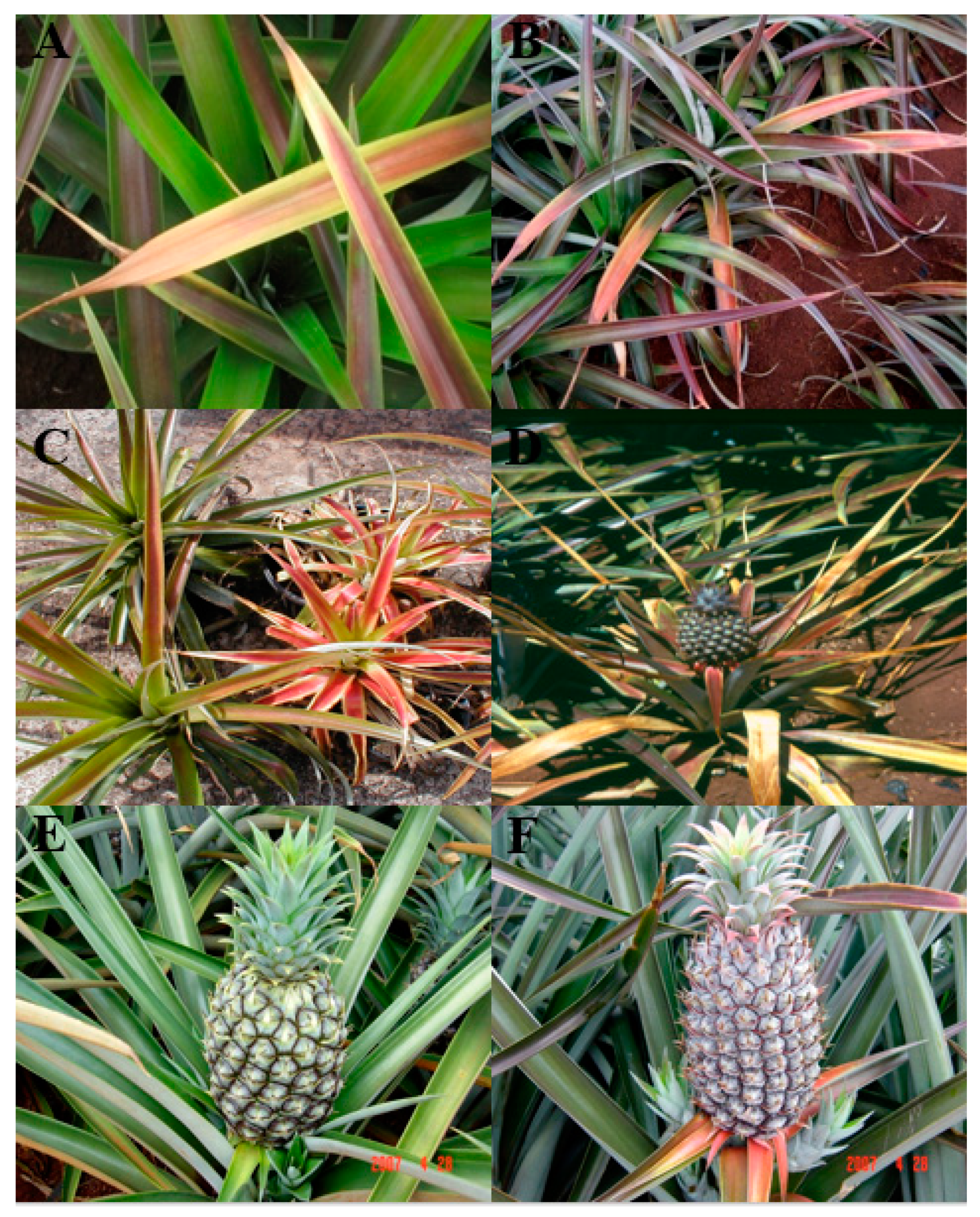
Figure 1. Symptoms of mealybug wilt disease in commercial pineapple hybrids and traditional Smooth Cayenne “Champaka” cultivar. (A) Pineapple Research Institute (PRI) hybrid 73–114; (B) PRI 73–50; (C) typical symptoms, including reddening of leaves with downward curling of margin tips, leaf-tip dieback; (D) and pronounced wilting of mature leaves. (E) Hybrid variety PRI 73-114, and (F) hybrid variety PRI 73–50.
Horticulturae 04 00052 g001
Figure 2. Pineapple mealybugs (A) Dysmicoccus brevipes; (B) Dysmicoccus neobrevipes.
Horticulturae 04 00052 g002
Figure 3. Mutualistic association of ants and mealybugs. (A,B) Big-headed ants tending mealybugs.
Horticulturae 04 00052 g003
Figure 4. (A) Virus particles of PMWaV-2 decorated by PMWaV-2-specific monoclonal antibodies (MAbs) observed under electron microscopy; (B) particles of PMWaV-2 from a mealybug wilt of pineapple (MWP) symptomatic plant; (C) typical symptoms of mealybug wilt disease are expressed only in the plant with PMWaV-2 and mealybug exposure, while others appear to be healthy. Left to Right: PMWaV-free plant exposed to mealybugs; PMWaV-1 and PMWaV-2 infected plant not exposed to mealybugs; PMWaV-2-infected plant not exposed to mealybug, and PMWaV-2 infected plant exposed to mealybug feedings.
Horticulturae 04 00052 g004
Figure 5. Genome organization of PMWaV-1, PMWaV-2, PMWaV-3, PMWaV-4, and PMWaV-5. Abbreviations: MTR methyltransferase domain; HEL helicase domain; RdRp RNA-dependent RNA polymerase; Hsp70 heat shock protein 70; CP coat protein; CPd coat protein duplicate. The scale gives approximate size in kilobases.
Horticulturae 04 00052 g005
Figure 6. Phylogenetic analysis of ampeloviruses.
Horticulturae 04 00052 g006
Figure 7. Top: a symptomless pineapple plant infected by PMWaV-1, and its associated genome organization. Below: a symptomatic pineapple plant infected by PMWaV-2, exhibiting typical wilting symptoms and its genome organizations. In the genome organization, boxes represent open reading frames (ORFs), while homologous genes or domains are shown with the same pattern for both viruses. Red boxes represent ORFs that were identified asRNA silencing uppressors.
Horticulturae 04 00052 g007