


Highlights
- Exogenous allantoin increases water-use efficiency (WUE) in cucumber plants while also enhancing drought tolerance by promoting cuticular wax deposition.
- Multi-omics analyses identify CsCER1, encoding a very-long-chain aldehyde decarbonylase, as a core allantoin-responsive gene essential for cuticular wax biosynthesis.
- VIGS-mediated silencing of CsCER1 significantly reduces allantoin-induced drought tolerance, demonstrating that CsCER1 is a key functional gene for allantoin-mediated drought resistance.
Abstract
Cucumber (Cucumis sativus L.) is an economically important vegetable crop worldwide, but its yield and quality improvement are often constrained by drought stress. To investigate the physiological and molecular mechanisms by which exogenous allantoin enhances drought tolerance in cucumber, cucumber seedlings were sprayed with 6 mM allantoin solution once (A1), three times (A3), or five times (A5), while control plants were sprayed with distilled water (CK1, CK3, CK5). Each treatment consisted of three biological replicates. After treatment, drought stress was simulated by irrigating with 20% polyethylene glycol 6000 (PEG-6000) solution. The results showed that the protective effect of exogenous allantoin against drought stress was cumulative. After five applications (A5), the net photosynthetic rate (Pn) and water-use efficiency (WUE) of the plants were significantly higher than those of the corresponding control (CK5) (p < 0.01). The detached leaf water loss rate progressively decreased with an increasing number of allantoin applications, while the total leaf wax content increased approximately 2-fold (p < 0.01). Measurements of wax content in different plant tissues indicated that allantoin mainly induced wax accumulation in aboveground organs (leaf, stem, and fruit epidermis), and this effect was validated in three commercial varieties. Integrated transcriptomic and metabolomic analyses revealed that the cucumber CsCER1 gene (encoding a very-long-chain aldehyde decarbonylase) is a core allantoin-responsive gene. After silencing CsCER1 using virus-induced gene silencing (VIGS), the allantoin-induced wax accumulation and drought tolerance were almost completely lost: the wilting severity and detached leaf water loss rate of the silenced plants were comparable to those of the empty vector control, and no significant increase in wax content was observed. This study reveals a novel mechanism by which exogenous allantoin enhances drought tolerance in cucumber through activating CsCER1-mediated cuticular wax synthesis, providing a theoretical basis for the chemical regulation of drought tolerance in cucurbit crops.
1. Introduction
Drought stress is one of the major abiotic factors limiting global agricultural productivity and threatening food security [1,2]. Cucumber (Cucumis sativus L.), an economically important vegetable crop widely cultivated worldwide, has a shallow root system and large leaf area, making it highly sensitive to water deficit [3,4]. Drought stress not only inhibits photosynthesis and vegetative growth [5,6] but also leads to yield reduction and quality deterioration (e.g., fruit bitterness and deformation) [7,8]. Therefore, improving drought tolerance in cucumber is of great theoretical and practical significance.
During long-term evolution, plants have developed complex drought adaptation mechanisms, including osmotic adjustment, antioxidant defense, and hormonal signaling. According to the classic “growth-defense trade-off” theory, under stress conditions, plants may allocate limited resources preferentially to defense systems at the expense of vegetative growth [9,10]. Recent studies have shown that plants can regulate the balance between growth and defense through intricate hormonal signaling networks, enabling enhanced defense without growth inhibition [11,12]. Among these, the jasmonic acid (JA) signaling pathway plays a central regulatory role in this process [13]. Notably, allantoin, an intermediate of purine metabolism, has been demonstrated to activate the JA signaling pathway in an MYC2-dependent and ABA-dependent manner [14]. This suggests that exogenous allantoin may serve as an external chemical signal capable of modulating both plant growth and defense, although its specific role in cucumber remains to be verified.
Allantoin, a nitrogen-rich intermediate of purine catabolism, is widely recognized as an important metabolite involved in plant stress resistance [15]. A review by Kaur et al. indicated that the accumulation of allantoin in plants positively correlates with tolerance to various abiotic stresses, including drought, salinity, low temperature, and heavy metals; exogenous allantoin can act as a signaling molecule to activate ABA and JA pathways, thereby enhancing overall plant resistance [16]. Watanabe et al. confirmed in Arabidopsis that allantoin synergistically activates ABA metabolism, promotes ABA accumulation and stress-responsive gene expression, and significantly enhances plant tolerance to drought and salt stress [17]. In rapeseed, exogenous allantoin improves plant drought tolerance by enhancing the antioxidant defense system and maintaining water balance, thereby improving plant growth, biomass, and yield [18]. Lu et al. found that exogenous allantoin treatment in rice significantly accumulates drought-responsive metabolites such as proline and soluble sugars, reduces ROS levels, and upregulates the expression of ROS-scavenging enzyme genes including POD, CATA, and APX8 [19]. In leguminous plants, manipulation of nodule ureide permease 1 (UPS1) to enhance ureide transport from nodules to aboveground parts was shown to significantly improve nitrogen supply under drought stress, promote phloem loading and transport of sucrose, and enhance overall carbon acquisition and assimilation capacity [20,21]. This suggests that allantoin, as an important member of the ureide family, may not be confined to its traditional roles in antioxidant defense and signal activation, but might also participate in the coordinated regulation of carbon-nitrogen metabolism.
In the structural defenses of plants against drought, the cuticular wax covering the surface of above-ground organs serves as the first line of defense against non-stomatal water loss and various environmental stresses [22,23]. Cuticular wax is a complex mixture of lipids composed of very-long-chain fatty acids (VLCFAs) and their derivatives [24]. Among the various lipid components, very-long-chain (VLC) alkanes possess strong hydrophobicity and serve as the core constituents of the cuticle’s highly efficient water-repellent barrier [25,26]. The alkane synthase ECERIFERUM1 (CER1) has been shown to participate in the biosynthesis of very-long-chain alkanes, playing a crucial role in the accumulation of cuticular wax and directly influencing the plant’s response to abiotic stresses [27,28]. Overexpression of CER1 in Arabidopsis significantly promotes alkane accumulation and enhances cuticular barrier function [29]. In cucumber, CsCER1 significantly influences the biosynthesis of very-long-chain alkanes, as well as epidermal permeability and drought tolerance [30]. Although CsCER1-mediated alkane biosynthesis is crucial for plant drought tolerance, it remains unclear whether this carbon-intensive lipid metabolic process is induced by exogenous chemical signals, such as allantoin.
In this study, the cucumber variety ‘Shengqiu No. 2-1’ was used as the main experimental material to investigate the alleviating effect of exogenous allantoin on drought stress in cucumber and its molecular mechanism. The specific research contents include: (1) through physiological index measurements, to determine the effects of exogenous allantoin on photosynthetic characteristics, water-use efficiency, and cuticular wax accumulation in cucumber under drought stress; (2) using integrated transcriptomic and metabolomic analyses, to screen the core pathways and key genes responsive to allantoin; (3) via virus-induced gene silencing (VIGS), to verify the essential function of CsCER1 in allantoin-induced drought tolerance; and (4) to explore the universality of this effect across different cucumber varieties. This study aims to reveal a novel mechanism by which exogenous allantoin enhances drought tolerance in cucumber through activating CsCER1-mediated cuticular wax synthesis, thereby providing a theoretical basis and technical support for synergistically improving the growth potential and drought resistance of cucumber through chemical regulation.
2. Materials and Methods
2.1. Plant Materials and Growth Conditions
The experiment was conducted in a controlled growth chamber at the College of Advanced Agriculture and Ecological Environment, Heilongjiang University. The main cucumber (Cucumis sativus L.) variety used was ‘Shengqiu No. 2-1’. To verify the universality of allantoin-induced wax accumulation, three commercial varieties were also employed: ‘H19129-1-3’, ‘Heijian-5’, and ‘Qiangci Heici-1’. Seeds were surface-sterilized and germinated, then sown in plastic pots filled with a substrate mixture of potting soil and vermiculite (3:1, v/v). The growth chamber conditions were set as follows: 16 h light/8 h dark photoperiod, day/night temperatures of 25 °C/20 °C, and relative humidity of 60% [31]. The pots were arranged randomly and repositioned weekly to avoid positional effects. Plants were watered normally until they reached the two-true-leaf stage, after which treatments were initiated.
2.2. Experiment on Exogenous Allantoin Treatment and Drought Stress
To determine the optimal concentration of exogenous allantoin (Yousuo, Linyi, China), a preliminary experiment was performed. Five concentration gradients (0 (control), 3, 6, 9, and 12 mM) were tested by foliar spraying every 3 days for three consecutive applications, and overall growth status was used as the evaluation criterion. Based on the preliminary results, 6 mM was selected as the optimal concentration for all subsequent experiments (Figure S1). The formal experiment used a completely randomized design with three biological replicates per treatment, each replicate consisting of ten plants. Cucumber seedlings of uniform growth were selected and sprayed with a 6 mM allantoin aqueous solution containing 0.05% Tween-20 as a surfactant; spraying was performed until droplets appeared on both the adaxial and abaxial leaf surfaces, and the solution also dripped into the root-zone soil. Control plants were sprayed with an equal volume of distilled water containing 0.05% Tween-20. Allantoin treatments were administered as follows: once (A1), three times (A3), and five times (A5). The corresponding distilled water control groups were designated CK1, CK3, and CK5. Spraying was carried out every 3 days at 8:00 A.M. Twenty-four hours after each treatment, the third fully expanded leaf from the top was collected for subsequent measurements.
Drought stress was imposed by root irrigation with a 20% (w/v) polyethylene glycol 6000 (PEG-6000, Beijing, Solebao, China) solution [32]. The volume applied was sufficient to thoroughly saturate the root-zone substrate until a small amount of solution drained from the bottom of the pot. During the treatment period, normal watering was stopped. After seven days of treatment, the wilting phenotype of the plants was recorded.
2.3. Measurement of Photosynthetic Gas Exchange Parameters and Physiological Indices
Photosynthetic gas exchange parameters were measured on the third fully expanded true leaf using a CI-340 portable photosynthesis system (CID Bio-Science, Camas, WA, USA) between 9:00 and 11:00 A.M. Net photosynthetic rate (Pn), transpiration rate (E), stomatal conductance (Gs), and intercellular CO2 concentration (Ci) were recorded, and instantaneous water-use efficiency (WUE = Pn/E) was calculated. Three plants per treatment were measured.
Chlorophyll content was determined using the ethanol extraction method. Fresh leaf samples (0.1 g) were cut into small pieces, placed in 10 mL centrifuge tubes containing 10 mL of 95% (v/v) ethanol, and soaked in the dark for 48 h until the leaves became completely colorless. The absorbance of the extract was measured at 665 nm and 649 nm using a UV-visible spectrophotometer (UV-1800, Shimadzu, Kyoto, Japan) [33]. Chlorophyll a, chlorophyll b, and total chlorophyll concentrations were calculated according to the Arnon formula as follows:
Chlorophyll a (mg/L) = 13.95 × A665 − 6.88 × A649
Chlorophyll b (mg/L) = 24.96 × A649 − 7.32 × A665
Total chlorophyll = chlorophyll a + chlorophyll b
2.4. Determination of Water Loss Rate of Detached Leaves
Fully expanded leaves from the same position of control and treatment groups (including VIGS-silenced plants) were excised, and their initial fresh weight (W0) was recorded immediately. The leaves were then placed on a laboratory bench under ambient conditions (temperature 25 ± 1 °C, relative humidity 50–60%) and weighed every hour from 0 to 6 h (Wt) [34]. Cumulative water loss rate (%) was calculated as (W0 − Wt)/W0 × 100%. Each treatment included three biological replicates.
2.5. Determination of Total Wax Content and Scanning Electron Microscopy
Leaf samples were measured for surface area, then placed in 50 mL glass tubes containing 30 mL of chloroform and shaken at room temperature for 30 s to completely dissolve the cuticular wax layer. The extract was transferred to a pre-weighed vial, and the extraction was repeated once. The combined extracts were evaporated to dryness under a gentle stream of nitrogen [35]. Total wax content was expressed as both micrograms per square centimeter (μg/cm2) and milligrams per gram fresh weight (mg/g FW). For ultrastructural analysis, samples were dehydrated through a graded ethanol series (60%, 70%, 80%, 90%, 100%; 10 min each) and then critical-point dried (Balzers CPD 030, Balzers, Liechtenstein). The dried specimens were mounted on aluminum stubs with double-sided conductive carbon tape, sputter-coated with gold (Au, 2 min, 20 mA), and examined with an EVO-LS10 scanning electron microscope (Oberkochen, Germany) to capture images of the cuticular ultrastructure.
2.6. Transcriptomic Analysis
Total RNA was extracted from leaves (three biological replicates per treatment) using a Plant Total RNA Extraction Kit (Tiangen, Beijing, China). RNA quality and concentration were assessed by agarose gel electrophoresis and spectrophotometry (NanoDrop 2000, Thermo Fisher, Waltham, MA, USA). Libraries were prepared using the Illumina Stranded mRNA Prep Kit and sequenced on a NovaSeq X Plus platform (Illumina, San Diego, CA, USA) with 150 bp paired-end reads. Each sample generated >6 Gb of clean data (Q30 ≥ 96.3%). Raw reads were processed with fastp (v0.20.0) to remove adapter sequences and low-quality reads. The resulting clean reads were aligned to the cucumber reference genome (Chinese Long v3, Cucumber Genome Database) using HISAT2 (v2.1.0). Gene expression levels were quantified as transcripts per million (TPM) using RSEM (v1.3.1) [36]. Differentially expressed genes (DEGs) were identified with DESeq2 (v1.30.0) using the criteria: false discovery rate (FDR) < 0.05 and |log2 fold change| ≥ 1.
2.7. Metabolomics Analysis
Samples (six biological replicates per treatment) were ground in liquid nitrogen, and metabolites were extracted using an extraction buffer containing an internal standard (methanol–water = 4:1, v/v). After centrifugation, the supernatant was analyzed by LC-MS/MS using an ultra-high-performance liquid chromatography-tandem mass spectrometry system (UHPLC-Orbitrap Exploris 240, Waltham, MA, USA). Raw data were imported into Progenesis QI software (Version 3.0, Waters Corporation, Milford, MA, USA) for baseline filtering, peak alignment, and integration, and metabolites were annotated by comparison with the HMDB, Metlin, and Majorbio in-house metabolite databases [37]. Principal component analysis (PCA) and orthogonal partial least squares discriminant analysis (OPLS-DA) were performed using the ropls package in R. Significantly differentially accumulated metabolites (DAMs) were identified based on variable importance in projection (VIP) scores from the OPLS-DA model and Student’s t-test p-values; metabolites with VIP > 1 and p < 0.05 were considered significant.
2.8. Multi-Omics Integrated Analysis
Transcriptomic and metabolomic data matrices (all DEGs and DAMs from the A3 vs. CK3 and A5 vs. CK5 comparisons) were imported into the Majorbio Cloud Platform (https://www.majorbio.com). O2PLS analysis was performed to assess the covariance between the two datasets, and model quality was evaluated by R2X, R2Y, and Q2 values from cross-validation. Gene–metabolite correlation networks were constructed using Pearson’s correlation coefficients (R > 0.8, p < 0.01). Combined KEGG enrichment heatmaps were generated to identify pathways significantly enriched in both transcriptomic and metabolomic datasets.
2.9. Virus-Induced Gene Silencing
A VIGS system was used to validate the function of CsCER1 (CsaV3_6G006560) in ‘Shengqiu No. 2-1’ cucumber. Specific primers were designed based on the non-conserved region of CsCER1 to amplify a 436-bp target fragment, which was confirmed by BLAST against the cucumber genome to have no off-target effects. The fragment was cloned into the pV190 vector to generate the pV190-CsCER1 silencing recombinant plasmid [38].
The vectors pV190 (empty vector, EV), pV190-PDS (phytoene desaturase, used as a positive control), and pV190-CsCER1 were separately transformed into Agrobacterium tumefaciens strain GV3101. The agrobacterium cultures carrying the respective pV190 vectors were harvested by centrifugation (final OD600 = 0.6) and resuspended in infiltration buffer containing 10 mM MES, 10 mM MgCl2, and 100 μM acetosyringone. After dark incubation for 3 h, cucumber seeds were vacuum-infiltrated. When the pV190-PDS plants exhibited a clear photobleaching phenotype (approximately 2–3 weeks after inoculation), the pV190-EV and pV190-CsCER1 plants were subjected to allantoin treatment and 20% PEG-induced drought stress, followed by phenotype recording, qRT-PCR validation of silencing efficiency, measurement of detached leaf water loss rate, and determination of wax content. This VIGS method has been optimized for cucumber [39].
2.10. RNA Extraction and qRT-PCR Analysis
Total RNA was extracted from different cucumber tissues (roots, stems, leaves, fruit epidermis) and at different treatment stages using a Plant Total RNA Extraction Kit (Tiangen, China). First-strand cDNA was synthesized using HiScript III All-in-one RT SuperMix (Vazyme, Nanjing, China). The cucumber Actin gene (CsaV3_6G043400) was used as the internal reference, and qPCR was performed using SYBR Green Master Mix on an Agilent AriaMx Real-Time PCR System. Relative expression levels were calculated using the 2−ΔΔCt method. Three biological replicates were used for each sample, and each replicate was run in triplicate technical replicates.
2.11. Data Statistics and Analysis
All experiments included at least three independent biological replicates. Data were preliminarily organized using Microsoft Excel 2021 and subjected to statistical analysis and graph preparation using GraphPad Prism 10.0 software. Data are presented as mean ± standard deviation (Mean ± SD). Comparisons between two groups were performed using independent two-tailed Student’s t-tests, and multiple comparisons were performed using one-way analysis of variance (ANOVA), followed by Tukey’s post hoc test. Significance levels are indicated as * p < 0.05, ** p < 0.01, *** p < 0.001, **** p < 0.0001; “ns” denotes no significant difference.
3. Results
3.1. Exogenous Allantoin Enhances Plant Photosynthetic Capacity and Water-Use Efficiency
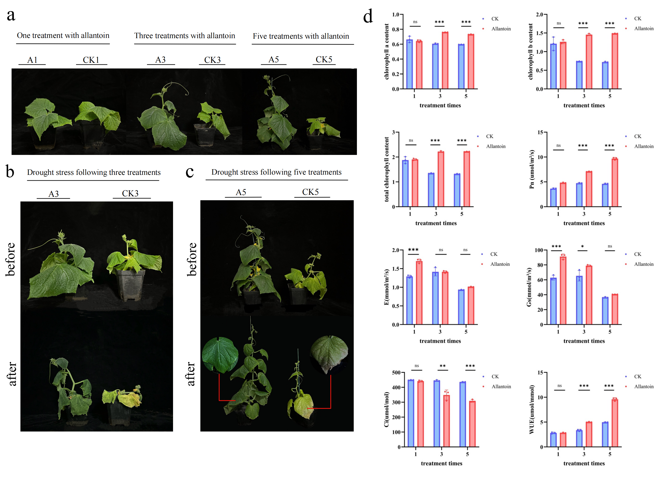
Following consecutive allantoin treatments, cucumber seedlings exhibited a glossy, deep-green phenotype with erect, fully expanded leaves (Figure 1a). We then observed phenotypic changes in plants under drought stress following one, three, and five applications of allantoin and found that drought resistance had already begun to emerge after the third treatment (A3). Compared with the control group (CK3), plants in the A3 treatment showed a significant delay in wilting during the early stages of drought. As the number of treatments increased to five (A5), the leaves of the control group (CK5) under drought stress exhibited yellowing due to water loss, curled edges, and tissue damage, while plants in the A5 treatment still maintained good physiological status (Figure 1b,c). This demonstrates that the protective effect of allantoin is cumulative. Drought tolerance of the plants increased progressively with the number of allantoin applications.
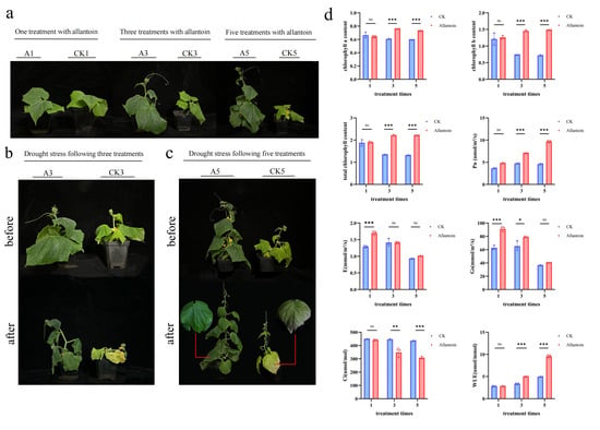
Figure 1.
Effects of exogenous allantoin treatment on the photosynthetic capacity and drought-tolerance phenotype of cucumber. (a) Overall phenotype of plants treated with 1, 3, or 5 applications of allantoin (A1, A3, A5) and their respective control groups (CK1, CK3, CK5), (b) Comparison of wilting phenotypes 7 days after 20% PEG drought stress treatment (A3 vs. CK3). (c) Comparison of wilting phenotypes 7 days after 20% PEG drought stress treatment (A5 vs. CK5). (d) Leaf chlorophyll a, chlorophyll b, and total chlorophyll content, net photosynthetic rate (Pn), transpiration rate (E), stomatal conductance (Gs), intercellular CO2 concentration (Ci), and water-use efficiency (WUE) in each treatment group. Data are presented as mean ± standard deviation (n = 3). * p < 0.05, ** p < 0.01, *** p < 0.001; ns indicates no significant difference.
Measurements of photosynthetic physiological parameters further supported the above phenotypic observations (Figure 1d). As the number of treatments increased, allantoin significantly elevated the levels of chlorophyll a, chlorophyll b, and total chlorophyll in the leaves, thereby enhancing the plants’ light-capturing capacity. Photosynthetic gas exchange parameters indicated that the net photosynthetic rate (Pn) of both A3 and A5 plants was significantly higher than that of the control at the same time point. In the A5 treatment, Pn increased substantially by 2.0-fold and intercellular CO2 concentration (Ci) decreased significantly, while transpiration rate (E) and stomatal conductance (Gs) showed no significant changes compared to the control group, significantly improving leaf water-use efficiency (WUE = Pn/E) by 1.9-fold. This suggests the possible existence of a non-stomatal water retention mechanism.
3.2. Multi-Omics Analysis Reveals That Allantoin Activates Lipid Metabolic Pathways
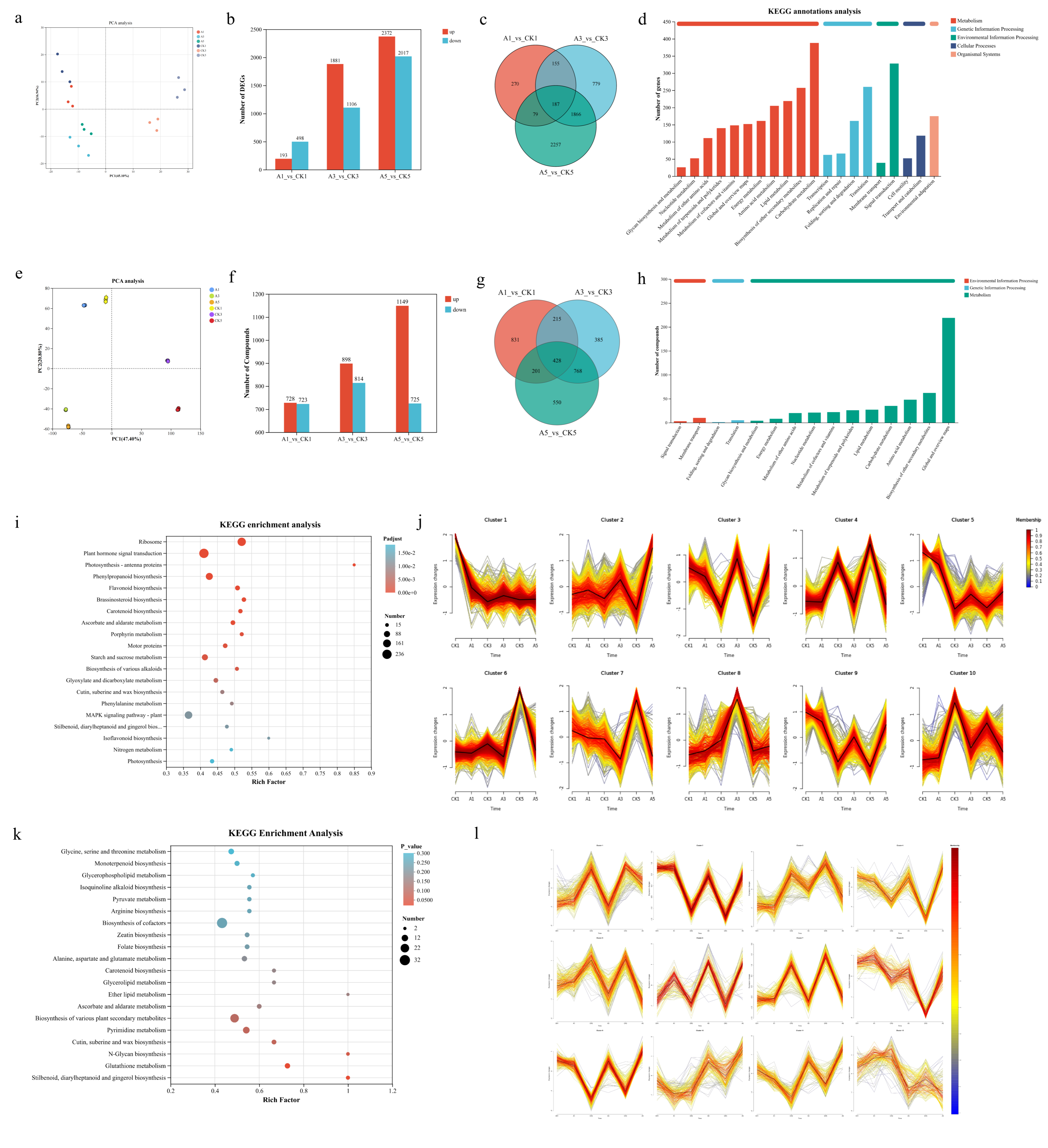
Based on the physiological manifestation of high water-use efficiency and the oil-green luster phenotype observed in the treated group, we hypothesize that allantoin may drive the directed conversion of photosynthetic products into hydrophobic compounds on the leaf surface. To test this hypothesis, we conducted integrated transcriptomic and metabolomic analyses of leaves at treatment periods 1, 3, and 5 (CK vs. allantoin). Principal component analysis (PCA) indicated that while A1 and CK1 clustered closely, the A3 and A5 groups were distinctly separated from their respective controls (Figure 2a,e). The first two principal components accounted for 45.1% and 16.94% of the variance in the transcriptomic data, and 47.4% and 20.8% in the metabolomic data. This suggests that allantoin-induced transcriptional and metabolic changes are cumulative, consistent with phenotypic observations. The number of differentially expressed genes (DEGs) and differentially accumulated metabolites (DAMs) exhibited a significant time-dependent increase. In the A1 vs. CK1 phase, only 691 DEGs and 1451 DAMs were identified; whereas in the A3 vs. CK3 and A5 vs. CK5 phases, the number of DEGs increased to 2987 and 4389, respectively, and the number of DAMs increased to 1712 and 1874, respectively (Figure 2b,f). Venn diagram analysis further highlighted that a core set of genes and metabolites remained consistently regulated in A3 and A5 (Figure 2c,g), indicating that continuous allantoin treatment triggered extensive remodeling of transcriptional and metabolic networks.
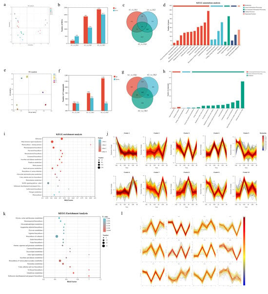
Figure 2.
Integrated transcriptomic and metabolomic analysis of cucumber leaves treated with exogenous allantoin. (a) Principal component analysis (PCA) of transcriptomic data. (b) Statistics on the number of differentially expressed genes (DEGs) in each comparison group (A1 vs. CK1, A3 vs. CK3, A5 vs. CK5) (upregulated/downregulated). (c) Venn diagram analysis of DEGs across the three time points. (d) KEGG annotation analysis of DEGs common to T3 and T5. (e) Principal component analysis (PCA) of metabolomic data. (f) Statistics on the number of differentially expressed metabolites (DAMs) in each comparison group (A1 vs. CK1, A3 vs. CK3, A5 vs. CK5) (upregulated/downregulated). (g) Venn diagram analysis of DAMs across the three time points. (h) KEGG annotation analysis of DAMs common to T3 and T5. (i) KEGG enrichment bubble plot of common DEGs between T3 and T5. (j) Temporal clustering analysis of DEGs. (k) KEGG enrichment bubble plot of common DAMs between T3 and T5. (l) Temporal clustering analysis of DAMs. Data are presented as mean ± standard deviation (n = 3).
We performed KEGG functional pathway and enrichment analyses on the common differentially expressed genes and metabolites from the T3 and T5 periods. The results revealed specific enrichment in pathways related to lipid metabolism and structural defense. In the transcriptomic dataset, “Metabolism” emerged as the most representative category, containing the largest number of annotated genes (450), with “Carbohydrate metabolism” and “Lipid metabolism” showing high levels of gene enrichment (Figure 2d). In the metabolomics dataset, specific clusters related to carbon and lipid fluxes, such as “Amino acid metabolism,” “Carbohydrate metabolism,” and “Lipid metabolism,” showed a significant accumulation of differentially expressed metabolites (Figure 2h). In the transcriptomic enrichment analysis (Figure 2i), the “Photosynthesis—antenna proteins” pathway was significantly enriched, indicating that the light-harvesting system of photosynthesis was strongly upregulated, consistent with the enhanced photosynthetic rates observed in plants from the A5 treatment group. Concurrently, pathways related to structural defense were also significantly enriched. “Cutin, suberin, and wax biosynthesis” emerged as a significant pathway (Rich Factor = 0.46). Additionally, biosynthetic pathways for lipid derivatives such as carotenoids and brassinosteroids also exhibited significant enrichment. In the metabolomic enrichment analysis (Figure 2k), “Cutin, suberin, and wax biosynthesis” was again identified as a significantly enriched pathway, confirming synchronized responses at both the transcriptional and metabolic levels. Furthermore, other lipid-related pathways, such as “triglyceride metabolism” and “ether lipid metabolism,” exhibited extremely high enrichment significance, pointing to a specific reorganization of lipid metabolism. To further elucidate the regulatory logic, we performed a time-series analysis and clustered the DEGs and DAMs. From time point T1 to T5, we identified clusters of genes (cluster 2, cluster 9) and metabolites (cluster 2, cluster 6, cluster 9) that showed a continuous increase (Figure 2j,l). Genes and metabolites within these clusters were significantly enriched in pathways such as unsaturated fatty acid biosynthesis, fatty acid biosynthesis, glycerol metabolism, and nitrogen metabolism. These results indicate that allantoin treatment is closely associated with the activation of lipid metabolic pathways.
3.3. Integrated Transcriptomic and Metabolomic Analysis Identifies Cutin, Suberin, and Wax Biosynthesis as Key Responsive Pathways
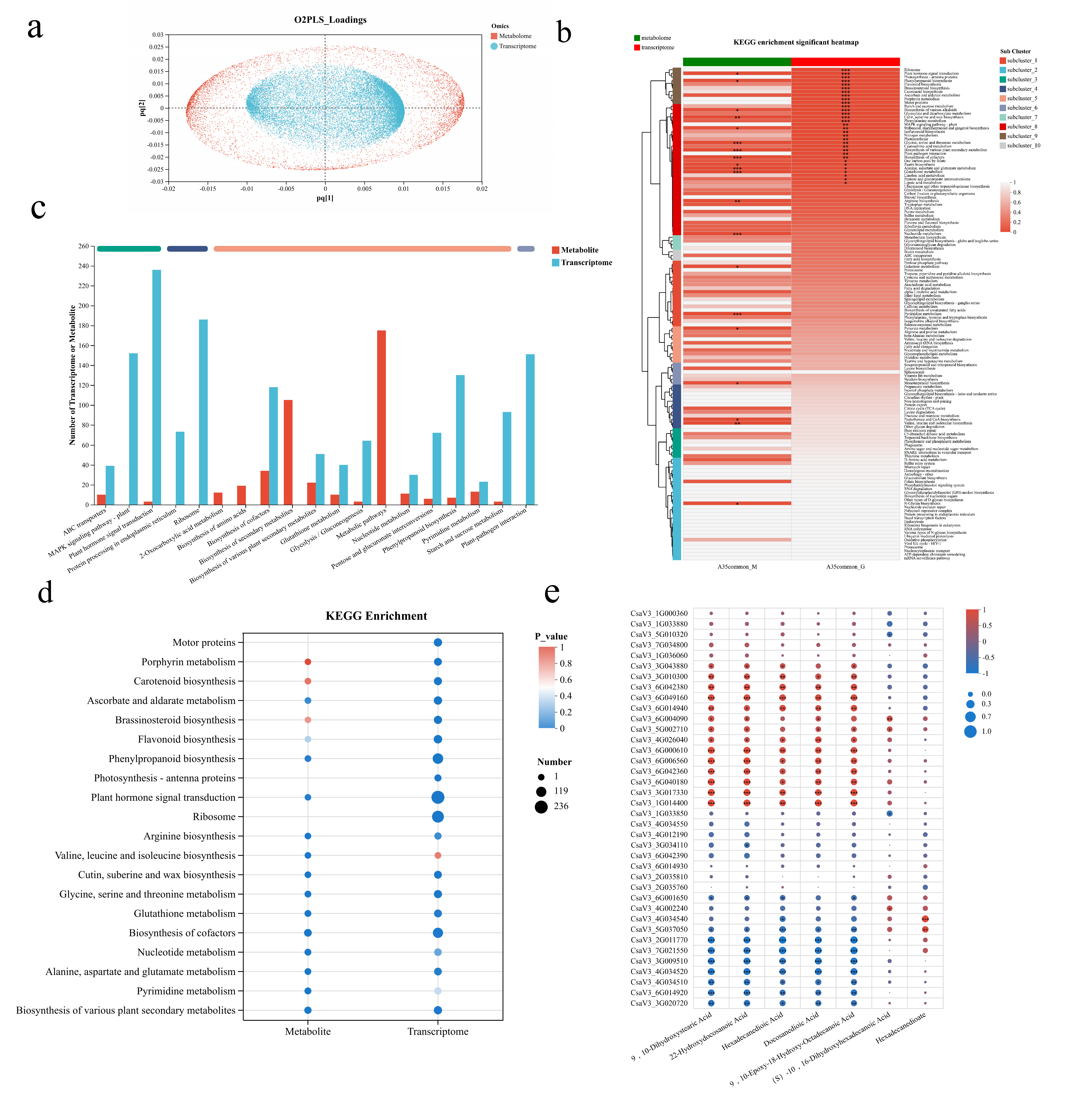
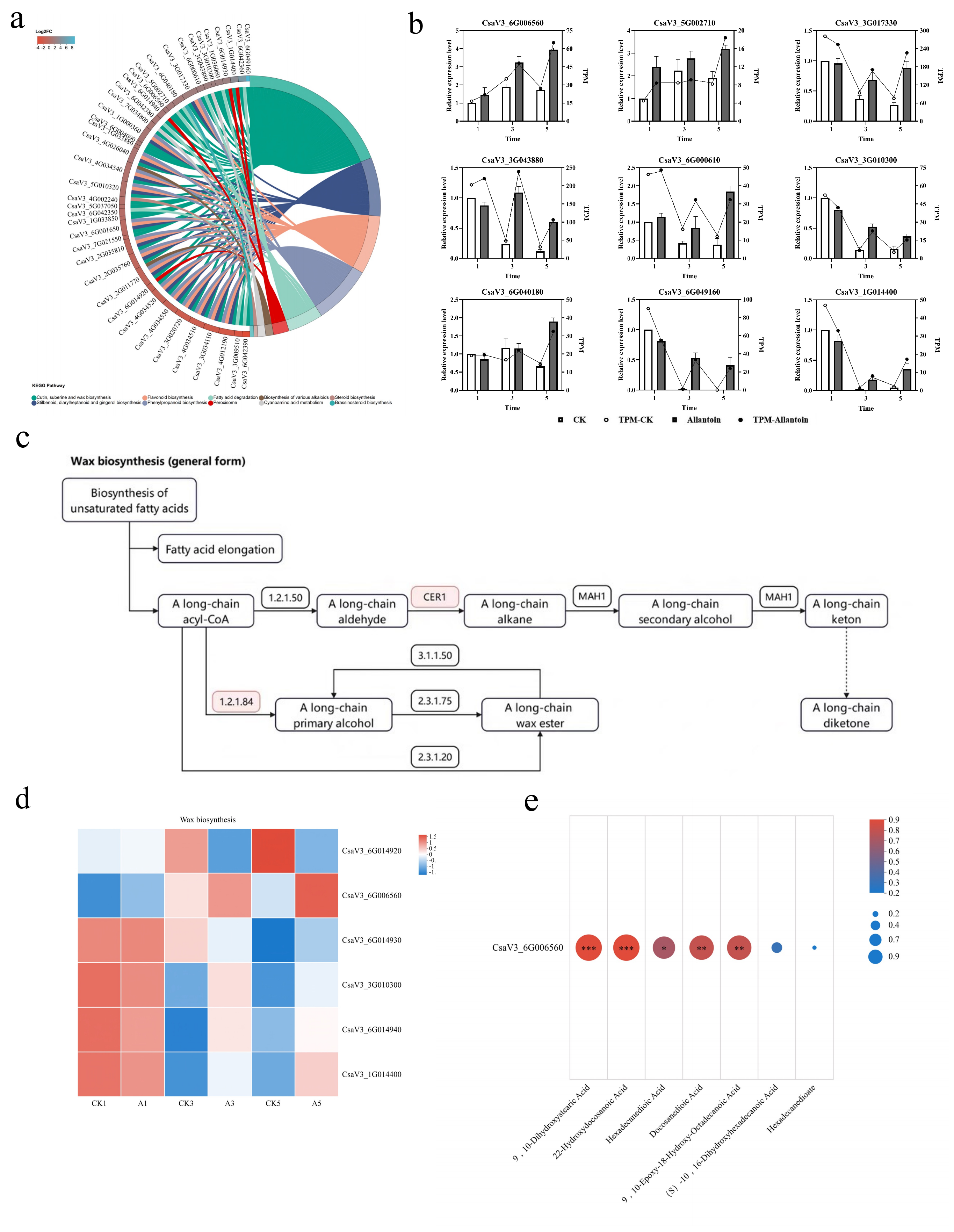
To identify specific biological pathways within lipid metabolism, we conducted an integrated analysis of differentially expressed genes and metabolites at the T3 and T5 stages. O2PLS analysis revealed a high degree of covariance between the transcriptomic and metabolomic datasets (R2X = 0.9504, R2Y = 0.756, R2Xjoint = 0.9365, R2Yjoint = 0.756, R2Xhat = 0.9208, R2Yhat = 0.7403, R2Xpred = 0.9833, R2Ypred = 0.9792), indicating a close regulatory relationship between gene expression and metabolic accumulation under allantoin treatment (Figure 3a). Among the KEGG pathway annotations, the “plant hormone signaling” category contained the highest number of genes, while the “metabolic pathways” category contained the highest number of metabolites (Figure 3c). Joint KEGG enrichment analysis showed that the pathways “glutathione metabolism,” “glycine, serine and threonine metabolism,” and “cutin, suberin and wax biosynthesis” were significantly enriched in both the transcriptomic and metabolomic datasets (Figure 3d). Joint KEGG enrichment heatmap analysis further indicated that the lipid metabolism pathway “cutin, suberin and wax biosynthesis” exhibited the highest significance (Figure 3b). Therefore, we conclude that allantoin treatment is closely associated with the activation of this structural defense-related pathway. Gene–metabolite correlation analysis (Figure 3e) revealed that a group of upregulated structural genes (e.g., CsaV3_1G014400, CsaV3_6G006560, CsaV3_6G040180) showed strong positive correlations (R > 0.8) with various long-chain fatty acids and their derivatives (including 9,10-dihydroxystearic acid, 22-hydroxydocosanoic acid, hexadecanedioic acid, etc.). These metabolites are intermediates or monomers in the cutin, suberin, and wax biosynthesis pathways, providing key insights into the molecular mechanism by which allantoin enhances the physical barrier function of leaves.
Figure 3.
Integrated transcriptomic and metabolomic analysis identifies the core pathway for cutin, suberin, and wax biosynthesis in response to allantoin. (a) O2PLS joint analysis plot for T3 and T5 stages. (b) Heatmap showing the correlation of KEGG-enriched pathways from the joint analysis. (c) Statistical classification of KEGG pathway annotations for DEGs and DAMs. (d) KEGG enrichment analysis of DEGs and DAMs. (e) Gene–metabolite correlation heatmap of the cutin, suberin and wax biosynthesis pathways, showing strong positive correlations (R > 0.8) between upregulated genes associated with wax biosynthesis (CsaV3_6G006560, CsaV3_3G010300, CsaV3_1G014400, etc.) and very-long-chain fatty acid derivatives. Data are presented as mean ± standard deviation (n = 3). * p < 0.05, ** p < 0.01, *** p < 0.001.
3.4. CsCER1 Is a Key Responsive Gene in Allantoin-Induced Cuticle Wax Biosynthesis
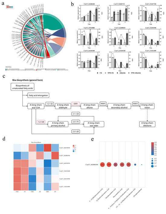
To further investigate the molecular basis of allantoin-induced drought tolerance, we mapped all differentially expressed genes in the “cutin, suberin, and wax biosynthesis” pathway at the T5 stage onto an enrichment string plot and ranked them by log2FC values (Figure 4a). Transcripts with very low expression at this stage were excluded, and nine candidate genes with significant upregulation were retained (Figure 4b). Most of these genes are annotated as involved in wax biosynthesis, as confirmed by qRT-PCR (Figure 4b). The KEGG wax biosynthesis pathway diagram shows two allantoin-responsive routes: the primary alcohol synthesis pathway (reduction pathway) catalyzed by fatty acyl-CoA reductase, and the alkane synthesis pathway (decarboxylation pathway) involving the enzyme CsCER1 (Figure 4c). Heatmap analysis of wax-related DEGs (Figure 4d) indicated that the expression pattern of CsaV3_6G006560 (annotated as CsCER1) was most closely associated with the cumulative drought-tolerance phenotype induced by allantoin: a weak response at A1, followed by stepwise upregulation at A3 and A5. Gene–metabolite correlation analysis (Figure 4e) showed that the expression level of CsCER1 was significantly positively correlated (R > 0.8) with several long-chain fatty acids and their derivatives in the pathway. These results suggest that CsCER1 is a key responsive gene involved in allantoin-induced cuticular wax accumulation and drought tolerance in cucumber.
Figure 4.
Multi-omics integrated analysis identifies CsCER1 as a key gene in allantoin-induced cuticle wax accumulation. (a) Enrichment string plot of differentially expressed genes in the “cutin, suberin and wax Biosynthesis” pathway at the T5 stage, sorted by log2FC. (b) qRT-PCR validation of the temporal expression of nine candidate genes following 1, 3, and 5 allantoin treatments. (c) KEGG pathway diagram of allin-activated wax biosynthesis; pink annotations indicate upregulated genes in the pathway. (d) Heatmap of wax-related DEGs in the two pathways; CsaV3_6G006560 (CsCER1) exhibits the most significant stepwise upregulation at the A3 and A5 time points. (e) Correlation analysis network diagram of CsCER1 expression abundance with key differential metabolites in the pathway. Data are presented as mean ± standard deviation (n = 3). * p < 0.05, ** p < 0.01, *** p < 0.001.
3.5. Cumulative Induction of CsCER1 Expression in Aboveground Parts of Cucumber and Universal Accumulation of Cuticular Wax by Allantoin
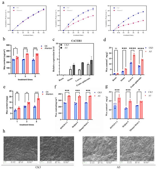
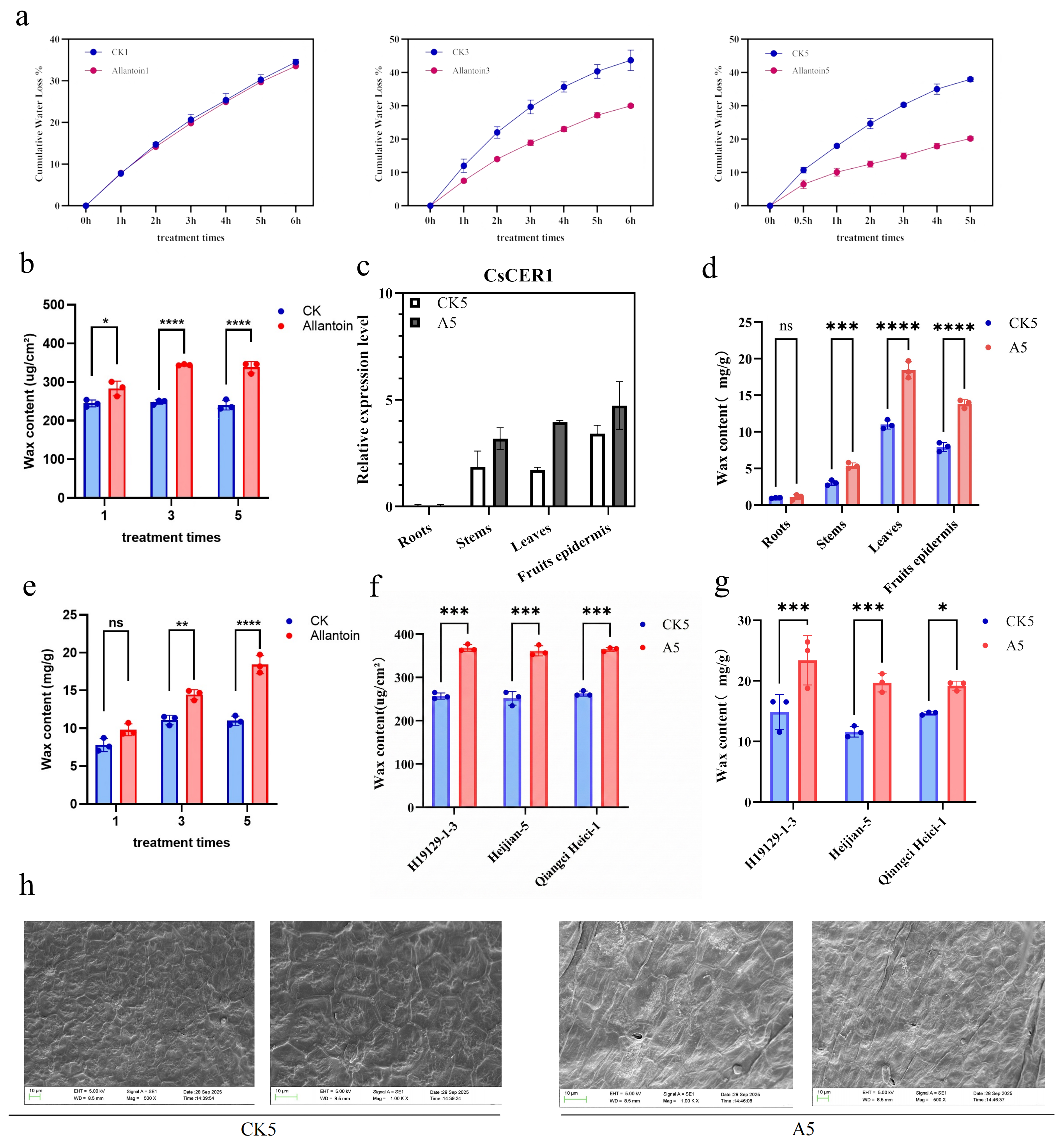
We evaluated the cumulative effects of exogenous allantoin application (one, three, and five times) on leaf water retention and cuticular wax biosynthesis. Consecutive allantoin treatments progressively enhanced leaf water retention capacity, with the A5 group exhibiting the lowest water loss rate in isolated leaves (Figure 5a). Concurrently, total wax content increased cumulatively with the number of allantoin applications, peaking after the fifth application (Figure 5b,e). Therefore, the A5 treatment was adopted as the standard induction condition for subsequent experiments.
Figure 5.
(a) Cumulative water loss rates of isolated leaves after 1, 3, and 5 allantoin treatments. (b,e) Total wax content in leaves, determined by surface area (μg/cm2) and fresh weight (mg/g), respectively. (c) Relative expression levels of CsCER1 in different tissues (root, stem, leaf, and fruit epidermis) under CK5 and A5 treatments. (d) Tissue-specific total wax content corresponding to figure (c). (f,g) Broad-spectrum validation of allantoin-induced wax accumulation in common cucumber varieties (H19129-1-3, Heijian-5, Qiangci Heici-1). (h) Scanning electron microscope (SEM) images of cucumber leaves showing irregular epidermal wax crystal deposition in the A5-treated group compared to the CK5-treated group. Data are presented as mean ± standard deviation (n = 3). * p < 0.05, ** p < 0.01, *** p < 0.001, **** p < 0.0001.
We then analyzed the tissue distribution of allantoin-induced CsCER1 expression. qRT-PCR analysis revealed that allantoin significantly upregulated the expression of CsCER1, a core gene in cuticular wax biosynthesis, primarily in aboveground tissues (especially leaf and fruit epidermis), while root expression remained largely unaffected (Figure 5c). This transcriptional pattern is fully consistent with tissue-specific wax accumulation: total wax content was significantly elevated in the stems, leaves, and fruit epidermis of A5 plants, with leaves exhibiting the highest accumulation (Figure 5d). These results indicate that allantoin selectively strengthens the cuticular barrier of aboveground organs subject to transpiration.
To verify whether this strengthening mechanism is universal, we performed quantitative analyses of leaf wax content in three commercial varieties (H19129-1-3, Heijian-5, and Qiangci Heici-1). Notably, allantoin treatment significantly induced epidermal wax accumulation in all tested varieties (Figure 5f,g). Scanning electron microscopy (SEM) observations further provided morphological evidence of cuticle remodeling (Figure 5h). The leaf epidermis of CK5 was relatively flat, whereas the epidermis of allantoin-treated leaves (A5) was densely covered with an amorphous waxy layer, appearing as white, flocculent deposits, with increased surface roughness. This provides a structural basis for enhanced water retention capacity in leaves, further supporting allantoin-mediated drought tolerance. Nevertheless, the varietal validation was limited to wax content, and further studies are required to evaluate physiological drought tolerance in these varieties.
3.6. Silencing CsCER1 Impairs Allantoin-Induced Cuticular Wax Accumulation and Drought Tolerance in Cucumber
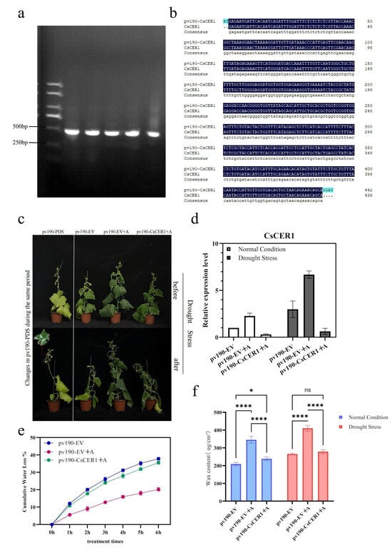
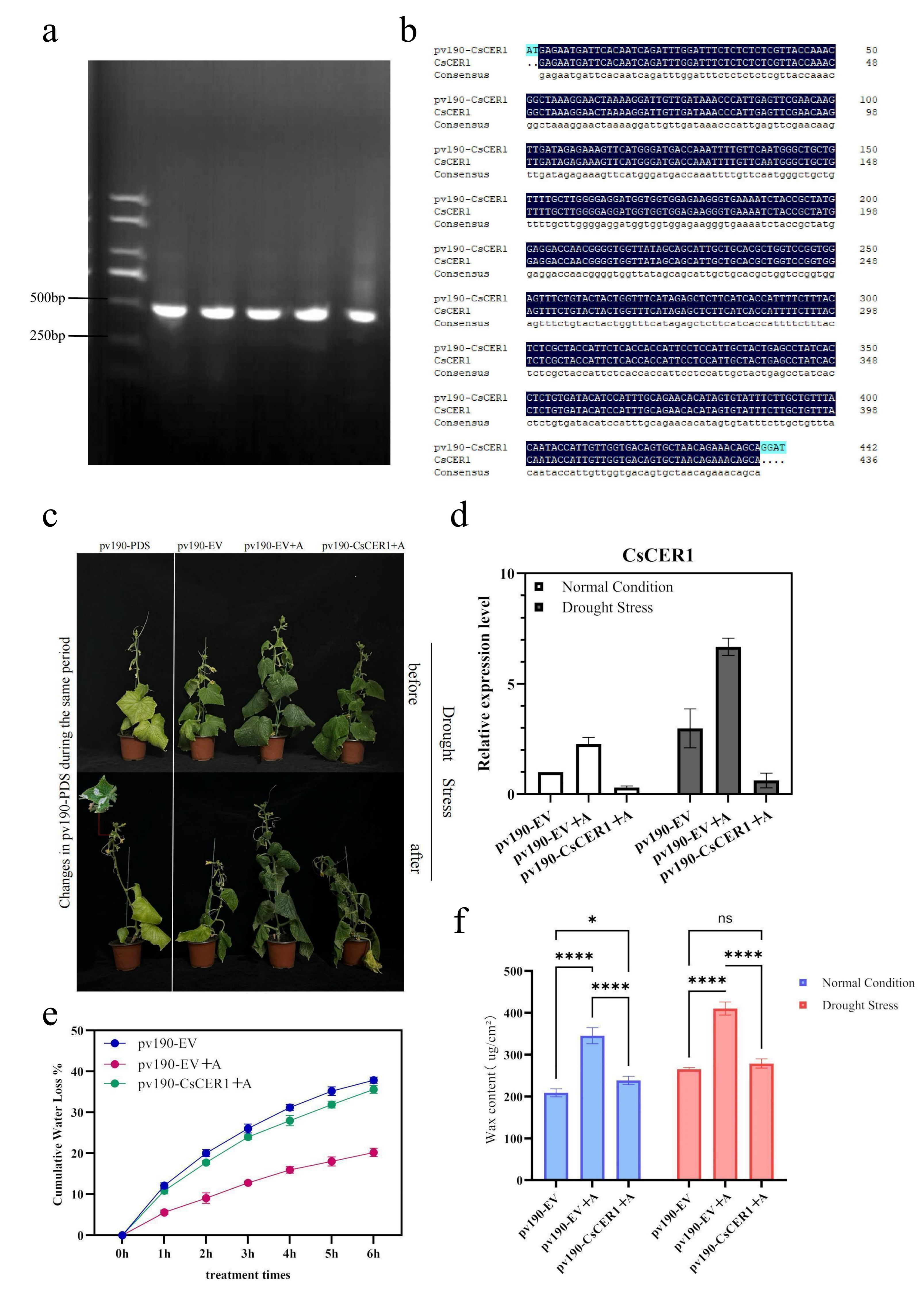
To further validate the biological function of CsCER1 in allantoin‑mediated drought tolerance, a VIGS approach was employed. A 436 bp fragment targeting the non‑conserved region of the CsCER1 CDS was amplified by PCR (Figure 6a) and cloned into the BamHI‑linearized pV190 vector via homologous recombination. The resulting construct pV190-CsCER1 was verified by Sanger sequencing, which confirmed that the inserted fragment matched the target sequence with the correct orientation and no mutations (Figure 6b). This construct was then used for Agrobacterium‑mediated VIGS assays. First, the photobleaching phenotype observed in pv190-PDS plants provided direct evidence of successful systemic infection. qRT-PCR analysis revealed that under both normal and drought conditions, CsCER1 transcription levels in pv190-CsCER1+A plants were significantly lower than those in the empty vector control (pv190-EV) and the allantoin-treated control (pv190-EV+A) (Figure 6d), The silencing efficiency ranged from 70% to 75%, confirming successful CsCER1 gene silencing. Compared with the empty vector control (pv190-EV), the allantoin-treated group (pv190-EV+A) effectively prevented drought-induced wilting; whereas pv190-CsCER1+A plants exhibited severe wilting consistent with the control group (Figure 6c). Consistent with the phenotypic observations, detached leaf water loss rate measurements showed that the water loss rate of pv190-CsCER1+A plants was significantly accelerated, comparable to that of the pv190-EV group, and failed to maintain the strong water retention capacity of the pv190-EV+A group (Figure 6e). These results demonstrate that the CsCER1 gene plays an irreplaceable role in allantoin-mediated plant drought tolerance.
Figure 6.
Silencing of the CsCER1 gene significantly reduced allantoin-induced drought tolerance and cuticular wax accumulation in cucumber. (a) PCR amplification (b) Sequencing validation (c) Phenotypic comparison of the VIGS control group (pv190-PDS), empty vector control group (pv190-EV), allantoin-treated control group (pv190-EV+A), and CsCER1-silenced plants (pv190-CsCER1+A) before and after drought stress induced by a 20% PEG solution. (d) Relative expression levels of the CsCER1 gene under normal and drought conditions (determined by RT-qPCR). (e) Cumulative water loss rate of detached leaves after 6 h (%). (f) Total cuticular wax content per unit leaf area (μg/cm²). Data represent the mean ± standard deviation of three biological replicates. Data are presented as mean ± standard deviation (n = 3). * p < 0.05, **** p < 0.0001.
To further elucidate the biochemical basis of this loss of function, we measured total cuticular wax content (μg/cm2). Under normal conditions, total wax content in pv190-CsCER1+A plants was significantly reduced compared to pv190-EV+A. The wax content in pv190-CsCER1+A plants was slightly higher than that in pv190-EV but significantly lower than that in pv190-EV+A, indicating that CsCER1 is the primary pathway for allantoin-induced wax accumulation. Under drought conditions, the total wax content of pv190-CsCER1+A plants showed no statistically significant difference (ns) compared to pv190-EV and was significantly lower than that of the highly drought-tolerant pv190-EV+A plants (Figure 6f). This indicates that CsCER1-dependent wax biosynthesis is crucial for allantoin-induced reinforcement of the cucumber epidermal barrier and subsequent drought tolerance.
4. Discussion
4.1. Allantoin Synergistically Promotes Enhanced Photosynthesis and Cuticular Wax Synthesis
The allocation of finite carbon resources between growth and defense is a fundamental concept in plant biology, often referred to as the “growth–defense trade-off” [40,41]. In this study, allantoin treatment significantly increased both net photosynthetic rate (Pn) and cuticular wax content, while treated plants showed no visible growth retardation compared with controls, suggesting that allantoin may partially mitigate this trade-off. Moreover, allantoin may directly enhance the efficiency of light reactions by increasing light capture capacity (e.g., upregulation of photosynthesis-antenna protein genes) and chlorophyll content. At the same time, the thickened cuticular wax layer acts as a physical barrier that reduces cuticular transpiration, allowing leaves to maintain higher water content under drought stress and thereby protecting the photosynthetic apparatus. Notably, stomatal conductance (Gs) in the A5 treatment group did not differ significantly from that of the control, yet Pn was markedly elevated, indicating that the enhanced photosynthesis was not driven by increased CO2 supply via stomatal opening. Allantoin has been shown to promote ABA accumulation and enhance drought tolerance by activating ABA metabolism [17]; ABA-mediated stomatal regulation is a dynamic process [42]. Upon stomatal closure, water loss is immediately restricted, but CO2 supply is simultaneously reduced, which is detrimental to sustained photosynthesis [43]. The observation that Gs remained stable while Pn continued to increase suggests that the enhancement in photosynthesis more likely originates from increased light-capturing capacity due to higher chlorophyll content, as well as the maintenance of leaf water homeostasis by the non-stomatal barrier formed by cuticular waxes. Cuticular wax constitutes the first line of defense against non-stomatal water loss, and its chemical composition directly determines the efficiency of the epidermal permeability barrier [44]. Previous studies have shown that cuticular wax accumulation can simultaneously reduce water loss and improve photosynthesis [45,46]. In Kentucky bluegrass, overexpression of PpKCS6 significantly increased photosynthetic efficiency and water-use efficiency under drought stress by regulating wax synthesis [47]. Following allantoin treatment, leaf wax content increased markedly, restricting non-stomatal water loss, persistently reducing cuticular transpiration, and thereby sustaining photosynthetic stability and enhancing water-use efficiency over a longer period under drought conditions. The allantoin-induced upregulation of CsCER1 and accumulation of cuticular wax together constitute a potentially more structurally persistent drought tolerance mechanism.
4.2. CsCER1 Is a Key Responsive Gene for Allantoin-Induced Wax Accumulation
The biosynthesis of plant cuticular wax mainly relies on two pathways: the decarbonylation pathway (producing very-long-chain alkanes) and the reduction pathway (producing primary alcohols) [48]. These two pathways do not operate independently; Li et al. [49] identified the SOH1–CER3–CER1 regulatory module in Arabidopsis, revealing that the allocation of carbon flux between the two pathways is finely tuned by environmental signals. Under drought conditions, this module preferentially directs wax precursors toward the decarbonylation pathway dominated by CER1, driving massive alkane accumulation while reducing primary alcohol content. Very-long-chain alkanes are the core components of the cuticular water barrier [50,51].
CsCER1 encodes a very-long-chain aldehyde decarbonylase that catalyzes the conversion of aldehydes to alkanes [29]. In cucumber, Wang et al. [30] demonstrated that CsCER1 is specifically expressed in epidermal cells, induced by drought and ABA, and essential for the biosynthesis of very-long-chain alkanes. Bernard et al. [50] reconstituted the alkane biosynthesis pathway in yeast, directly confirming that CER1 and CER3 are core components of the synthesis complex. Regarding the upstream regulation of CsCER1, existing studies have largely focused on drought and endogenous hormone signals: the R2R3-MYB transcription factors MYB94 and MYB96 activate wax synthesis genes, including CER1 [52,53]; members of the AP2/ERF family, DREB26 and ERF12, also participate in transcriptional regulation [54]; in citrus, the CsMYB44-csi-miR0008-CsCER1 module regulates wax accumulation under drought [55]. However, whether exogenous metabolites can directly drive CsCER1 expression has not been reported previously.
The multi-omics analysis in this study showed that allantoin treatment upregulated both wax biosynthetic pathways, with a stronger response in the decarbonylation pathway. VIGS functional validation demonstrated that CsCER1 is required for allantoin-induced drought tolerance. It should be noted, however, that the current data do not prove that allantoin directly activates CsCER1; regulation could be indirect (e.g., through JA signaling). Direct regulation remains to be tested by promoter-binding assays (e.g., EMSA, dual-luciferase reporter assays).
4.3. CsCER1 Is Required for Allantoin-Induced Drought Tolerance
VIGS silencing experiments showed that pv190-CsCER1+A plants lost allantoin-induced drought tolerance under drought stress: their wilting severity and detached leaf water loss rates were comparable to those of the empty vector control (pv190-EV), and wax content did not increase. These results indicate that CsCER1 is required for allantoin-driven cuticular wax accumulation and enhanced drought tolerance. Notably, under normal conditions, the total wax content of pv190-CsCER1+A plants was slightly higher than that of pv190-EV (p < 0.05). This suggests that when the decarbonylation pathway is blocked by CsCER1 silencing, the acyl-reduction pathway (alcohol synthesis bypass) may be partially compensatorily activated. However, because alcohol wax components were not quantified, this remains speculative. Under drought stress, any such compensation is insufficient to maintain an effective barrier, as wax content in the silenced plants showed no significant difference from that of pv190-EV, and drought tolerance was not improved. Thus, under drought conditions, the decarbonylation pathway centered on alkane synthesis appears necessary for allantoin-induced wax accumulation.
The VIGS silencing results in this study are highly consistent with the phenotypes of CER1 loss-of-function mutants in other species: silencing CER1 in maize [56], tomato [57], and citrus [58] all led to a significant reduction in plant drought tolerance, further supporting the central role of CER1-mediated alkane synthesis in the plant cuticular barrier. Together, these findings indicate that CER1-mediated alkane synthesis is a key component of the plant cuticular barrier and that its loss cannot be fully compensated by other pathways. This functional conservation highlights the critical role of alkane waxes in the adaptation of plants to terrestrial environments.
4.4. Allantoin-Induced Wax Deposition in Aboveground Parts and Its Varietal Universality
Spatial expression analysis revealed that allantoin induced CsCER1 expression most strongly in leaf and fruit epidermis, moderately in stems, and barely in roots (Figure 5c). This is consistent with the biological function of cuticular wax, which provides a hydrophobic seal for aboveground organs to limit non-stomatal water loss, protect against UV radiation and pathogen attack, and deter herbivory [38,46]. Wax accumulation in the root epidermis would impair water and mineral nutrient uptake. Therefore, the tissue-specificity of allantoin-induced CsCER1 expression reflects an adaptive regulatory mechanism that enhances aboveground water conservation while preserving root absorptive function.
The allantoin-induced increase in leaf wax content was confirmed in three commercial cucumber varieties (H19129-1-3, Heijian-5, and Qiangci Heici-1). However, as noted in the Results section (Section 3.6), varietal universality was assessed only for wax content; drought tolerance physiological parameters (e.g., water loss rate, photosynthetic rate) were not measured in these varieties. Therefore, the conclusion of universality is currently limited to wax accumulation. Genetic improvement in cuticular wax traits has been an important direction for drought-tolerant crop breeding [59]. Traditional approaches rely on screening natural high-wax germplasm or creating wax-overexpressing lines, which can be time-consuming and may negatively impact growth and yield. The present study offers a new concept: exogenous application of the natural metabolite allantoin to coordinately improve photosynthetic capacity and wax-based barrier function, thereby promoting cuticular wax accumulation. Foliar spraying of allantoin is simple to perform, and its concentration, frequency, and timing can be adjusted according to production needs. Nevertheless, field validation is required before any practical agricultural application.
5. Conclusions
This study preliminarily reveals a mechanism by which exogenous allantoin enhances drought tolerance in cucumber through activating CsCER1-mediated cuticular wax synthesis. Allantoin treatment, while improving photosynthetic capacity, upregulates CsCER1 expression, drives the accumulation of cuticular wax, forms a dense wax layer, significantly reduces non-stomatal water loss, and enhances plant drought tolerance. This finding provides a preliminary theoretical basis for improving drought tolerance in cucurbit crops through chemical regulation (Figure 7).
Figure 7.
Working model of how exogenous allantoin enhances cucumber drought tolerance by activating CsCER1-mediated cuticular wax synthesis.
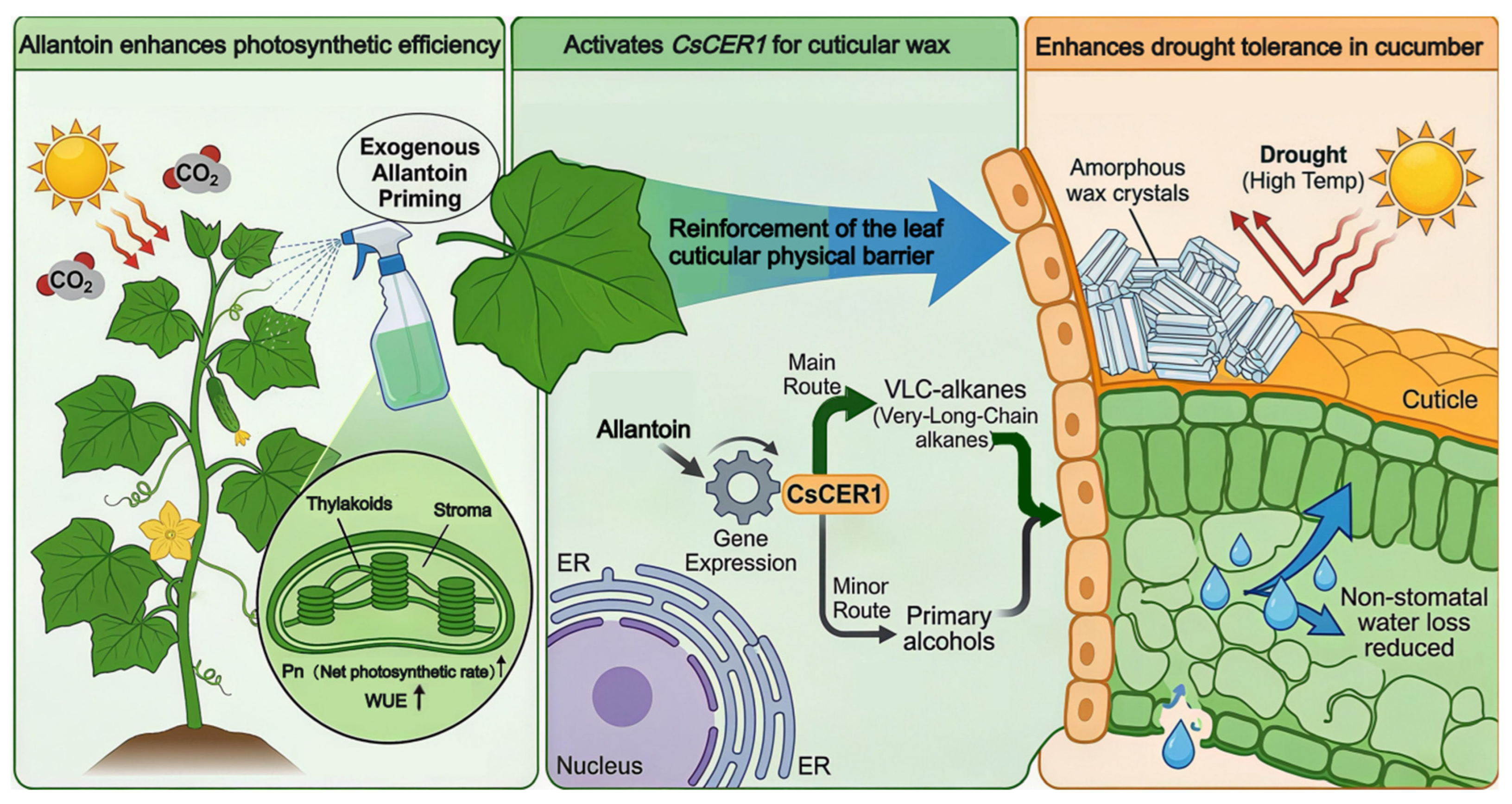
Following exogenous allantoin treatment, cucumbers exhibit increased net photosynthetic rate (Pn) and water-use efficiency (WUE) while simultaneously showing upregulation of CsCER1 transcription in aboveground tissues, which activates the decarbonylation pathway, leading to accumulation of cuticular wax and formation of a dense, amorphous wax layer. This significantly reduces non-stomatal water loss and enhances the plant’s overall drought tolerance.
Supplementary Materials
The following supporting information can be downloaded at: https://www.mdpi.com/article/10.3390/horticulturae12070798/s1, Figure S1: Phenotypes of cucumber seedlings treated with different concentrations of allantoin (0, 3, 6, 9, and 12 mM).
Author Contributions
Conceptualization: W.W., T.Z.; Methodology: W.W., T.Z.; Software: W.W.; Validation: W.W.; Formal analysis: W.W.; Investigation: W.W., C.Y.; Resources: D.L., T.Z., G.F.; Data curation: W.W.; Writing—original draft preparation: W.W.; Writing—review and editing: W.W., X.Y., C.L., Z.Y., D.L., T.Z., G.F.; Visualization: W.W.; Supervision: T.Z., G.F.; Project administration: T.Z., G.F.; Funding acquisition: T.Z., G.F. All authors have read and agreed to the published version of the manuscript.
Funding
Heilongjiang Province “Double First-Class” Discipline Collaborative Innovation Achievement Project LJGXCG2023-013. Basic Research Operating Funds for Provincial Undergraduate Universities in Heilongjiang Province 2024-KYYWF-0147.
Data Availability Statement
The data presented in this study are openly available in [https://www.majorbio.com/].
Conflicts of Interest
Authors Taifeng Zhang and Dajun Liu are employed by the company Heilongjiang Junyi Agricultural Limited Liability Company, Harbin 150000, China. The remaining authors declare that this research was conducted in the absence of any commercial or financial relationships that could be construed as potential conflicts of interest.
References
- Benmrid, B.; Ghoulam, C.; Zeroual, Y.; Kouisni, L.; Bargaz, A. Bioinoculants as a means of increasing crop tolerance to drought and phosphorus deficiency in legume-cereal intercropping systems. Commun. Biol. 2023, 6, 1016. [Google Scholar] [CrossRef] [Scilit] [PubMed]
- Janni, M.; Maestri, E.; Gullì, M.; Marmiroli, M.; Marmiroli, N. Plant responses to climate change, how global warming may impact on food security: A critical review. Front. Plant Sci. 2024, 14, 1297569. [Google Scholar] [CrossRef] [Scilit] [PubMed]
- Das, A.; Munshi, A.D.; Raju, D.; Kumar, S.; Singh, D.; Talukdar, A.; Hongal, D.; Iquebal, M.A.; Kumar, S.; Bhattacharya, R.C.; et al. Key physiological traits for drought tolerance identified through phenotyping a large set of slicing cucumber (Cucumis sativus L.) genotypes under field and water-stress conditions. Genet. Resour. Crop Evol. 2024, 71, 1855–1868. [Google Scholar] [CrossRef] [Scilit]
- Nandi, S.; Das, A.; Munshi, A.D.; Vikrant, V.; Kumari, K.; Choudhary, H.; Sureja, A.K.; Talukdar, A.; Raju, D.; Singh, B.; et al. Tissue‑specific response in seedling stage reveals key physio‑biochemical and molecular network associated with drought tolerance in cucumber. Sci. Hortic. 2025, 342, 114009. [Google Scholar] [CrossRef] [Scilit]
- Wang, S.; Ma, Q.; Li, C.; Li, Y.; Wang, Y.; Zhang, Y. Chloroplast responses to drought: Integrative mechanisms and mitigation strategies. Int. J. Mol. Sci. 2025, 26, 11872. [Google Scholar] [CrossRef] [Scilit] [PubMed]
- Chaves, M.M.; Flexas, J.; Pinheiro, C. Photosynthesis under drought and salt stress: Regulation mechanisms from whole plant to cell. Ann. Bot. 2009, 103, 551–560. [Google Scholar] [CrossRef] [Scilit]
- Shang, Y.; Ma, Y.; Zhou, Y.; Zhang, , H.; Duan, L.; Chen, H.; Zeng, J.; Zhou, Q.; Wang, S.; Gu, W.; et al. Biosynthesis, regulation, and domestication of bitterness in cucumber. Science 2014, 346, 1084–1088. [Google Scholar] [CrossRef] [Scilit] [PubMed]
- Smiti, K.; Mina, U.; Verma, M.; Arya, L. Impact of climate extremes and other key abiotic stresses on cucurbits: A systematic review. Vegetos 2025, 1–21. [Google Scholar] [CrossRef] [Scilit]
- Huot, B.; Yao, J.; Montgomery, B.L.; He, S.Y. Growth–defense tradeoffs in plants: A balancing act to optimize fitness. Mol. Plant 2014, 7, 1267–1287. [Google Scholar] [CrossRef] [Scilit] [PubMed]
- Panda, S.; Kazachkova, Y.; Aharoni, A. Catch-22 in specialized metabolism: Balancing defense and growth. J. Exp. Bot. 2021, 72, 6027–6041. [Google Scholar] [CrossRef] [Scilit] [PubMed]
- Campos, M.L.; Yoshida, Y.; Major, I.T.; Ferreira, D.O.; Weraduwage, S.M.; Froehlich, J.E.; Johnson, B.F.; Kramer, D.M.; Jander, G.; Sharkey, T.D.; et al. Rewiring of jasmonate and phytochrome B signalling uncouples plant growth-defense tradeoffs. Nat. Commun. 2016, 7, 12570. [Google Scholar] [CrossRef] [Scilit] [PubMed]
- Majchrzak, L.; Xiao, J.; Li, R.; Zhang, Y.; Wang, X.; Chen, Y.; Liu, Z.; Yang, H. A synthetic jasmonate receptor agonist uncouples the growth–defense trade-off in rice. Proc. Natl. Acad. Sci. USA 2025, 122, e2505675122. [Google Scholar] [CrossRef] [Scilit] [PubMed]
- Li, C.; Xu, M.X.; Cai, X.; Wang, Y.; Liu, J.; Sun, Y.; Chen, R.; Wu, J.; Zhang, X. Jasmonate signaling pathway modulates plant defense, growth, and their trade-offs. Int. J. Mol. Sci. 2022, 23, 3945. [Google Scholar] [CrossRef] [Scilit] [PubMed]
- Takagi, H.; Ishiga, Y.; Watanabe, S.; Matsumoto, M.; Hakomori, Y.; Shimada, H.; Sakamoto, A. Allantoin, a stress-related purine metabolite, can activate jasmonate signaling in a MYC2-regulated and abscisic acid-dependent manner. J. Exp. Bot. 2016, 67, 2519–2532. [Google Scholar] [CrossRef] [Scilit] [PubMed]
- Kaur, R.; Chandra, J.; Varghese, B.; Keshavkant, S. Allantoin: A potential compound for the mitigation of adverse effects of abiotic stresses in plants. Plants 2023, 12, 3059. [Google Scholar] [CrossRef] [Scilit] [PubMed]
- Kaur, H.; Chowrasia, S.; Gaur, V.S.; Mondal, T.K. Allantoin: Emerging role in plant abiotic stress tolerance. Plant Mol. Biol. Rep. 2021, 39, 648–661. [Google Scholar] [CrossRef] [Scilit]
- Watanabe, S.; Matsumoto, M.; Hakomori, Y.; Takagi, H.; Shimada, H.; Sakamoto, A. The purine metabolite allantoin enhances abiotic stress tolerance through synergistic activation of abscisic acid metabolism. Plant Cell Environ. 2014, 37, 1022–1036. [Google Scholar] [CrossRef] [Scilit] [PubMed]
- Raihan, M.R.H.; Rahman, M.; Rastogi, A.; Fujita, M.; Hasanuzzaman, M. Exogenous allantoin confers rapeseed (Brassica campestris) tolerance to simulated drought by improving antioxidant metabolism and physiology. Antioxidants 2023, 12, 1508. [Google Scholar] [CrossRef] [Scilit] [PubMed]
- Lu, S.; Jia, Z.C.; Meng, X.F.; Chen, Y.; Zhang, L.; Wang, P.; Liu, Q.; Zhao, S.; Li, X. Combined metabolomic and transcriptomic analysis reveals allantoin enhances drought tolerance in rice. Int. J. Mol. Sci. 2022, 23, 14172. [Google Scholar] [CrossRef] [Scilit] [PubMed]
- Thu, S.W.; Collier, R.; Gandin, A.; Sitton, C.C.; Tegeder, M. Role of ureides in source-to-sink transport of photoassimilates in non-fixing soybean. J. Exp. Bot. 2020, 71, 4495–4511. [Google Scholar] [CrossRef] [Scilit] [PubMed]
- Thu, S.W.; Tegeder, M. Enhanced ureide partitioning improves soybean performance under drought stress. J. Exp. Bot. 2025, 76, 2786–2808. [Google Scholar] [CrossRef] [Scilit] [PubMed]
- Arya, G.C.; Sarkar, S.; Manasherova, E.; Aharoni, A.; Cohen, H. The plant cuticle: An ancient guardian barrier set against long-standing rivals. Front. Plant Sci. 2021, 12, 663165. [Google Scholar] [CrossRef] [Scilit] [PubMed]
- Lewandowska, M.; Keyl, A.; Feussner, I. Wax biosynthesis in response to danger: Its regulation upon abiotic and biotic stress. New Phytol. 2020, 227, 698–713. [Google Scholar] [CrossRef] [Scilit] [PubMed]
- Wu, W.; Jiang, B.; Liu, R.; Han, Y.; Fang, X.; Mu, H.; Farag, M.A.; Simal-Gandara, J.; Prieto, M.A.; Chen, H.; et al. Structures and functions of cuticular wax in postharvest fruit and its regulation: A comprehensive review with future perspectives. Engineering 2023, 23, 118–129. [Google Scholar] [CrossRef] [Scilit]
- Pascal, S.; Bernard, A.; Deslous, P.; Gronnier, J.; Fournier-Goss, A.; Domergue, F.; Rowland, O.; Joubès, J. Arabidopsis CER1-LIKE1 functions in a cuticular very-long-chain alkane-forming complex. Plant Physiol. 2019, 179, 415–432. [Google Scholar] [CrossRef] [Scilit]
- Youssif, N.E.E.; Yang, B.; Huang, H.; Amar, M.H.; Ezzat, M.; Belal, M.; Zaghlool, S.A.M.; Zhao, H.; Fu, D.; Lü, S. Comparative genomic analysis and functional identification of CER1 and CER3 homologs in rice wax synthesis. Biology 2026, 15, 166. [Google Scholar] [CrossRef] [Scilit] [PubMed]
- Gao, Y.; Zhang, Z.; Cheng, J.; Xian, X.; Li, C.; Wang, Y. Genome-wide identification of the CER1 gene family in apple and response of MdCER1-1 to drought stress. Funct. Integr. Genom. 2023, 23, 17. [Google Scholar] [CrossRef] [Scilit]
- Zhu, H.-X.; Huang, J.-F.; Cao, J.-F.; Wang, L.-J.; Huang, J.-Q. SPL1 positively regulates cuticular ridge wax biosynthesis in Arabidopsis. Plant Stress 2025, 16, 100872. [Google Scholar] [CrossRef] [Scilit]
- Bourdenx, B.; Bernard, A.; Domergue, F.; Pascal, S.; Léger, A.; Roby, D.; Pervent, M.; Vile, D.; Haslam, R.P.; Napier, J.A.; et al. Overexpression of Arabidopsis ECERIFERUM1 promotes wax very-long-chain alkane biosynthesis and influences plant response to biotic and abiotic stresses. Plant Physiol. 2011, 156, 29–45. [Google Scholar] [CrossRef] [Scilit] [PubMed]
- Wang, W.; Zhang, Y.; Xu, C.; Ren, J.; Liu, X.; Black, K.; Gai, X.; Wang, Q.; Ren, H. Cucumber ECERIFERUM1 (CsCER1), which influences the cuticle properties and drought tolerance of cucumber, plays a key role in VLC alkanes biosynthesis. Plant Mol. Biol. 2015, 87, 219–233. [Google Scholar] [CrossRef] [Scilit]
- An, S.; Hwang, H.; Chun, C.; Jang, Y.; Lee, H.J.; Wi, S.H.; Yeo, K.H.; Yu, I.; Kwack, Y. Evaluation of air temperature, photoperiod and light intensity conditions to produce cucumber scions and rootstocks in a plant factory with artificial lighting. Horticulturae 2021, 7, 102. [Google Scholar] [CrossRef] [Scilit]
- Li, S.; Huang, M.; Di, Q.; Ji, T.; Wang, X.; Wei, M.; Shi, Q.; Li, Y.; Gong, B.; Yang, F. The functions of a cucumber phospholipase D alpha gene (CsPLDα) in growth and tolerance to hyperosmotic stress. Plant Physiol. Biochem. 2015, 97, 175–186. [Google Scholar] [CrossRef] [Scilit] [PubMed]
- Koes, R.; Verweij, W.; Quattrocchio, F. Flavonoids: A colorful model for the regulation and evolution of biochemical pathways. Trends Plant Sci. 2005, 10, 236–242. [Google Scholar] [CrossRef] [Scilit] [PubMed]
- Zhou, L.; Ni, E.; Yang, J.; Zhou, H.; Liang, H.; Li, J.; Jiang, D.; Wang, Z.; Liu, Z.; Zhuang, C. Rice OsGL1-6 is involved in leaf cuticular wax accumulation and drought resistance. PLoS ONE 2013, 8, e65139. [Google Scholar] [CrossRef] [Scilit] [PubMed]
- Kim, R.J.; Lee, S.B.; Pandey, G.; Suh, M.C. Functional conservation of an AP2/ERF transcription factor in cuticle formation suggests an important role in the terrestrialization of early land plants. J. Exp. Bot. 2022, 73, 7450–7466. [Google Scholar] [CrossRef] [Scilit] [PubMed]
- Li, B.; Dewey, C.N. RSEM: Accurate transcript quantification from RNA-seq data with or without a reference genome. BMC Bioinform. 2011, 12, 323. [Google Scholar] [CrossRef] [Scilit] [PubMed]
- Ren, Y.; Yu, G.; Shi, C.; Liu, L.; Guo, Q.; Han, C.; Zhang, D.; Zhang, L.; Liu, B.; Gao, H. Majorbio cloud: A one-stop, comprehensive bioinformatic platform for multiomics analyses. iMeta 2022, 1, e12. [Google Scholar] [CrossRef] [Scilit] [PubMed]
- Song, J.; Zhu, Z.; Zhang, T.; Meng, X.; Zhang, W.; Gao, P. Genome-wide identification, evolutionary analysis, and functional studies of APX genes in melon (Cucumis melo L.). Int. J. Mol. Sci. 2023, 24, 17571. [Google Scholar] [CrossRef] [Scilit] [PubMed]
- Wan, L.L.; Wang, Z.R.; Tang, M.; Zhang, X.J.; Ren, J.; Zeng, H.X.; Zhang, N.; Wei, J.Q.; Xiong, J.S. Optimization of CGMMV-VIGS System in Cucurbit Crops. Chin. Agric. Sci. Bull. 2024, 40, 40–47. [Google Scholar] [CrossRef]
- Sestari, I.; Campos, M.L. Into a dilemma of plants: The antagonism between chemical defenses and growth. Plant Mol. Biol. 2022, 109, 469–482. [Google Scholar] [CrossRef] [Scilit] [PubMed]
- Züst, T.; Agrawal, A.A. Trade-offs between plant growth and defense against insect herbivory: An emerging mechanistic synthesis. Annu. Rev. Plant Biol. 2017, 68, 513–534. [Google Scholar] [CrossRef] [Scilit] [PubMed]
- Wilkinson, S.; Davies, W.J. ABA-based chemical signalling: The co-ordination of responses to stress in plants. Plant Cell Environ. 2002, 25, 195–210. [Google Scholar] [CrossRef] [Scilit] [PubMed]
- Flexas, J. Drought-inhibition of photosynthesis in C3 plants: Stomatal and non-stomatal limitations revisited. Ann. Bot. 2002, 89, 183–189. [Google Scholar] [CrossRef] [Scilit] [PubMed]
- Riederer, M.; Schreiber, L. Protecting against water loss: Analysis of the barrier properties of plant cuticles. J. Exp. Bot. 2001, 52, 2023–2032. [Google Scholar] [CrossRef] [Scilit] [PubMed]
- Guo, J.; Xu, W.; Yu, X.; Shen, H.; Li, H.; Cheng, D.; Liu, A.; Liu, J.; Liu, C.; Zhao, S.; et al. Cuticular wax accumulation is associated with drought tolerance in wheat near-isogenic lines. Front. Plant Sci. 2016, 7, 1809. [Google Scholar] [CrossRef] [Scilit] [PubMed]
- Han, D.; Lu, J.; Zhao, C.; Ali, S.; Jiang, Z. The role and regulatory mechanisms of cuticular wax in crop stress tolerance and yield. Plants 2026, 15, 554. [Google Scholar] [CrossRef] [Scilit] [PubMed]
- Jiang, J.; Zheng, X.; He, T.; Liu, X.; Zhao, Q.; Tan, W.; Xiong, L.; Li, B.; Yin, H.; Agyei, G.D.; et al. The function of PpKCS6 in regulating cuticular wax synthesis and drought resistance of Kentucky bluegrass. Plant Cell Environ. 2025, 48, 4643–4655. [Google Scholar] [CrossRef] [Scilit] [PubMed]
- Wen, J.; Xia, W.; Wang, Y.; Li, J.; Guo, R.; Zhao, Y.; Fen, J.; Duan, X.; Wei, G.; Wang, G.; et al. Pathway elucidation and heterologous reconstitution of the long-chain alkane pentadecane biosynthesis from Pogostemon cablin. Plant Biotechnol. J. 2025, 23, 564–578. [Google Scholar] [CrossRef] [Scilit] [PubMed]
- Li, S.; Zhang, X.; Huang, H.; Yin, M.; Jenks, M.A.; Kosma, D.K.; Yang, P.; Yang, X.; Zhao, H.; Lü, S. Deciphering the core shunt mechanism in Arabidopsis cuticular wax biosynthesis and its role in plant environmental adaptation. Nat. Plants 2025, 11, 165–175. [Google Scholar] [CrossRef] [Scilit] [PubMed]
- Bernard, A.; Domergue, F.; Pascal, S.; Jetter, R.; Renne, C.; Faure, J.-D.; Haslam, R.P.; Napier, J.A.; Lessire, R.; Joubès, J. Reconstitution of plant alkane biosynthesis in yeast demonstrates that Arabidopsis ECERIFERUM1 and ECERIFERUM3 are core components of a very-long-chain alkane synthesis complex. Plant Cell 2012, 24, 3106–3118. [Google Scholar] [CrossRef] [Scilit] [PubMed]
- Busta, L.; Jetter, R. Moving beyond the ubiquitous: The diversity and biosynthesis of specialty compounds in plant cuticular waxes. Phytochem. Rev. 2018, 17, 1275–1304. [Google Scholar] [CrossRef] [Scilit]
- Lee, S.B.; Kim, H.U.; Suh, M.C. MYB94 and MYB96 additively activate cuticular wax biosynthesis in Arabidopsis. Plant Cell Physiol. 2016, 57, 2300–2311. [Google Scholar] [CrossRef] [Scilit] [PubMed]
- Seo, P.J.; Lee, S.B.; Suh, M.C.; Park, M.-J.; Go, Y.S.; Park, C.-M. The MYB96 transcription factor regulates cuticular wax biosynthesis under drought conditions in Arabidopsis. Plant Cell 2011, 23, 1138–1152. [Google Scholar] [CrossRef] [Scilit] [PubMed]
- Urano, K.; Oshima, Y.; Ishikawa, T.; Kajino, T.; Sakamoto, S.; Sato, M.; Toyooka, K.; Fujita, M.; Kawai-Yamada, M.; Taji, T.; et al. Arabidopsis DREB26/ERF12 and its close relatives regulate cuticular wax biosynthesis under drought stress condition. Plant J. 2024, 120, 2057–2075. [Google Scholar] [CrossRef] [Scilit] [PubMed]
- Xie, J.; Yang, L.; Hu, W.; Song, J.; Kuang, L.; Huang, Y.; Liu, D.; Liu, Y. The CsMYB44-csi-MIR0008-CsCER1 module regulates cuticular wax biosynthesis and drought tolerance in citrus. New Phytol. 2025, 246, 1757–1779. [Google Scholar] [CrossRef] [Scilit] [PubMed]
- Zhao, Y.; Liu, Q.; Wang, X.; Zhang, W.; Xu, W.; Zhang, Y.; Liu, B. ZmCER1, a putative ECERIFERUM 1 protein in maize, functions in cuticular wax biosynthesis and bulliform cell development. Crop J. 2024, 12, 743–752. [Google Scholar] [CrossRef] [Scilit]
- Wu, H.; Liu, L.; Chen, Y.; Liu, T.; Jiang, Q.; Wei, Z.; Li, C.; Wang, Z. Tomato SlCER1–1 catalyzes the synthesis of wax alkanes, increasing drought tolerance and fruit storability. Hortic. Res. 2022, 9, uhac004. [Google Scholar] [CrossRef] [Scilit] [PubMed]
- Dang, Q.H.; Suh, M.C. How plants adapt surface lipids to environmental changes. Nat. Plants 2025, 11, 159–160. [Google Scholar] [CrossRef] [Scilit] [PubMed]
- Liu, Y.; Chen, B.; Qin, Z.; Jiang, P.; Yang, Y.; Wang, C.; Xing, T.; Li, F.; Du, L.; Li, S.; et al. TaFAR5-TaFAR3 module regulates cuticular wax biosynthesis and drought tolerance in wheat. New Phytol. 2025, 248, 1802–1821. [Google Scholar] [CrossRef] [Scilit] [PubMed]
Disclaimer/Publisher’s Note: The statements, opinions and data contained in all publications are solely those of the individual author(s) and contributor(s) and not of MDPI and/or the editor(s). MDPI and/or the editor(s) disclaim responsibility for any injury to people or property resulting from any ideas, methods, instructions or products referred to in the content. |
© 2026 by the authors. Licensee MDPI, Basel, Switzerland. This article is an open access article distributed under the terms and conditions of the Creative Commons Attribution (CC BY) license.