Article

Regulation on the Induction of Protocorm-like Bodies and Callus in Dendrobium officinale

1
Key Laboratory for Economic Plants and Biotechnology, Kunming Institute of Botany, Chinese Academy of Sciences, Kunming 650201, China
2
Yunnan Key Laboratory for Wild Plant Resources, Kunming 650201, China
3
University of Chinese Academy of Sciences, Beijing 100049, China
*
Author to whom correspondence should be addressed.
Horticulturae 2026, 12(5), 555; https://doi.org/10.3390/horticulturae12050555
Submission received: 8 April 2026 / Revised: 28 April 2026 / Accepted: 30 April 2026 / Published: 2 May 2026
(This article belongs to the Special Issue In Vitro Conservation and Rapid Propagation of Horticultural Crops)

Abstract

As globally important ornamental and medicinal plants, orchids exhibit significant differences in the difficulty and pathways of in vitro regeneration. Most orchid species can directly form protocorm-like bodies (PLBs) through the differentiation of shoot tips or other explants, which then regenerate into new plantlets, while some species form callus through explant dedifferentiation followed by PLB differentiation from the callus. At present, the regenerative mechanisms underlying PLB and callus in orchids, as well as the key factors influencing their differentiation, remain poorly elucidated. In this study, seedlings of Dendrobium officinale obtained from aseptic seed germination were used to investigate the effects of explant type, 2,4-D concentration, temperature, light intensity and photoperiod on the induction of PLBs and callus. The results showed that there were no significant differences in callus induction among the tested explants in D. officinale, whereas stem nodal segments were more suitable for PLB induction. For both internodal and nodal segments, the incidence rate of callus formation was higher than that of PLBs. The concentration of 2,4-D influenced the induction direction of the explants; higher concentration promoted PLB induction, while lower concentration was sufficient for callus formation. Low temperature and low light intensity inhibited PLB induction while promoting callus formation in D. officinale. High temperature and intense light partially caused desiccation of explants. A temperature of 25/22 °C (day/night) and a photosynthetic photon flux density of 50 µmol m−2 s−1 were more suitable for callus or PLB induction in D. officinale. A shorter photoperiod favored callus induction, while a longer photoperiod was beneficial for PLB induction. This study reveals the differences in influencing factors for PLB and callus induction in D. officinale, providing important insights for the propagation of orchid seedlings and laying a significant foundation for elucidating the mechanisms of PLB and callus induction.

1. Introduction

Orchidaceae is the most species-rich family of angiosperms on Earth, comprising approximately 736 genera and 29,524 species, which are widely distributed in various ecosystems except polar regions and extreme deserts [1,2]. To adapt to diverse habitats, orchid species have evolved numerous unique adaptive and developmental traits and formed close interactions with fungi and pollinators [3,4,5]. For this reason, since Darwin’s time, orchids have consistently attracted widespread attention in evolutionary biology [1,2]. In addition, Orchids have important ornamental, medicinal and edible values [6,7]. Their rich flower shapes, colors, fragrance, and long-lasting blooms have attracted numerous enthusiasts worldwide. In the ornamental flower trade, genera such as Phalaenopsis, Cymbidium, Dendrobium, Oncidium, Cattleya and Paphiopedilum occupy important positions [7]. Furthermore, over 250 orchid species are utilized as medicinal herbs worldwide [6]. Among these, more than 10 species are officially recorded in the Pharmacopoeia of the People’s Republic of China [8]. The pods of Vanilla planifolia are rich in vanillin, serving as an important source of natural flavor widely used in the food, beverage and cosmetic industries [9].
Given their high economic values in horticultural and medicinal industries, numerous orchid species are severely threatened by over-collection and illegal trade, coupled with extensive habitat destruction. In addition, orchids have a special dependence on fungi and pollinators, which makes the reproduction and survival of many species extremely vulnerable to climate change and human activities [3,5]. These render orchids one of the key plant families prioritized for global conservation [10,11]. At present, all wild orchid species are listed under the protection of the Convention on International Trade in Endangered Species of Wild Fauna and Flora (https://cites.org/, accessed on 7 April 2026). In the List of China National Key Protected Wild Plants released in 2021, approximately 350 orchid species are included, accounting for nearly one-third of the total species listed (https://www.gov.cn/zhengce/zhengceku/2021-09/09/content_5636409.htm, accessed on 7 April 2026).
Establishing seedling propagation systems for orchids is of great significance for their species conservation, commercial cultivation, and gene function research [10,12,13]. After more than a century of research, remarkable progress has been made in asymbiotic seed germination, symbiotic seed germination and in vitro culture of orchids [12,14,15]. However, due to their unique developmental mechanisms and biological characteristics, artificial propagation of some orchids remains challenging [15,16]. Compared to seed-propagated seedlings, plants regenerated from explants like shoot tips or buds generally exhibit higher genetic stability and trait consistency, offering advantages in commercial production [17,18].
For many plant species, callus is first formed through dedifferentiation of explants such as shoot tips, buds and leaves, followed by de novo regeneration of shoots or roots to develop into complete plants [19]. Protocorm-like bodies (PLBs) are unique structures formed during in vitro regeneration of orchids, which are generated through direct or indirect pathways and subsequently differentiate and regenerate into plantlets [15,16]. In Paphiopedilum, PLBs originate from the meristematic cell layer on the callus surface and exhibit distinct growth polarity. The leaf primordia of regenerated shoots differentiate from the apical meristematic cells of PLBs [20]. The direct pathway for PLB formation involves the direct dedifferentiation of parenchyma cells in explant meristems into PLBs. The indirect pathway involves initial dedifferentiation of parenchyma cells into callus, followed by proliferation and differentiation of the callus to form PLBs [21,22]. For example, protocorms obtained from seed germination of Paphiopedilum hybrids can be induced to form callus, which then further differentiates into PLBs and ultimately regenerates plantlets [23]. However, callus induction is not commonly employed during in vitro regeneration of orchids [15], with the majority of species relying primarily on the PLB pathway for plantlet regeneration [12,17]. Direct PLB induction exhibits relatively low efficiency for seedling regeneration. In contrast, pre-induction via callus formation enables each explant to ultimately produce more PLBs, thereby markedly improving propagation efficiency. Accordingly, exploring the regulatory mechanisms underlying callus induction holds great theoretical and practical significance.
Although PLBs are morphologically similar to protocorms formed from seed germination, they are induced from in vitro tissues [17]. The development of PLB in Phalaenopsis aphrodite does not follow the somatic embryogenesis program but is more similar to the shoot organ regeneration process [14,24,25,26]. The shoot organogenesis pathway mediated by the class I KNOTTED-LIKE homeobox gene SHOOT MERISTEMLESS (STM) may be a key mechanism initiating PLB formation [25,27]. Overexpression of STM can promote PLB regeneration in explants of P. aphrodite and D. officinale [26,27]. Although plant propagation through PLB regeneration systems has become a conventional method in the commercial production of orchids [14,18,28], the formation mechanisms of PLBs and callus during in vitro regeneration of orchids remain unclear [28,29].
In vitro regeneration of plants is affected by genotype, explant type, plant hormones and culture conditions [20,22,30,31,32,33,34,35]. Generally, young tissues have higher plant regeneration potential. For example, in Renanthera, young leaves can be induced to form callus and PLBs, but the frequency of callus formation in the leaf base segment is higher than that in the leaf tip segment [31]. Hormones such as auxins and cytokinins play key regulatory roles in cell fate determination during organ regeneration, and the induction effect of callus can be altered by adjusting the ratio of exogenous auxins to cytokinins [15,16,33,36]. On callus induction medium, the Aux/IAA protein IAA14, by degrading auxin signaling, activates auxin response factors ARF7 and ARF19, which upregulate the expression of genes such as LBD16, LBD17, LBD18 and LBD29, thereby promoting callus formation [37,38]. TaLAX PANICLE1 (TaLAX1) can upregulate the expression of the TaGROWTH-REGULATING FACTOR (TaGRF) and TaGRF-INTERACTING FACTOR 1 (TaGIF1) genes, enhancing cytokinin accumulation and auxin response, thereby promoting wheat callus proliferation and shoot regeneration [39]. PLB induction is primarily regulated by cytokinins [18,40,41]. In Phalaenopsis leaves, embryogenic callus formation can be observed on leaf segments after 60 days of dark treatment, and green PLB differentiation becomes visible 15 days after transfer to light conditions [30]. Remarkable progress has been made in understanding callus induction and shoot regeneration mechanisms in economic and food crops [34,42,43], while the key factors and mechanisms regulating PLB and callus induction in orchids remain unclear [20].
Dendrobium officinale possesses important medicinal and edible values [44], and large-scale cultivation has now been achieved. A previous study showed that D. officinale can regenerate into complete plants through both PLB and callus pathways [21]. Therefore, this study employed D. officinale seedlings derived from aseptic seed germination to investigate the effects of different stem segment positions, exogenous auxin, temperature, and light conditions on callus and PLB induction. This study aims to elucidate the main influencing factors governing their induction, provide a theoretical basis for efficient seedling propagation, and lay a foundation for dissecting the developmental mechanisms of PLBs and callus.

2. Materials and Methods

2.1. Experimental Materials

This study used aseptically germinated seedlings of D. officinale as experimental material. First, mature capsules of D. officinale were surface-sterilized in 75% ethanol for 15 min, followed by treatment with 0.5% mercuric chloride for 10 min, and finally rinsed 3–5 times with sterile distilled water. After disinfection, the seeds were sown on a seed germination medium containing full-strength Murashige and Skoog (MS) formulation [45], supplemented with 0.5 mg/L 6-BA, 0.2 mg/L NAA, 30 g/L sucrose, 7 g/L gelidium agar (Sinuohui Biotechnology Co., Ltd., Nantong, China), and 80 g/L potato homogenate. The pH of the medium was adjusted to 5.8. Culture conditions were maintained at a photosynthetic photon flux density (PPFD) of 50 µmol m−2 s−1 supplied by LED lamps, a 12 h/12 h light/dark photoperiod, and a constant temperature of 25 ± 1 °C. After seed germination and protocorm formation, the protocorms were transferred to a differentiation medium consisting of MS + 0.2 mg/L 6-BA + 0.2 mg/L NAA + 25 g/L sucrose + 7 g/L agar + 80 g/L potato homogenate + 1 g/L activated charcoal, with pH 5.8, under the same culture conditions. After 60 days of culture, the seedlings were transferred to a rooting medium containing MS medium supplemented with 0.1 mg/L NAA, 0.1 mg/L KT, 0.5 mg/L IBA, 7 g/L agar, 50 g/L potato homogenate, 50 g/L banana homogenate, and 2 g/L activated charcoal (pH 5.8). All plant materials were cultivated under consistent environmental conditions prior to subsequent experiments.
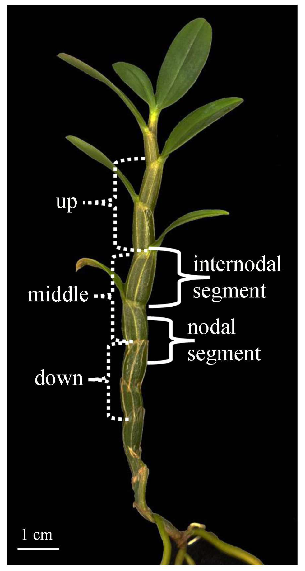
The robust and uniformly sized seedlings with 7–8 nodes were selected as experimental materials. The roots, leaves and leaf sheaths of D. officinale seedlings were excised, and stems were used as explants. Starting from the first intact node at the stem apex, the stem was divided into upper, middle and lower sections. Internodal segments and nodal segments were excised as explants (Figure 1) with a length of 1 cm. According to different experimental objectives, the stems of D. officinale were cut and treated accordingly. Each culture dish was inoculated with about 20 explants, with 100 explants per treatment and three replicates. The explants were cultured in artificial climate light incubator, with LED lamps as the light source (MGC-1000HP-2, Yiheng, Shanghai, China). Light intensity was measured in the center of the incubator by LI-1400 (Li-Cor Biosciences, Lincoln, NE, USA). After 40 days of culture, the explants were photographed, and the induction rates of PLBs and callus were recorded.

2.2. Differences in PLB and Callus Induction Among Different Stem Explant Types and Positions

The explants derived from internodal and nodal segments were inoculated on induction medium (MS + 2.0 mg/L 2,4-D + 30 g/L sucrose + 6.5 g/L agar, pH = 5.8). The culture conditions included a PPFD of 50 µmol m−2 s−1, a 12 h/12 h (day/night) photoperiod, and a temperature of 25 ± 1 °C.

2.3. Effect of 2,4-Dichlorophenoxyacetic Acid (2,4-D) Concentration

According to the results of Section 2.2, we selected the nodal and internodal segments as explants to investigated the effect of 2,4-D concentration on PLB and callus induction. The concentrations of 2,4-D were set at 1.0 mg/L, 2.0 mg/L and 3.0 mg/L, with the remaining medium components being MS + 30 g/L sucrose + 6.5 g/L agar (pH = 5.8). The culture conditions included a PPFD of 50 µmol m−2 s−1, a 12 h/12 h day/night photoperiod, and a temperature of 25 ± 1 °C.

2.4. Effect of Temperature on PLB and Callus Induction

Three temperature gradients were set at 18/15 °C (day/night), 25/22 °C and 32/29 °C. The middle nodal segments were transferred to induction medium (MS + 2.0 mg/L 2,4-D + 30 g/L sucrose + 6.5 g/L agar, pH = 5.8). Cultures were maintained under different temperature gradients, with a PPFD of 50 µmol m−2 s−1 and a photoperiod of 12 h/12 h (day/night).

2.5. Effect of Light Intensity

Three light intensity levels were established, with the PPFD set at 25, 50 and 75 µmol m−2 s−1, respectively. The middle nodal segments were transferred to induction medium (MS + 2.0 mg/L 2,4-D + 30 g/L sucrose + 6.5 g/L agar, pH = 5.8). Cultures were maintained under different light intensities with a 12 h/12 h photoperiod at 25 ± 1 °C.

2.6. Photoperiod Effect

Three photoperiod treatments were set as 3/21 h, 12/12 h and 21/3 h (day/night). The middle nodal segments were transferred to induction medium (MS + 2.0 mg/L 2,4-D + 30 g/L sucrose + 6.5 g/L agar, pH = 5.8), and cultured under different photoperiods, with a PPFD of 50 µmol m−2 s−1 and temperature of 25 ± 1 °C.

2.7. Scanning Electron Microscopy (SEM)

At different time points after culture, fresh plant materials were selected, rinsed with distilled water, air-dried naturally, and observed for surface morphological characteristics under a Leica S8 APO stereomicroscope (Leica Microsystems, Wetzlar, Germany). Meanwhile, the samples were cut into small pieces, fixed overnight in 2.5% glutaraldehyde at 4 °C, rinsed with phosphate buffer, dehydrated in a graded ethanol series, and subjected to critical point drying. The ethanol gradient dehydration procedure was as follows: 50% ethanol (30 min) → 70% ethanol (60 min) → 85% ethanol (30 min) → 95% ethanol (twice, 30 min each) → absolute ethanol (twice, 30 min each). The dried samples were mounted on specimen stubs and sputter-coated with gold, then observed and photographed for ultrastructural features using a ZEISS Sigma 560 scanning electron microscope (Carl Zeiss Microscopy GmbH, Oberkochen, Germany). After 40 days of culture, the callus and PLBs induced from explants were clearly distinguishable. Therefore, the factors affecting the induction of callus and PLBs were investigated by calculating their induction rates at 40 days of culture.

2.8. Data Analysis

The formation of callus and PLBs in explants under each treatment was counted at 40 days of culture, and the PLB and callus induction rates were calculated, respectively. PLB induction rate = (Number of explants with PLB induction/total number of inoculated explants) × 100%; Callus induction rate = (Number of explants with callus induction/total number of inoculated explants) × 100%. Data were organized using Microsoft Excel 2016. After testing for homogeneity of variance, one-way analysis of variance (ANOVA) was used to compare the differences in PLB and callus induction rates among different treatments. Tukey’s post hoc test was performed for multiple comparisons, with the significance level set at p < 0.05 (SPSS Inc., Chicago, IL, USA). In each figure, values with different superscript letters indicate significant differences among treatments, and error bars denote the standard deviation (SD) of three replicates.

3. Results

3.1. Induction Differences in Callus and PLB Among Different Stem Explant Types and Positions

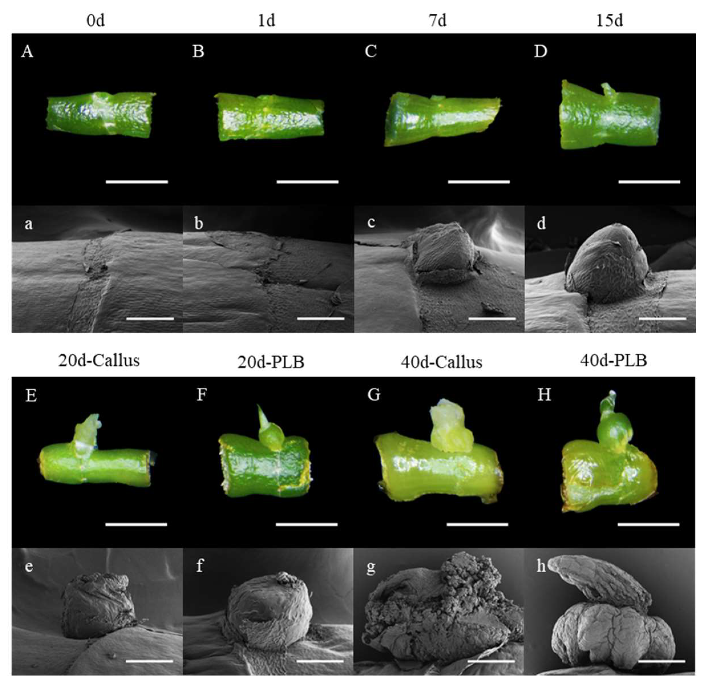
After 7 days of culture on induction medium, the explants began to protrude from the epidermis at the internodal axils, forming regenerative initials morphologically similar to the initiation of growing buds or adventitious buds (Figure 2). Under SEM, the surface cells were observed to be neatly arranged and uniform in size. At 15 days of culture, distinct protrusions were visible on the explant internodes by the naked eye, covered by a degenerated epidermis.
At 20 days of culture, the two regenerants could be clearly distinguished. Induced callus was pale yellowish-green with granular surface protrusions and vigorous growth. Under SEM, obvious proliferation of apical cells was observed, with vigorous cell division, uniform cell size, and complete degeneration of the epidermal layer. Induced PLBs appeared as green or dark green spherical structures with a smooth epidermis and polar growth polarity, clearly differentiating between the base and apex. No apical cell proliferation was observed under SEM (Figure 2).
At 40 days of culture, the differences between the two regenerants became even more pronounced. Callus exhibited translucent, pale yellowish-green irregular structures with distinct surface protrusions. Under SEM, cell proliferation was more intense, completely breaking through the degenerated epidermis, resembling disorderly proliferating meristem-like cells. PLBs were fully enlarged into spherical structures with increased volume, and their apices resembled degenerated leaf primordia. No obvious cell proliferation on the surface or apex was observed under SEM (Figure 2).
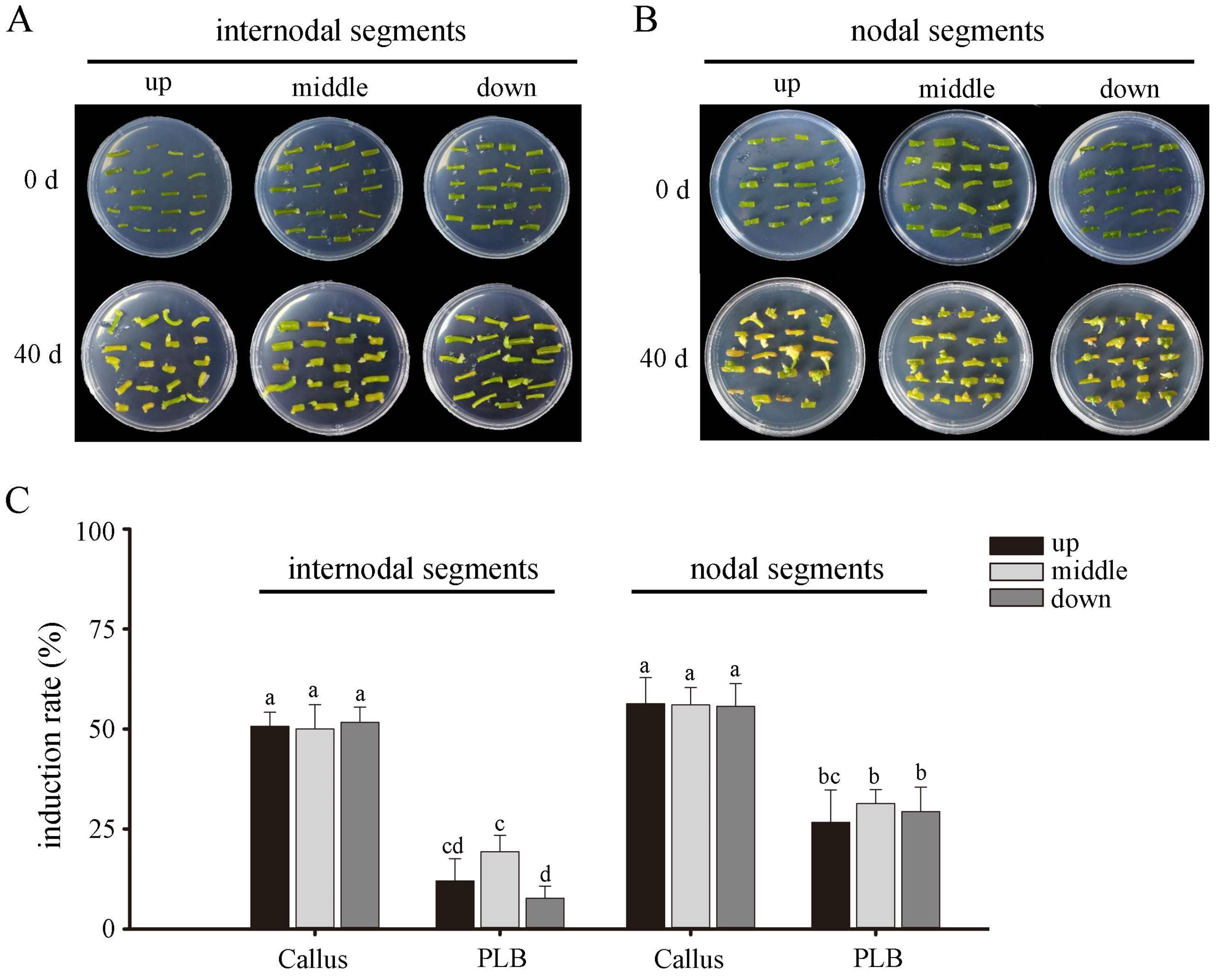
After 40 days of culture, no significant difference in the callus induction rate was detected between nodal and internodal segments collected from different positions (Figure 3). For PLB induction rate, there was no obvious difference among nodal segments at different positions, but the induction rate of middle internodal segments was higher than that of upper internodal segments. The PLB induction rate of nodal segments (29.11 ± 2.34%) was higher than that of internodal segments (13.00 ± 6.34%). Therefore, nodal segments were more suitable as explants for callus or PLB induction in D. officinale than internodal segments, especially the middle nodal segments. In addition, the induction rate of callus was significantly higher than that of PLB in both internodal and nodal segments.

3.2. Effect of 2,4-D Concentration on Callus and PLB Induction

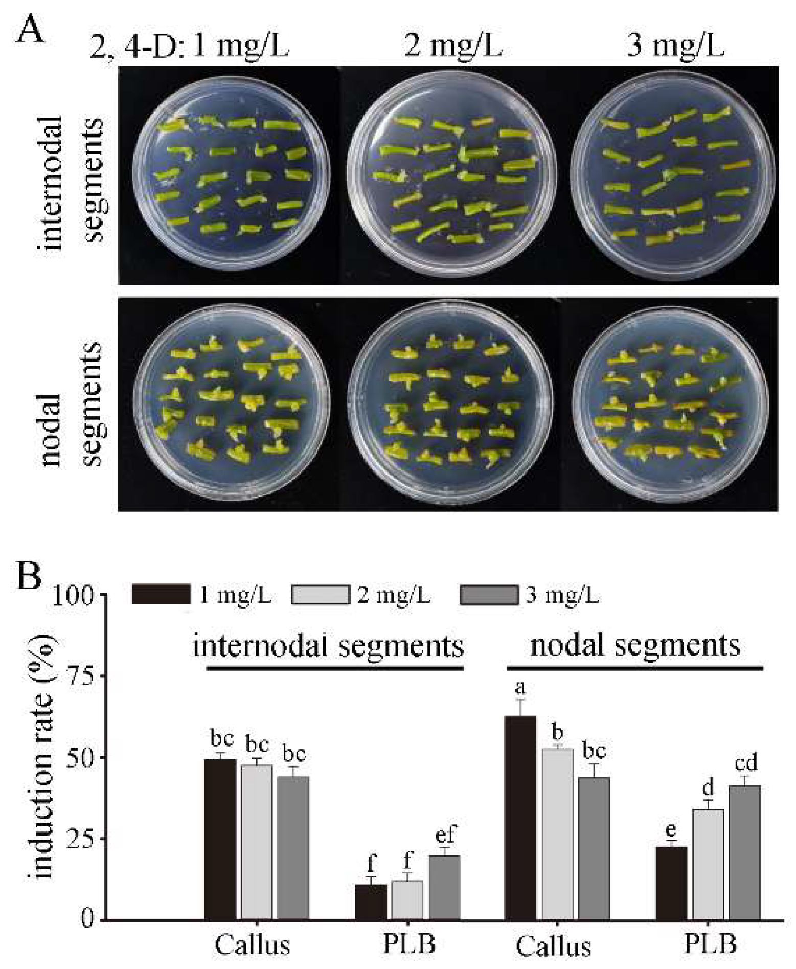
2,4-D concentration had a significant effect on the induction of PLBs and callus in internodal and nodal segments of D. officinale (Figure 4). After 40 days of culture, within the tested concentration range of 2,4-D, the callus induction rates of nodal and internodal segments decreased as 2,4-D concentration increased, while the PLB induction rates showed an opposite trend. At a concentration of 1.0 mg/L, the callus induction rate of internodal segments (49.44 ± 1.92%) was lower than that of nodal segments (62.78 ± 5.36%), while there was no significant difference at other concentrations. The PLB induction rate of nodal segments was higher than that of internodal segments at all concentrations. These results indicated that 2,4-D concentration affected the induction direction of explants. Callus induction required a lower concentration of 2,4-D than PLB induction.

3.3. Induction Responses of PLBs and Callus to Different Temperatures

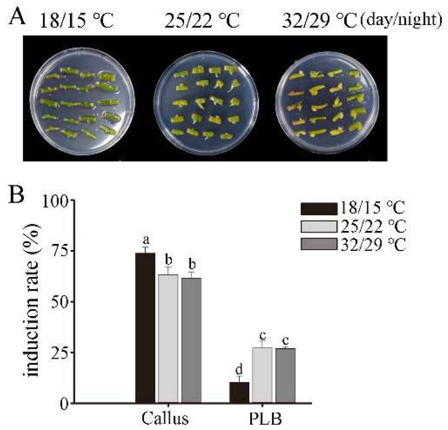
Temperature had a significant effect on the induction of PLBs and callus in nodal segments (Figure 5). Within the tested temperature regimes, the callus induction rate at 18/15 °C was significantly higher than that at 25/22 °C and 32/29 °C, while the PLB induction rate showed the opposite trend. There was no significant difference in both callus and PLB induction rates between 25/22 °C and 32/29 °C. However, the induction speed of callus or PLB was slow at 18/15 °C, and no mature callus or PLBs were observed after 40 days of culture. At 32/29 °C, partial desiccation of explants occurred in the later stage of culture. Thus, 25/22 °C was more suitable for the induction of callus and PLBs in D. officinale.

3.4. Regulatory Role of Light on PLBs and Callus Induction

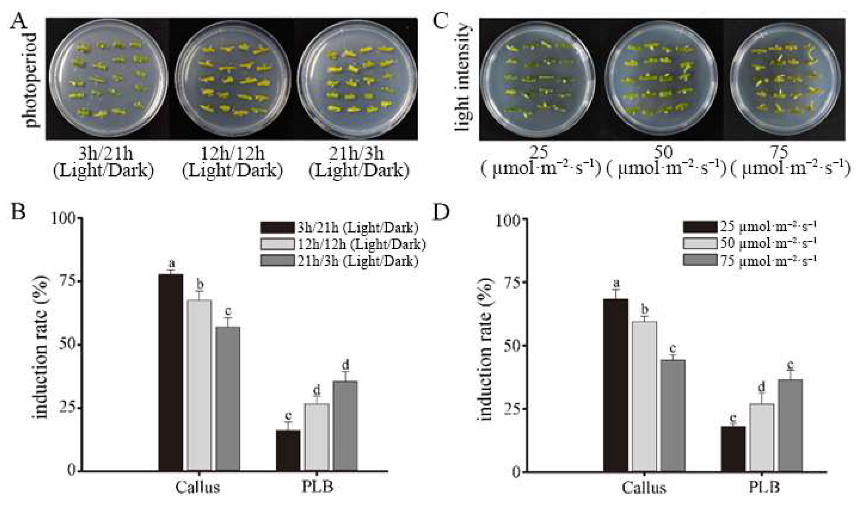
Both photoperiod and light intensity significantly affected the induction of PLBs and callus in nodal segments of D. officinale (Figure 6). Within the tested range, callus induction rate decreased significantly as light duration increased, whereas PLB induction rate showed an opposite trend. In addition, the induction process was delayed under the 3 h light treatment. Thus, a photoperiod of 6 h was beneficial for callus induction, while a longer photoperiod (21 h) favored PLB induction. The callus induction rate gradually decreased with the increase in light intensity, while the PLB induction rate gradually increased. However, the induction rate was slow at 25 µmol m−2 s−1, and no mature callus or PLBs were observed after 40 days of culture. Although the PLB induction rate was high at 75 µmol m−2 s−1, partial desiccation of explants occurred in the later stage of culture. Thus, 50 µmol m−2 s−1 was more suitable for the induction of callus or PLBs in D. officinale.

4. Discussion

This study found that explant type, 2,4-D concentration, temperature, light intensity and photoperiod significantly affected the induction of PLBs and callus in D. officinale. Middle nodal segments, a day/night temperature of 25/22 °C and a light intensity of 50 µmol m−2 s−1 were more suitable for the induction of callus and PLBs. However, callus induction required lower 2,4-D concentration and shorter photoperiod than PLB induction.

4.1. Variations in PLB and Callus Induction as Affected by Explant Type and Position

Explant type represents a critical determinant of orchid propagation, whereas the most suitable explants differ considerably across orchid species [12,21]. In D. officinale, stem nodal segments, especially those from the middle section, are more suitable as explants for inducing callus or PLBs compared to internodal segments, with higher induction rates. A previous study suggests that seedling nodal segments of Paphiopedilum can be induced to form embryogenic callus, subsequently developing into PLBs [46]. Compared with leaves, stems and roots, protocorms of Paphiopedilum are more easily induced to form PLBs through embryogenic callus [23]. In Renanthera, the callus induction rate of leaf base segments is significantly higher than that of leaf tip segments [31]. Different explants vary greatly in meristem content, cell differentiation status, endogenous hormone levels and phenolic exudation, which determine their regeneration capacity [17]. Generally, stem nodal segments are rich in meristematic cells, procambial cells and active parenchyma cells, serving as major sites for cytokinin, auxin, and nutrient accumulation and transport [17]. Thus, the differences in callus and PLB induction among different explant types and positions of D. officinale are caused by their cell differentiation degree, meristem content, and endogenous hormone level.

4.2. Responses of PLB and Callus Induction to Auxin Concentrations

This study found that 2,4-D concentration affected the induction direction of stem explants in D. officinale. Within the tested range, the callus induction rates of both internodal and nodal stem segments of D. officinale declined with increasing 2,4-D concentrations, whereas the PLB induction rates showed a rising trend. In addition, the suitable 2,4-D concentration for callus induction was lower than that for PLB induction. This result is different from that of Wu et al. [47], who found that a relatively high level of 2,4-D is beneficial for the initial induction of PLBs but not for the further development of PLBs in the middle and later stages using leaf base segments of Cymbidium hybridum as explants. This may be related to the endogenous hormone levels of leaves and stems.
Hormone is the most important factor controlling plant regeneration. The induction of PLBs and callus involves cell dedifferentiation and redifferentiation, in which hormones such as auxin and cytokinin play important roles. Generally, cytokinins promote cell division, organ differentiation and adventitious shoot proliferation, while auxins facilitate cell dedifferentiation and callus formation [17,32]. A suitable combination and ratio of cytokinin and auxin can control the induction, proliferation and differentiation of PLBs [17,18]. However, the effects of hormones vary among different species [17,47,48]. For example, in Catasetum fimbriatum, exogenous auxin drastically reduces PLB formation but significantly promotes callus development at the concentrations tested. 6-BA shows no effect on callus formation, yet markedly accelerates root tip conversion and increases the average number of PLBs [48].
Hormones regulate callus formation and organ regeneration by modulating gene expression. For example, genes such as GROWTH-REGULATING FACTOR 5 (AtGRF5), TaLAX PANICLE1 (TaLAX1) and ZmHSFC1 can promote plant callus formation and shoot regeneration through auxin and cytokinin signaling pathways [35,39]. Auxin-induced callus formation is regulated by ARF transcription factors. Auxin triggers the expression of ARF7 and ARF19, which in turn activate the downstream genes LBD16, LBD17, LBD18 and LBD29. Subsequently, LBD transcription factors further induce the expression of E2Fa to modulate cell cycle progression, ultimately promoting callus formation in plant explants [49]. 2,4-D may inhibit PLB differentiation by mediating the upregulation of PaphWOX8 and PaphWOX11 expression [20].

4.3. Temperature Impacts PLB and Callus Formation

Temperature significantly affected PLB and callus induction in stem nodal segments, with 25/22 °C being more suitable for callus or PLB induction in D. officinale. Excessively high or low temperature can affect cell division and differentiation, thereby inhibiting the development of callus and PLBs. For example, in Arabidopsis thaliana, low temperature regulates cellular pluripotency and cell fate transition of callus via the cytokinin signaling pathway and represses the upregulated expression of WUSCHEL (WUS), SHOOT MERISTEMLESS (STM) and CLAVATA3 (CLV3), thereby affecting callus formation [50]. Relatively high temperature promotes the enhanced expression of CUC1 (encoding a transcription factor involved in shoot meristem formation) and YUCCA (YUC) genes (encoding auxin biosynthesis enzymes), thereby promoting callus formation [51]. High temperature induces the expression of the PHYTOCHROME-INTERACTING FACTOR 4 (PIF4) gene, subsequently activating genes related to auxin biosynthesis, brassinosteroid biosynthesis, and growth-promoting factors [52,53]. Low temperature (16 °C) can affect the dynamic changes in H3K27me3-H2A.Z by inhibiting the expression of the transcription factor TOE1, thus altering cell developmental fate. The prc2 mutant develops into callus at normal temperature (22 °C) but forms relatively normal structures under low-temperature conditions [42]. Here, low temperature led to slow induction speeds of callus or PLBs, with no mature callus or PLBs observed after 40 days of culture, while high temperature caused partial desiccation of explants. However, little is known about the effect of temperature on the regeneration potential of callus and cell fate transition [50].

4.4. Effect of Light on PLB and Callus Induction in Dendrobium Officinale

This study found that light intensity and photoperiod significantly affected the induction of PLBs and callus in D. officinale. A light intensity of 50 µmol m−2 s−1 was more suitable for the induction of callus or PLBs, and callus induction required a shorter photoperiod than PLB induction. Previous studies have shown that light intensity and photoperiod have a significant effect on the growth and proliferation of PLBs in D. officinale [54,55]. An appropriate light intensity can improve the photosynthetic rate of PLBs, thus increasing the accumulation of assimilates and promoting the growth of PLBs [54]. In Phalaenopsis leaves, embryogenic tissue formation in leaf segments can be observed after 60 days of dark treatment, and PLBs are formed about 15 days after transfer to a 14 h/d light condition [30]. However, appropriately increasing light intensity can effectively promote explant regeneration in Phalaenopsis [40,56]. Light intensity and duration can influence PLB and callus induction by regulating the expression of genes related to energy metabolism and hormone metabolism [18,57]. An appropriate light intensity and photoperiod can increase the accumulation of photosynthetic products to meet the energy demand during the regeneration process. In tomato, sucrose produced by green cells can be transported to shoot primordia and converted into glucose, which activates the glucose TOR signaling pathway to promote the differentiation of shoot primordia [58].

5. Conclusions

This study revealed the differences in the regulation of PLB and callus induction in D. officinale by explant type and position, auxin concentration, temperature, light intensity and photoperiod. It was found that stem nodal segments were more suitable as explants for callus or PLB induction. 2,4-D concentration affected the induction direction of explants: a relatively high concentration of 2,4-D promoted PLB induction, while callus induction required a lower concentration. Low temperature and low light intensity suppressed PLB induction yet promoted callus formation in D. officinale by slowing developmental progression. In contrast, high temperature and strong light triggered explant desiccation and reduced the induction efficiency. A shorter photoperiod favored callus induction, while a longer photoperiod was beneficial for PLB induction. This study is of great significance for guiding the seedling propagation of D. officinale and lays an important foundation for elucidating the induction mechanisms of PLBs and callus. However, the interactive effects of phytohormones and environmental factors on callus and PLB induction in D. officinale remain to be further elucidated. Future research should also focus on clarifying the molecular mechanisms underlying the induction of callus and PLB, as well as the molecular regulatory mechanisms through which temperature and light influence these two regeneration processes.

Author Contributions

Conceptualization, G.-G.X. and S.-B.Z.; methodology, G.-G.X., W.C. and X.-M.D.; investigation, G.-G.X. and W.C.; visualization, G.-G.X. and X.-M.D.; Writing—original draft preparation, G.-G.X. and S.-B.Z.; writing—review and editing, W.C., X.-M.D. and S.-B.Z.; supervision, S.-B.Z.; funding acquisition, W.C. and S.-B.Z. All authors have read and agreed to the published version of the manuscript.

Funding

This work was financially supported by the National Key Research and Development Program of China (2024YFF1306703); the Key Research and Development Program of Yunnan Province (202403AC100028); CAS Technology Talent Program (E4233811T1); the National Natural Science Foundation of China (32570438); and the High-level Talent Support Plan of Yunnan Province (YNWR-CYJS-2020-023).

Data Availability Statement

The original contributions presented in this study are included in the article. Further inquiries can be directed to the corresponding author.

Conflicts of Interest

The authors declare no conflicts of interest.

References

  1. Givnish, T.J.; Spalink, D.; Ames, M.; Lyon, S.P.; Hunter, S.J.; Zuluaga, A.; Iles, W.J.D.; Clements, M.A.; Arroyo, M.T.K.; Leebens-Mack, J.; et al. Orchid phylogenomics and multiple drivers of their extraordinary diversification. Proc. R. Soc. B-Biol. Sci. 2015, 282, 1553. [Google Scholar] [CrossRef]
  2. Pérez-Escobar, O.A.; Bogarín, D.; Przelomska, N.A.S.; Ackerman, J.D.; Balbuena, J.A.; Bellot, S.; Bühlmann, R.P.; Cabrera, B.; Cano, J.A.; Charitonidou, M.; et al. The origin and speciation of orchids. New Phytol. 2024, 242, 700–716. [Google Scholar] [CrossRef] [PubMed]
  3. Waterman, R.J.; Bidartondo, M.I. Deception above, deception below: Linking pollination and mycorrhizal biology of orchids. J. Exp. Bot. 2008, 59, 1085–1096. [Google Scholar] [CrossRef] [PubMed]
  4. Mondragón-Palomino, M.; Theißen, G. Why are orchid flowers so diverse? Reduction of evolutionary constraints by paralogues of class B floral homeotic genes. Ann. Bot. 2009, 104, 583–594. [Google Scholar] [CrossRef] [PubMed]
  5. Zhang, S.; Yang, Y.; Li, J.; Qin, J.; Zhang, W.; Huang, W.; Hu, H. Physiological diversity of orchids. Plant Diver 2018, 40, 196–208. [Google Scholar] [CrossRef]
  6. Teoh, E.S. Medicinal Orchids of Asia; Springer International Publishing: Cham, Switzerland, 2016. [Google Scholar] [CrossRef]
  7. Yuan, S.C.; Lekawatana, S.; Amore, T.D.; Chen, F.C.; Chin, S.W.; Vega, D.M.; Wang, Y.T. The Global Orchid Market. In The Orchid Genome; Chen, F.C., Chin, S.W., Eds.; Springer: Cham, Switzerland, 2021; pp. 1–28. [Google Scholar] [CrossRef]
  8. Chinese Pharmacopoeia Commission. Pharmacopoeia of the People’s Republic of China; China Medical Science Press: Beijing, China, 2025; Volume 1, pp. 1–416. Available online: https://www.gov.cn/zhengce/zhengceku/202503/P020250326411104895580.pdf (accessed on 7 April 2026).
  9. Baqueiro-Peña, I.; Guerrero-Beltrán, J.Á. Vanilla (Vanilla planifolia Andr.), its residues and other industrial by-products for recovering high value flavor molecules: A review. J. Appl. Res. Med. Aromat. Plants 2017, 6, 1–9. [Google Scholar] [CrossRef]
  10. Phillips, R.D.; Reiter, N.; Peakall, R. Orchid conservation: From theory to practice. Ann. Bot. 2020, 126, 345–362. [Google Scholar] [CrossRef]
  11. Ticktin, T.; Mondragón, D.; Lopez-Toledo, L.; Dutra-Elliott, D.; Aguirre-León, E.; Hernández-Apolinar, M. Synthesis of wild orchid trade and demography provides new insight on conservation strategies. Conser. Lett. 2020, 13, e12697. [Google Scholar] [CrossRef]
  12. Yeung, E.C.-T.; Lee, Y.-I. Orchid Propagation: The Biology and Biotechnology of the Protocorm; Humana Press: Totowa, NJ, USA, 2024. [Google Scholar] [CrossRef]
  13. Tiwari, P.; Sharma, A.; Bose, S.K.; Park, K.-I. Advances in orchid biology: Biotechnological achievements, translational success, and commercial outcomes. Horticulturae 2024, 10, 152. [Google Scholar] [CrossRef]
  14. Chen, J.C.; Tong, C.G.; Lin, H.Y.; Fang, S.C. Phalaenopsis LEAFY COTYLEDON1-induced somatic embryonic structures are morphologically distinct from protocorm-like bodies. Front. Plant Sci. 2019, 10, 1594. [Google Scholar] [CrossRef]
  15. Yao, L.; Huang, J.; Zhang, S. An improved protocol for asymbiotic seed germination and seedling development of Paphiopedilum tigrinum. Horticulturae 2021, 7, 298. [Google Scholar] [CrossRef]
  16. Zeng, S.; Wang, J.; Wu, K.; Teixeira da Silva, J.A.; Zhang, J.; Duan, J. In vitro propagation of Paphiopedilum hangianum Perner & Gruss. Sci. Hortic. 2013, 151, 147–156. [Google Scholar] [CrossRef]
  17. Chugh, S.; Guha, S.; Rao, I.U. Micropropagation of orchids: A review on the potential of different explants. Sci. Hortic. 2009, 122, 507–520. [Google Scholar] [CrossRef]
  18. Cardoso, J.C.; Zanello, C.A.; Chen, J.T. An overview of orchid protocorm-like bodies: Mass propagation, biotechnology, molecular aspects, and breeding. Int. J. Mol. Sci. 2020, 21, 985. [Google Scholar] [CrossRef]
  19. Xu, Z.H.; Zhang, X.S.; Su, Y.H.; Hu, Y.X.; Xu, L.; Wang, J.W. Plant cell totipotency and regeneration. Sci. Sin. Vitae 2019, 49, 1282–1300, (In Chinese with English Abstract). Available online: https://www.sciengine.com/SSV/doi/10.1360/SSV-2019-0199 (accessed on 7 April 2026).
  20. Guo, B. Study on Establishment of Regeneration System of Protocorm-Like Bodies and Its Regulation Mechanism in Paphiopedilum. Ph.D. Dissertation, University of Chinese Academy of Sciences, Beijing, China, 2022. [Google Scholar]
  21. Zou, J.; Wang, Y.; Zhao, W.; Yin, Z.; Du, J.; Sun, C. Research progress of protocorm and protocorm-like body in Orchidaceae plants. Acta Agric. Zhejiangensis 2025, 37, 1372–1389, (In Chinese with English Abstract). [Google Scholar]
  22. Teixeira da Silva, J.A.; Singh, N.; Tanaka, M. Priming biotic factors for optimal protocorm-like body and callus induction in hybrid Cymbidium (Orchidaceae), and assessment of cytogenetic stability in regenerated plants. Plant Cell Tiss. Org. 2006, 84, 135–144. [Google Scholar] [CrossRef]
  23. Lin, Y.H.; Chang, C.; Chang, W.C. Plant regeneration from callus culture of a paphiopedilum hybrid. Plant Cell Tiss. Org. 2000, 62, 21–25. [Google Scholar] [CrossRef]
  24. Lee, Y.I.; Hsu, S.T.; Yeung, E.C. Orchid protocorm-like bodies are somatic embryos. Am. J. Bot. 2013, 100, 2121–2131. [Google Scholar] [CrossRef]
  25. Fang, S.C.; Chen, J.C.; Wei, M.J. Protocorms and protocorm-like bodies are molecularly distinct from zygotic embryonic tissues in Phalaenopsis aphrodite. Plant Physiol. 2016, 171, 2682–2700. [Google Scholar] [CrossRef] [PubMed]
  26. Fang, S.-C.; Chen, J.-C.; Chang, P.-Y.; Lin, H.-Y. Co-option of the SHOOT MERISTEMLESS network regulates protocorm-like body development in Phalaenopsis aphrodite. Plant Physiol. 2022, 190, 127–145. [Google Scholar] [CrossRef] [PubMed]
  27. Zeng, D.; Si, C.; Zhang, M.; Duan, J.; He, C. ERF5 enhances protocorm-like body regeneration via enhancement of STM expression in Dendrobium orchid. J. Integr. Plant Biol. 2023, 65, 2071–2085. [Google Scholar] [CrossRef]
  28. Paek, K.Y.; Hahn, E.J.; Park, S.Y. Micropropagation of Phalaenopsis orchids via protocorms and protocorm-like bodies. In Plant Embryo Culture; Thorpe, T., Yeung, E., Eds.; Methods in Molecular Biology; Humana Press: Totowa, NJ, USA, 2011; Volume 710, pp. 293–306. [Google Scholar] [CrossRef]
  29. Yeung, E.C. A perspective on orchid seed and protocorm development. Bot. Stud. 2017, 58, 33. [Google Scholar] [CrossRef] [PubMed]
  30. Gow, W.; Chen, J.; Chang, W. Effects of genotype, light regime, explant position, and orientation on direct somatic embryogenesis from leaf explants of Phalaenopsis orchid. Acta Physiol. Plant 2009, 31, 363–369. [Google Scholar] [CrossRef]
  31. Wu, K.; Zeng, S.; Teixeira da Silva, J.A.; Chen, Z.; Zhang, J.; Yang, Y.; Duan, J. Efficient regeneration of renanthera tom thumb ‘qilin’ from leaf explants. Sci. Hortic. 2012, 135, 194–201. [Google Scholar] [CrossRef]
  32. Novak, S.D.; Luna, L.J.; Gamage, R.N. Role of auxin in orchid development. Plant Signal. Behav. 2014, 9, e972277. [Google Scholar] [CrossRef]
  33. Bustillo-Avendaño, E.; Ibáñez, S.; Sanz, O.; Barros, J.A.S.; Gude, I.; Perianez-Rodriguez, J.; Micol, J.L.; Del Pozo, J.C.; Moreno-Risueno, M.A.; Pérez-Pérez, J.M. Regulation of hormonal control, cell reprogramming, and patterning during de novo root organogenesis. Plant Physiol. 2018, 176, 1709–1727. [Google Scholar] [CrossRef]
  34. Wang, K.; Shi, L.; Liang, X.; Zhao, P.; Wang, W.; Liu, J.; Chang, Y.; Hiei, Y.; Yanagihara, C.; Du, L.; et al. The gene TaWOX5 overcomes genotype dependency in wheat genetic transformation. Nat. Plants 2022, 8, 110–117. [Google Scholar] [CrossRef] [PubMed]
  35. Li, Y.; Geng, Y.; Qing, X.; Yin, X.; Xu, F.; Huang, L.; Zhu, R.; Chen, B.; Yang, T.; Song, W.; et al. ZmHSCF1 enhances Agrobacterium-mediated transformation frequency in commercial maize inbred lines by promoting embryogenic callus proliferation. Plant Commun. 2025, 6, 101525. [Google Scholar] [CrossRef]
  36. Ikeuchi, M.; Sugimoto, K.; Iwase, A. Plant callus: Mechanisms of induction and repression. Plant Cell 2013, 25, 3159–3173. [Google Scholar] [CrossRef]
  37. Okushima, Y.; Fukaki, H.; Onoda, M.; Theologis, A.; Tasaka, M. ARF7 and ARF19 regulate lateral root formation via direct activation of LBD/ASL genes in Arabidopsis. Plant Cell 2007, 19, 118–130. [Google Scholar] [CrossRef]
  38. Fan, M.; Xu, C.; Xu, K.; Hu, Y. LATERAL ORGAN BOUNDARIES DOMAIN transcription factors direct callus formation in Arabidopsis regeneration. Cell Res. 2012, 22, 1169–1180. [Google Scholar] [CrossRef] [PubMed]
  39. Yu, Y.; Yu, H.; Peng, J.; Yao, W.J.; Wang, Y.P.; Zhang, F.L.; Wang, S.R.; Zhao, Y.; Zhao, X.Y.; Zhang, X.S.; et al. Enhancing wheat regeneration and genetic transformation through overexpression of TaLAX1. Plant Commun. 2024, 5, 100738. [Google Scholar] [CrossRef]
  40. Park, S.Y.; Murthy, H.N.; Paek, K.Y. Rapid propagation of Phalaenopsis from flower stalk-derived leaves. Vitr. Cell. Dev. Biol.-Plant 2002, 38, 168–172. [Google Scholar] [CrossRef]
  41. Zanello, C.A.; Duarte, W.N.; Gomes, D.M.; Cardoso, J.C. Micropropagation from inflorescence nodal segments of Phalaenopsis and acclimatization of plantlets using different substrates. Horticulturae 2022, 8, 340. [Google Scholar] [CrossRef]
  42. Zhu, K.; Chen, J.; Zhao, L.; Lu, F.; Deng, J.; Lin, X.; He, C.; Wagner, D.; Xiao, J. Dynamic control of H2A.Zub and H3K27me3 by ambient temperature during cell fate determination in Arabidopsis. Dev. Cell. 2025, 60, 2192–2208.e5. [Google Scholar] [CrossRef]
  43. Yang, W.; Zhai, H.; Wu, F.; Deng, L.; Chao, Y.; Meng, X.; Chen, Q.; Liu, C.; Bie, X.; Sun, C.; et al. Peptide REF1 is a local wound signal promoting plant regeneration. Cell 2024, 187, 3024–3038.e14. [Google Scholar] [CrossRef] [PubMed]
  44. Tu, J.; Xu, L.; Guo, Y.; Zhang, M.; Gan, M.; Bao, X.; Yang, R.; Liu, H.; Lin, F. Dendrobium officinale-derived nanovesicles: A natural therapy for comprehensive regulation of angiogenesis, inflammation, and tissue repair to enhance skin wound healing. Bioresour. Bioprocess. 2025, 12, 74. [Google Scholar] [CrossRef]
  45. Murashige, T.; Skoog, F. A revised medium for rapid growth and bio assays with tobacco tissue cultures. Plant Physiol. 1962, 15, 473–497. [Google Scholar] [CrossRef]
  46. Istiqomah, N.; Indriani, H.; Wijaya, Y.I.F.; Safitri; Yalapuspita, D.C.; Handini, E.; Diantina, S.; Aprilianti, P.; Semiarti, E. Clonal propagation of rare orchid species Paphiopedilum spp. (Orchidaceae) to save Indonesian biodiversity. S. Afr. J. Bot. 2024, 172, 779–785. [Google Scholar] [CrossRef]
  47. Wu, K.; Huang, M.; Wang, M. Protocorm-like body induced from leaf-segments of vitro-seedlings of Cymbidium hybridum. Mol. Plant Breed. 2007, 5, 341–346, (In Chinese with English Abstract). Available online: https://kns.cnki.net/kcms2/article/abstract?v=3AJmsS-kAvj0FYFAPR7pi5x4bRqOcTAx-xTizjCkRfGNcuIYg7ZrZA1LE50X5ldLs6Rj-rhdmEiRHQYo-xwcnG37uzadR_NYMIV5kcqAmjt_BfKVKZbVTlqCVArpKxBAKtJYYQdRlIbsuLEjdJxJF7igM5Rvvh_Z&uniplatform=NZKPT (accessed on 7 April 2026).
  48. Colli, S.; Kerbauy, G.B. Direct root tip conversion of Catasetum into protocorm-like bodies. Effects of auxin and cytokinin. Plant Cell Tiss. Organ Cult. 1993, 33, 39–44. [Google Scholar] [CrossRef]
  49. Iwase, A.; Mitsuda, N.; Koyama, T.; Hiratsu, K.; Kojima, M.; Arai, T.; Inoue, Y.; Seki, M.; Sakakibara, H.; Sugimoto, K.; et al. The AP2/ERF transcription factor WIND1 controls cell dedifferentiation in Arabidopsis. Curr. Biol. 2011, 21, 508–514. [Google Scholar] [CrossRef]
  50. Wu, S.S. Study on Low-Temperature Treatment Affecting Callus Regeneration. Master’s Thesis, Shanghai Normal University, Shanghai, China, 2023. (In Chinese with English Abstract). [Google Scholar]
  51. Lambolez, A.; Kawamura, A.; Takahashi, T.; Rymen, B.; Iwase, A.; Favero, D.S.; Ikeuchi, M.; Suzuki, T.; Cortijo, S.; Jaeger, K.E.; et al. Warm temperature promotes shoot regeneration in Arabidopsis thaliana. Plant Cell Physiol. 2022, 63, 618–634. [Google Scholar] [CrossRef]
  52. Koini, M.A.; Alvey, L.; Allen, T.; Tilley, C.A.; Harberd, N.P.; Whitelam, G.C.; Franklin, K.A. High temperature-mediated adaptations in plant architecture require the bHLH transcription factor PIF4. Curr. Biol. 2009, 19, 408–413. [Google Scholar] [CrossRef] [PubMed]
  53. Franklin, K.A.; Lee, S.H.; Patel, D.; Kumar, S.V.; Spartz, A.K.; Gu, C.; Ye, S.; Yu, P.; Breen, G.; Cohen, J.D.; et al. PHYTOCHROME-INTERACTING FACTOR 4 (PIF4) regulates auxin biosynthesis at high temperature. Proc. Natl. Acad. Sci. USA 2011, 108, 20231–20235. [Google Scholar] [CrossRef] [PubMed]
  54. Xu, B.-Q.; Cui, Y.-Y.; Guo, C.; Xia, G.-H. Dynamic variation of biomass and content of polysaccharide and alkaloid in protocorm like bodies from Dendrobium officinale at different light intensities and incubation time. Chin. Tradit. Herb. Drugs 2012, 43, 355–359, (In Chinese with English Abstract). Available online: https://www.tiprpress.com/zcy/article/abstract/201202035?st=article_issue (accessed on 7 April 2026).
  55. Wang, Y.L.; Lv, Y.P.; Mou, H.J.; Zhou, D.J.; Chen, Z.; Chen, L.P.; Chen, J.P. Effect of culture environment on the growth of Dendrobium candidum in tissue culture. J. Zhejiang Agric. Sci. 2014, 7, 1021–1023, (In Chinese with English Abstract). Available online: https://link.cnki.net/doi/10.16178/j.issn.0528-9017.2014.07.028 (accessed on 7 April 2026).
  56. Khoddamzadeh, A.A.; Sinniah, U.R.; Kadir, M.A.; Kadzimin, S.B.; Mahmood, M.; Sreeramanan, S. In vitro induction and proliferation of protocorm-like bodies (PLBs) from leaf segments of Phalaenopsis bellina (Rchb.f.) Christenson. Plant Growth Regul. 2011, 65, 381–387. [Google Scholar] [CrossRef]
  57. Khan, T.; Abbasi, B.H.; Khan, M.A. The interplay between light, plant growth regulators and elicitors on growth and secondary metabolism in cell cultures of Fagonia indica. J. Photochem. Photobiol. B Biol. 2018, 185, 153–160. [Google Scholar] [CrossRef]
  58. Song, X.; Guo, P.; Xia, K.; Wang, M.; Liu, Y.; Chen, L.; Zhang, J.; Xu, M.; Liu, N.; Yue, Z.; et al. Spatial transcriptomics reveals light-induced chlorenchyma cells involved in promoting shoot regeneration in tomato callus. Proc. Natl. Acad. Sci. USA 2023, 120, e2310163120. [Google Scholar] [CrossRef]
Figure 1. Schematic diagram of explants of Dendrobium officinale used in this study.
Horticulturae 12 00555 g001
Figure 2. Callus and PLB regeneration process from stem segments of Dendrobium officinale under a PPFD of 50 µmol m−2 s−1, a 12 h/12 h day/night photoperiod, and a temperature of 25 ± 1 °C. (AH), morphological characteristics during the development of stem node explants (scale bars = 1 cm); (ah), ultrastructural characteristics under SEM (scale bars = 500 μm). (A,a), Schematic diagram of the stem node explant used in this study; (B,b), No obvious structural changes observed at 1 day of culture; (C,c), Protrusions at the internodes visible under SEM at 7 days of culture; (D,d), Distinct protrusions formed on the internodes at 15 days of culture, which were more obvious under SEM; (E,e), Callus observed at 20 days of culture, pale yellowish-green in color with granular surface protrusions and obvious apical cell division; (F,f), PLBs observed at 20 days of culture, green or dark green and spherical in shape with relatively smooth surfaces and obvious apical polarity; (G,g), Callus obtained at 40 days of culture, translucent pale yellowish-green, irregular in shape, enlarged in volume, and with intensified apical cell proliferation; (H,h), PLBs obtained at 40 days of culture, presenting a typical green spherical structure, further enlarged in volume, with no obvious leaf primordia differentiated at the apex.
Horticulturae 12 00555 g002
Figure 3. Effect of stem explant types and positions on callus and PLB induction under a PPFD of 50 µmol m−2 s−1, a 12 h/12 h day/night photoperiod (day/night), and a temperature of 25 ± 1 °C. (A,B) Induction of callus and PLBs in internodal (A) and nodal (B) segments at different positions; (C) Differences in the induction of callus and PLBs between internodes and nodes segments from different positions. Each value represents the mean ± SD of three replicates. Different letters above the bars indicate significant differences among different treatments (p < 0.05).
Horticulturae 12 00555 g003
Figure 4. Effect of 2,4-D concentrations on the induction of callus and PLBs in internodal and nodal segments of Dendrobium officinale under a PPFD of 50 µmol m−2 s−1, a 12 h/12 h day/night photoperiod, and a temperature of 25 ± 1 °C. (A), Phenotypes induced from internodal and nodal segments at different 2,4-D concentrations; (B), Differences in callus and PLB induction rates from internodal and nodal segments at different 2,4-D concentrations. Each value represents the mean ± SD of three replicates. Different letters above the bars indicate significant differences among different treatments (p < 0.05).
Horticulturae 12 00555 g004
Figure 5. Effects of temperature on PLB and callus induction in nodal segments of Dendrobium officinale under a PPFD of 50 µmol m−2 s−1 and a 12 h/12 h day–night photoperiod. Each value represents the mean ± SD of three replicates. (A), Phenotypes induced from nodal segment at different temperatures; (B), Differences in callus and PLB induction rates from nodal segment at different temperatures. Different letters above the bars indicate significant differences among different treatments (p < 0.05).
Horticulturae 12 00555 g005
Figure 6. Effects of different photoperiods (A,B) at a PPFD of 50 µmol·m−2·s−1 and a temperature of 25 ± 1 °C, and different light intensities (C,D) under a 12 h/12 h day/night photoperiod at 25 ± 1 °C, on callus and PLB induction in Dendrobium officinale. Each value represents the mean ± SD of three replicates. Different letters above the bars indicate significant differences among different treatments (p < 0.05).
Horticulturae 12 00555 g006
Disclaimer/Publisher’s Note: The statements, opinions and data contained in all publications are solely those of the individual author(s) and contributor(s) and not of MDPI and/or the editor(s). MDPI and/or the editor(s) disclaim responsibility for any injury to people or property resulting from any ideas, methods, instructions or products referred to in the content.