Article

In Vitro Propagation System for Proiphys amboinensis Using Twin-Scale Explants and Genetic Fidelity Assessment

by
Kornkanok Chamchusri
1,
Piyanuch Sornchai
2,
Pitchaporn Wannitikul
2,
Panumart Rithichai
1 and
Yaowapha Jirakiattikul
1,*
1
Department of Agricultural Technology, Faculty of Science and Technology, Thammasat University, Pathum Thani 12120, Thailand
2
Research and Development of Genetically Modified Plant and Microbe Detection Group, Biotechnology Research and Development Office, Department of Agriculture, Bangkok 10900, Thailand
*
Author to whom correspondence should be addressed.
Horticulturae 2026, 12(3), 317; https://doi.org/10.3390/horticulturae12030317
Submission received: 21 January 2026 / Revised: 3 March 2026 / Accepted: 4 March 2026 / Published: 6 March 2026
(This article belongs to the Special Issue Micropropagation and Cultivation of Ornamental Species)

Abstract

Proiphys amboinensis has considerable potential as a commercial ornamental plant due to its attractive foliage, distinctive flowers, and long flowering period. This study established an in vitro micropropagation protocol and evaluated the genetic fidelity of regenerated bulblets using Inter Simple Sequence Repeat (ISSR) markers and flow cytometry. Twin-scale explants were cultured on Murashige and Skoog (MS) medium supplemented with 0, 0.5, 1.0, and 2.0 mg/L N6-benzyladenine (BA) for 12 weeks. Bulblet formation efficiency ranged from 60.00 ± 16.33% to 70.00 ± 11.55%, with no significant differences among treatments. A significant increase in bulblet number was observed at 1.0 mg/L BA compared with the control and 0.5 mg/L BA; however, bulblet fresh weight did not differ significantly among these treatments. Sucrose concentrations (30–90 g/L) had no significant effects on bulblet weight and diameter. Root induction was evaluated using indole-3-butyric acid (IBA) and α-naphthaleneacetic acid (NAA) at concentrations of 0–1.0 mg/L, with 0.5 mg/L IBA identified as the optimal treatment. Following acclimatization, regenerated bulblets exhibited high survival rates (90–100%). ISSR and flow cytometric analyses revealed no detectable genetic variation, with a consistent genome size between regenerated bulblets and the mother plants, indicating high genetic uniformity. The protocol provides a micropropagation system for P. amboinensis with high genetic fidelity, supporting its commercial and research potential.

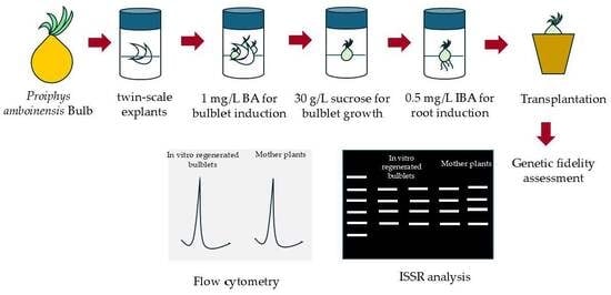
Graphical Abstract

1. Introduction

Proiphys amboinensis (syn. Eurycles amboinense) is a bulbous species belonging to the family Amaryllidaceae and is commonly known as the Cardwell lily, Brisbane lily, or Northern Christmas lily [1,2]. The species is native to Thailand and is widely distributed throughout Southeast Asia, including Indonesia, the Philippines, and Vietnam, as well as northern Australia [2,3]. In recent years, P. amboinensis has attracted increasing attention for its medicinal potential. Its leaves are traditionally used in Papua Island, Indonesia, for the treatment of malaria [4], while bulb scales have been reported to contain lycorine, an Amaryllidaceae alkaloid exhibiting acetylcholinesterase inhibitory activity that may contribute to the alleviation of Alzheimer’s disease-related symptoms [5]. Although the pharmacological properties of this species remain incompletely explored, existing evidence highlights its promising therapeutic potential.
In addition to its medicinal relevance, P. amboinensis is highly valued as an ornamental plant due to its distinctive morphology and prolonged flowering period. The plant produces a tunicate, ovoid bulb measuring 5–8 cm in diameter and bears broad ovate to nearly circular leaves, 10–30 cm wide and 8–35 cm long, with long petioles ranging from 15 to 60 cm. The inflorescence consists of 5–25 white flowers arranged in an umbel on a scape 15–100 cm in length [1,2,3]. Flowering occurs gradually, with individual flowers opening sequentially over an extended period, thereby enhancing its ornamental display [6]. In Thailand, the combination of attractive foliage, prominent floral traits, and prolonged flowering enhances the suitability of the species for development as a commercial ornamental crop [7]. The successful commercialization and sustainable utilization of P. amboinensis require efficient propagation systems capable of producing large quantities of high-quality, genetically uniform planting material to support commercial production and further research on flower production, plant physiology, and postharvest management of flowers and mother bulbs. Conventional propagation through seeds is impractical for commercial bulb production due to high genetic variability, low seed viability, and specific germination requirements. Vegetative propagation via bulb division or cutting is also constrained by low multiplication rates, long production cycles, and an increased risk of pathogen transmission [8]. These limitations necessitate the development of alternative propagation strategies.
Plant tissue culture offers a reliable and efficient approach for the rapid multiplication of elite genotypes and the production of uniform, disease-free planting material [8]. In vitro propagation has been successfully applied to numerous Amaryllidaceae species, including Brunsvigia undulata [9], Crinum malabaricum [10], Lycoris sprengeri [11], Narcissus tazetta [12], and N. pallidulus [13]. Among various explant types, twin-scale explants, which consist of two adjacent bulb scales attached to a portion of the basal plate, are particularly suitable for inducing adventitious bulblet formation in various bulbous plants within the Amaryllidaceae family, such as B. undulata [9], C. variabile [14], and Hippeastrum vittatum [15]. Bulblet induction, rooting, and subsequent growth under aseptic conditions are strongly influenced by plant growth regulators and carbohydrate sources, particularly sucrose [16]. However, the effects of plant growth regulators on in vitro bulblet induction from twin-scale explants, the influence of sucrose concentration on bulblet enlargement, and the subsequent rooting of regenerated bulblets in P. amboinensis have not yet been investigated. Cytokinins, particularly BA, play a central role in adventitious shoot organogenesis by promoting cell division and facilitating the establishment of meristematic zones that give rise to adventitious buds [17]. Sucrose is a fundamental component of tissue culture media, functioning as both a primary carbon source and an osmotic regulator that supports bulblet enlargement and biomass accumulation [16], thereby enhancing the potential for bulblet multiplication through the subdivision of well-developed bulblets. Root formation during in vitro regeneration is predominantly governed by auxins, which regulate cell differentiation, root primordia initiation, and adventitious root development [18]. Based on these established physiological roles, the selection of BA concentrations, sucrose levels, and auxin type and concentration in this study was designed within a hypothesis-driven and stage-specific framework to elucidate their distinct functions during bulblet induction, growth, and enlargement, and rooting. In addition to optimizing regeneration efficiency, the assessment of genetic stability was incorporated as a critical component of the experimental design, as somaclonal variation may arise during in vitro culture. Inter simple sequence repeat (ISSR) markers and flow cytometry have been widely employed as complementary tools for evaluating clonal fidelity and genome integrity in tissue culture-derived bulbous plants and other species, including Zephyranthes grandiflora [19], Polianthes tuberosa [20], Galanthus spp. [21], and Uvaria hamiltonii [22]. Such an integrated and sequential approach, encompassing physiological optimization and genetic fidelity assessment, is essential for developing an efficient, biologically grounded, and genetically reliable micropropagation protocol for bulbous ornamental plants. Therefore, the objectives of this study were to (i) evaluate the effect of BA on bulblet induction from twin-scale explants, (ii) investigate the influence of sucrose concentration on bulblet growth and enlargement, (iii) optimize the rooting of regenerated bulblets, and (iv) assess the genetic stability of in vitro regenerated bulblets of P. amboinensis using ISSR markers and flow cytometry. The findings of this study are expected to support the development of a standardized in vitro propagation system for P. amboinensis, with practical applications in bulb and flower production as well as future physiological and molecular studies in Amaryllidaceae.

2. Materials and Methods

2.1. Medium and Culture Conditions

All experiments were conducted using Murashige and Skoog (MS) basal medium. Plant growth regulators and sucrose were added to the medium at concentrations specified for each experiment. The medium was solidified with 8.5 g/L agar, adjusted to pH 5.6–5.8 with 1 N NaOH, and sterilized by autoclaving at 121 °C for 15 min. Cultures were incubated at 25 ± 2 °C under a 16 h photoperiod with illumination from cool white fluorescent lamps at a light intensity of approximately 3000 lux (equivalent to about 45–55 µmol/m2/s PPFD). Unless otherwise specified, all treatments were maintained under the same culture conditions. Cultures were subcultured at 6-week intervals to maintain viability and uniform growth prior to subsequent experimental treatments.

2.2. Plant Materials and Sterilization Method

Bulbs of P. amboinensis were obtained from a local farmer in Chiang Mai Province, Thailand. The experiment commenced in October 2023, and the bulbs were in the vegetative (pre-flowering) stage at the time of culture initiation. The bulbs were thoroughly cleaned to remove adhering soil, and the roots were trimmed. After removing the outer dry scales, the bulbs were washed and transversely cut in half. The lower half containing the basal plate was further longitudinally divided into four equal wedge-shaped sections, each retaining a portion of the basal plate. The bulb sections were immersed in 70% (v/v) ethanol for approximately 1 min, followed by sonication in a tetracycline solution (500 mg per 100 mL sterile distilled water) for 30 min to reduce bacterial contamination. Surface sterilization was subsequently performed using a two-step sodium hypochlorite (NaOCl) treatment: first in 25% (v/v) NaOCl for 25 min and then in 20% (v/v) NaOCl for 20 min, with a few drops of Tween 20 added to each solution. The explants were then rinsed twice with sterile distilled water, each rinse lasting 1 min. Following surface sterilization, twin-scale explants approximately 1 cm in length were excised and cultured on MS medium containing 30 g/L sucrose without plant growth regulators for two weeks. Only contamination-free explants were subsequently selected for the bulblet induction experiments.

2.3. Effect of BA Concentration on In Vitro Bulblet Induction

The sterile explants were cultured on MS medium supplemented with 30 g/L sucrose and various concentrations of BA: 0, 0.5, 1.0, and 2.0 mg/L. The experiment followed a completely randomized design (CRD) with four treatments and four replicates. Each replicate consisted of five 4-oz (approximately 118 mL) glass culture bottles containing 15 mL of solid MS medium, with one twin-scale explant per bottle. Cultures were maintained in a culture room for 12 weeks. Data were collected on bulblet formation efficiency, the number of bulblets, and mean bulblet fresh weight (mg). Bulblet formation efficiency (%) was calculated as follows:
bulblet   formation   efficiency   ( % ) = Number   of   twin scales   with   regenerated   bulblets Total   number   of   cultured   twin scale   explants × 100

2.4. Effect of Sucrose Concentration on In Vitro Bulblet Growth and Enlargement

In vitro regenerated bulblets were cultured on solid MS medium supplemented with four sucrose concentrations (30, 45, 60, and 90 g/L) for six weeks. The experiment was arranged in a CRD with four treatments and five replicates. Each replicate consisted of four culture bottles, with one bulblet per bottle. Mean bulblet fresh weight (mg) and mean bulblet diameter (mm) were measured before and after the culture period to assess changes during in vitro development.

2.5. Effects of IBA and NAA Concentrations on Bulblet Root Induction and Acclimatization

In vitro regenerated bulblets were cultured on solid MS medium supplemented with 30 g/L sucrose and either IBA or NAA, each at concentrations of 0.5 and 1.0 mg/L. MS medium containing 30 g/L sucrose without plant growth regulators was used as the control. The experiment was arranged in a CRD with five treatments and four replicates. Each replicate consisted of five bottles, with one bulblet per bottle. Rooting percentage, number of roots, and root length were recorded after four weeks of culture. Rooting percentage was calculated using the following equation:
rooting   ( % )   = Number   of   bulblets   with   roots Total   number   of   cultured   bulblets × 100
Rooted bulblets were removed from the culture vessels and gently rinsed under tap water to remove residual agar. The bulblets were then transferred to clear plastic boxes (17 × 24 × 9 cm) containing moist cocopeat (pH 5.6) as the growing substrate. The boxes were covered to maintain high humidity for two weeks. Subsequently, the covers were removed, and the plants were maintained under shaded conditions for an additional one week. The survival rate was then calculated as follows:
S u r v i v a l   r a t e   ( % )   = N u m b e r   o f   s u r v i v i n g   b u l b l e t s T o t a l   n u m b e r   o f   t r a n s p l a n t e d   b u l b l e t s × 100

2.6. Clonal Fidelity Assessment

2.6.1. ISSR Analysis

ISSR analysis was performed on nine randomly selected in vitro regenerated bulblets and four mother plants. Approximately 300–400 mg of leaf tissue from 12-week-old plantlets was finely ground in liquid nitrogen. The resulting powder was mixed with 400 µL of 2× CTAB extraction buffer supplemented with 4 µL of β-mercaptoethanol and incubated at 60 °C for 1 h. The mixture was subsequently extracted with 400 µL of chloroform: isoamyl alcohol (24:1, v/v) and centrifuged at 12,000 rpm for 10 min. The upper aqueous phase was transferred to a new tube, combined with 400 µL of isopropanol, and incubated at −20 °C for 30 min to precipitate DNA. The precipitated DNA was collected by centrifugation at 12,000 rpm for 10 min, washed with 500 µL of 70% ethanol, air-dried, and dissolved in 50 µL of ultrapure water. DNA concentration and purity were assessed using a NanoDrop spectrophotometer (NanoDrop Eight, Thermo Scientific, Wilmington, DE, USA).
PCR amplification was performed in a 20 µL reaction mixture containing 50 ng of genomic DNA, 0.5 µM of a single ISSR primer (Table S1), and 10 µL of 2× Phanta Max Master Mix. The PCR conditions were as follows: initial denaturation at 95 °C for 5 min, followed by 35 cycles of denaturation at 95 °C for 15 s, annealing at 55 °C for 30 s, and extension at 72 °C for 1 min, with a final extension at 72 °C for 3 min. The amplification products (10 µL each) were separated on a 2% (w/v) agarose gel at 120 V for 65 min. DNA fragments were visualized under UV illumination using a transilluminator.

2.6.2. Flow Cytometry

Nuclei were obtained from 20 to 50 mg of young leaves by cutting the tissue into strips (approximately 0.5 mm wide) and immersed them in 500 µL of Quantum Stain NA UV 2 (A). Polyvinylpyrrolidone (PVP) (approximately 0.01 g) was added, and the tissue was finely chopped using a razor blade. The resulting suspension was aspirated and filtered through a 50-µm filter into a test tube. Subsequently, 0.5 mL of Quantum Stain NA UV 2 (B) was added, after which the suspension was filtered again and incubated for 4 min. Nuclear DNA content was measured using a Quantum Analysis Flow Cytometer (Quantum P, Quantum Analysis GmbH, Münster, Germany), and approximately 5000 nuclei were analyzed per sample. Due to the lack of published genome size data for P. amboinensis, rice (Oryza sativa, 2n), a species with a well-characterized genome size and stable ploidy level, was used as an external reference standard provided by the Rice Science Center, Kamphaeng Saen, Nakhon Pathom, Thailand. Ploidy levels and relative nuclear DNA content were then assessed by comparison with this diploid reference. Nuclear suspensions of both the samples and the rice standard were prepared and analyzed under identical instrument settings to ensure comparability of fluorescence signals. The 2C nuclear DNA content of the samples was estimated based on the ratio of the mean fluorescence intensity of the G1 peak of the sample to that of the external standard, using the following equation:
2 C   D N A   c o n t e n t   ( s a m p l e ) = M e a n   G 1   p e a k   o f   s a m p l e M e a n   G 1   p e a k   o f   s t a n d a r d × 2 C   D N A   c o n t e n t   o f   t h e   s t a n d a r d
In addition, mother plants of P. amboinensis were analyzed alongside the in vitro-derived bulblets and served as an internal reference for relative DNA content comparison and ploidy stability assessment. This approach allowed verification of genome size consistency among regenerated plants, even in the absence of previously reported absolute genome size data for this species.

2.7. Data Analysis

Analysis of variance (ANOVA) was used to analyze the data with a CRD. Mean comparisons were performed using Tukey’s honestly significant difference (HSD) test at p < 0.05 using SPSS Statistics version 23.0. Pearson’s correlation analysis was conducted to assess the relationships among the measured variables, and statistical significance was determined at p < 0.05.

3. Results

3.1. In Vitro Bulblet Induction

Bulblet formation was observed in twin-scale explants after 12 weeks of culture on MS medium supplemented with different BA concentrations including the control treatment (Figure 1). The regenerated bulblets originated from the basal plate tissue located between the two adjacent scales. No callus formation was observed in any treatment. The bulblet formation efficiency ranged from 60.00 ± 16.33% to 70.00 ± 11.55%, and no significant differences were detected among treatments (Figure 2a). In contrast, BA concentration significantly affected both the number and fresh weight of regenerated bulblets. The highest number of bulblets (2.40 ± 0.28) was obtained on MS medium supplemented with 1.0 mg/L BA, which did not differ significantly from that obtained on 2.0 mg/L BA (1.90 ± 0.14). Both treatments produced significantly more bulblets than the hormone-free control (0.93 ± 0.23) (Figure 2b). Regarding bulblet fresh weight, regenerated bulblets from explants cultured on MS medium supplemented with 0 and 0.5 mg/L BA exhibited significantly higher fresh weights (44.36 ± 6.35 mg and 46.87 ± 7.14 mg, respectively) than those cultured at 2.0 mg/L BA (22.47 ± 3.17 mg), while no significant difference was observed compared with those cultured at 1.0 mg/L BA (31.01 ± 5.84 mg) (Figure 2c). Pearson’s correlation analysis revealed a significant negative correlation between bulblet number and bulblet fresh weight (r = −0.57, p < 0.05), indicating that treatments promoting higher bulblet proliferation were generally associated with lower individual bulblet weight.

3.2. Sucrose Requirements for In Vitro Bulblet Growth and Enlargement

No statistically significant differences were detected in final bulblet fresh weight or diameter among treatments when bulblets were cultured on MS medium supplemented with sucrose concentrations ranging from 30 to 90 g/L (Figure 3 and Table 1). Final bulblet fresh weight ranged from 130.79 ± 9.56 to 136.07 ± 8.56 mg, while bulblet diameter ranged from 4.35 ± 0.80 to 4.65 ± 0.34 mm. In addition, higher sucrose concentrations, particularly 60 and 90 g/L, were associated with reduced leaf size in bulblets grown in vitro (Figure 3).

3.3. In Vitro Root Induction and Acclimatization

Root induction occurred in bulblets cultured on MS medium both with and without IBA or NAA (Figure 4 and Figure 5). However, significant differences were detected among treatments with respect to rooting percentage, number of roots, and root length. MS medium supplemented with 1.0 mg/L NAA resulted in the lowest rooting percentage (58.33 ± 9.62%), whereas all other treatments exhibited significantly higher rooting percentages, ranging from 95.83 ± 8.33% to 100.00 ± 0.00% (Figure 5a).
The highest number of roots was obtained on MS medium supplemented with 1.0 mg/L IBA (6.78 ± 0.69), which was not significantly different from that recorded with 0.5 mg/L NAA (5.63 ± 0.28) (Figure 5b). In contrast, bulblets cultured on MS medium supplemented with 1.0 mg/L NAA produced the lowest number of roots (1.17 ± 0.44), which was not statistically different from that of the control (1.71 ± 0.44).
In terms of root length, the shortest roots were observed in bulblets cultured on MS medium supplemented with 1.0 mg/L NAA (0.38 ± 0.02 cm), while those treated with 0.5 mg/L NAA also exhibited relatively short roots (0.62 ± 0.13 cm) (Figure 5c). Conversely, the longest roots (4.72 ± 0.31 cm) were obtained from bulblets cultured on hormone-free MS medium. Rooted bulblets were successfully transplanted (Figure 6), and survival rates ranged from 90.00% to 100.00% across all treatments (Figure 5d).

3.4. Clonal Fidelity Assessment

3.4.1. ISSR Analysis

Clear and reproducible ISSR amplification patterns were obtained using primers ISSR1, ISSR2, ISSR5, and ISSR18 in both the parental plants and in vitro-derived progeny (Figure 7). The majority of regenerated plantlets (A1–A9) displayed banding profiles identical to those of the parental lines (P1–P4), with exclusively monomorphic amplification patterns observed across all ISSR primers. Using primer ISSR1, major DNA fragments were consistently detected at approximately 380 bp, 490 bp, 600 bp, 800 bp, 920 bp, 1300 bp, 1950 bp, and 2000 bp. Primer ISSR2 generated predominant bands at around 450 bp, 700 bp, 720 bp, 1200 bp, 2000 bp, and 2500 bp. ISSR5 predominant bands appeared around 310 bp, 380 bp, 590 bp, 610 bp, 800 bp, 900 bp,1000 bp, and 1500 bp, while ISSR18 produced distinct fragments at approximately 400 bp, 570 bp, 700 bp, 1500 bp, and 2000 bp (Table 2). No polymorphic bands were detected between the in vitro-derived progeny and their corresponding parental plants, indicating the absence of detectable genetic variation at the loci amplified by the selected ISSR markers.

3.4.2. Flow Cytometry

Flow cytometric analysis revealed that in vitro regenerated bulblets and the mother plants exhibited identical ploidy levels (2n), as determined by comparison with a diploid rice (2n) reference standard. The mean nuclear DNA content of the regenerated bulblets was 2.51 pg/2C, ranging from 2.11 to 3.07 pg/2C, which was not significantly different from that of the mother plants (2.43–3.00 pg/2C) (Figure 8). These results suggest that in vitro propagation from twin-scale explants of P. amboinensis did not alter ploidy levels and did not result in detectable changes in nuclear DNA content.

4. Discussion

Micropropagation is a well-established vegetative propagation technique that offers an efficient and cost-effective approach for the rapid multiplication of a wide range of plant species, including bulbous plants [23]. However, for P. amboinensis, this technique has not previously been optimized for reliable application in either commercial production or research. The present study represents the first report of an in vitro propagation protocol for P. amboinensis using twin-scale explants.
In bulbous crops, particularly when explants are derived from bulb tissues, endogenous contamination frequently represents a major constraint; consequently, the use of antibiotics has become a common practice [23]. The application of antibiotics in plant tissue culture has been described as a supplementary approach for controlling bacterial contaminants. For example, tetracycline has been shown to be effective against explant-associated bacteria in micropropagation systems [24]. However, tetracyclines may interfere with organelle function due to the similarities between bacterial and chloroplast/mitochondrial translation machinery [25,26]. In our preliminary establishment trials, recurrent bacterial contamination was consistently observed in bulb-derived explants despite standard surface sterilization procedures, suggesting the presence of endogenous or difficult-to-eliminate bacterial populations. This persistent contamination substantially reduced explant survival and successful establishment, thus requiring the implementation of an additional contamination control strategy. In the present study, tetracycline was applied only during the surface sterilization procedure to reduce contamination. This temporary treatment significantly increased the number of clean twin-scale explants used for the initial bulblet induction experiment. The explants were subsequently transferred to antibiotic-free media for further culture stages. Following transfer to antibiotic-free medium, cultures were carefully monitored over successive subcultures to detect any delayed or latent contamination, and none was observed under the experimental conditions. Furthermore, no visible symptoms of phytotoxicity, such as growth inhibition, chlorosis, or abnormal morphogenesis, were detected in the regenerated bulblets. However, when alternative explant sources, such as young inflorescences, seeds, embryos, or young leaves, do not exhibit persistent contamination issues, the use of antibiotics during culture establishment may not be necessary.
Adventitious bulblet formation originating from basal plate tissues was successfully achieved in both the presence and absence of BA. This response indicates that twin-scale explants of P. amboinensis possess an inherent regenerative capacity. Similar observations have been widely reported in Amaryllidaceae species, in which twin-scale explants are favored for direct bulblet regeneration due to the presence of pre-existing meristematic tissues at the junction between the bulb scales and the basal plate [14]. Nevertheless, supplementation with BA at concentrations of 1.0–2.0 mg/L significantly increased the number of regenerated bulblets compared with the hormone-free control, demonstrating the stimulatory role of exogenous cytokinin in enhancing organogenic responses in P. amboinensis. BA is well recognized for promoting cell division, shoot induction, and organ formation in numerous plant species [16,27]. The present findings are consistent with previous reports in other bulbous plants, including C. variabile [14] and Pancratium maritimum [28], in which cytokinin-based treatments enhanced bulblet formation. Despite this promotive effect of BA, the relatively high standard deviations observed in bulblet induction suggest a degree of biological variability among twin-scale explants. In this study, explants were carefully selected to be as uniform as possible in terms of size and physiological condition prior to culture in order to minimize experimental variation. The heterogeneous response is therefore more likely attributable to inherent physiological differences among explants, including variations in endogenous hormone balance, nutrient reserves, and cellular competence for organogenesis [29,30,31], all of which are known to influence in vitro morphogenic responses. Higher BA concentrations increased the number of regenerated bulblets; however, bulblet fresh weight was reduced compared with the control and the 0.5 mg/L BA treatment. Consistent with this pattern, Pearson’s correlation analysis revealed a significant negative correlation between bulblet number and bulblet fresh weight, indicating that enhanced bulblet proliferation was associated with reduced individual bulblet biomass. This inverse relationship is physiologically plausible and likely reflects competition for assimilates among a greater number of developing bulblets under elevated cytokinin levels, thereby limiting individual bulblet enlargement. Based on these considerations, 1.0 mg/L BA was identified as the most suitable concentration for bulblet induction in P. amboinensis, providing an optimal balance between multiplication rate and bulblet growth. However, bulblet size remained suboptimal under this condition. Therefore, promoting bulblet enlargement through sucrose supplementation was considered an integral component of the propagation strategy in this study, as larger bulblets are more suitable for subsequent division and further multiplication. It was hypothesized that increasing sucrose availability would enhance bulblet enlargement and promote overall bulblet growth. Accordingly, the effect of sucrose concentration on bulblet growth and enlargement was subsequently investigated.
Sucrose is the most widely used carbohydrate source in plant tissue culture, functioning not only as an energy supply but also as an osmotic regulator. Beyond these fundamental roles, sucrose has been recognized as a signaling molecule involved in regulating plant growth and development [16]. In bulbous plants, sucrose is commonly converted into starch and stored in bulb scales; thus, carbohydrate availability in the culture medium is considered a key determinant of bulblet biomass accumulation [32]. Gao et al. [33] demonstrated that sucrose promotes bulblet initiation and swelling through sucrose-specific signaling pathways in addition to acting as a carbon source, although the precise molecular mechanisms underlying these effects remain unclear [16]. In the present study, however, increasing sucrose concentrations from 30 to 90 g/L did not result in significant improvements in bulblet fresh weight or diameter in P. amboinensis, indicating that carbohydrate supply was not a limiting factor for bulblet growth under the conditions tested. Furthermore, reduced leaf development at higher sucrose concentrations (60–90 g/L) suggests that excessive sucrose may impose osmotic stress or negatively affect morphogenesis by lowering the osmotic potential of the medium. Similar responses have been reported in Albuca bracteata, where elevated sucrose levels did not enhance bulblet weight or diameter [34], and in P. maritimum, in which the highest sucrose concentration tested (80 g/L) suppressed leaf formation in bulblets [35]. In contrast, positive effects of increased sucrose concentration on bulb diameter or weight have been documented in other bulbous species, including Cyrtanthus ‘Orange Gem’ × C. eucallus [36], Hyacinthus orientalis [37], and Z. irwiniana [38]. Collectively, these findings indicate that the response of bulblet growth to sucrose concentration is highly species-dependent, reflecting differences in carbohydrate metabolism and physiological regulation. From a practical perspective, the results suggest that a sucrose concentration of 30 g/L is sufficient for bulblet growth in P. amboinensis and offers advantages in terms of cost efficiency. In addition to sucrose availability, bulblet proliferation is strongly influenced by plant growth regulators. A systematic evaluation of their types, combinations, and concentrations may further enhance multiplication rates and overall propagation efficiency. Therefore, future studies should focus on optimizing plant growth regulator regimes to improve the in vitro multiplication of this species.
During the sucrose experiment, bulblets were able to initiate roots on hormone-free MS medium; however, root formation was limited to only one or two thin roots per bulblet. Such fragile roots are prone to mechanical damage during transplantation, which may negatively affect bulblet survival during acclimatization. Therefore, the effect of exogenous auxins on root induction was further examined to improve rooting efficiency and root quality. Auxins play a central role in root initiation and development by regulating cell division, elongation, and differentiation in root primordia [39]. Among synthetic auxins, IBA and NAA are commonly employed for in vitro rooting due to their effectiveness and stability [27]. In the present study, both auxin type and concentration significantly influenced rooting responses in P. amboinensis. Although bulblets cultured on hormone-free medium exhibited a relatively high rooting percentage (95%) and produced long, slender roots, the number of roots per bulblet was low. This response likely reflects the activity of endogenous auxins present in the bulblets; however, such endogenous levels may not be sufficient to support robust root system development. Supplementation with IBA and NAA at 0.5 and 1.0 mg/L resulted in differential rooting responses. Bulblets cultured on 0.5 mg/L IBA achieved 100% rooting with roots of satisfactory length, whereas 1.0 mg/L IBA induced the highest number of roots but resulted in shorter roots and a slightly reduced rooting percentage (95%). In contrast, both NAA treatments produced significantly shorter roots, with 1.0 mg/L NAA showing the lowest rooting percentage and the fewest roots per bulblet. These results suggest that excessive auxin levels, particularly NAA, may inhibit normal root elongation, consistent with previous findings demonstrating that high auxin concentrations suppress lateral root formation and primary root elongation [40]. From a horticultural perspective, root quality and uniform rooting are more critical than maximizing root number alone, as they directly affect transplant success and plant establishment. Considering both rooting efficiency and root morphology, 0.5 mg/L IBA was identified as the most reliable treatment for root induction in P. amboinensis. Although this concentration did not produce the highest number of roots, the achievement of complete rooting combined with adequate root length provides a clear advantage for uniform acclimatization and large-scale propagation. The superior performance of IBA may be attributed to its greater stability under in vitro conditions and its ability to enhance endogenous indole-3-acetic acid (IAA) levels at the rooting site, thereby promoting root primordium differentiation [33,41]. Similar preferences for IBA in rooting responses have been documented in other bulbous species, including P. tuberosa [20], Muscari muscarimi [42], Lilium monodelphum var. armenium [43], and C. malabaricum [10]. The rooted plantlets were successfully acclimatized under greenhouse conditions and exhibited high survival rates, demonstrating good adaptation to ex vitro environments and confirming the practical applicability of the optimized rooting protocol for horticultural production.
In the present study, genetic stability was evaluated at the early regeneration stage to provide preliminary evidence of clonal fidelity under the established in vitro conditions. Although somaclonal variation may not be phenotypically detectable at early developmental stages, it can originate during the initial phases of tissue culture as a consequence of factors such as explant source, plant growth regulator exposure, and culture-induced stress [44,45]. Accordingly, early molecular assessment should be interpreted as a diagnostic and precautionary measure rather than conclusive evidence of long-term genetic stability. This approach is particularly critical in micropropagation systems, where the early detection of genetic variants contributes to quality assurance and minimizes the large-scale propagation of off-type plantlets, while long-term and multi-stage evaluations remain necessary to fully confirm clonal fidelity. Molecular marker systems, including ISSR, SSR (simple sequence repeat), and AFLP (amplified fragment length polymorphism), are extensively utilized for the early detection of subtle genetic alterations in micropropagated plants. SSR markers offer locus-specific and co-dominant resolution; however, their application requires prior genomic information and the development of species-specific primers [45]. Similarly, AFLP and sequencing-based approaches provide high discriminatory power but are generally more labor-intensive, technically demanding, and costly for routine screening [45,46]. In contrast, ISSR markers are considered highly suitable for clonal fidelity assessment due to their multilocus coverage, reproducibility, and independence from prior sequence information. These advantages make ISSR particularly effective for non-model or underexplored species with limited genomic resources [47]. Therefore, ISSR markers were selected as an appropriate tool for evaluating genetic stability in this study. To enhance genome coverage and increase the robustness of the assessment, multiple ISSR primers were employed. The use of several primers targeting different microsatellite regions reduces the likelihood of overlooking potential somaclonal variation that might remain undetected when only a limited number of loci are analyzed. The application of multiple primers enables a more comprehensive evaluation of genetic fidelity among regenerated plantlets. The results revealed no detectable polymorphism between in vitro regenerated bulblets and the mother plants, as all samples exhibited identical and exclusively monomorphic banding patterns across the four ISSR primers. These findings indicate that the direct regeneration pathway effectively maintained genetic fidelity at the DNA sequence level, thereby supporting the production of true-to-type plantlets. However, as ISSR markers are dominant in nature, their resolution is restricted to the amplified loci, and they cannot distinguish between homozygous and heterozygous alleles [48,49]. Thus, minor genetic alterations occurring outside the amplified regions cannot be entirely excluded.
To address this limitation, ISSR analysis was complemented with flow cytometric evaluation, allowing genetic stability to be assessed at both the sequence and genome levels. Flow cytometry confirmed ploidy stability among the regenerated bulblets, as all samples exhibited identical nuclear DNA content and maintained a consistent diploid (2n) status relative to the mother plants. These results indicate the absence of detectable genome size variation or chromosomal alterations. This finding is particularly significant, given that ploidy alterations are commonly associated with somaclonal variation arising from prolonged in vitro culture or excessive exposure to plant growth regulators [50]. The concordant results obtained from ISSR marker analysis and flow cytometry provide strong and complementary evidence that the tissue culture protocol applied in this study did not induce detectable genetic or genomic variation in regenerated bulblets. Comparable levels of clonal fidelity confirmed using ISSR markers and/or flow cytometry have been reported in other micropropagated species, including P. tuberosa [20], Hibiscus sabdariffa [51], Lycium chinense [52], Salix myrtilloides [53], Hypolepis punctata [54], and Fritillaria dagana [55].
This study establishes a bulblet-based regeneration protocol for P. amboinensis, a species for which in vitro propagation systems remain limited. The integrated optimization of key regeneration stages, including bulblet induction, growth, and rooting, ensures reliable plantlet development and multiplication under in vitro conditions. In addition, the complementary application of ISSR markers and flow cytometry provides supportive evidence of clonal fidelity at both the DNA and genome levels. Taken together, the developed protocol not only addresses the existing gap in species-specific micropropagation but also offers a practical and transferable framework for regeneration and genetic stability assessment in other bulbous plants with similar propagation characteristics.

5. Conclusions

An efficient and genetically stable in vitro propagation system for P. amboinensis was successfully established using twin-scale explants. Bulblet induction was highest with 1.0 mg/L BA, while sucrose concentrations from 30 to 90 g/L did not significantly affect bulblet growth, indicating that 30 g/L is sufficient for in vitro culture. Rooting was most reliable with 0.5 mg/L IBA, achieving 100% rooting and producing long, healthy roots. ISSR marker and flow cytometric analyses revealed no detectable genetic variation and consistent genome size among regenerated bulblets, indicating a high level of clonal fidelity. Overall, this propagation system provides a reliable and uniform method for bulb production, supporting commercial ornamental cultivation and future physiological, biochemical, and molecular studies in Amaryllidaceae species.

Supplementary Materials

The following supporting information can be downloaded at: https://www.mdpi.com/article/10.3390/horticulturae12030317/s1, Table S1: Genetic homogeneity analysis of in vitro regenerated P. amboinensis using ISSR primers.

Author Contributions

Conceptualization, Y.J., P.R. and P.S.; investigation, K.C., Y.J., P.S. and P.W.; data curation, K.C. and Y.J., writing—original draft preparation, Y.J., P.S. and P.W.; writing—review and editing, Y.J., P.R., P.S. and P.W.; funding acquisition, Y.J. and P.R. All authors have read and agreed to the published version of the manuscript.

Funding

This research was funded by Faculty of Science and Technology, Thammasat University, Contract No. SciGR27/2568.

Data Availability Statement

The original contributions presented in this study are included in the article/Supplementary Materials. Further inquiries can be directed to the corresponding author.

Acknowledgments

The authors gratefully acknowledge the Faculty of Science and Technology, Thammasat University, for providing research funding and facilities. The authors also thank Janejaree Inuthai, Department of Biotechnology, Faculty of Science and Technology, Thammasat University, for providing valuable information on P. amboinensis.

Conflicts of Interest

The authors declare no conflicts of interest.

Abbreviations

The following abbreviations are used in this manuscript:
ISSRInter Simple Sequence Repeat
SSRSimple Sequence Repeat
AFLPamplified fragment length polymorphism
MS mediumMurashige and Skoog medium
BAN6-benzyladenine
IBAindole-3-butyric acid
NAAα-naphthaleneacetic acid
NaOClsodium hypochlorite

References

  1. Proiphys amboinensis (L.) Herb. Available online: https://apps.lucidcentral.org/rainforest (accessed on 31 July 2025).
  2. Wakhidah, A.Z.; Mustaqim, W.A. Proiphys amboinensis (L.) Herb. Amaryllidaceae. In Ethnobotany of the Mountain Regions of Southeast Asia; Ethnobotany of Mountain Regions; Franco, F.M., Ed.; Springer: Cham, Switzerland, 2021; pp. 1–6. [Google Scholar] [CrossRef]
  3. Phan, K.L.; Averyanov, L.V.; Regalado, J.; Nguyen, T.H. The diversity of the flora of Vietnam 22. Proiphys Herb. and P. amboinensis (L.) Herb., genus and species new occurrence for the flora. J. Appl. Genet. 2005, 4, 12–15. [Google Scholar]
  4. Budiarti, M.; Maruzy, A.; Mujahid, R.; Sari, A.N.; Jokopriyambodo, W.; Widayat, T.; Wahyono, S. The use of antimalarial plants as traditional treatment in Papua Island, Indonesia. Heliyon 2020, 6, e05562. [Google Scholar] [CrossRef]
  5. Jensen, B.S.; Christensen, S.B.; Jäger, A.K.; Rønsted, N. Amaryllidaceae alkaloids from the Australasian tribe Calostemmateae with acetylcholinesterase inhibitory activity. Biochem. Syst. Ecol. 2011, 39, 153–155. [Google Scholar] [CrossRef]
  6. Proiphys amboinensis. Available online: https://www.botanybrisbane.com/plants/amaryllidaceae-s-l/proiphys-amboinensis/ (accessed on 31 July 2025).
  7. Lekawatana, S.; Pituck, O. New floricultural crops in Thailand. Acta Hortic. 1998, 454, 59–64. [Google Scholar] [CrossRef]
  8. Yasemin, S.; Beruto, M. A review on flower bulb micropropagation: Challenges and opportunities. Horticulturae 2024, 10, 284. [Google Scholar] [CrossRef]
  9. Rice, L.J.; Finnie, J.F.; Van Staden, J. In vitro bulblet production of Brunsvigia undulata from twin-scales. S. Afr. J. Bot. 2011, 77, 305–312. [Google Scholar] [CrossRef]
  10. Chahal, J.; Kaura, H.; Lekhak, A.A.; Yadav, S.; Sharma, V.; Singh, S.; Pandey, A.K.; Kumar, R. Meta-topolin-mediated regeneration and accumulation of phenolic acids in the critically endangered medicinal plant Crinum malabaricum (Amaryllidaceae): A potent source of galanthamine. S. Afr. J. Bot. 2022, 149, 853–859. [Google Scholar] [CrossRef]
  11. Ren, Z.; Lin, Y.; Lv, X.; Zhang, J.; Zhang, D.; Gao, C.; Wu, Y.; Xia, Y. Clonal bulblet regeneration and endophytic communities profiling of Lycoris sprengeri, an economically valuable bulbous plant of pharmaceutical and ornamental value. Sci. Hortic. 2021, 279, 109856. [Google Scholar] [CrossRef]
  12. Khonakdari, M.R.; Rezadoost, H.; Heydari, R.; Mirjalili, M.H. Effect of photoperiod and plant growth regulators on in vitro mass bulblet proliferation of Narcissus tazzeta L. (Amaryllidaceae), a potential source of galantamine. Plant Cell Tissue Organ Cult. 2020, 142, 187–199. [Google Scholar] [CrossRef]
  13. Berkov, S.; Georgieva, L.; Sidjimova, B.; Nikolova, M.; Stanilova, M.; Bastida, J. In vitro propagation and biosynthesis of Sceletium-type alkaloids in Narcissus pallidulus and Narcissus cv. Hawera. S. Afr. J. Bot. 2021, 136, 190–194. [Google Scholar] [CrossRef]
  14. Fennell, C.W.; Crouch, N.R.; Van Staden, J. Micropropagation of the River Lily, Crinum ariable (Amaryllidaceae). S. Afr. J. Bot. 2001, 67, 74–77. [Google Scholar] [CrossRef]
  15. Zhang, W.; Song, L.; da Silva, J.A.T.; Suna, H. Effects of temperature, plant growth regulators and substrates and changes in carbohydrate content during bulblet formation by twin scale propagation in Hippeastrum vittatum ‘Red lion’. Sci. Hortic. 2013, 160, 230–237. [Google Scholar] [CrossRef]
  16. Li, K.; Ren, H.; Zhao, W.; Zhao, X.; Gan, C. Factors affecting bulblet multiplication in bulbous plants. Sci. Hortic. 2023, 312, 111837. [Google Scholar] [CrossRef]
  17. Šmeringai, J.; Schrumpfová, P.P.; Pernisová, M. Cytokinins-regulators of de novo shoot organogenesis. Front. Plant Sci. 2023, 14, 1239133. [Google Scholar] [CrossRef]
  18. Lakehala, A.; Bellinia, C. Control of adventitious root formation: Insights into synergistic and antagonistic hormonal interactions. Physiol. Plant. 2019, 165, 90–100. [Google Scholar] [CrossRef]
  19. Gangopadhyay, M.; Chakraborty, D.; Dewanjee, S.; Bhattacharya, S. Clonal propagation of Zephyranthes grandiflora using bulbs as explants. Biol. Plant. 2010, 54, 793–797. [Google Scholar] [CrossRef]
  20. Nalousi, A.M.; Hatamzadeh, A.; Azadi, P.; Mohsenpour, M.; Lahiji, H.S. A procedure for indirect shoot organogenesis of Polianthes tuberosa L. and analysis of genetic stability using ISSR markers in regenerated plants. Sci. Hortic. 2019, 244, 315–321. [Google Scholar] [CrossRef]
  21. Semerdjieva, I.; Sidjimova, B.; Yankova-Tsvetkova, E.; Kostova, M.; Zheljazkov, V.D. Study on Galanthus species in the Bulgarian flora. Heliyon 2019, 5, e03021. [Google Scholar] [CrossRef]
  22. Mohanty, S.P.; Das, P.K.; Acharya, L.; Panda, P.C.; Barik, D.P. An efficient micropropagation protocol, establishment of genetic and biochemical fidelity of a rare medicinal plant, Uvaria hamiltonii Hook. f. & Thomson. S. Afr. J. Bot. 2025, 179, 389–399. [Google Scholar] [CrossRef]
  23. de Klerk, G.J. Micropropagation of bulbous crops: Technology and present state. Floric. Ornam. Biotechnol. 2012, 6, 1–8. [Google Scholar]
  24. Liang, C.; Wu, R.; Han, Y.; Wan, T.; Cai, Y. Optimizing suitable antibiotics for bacterium control in micropropagation of cherry rootstock using a modified leaf disk diffusion method and e test. Plants 2019, 8, 66. [Google Scholar] [CrossRef] [PubMed]
  25. Wang, X.; Ryu, D.; Houtkooper, R.H.; Auwerx, J. Antibiotic use and abuse: A threat to mitochondria and chloroplasts with impact on research, health, and environment. Bioessays 2015, 37, 1045–1053. [Google Scholar] [CrossRef] [PubMed]
  26. Khan, K.Y.; Ali, B.; Zhang, S.; Stoffella, P.J.; Shi, S.; Xia, Q.; Cui, X.; Ali, Z.; Guo, Y. Phytotoxic effects on chloroplast and UHPLC-HRMS based untargeted metabolomic responses in Allium tuberosum Rottler ex Sprengel (Chinese leek) exposed to antibiotics. Ecotoxicol. Environ. Saf. 2022, 234, 113418. [Google Scholar] [CrossRef] [PubMed]
  27. Phillips, G.C.; Garda, M. Plant tissue culture media and practices: An overview. Vitr. Cell. Dev. Biol. Plant 2019, 55, 242–257. [Google Scholar] [CrossRef]
  28. Dragassaki, M.; Economou, A.S.; Vlahos, J.C. Bulblet formation in vitro and plantlet survival extra vitrum in Pancratium maritimum L. Acta Hortic. 2003, 616, 347–352. [Google Scholar] [CrossRef]
  29. Long, Y.; Yang, Y.; Pan, G.; Shen, Y. New insights into tissue culture plant-regeneration mechanisms. Front. Plant Sci. 2022, 13, 926752. [Google Scholar] [CrossRef]
  30. Pasternak, T.P.; Steinmacher, D. Plant growth regulation in cell and tissue culture in vitro. Plants 2024, 13, 327. [Google Scholar] [CrossRef]
  31. Zhu, L.; Zhou, L.; Li, J.; Chen, Z.; Wang, M.; Li, B.; Xu, S.; Luo, J.; Zeng, T.; Wang, C. Regeneration of ornamental plants: Current status and prospects. Ornam. Plant Res. 2024, 4, e022. [Google Scholar] [CrossRef]
  32. Santos, A.; Fidalgo, F.; Santos, I. In vitro propagation of Hyacinthus orientalis cv. Jan Boss from bulb twin-scale explants. In Floriculture, Ornamental and Plant Biotechnology: Advances and Topical Issues; da Silva, J.A.T., Ed.; Global Science Books, Ltd.: Isleworth, UK, 2006; Volume 2, pp. 561–563. [Google Scholar]
  33. Gao, S.; Zhu, Y.; Zhou, L.; Fu, X.; Lei, T.; Chen, Q.; Yu, X.; Zhou, Y.; Li, W.; Hu, J.; et al. Sucrose signaling function on the formation and swelling of bulblets of Lilium sargentiae E. H. Wilson. Plant Cell Tissue Organ Cult. 2018, 135, 143–153. [Google Scholar] [CrossRef]
  34. Ascough, G.D.; Van Staden, J. Micropropagation of Albuca bracteata and A. nelsonii—Indigenous ornamentals with medicinal value. S. Afr. J. Bot. 2010, 76, 579–584. [Google Scholar] [CrossRef][Green Version]
  35. Alexopoulos, A.A.; Mavrommati, E.; Kartsonas, E.; Petropoulos, S.A. Effect of temperature and sucrose on in vitro seed germination and bulblet production of Pancratium maritimum L. Agronomy 2022, 12, 2786. [Google Scholar] [CrossRef]
  36. Hong, J.; Lee, A.K. Micropropagation of Cyrtanthus ‘Orange Gem’ × C. eucallus hybrid. Sci. Hortic. 2012, 142, 174–179. [Google Scholar] [CrossRef]
  37. Kizil, S.; Sogut, T.; Acay, U.; Sarihan, B.; Sesiz, U.; Khawar, K.M. Effects of sucrose concentration on increase in bulb size of in vitro regenerated hyacinth (Hyacinthus orientalis L.) bulblets. Sci. Bull. Ser. F Biotechnol. 2017, 21, 51–55. [Google Scholar]
  38. Dewes Neto, B.; Gomes-Copeland, K.K.P.; Silveira, D.; Gomes, S.M.; Craesmeyer, J.M.M.; de Castro Nizio, D.A.; Fagg, C.W. Influence of sucrose and activated charcoal on phytochemistry and vegetative growth in Zephyranthes irwiniana (Ravenna) Nic. García (Amaryllidaceae). Plants 2024, 13, 569. [Google Scholar] [CrossRef]
  39. Adem, M.; Sharma, L.; Shekhawat, G.S.; Ŝafranek, M.; Jásik, J. Auxin signaling, transport, and regulation during adventitious root formation. Curr. Plant Biol. 2024, 40, 100385. [Google Scholar] [CrossRef]
  40. Ivanchenko, M.G.; Mendivil, S.N.; Dubrovsky, J.G. Auxin-induced inhibition of lateral root initiation contributes to root system shaping in Arabidopsis thaliana. Plant J. 2010, 64, 740–752. [Google Scholar] [CrossRef] [PubMed]
  41. Pacurar, D.I.; Perrone, I.; Bellini, C. Auxin is a central player in the hormone cross-talks that control adventitious rooting. Physiol. Plant. 2014, 151, 83–96. [Google Scholar] [CrossRef]
  42. Ozel, C.A.; Khawar, K.M.; Unal, F. Factors affecting efficient in vitro micropropagation of Muscari muscarimi Medikus using twin bulb scale. Saudi J. Biol. Sci. 2015, 22, 132–138. [Google Scholar] [CrossRef]
  43. Azeri, F.; Öztürk, G. Microbulb and plantlet formation of a native bulbous flower, Lilium monodelphum M. Bieb var. Armenum, through tissue culture propagation. Biotechnol. Rep. 2021, 32, e00665. [Google Scholar] [CrossRef]
  44. Ferreira, M.d.S.; Rocha, A.d.J.; Nascimento, F.d.S.; Oliveira, W.D.d.S.; Soares, J.M.d.S.; Rebouças, T.A.; Morais Lino, L.S.; Haddad, F.; Ferreira, C.F.; Santos-Serejo, J.A.d.; et al. The role of somaclonal variation in plant genetic improvement: A systematic review. Agronomy 2023, 13, 730. [Google Scholar] [CrossRef]
  45. Majumder, S.; Igamberdiev, A.U.; Debnath, S.C. Somaclonal variation and clonal fidelity in commercial micropropagation: Challenges and perspectives. Agronomy 2025, 15, 1489. [Google Scholar] [CrossRef]
  46. Li, X.; Hu, Z.; Yu, W.; Xie, H.; Wang, X.; Huang, P.; Zhang, X.; Yang, J.; Li, Y.; Zhao, W.; et al. Advances and challenges in plant molecular marker technologies and their applications in the artificial intelligence empowered era. Front. Plant Sci. 2025, 16, 1757949. [Google Scholar] [CrossRef]
  47. Reddy, M.P.; Sarla, N.; Siddiq, E.A. Inter simple sequence repeat (ISSR) polymorphism and its application in plant breeding. Euphytica 2002, 128, 9–17. [Google Scholar] [CrossRef]
  48. Blank, A.F.; de Andrade Brito, F.; Silva-Mann, R.; de Castro Nizio, D.A.; de Fátima Arrigoni-Blank, M. Molecular analyses used in genetic diversity studies of bioactive plants of Verbenaceae and Lamiaceae: A review. Ann. Rev. Res. 2018, 3, 555607. [Google Scholar] [CrossRef]
  49. Eren, B. ISSR-based genetic diversity and structure of Medicago sativa L. populations from the Aras Basin, a crossroad of gene centers. Life 2026, 16, 21. [Google Scholar] [CrossRef]
  50. Bairu, M.W.; Aremu, A.O.; Van Staden, J. Somaclonal variation in plants: Causes and detection methods. Plant Growth Regul. 2011, 63, 147–173. [Google Scholar] [CrossRef]
  51. Konar, S.; Karmakar, J.; Ray, A.; Adhikari, S.; Bandyopadhyay, T.K. Regeneration of plantlets through somatic embryogenesis from root derived calli of Hibiscus sabdariffa L. (roselle) and assessment of genetic stability by flow cytometry and ISSR analysis. PLoS ONE 2018, 13, e0202324. [Google Scholar] [CrossRef]
  52. Jung, W.S.; Chung, I.M.; Kim, S.H.; Chi, H.Y.; Yu, C.Y.; Ghimire, B.K. Direct shoot organogenesis from Lycium chinense Miller leaf explants and assessment of genetic stability using ISSR markers. Agronomy 2021, 11, 503. [Google Scholar] [CrossRef]
  53. Parzymies, M.; Pogorzelec, M.; Głębocka, K.; Sliwinska, E. Micropropagation protocol and genetic stability of the Salix myrtilloides plants cultivated in vitro. Biology 2023, 12, 168. [Google Scholar] [CrossRef]
  54. Wang, X.; Yan, X.; Zheng, K.; Shen, H.; Cao, J.; Zhou, Q.; Zhu, M. Efficient in vitro plantlet regeneration from stolon explants and genetic stability assessment using ISSR markers in the ornamental fern Hypolepis punctata. Plants 2025, 14, 2569. [Google Scholar] [CrossRef]
  55. Muraseva, D.S.; Kobozeva, E.V.; Novikova, T.I. Assessment of genetic fidelity of Fritillaria dagana (Liliaceae) regenerated plants using ISSR markers. BIO Web Conf. 2018, 11, 29. [Google Scholar] [CrossRef][Green Version]
Figure 1. Formation of bulblets in P. amboinensis cultured from twin-scale explants on MS medium supplemented with 0–2.0 mg/L BA for 12 weeks. Scale bar = 1 cm.
Horticulturae 12 00317 g001
Figure 2. Induction of bulblets from twin bulb scales of P. amboinensis cultured on MS medium containing 0–2.0 mg/L BA for 12 weeks: (a) bulblet formation efficiency (%), (b) number of bulblets, and (c) bulblet weight (mg). The data represent the means of four replicates, and error bars indicate ± SD. Different letters within each panel indicate significant differences among treatments according to Tukey’s honestly significant difference (HSD) test at p < 0.05. The absence of letters indicates that no significant differences were detected.
Horticulturae 12 00317 g002
Figure 3. Bulblet growth of P. amboinensis after six weeks of culture on MS medium supplemented with 30–90 g/L sucrose. Scale bar = 1 cm.
Horticulturae 12 00317 g003
Figure 4. Development of roots in P. amboinensis bulblets cultured on MS medium supplemented with 0–1.0 mg/L IBA or NAA. Scale bar = 1 cm.
Horticulturae 12 00317 g004
Figure 5. Induction of roots in P. amboinensis cultured on MS medium supplemented with 0–1.0 mg/L IBA or NAA for four weeks: (a) rooting percentage, (b) number of roots, (c) root length (cm), and (d) survival rate (%) after acclimatization. The data represent the means of four replicates, and error bars indicate ± SD. Different letters within each panel indicate significant differences among treatments according to Tukey’s honestly significant difference (HSD) test at p < 0.05. The absence of letters indicates that no significant differences were detected.
Horticulturae 12 00317 g005
Figure 6. Growth of P. amboinensis bulblets at two (left) and three weeks (right) after transplantation.
Horticulturae 12 00317 g006
Figure 7. Genetic fidelity assessment of P. amboinensis using different ISSR primers. Lane M: VC DNA ladder mix; lanes A1–A9: in vitro regenerated bulblets; and lanes P1–P4: mother plants.
Horticulturae 12 00317 g007
Figure 8. Flow cytometric histograms of P. amboinensis: (a) in vitro regenerated bulblets and (b) mother plants.
Horticulturae 12 00317 g008
Table 1. Mean bulblet fresh weight and diameter of P. amboinensis before and after six weeks of culture on MS medium supplemented with varying concentrations of sucrose.
Sucrose Conc. (g/L)Initial BulbletFinal Bulblet
Weight (mg)Diameter (mm)Weight (mg)Diameter (mm)
3036.88 ± 3.453.50 ± 0.31130.79 ± 9.564.44 ± 0.43
4535.84 ± 4.983.45 ± 0.21136.07 ± 8.564.35 ± 0.80
6037.61 ± 4.293.30 ± 0.21134.96 ± 5.424.60 ± 0.29
9036.47 ± 5.183.25 ± 0.35131.31 ± 5.664.65 ± 0.34
F-testnsnsnsns
ns: not significantly different.
Table 2. Genetic homogeneity analysis of in vitro regenerated bulblets and mother plants of P. amboinensis using ISSR primers.
No.DNA Sequence (5′3′)Number of
Scorable Bands
Band Length of Amplicons
ISSR1AGAGAGAGAGAGAGCTG8380 bp, 490 bp, 600 bp, 800 bp, 920 bp, 1300 bp, 1950 bp, and 2000 bp
ISSR2AGAGAGAGAGAGAGAGYT6450 bp, 700 bp, 720 bp, 1200 bp, 2000 bp, and 2500 bp
ISSR5CACACACACACACARG8310 bp, 380 bp, 590 bp, 610 bp, 800 bp, 900 bp, 1000 bp and 1500 bp
ISSR18AGAGAGAGAGAGAGAGC5400 bp, 570 bp, 700 bp, 1500 bp, and 2000 bp
Disclaimer/Publisher’s Note: The statements, opinions and data contained in all publications are solely those of the individual author(s) and contributor(s) and not of MDPI and/or the editor(s). MDPI and/or the editor(s) disclaim responsibility for any injury to people or property resulting from any ideas, methods, instructions or products referred to in the content.