Article

Habitat Characteristics and Root Mycobiome Diversity of Cypripedium shanxiense S. C. Chen in the Changbai Mountains


College of Horticulture, Jilin Agricultural University, 2888 Xincheng Street, Changchun 130118, China
*
Author to whom correspondence should be addressed.
Horticulturae 2026, 12(2), 199; https://doi.org/10.3390/horticulturae12020199
Submission received: 17 January 2026 / Revised: 1 February 2026 / Accepted: 4 February 2026 / Published: 5 February 2026

Abstract

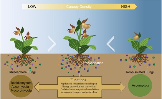
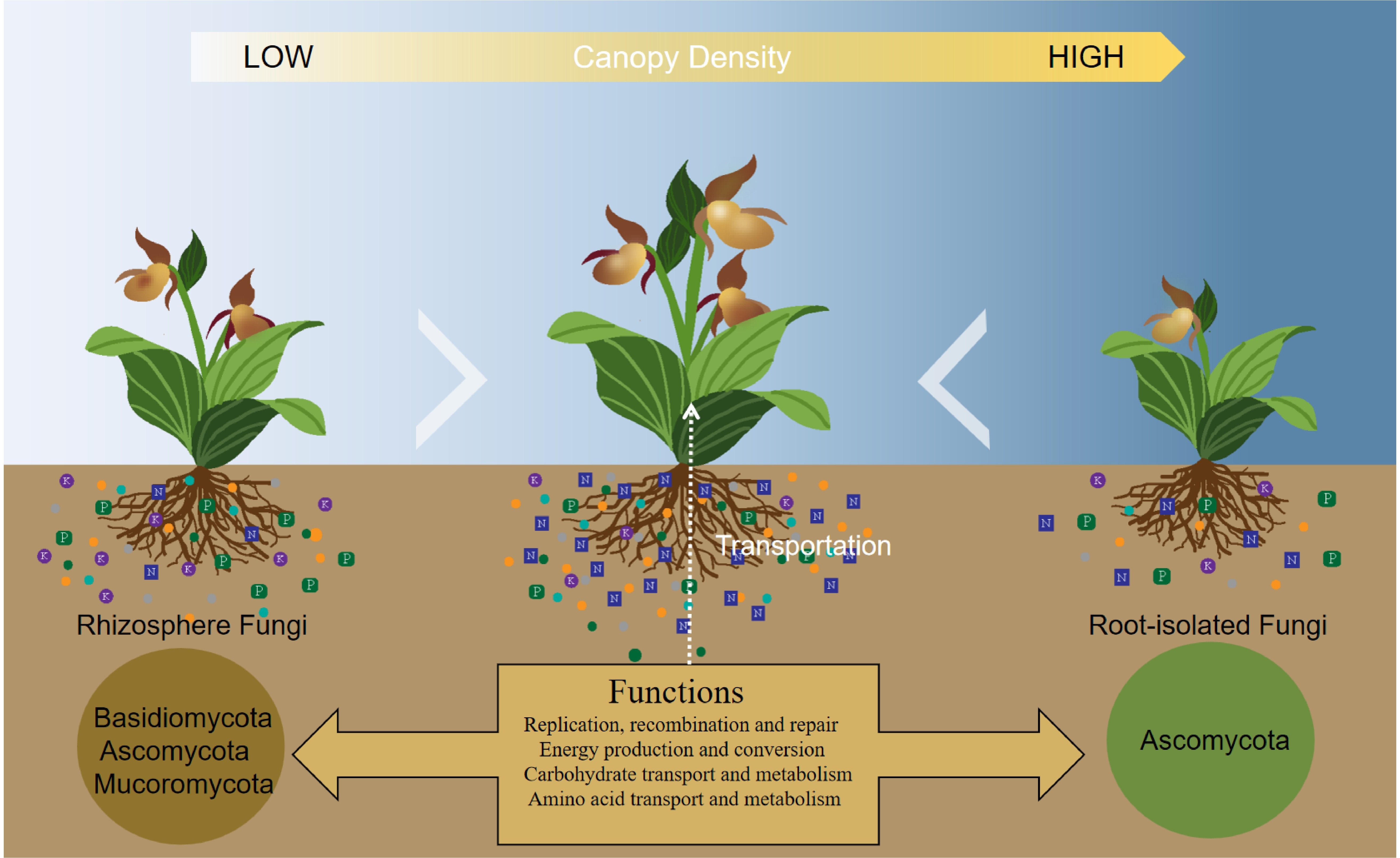
Cypripedium shanxiense S. C. Chen has high ornamental value; it relies on specific habitats and fungi. Wild C. shanxiense populations need urgent conservation because they are declining rapidly. This study investigated three wild C. shanxiense populations under different canopy densities in the Changbai Mountains, analyzing habitat characteristics and plant morphology. Tissue isolation methods, molecular identification techniques, and metagenomic approaches were applied separately to purify root-colonizing fungi and to investigate the composition and functions of rhizosphere fungi, thereby revealing the diversity of root mycobiome in C. shanxiense. Results revealed that C. shanxiense achieved the best growth when the canopy density was 85.29%, and the lowest growth was under 96.13% canopy density. Soil phosphorus and potassium contents reached their highest levels under 69.33% canopy density, while soil nitrogen and organic matter contents peaked at 85.29%. Soil organic matter and available nitrogen constitute the core nutrient factors for the growth of C. shanxiense. A total of 16 fungal strains were mainly enriched in the roots, all belonging to Ascomycota. Including numerous growth-promoting fungi and pathogenic fungi. The rhizosphere fungi were mainly enriched with Basidiomycota and Ascomycota. Functional genes related to replication, recombination, and repair, and Glycoside Hydrolases. This study clarifies the optimal growth conditions of this species and the dominant rhizosphere and root fungi, providing a scientific basis for the ecological restoration and conservation of rare species.

Graphical Abstract

1. Introduction

Orchidaceae is one of the most species-rich families of angiosperms [1]. It has a global distribution across diverse ecosystems, spanning tropical rainforests, temperate forests, and alpine meadows [2]. Orchid populations are strongly regulated by microenvironmental factors [3], including temperature, humidity, soil physicochemical properties, and canopy density [4]. As a key ecological group in Orchidaceae, terrestrial orchids are particularly close to microbial communities [5]. The composition and functional traits of fungi are especially pivotal to their survival and reproduction [6]. Fungi serve as essential energy sources for orchid seed germination [7]. Orchid seeds lack endosperm and cannot germinate independently under natural conditions. This unique biological characteristic inherently restricts the self-reproduction capacity of orchids [8]. Furthermore, habitat destruction and illegal harvesting of wild orchids often lead to accompanies these collection activities [9]. These anthropogenic disturbances have placed wild orchid populations in increasingly precarious survival conditions. Currently, wild orchid species with high ornamental and economic value are not fully protected in natural habitats. This has led to severe degradation and threats to wild orchid resources [10]. Notably, Orchidaceae accounts for over 73% of the species listed in the Convention on International Trade in Endangered Species of Wild Fauna and Flora (CITES), making it the most closely monitored and protected plant group [11].
Rhizosphere fungi exert key ecological functions by regulating plant fungal communities, thereby promoting host growth and enhancing nutrient acquisition [12]. Typical orchid rhizosphere fungi primarily belong to Basidiomycota, including taxa from Sebacinales, Ceratobasidiaceae, and Tulasnellaceae. These fungi form characteristic spiral mycelial structures on host roots [13]. Rhizosphere soil directly connects with plant root systems. Some rhizosphere fungi invade root cortex cells or colonize the rhizosphere environment surrounding roots, establishing mutualistic symbiotic relationships with host plants [14]. Consequently, the community composition and function of rhizosphere fungi influence the colonization efficiency and interaction stability of root fungi. These fungi also perform crucial ecological functions, such as modifying soil properties, enhancing plant tolerance to adverse environments, and inhibiting pathogen proliferation [15,16]. Dark septate fungi are commonly associated with plant roots, aiding survival and reproduction in harsh conditions, including drought, cold, nutrient-poor soils, alpine or subalpine ecosystems, high salinity, heavy metal pollution, and polar regions [17]. The habitat adaptability of Dendrobium officinale is regulated by beneficial fungi, whose colonization effectively enhances the plant’s tolerance to environmental fluctuations [18]. Key fungi identified from D. officinale roots included Epulorhiza, Tulasnella, and Sebacinales taxa. These fungi promoted seed germination, improved nitrogen uptake efficiency, and stimulated polysaccharide accumulation [19]. Two fungal strains isolated from Camellia oleifera significantly improved orchid rhizosphere soil fertility. They also promoted soil material cycling and energy flow. These strains increased beneficial microbe abundance while reducing pathogen levels, thereby establishing a microenvironment more conducive to orchid growth [20].
C. shanxiense is a typical terrestrial orchid species inhabiting temperate forest understories. It is light-sensitive and thrives in shaded environments [21]. With sparse populations and a restricted distribution range, this species is classified as rare and endangered, warranting high conservation priority. Changbai Mountain is widely recognized as one of the world’s biodiversity hotspots [22], where multiple wild C. shanxiense populations have been discovered during field surveys. This unique habitat provides valuable natural samples for further studies on the ecological habits of C. shanxiense and the assembly of its associated fungi. In addition to its ecological significance, C. shanxiense holds considerable ornamental value due to its distinctive floral morphology and coloration. However, its endangered status necessitates urgent conservation and research efforts. Therefore, this study conducted environmental surveys on wild C. shanxiense populations distributed across different canopy density levels in Jilin Province, China. The surveys focused on analyzing habitat characteristics, plant morphological traits, soil environmental factors, and associated plant species. Additionally, through metagenomic sequencing analysis of rhizosphere soils across a gradient of canopy densities, the community structure and functional traits were investigated. Through tissue isolation and molecular identification techniques, the fungal species associated with the roots of C. shanxiense were confirmed. This research aims to reveal the survival strategies and ecological adaptation mechanisms of C. shanxiense under specific environmental conditions, while also providing new insights and a theoretical basis for the conservation biology of rare and endangered plants. Exploring the fungal preference and functional characteristics of fungi associated with C. shanxiense will provide a basis for the effective conservation and sustainable utilization of this orchid species.

2. Materials and Methods

2.1. Sample Collection and Environmental Survey

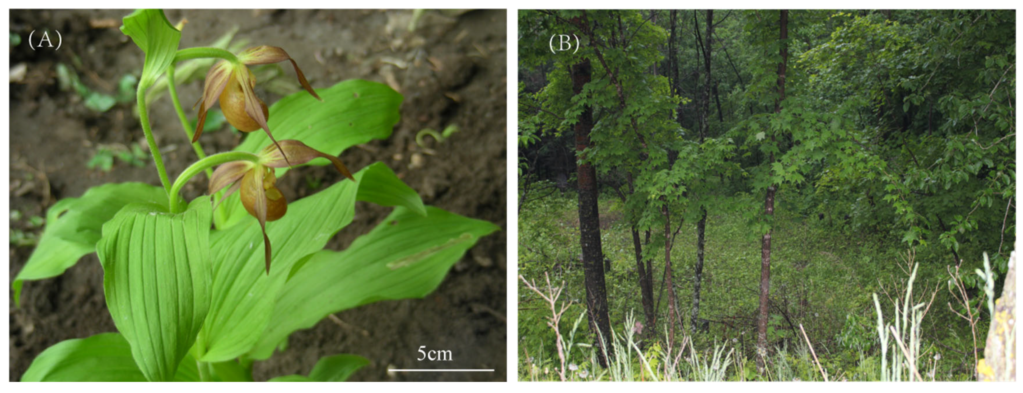
A total of 12 natural populations of C. shanxiense were identified during field surveys conducted by our research team in the Changbai Mountains. To minimize the interference of other significant environmental variations on the survey results, three sampling sites in Linjiang City were selected for the formal experiment. Linjiang City (126°92′ E, 41°81′ N), where the average elevation is 776 m, the annual mean temperature is 6 °C, and the annual mean precipitation is 824 mm. The field survey was carried out in June, during which the average maximum temperature reached 26 °C, the average minimum temperature was 13 °C, and the average rainfall stood at 115 mm. Sampling plots were situated under broad-leaved forests and along forest edges. Figure 1A depicts C. shanxiense in its natural state, while Figure 1B presents its habitat conditions. Five sets of rhizosphere soil samples (10 g each) were collected from each plot. The soil layer was gently excavated until the fibrous roots of C. shanxiense were exposed, and the soil adhering to the roots was collected as rhizosphere soil. After removing sand and gravel particles, the rhizosphere soil samples from the same plots were thoroughly mixed and subjected to metagenomic sequencing. 5 cm root segments were collected from the root tips, with 2–3 roots sampled per plant, for the isolation and purification of endophytic fungi. All sampling and surveys conducted in this study were performed without affecting the normal growth conditions of C. shanxiense.
Five 5 m × 5 m plots were established, with each plot centered on a surveyed wild C. shanxiense population. Each plot was divided into 4 subplots of 2.5 m × 2.5 m, and the vertex of each subplot was set as a measurement point [23]. The average value of measurements from these points was used as the environmental indicator for the corresponding C. shanxiense population. Environmental factors, including altitude, canopy density, temperature, and humidity, were measured using a GPS receiver, a canopy analyzer, and a thermo-hygrometer (DELIXI ELECTRIC, China), respectively. Herbaceous and woody plants within the sampling plots were recorded as companion plants of C. shanxiense. After removing dead branches and fallen leaves around the rhizosphere of C. shanxiense, rhizosphere soil samples were collected using the quadrat sampling method. The soil samples were naturally air-dried, thoroughly mixed, and then sieved through 20-mesh and 100-mesh sieves sequentially for the determination of soil physicochemical properties. The determination methods for soil parameters were as follows [24,25]:
Available phosphorus (AP): 0.5 mol·L−1 NaHCO3 extraction method;
Available potassium (AK): NH4OAc extraction followed by flame photometry;
Alkali-hydrolyzable nitrogen (AN): Alkali-hydrolyzable diffusion method;
Total phosphorus (TP): HClO4-H2SO4 digestion method;
Total potassium (TK): NaOH fusion followed by flame photometry;
Total nitrogen (TN): Kjeldahl method;
Organic matter (OM): Potassium dichromate external heating method;
pH: Determined using a pH meter.
Eight soil physicochemical parameters were measured in total, with three technical replicates set for each parameter.

2.2. Isolation and Purification of Root-Isolated Fungi

The collected root samples were immersed in a 2% sodium hypochlorite solution for 3 min, rinsed repeatedly with sterile distilled water several times, and then immersed in 75% ethanol for 30 s. After thorough rinsing with sterile distilled water once more, the samples were dried using sterile filter paper. Sterilized roots were cut into 0.2 cm segments, which were then inoculated onto isolation medium supplemented with antibiotics (200 g/L potato, 20 g/L glucose, 20 g/L agar, kanamycin, and ampicillin). After 6–7 days of incubation, when fungal mycelia covered approximately 2/3 of the root segment area, edge portions of different colonies were picked and inoculated onto fresh potato dextrose agar (PDA) medium (200 g/L potato, 20 g/L glucose, 20 g/L agar). Inoculated PDA plates were incubated in a 25 °C constant-temperature incubator in the dark. This purification procedure was repeated continuously until pure fungal cultures were obtained. To verify the effectiveness of root surface sterilization, the final rinse water (sterile distilled water used for the last root rinse) was also inoculated onto the isolation medium. No fungal growth on these control plates confirmed successful surface disinfection.

2.3. Molecular Identification of Root-Isolated Fungi

Genomic DNA of purified fungal isolates was extracted using the Ezup Column-Based Fungal Genomic DNA Extraction Kit supplied by Sangon Biotech Co., Ltd., Shanghai, China. For molecular identification of the mycorrhizal fungi isolated from C. shanxiense, the universal primer pair ITS1/ITS4 was selected to amplify the fungal internal transcribed spacer (ITS) region. Polymerase chain reaction (PCR) amplification was performed under the following conditions: initial denaturation at 95 °C for 5 min, followed by 35 cycles of denaturation at 95 °C for 30 s, annealing at 58 °C for 30 s, and extension at 72 °C for 1 min, and a final extension step at 72 °C for 7 min. A 3 μL aliquot of each PCR product was subjected to 1% agarose gel electrophoresis for fragment detection and verification. PCR products showing bright single bands were submitted to a commercial sequencing service provider for Sanger sequencing. Resultant sequences were subjected to BLAST analysis against the nucleotide database of the National Center for Biotechnology Information (NCBI). Mycorrhizal fungi were identified by combining the sequence alignment results with morphological observation data.

2.4. Metagenomic Data Processing

A total of 50 g of filtered rhizosphere soil was collected for metagenomic sequencing. Metagenomic sequencing was performed by Personalbio Technology Co., Ltd., Shanghai, China. The total DNA of rhizosphere soil microorganisms was extracted via the de novo sequencing method using the Mag-Bind Soil DNA Kit purchased from OMEGA Bio-tek, Inc. Subsequently, the concentration and purity of the extracted DNA were determined. Specifically, the DNA concentration was quantified with a Qubit 4 Fluorometer (Qubit™ 4 Fluorometer, with WiFi: Q33238; Qubit™ Assay Tubes: Q32856; Qubit™ 1X dsDNA HS Assay Kit: Q33231), and the genomic sequencing libraries were constructed following the standard experimental protocol described in the Illumina TruSeq DNA Sample Preparation Guide. The quality of the sequencing data before and after quality control was assessed using the fastp software, generating the Raw Data Statistics Table and Valid Data Statistics Table, respectively (Table 1). In total, 380,086,460 valid raw reads were obtained. For species annotation of the valid reads, Kaiju software was employed to align each read against the prokaryotic and eukaryotic microbial protein sequences in the NCBI-nr database, as well as the viral protein sequences in the RNDB-prot database. By counting the number of reads annotated to each species and collating the corresponding species annotation information, species abundance tables at different taxonomic levels were generated. Finally, the fungal genes were selected for subsequent visualization analysis.

2.5. Data Analysis

One-way analysis of variance (ANOVA) was performed in SPSS 27.0 to analyze soil nutrient contents, morphological indicators, and physiological parameters. Redundancy analysis (RDA) plots were generated using Wekemo Bioincloud (https://www.genescloud.cn (accessed on 3 February 2026)) [26]. Circos, Column, Veen, LDA Effect Size (LEFse), and Heatmap were all generated using R 4.5.2. Image integration was conducted with Adobe Illustrator 29.0. Phylogenetic trees were constructed using the neighbor-joining method in MEGA 11.0.13 software.

3. Results

3.1. Environmental Characteristics and Morphological Traits of C. shanxiense

In this study, environmental factors and plant growth indicators were measured and analyzed for 3 wild C. shanxiense plots in different canopy densities (Table 2). P1 had the lowest canopy density of 69.33% and exhibited intermediate plant growth performance. P2 with a canopy density of 85.29% showed the optimal growth of C. shanxiense, while P3 with a canopy density as high as 96.13% displayed the most unfavorable growth.
Rhizosphere soil physicochemical properties of C. shanxiense were measured in three plots (Table 3). Significant differences were observed in all eight measured soil physicochemical indicators in three plots. Specifically, P1 had the highest contents of TP, TK, and AK. P2 exhibited significantly higher contents of OM, TN, and AN compared with the other two plots. P3 had higher AP content than P1 and P2, along with the highest soil pH.
The habitat of C. shanxiense primarily consists of forest understories or grassy slopes, where it associates with a diverse array of plant species. Statistical analysis of woody and herbaceous plants in three plots revealed a total of six woody plant species and seven herbaceous plant species, summarized in Table 4.

3.2. Correlation Analysis of Morphology and Environment

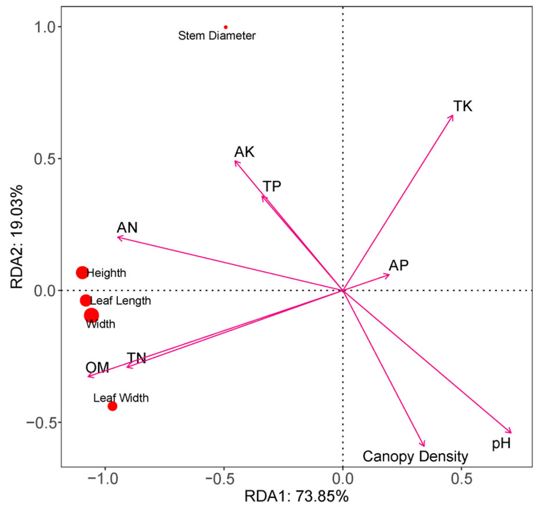
The results of the RDA were presented in Figure 2. Red dots represented plant growth indicators, whereas arrows denoted environmental factors. RDA1 explained 73.85% of the total variance, and RDA2 accounted for 19.03%, resulting in an overall explanatory rate of 92.88%. The selected environmental factors effectively explained their impacts on the measured plant growth indicators. AN, TN, and OM exhibited small vector angles with plant height, width, and leaf length and width; additionally, the angles among these three factors were narrow, indicating that they synergistically regulated the aboveground vegetative growth of plants. AK and TP showed small vector angles with stem diameter, suggesting a positive correlation with stem diameter. In contrast, TK formed large vector angles with most growth indicators, implying a weak regulatory effect on the plant growth indicators investigated in this study.

3.3. Morphological Description of C. shanxiense Root-Isolated Fungi

A total of 16 strains were isolated from C. shanxiense, and their morphological characteristics were described (Table 5). The colony textures were predominantly characterized as velvety, felt-like, powdery, waxy, or cottony. The felt-like texture was the most common colony type, with tightly packed and non-dispersive hyphae. Waxy mycelia were nearly transparent, and their morphology could only be observed within the culture medium. Colony colors ranged from white, pale yellow, and olive green to brown, black, and orange. Most colonies were circular in shape, while a few displayed irregular and scattered growth patterns.

3.4. Molecular Identification of C. shanxiense Root-Isolated Fungi

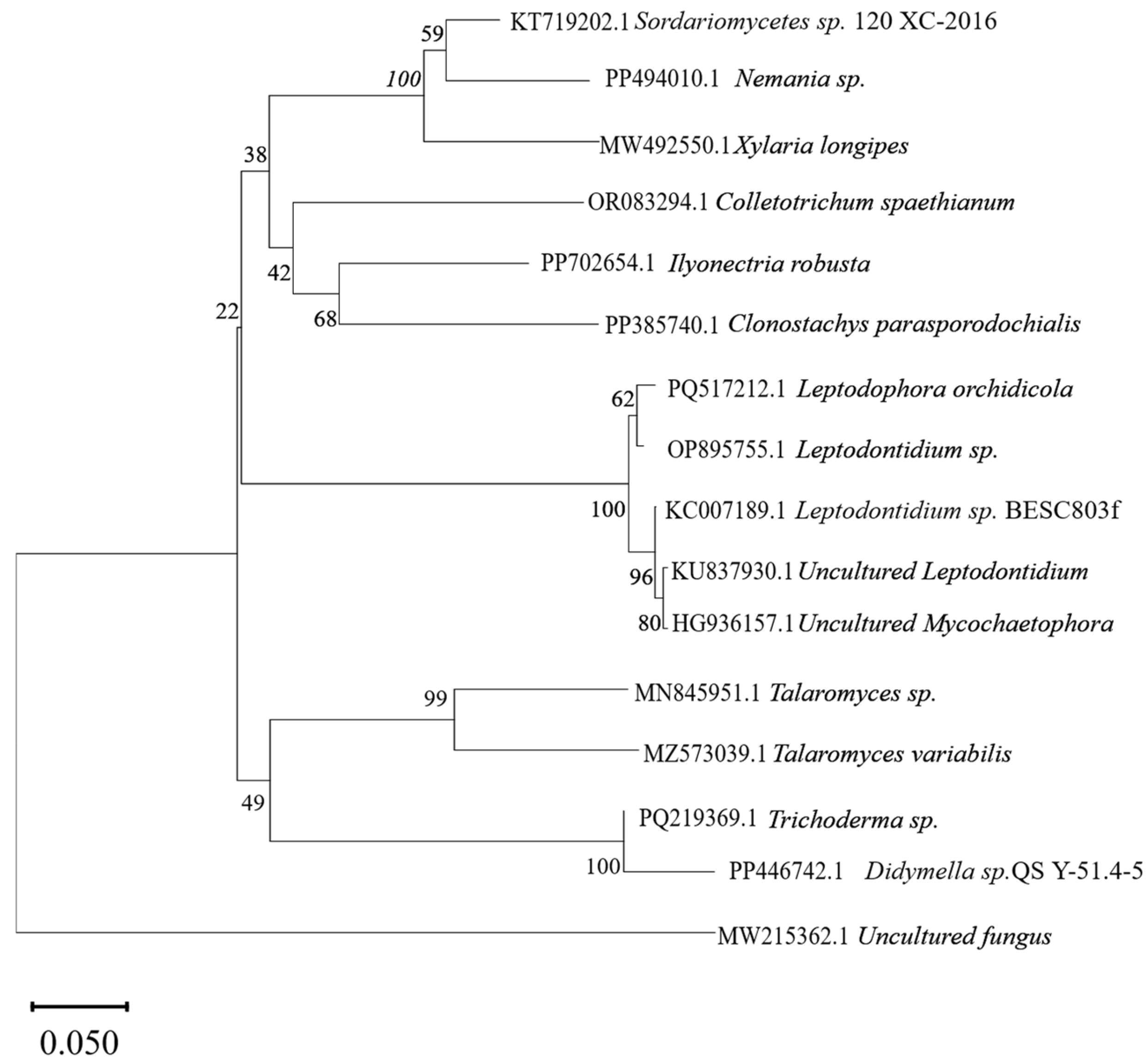
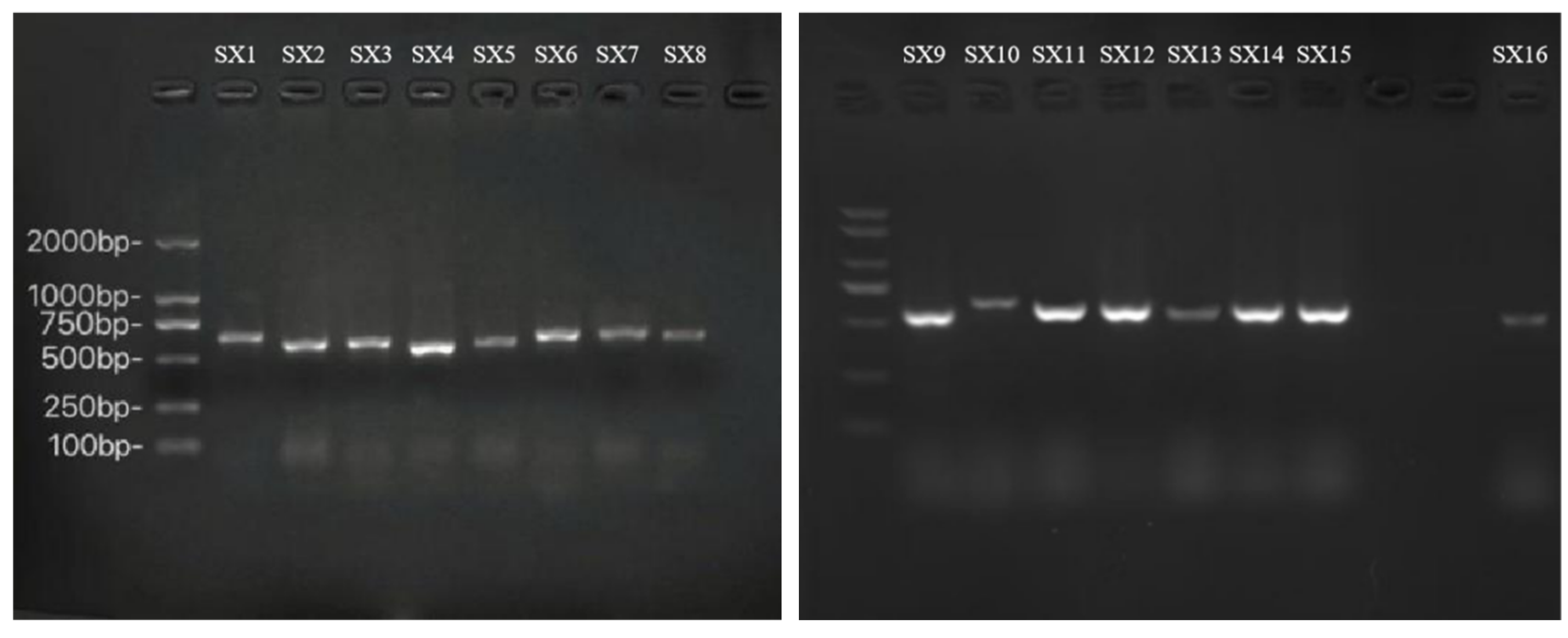
DNA was extracted from the mycelium of 16 fungal strains isolated from the roots of C. shanxiense (Figure A1). All sequences satisfied the quality criteria for Sanger sequencing. The sequencing results were uploaded to the NCBI database for BLAST alignment. The identification results of the 16 root-isolated fungal strains are presented in Table A1. All strains belonged to the Ascomycota (Table A2), with two strains unidentifiable at the class level. The remaining 14 strains were classified into four classes (Leotiomycetes, Eurotiomycetes, Sordariomycetes, and Dothideomycetes), encompassing six orders and seven families. Among these, seven strains could not be identified to the species level. Three additional specimens were identified as fungi not previously cultured. To clarify the phylogenetic positions of the isolated strains, a phylogenetic tree was constructed based on the ITS sequences using the neighbor-joining method (Figure 3). The results showed that Sordariomycetes sp. 120 XC-2016, Nemania sp., and Xylaria longipes clustered together with bootstrap support values of 59%, 100%, and 38%, respectively. Colletotrichum spaethianum, Ilyonectria robusta, and Clonostachys parasporodochialis formed a subclade with bootstrap support values of 42% and 68% for the respective nodes. Leptodophora orchidicola, Leptodontidium sp., Leptodontidium sp. BESC803f, uncultured Leptodontidium, and uncultured Mycochaetophora clustered into a clade with bootstrap support values of 62%, 100%, 96%, and 80% at the corresponding nodes. Talaromyces sp. and Talaromyces variabilis formed a well-supported clade (99% bootstrap value) and further clustered with Trichoderma sp. and Didymella sp. QSY-51.4-5 into a major clade with 49% bootstrap support. The uncultured fungus formed an independent outgroup. The evolutionary distance scale of the phylogenetic tree was 0.050. The support rates of each branch reflect the reliability of clustering, providing molecular biological evidence for strain classification and phylogenetic relationship analysis.

3.5. Different Classes Root-Isolated Fungi in C. shanxiense

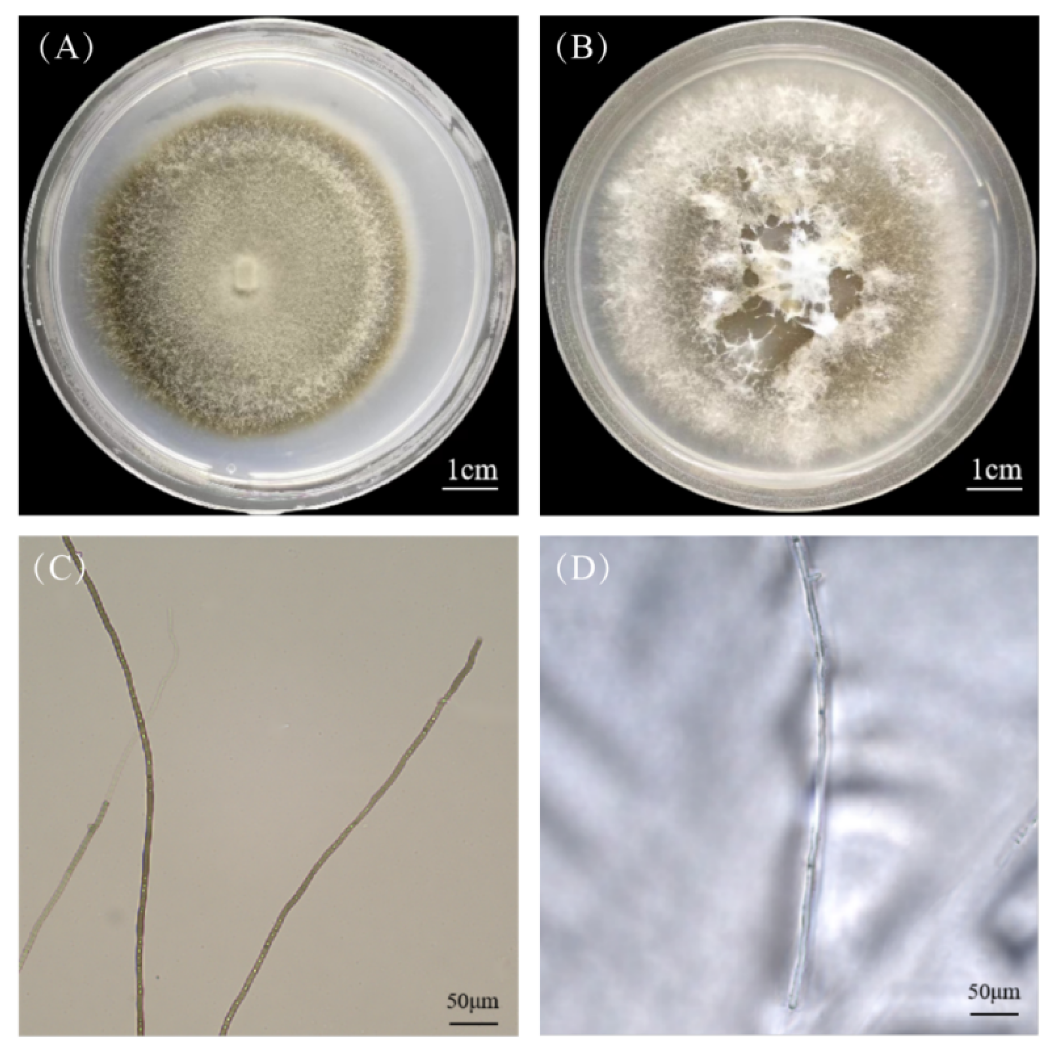
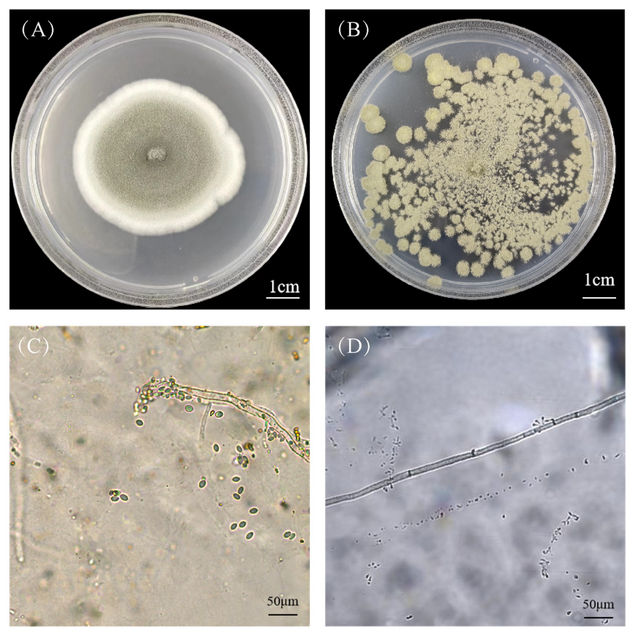
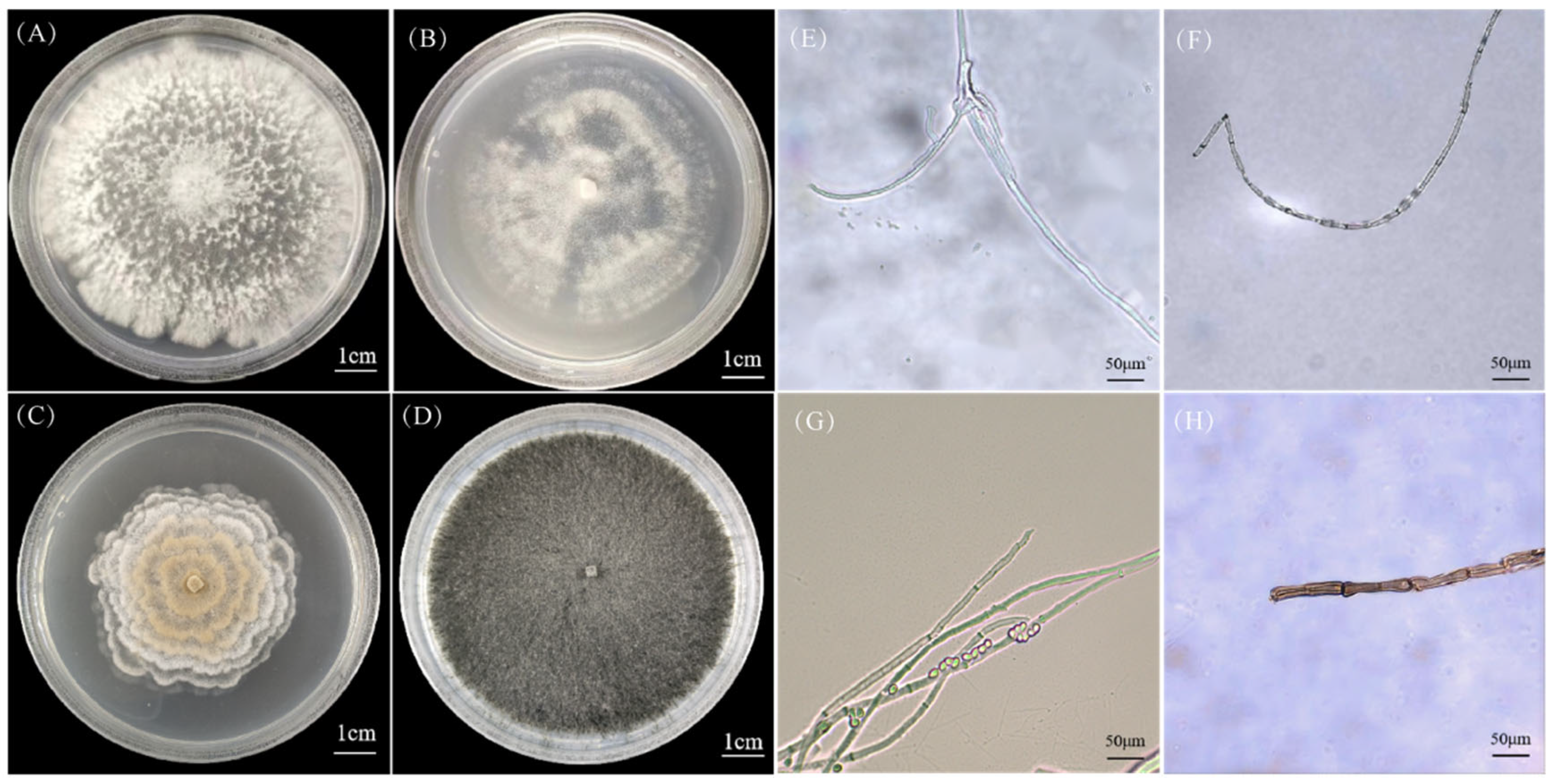
After comprehensive identification based on morphological and molecular characteristics, the colony and mycelial morphologies of the strains were observed and described. Nine strains representing four different classes with distinct morphological variations, along with their mycelial structures (SX1, SX2, SX3, SX7, SX9, SX12, SX14, SX16), were selected for presentation.
Figure 4 shows two strains (SX1 and SX7) belonging to Leotiomycetes. Figure 4A,C illustrate the colony morphology and mycelium structure of SX1. Colonies were regular and circular, with dense mycelium exhibiting a velvety appearance. The central portion of the colony was pale yellow, while the outer region was brown. ITS sequence alignment revealed 99.83% similarity to Leptodontidium sp. BESC803f (KC007189.1).
Figure 4B,D illustrate the colony morphology and mycelial structure of strain SX7, respectively. The colony was regular and circular, with a cottony texture and an overall white coloration. ITS sequence alignment showed that SX7 shared 100.00% similarity with Ilyonectria robusta (PP702654.1).
Figure 5 shows two strains (SX2 and SX14) belonging to Eurotiomycetes. Figure 5A,C illustrate the colony morphology and mycelial structure of SX2, respectively. The colonies were regular and circular, with dense mycelia expanding gradually outward and exhibiting a felt-like texture. The central portion of the colonies was olive green, while the outer edges were white. ITS sequence alignment revealed that SX2 shared 99.63% similarity with Talaromyces sp. (MN845951.1).
Figure 5B,D illustrate the colony morphology and mycelial structure of SX14, respectively. SX14 formed multiple irregular circular olive green colonies on the medium, with a powdery and relatively flat surface. The grayish–white mycelia were slender and filamentous, with distinct septa and a smooth surface. Small conidia were distributed along the hyphae. ITS sequence alignment showed that SX14 shared 99.64% similarity with Talaromyces variabilis (MZ573039.1).
Figure 6 shows four strains (SX3, SX9, SX12, and SX16) belonging to Sordariomycetes. Figure 6A,E illustrate the colony morphology and mycelial structure of strain SX3, respectively. The colonies were irregularly circular with a velvety texture and maintained a uniform white coloration throughout cultivation. ITS sequence alignment revealed that SX3 shared 99.63% similarity with Xylaria longipes (MW492550.1).
Figure 6B,F illustrate the colony morphology and mycelial structure of SX9, respectively. The colony was generally circular, with mycelia spreading outward in irregular circular patterns within the medium. A pale yellow powdery substance formed on the colony surface. The mycelia were robust, filamentous, moderately curved, transparent, smooth, septate, and branched. ITS sequence alignment showed that SX9 shared 99.44% similarity with Clonostachys parasporodochialis (PP385740.1).
Figure 6C,G illustrate the colony morphology and mycelial structure of SX12, respectively. The colony had a waxy texture, with a distinct orange center and white periphery, expanding in a concentric ring pattern to form a petal-like appearance. The mycelia were slender and filamentous, smooth, with clearly visible septa, and pale green in color. Green spores, spherical or irregularly oval in shape, were distributed along the mycelia. ITS sequence alignment revealed that SX12 shared 99.65% similarity with Nemania sp. (PP494010.1).
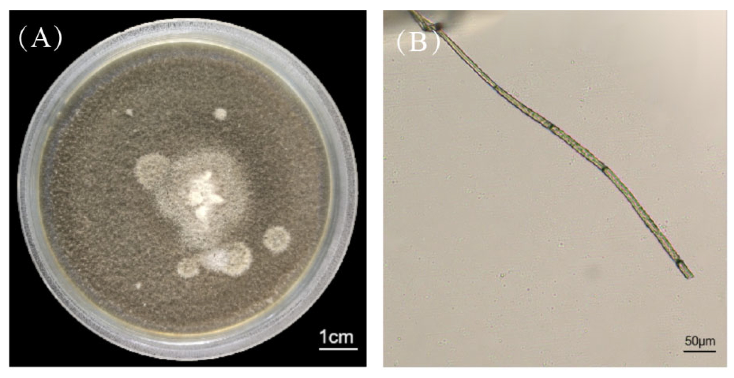
Figure 6D,H illustrate the morphological characteristics of SX16, respectively. The colonies were felt-like, regularly circular, with an uneven surface exhibiting a granular texture, and displayed a uniform black coloration. The brownish-gray mycelia were rod-like, with distinct septa and a smooth surface. ITS sequence alignment revealed that SX16 shared 100.00% similarity with Colletotrichum spaethianum (OR083294.1).
Figure 7 shows SX15, which is classified into Dothideomycetes. Figure 7A,B illustrate the colony morphology and mycelial structure of SX15, respectively. The colony was regularly circular with a felt-like texture, displaying an overall deep brown color and producing white mycelia. ITS sequence alignment revealed that SX15 shared 99.22% similarity with Didymella sp. QSY-51.4-5 (PP446742.1).

3.6. Analysis of the Composition and Function of Rhizosphere Fungi

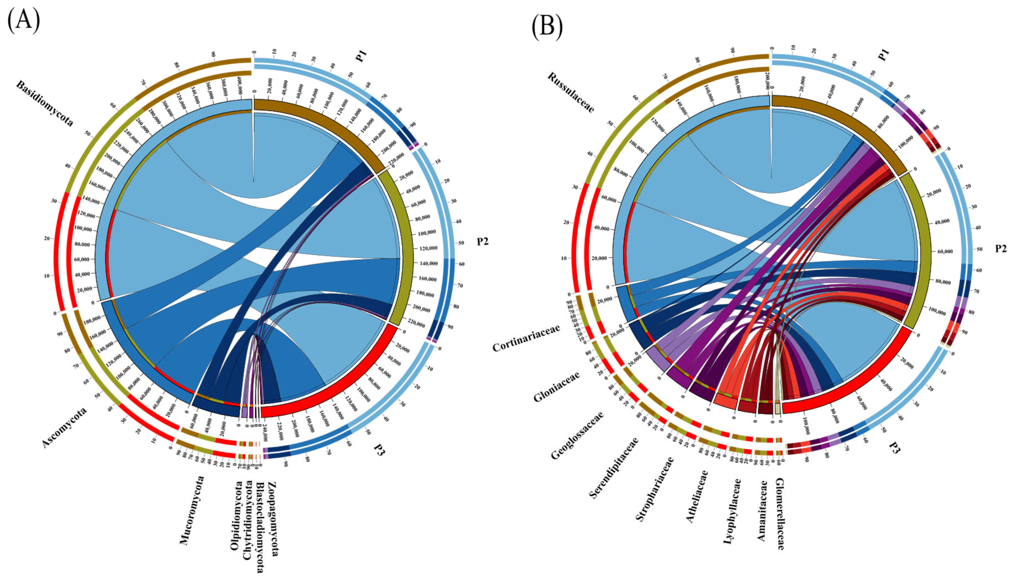
Circos diagram of the rhizosphere fungal community in C. shanxiense showed composition and distribution characteristics at different taxonomic levels (Figure 8). At the Phylum level (Figure 8A), rhizosphere fungi primarily comprised Basidiomycota, Ascomycota, and other low-abundance groups, including Mucoromycota and Olpidiomycota, with distinct association patterns in fungal composition between groups. At the family level (Figure 8B), the dominant families included Russulaceae, Cortinariaceae, and Gloniaceae. The composition of rhizosphere fungi at the family level was more complex in 3 plots, reflecting specific distribution patterns of fungal communities at this taxonomic level within the rhizosphere environment.
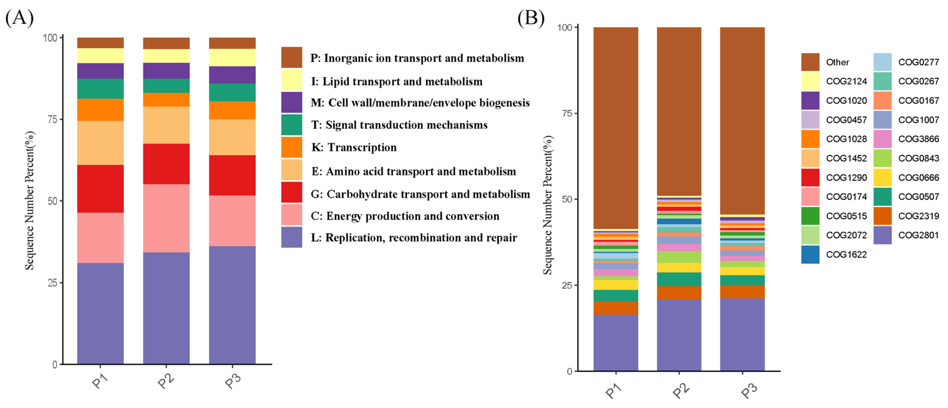
The Unigene set obtained from metagenomic sequencing was aligned against the eggNOG database, yielding 29,569 annotated genes. These genes were screened for Clusters of Orthologous Groups of proteins (COG) functions, and 5125 genes were selected for subsequent analysis. Analysis of the top 20 functional genes by abundance and nine dominant functional categories (Figure 9) revealed that rhizosphere fungi were primarily involved in the following functional categories (Figure 9A): L (replication, recombination, and repair), C (energy production and conversion), and G (carbohydrate transport and metabolism). Specifically (Figure 9B), rhizosphere fungi were enriched in the COG functional genes COG2801, COG2319, COG0507, COG0666, COG0843, and COG3866. Among these, COG2801 and COG0507 belonged to L, which were primarily involved in transposition and the preparation of double-strand DNA breaks for recombinational DNA repair, COG2319 was mainly associated with anaphase-promoting complex binding, COG0666 and COG3866 belonged to G, which were primarily involved in the response to abiotic stimuli and pectate lyase function, respectively, while COG0843 was associated with C, primarily involved in heme-copper terminal oxidase activity.
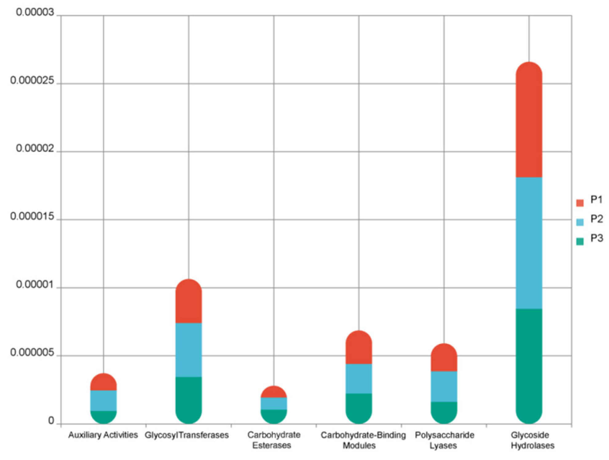
Analysis of carbohydrate-active enzymes (CAZy) functional genes was performed on the rhizosphere fungi of C. shanxiense (Figure 10). CAZy annotation results in 3 plots revealed significant differences in the abundance distribution of different CAZy enzyme classes. Among all CAZy enzyme classes, Glycoside Hydrolases (GHs) exhibited the highest abundance, which was far greater than that of other enzyme types, while Carbohydrate Esterases (CEs) showed the lowest abundance. No significant differences in the abundance of carbohydrate-active enzyme genes were observed in 3 plots for the rhizosphere fungi associated with C. shanxiense.

3.7. Differential Analysis

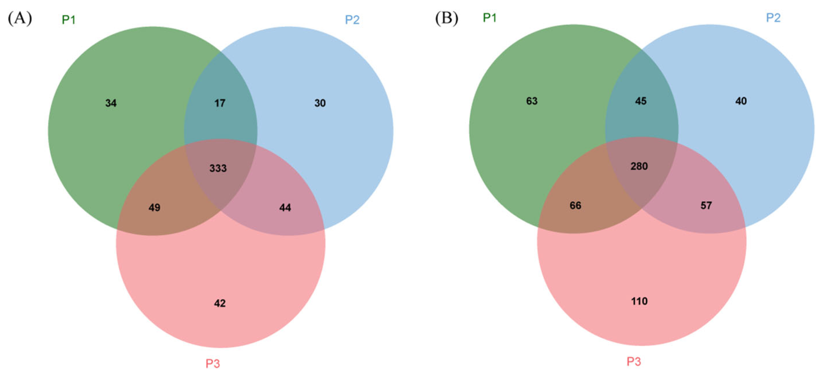
The species and functional gene composition characteristics of rhizosphere fungi in three plots of C. shanxiense were analyzed (Figure 11A,B). At the species level, the numbers of endemic species in P1, P2, and P3 were 34, 30, and 42, respectively. The number of shared species between P1 and P2, P1 and P3, and P2 and P3 was 17, 49, and 44, respectively, and the total number of shared species across the three plots reached 333. At the functional gene level, the number of unique functional genes in P1, P2, and P3 was 63, 40, and 110, respectively. The number of shared functional genes between P1 and P2, P1 and P3, and P2 and P3 was 45, 66, and 57, respectively, with the total number of shared functional genes in the three plots reaching 280. Overall, P1 exhibited intermediate levels of endemic species and functional genes, while P3 showed the highest abundances of both endemic species and functional genes in the three plots. In contrast, P2 recorded the lowest numbers of endemic species and functional genes.
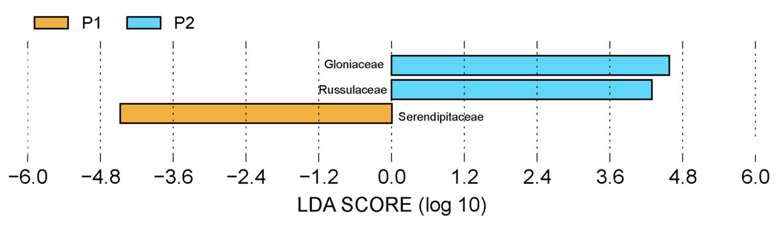
LEfSe analysis (LDA score > 4) identified significantly different fungal taxa in rhizosphere communities across different plots of C. shanxiense (Figure 12). Specifically, Serendipitaceae was a statistically enriched taxon in P1, whereas Gloniaceae and Russulaceae were significantly enriched taxa in P2. These taxa served as biomarkers distinguishing rhizosphere fungal community structures across plots, reflecting the selective preferences of distinct rhizosphere microenvironments for specific fungal taxa.

3.8. Correlation Analysis of Rhizosphere Fungi and Environment

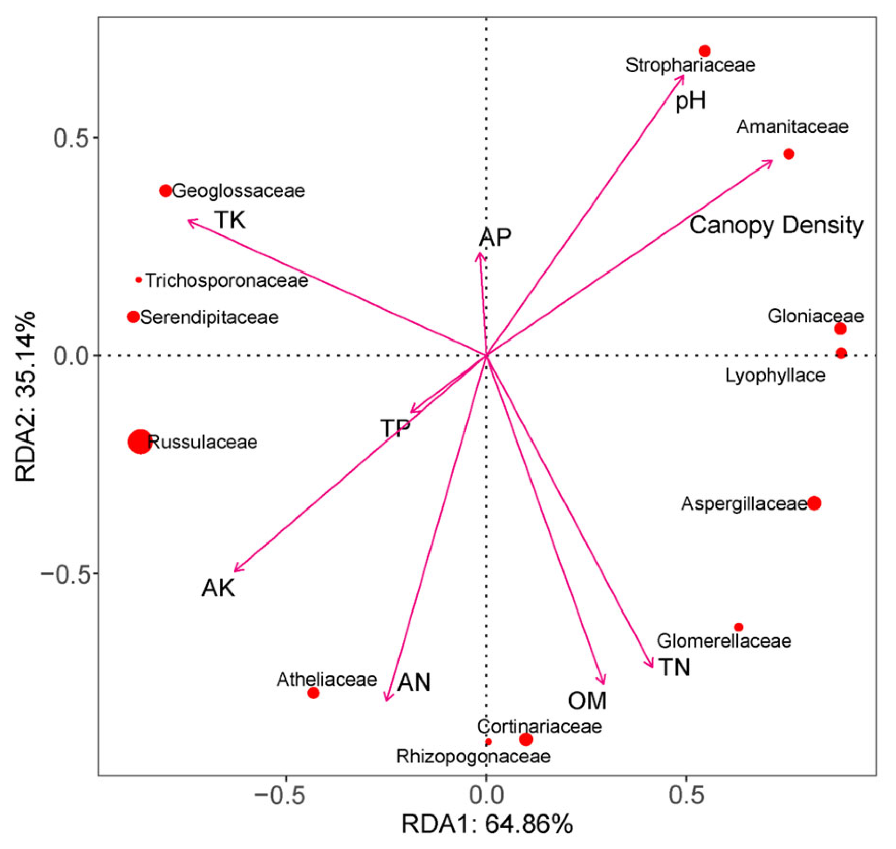
RDA results (Figure 13) indicated that RDA1 explained 64.86% of the variance, while RDA2 explained 35.14%. Significant associations existed between the rhizosphere fungal communities of C. shanxiense and environmental factors. Specifically, Geoglossaceae, Serendipitaceae, and Russulaceae showed significant positive correlations with AK and TK; Strophariaceae and Amanitaceae exhibited positive correlations with pH and canopy density; Atheliaceae and other taxa were closely associated with AN; while Cortinariaceae, Rhizopogonaceae, and other groups showed strong associations with OM.
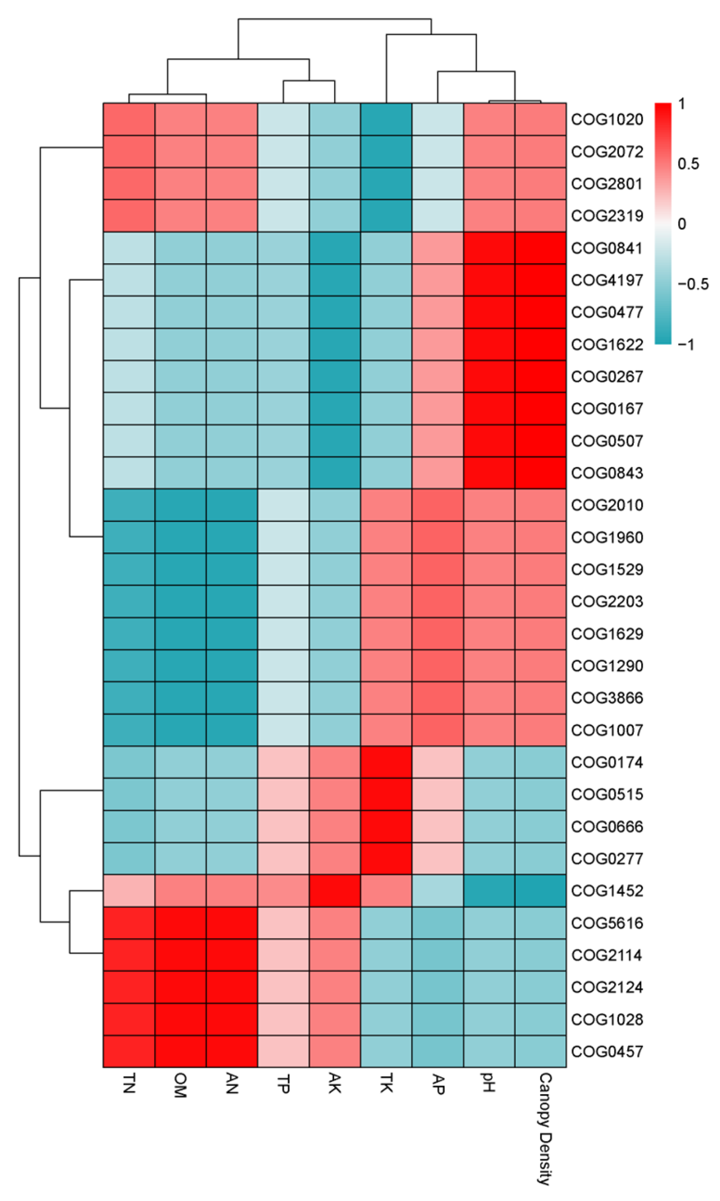
Heatmap analysis of the correlation between eggNOG annotation functions and environmental factors (Figure 14) revealed strong associations between different COG functional groups and environmental factors in the rhizosphere of C. shanxiense. Functional groups represented by COG1020 and COG2072 showed significant positive correlations with AP; functional groups such as COG0841 and COG4197 exhibited significant positive correlations with factors including TN, OM, and TP. Conversely, groups such as COG2114 and COG2124 displayed significant negative correlations with TN and OM. Additionally, COG1452 demonstrated clear associations with pH and canopy density. These results indicated that soil physicochemical factors (N, OM, and pH) and the habitat factor (canopy density) significantly regulated the distribution of core functional gene clusters in the rhizosphere of C. shanxiense.

4. Discussion

C. shanxiense survival and reproduction are highly dependent on specific habitat conditions and its symbiotic relationship with fungi [27]. In this study, canopy density significantly influenced the growth and development of C. shanxiense. Its growth under 85.29% canopy density exhibited optimal morphological traits, including plant height, crown spread, and leaf length. By contrast, growth was unfavorable under a high canopy density of 96.13%. This indicated that moderate shading promoted photosynthesis and energy accumulation in C. shanxiense, whereas excessive shading might lead to insufficient light intensity, limiting its carbon assimilation capacity [28] and consequently affecting biomass accumulation [29]. This phenomenon aligned with the light response mechanisms of most orchids [30]. This study further quantified the optimal canopy density range for C. shanxiense under natural conditions, providing actionable thresholds for habitat management.
Soil physicochemical properties serve as crucial indicators for evaluating habitat suitability for orchids, with nutrient content and pH directly influencing root absorption efficiency and symbiotic microbial activity [31]. In this study, the rhizosphere soil of C. shanxiense exhibited weak acidity, with a pH ranging from 6.49 to 6.77, which was consistent with the preference for acidic soils among most terrestrial orchids [32]. Plants grown under 85.29% canopy density exhibited the most favorable morphological traits and higher contents of soil organic matter, total nitrogen, and alkali-hydrolyzable nitrogen compared with those in other plots. Their available potassium content remained at a moderate level, indicating that soil organic matter and nitrogen supply were key nutrient-limiting factors for C. shanxiense growth. Meanwhile, organic matter accumulation promoted the proliferation of microorganisms and nutrient transformation [33,34]. Under high canopy density conditions, organic matter accumulation and alkaline-hydrolyzable nitrogen content were relatively low. This might be attributed to insufficient available nitrogen supply caused by enhanced microbial nitrogen immobilization [35]. Furthermore, no significant differences in soil phosphorus content were observed in 3 plots. This suggested that microbial activation of phosphorus might have compensated for natural variations in soil phosphorus pools, providing a starting point for further investigations into microbial nutrient transformation functions.
This study isolated and identified 16 fungal species from the roots of C. shanxiense. However, at the phylum level, they all belonged to the Ascomycota. This composition differed from previously reported fungal assemblages associated with temperate terrestrial orchids. Most studies indicated that Basidiomycota were the dominant symbiotic fungi of orchids [36]; however, no Basidiomycota were isolated in this study. This discrepancy might be related to the sampling season, altitude, and vegetation. The dominant status of Ascomycota in C. shanxiense roots might result from their superior acid tolerance and organic matter decomposition capacity [37]. These traits were highly compatible with the soil characteristics of the study area. Notably, Leptodontidium sp. and Clonostachys parasporodochialis have been shown to enhance nutrient uptake in orchids and inhibit pathogen colonization [38,39]. Leptodophora orchidicola is an orchid-specific symbiotic fungus. Its presence in the roots of C. shanxiense further confirms the specialized symbiotic association between orchids and fungi [40]. These fungi also exhibited lignin-degrading capabilities [41] and thus might participate in rhizosphere litter degradation and carbon cycling processes in the C. shanxiense rhizosphere. Talaromyces, which belongs to Eurotiomycetes, produces stress-resistant conidia that aid plants in coping with adverse environments [42]. This resembled the functional characteristics of endophytic fungal communities in orchid species such as Dendrobium and Bletilla striata [43,44], confirming the functional conservation of orchid–fungal symbiotic systems. This discovery enriched the diversity database of fungi associated with orchids. However, this study also isolated the pathogenic fungus Ilyonectria robusta and Colletotrichum spaethianum, which cause root rot in various plant species [45,46]. Their presence in C. shanxiense roots suggests a potential “symbiotic-pathogenic” dynamic equilibrium within natural populations. This finding emphasized that conservation and artificial introduction efforts must not only screen for beneficial strains but also monitor the structure of root microbial communities to mitigate potential pathogen threats.
The functional specificity of fungi colonizing roots is central to understanding the mechanisms underlying orchid endangerment [47]. In this study, velvety and fluffy colonies accounted for the highest proportion at 75%. This colony structure facilitated hyphal attachment and nutrient exchange, while the transparent hyphae of waxy colonies might enhance adaptation to low-light environments. Such strains gained a competitive advantage in nutrient acquisition, while other strains might exert defensive functions by producing secondary metabolites [48]. The diversity of colony colors, including white, pale yellow, and black, correlates with pigment synthesis. Dark-colored colonies might produce pigments to resist UV damage [49], which represented a morphological adaptation suited to the light conditions under low canopy density. These findings indicated that the root fungal communities of C. shanxiense had developed synergistic adaptation mechanisms to heterogeneous habitats through differentiation in morphology and growth strategies.
The community composition and function of dominant rhizosphere fungi in C. shanxiense remained unchanged under varying canopy density. At the phylum level, Ascomycota and Basidiomycota dominated, while Russulaceae predominated at the family level. From the perspective of carbohydrate-active enzymes, rhizosphere fungi primarily participated in Glycoside Hydrolase processes. Studies on autotrophic-heterotrophic nutritional mode switching in orchids revealed that trehalosidase genes were highly enriched in fully heterotrophic orchids, indicating carbon acquisition from fungi [50]. This suggested that rhizosphere fungi associated with C. shanxiense might supply trehalose for its growth and participate in carbohydrate transport. Comparative studies of root sections from Cypripedium macranthos [51] under varying canopy density levels revealed that increased canopy density also heightened fungal infection rates on roots. This indicated that high canopy density enhanced fungal activity both in the rhizosphere and within the roots of Cypripedium.
This study has certain limitations. The tissue block isolation method failed to obtain dominant rhizosphere fungi, possibly because some fungi were strongly endophytic and difficult to culture in vitro [52]. Additionally, functional validation experiments for the isolated fungi were not performed, meaning their growth-promoting effects on C. shanxiense remained to be confirmed through systematic inoculation trials. Therefore, the fungi isolated from the roots cannot be identified as the symbiotic fungi of C. shanxiense. Moreover, sampling was only carried out during one growing season, resulting in unclear seasonal dynamics of the fungal community. Future research should focus on screening highly effective growth-promoting fungal strains through inoculation trials for application in artificial propagation, investigating the regulatory mechanisms of canopy density and soil nutrients on the fungal community structure, and conducting long-term, fixed-site monitoring to reveal the association patterns between C. shanxiense population dynamics and symbiotic systems.

5. Conclusions

This study systematically clarified the optimal habitat conditions of the endangered species C. shanxiense in the Changbai Mountain, as well as the composition, structure, and functional characteristics of its root mycobiome. Canopy density and soil nutrients were identified as the core regulating factors for its growth, under 85.29% canopy density being the optimal growth condition. Both excessive and insufficient shading were found to inhibit the morphological development of C. shanxiense. Soil organic matter and nitrogen are key factors influencing not only plant growth but also the dynamics of the fungal community. 16 fungal strains isolated from C. shanxiense roots all belong to Ascomycota, covering 3 classes, 6 orders, and 7 families. The dominant genera include Leptodontidium, Talaromyces, and Xylaria. These fungi exhibited functional diversity, with some strains promoting nutrient uptake, suppressing pathogens, or enhancing plant stress resistance. Pathogenic fungi such as Ilyonectria robusta and Colletotrichum spaethianum were also present, forming a “symbiotic-pathogenic” dynamic equilibrium. Basidiomycota and Ascomycota were the dominant phyla in the rhizosphere fungal community, and these fungi were involved in carbohydrate transport processes of C. shanxiense, providing carbon sources essential for plant growth. This study elucidated the survival strategy of C. shanxiense from a plant–microbe–environment interaction perspective, enriching the fungal diversity database of the subterranean parts of temperate orchids. It provided critical theoretical foundations and fungal strain resources for the conservation, artificial propagation, and ecological restoration of this endangered species, while offering new insights into the microecological adaptation mechanisms of endangered orchids.

Author Contributions

Conceptualization, L.C., formal analysis, Y.S. (Yuze Shan) and J.Y., data curation, Y.S. (Yuze Shan), N.J. and Y.X., methodology, S.W. (Sulei Wu) and Q.W., resources, S.W. (Shizhuo Wang), M.Z. and Y.Y., software, Q.C. and Y.S. (Yuze Shan), validation, L.C. and Y.S. (Yuze Shan), visualization, Y.S. (Yue Sun) and D.Z., writing—original draft preparation, Y.S. (Yuze Shan), writing—review and editing, L.C. All authors have read and agreed to the published version of the manuscript.

Funding

This work was supported by the Natural Science Foundation of Jilin Provincial Science and Technology Department, Jilin Province, China. (Grant No. 20240101197JC).

Data Availability Statement

The raw data supporting the conclusions of this article will be made available by the authors on request.

Conflicts of Interest

All plot surveys and sample collection in this paper were conducted within reasonable limits. The authors declare no conflicts of interest.

Abbreviations

The following abbreviations are used in this manuscript:
CITESConvention on International Trade in Endangered Species of Wild Fauna and Flora
APAvailable phosphorus
AKAvailable potassium
ANAlkali-hydrolyzable nitrogen
TPTotal phosphorus
TKTotal potassium
TNTotal nitrogen
OMOrganic matter
UVUltraviolet
PDAPotato dextrose agar
ITSInternal transcribed spacer
PCRPolymerase chain reaction
NCBINational Center for Biotechnology Information
ANOVAOne-way analysis of variance
RDARedundancy analysis
LEFseLDA Effect Size
CAZyCarbohydrate-active enzymes
GHsGlycoside Hydrolases
CEsCarbohydrate Esterases
COGClusters of Orthologous Groups of proteins

Appendix A

Molecular Identification of C. shanxiense Root-Isolated Fungi

The internal transcribed spacer region of the fungal genome was amplified using PCR. The amplified ITS sequences spanned a length range of 500 to 750 bp, with clear single specific bands visualized at approximately 500 bp (Figure A1).
Figure A1. Results of DNA gel electrophoresis for fungi.
Horticulturae 12 00199 g0a1
Table A1. BLAST alignment results of 16 root-isolated fungal strains.
Strain NumberSimilar StrainsSequence LengthLogin IDCoverageE-ValueConfidence Probability
SX1Leptodontidium sp. BESC803f1136KC007189.196.00%099.83%
SX2Talaromyces sp.561MN845951.195.00%099.63%
SX3Xylaria longipes573MW492550.198.00%099.63%
SX4Trichoderma sp.516PQ219369.197.00%097.01%
SX5uncultured Leptodontidium634KU837930.196.00%099.14%
SX6uncultured Mycochaetophora893HG936157.198.00%099.66%
SX7Ilyonectria robusta518PP702654.195.00%0100.00%
SX8Sordariomycetes sp. 120 XC-2016510KT719202.192.00%0100.00%
SX9Clonostachys parasporodochialis551PP385740.198.00%099.44%
SX10Leptodophora orchidicola630PQ517212.196.00%099.66%
SX11uncultured fungus885MW215362.114%2 × 10−3291.15%
SX12Nemania sp.579PP494010.197.00%099.65%
SX13Leptodontidium sp.635OP895755.197.00%099.66%
SX14Talaromyces variabilis579MZ573039.198.00%099.64%
SX15Didymella sp. QSY-51.4-5569PP446742.198.00%099.22%
SX16Colletotrichum spaethianum566OR083294.198.00%0100.00%
Table A2. BLAST comparison of strain classification.
Strain NumberSimilar StrainsPhylumClassOrderFamily
SX1Leptodontidium sp. BESC803fAscomycotaLeotiomycetesHelotialesunclassified
SX2Talaromyces sp.AscomycotaEurotiomycetesEurotialesTrichocomaceae
SX3Xylaria longipesAscomycotaSordariomycetesXylarialesXylariaceae
SX4Trichoderma sp.AscomycotaSordariomycetesHypocrealesHypocreaceae
SX5uncultured LeptodontidiumAscomycotaLeotiomycetesHelotialesunclassified
SX6uncultured MycochaetophoraAscomycotaLeotiomycetesHelotialesunclassified
SX7Ilyonectria robustaAscomycotaLeotiomycetesHelotialesunclassified
SX8Sordariomycetes sp. 120 XC-2016AscomycotaSordariomycetesunclassifiedunclassified
SX9Clonostachys parasporodochialisAscomycotaSordariomycetesHypocrealesBionectriaceae
SX10Leptodophora orchidicolaAscomycotaunclassifiedunclassifiedunclassified
SX11uncultured fungusAscomycotaunclassifiedunclassifiedunclassified
SX12Nemania sp.AscomycotaSordariomycetesXylarialesXylariaceae
SX13Leptodontidium sp.AscomycotaLeotiomycetesHelotialesunclassified
SX14Talaromyces variabilisAscomycotaEurotiomycetesEurotialesTrichocomaceae
SX15Didymella sp. QSY-51.4-5AscomycotaDothideomycetePleosporaleDidymellaceae
SX16Colletotrichum spaethianumAscomycotaSordariomycetesGlomerellaleGlomerellacea

References

  1. Chase, M.W.; Cameron, K.M.; Freudenstein, J.V.; Pridgeon, A.M.; Salazar, G.; van den Berg, C.; Schuiteman, A. An updated classification of Orchidaceae. Bot. J. Linn. Soc. 2015, 177, 151–174. [Google Scholar] [CrossRef]
  2. Chagona, P.; Njeru, S.N.; Mwamburi, L.A. Phytochemical analysis and antibacterial activity of the Kenyan wild orchids. Micro Environer 2021, 1, 93–100. [Google Scholar] [CrossRef]
  3. Hussain, K.; Dar, M.E.U.I.; Khan, A.M.; Iqbal, T.; Mehmood, A.; Habib, T.; Moussa, I.M.; Casini, R.; Elansary, H.O. Temperature, topography, woody vegetation cover and anthropogenic disturbance shape the orchids distribution in the western Himalaya. S. Afr. J. Bot. 2024, 166, 344–359. [Google Scholar] [CrossRef]
  4. Valentin, D.N.; Adamo, M.; Richiardi, C.; Mammola, S.; Kull, T. Impact of climate and land use change on the distribution of orchids in Estonia. Plant Ecol. 2025, 226, 831–844. [Google Scholar] [CrossRef]
  5. Lin, M.; Xiong, H.; Xiang, X.; Zhou, Z.; Liang, L.; Mei, Z. The Effect of Plant Geographical Location and Developmen tal Stage on Root-Associated Microbiomes of Gymnadenia conopsea. Front. Microbiol. 2020, 11, 1257. [Google Scholar] [CrossRef]
  6. Miyauchi, S.; Kiss, E.; Kuo, A.; Drula, E.; Kohler, A.; Sánchez-García, M.; Morin, E.; Andreopoulos, B.; Barry, K.W.; Bonito, G.; et al. Large-scale genome sequencing of mycorrhizal fungi provides insights into the early evolution of symbiotic traits. Nat. Commun. 2020, 11, 5125. [Google Scholar] [CrossRef]
  7. Shi, J.; Wang, X.; Wang, E. Mycorrhizal symbiosis in plant growth and stress adaptation: From genes to ecosystems. Annu. Rev. Plant Biol. 2023, 74, 569–607. [Google Scholar] [CrossRef]
  8. Yang, F.S.; Sun, A.H.; Zhu, J.; Downing, J.; Song, X.Q.; Liu, H. Impacts of host trees and sowing conditions on germination success and a simple ex situ approach to generate symbiotic seedlings of a rare epiphytic orchid endemic to Hainan island China. Bot. Rev. 2017, 83, 74–86. [Google Scholar] [CrossRef]
  9. Gale, S.W.; Fischer, G.A.; Cribb, P.J.; Fay, M.F. Orchid conservation: Bridging the gap between science and practice. Bot. J. Linn. Soc. 2018, 186, 425–434. [Google Scholar] [CrossRef]
  10. Wang, Q.X.; Shao, S.C.; Su, Y.; Hu, X.; Shen, Y.; Zhao, D.K. A novel case of autogamy and cleistogamy in Dendrobium wangliangii: A rare orchid distributed in the dry-hot valley. Ecol. Evol. 2019, 9, 12906–12914. [Google Scholar] [CrossRef]
  11. Robinson, J.E.; Fraser, I.M.; Balcombe, K.G.; Roberts, D.L. A hedonic analysis of the market for Southeast Asian slipper orchids (Paphiopedilum). Biodivers. Conserv. 2025, 34, 1625–1645. [Google Scholar] [CrossRef]
  12. Xun, W.; Liu, Y.; Ma, A.; Yan, H.; Miao, Y.; Shao, J.; Zhang, N.; Xu, Z.; Shen, Q.; Zhang, R. Dissection of rhizosphere microbiome and exploiting strategies for sustainable agriculture. New Phytol. 2024, 242, 2401–2410. [Google Scholar] [CrossRef] [PubMed]
  13. Dearnaley, J.D.W.; Martos, F.; Selosse, M.A. 12 Orchid Mycorrhizas: Molecular Ecology, Physiology, Evolution and Conservation Aspects. In Fungal Associations; Hock, B., Ed.; Springer: Berlin/Heidelberg, Germany, 2012; Volume 9, pp. 207–230. [Google Scholar] [CrossRef]
  14. Gupta, M.M. Arbuscular Mycorrhizal Fungi: The Potential Soil Health Indicators. In Soil Health; Springer: Cham, Switzerland, 2022; Volume 59, pp. 183–195. [Google Scholar] [CrossRef]
  15. Chen, J.; Wang, H.; Guo, S. Isolation and identificationof endophytic and mycorrhizal fungi from seeds androots of Dendrobium (Orchidaceae). Mycorrhiza 2012, 22, 297–307. [Google Scholar] [CrossRef] [PubMed]
  16. Shahzad, R.; Khan, A.L.; Bilal, S.; Asaf, S.; Lee, I.-J. What is there in seeds? Vertically transmitted endophytic resources forsustainable improvement in plant growth. Front. Plant Sci. 2018, 9, 24. [Google Scholar] [CrossRef]
  17. Massenssini, A.M.; Bonduki, V.H.A.; Tótola, M.R.; Ferreira, F.A.; Costa, M.D. Arbuscular mycorrhizal associations and occurrence of dark septate endophytes in the roots of Brazilian weed plants. Mycorrhiza 2014, 24, 153–159. [Google Scholar] [CrossRef] [PubMed]
  18. Zhang, S.; Li, J.; Shen, Y.; Korkor, L.N.; Pu, Q.; Lu, J.; Shakeela, B.; Kong, D.; Li, O.; Zeng, G.; et al. Physiological responses of Dendrobium officinale under exposure to cold stress with two cultivars. Phyton Int. J. Exp. Bot. 2020, 89, 599–617. [Google Scholar] [CrossRef]
  19. Wu, Y.H.; Chen, X.G.; Li, N.Q.; Li, T.Q.; Anbazhakan, R.; Gao, J.Y. Core Mycorrhizal Fungi Promote Seedling Growth in Dendrobium officinale: An Important Medicinal Orchid. Plants 2025, 14, 1024. [Google Scholar] [CrossRef]
  20. Wei, M.; Zhang, M.; Huang, G.; Yuan, Y.; Fu, C.; Yu, L. Coculture with two Bacillus velezensis strains enhances the growth of Anoectochilus plants via promoting nutrient assimilation and regulating rhizosphere microbial community. Ind. Crops Prod. 2020, 154, 112697. [Google Scholar] [CrossRef]
  21. Chen, L.; Li, S.; Li, Y.; Zhang, Y.; Bai, Y.; Cong, H.; Zhou, Y. Comparative study of Cypripedium plant photosynthetic characteristics from Changbai mountain. Horticulturae 2023, 9, 358. [Google Scholar] [CrossRef]
  22. Qiao, S.; Bai, X.; Piątek, J.; Wołowski, K.; Bu, Z.J.; Chen, X. Morphotypes of chrysophycean stomatocysts from four peatlands in the Changbai Mountains, northeastern China. Phytotaxa 2025, 700, 147–195. [Google Scholar] [CrossRef]
  23. Jarillo-Rodríguez, J.; Castillo-Gallegos, E.; López-Ortiz, S. Forage Production in Urochloa Hybrids under Sun and Shade Conditions of Melia azedarach L. Trop. Grassl.-Forrajes Trop. 2023, 11, 210–219. [Google Scholar] [CrossRef]
  24. Kennedy, A.C.; Papendick, R.I. Microbial characteristics of soil quality. J. Soil Water Conserv. 1995, 50, 243–248. [Google Scholar] [CrossRef]
  25. Rowland, A.P.; Grimshaw, H.M. A wet oxidation procedure suitable for total nitrogen and phosphorus in soil. Commun. Soil Sci. Plant Anal. 1985, 16, 551–560. [Google Scholar] [CrossRef]
  26. Gao, Y.; Zhang, G.; Jiang, S.; Liu, Y.-X. Wekemo Bioincloud: A user-friendly platform for meta-omics data analyses. iMeta 2024, 3, 175. [Google Scholar] [CrossRef]
  27. Cho, G.; Gang, G.H.; Jung, H.Y.; Kwak, Y.S. Exploration of Mycobiota in Cypripedium japonicum, an Endangered Species. Mycobiology 2022, 50, 142–149. [Google Scholar] [CrossRef]
  28. Parkash, V.; Snider, J.L.; Virk, G.; Dhillon, K.K.; Lee, J.M. Diffusional and Biochemical Limitations to Photosynthesis Under Water Deficit for Field-Grown Cotton. Physiol. Plant 2024, 176, 14281. [Google Scholar] [CrossRef]
  29. Wan, Y.-X.; Liang, S.-M.; Wu, Q.-S.; Hashem, A.; Abd Allah, E.F.; Zou, Y.-N. Serendipita indica Accelerates Chlorophyll Synthesis Pathway and Photosynthetic Efficiency in Trifoliate Orange Subjected to Water Deficit. Sci. Hortic. 2024, 338, 113667. [Google Scholar] [CrossRef]
  30. Zhang, S.B.; Hu, H.; Xu, K.; Li, Z.R. Photosynthetic performances of five Cypripedium species after transplanting. Photosynthetica 2006, 44, 425–432. [Google Scholar] [CrossRef]
  31. Bell, J.; Yokoya, K.; Kendon, J.P.; Sarasan, V. Diversity of root-associated culturable fungi of Cephalanthera rubra (Orchidaceae) in relation to soil characteristics. PeerJ 2020, 8, 8695. [Google Scholar] [CrossRef]
  32. Račko, M.; Ozdín, D.; Kučerová, G.; Jurkovič, Ľ.; Vaculík, M. Occurrence and uptake of heavy metals by selected terrestrial orchids in extreme conditions of initial soils on previous mining sites. Biologia 2021, 76, 2113–2122. [Google Scholar] [CrossRef]
  33. Hartmann, M.; Six, J. Soil structure and microbiome functions in agroecosystems. Nat. Rev. Earth Environ. 2023, 4, 4–18. [Google Scholar] [CrossRef]
  34. Gentile, L.; Floudas, D.; Olsson, U.; Persson, P.; Tunlid, A. Fungal Decomposition and Transformation of Molecular and Colloidal Fractions of Dissolved Organic Matter Extracted from Boreal Forest Soil. Soil Biol. Biochem. 2024, 195, 109473. [Google Scholar] [CrossRef]
  35. Xue, S.; Yi, X.; Peng, J.; Bak, F.; Zhang, L.; Duan, G.; Liesack, W.; Zhu, Y. Fulvic Acid Enhances Nitrogen Fixation and Retention in Paddy Soils through Microbial-Coupled Carbon and Nitrogen Cycling. Environ. Sci. Technol. 2024, 58, 18777–18787. [Google Scholar] [CrossRef]
  36. Yukawa, T.; Ogura-Tsujita, Y.; Shefferson, R.P.; Yokoyama, J. Mycorrhizal diversity in Apostasia (Orchidaceae) indicates the origin and evolution of orchid mycorrhiza. Am. J. Bot. 2009, 96, 1997–2009. [Google Scholar] [CrossRef] [PubMed]
  37. Li, K.; Xing, X.; Wang, S.; Liao, R.; Hassan, M.U.; Aamer, M.; Barbanti, L.; Wen, T.; Xu, H. Organic fertilisation enhances network complexity among bacteria, fungi, and protists by improving organic matter and phosphorus in acidic agricultural soils. Eur. J. Soil Biol. 2024, 122, 103649. [Google Scholar] [CrossRef]
  38. Singh, V.; Gamage, H.; Jones, A.; Wood, H.V.; Bruning, B.; James, A.; Van Drie, P.; Purushotham, N.; Oppenheimer, R.; Dalal, R.C. Fungal endophytes influence soil organic carbon and nitrogen fractions promoting carbon sequestration and improving grain yield in soybean. Sci. Rep. 2025, 15, 11402. [Google Scholar] [CrossRef] [PubMed]
  39. Reyes-Estebanez, M.; Mendoza-de Gives, P. The Genus Clonostachys (Bionectria) as a Potential Tool Against Agricultural Pest and Other Biotechnological Applications: A Review. Microbiol. Res. 2025, 16, 86. [Google Scholar] [CrossRef]
  40. Fernández, M.; Kaur, J.; Sharma, J. Co-occurring epiphytic orchids have specialized mycorrhizal fungal niches that are also linked to ontogeny. Mycorrhiza 2023, 33, 87–105. [Google Scholar] [CrossRef]
  41. Büttner, E.; Gebauer, A.M.; Hofrichter, M.; Liers, C.; Kellner, H. Draft Genome Sequence of Xylaria longipes DSM 107183, a Saprotrophic Ascomycete Colonizing Hardwood. Microbiol. Resour. Announc. 2019, 8, 12. [Google Scholar] [CrossRef]
  42. Chandra, P.; Rai, A.K.; Basak, N.; Sundha, P.; Bedwal, S.; Prajapat, K.; Yadav, R.K. Mitigating Salinity Stress in Crops using Talaromyces sp.: Impacts on Phosphorus Solubilization, Growth and P Nutrition. J. Soil Sci. Plant Nutr. 2025, 25, 4212–4224. [Google Scholar] [CrossRef]
  43. Li, C.; Qiu, S.; Yang, C.; Zhou, R.; Yuan, Y. Endophytic microbes in Dendrobium: Role in endophytic microbial function and accumulation of medicinal compounds. J. Sci. Food Agric. 2025, 12, 8380–8391. [Google Scholar] [CrossRef]
  44. Chen, J.; Li, L.; Tian, P.; Xiang, W.; Lu, X.; Huang, R.; Li, L. Fungal endophytes from medicinal plant Bletilla striata (Thunb.) Reichb. F. promote the host plant growth and phenolic accumulation. S. Afr. J. Bot. 2022, 143, 25–32. [Google Scholar] [CrossRef]
  45. Behdarvandi, B.; Hsiang, T.; Valliani, M.; Goodwin, P.H. Differences in Saprophytic Growth, Virulence, Genomes, and Secretomes of Ilyonectria robusta and I. mors-panacis Isolates from Roots of American Ginseng (Panax quinquefolius). Horticulturae 2023, 9, 713. [Google Scholar] [CrossRef]
  46. Liu, L.; Zhang, L.; Qiu, P.; Wang, Y.; Liu, Y.; Li, Y.; Gao, J.; Hsiang, T. Leaf spot of Polygonatum odoratum caused by Colletotrichum spaethianum. J. Gen. Plant Pathol. 2020, 86, 157–161. [Google Scholar] [CrossRef]
  47. McCormick, M.K.; Jacquemyn, H. What constrains the distribution of orchid populations. New Phytol. 2014, 202, 392–400. [Google Scholar] [CrossRef]
  48. Narayanan, Z.; Glick, B.R. Secondary Metabolites Produced by Plant Growth-Promoting Bacterial Endophytes. Microorganisms 2022, 10, 2008. [Google Scholar] [CrossRef]
  49. Singh, D.; Deepshikha; Chaturvedi, V.; Verma, P. The amazing world of biological pigments: A review on microbial melanins. Dye Pigment. 2025, 237, 112711. [Google Scholar] [CrossRef]
  50. Li, M.-H.; Liu, K.-W.; Li, Z.; Lu, H.-C.; Ye, Q.-L.; Zhang, D.; Wang, J.-Y.; Li, Y.-F.; Zhong, Z.-M.; Liu, X.; et al. Genomes of leafy and leafless Platanthera orchids illuminate the evolution of mycoheterotrophy. Nat. Plants 2022, 8, 373–388. [Google Scholar] [CrossRef]
  51. Chen, L.; Liu, W.; Jiang, N.; Xiao, Y.; Shan, Y.; Wang, S.; Wu, S.; Wang, Q.; Yu, J.; Zhang, Y.; et al. Population Dynamics of Cypripedium macranthos Sw. and Its Interactions with Environmental Factors in the Changbai Mountains. Agronomy 2025, 15, 68. [Google Scholar] [CrossRef]
  52. Liao, C.; Doilom, M.; Jeewon, R.; Hyde, K.D.; Manawasinghe, I.S.; Chethana, K.W.T.; Balasuriya, A.; Thakshila, S.A.D.; Luo, M.; Mapook, A.; et al. Challenges and update on fungal endophytes: Classification, definition, diversity, ecology, evolution and functions. Fungal Divers. 2025, 131, 301–367. [Google Scholar] [CrossRef]
Figure 1. (A) Morphological characteristics of C. shanxiense. (B) Natural habitats of C. shanxiense.
Horticulturae 12 00199 g001
Figure 2. RDA of environment and plant morphology.
Horticulturae 12 00199 g002
Figure 3. Phylogenetic tree of isolated strains.
Horticulturae 12 00199 g003
Figure 4. (A) Morphology of SX1 fungal colony. (B) Morphology of SX7 fungal colony. (C) Microscopic morphology of SX1 mycelium. (D) Microscopic morphology of SX7 mycelium.
Horticulturae 12 00199 g004
Figure 5. (A) Morphology of SX2 fungal colony. (B) Morphology of SX14 fungal colony. (C) Microscopic morphology of SX2 mycelium. (D) Microscopic morphology of SX14 mycelium.
Horticulturae 12 00199 g005
Figure 6. (A) Morphology of SX3 fungal colony. (B) Morphology of SX9 fungal colony. (C) Morphology of SX12 fungal colony. (D) Morphology of SX16 fungal colony. (E) Microscopic morphology of SX3 mycelium. (F) Microscopic morphology of SX9 mycelium. (G) Microscopic morphology of SX12 mycelium. (H) Microscopic morphology of SX16 mycelium.
Horticulturae 12 00199 g006
Figure 7. (A) Morphology of SX15 fungal colony. (B) Microscopic morphology of SX15 mycelium.
Horticulturae 12 00199 g007
Figure 8. (A) Phylum-level Circos diagram of rhizosphere fungal communities. (B) Family-level Circos diagram of rhizosphere fungal communities.
Horticulturae 12 00199 g008
Figure 9. Rhizosphere fungi eggNOG annotation: (A) functional categories; (B) specific functions.
Horticulturae 12 00199 g009
Figure 10. CAZy analysis of three plots.
Horticulturae 12 00199 g010
Figure 11. (A) Species-level Venn diagram of rhizosphere fungi in three plots. (B) Functional gene-level Venn diagram of rhizosphere fungi in three plots.
Horticulturae 12 00199 g011
Figure 12. LEfse analysis of rhizosphere fungi in three plots.
Horticulturae 12 00199 g012
Figure 13. RDA of environment and rhizosphere fungi.
Horticulturae 12 00199 g013
Figure 14. Heatmap of eggNOG feature–environment associations.
Horticulturae 12 00199 g014
Table 1. Basic sequencing information.
PlotRaw ReadsRaw BasesClean ReadsClean BasesClean_Q20 (%)Clean_Q30 (%)Clean_GC (%)Effective (%)
P1111,283,90016,692,585,000105,833,60215,627,316,55710098.446293.62
P2145,261,32821,789,199,200134,654,45219,698,580,77610098.1261.590.41
P3123,541,23218,531,184,800113,150,86416,452,009,23510097.516288.78
Table 2. Environmental and morphological characteristics of C. shanxiense.
PlotCanopy Density (%)Temperature
(°C)
Humidity
(%)
Elevatio
(m)
Height
(cm)
Width
(cm)
Stem Diameter
(cm)
Leaf Length
(cm)
Leaf Width
(cm)
P169.3322.4091.9059215.83 ± 1.76 ab22.67 ± 2.52 a0.30 ± 0.00 a12.83 ± 0.28 c5.87 ± 0.51 a
P285.2923.7090.7060919.17 ± 4.25 a25.5 ± 3.91 a0.27 ± 0.06 a15.33 ± 1.76 b7.67 ± 1.04 a
P396.1322.6095.9060811.17 ± 1.26 b19.67 ± 2.2 a0.23 ± 0.06 a10.67 ± 0.58 a5.83 ± 1.04 a
Note: Different letters after the values in the same column indicate that the difference between samples reaches a significant level of p < 0.05.
Table 3. Physical and chemical properties of the rhizosphere soil of C. shanxiense.
PlotOrganic
Matter
(g·kg−1)
Total
Nitrogen
(g·kg−1)
Total
Phosphorus
(g·kg−1)
Total
Potassium
(g·kg−1)
Alkali
Hydrolyzed
Nitrogen
(mg·kg−1)
Available
Phosphorus
(mg·kg−1)
Available
Potassium
(mg·kg−1)
pH
P1204.42 ± 10.09 b3.03 ± 0.06 b0.83 ± 0.017 a24.69 ± 0.15 a345.94 ± 12.40 b7.30 ± 2.00 a289.30 ± 14.00 a6.49 ± 0.04 c
P2281.36 ± 14.92 a3.88 ± 0.04 a0.80 ± 0.05 a20.63 ± 0.61 c365.17 ± 2.19 a6.56 ± 0.53 a253.63 ± 5.51 b6.56 ± 0.02 b
P3177.83 ± 6.18 c2.93 ± 0.10 b0.77 ± 0.07 a22.55 ± 0.39 b220.16 ± 9.96 c8.10 ± 0.57 a211.63 ± 2.52 c6.77 ± 0.03 a
Note: Different letters after the values in the same column indicate that the difference between samples reaches a significant level of p < 0.05.
Table 4. Main associated plants of C. shanxiense.
Main Associated Plants
Woody PlantsTilia tuan Szyszyl.
Quercus mongolica Fisch. ex Ledeb.
Salix raddeana Lacksch. ex Nasarow
Acer pseudosieboldianum (Pax) Kom.
Juglans mandshurica Maxim.
Acer pictum subsp. Mono (Maxim.) Ohashi
Herbaceous PlantsVicia unijuga A. Br.
Phedimus aizoon (L.) ‘t Hart.
Clematis florida Thunb.
Brachybotrys paridiformis Maxim. ex Oliv.
Arisaema amurense Maxim.
Hylomecon japonica (Thunb.) Prantl and Kündig
Convallaria keiskei Miq.
Table 5. Preliminary morphological identification of root-isolated strains.
Strain NumberColony TextureColony ColorColony Margin
SX1FluffyCenter: Light yellow
Outer: Brown
Rules
SX2VelvetyCenter: Olive green
Outer: White
Rules
SX3FluffyWhiteIrregular
SX4FluffyWhiteRules
SX5FluffyCentral: Yellowish–green
Outer: Olive green
Rules
SX6FluffyCentral: Grayish–white
Outer: Dark gray
Rules
SX7Cotton-likeWhiteRules
SX8Thin and waxyPale yellowIrregular
SX9PowderedPale yellowRules
SX10PowderedPale yellowRules
SX11FluffyGrayish–whiteRules
SX12Thin and waxyCenter: Orange
Outer: White
Irregular
SX13FluffyDark grayRules
SX14PowderedOlive greenIrregular
SX15VelvetyDark brown with white mycelium growing on topRules
SX16VelvetyBlackRules
Disclaimer/Publisher’s Note: The statements, opinions and data contained in all publications are solely those of the individual author(s) and contributor(s) and not of MDPI and/or the editor(s). MDPI and/or the editor(s) disclaim responsibility for any injury to people or property resulting from any ideas, methods, instructions or products referred to in the content.