Article

Effect of Nanoparticles on the Development of Bacterial Speck in Tomato (Solanum lycopersicum L.) and Chili Variegation (Capsicum annuum L.)

by
Edgar Alejandro Ruiz-Ramirez
1,
Daniel Leobardo Ochoa-Martínez
1,
Gilberto Velázquez-Juárez
2,
Reyna Isabel Rojas-Martinez
1,* and
Victor Manuel Zuñiga-Mayo
3,*
1
Posgrado en Fitosanidad-Fitopatología, Colegio de Postgraduados, Carretera México-Texcoco Km. 36.5, Montecillo, Texcoco C.P. 56230, Estado de México, Mexico
2
Departamento de Química, Centro Universitario de Ciencias Exactas e Ingenierías, Universidad de Guadalajara, Blvd. Marcelino García Barragán #1421, Guadalajara C.P. 44430, Jalisco, Mexico
3
SECIHTI-Posgrado en Fitosanidad-Fitopatología, Colegio de Postgraduados, Carretera México-Texcoco Km. 36.5, Montecillo, Texcoco C.P. 56264, Estado de México, Mexico
*
Authors to whom correspondence should be addressed.
Horticulturae 2025, 11(8), 907; https://doi.org/10.3390/horticulturae11080907 (registering DOI)
Submission received: 13 June 2025 / Revised: 16 July 2025 / Accepted: 21 July 2025 / Published: 4 August 2025

Abstract

Among the new strategies for managing diseases in agricultural crops is the application of metallic nanoparticles due to their ability to inhibit the development of phytopathogenic microorganisms and to induce plant defense responses. Therefore, this research evaluated the effects of silver (AgNPs), zinc oxide (ZnONPs), and silicon dioxide (SiO2NPs) nanoparticles on symptom progression and physiological parameters in two pathosystems: Pseudomonas syringae pv. tomato (Psto) in tomato (pathosystem one, culturable pathogen) and Candidatus Liberibacter solanacearum (CaLso) in pepper plants (pathosystem two, non-culturable pathogen). For in vitro pathosystem one assays, SiO2NPs did not inhibit Psto growth. The minimum inhibitory concentration (MIC) was 31.67 ppm for AgNPs and 194.3 ppm for ZnONPs. Furthermore, the minimum lethal concentration (MLC) for AgNPs was 100 ppm, while for ZnONPs, it was 1000 ppm. For in planta assays, ZnONPs, AgNPs, and SiO2NPs reduced the number of lesions per leaf, but only ZnONPs significantly decreased the severity. Regarding pathosystem two, AgNPs, ZnONPs, and SiO2NPs application delayed symptom progression. However, only AgNPs significantly reduced severity percentage. Moreover, treatments with AgNPs and SiO2NPs increased the plant height and dry weight compared to the results for the control.

1. Introduction

The Solanaceae family is of great economic importance in global agriculture, as it includes different crop species such as tomato (Solanum lycopersicum L.) and chili (Capsicum spp.). In 2023, the tomato became the most produced vegetable worldwide, with 192.31 million tons harvested on an area of 5.41 million hectares and an economic value of USD 100 billion [1,2]. Tomatoes are affected by different types of pathogens, such as viruses, fungi, and bacteria. The latter are associated with different diseases such as bacterial speck caused by Pseudomonas syringae pv. tomato (Psto), which generates significant losses in crop yields [3,4].
Another important vegetable is the chili pepper, the name given to the fruit of the five domesticated species of the Capsicum genus: C. baccatum, C. frutescens, C. pubescens, C. chinense, and C. annuum, the latter being the species with the most cultivated varieties. In 2023, chili ranked eighth in vegetable production worldwide, with 38.31 million tons harvested on an area of 2.06 million hectares and an economic value of USD 34.92 billion [1,2]. Chili production is reduced by several bacterial pathogens. In recent years, the non-culturable bacteria Candidatus Liberibacter solanacearum (CaLso), the pathogen associated with chili variegation, has gained relevance due to its negative impact on this crop. CaLso is a phloem-limited bacteria transmitted by the insect vector Bactericera cockerelli [5,6].
Currently, the management of bacterial diseases is carried out primarily through chemical control, which leads to the emergence of resistant bacterial strains, negatively impacts the environment, and it is a potential risk to human health. In this context, the development of nanomaterials such as nanoparticles is emerging as an alternative for the management of different plant pathogens. Nanoparticles are structures smaller than 1–100 nanometers in all their three dimensions, where the lengths of the longest and the shortest axes of the nano-object do not differ significantly [7]. According to their composition, nanoparticles are classified into three categories: carbon-based, organic, and inorganic; the latter can be metal-based, such as aluminum (Al), copper (Cu), gold (Au), and silver (Ag), or metal oxide-based, such as aluminum oxide (Al2O3), titanium oxide (TiO2), silicon dioxide (SiO2), and zinc oxide (ZnO) [8,9]. Nanoparticles exhibit a high surface-to-volume ratio; this property, along with other unique physicochemical characteristics, gives them high efficiency in adhering to and even penetrating cellular membranes of various microorganisms. Due to these characteristics, nanoparticles have demonstrated potential as control agents against multiple groups of pathogens, including fungi, nematodes, bacteria, and even viruses [10,11,12,13].
In general, studies on plant pathogens have focused on pathosystems where the causal agent can be isolated and cultured in artificial media, since in vitro assays are important for determining the biocidal potential of nanoparticles. These tests allow key parameters, such as minimum inhibitory concentration (MIC) and minimum lethal concentration (MLC), to be established, which form the basis for understanding the potential of a nanoparticle as a control agent. Despite the increasing number of studies in various pathosystems, there are few investigations focused on non-culturable bacteria like CaLso [14].
Therefore, the objective of this study was to evaluate the effect of AgNPs, ZnONPs, and SiO2NPs on symptom progression in two bacterial diseases: bacterial speck caused by Pseudomonas syringae pv. tomato (Psto) in Solanum lycopersicum and chili variegation caused by Candidatus Liberibacter solanacearum (CaLso) in Capsicum annuum L.

2. Materials and Methods

2.1. Plant Material

Seeds of tomato plants cv. Rio Grande (Pathosystem one) and jalapeño pepper cv. Centella (Pathosystem two) were germinated in peat moss at temperatures between 22 ± 2 °C, under a 12 h light period. At 20 days post-germination, they were transplanted into 3-inch pots filled again with peat moss as substrate.

2.2. Growth Kinetics

The growth kinetics were measured using a broth dilution method in 96-well microtiter plates (Greiner CELLSTAR®, Greiner Bio-One GmbH, Kremsmünster, Austria). To evaluate the ability of Ag+s, ZnONPs, and SiO2NPs to inhibit the in vitro growth of Pseudomonas syringae pv. tomato, a preliminary growth kinetics assay was conducted using three concentrations (100, 200, and 300 ppm). These concentrations were selected based on previous studies reporting effective antimicrobial activity of similar nanoparticles against phytopathogenic bacteria, including species of the genus Pseudomonas.
The used Pseudomonas syringae pv. tomato inoculum consisted of a 16 h culture in Luria-Bertani (LB) broth under constant agitation at 24–26 °C. Purity was verified by plating on King’s B medium and counting CFU/mL using the serial dilution technique, resulting in a final concentration of 9.08 × 10⁸ CFU/mL. Each treatment was prepared in 1.6 mL Eppendorf tubes containing 100 µL of 10× LB broth, 10 µL of bacterial inoculum, and nanoparticles diluted to the desired concentration from a stock solution, increased to 1 mL with sterile distilled water. Controls included blanks with nanoparticles, water, and broth alone to ensure that the observed effects were exclusive to the bacterial response. Each condition was distributed into four wells (technical replicates) of 200 µL each. The plate was analyzed using a UV/VIS spectrophotometer (Synergy 2, Biotek®, Winooski, VT, USA), programmed to record readings every 10 min for 24 h at a 600 nm wavelength and 27 °C with continuous shaking. After 24 h of incubation, resowing was performed using bacteriological loops onto Petri dishes with King’s B medium. This procedure aimed to verify microbial purity in growing cultures and confirm bacteriostatic–bactericidal effects in treatments where no microbial growth was observed.

2.3. Minimum Inhibitory Concentration (MIC)

Experimental conditions were similar to those used in the growth kinetics assay, but different concentrations of AgNPs (0.5, 1, 5, 10, 15, 20, 30, 50, and 100 ppm) and ZnONPs (50, 100, 150, 200, 300, 400, and 600 ppm) were tested. To generate inhibition curves and calculate the minimum inhibitory concentration (MIC), final optical density values were analyzed using the modified Gompertz function [15] with GraphPad Prism 10 statistical software. The bacteria P. syringae pv. tomato was incubated for 24 h in LB broth supplemented with AgNPs or ZnONPs at minimum inhibitory concentration; serial dilutions were then performed to estimate the concentration of viable bacteria (CFU), following the protocol described in ref. [16].

2.4. Minimum Lethal Concentration (MLC)

For AgNPs, concentrations of 40, 50, 60, 70, 80, 90, 100, and 110 ppm were tested. For ZnONPs, concentrations of 200, 300, 400, 500, 600, 700, 800, 900, 1000, and 1200 ppm were evaluated. No MLC calculation was performed for SiO2 nanoparticles. Conditions were the same as those used in the MIC experiment, except that the Eppendorf tubes were fixed horizontally on a metal rack, covered with plastic film, and placed in an orbital shaker (DLAB, PROLAB, Tlajomulco de Zuñiga, Jalisco, Mexico) at 450 rpm. After 24 h of incubation, resowing was performed using bacteriological loops onto Petri dishes with King’s B medium.

2.5. Nanoparticles Application

AgNPs stabilized with 0.2% food grade polyvinylpyrrolidone (PVP) were purchased from Investigación y Desarrollo de Nanomateriales S.A. de C.V. (San Luis Potosi, México), product code Ag-ID01; these nanoparticles were synthesized using a wet chemical reduction method, resulting in spherical particles with an average size of 20 nm. The suspension was supplied at a concentration of 3400 ppm. ZnONPs were donated by Investigación y Desarrollo de Nano-materiales S.A. de C.V.; these nanoparticles were synthesized via the hydrothermal method following the methodology described in ref. [17], resulting in a fine white powder composed of hexagonal Wurtzite-type NPs with an average size of 50 nm and 99.7% purity. According to the manufacturer, both AgNPs and ZnONPs were characterized using analytical techniques. Particle morphology and size were evaluated through scanning electron microscopy (SEM) and transmission electron microscopy (TEM). Elemental composition was confirmed by energy-dispersive X-ray spectroscopy (EDX), and crystalline structure was determined by X-ray diffraction (XRD). SiO2NPs were obtained from Sigma-Aldrich (St. Louis, MO, USA), product code 637238-50G; according to the manufacturer’s specifications, the particles display a size range of 10 to 20 nm and 99.5% purity.
Since the prevention of diseases is an important aspect in plant pathology, this study analyzed the effect of nanoparticles as a preventive treatment. For this purpose, two applications of nanoparticles were performed, at 48 and 24 h before inoculation. Each plant was foliar sprayed with 8 mL of nanoparticle solution. Initially, based on data obtained from in vitro assays and previous studies, it was determined to use 100 ppm AgNPs and 150 ppm ZnONPs or SiO2NPs. However, for pathosystem one, no conclusive results were obtained; therefore, changes were made to the concentrations, using 250 ppm for all three nanoparticles. All preparations were created with distilled water and sonicated in a Branson® M1800 (Emerson Electric Co., St. Louis, MO, USA) ultrasonic bath for 30 min to promote dispersion. Finally, a commercial dispersant (Inex-A®) was added at 1 mL/L to ensure uniform distribution over the foliage.

2.6. Pathogens Inoculation

For pathosystem one, five mL of bacterial suspension at 1 × 10⁸ CFU/mL were applied to all leaves by foliar spraying. Bacteria had been grown for 48 h on King’s B medium. During the first 48 h after inoculation, plants were kept inside plastic chambers at a temperature ranging between 25–30 °C. Sterile distilled water was also sprayed on foliage during the first two days to maintain high relative humidity. For pathosystem two, transmission of the bacterium was achieved by placing 10 Bactericera cockerelli adults per plant, sourced from CaLso-positive colonies. An anti-aphid mesh (25 × 15 cm) was placed over each plant. Seven days after exposure, insects, nymphs, and eggs were removed and treated with Confidor® at 1 mL/L to eliminate remaining pests.

2.7. Disease Severity

For pathosystem one, 14 days after inoculation, representative leaves from the third branch were scanned using an Epson L3150 (Suwa, Nagano, Japan) scanner. Images were saved in JPEG format. Subsequently, images were processed with GIMP software (version 2.10.12), transformed into grayscale (low intensity for healthy tissue; high intensity for diseased tissue). Using ImageJ (version 1.53K), total pixels of the leaf and those corresponding to diseased tissue, as well as total lesion count, were quantified. The severity percentage was calculated using the following equation:
Severity (%) = (Lesion pixels/Total leaf pixels) × 100.
For pathosystem two, severity was assessed weekly for nine weeks post-inoculation with B. cockerelli, using the scale shown in Table 1.
The area under the disease progression curve (AUDPC) was calculated following the methodology proposed by ref. [18]. For this purpose, weekly severity assessments were conducted in all experimental units for nine weeks after inoculation.

2.8. Dry Weight

Roots were washed with tap water to remove any substrate residue, and excess moisture was removed with a paper towel. Each plant was wrapped in kraft paper bags and placed in a drying oven at 50 °C for 7 days. From the fifth day onward, samples were weighed every 24 h to ensure that no weight variation occurred. Once confirmed, the dry weight of each experimental unit was recorded.

2.9. Statistical Analysis

For pathosystem one, a completely randomized design with five replicates per treatment was used. Data on height and dry weight, lesion count, and severity percentage were subjected to ANOVA (p ≤ 0.05) and Tukey’s multiple comparison test using GraphPad Prism 10. For pathosystem two, a completely randomized design with five replicates per treatment was used. Height and dry weight data were subjected to ANOVA (p ≤ 0.05) and Tukey’s multiple comparison test using GraphPad Prism (Version 10.4.1) 10. For severity analysis, the non-parametric Kruskal–Wallis test was employed.

3. Results

3.1. Pathosystem One

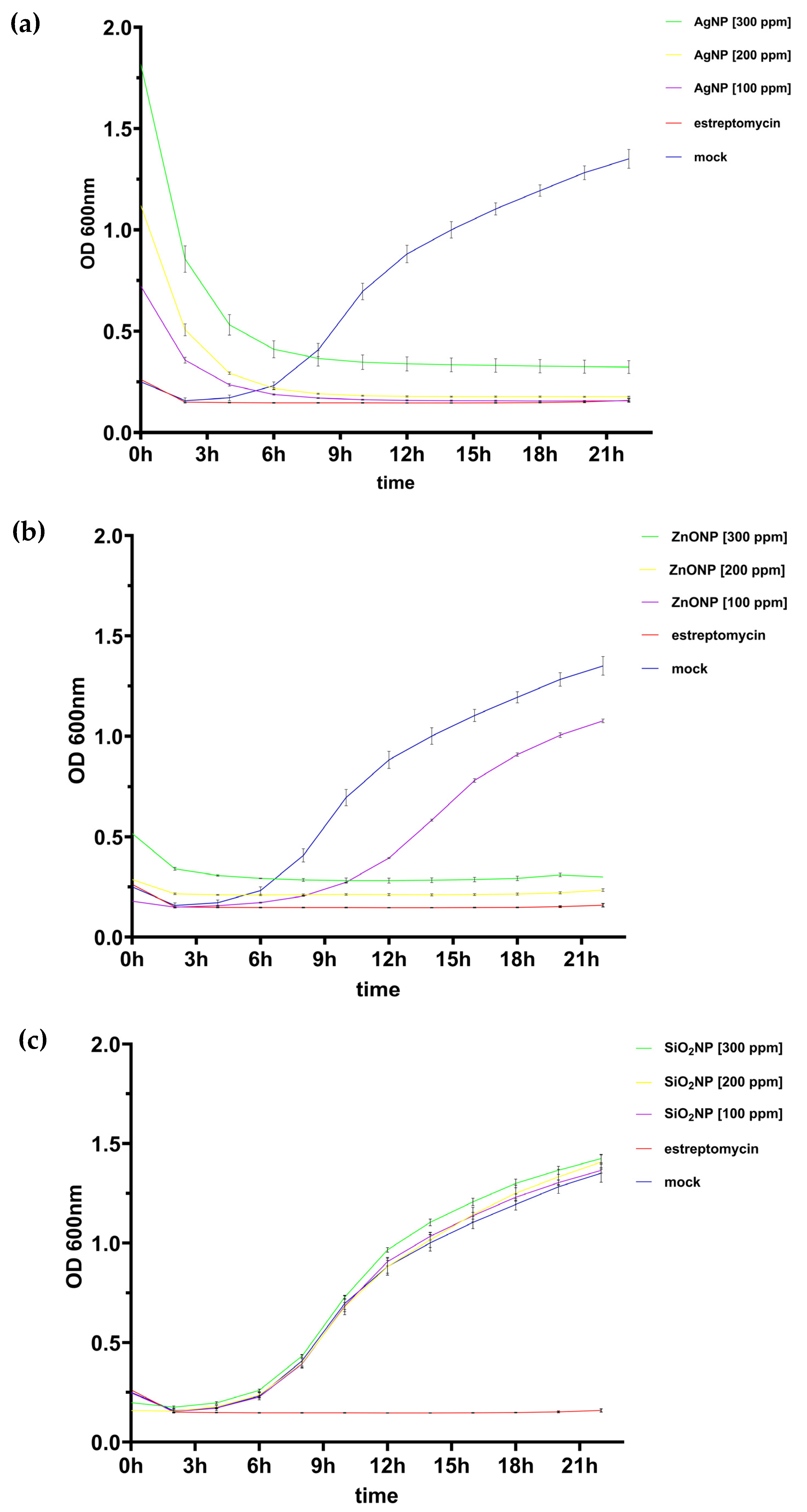
3.1.1. Growth Kinetics of Pseudomonas syringae pv. tomato

To determine whether AgNPs, ZnONPs, and SiO2NPs inhibit Pseudomonas syringae pv. tomato growth in vitro, a growth kinetics assay was conducted using low, medium, and high nanoparticle concentrations, comparable to the methods used in previous studies. Streptomycin served as the negative control. Results indicated that Ag nanoparticles inhibited bacterial growth starting at 100 ppm (Figure 1a). ZnO nanoparticles showed inhibition at 200 and 300 ppm (Figure 1b), while SiO2 showed no inhibitory effect at any concentration tested (Figure 1c). This preliminary experiment helped establish the range of concentrations for the MIC experiment.

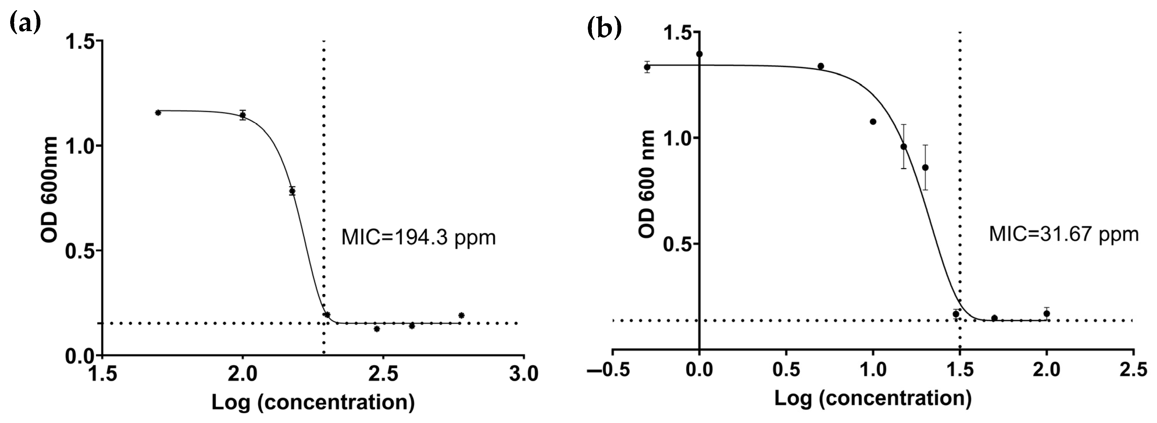
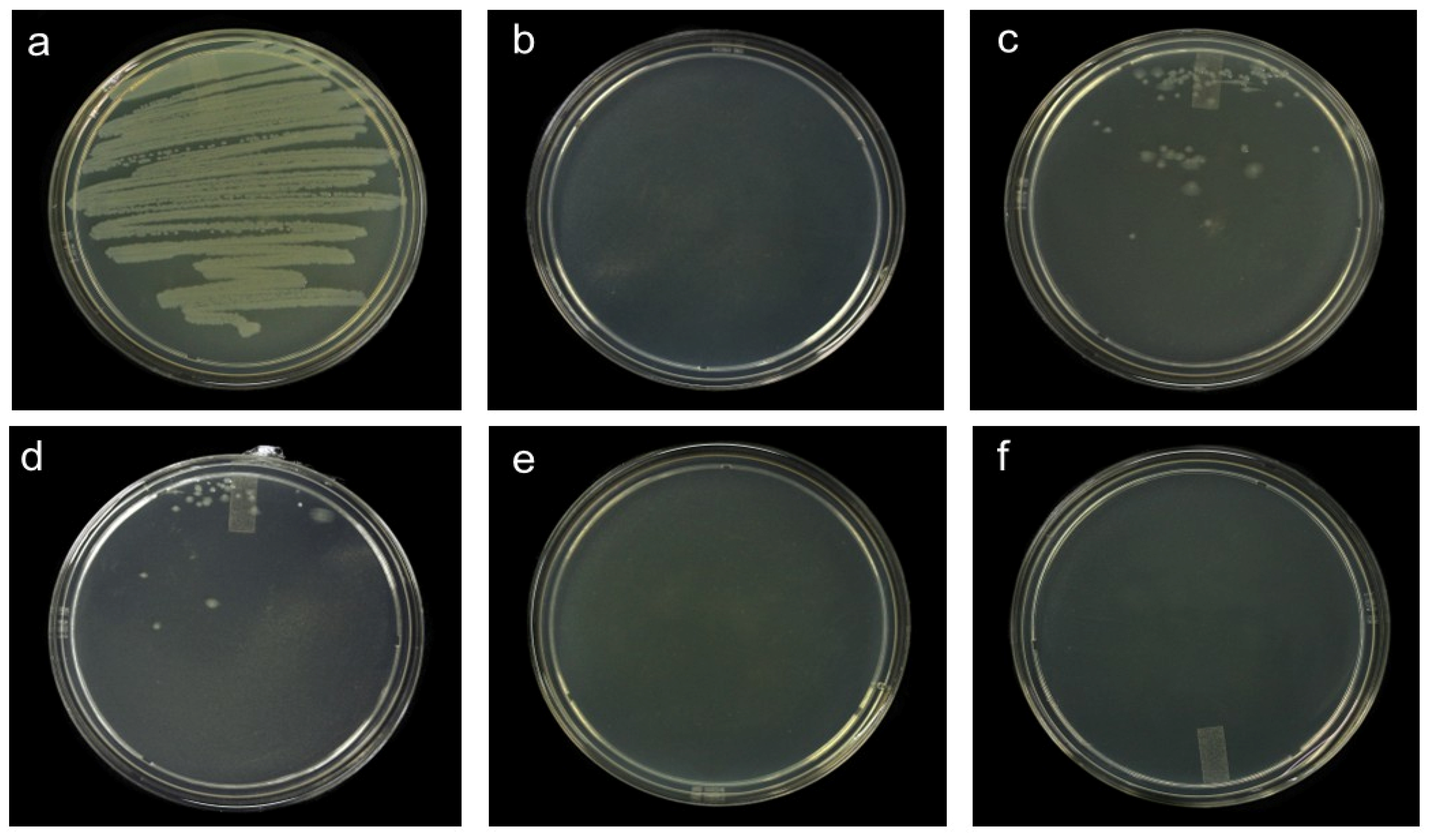
3.1.2. Minimum Inhibitory Concentration (MIC) and Minimum Lethal Concentration (MLC)

The MIC was defined as the lowest nanoparticle concentration causing a reduction in optical density at 600 nm after 24 h of exposure. Results showed that P. syringae pv. tomato growth was inhibited starting at 194.3 ppm for ZnONPs (Figure 2a) and 31.67 ppm for AgNPs (Figure 2b). Upon plating P. syringae pv. tomato on King’s B medium after 24 h of exposure to MIC-level ZnONPs and AgNPs, bacterial growth was observed (Figure 3c,d), suggesting a bacteriostatic rather than bactericidal effect. A colony forming unit (CFU) count test showed that after 24 h of incubation, the P. syringae pv. tomato without nanoparticles displayed a concentration of 3.9 × 10⁸ ± 141.6 CFU, while the AgNP and ZnONP treatments showed a concentration of 1.16 × 10⁴ ± 48.2 and 1.6 × 10⁶ ± 296.76 CFU respectively, indicating a significant reduction in bacterial growth (Table 2).
At concentrations equal to or higher than 100 ppm for AgNPs and 1000 ppm for ZnONPs, cell death was evident, as no bacterial growth was observed upon plating (Figure 3).

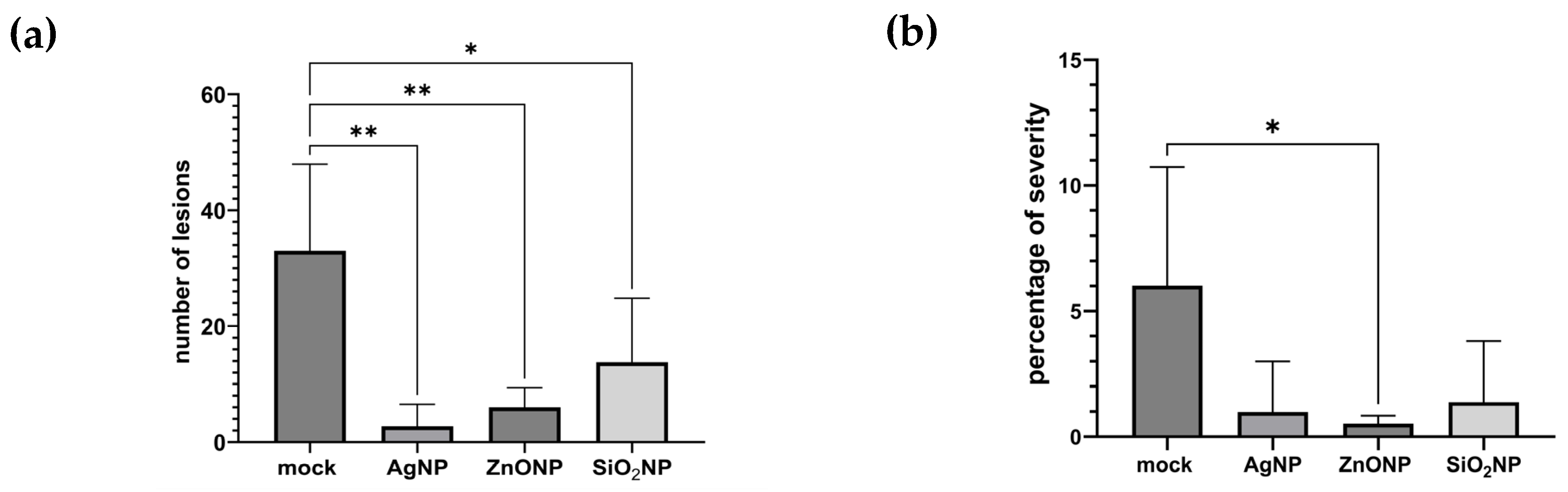
3.1.3. Symptoms, Lesion Count, and Severity

Six days post-inoculation, symptoms began to appear. Leaves showed small dark brown to black lesions surrounded by chlorotic halos, consistent with symptoms caused by Pseudomonas syringae pv. tomato [19]. Treatments with nanoparticles resulted in fewer lesions compared to those in the control (Figure 4a). Statistical analyses showed that ZnO nanoparticles significantly reduced severity percentage compared to that of the control (Figure 4b).

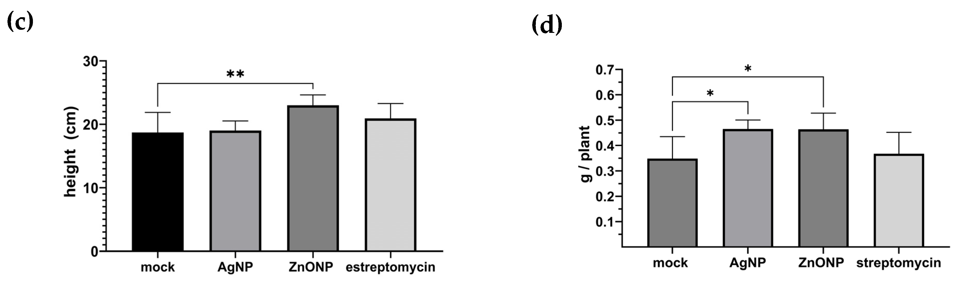
3.1.4. Plant Height and Dry Weight

An increase in plant height was recorded for ZnONP treatment compared to the control; no such effect was observed for AgNPs and streptomycin (Figure 4c). Further, dry weight was higher in ZnONP and AgNP treatments (Figure 4d).

3.2. Pathosystem Two

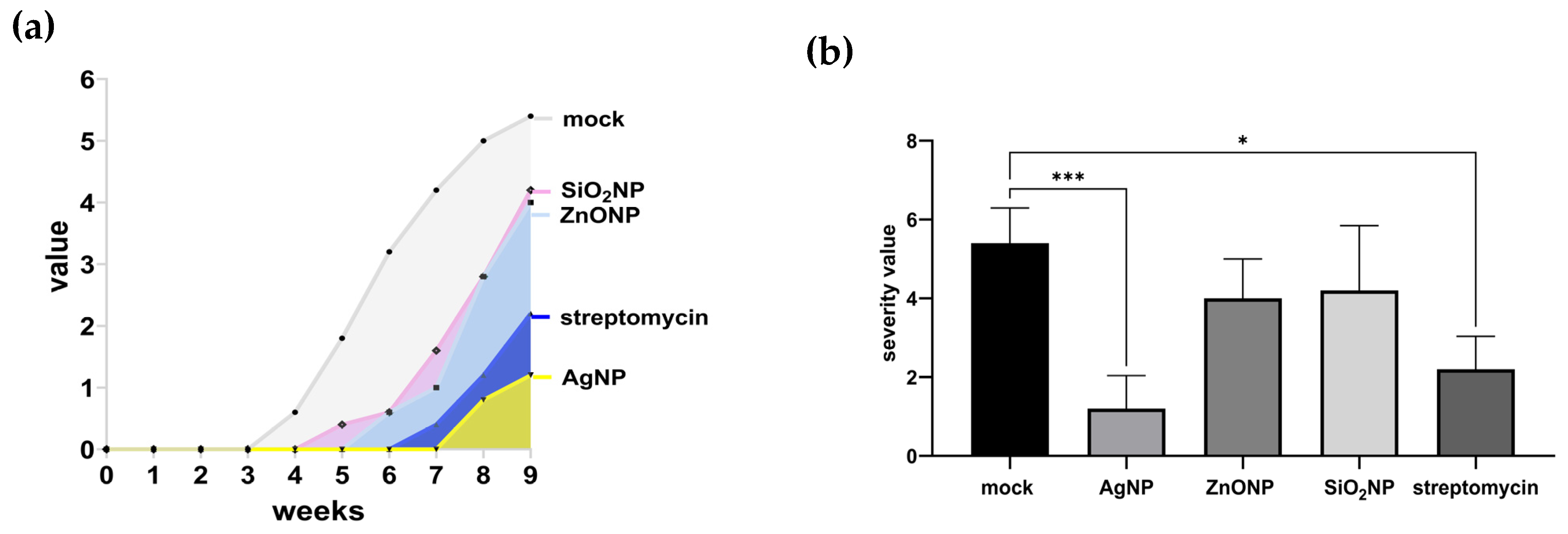
3.2.1. Symptom Progression (Area Under the Disease Progression Curve) and Severity

Thirty days after inoculation with B. cockerelli, symptoms began to manifest, particularly in controls, showing typical disease features such as stunted growth, generalized chlorosis, necrotic interveinal lesions, reduced leaf area, and young leaf deformation. In order to determine the effect of nanoparticles on pathosystem two, the progression of chili variegation over time was evaluated according to the proposed severity scale (Table 1), and the area under the disease progression curve (AUDPC) was calculated. The AUDPC value was 18.4 in control treatment, indicating the highest symptom progression in our evaluated conditions. The AUDPC values of SiO2NPs and ZnONPs were 7.5 and 6.4, respectively, indicating an intermediate symptom progression. The lowest symptom progression was observed in AgNP treatment followed, by streptomycin treatment, with AUDPC values of 1.4 and 2.7, respectively (Figure 5a). Final severity was analyzed using the non-parametric Kruskal–Wallis test. A significant difference was observed between Ag-treated and control plants. Treatments with ZnO and SiO2 nanoparticles showed no significant differences compared to results for the control. Streptomycin showed an effect comparable to that of Ag nanoparticles (Figure 5b).

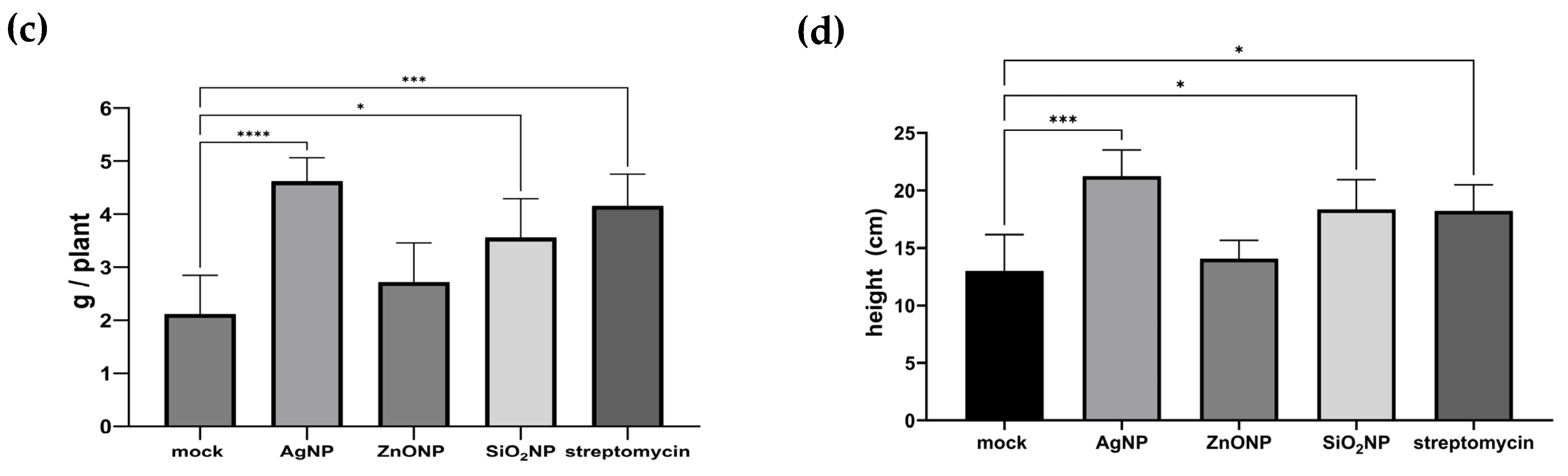
3.2.2. Plant Height and Dry Weight

Regarding dry weight, plants treated with AgNPs and streptomycin showed the highest values, followed by those treated with SiO2NPs. No significant differences were found between ZnONP treatment and the control (Figure 5c). Statistical analysis indicates that AgNP, SiO2NP, and streptomycin treatments significantly increased plant height compared to that of the control. In contrast, ZnONPs showed no significant differences compared to the control (Figure 5d).

4. Discussion

4.1. Pathosystem One

In vitro assays indicate that SiO2 nanoparticles at 300 ppm do not exert a bacteriostatic or bactericidal effect against Pseudomonas syringae pv. tomato. This observation aligns with previous findings. An in vitro assay indicated that SiO2NPs at 100 ppm did not inhibit the growth of Pseudomonas syringae [20]. Another work reported no growth inhibition of Ralstonia solanacearum after 24 h of exposure to 250 ppm SiO2NPs in 96-well plates [21]. In contrast, a study showed that Whatman filter paper disks impregnated with 200 ppm SiO2NP suspension inhibited bacterial growth of different pathogens, including P. syringae pv. tomato, R. solanacearum Xanthomonas campestris pv. vesicatoria, and Pectobacterium carotovorum subsp. carotovorum [22]. This discrepancy could be due to differences in methodologies, such as disk diffusion versus well-plate exposure, experimental conditions, or specific nanoparticle characteristics. This reinforces the need to standardize experimental conditions when evaluating the antimicrobial activity of SiO2 nanoparticles.
ZnO nanoparticles at 194.3 ppm exhibited a bacteriostatic effect on P. syringae pv. tomato, inhibiting its growth without causing total cell death; since the minimum lethal concentration was recorded at 1000 ppm. Our results are consistent with those of previous studies, where the inhibitory effect of ZnONPs on Pseudomonas syringae pathovars at different concentrations has been reported. A paper disc dipped in 100, 200, and 300 ppm ZnONPs inhibited the growth of P. syringae pv. tomato [23,24]. Using the same methodology, it was observed that 250 and 500 ppm ZnONPs inhibited the growth of P. syringae pv. aptata [25]. Recently, a microplate broth-dilution assay showed that ZnONPs suppressed bacterial growth of P. syringae pv. tomato in a concentration range of 30 to 100 ppm [26]. Although these studies did not test whether bacteria incubated with ZnONPs had the capacity to grow if transferred to a ZnONP-free media, the growth kinetics shown by Orfei et al. suggests that the observed effect was bacteriostatic rather than bactericidal, similar to that observed in this work. However, it has also been reported that rod-shaped ZnONPs at 100, 250, and 500 ppm did not inhibit biofilm formation or bacterial growth of nine different pathovars of P. syringae [27]. Again, the difference in results may be due to specific nanoparticle characteristics, differences in methodologies, or experimental conditions.
Since the 19th century, AgNPs have shown remarkable antimicrobial activity, in both ionic and nanoparticle forms, as a medical and antibacterial agent [28]. In the last decade, several studies have reported that AgNPs inhibit the growth of P. syringae over a wide concentration range from 1 ppm to 3500 ppm. In the well-diffusion assay, inhibition of bacterial growth was observed between 12.5 and 100 ppm [29,30], while in the disk diffusion assay, the inhibition range was reported between 100 and 3500 ppm [31,32]. Furthermore, through microplate assays, MICs between 1 and 40 ppm have been reported [33,34,35]. During this work, an MIC of 31.67 ppm was calculated for AgNPs, which is consistent with the results in previous works. Although the aforementioned studies did not determine the minimum lethal concentration, this parameter was determined by Danish et al., obtaining a value of 400 ppm, four times higher than the MLC of 100 ppm recorded in this work.
In tomato plants treated with 250 ppm ZnONPs, AgNPs, or SiO2, a reduction in the lesions number and the percentage of severity was observed. However, regarding to percentage of severity, the difference between AgNPs and SiO2 was not significant compared to the control. These two treatments exhibited a higher standard deviation, indicating greater variability between experimental units. This prevents a clear assessment of the effect of these treatments. The observed positive effect of different nanoparticles on P. syringae-induced symptoms in tomato in this study aligns with previously published findings. It has been reported that tomato plants treated with ZnONPs showed a reduction in disease severity and bacterial proliferation through direct antibacterial activity and by regulating the expression of plant defense genes [23,26]. ZnONPs can also reduce disease severity caused by Pseudomonas syringae pv. tabaci in tobacco. The mode of action involves inhibition or alteration of motility, biofilm formation, metabolism, virulence, and cell envelope integrity of the pathogen [36]. Furthermore, beetroot plants treated with ZnONPs showed a reduction in disease severity caused by P. syringae pv. aptata and an increase in growth, as well as in the content of chlorophyll and carotenoids [25].
In the case of AgNPs, it has been reported that peach trees treated with 100 ppm of AgNPs showed a reduction in the severity of the disease caused by P. syringae pv. syringae [34]. Another study showed that treatments with 5 and 100 ppm of AgNPs significantly reduced the damage caused by P. syringae in Arabidopsis thaliana and tomato plants, respectively, compared to the control [37,38]. Also, it was shown that the symptoms associated with P. syringae pv. tabaci are significantly reduced in Nicotiana benthamiana leaves pretreated with 1.2 ppm of AgNPs. The authors show that the effect of AgNPs on the pathogen can occur through direct antimicrobial activity or indirectly by inducing the expression of defense genes and modifying antioxidant enzymatic activity [39]. In this study, in vitro data suggest that ZnONPs and AgNPs may be exerting their effect on P. syringae pv. tomato directly, since both nanoparticles displayed a bacteriostatic effect. These types of nanoparticles can interact directly with the bacterial membrane through electrostatic attraction between negatively charged bacterial cells and positively charged nanoparticles. This interaction modifies membrane permeability, leading to the leakage of cellular components. Furthermore, they can cross the membrane and destabilize molecules such as DNA and proteins, affecting essential cellular functions [40,41,42,43]. Thus, it is hypothesized that Ag and ZnO nanoparticles interact directly with P. syringae pv. tomato on leaf surfaces, exerting a bacteriostatic effect that reduces lesion number and severity. However, according to in vivo results, we cannot rule out the possibility that the ZnONP and AgNP application activates defense mechanisms in plants, as previously reported [23,26,39]. Therefore, the effects observed in this work may be the result of both direct and indirect mechanisms.
Regarding SiO2NPs, a study reported that applications of SiO2NPs at a concentration range of 25 to 1600 ppm indirectly suppress P. syringae growth in A. thaliana by promoting systemic acquired resistance [20]. Also, pea plants treated with 100 ppm SiO2NPs showed a decrease in the severity of symptoms caused by P. syringae pv. pisi compared to the control. This effect was greater when the SiO2NPs treatment was combined with Rhizobium leguminosarum inoculation [44]. Additionally, a treatment with 200 ppm of SiO2NPs in tomato plants partially suppressed the disease caused by P. syringae pv. tomato, increasing enzymatic antioxidant activities, as well as the contents of chlorophyll, carotenoids, and proline [24]. Moreover, it has been reported that the exogenous application of SiO2NPs at 150 ppm reduced severity of bacterial leaf spot in sweet pepper caused by Xanthomonas vesicatoria [45], while treatment with SiO2NPs at 450 and 650 ppm in tomato reduced wilt severity caused by Ralstonia Solanacearum [46]. In the case of SiO2NPs, the in vitro results indicate that they have no antimicrobial activity. Therefore, the effect on the lesion number caused by P. syringae pv. syringae observed in this work suggests that this occurs through an indirect mechanism [20,24].
An increase in plant height and dry weight was recorded for ZnO treatments, which aligns with Zn’s role as an essential micronutrient. As a regulatory cofactor and structural component of several enzymes and proteins, Zn is fundamental in plant metabolism, including photosynthesis, phytohormone synthesis, and antioxidant systems [47].

4.2. Pathosystem Two

To our knowledge, this is the first report demonstrating that nanoparticles can delay symptom progression and reduce disease severity in this pathosystem. The use of nanoparticles against non-culturable bacteria has been scarcely explored in scientific literature. The non-cultivable nature of Candidatus Liberibacter solanacearum and other phloem-limited bacteria represents a major limitation for in vitro study, hindering the development of effective control strategies [48,49].
Studies have shown that AgNPs [50,51,52], ZnONPs [53,54,55], and SiO2NPs [56,57] can enter foliar tissue through stomata and subsequently translocate to sieve elements of the phloem [58], the site parasitized by CaLso. Thus, a possible explanation for the observed effect could relate to the bactericidal activity of nanoparticles within the vascular system. However, this hypothesis requires experimental validation. Future studies should focus on examining the interaction between nanoparticles and CaLso within the phloem, including localization, accumulation, and modes of action in plant vascular tissues. Additionally, statistically significant increases in plant height and dry weight were observed in plants treated with AgNPs. This finding aligns with those in ref. [39], which reported increased dry weight in Nicotiana benthamiana treated with Ag nanoparticles and inoculated with Pseudomonas syringae pv. tabaci. Both our findings and previous reports support the hypothesis that Ag nanoparticles can stimulate vegetative growth under biotic stress conditions.
ZnONPs and SiO2NPs delayed symptom onset; however, after nine weeks of evaluation, severity levels showed no statistically significant differences compared to the control. Despite this, SiO2NP treatment increased the height and dry weight in CaLso-infected plants compared to the control. The positive effect on these growth parameters is related to previous reports in the pea-P. syringae, tomato-P. syringae, and tomato-R. solanacearum pathosystems [22,44,46]. On the other hand, the effect of ZnO nanoparticles on non-culturable bacteria like CaLso has been scarcely studied. A ZnO-based nanoformulation has been evaluated in vitro against Liberibacter crescens (the closest model organism to CaLso), and a bacteriostatic effect starting at 150 ppm and a bactericidal effect above 175 ppm was observed [59]. Therefore, results from this study must be interpreted cautiously, as only a single concentration was tested using a specific application route and two pre-inoculation applications. For future research, different concentrations, administration routes, and application schemes are suggested to optimize ZnO nanoparticle efficacy against CLso.
Following a comprehensive review of the literature on nanoparticle use in controlling various plant pathogens, we identified opportunities to explore their potential in managing diseases caused by non-culturable bacteria. Examples include Candidatus Liberibacter solanacearum (this study); Ca. Liberibacter asiaticus; Ca. Liberibacter americanus; and Ca. Liberibacter africanus, associated with citrus greening, a devastating disease that has caused massive losses in global citrus production [49]. Research in this line will help develop new methodologies and generate key technical information, such as optimal application strategies, timing, and effective concentrations. This information will contribute to the design of innovative bacterial disease management strategies, improving the efficiency of phytosanitary management programs.

5. Conclusions

Results indicate that ZnONPs nanoparticles have a positive effect on mitigating damage caused by P. syringae pv. tomato in tomato plants, significantly reducing lesion number and severity while promoting plant growth. This highlights the promising potential of ZnO as a phytosanitary management alternative in this pathosystem. On the other hand, although AgNPs and SiONPs only reduce the number of lesions, they could help mitigate the damage caused by this pathogen.
Regarding the second pathosystem, we show that AgNPs are more effective in reducing chili variegation severity compared to ZnONPs and SiO2NPs. Moreover, AgNPs and SiO2NPs enhanced plant growth by significantly increasing plant height and dry weight. These findings suggest that AgNPs and SiO2NPs not only exhibit potential as phytosanitary control agents but may also act as bio-stimulants under biotic stress conditions. In addition, future work could include treatments with combined applications of nanoparticles to test their effectiveness against these or other pathogens.

Author Contributions

E.A.R.-R. and V.M.Z.-M. performed the assays, prepared the figures, and wrote the manuscript; V.M.Z.-M. and R.I.R.-M. conceived the study; E.A.R.-R. performed the statistical analysis of the data; E.A.R.-R., V.M.Z.-M., R.I.R.-M., D.L.O.-M. and G.V.-J. performed the manuscript review and organized the overall experimental design. All authors have read and agreed to the published version of the manuscript.

Funding

This research was funded by the Investigadoras e Investigadores por México program (SECIHTI), grant number 5016; and SECIHTI, project number CF-2023-G-728.

Data Availability Statement

The original contributions presented in this study are included in the article. Further inquiries can be directed to the corresponding authors.

Acknowledgments

E.A.R.-R. is grateful for financial support provided by Consejo Nacional de Humanidades, Ciencia y Tecnología (CONAHCYT, Mexico), now Secretaría de Ciencias, Humanidades, Tecnología, e Innovaciòn (SECIHTI).

Conflicts of Interest

The authors declare no conflicts of interest.

References

  1. FAO. Agricultural Production Statistics 2010–2023; FAO: Rome, Italy, 2024. [Google Scholar]
  2. FAO. FAOSTAT: Statistical Database. Available online: https://www.fao.org/faostat/en/ (accessed on 9 June 2025).
  3. Griffin, K.; Gambley, C.; Brown, P.; Li, Y. Copper-tolerance in Pseudomonas syringae pv. tomato and Xanthomonas spp. and the control of diseases associated with these pathogens in tomato and pepper. A systematic literature review. Crop Prot. 2017, 96, 144–150. [Google Scholar] [CrossRef]
  4. El-Fatah, B.E.S.A.; Imran, M.; Abo-Elyousr, K.A.M.; Mahmoud, A.F. Isolation of Pseudomonas syringae pv. tomato strains causing bacterial speck disease of tomato and marker-based monitoring for their virulence. Mol. Biol. Rep. 2023, 50, 4917–4930. [Google Scholar] [CrossRef] [PubMed]
  5. Camacho-Tapia, M.; Rojas-Martínez, R.I.; Zavaleta-Mejía, E.; Hernández-Deheza, M.G.; Carrillo-Salazar, J.A.; Rebollar-Alviter, A.; Ochoa-Martínez, D.L. Aetiology of Chili Pepper Variegation from Yurecuaro, Mexico. J. Plant Pathol. 2011, 93, 331–335. [Google Scholar]
  6. Velásquez-Valle, R.; Reveles-Torres, L.R.; Mena-Covarrubias, J.; Salas-Muñoz, S.; Mauricio-Castillo, J.A. Outbreak of Candidatus Liberibacter solanacearum in Dried Chile Pepper in Durango, Mexico. Agrofaz 2014, 14, 93–104. [Google Scholar]
  7. Boholm, M.; Arvidsson, R. A Definition Framework for the Terms Nanomaterial and Nanoparticle. Nanoethics 2016, 10, 25–40. [Google Scholar] [CrossRef]
  8. Joudeh, N.; Linke, D. Nanoparticle Classification, Physicochemical Properties, Characterization, and Applications: A Comprehensive Review for Biologists. J. Nanobiotechnol. 2022, 20, 262. [Google Scholar] [CrossRef]
  9. Khan, Y.; Sadia, H.; Zeeshan, S.; Shah, A.; Khan, M.N.; Shah, A.A.; Ullah, N.; Ullah, M.F.; Bibi, H.; Bafakeeh, O.T.; et al. Classsification, Synthetic, and Characterization Approaches to Nanoparticles, and Their Applications in Various Fields of Nanotechnology: A Review. Catalysts 2022, 12, 1386. [Google Scholar] [CrossRef]
  10. Abdelkhalek, A.; Al-Askar, A.A. Green synthesized ZnO nanoparticles mediated by Mentha spicata extract induce plant systemic resistance against Tobacco Mosaic Virus. Appl. Sci. 2020, 10, 5504. [Google Scholar] [CrossRef]
  11. Morones, J.R.; Elechiguerra, J.L.; Camacho, A.; Holt, K.; Kouri, J.B.; Ramírez, J.T.; Yacaman, M.J. The Bactericidal Effect of Silver Nanoparticles. Nanotechnology 2005, 16, 2346–2353. [Google Scholar] [CrossRef]
  12. Kumar, A.; Choudhary, A.; Kaur, H.; Guha, S.; Mehta, S.; Husen, A. Potential Applications of Engineered Nanoparticles in Plant Disease Management: A Critical Update. Chemosphere 2022, 295, 133798. [Google Scholar] [CrossRef]
  13. Dutta, P.; Kumari, A.; Mahanta, M.; Upamanya, G.K.; Heisnam, P.; Borua, S.; Kaman, P.K.; Mishra, A.K.; Mallik, M.; Muthukrishnan, G.; et al. Nanotechnological Approaches for Management of Soil-Borne Plant Pathogens. Front. Plant Sci. 2023, 14, 1136233. [Google Scholar] [CrossRef] [PubMed]
  14. Stewart, E.J. Growing unculturable bacteria. J. Bacteriol. 2012, 194, 4151–4160. [Google Scholar] [CrossRef]
  15. Lambert, R.; Pearson, J. Effect of Storage Conditions on the Quality Characteristics of Onion. J. Appl. Microbiol. 2000, 88, 784–790. [Google Scholar] [CrossRef]
  16. Green, L.H.; Goldman, E. Practical Handbook of Microbiology; Elsevier: Boca Raton, FL, USA, 2021. [Google Scholar]
  17. Bharti, D.B.; Bharati, A.V. Synthesis of ZnO Nanoparticles Using a Hydrothermal Method and a Study Its Optical Activity. Luminescence 2016, 32, 317–320. [Google Scholar] [CrossRef]
  18. Madden, L.V.; Hughes, G.; van den Bosch, F. The Study of Plant Disease Epidemics; APS Press: St. Paul, MN, USA, 2007. [Google Scholar]
  19. Valenzuela, M.; Fuentes, B.; Alfaro, J.F.; Galvez, E.; Salinas, A.; Besoain, X.; Seeger, M. First report of bacterial speck caused by Pseudomonas syringae pv. tomato race 1 affecting tomato in different regions of Chile. Plant Dis. 2022, 106, 1979. [Google Scholar] [CrossRef]
  20. El-Shetehy, M.; Moradi, A.; Maceroni, M.; Reinhardt, D.; Petri-Fink, A.; Rothen-Rutishauser, B.; Mauch, F.; Schwab, F. Silica nanoparticles enhance disease resistance in Arabidopsis plants. Nat. Nanotechnol. 2021, 16, 344–353. [Google Scholar] [CrossRef]
  21. Ijaz, M.; Lv, L.; Ahmed, T.; Noman, M.; Manan, A.; Ijaz, R.; Hafeez, R.; Shahid, M.S.; Wang, D.; Ondrasek, G.; et al. Immunomodulating melatonin-decorated silica nanoparticles suppress bacterial wilt (Ralstonia solanacearum) in tomato (Solanum lycopersicum L.) through fine-tuning of oxidative signaling and rhizosphere bacterial community. J. Nanobiotechnol. 2024, 22, 617. [Google Scholar] [CrossRef]
  22. Parveen, A.; Siddiqui, Z.A. Impact of Silicon Dioxide Nanoparticles on Growth, Photosynthetic Pigments, Proline, Activities of Defense Enzymes and Some Bacterial and Fungal Pathogens of Tomato. Vegetos 2022, 35, 83–93. [Google Scholar] [CrossRef]
  23. Elsharkawy, M.; Derbalah, A.; Hamza, A.; El-Shaer, A. Zinc oxide nanostructures as a control strategy of bacterial speck of tomato caused by Pseudomonas syringae in Egypt. Environ. Sci. Pollut. Res. 2020, 27, 19049–19057. [Google Scholar] [CrossRef]
  24. Parveen, A.; Siddiqui, Z.A. Zinc Oxide Nanoparticles Affect Growth, Photosynthetic Pigments, Proline Content and Bacterial and Fungal Diseases of Tomato. Arch. Phytopathol. Plant Prot. 2021, 54, 1519–1538. [Google Scholar] [CrossRef]
  25. Siddiqui, Z.A.; Khan, M.R.; Abd_Allah, E.F.; Parveen, A. Titanium dioxide and zinc oxide nanoparticles affect some bacterial diseases, and growth and physiological changes of beetroot. Int. J. Veg. Sci. 2019, 25, 409–430. [Google Scholar] [CrossRef]
  26. Orfei, B.; Scian, A.; Del Buono, D.; Paglialunga, M.; Tolisano, C.; Priolo, D.; Moretti, C.; Buonaurio, R. Biogenic zinc oxide nanoparticles protect tomato plants against Pseudomonas syringae pv. tomato. Horticulturae 2025, 11, 431. [Google Scholar] [CrossRef]
  27. Loera-Muro, A.; Contreras-Arvizu, G.I.; Palestino, G.; Hern, L.G.; Luis, P.; Daniel, L.; Figueroa, M.; Hern, L. Effect of zinc oxide nanorods on biofilms of different pathovars of Pseudomonas syringae. Mater. Lett. 2024, 376, 137334. [Google Scholar] [CrossRef]
  28. Yang, C.; Zhong, Y.; Powell, C.A.; Doud, M.S.; Duan, Y.; Huang, Y.; Zhang, M. Antimicrobial Compounds Effective against Candidatus Liberibacter Asiaticus Discovered via Graft-Based Assay in Citrus. Sci. Rep. 2018, 8, 17288. [Google Scholar] [CrossRef]
  29. Velmurugan, P.; Lee, S.M.; Iydroose, M.; Lee, K.J.; Oh, B.T. Pine Cone-Mediated Green Synthesis of Silver Nanoparticles and Their Antibacterial Activity against Agricultural Pathogens. Appl. Microbiol. Biotechnol. 2013, 97, 361–368. [Google Scholar] [CrossRef] [PubMed]
  30. Das, A.; Deb, C.; Lahiri, S.; Kundu, S. Antimicrobial Effect of Green Synthesized Silver Nanoparticles on Plant Pathogenic Bacteria Pseudomonas syringae pv. tomato. J. Mycopathol. Res. 2023, 61, 2583–6315. [Google Scholar]
  31. Khan, A.U.; Khan, M.; Khan, M.M. Antifungal and antibacterial assay by silver nanoparticles synthesized from aqueous leaf extract of Trigonella foenum-graecum. Bionanoscience 2019, 9, 597–602. [Google Scholar] [CrossRef]
  32. Iqbal, M.; Raja, N.I.; Khan, S.A.; Ali, A.; Hanif, A.; Hussain, M.; Anwar, T.; Qureshi, H.; Saeed, M.; Rauf, A. Evaluation of Green Synthesized Silver Nanoparticles against Bacterial Pathogenic Strains of Plants. Pak. J. Bot. 2023, 55, 1967–1972. [Google Scholar] [CrossRef] [PubMed]
  33. Manosalva, N.; Tortella, G.; Cristina Diez, M.; Schalchli, H.; Seabra, A.B.; Durán, N.; Rubilar, O. Green Synthesis of Silver Nanoparticles: Effect of Synthesis Reaction Parameters on Antimicrobial Activity. World J. Microbiol. Biotechnol. 2019, 35, 88. [Google Scholar] [CrossRef]
  34. Shahryari, F.; Rabiei, Z.; Sadighian, S. Antibacterial activity of synthesized silver nanoparticles by sumac aqueous extract and silver-chitosan nanocomposite against Pseudomonas syringae pv. syringae. J. Plant Pathol. 2020, 102, 469–475. [Google Scholar] [CrossRef]
  35. Khan, M.A.; Dzimitrowicz, A.; Prusinski, M.; Motyka-Pomagruk, A.; Jamroz, P.; Cyganowski, P.; Łojkowska, E.; Sledz, W.; Pohl, P. Coffee-mediated synthesis of silver nanoparticles showing antibacterial properties against economically important phytopathogens from the genus Pseudomonas. Colloids Surf. A Physicochem. Eng. Asp. 2025, 718, 136802. [Google Scholar] [CrossRef]
  36. Fan, G.; Xiao, Q.; Li, Q.; Xia, Y.; Feng, H.; Ma, X.; Cai, L.; Sun, X. Antimicrobial mechanisms of ZnO nanoparticles to phytopathogen Pseudomonas syringae: Damage of cell envelope, suppression of metabolism, biofilm and motility, and stimulation of stomatal immunity on host plant. Pestic. Biochem. Physiol. 2023, 194, 105455. [Google Scholar] [CrossRef]
  37. Danish, M.; Shahid, M.; Ahamad, L.; Raees, K.; Atef Hatamleh, A.; Al-Dosary, M.A.; Mohamed, A.; Al-Wasel, Y.A.; Singh, U.B.; Danish, S. Nano-pesticidal potential of Cassia fistula leaf synthesized silver nanoparticles (Ag@CfL-NPs): Deciphering the phytopathogenic inhibition and growth augmentation in Solanum lycopersicum. Front. Microbiol. 2022, 13, 985852. [Google Scholar] [CrossRef]
  38. Paul, K.; Großkinsky, D.K.; Vass, I.; Roitsch, T. Silver nanoparticles affect Arabidopsis thaliana leaf tissue integrity and suppress Pseudomonas syringae infection symptoms in a dose-dependent manner. Bionanoscience 2022, 12, 332–338. [Google Scholar] [CrossRef]
  39. Jiang, L.; Xiang, S.; Lv, X.; Wang, X.; Li, F.; Liu, W.; Liu, C.; Ran, M.; Huang, J.; Xu, X.; et al. Biosynthesized silver nanoparticles inhibit Pseudomonas syringae pv. tabaci by directly destroying bacteria and inducing plant resistance in Nicotiana benthamiana. Phytopathol. Res. 2022, 4, 48. [Google Scholar] [CrossRef]
  40. Sondi, I.; Salopek-Sondi, B. Silver nanoparticles as antimicrobial agent: A case study on E. coli as a model for gram-negative bacteria. Colloid Interface Sci. 2004, 275, 177–182. [Google Scholar] [CrossRef] [PubMed]
  41. Wahab, M.A.; Luming, L.; Matin, M.A.; Karim, M.R.; Aijaz, M.O.; Alharbi, H.F.; Abdala, A.; Haque, R. Silver Micro-Nanoparticle-Based Nanoarchitectures: Synthesis Routes, Biomedical Applications, and Mechanisms of Action. Polymers 2021, 13, 2870. [Google Scholar] [CrossRef]
  42. Hancharova, M.; Halicka-Stępień, K.; Dupla, A.; Lesiak, A.; Sołoducho, J.; Cabaj, J. Antimicrobial Activity of Metal-Based Nanoparticles: A Mini-Review. BioMetals 2024, 37, 773–801. [Google Scholar] [CrossRef]
  43. Casals, E.; Gusta, M.F.; Bastus, N.; Rello, J.; Puntes, V. Silver Nanoparticles and Antibiotics: A Promising Synergistic Approach to Multidrug-Resistant Infections. Microorganisms 2025, 13, 952. [Google Scholar] [CrossRef] [PubMed]
  44. Kashyap, D.; Siddiqui, Z.A. Effect of silicon dioxide nanoparticles and Rhizobium leguminosarum alone and in combination on the growth and bacterial blight disease complex of pea caused by Meloidogyne incognita and Pseudomonas syringae pv. pisi. Arch. Phytopathol. Plant Prot. 2021, 54, 499–515. [Google Scholar] [CrossRef]
  45. Awad-Allah, E.F.A.; Shams, A.H.M.; Helaly, A.A. Suppression of Bacterial Leaf Spot by Green Synthesized Silica Nanoparticles and Antagonistic Yeast Improves Growth, Productivity and Quality of Sweet Pepper. Plants 2021, 10, 1689. [Google Scholar] [CrossRef]
  46. Wang, L.; Pan, T.; Gao, X.; An, J.; Ning, C.; Li, S.; Cai, K. Silica nanoparticles activate defense responses by reducing reactive oxygen species under Ralstonia solanacearum infection in tomato plants. NanoImpact 2022, 28, 100418. [Google Scholar] [CrossRef]
  47. Thounaojam, T.C.; Meetei, T.T.; Devi, Y.B.; Panda, S.K.; Upadhyaya, H. Zinc Oxide Nanoparticles (ZnO-NPs): A Promising Nanoparticle in Renovating Plant Science. Acta Physiol. Plant. 2021, 43, 136. [Google Scholar] [CrossRef]
  48. Levy, J.G.; Gross, R.; Mendoza-Herrera, A.; Tang, X.; Babilonia, K.; Shan, L.; Kuhl, J.C.; Dibble, M.S.; Xiao, F.; Tamborindeguy, C. Lso-HPE1, an Effector of “Candidatus Liberibacter Solanacearum”, Can Repress Plant Immune Response. Phytopathology 2020, 110, 648–655. [Google Scholar] [CrossRef]
  49. Merfa, M.V.; Pérez-López, E.; Naranjo, E.; Jain, M.; Gabriel, D.W.; De La Fuente, L. Progress and Obstacles in Culturing ‘Candidatus Liberibacter Asiaticus’, the Bacterium Associated with Huanglongbing. Phytopathology 2019, 109, 1092–1101. [Google Scholar] [CrossRef]
  50. Larue, C.; Castillo-Michel, H.; Sobanska, S.; Cécillon, L.; Bureau, S.; Barthès, V.; Ouerdane, L.; Carrière, M.; Sarret, G. Foliar exposure of the crop Lactuca sativa to silver nanoparticles: Evidence for internalization and changes in Ag speciation. J. Hazard. Mater. 2014, 264, 98–106. [Google Scholar] [CrossRef]
  51. Li, C.C.; Dang, F.; Li, M.; Zhu, M.; Zhong, H.; Hintelmann, H.; Zhou, D.M. Effects of Exposure Pathways on the Accumulation and Phytotoxicity of Silver Nanoparticles in Soybean and Rice. Nanotoxicology 2017, 11, 699–709. [Google Scholar] [CrossRef] [PubMed]
  52. Wu, J.; Wang, G.; Vijver, M.G.; Bosker, T.; Peijnenburg, W.J.G.M. Foliar versus Root Exposure of AgNPs to Lettuce: Phytotoxicity, Antioxidant Responses and Internal Translocation. Environ. Pollut. 2020, 261, 114117. [Google Scholar] [CrossRef] [PubMed]
  53. Wang, W.N.; Tarafdar, J.C.; Biswas, P. Nanoparticle Synthesis and Delivery by an Aerosol Route for Watermelon Plant Foliar Uptake. J. Nanoparticle Res. 2013, 15, 1417. [Google Scholar] [CrossRef]
  54. Raliya, R.; Nair, R.; Chavalmane, S.; Wang, W.N.; Biswas, P. Mechanistic evaluation of translocation and physiological impact of titanium dioxide and zinc oxide nanoparticles on the tomato (Solanum lycopersicum L.) plant. Metallomics 2015, 7, 1584–1594. [Google Scholar] [CrossRef]
  55. Cantu, J.M.; Ye, Y.; Hernandez-Viezcas, J.A.; Zuverza-Mena, N.; White, J.C.; Gardea-Torresdey, J.L. Tomato Fruit Nutritional Quality Is Altered by the Foliar Application of Various Metal Oxide Nanomaterials. Nanomaterials 2022, 12, 2349. [Google Scholar] [CrossRef] [PubMed]
  56. Hussain, H.I.; Yi, Z.; Rookes, J.E.; Kong, L.X.; Cahill, D.M. Mesoporous Silica Nanoparticles as a Biomolecule Delivery Vehicle in Plants. J. Nanoparticle Res. 2013, 15, 1676. [Google Scholar] [CrossRef]
  57. Zhao, P.; Cao, L.; Ma, D.; Zhou, Z.; Huang, Q.; Pan, C. Translocation, Distribution and Degradation of Prochloraz-Loaded Mesoporous Silica Nanoparticles in Cucumber Plants. Nanoscale 2018, 10, 1798–1806. [Google Scholar] [CrossRef] [PubMed]
  58. Francis, D.V.; Abdalla, A.K.; Mahakham, W.; Sarmah, A.K.; Ahmed, Z.F.R. Interaction of Plants and Metal Nanoparticles: Exploring Its Molecular Mechanisms for Sustainable Agriculture and Crop Improvement. Environ. Int. 2024, 190, 108859. [Google Scholar] [CrossRef] [PubMed]
  59. Naranjo, E.; Merfa, M.V.; Santra, S.; Ozcan, A.; Johnson, E.; Cobine, P.A.; De La Fuente, L. Zinkicide Is a ZnO-Based Nanoformulation with Bactericidal Activity against Liberibacter crescens in Batch Cultures and in Microfluidic Chambers Simulating Plant Vascular Systems. Appl. Environ. Microbiol. 2020, 86, e00788-20. [Google Scholar] [CrossRef]
Figure 1. In vitro growth of Pseudomonas syringae pv. tomato in LB broth and different nanoparticles at 100, 200 ppm, and 300 ppm. (a) All three concentrations tested for AgNPs inhibited bacterial growth, as evidenced by a decrease in optical density, similar to the negative control treated with streptomycin. (b) ZnONPs inhibited bacterial growth at 300 and 200 ppm, while no growth inhibition was observed at 100 ppm. (c) No inhibition of bacterial growth was observed at all SiO2NP concentrations evaluated.
Horticulturae 11 00907 g001
Figure 2. Minimum inhibitory concentration of nanoparticles for Pseudomonas syringae pv. tomato. (a) ZnONPs and (b) AgNPs. Both graphs show two clearly defined asymptotes: the lower graph corresponds to the inhibition of bacterial growth, while the upper graph reflects an increase in optical density, indicating bacterial growth activity. The y-axis represents the absorbance value at 600 nm, and the x-axis represents the logarithm of the nanoparticle concentration.
Horticulturae 11 00907 g002
Figure 3. In vitro growth of Pseudomonas syringae pv. tomato after 24 h of incubation with AgNPs and ZnONPs at the minimum inhibitory concentration and the minimum lethal concentration. (a) Positive control, (b) negative control, (c) AgNPs (31.67 ppm), (d) ZnONPs (194.3 ppm), (e) AgNPs (100 ppm), and (f) ZnONPs (1000 ppm).
Horticulturae 11 00907 g003
Figure 4. (a) Number of lesions and (b) percentage severity on tomato leaves treated with ZnONPs, AgNPs, and SiO2NPs and infected with Pseudomonas syringae pv. tomato. (c) Height and (d) dry weight of tomato plants treated with streptomycin, ZnONP, and AgNP nanoparticles and inoculated with Pseudomonas syringae pv. tomato. The asterisk indicates statistically significant differences between treatments (*: p < 0.05; **: p < 0.05).
Horticulturae 11 00907 g004aHorticulturae 11 00907 g004b
Figure 5. Effect of nanoparticles on symptom progression and severity of chili variegation associated with Candidatus Liberibacter solanacearum. (a) Area under the disease progression curve (AUDPC) of chili variegation in plants treated with different nanoparticles and streptomycin. Severity was evaluated weekly for nine weeks after inoculation with Bactericera cockerelli. (b) Final severity of chili variegation in plants treated with different nanoparticles and streptomycin. (c) Plant height (cm) and (d) total dry weight (g) were recorded at the end of the experiment for the plants treated with nanoparticles and streptomycin. The bars represent the obtained values ± standard error. The asterisk indicates statistically significant differences between treatments, determined by analysis of variance followed by a Tukey test (*: p < 0.05; ***: p < 0.005; ****: p < 0.005).
Horticulturae 11 00907 g005aHorticulturae 11 00907 g005b
Table 1. Severity scale for CaLso-positive pepper plants.
ValueSymptoms
0Asymptomatic plants
11/3 chlorosis (upper third)
22/3 chlorosis and reduced leaf area (upper third)
3Stunting, shortened internodes, flower bud abortion
4Generalized chlorosis and wilting
5Up to 50% defoliation
6More than 50% of defoliation and plant death
Table 2. Bacterial density of Pseudomonas syringae pv. tomato after 24 h of exposure to the minimum inhibitory concentration of ZnONPs and AgNPs. Colony counts were performed 48 h after plating on King’s B medium. Values represent the average of three replicates.
TreatmentUFC/mL
Positive control 3.9 × 10 8 ± 141.6 a
ZnONPs 1.16 × 10 4 ± 48.2 b
AgNPs 1.6 × 10 6 ± 296.76 b
Values followed by the same letter are not significantly different between treatments (Tukey’s test, α = 0.05).
Disclaimer/Publisher’s Note: The statements, opinions and data contained in all publications are solely those of the individual author(s) and contributor(s) and not of MDPI and/or the editor(s). MDPI and/or the editor(s) disclaim responsibility for any injury to people or property resulting from any ideas, methods, instructions or products referred to in the content.

