Review

Recent Advances in In Vitro Floral Induction in Tropical Orchids: Progress and Prospects in Vanilla Species

by
Obdulia Baltazar-Bernal
* and
José Luis Spinoso-Castillo
Colegio de Postgraduados, Campus Córdoba, Carretera Córdoba Veracruz, Amatlán de los Reyes Km 348, Veracruz 94953, Mexico
*
Author to whom correspondence should be addressed.
Horticulturae 2025, 11(7), 829; https://doi.org/10.3390/horticulturae11070829
Submission received: 2 May 2025 / Revised: 1 July 2025 / Accepted: 10 July 2025 / Published: 12 July 2025
(This article belongs to the Special Issue Orchids: Advances in Propagation, Cultivation and Breeding)

Abstract

Orchids and other flowering plants offer a wide range of floral traits. Within the Orchidaceae family, the Vanilla genus is one of the most valued plants in the commercial flavor industry. In vitro biotechnological approaches to Vanilla, such as germplasm conservation, massive propagation, and genetic engineering, have played a key role in breeding programs. There are, however, few studies that elucidate the physiological, molecular, and genetic aspects of vanilla orchid flowering and in vitro induction. This review’s main objective is to provide updated and complete data on in vitro floral induction and flowering of tropical and vanilla orchid species. A bibliographic search was carried out for scientific reports in academic databases (Scopus, Web of Science, PubMed, and ScienceDirect), and a total of 39 documents from 2014 and 2025 were analyzed. This review discusses the most important factors that affect in vitro flowering in Vanilla, including the monopodial genotypes, photoperiod, irradiance, temperature, nutrition, plant growth regulators, explant types, and culture methods. Consequently, this revision incorporates a number of studies on orchid in vitro flowering, with a focus on vanilla species. In conclusion, there still exists limited progress in Vanilla compared to other orchid species; however, the use of biotechnological techniques like in vitro flowering offers a fundamental framework for orchid breeding.

1. Introduction

Plants have two essential growth phases during their life cycle: a vegetative growth phase and a reproductive growth phase [1]. Flowers, the reproductive structures of angiosperms, are fundamental to form fruits and seeds for the completion of their life cycle [2]. Thereby, flowering is a key process of sexual propagation in plants and is regulated by internal (macro- and micro-nutrients, carbon sources, vitamins, plant hormones, etc.) factors and external (photoperiod, light, temperature, humidity, and plant growth regulators (PGRs)). However, seasons and environmental conditions largely limit plants’ ability to flower naturally [3]. In these conditions, biotechnological approaches could play a role in overcoming these restrictions. Besides in vitro propagation aiming for the production of a high genetic quality and phytosanitary plantlets, plant tissue culture (PTC) has several applications. In vitro conditions under control are exceptional for research on in vitro floral induction and flowering, due to the testing of specific environmental factors. In vitro flowering is a singular phenomenon in PTC.
The transition system from the vegetative to the floral stage and a consequently shorter flower production time are made possible by in vitro floral induction [4,5,6]. In vitro flowering offers several advantages, such as incubation of the plantlets in a sterilized and controlled environment and a reduction in the interference of abiotic and biotic stresses. Also, the plantlet’s size is relatively small within the incubation flask, allowing for the supplementation of PGR. Lastly, flowering can be induced if desired; however, different orchid species have varied in vitro flowering times [6]. Therefore, optimizing the orchid flowering duration, flower number, pollination, and production output requires an understanding of the mechanisms and issues influencing flowering [5,6]. The selection of explants for in vitro culture establishment and subsequent regeneration is another crucial aspect of in vitro flowering [7]. Current protocols use flower stalks, shoot and root tips, stem and rhizome segments, and seedlings.
Arabidopsis thaliana and Oryza sativa are model plants for in vitro flowering research and have been studied widely by using PTC, molecular, and genetics methods. Flowering plants like orchids (Orchidaceae) provide a huge diversity of floral morphology, size, color, and fragrance. Therefore, floral morphology is of great interest for research into flower development [8,9]. The success in the propagation and marketing of orchids has been reached via biotechnological and breeding approaches around the world. Vanilla planifolia Jacks. ex Andrews is a Mexican orchid; this species is commercially designated as natural vanilla and is one of the most expensive condiments around the world. However, most Vanilla species, such as V. cribbiana, V. hartii, V. pompona, V. planifolia, V. insignis, V. inodora, V. somai, V. phaeantha, and V. odorata, have been listed as Endangered under criteria A2cd; B2ab(i, ii, iii, v) by the International Union for Conservation of Nature (IUCN). One issue with orchids is that they have extensive juvenile periods before reaching the reproductive stage [9]. In tropical orchids, especially in Vanilla spp., in vitro flowering research is necessary for breeding programs to reduce the juvenile phase and synchrony of the flowering time.
To date, there are limited comprehensive reviews that have been published on the aspects we discuss in this review, including in vitro techniques, approaches, and factors for floral induction and flowering in Vanilla. This review’s primary objective is to present current and comprehensive information on in vitro floral induction and flowering of tropical and vanilla orchid species. Therefore, a bibliographic search was carried out for scientific reports in academic databases published between 2014 and 2025 in order to analyze the in vitro floral induction and flowering of tropical monopodial orchids and vanilla species. The information search was conducted in English, and it was based on the following keywords: in vitro flowering, monopodial orchids, ornamental orchids, vanilla, culture medium, plant growth regulators, and orchid biotechnology. The bibliographic search was conducted in Scopus, Web of Science, PubMed, and ScienceDirect. More than 50 papers were collected; however, only 39 were chosen due to their strong alignment with the goal of this study. The papers were independently analyzed and classified based on their topics.

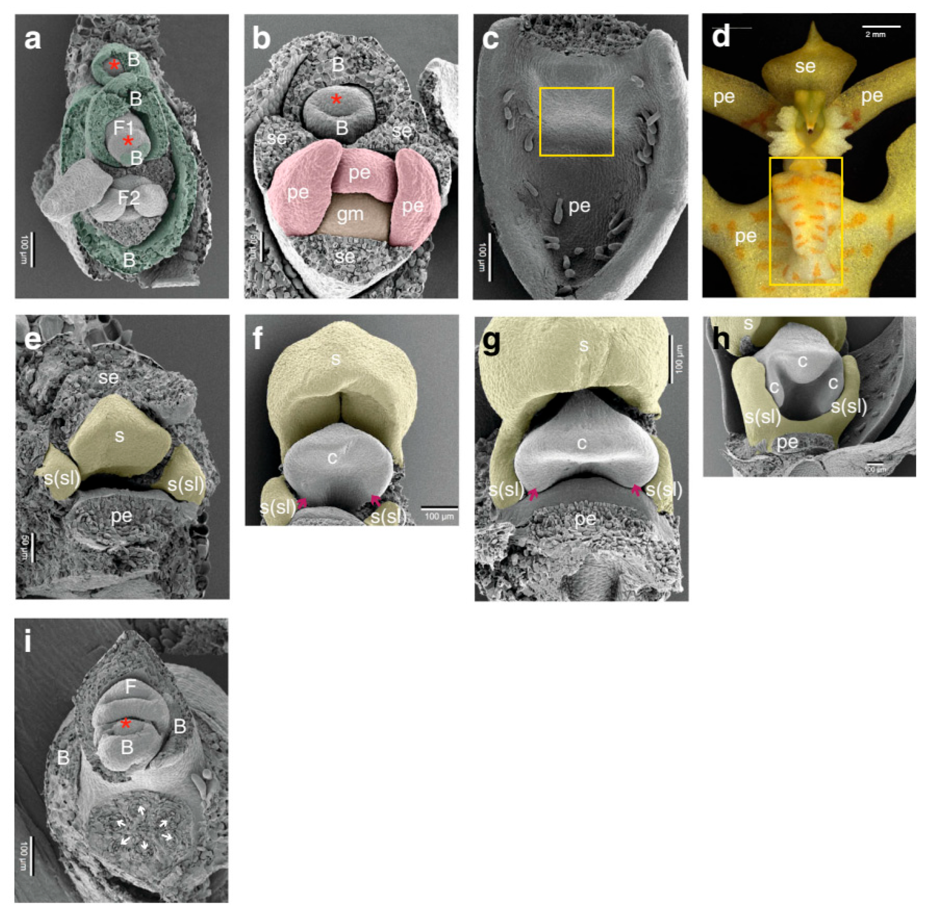
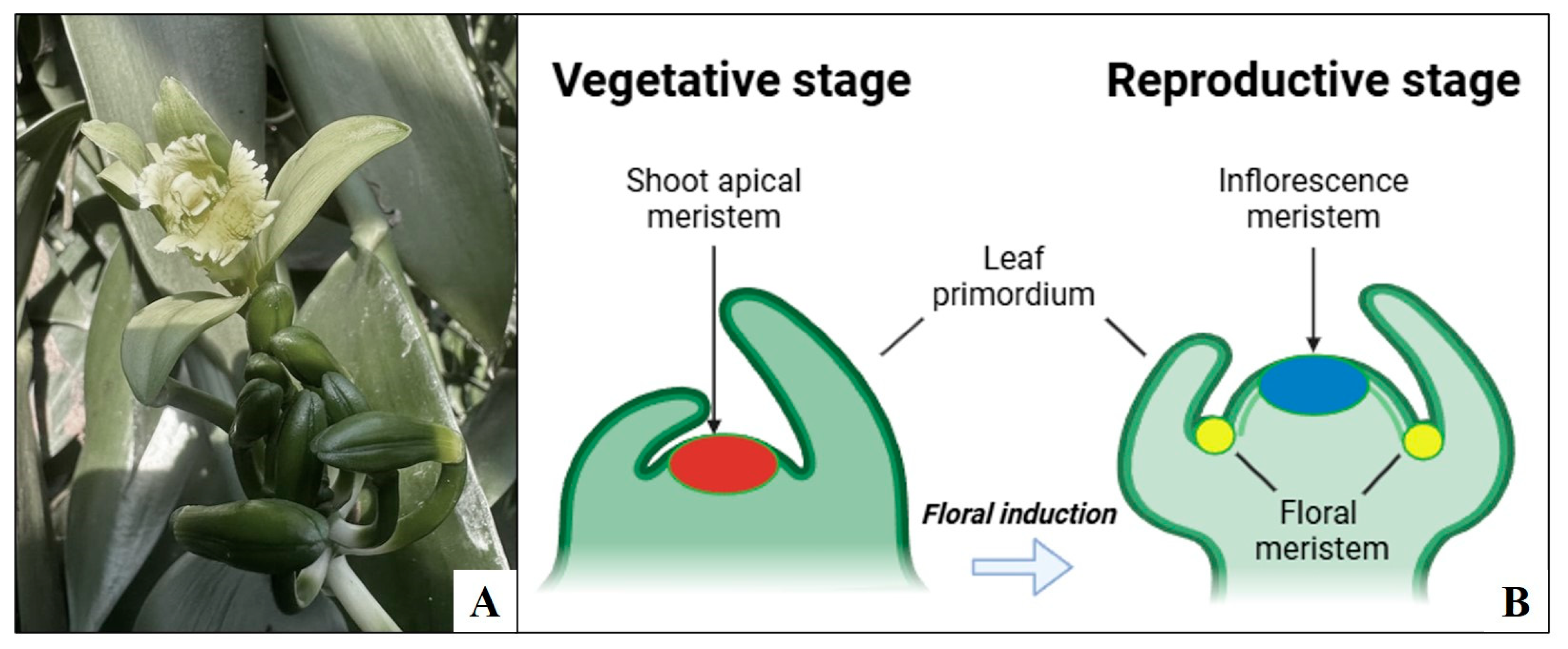
2. Floral Induction and Development in Tropical Orchids

In order to attract pollinators, the flowers of the Orchidaceae family present a high degree of speciation, with significant variances in floral traits, such as shape, color, size, and aroma [10]. In angiosperms, there is a significant variation in the morphology of flowers, but there are four distinct types of floral organs: sepals, petals, stamens, and carpels [11]. In theory, an orchid flower has five whorls of floral organs and two growth types (monopodial and sympodial). The stem apex of monopodial orchids, such as vanilla, develops vegetatively, while axillary meristems give rise to the inflorescences. After developing a succession of adjacent shoots, sympodial orchids eventually bloom and are replaced [11,12]. In order to fix carbon, orchids use crassulacean acid metabolism to adapt to arid environments; flowering consumes a lot of energy [13]. In orchids, the flowering is influenced by changes in the photoperiod, light, temperature, humidity, nutriments, carbon source, vitamins, and plant hormones. Orchids require 3 to 7 [14], 10, or even 16 years for the first bloom [15]. For the species to be maintained, the bud commitment time for the floral induction under ideal physiological conditions is essential [16]. Inflorescence in vanilla vines typically emerges after two years, usually in the third year of cultivation.
The floral ontogeny in Erycina pusilla has been described by Dirks-Mulder et al. [16]: early ontogeny starts from floral initiation up to the three-carpel-apex stage, and late ontogeny starts from the three-carpel-apex stage until anthesis. The development of an early-flowering technique would decrease the breeding time and commercial production costs. Such a method allows for the earlier assessment of particular traits of the flowers, like size, shape, and color. In addition, shortening of the juvenile phase can make a model system for studying the flowering induction and transition available. Once the traits are selected, the clone can be propagated by PTC techniques.

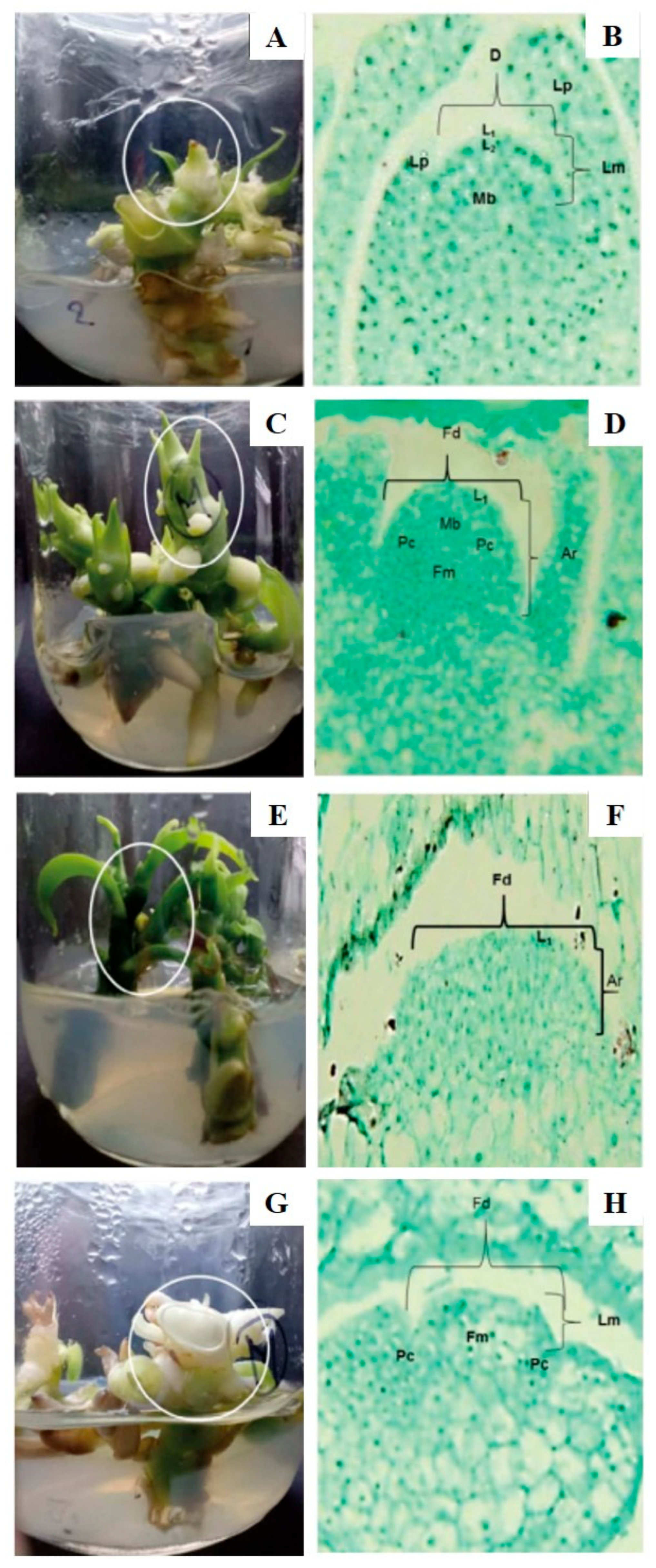
Floral Induction and Development in Vanilla spp.

Orchids are grown primarily as ornamentals; some are used as natural medications and food by different countries [17]. The genus Vanilla (Orchidaceae) consists of 140 species [18], and vanillin is the major compound and accepted flavoring. This genus is characterized by a monopodial growth habit, growth as perennial vines, thick and succulent stem, aerial axial roots growing at each node, absence of pseudobulbs, alternate leaves, axillary inflorescence, flowers with lips partially adnate to the column, and fruits with encrusted seeds [17,18]. V. planifolia Jacks. ex Andrews, V. tahithensis J.W. Moore, and V. pompona Schiede are important for commercial cultivation. V. planifolia is a species originated from Mexico and is the most widely marketed vanilla pod. Despite its importance, this crop has a number of restrictions that prevent it from reaching its full production potential.
To date, orchids are in the Red Data Book of the International Union of Conservation of Nature and Natural Resources and in the Appendix-II of Convention on International Trade in Endangered Species of Wild Fauna and Flora. In addition, V. planifolia is subject to Special Protection by the Mexican Government under NOM-059-SEMARNAT-2010. This calls for a need to standardize PTC techniques for massive propagation of high-quality planting material [19]. Due to the low genetic variation in V. planifolia populations, genetics studies in floral development and morphology must be conducted to detect populations diversity. However, the process of flowering of vanilla plants is highly sensitive to stress caused by abiotic and biotic factors [20]. In orchids, the flowering process is induced by changes in the photoperiod, light, temperature, humidity, nutriments, plant growth regulators, etc.
The flowers, in Vanilla spp., are zygomorphic, bisexual, epigynous, fragrant, yellow-green, and short pedicellate [21]. Inflorescences show a short duration, and each cluster contains around 20 flowers of large size with about 10 cm diameter. These flowers consist of three oblong–lanceolate sepals and two petals similar to sepals. The gynostemium or column is formed by the fusion of styles. The rostellum, a wing-shaped structure, separates the stamen from the sticky stigma, containing two pollen masses [21]. In autumn–winter seasons, flowering is activated by low temperatures [21]. In V. planifolia, flowers are ephemeral and usually last a single day. These flowers are hand-pollinated in cultivation areas due to a structure (called the rostellum) situated between the stigma and pollinia that avoids auto-pollination.
Studies on pollination requirements, reproductive strategies, and breeding systems are required. Traditional genetic improvement has predominantly served as the primary approach for V. planifolia breeding. However, further field results are still needed to show an advanced stage of these breeding works. In this context, biotechnological tools represent a valuable alternative for increasing vanilla production and for transferring important traits into V. planifolia. Effective protocols for in vitro propagation or micropropagation in vitro conservation, cryopreservation, somaclonal variations, protoplast culture, in vitro mutagenesis, genetic transformation, and genome editing of V. planifolia are available [18,19].

3. In Vitro Floral Induction in Tropical Orchids

In vitro propagation or micropropagation allows for the massive production of orchids at a low cost and scales up to industrial commercialization. Up to now, there are several studies on in vitro flowering in diverse plant genera [22]. In tropical orchids, in vitro flowering has been studied as well (Table 1). Several protocols have been improved for the in vitro flowering of numerous orchid species through the in vitro culture of different parts, such as shoot and root tips, flower stalk nodes, buds, and rhizome segments. Even though seeds and seedlings are the most favorable explant sources for in vitro propagation experiments. Techniques such as in vitro pollination have also been explored, though with low fertility rates. In vitro culture of orchids has been a leader in micropropagation commercially when it started almost a century ago with the in vitro germination of Cattleya and Phalaenopsis seeds [23].
Induction of the in vitro flowering of different orchid species had been reported for Phalaenopsis spp. [24], Oncidium spp. [25], Dendrobium spp. [26], and Cymbidium spp. [27,28]. In O. varicosum, Kerbauy [25] reported that seedlings produced flower stalks on PGR-free medium. In Phalaenopsis var Pink Leopard (“Petra”), Duan and Yazawa [24] found that flower induction was achieved by cytokinin and low nitrogen supply. In C. niveo-marginatum Mak, Kostenyuk et al. [27] reported that addition of cytokinin, root excision and limited nitrogen supply resulted in early in vitro flowering of C. niveo-marginatum. In addition, in vitro flowering is influenced by the levels and ratios of the two major components, carbohydrates and minerals [7]. High concentrations of nitrogen usually inhibited flowering and promoted vegetative growth, while the use of a half-strength mineral medium or a reduced nitrogen level enhanced in vitro flowering in Cymbidium and Doritis [24].
Table 1. In vitro floral induction and flowering in tropical orchids.
SpeciesExplant TypeCulture Conditions (Temperature [°C]/Photoperiod [h]/Irradiance [µmol m−2 s−1])Culture MediumFlower Induction/FormationSuccess Rate/Flower AbnormalitiesReferences
Dendrobium candidum
Wall. Ex Lindl.
Seed-derived protocorms25–27/12/13.5Modified MS + PGR (BA, NAA, and ABA)Induction of floral buds27–84% of success/many of the flowers showed abnormalities[29]
Dendrobium candidum
Wall. Ex Lindl.
Seed-derived protocorms25–27/12/13.5Modified MS + PGR (BA, NAA, and ABA)Flower initiation from protocorms or shoots46–83% of success/malformation of petals and lip[30]
Cymbidium niveo-marginatum MakRhizomes and
plantlets
25–26/16/50Modified MS + BAIn vitro floral induction and flowering90% of success/floral buds withered with TDZ[27]
Dendrobium huoshanaseSeedlings25 ± 2/12/27Modified MS + PGR (2,4-D and Zeatin)In vitro floral induction and floweringNot reported[31]
Geodorum densiflorum (Lam.)
Schltr.
Seedlings25 ± 2/16/13.5Modified MS + PGR (BA and IAA) + ACIn vitro floweringNot reported[32]
Psygmorchis pusilla Dodson and DresslerSeedlings22–32/dark 6, 8, 12, 16,
20 and 22/30
Modified VW + additives (ripe banana pulp, AC, and sucrose)Floral spike development and flower formationLow rate of success/flower buds did not open[33]
Phalaenopsis cygnus var Silky MoonPlantlets25 ± 2/16/35–40VW medium + BAFlower bud formation38–43% of success/no flower abnormality reported[34]
Dendrobium moniliformeSeedlings25 ± 2/12/50–60Modified MS + PGR (TDZ or PBZ and ABA)In vitro floral induction and normal flowering45–80% of success/many of the flowers showed abnormalities[35]
Dendrobium var Second LoveShoots26 ± 1/16/50–60Modified VW and MS + PGR (TDZ, BA, and [9R]iP)In vitro floral induction, transition and flowering65% of success/no flower abnormality reported[36]
Dendrobium var Madame Thong-InProtocorms26 ± 2/16/35Modified KC + BA + additives (CW and AC)In vitro floral induction, transition and abnormal flowering100% of success/75% of the flowers showed abnormalities[37]
Dendrobium var Chao PrayaProtocorms25 ± 2/16/40Modified KC + BA + CWIn vitro inflorescence induction and flowering, but some incomplete flowers were reported18–83% of success/many of the flowers were incomplete[38]
Dendrobium var Sonia 17Three-leaf stage plantlets25 ± 2/16/25Modified MS + BAIn vitro inflorescence induction and flowering, but incomplete
flower structures were reported
20–52% of success/many of the flowers were incomplete[39]
Psygmorchis pusillaSeedlings25 ± 2/16/30Modified VW + additives (ripe banana pulp, AC, and sucrose)Floral spike development and flower formationNot reported[40]
Dendrobium primulinumProtocorm-like bodies (PLBs)25 ± 2/12/40Modified MS + PGR (BA and NAA) + fresh apple juiceInduction of floral budsLow rate of success/flower buds did not open and deformed flower developed[41]
Dendrobium denndanumSeedlings22–24/12/27Modified MS + PGR (BA, NAA, ABA, and 2,4-D)Induction and formation of floral buds10% of success/flower buds did not open[42]
Bulbophyllum auricomum Lindl.Seedlings24/16/80Modified MS and KC + PGR (BA, KIN, NAA, and IBA) + CWIn vitro flowering but some flowers with asymmetrical sepals were reported50% of success/many of the flowers were incomplete[43]
Friederick’s
Dendrobium
Shoots26 ± 2/14/22Modified MS + PBZIn vitro induction of floral buds and normal flowering7–29% of success/normal morphology[44]
Dendrobium nobile Lindl.Seedlings25/12/60Modified MS + PGR (PBZ and TDZ)In vitro induction of floral buds and flowering, but deformed flowers were reported at high temperature33–97% of success/deformed flowers at 25 °C[45]
Dendrobium officinaleNon-rooted plantlets26 ± 1/16/13.5–24Modified MS + single-factor and multi-factor PGR treatmentsIn vitro induction of floral buds and normal flowering80–90% of success/no deformed flowers reported[46]
Oncidium var Gower RamseyPetal-bearing embryos from root
callus
26/16/36Modified MS + PGR (2,4-D, dicamba, NAA, IBA, 2iP, BA, kinetin, TDZ, and GA3)Normal in vitro flowering50% of success/abnormal petal-bearing embryos[47]
Dendrobium aphyllumPLBs25 ± 2/14/60Culture medium (MS, PM and KC) + PGR (BA and NAA) Normal in vitro flowering10% of success/no deformed flowers reported[48]
Dendrobium
wangliangii
Protocorms22 ± 2/16/36Modified MS + PGR (BA, NAA, and PBZ)Induction of floral buds5–20% of success/no deformed flowers reported[49]
Dendrobium var Chao Praya
Smile, Pinky
and Kiyomi
Beauty
Protocorms24/16/35Modified KC + BA + additives (sucrose and CW)Normal in vitro floweringNot reported/abnormal floral buds on the apex[50]
Dendrobium nobile typesSeedlings14/12/80 for
30 days, then at 25 ± 2/12/50
Modified MS + PGR (PBZ and TDZ) + CWFlower bud formation4–39% of success/no deformed flowers reported[51]
Dendrobium huoshanensePLBs25 ± 2/16/42–55Modified MS + PGR (2,4-D, TDZ, BA, NAA, and IBA)In vitro floweringNot reported/flowers showed a lack of the gynandrium[52]
Dendrobium Chao Praya SmileProtocorms24/16/--Modified KC + BA + CWInflorescence stalk formationNot reported/no deformed flowers reported[53]
Erycina pusillaSeedlings25 ± 2/12/6–55Modified MS + additives (sucrose, AC, peptone, potato powder, and CW)Normal in vitro floweringNot reported/no deformed flowers reported[54]
Dendrobium ovatumProtocorms and
PLBs
Prechilling at −20, 0, 4, and 15 for 2 h/dark 6, 12, 18, and 24/50Modified MS + Zeatin + CWNormal in vitro flowering10–25% of success/presence of lateral petals[55]
Cymbidium ensifolium, Cymbidium sinense,
and Cymbidium goeringii
Protocorms26 ± 2/12/40Culture medium (not reported) + PGR (BA and NAA) + additives (sucrose and AC)Abnormal in vitro floweringNot reported/leafless flowers[56]
Dendrobium nobileSeedlings26/12/40–56Modified KC + TDZ + additives (sucrose, AC, and CW)Normal in vitro flowering88% of success/no deformed flowers reported[57]
Vanilla planifolia Jacks.
ex Andrews
Shoots26 ± 2/--/55Modified KC + PGR (BA, TDZ, PBZ, and GA3) + additives (sucrose and CW)Floral differentiation0% of success/no abnormalities reported[58]
Cymbidium faberi RolfePLBsNot measuredModified MS + PGR (2,4-D, BA, IBA and NAA) + additives (sucrose, glucose, AC, CW, and peptone)In vitro flowering but changes in leaf and flower morphology were reportedLow rate of success/deformed flower developed and small or curled petals[59]
KC: Knudson C; MS: Murashige and Skoog; NP: New Phalaenopsis; PM: Phytamax; VW: Vacin–Went; ABA: Abscisic acid; IAA: Indole-3-acetic acid; IBA: Indole-3-butyric acid; BA: Benzyladenine; GA3: Gibberellic acid, KIN: Kinetin, NAA: Naphthaleneacetic acid, PBZ: Paclobutrazol, TDZ: Thidiazuron, 2,4-D: 2,4-Dichlorophenoxyacetic acid, 2iP: 6-(γ,γ-Dimethylallylamino)Purine, [9R]iP: Isopentenyladenosine; AC: activated charcoal; CW: coconut water.
Utilizing a suitable medium is also an important factor for orchid culture. Another significant factor in in vitro flowering is the medium’s physical state, such as whether it is liquid or solidified, and which most likely allows air exchanges [7,26]. In most of the studies in Table 1, benzyladenine is reported to be crucial for the induction of in vitro flowering. Sim et al. [37] stated that when additional cytokinin sources, such as coconut water, were present, benzyladenine promoted the early development of inflorescence stalks and stimulated the induction of flower buds. Wang et al. [30], in D. candidum, reported that benzyladenine alone can induce flowering, but the addition of auxin and cytokinin stimulates flower formation; however, auxin alone suppresses flower formation. For example, in Dendrobium hybrids like Chao Praya Smile, in vitro flowering rates reached up to 72% with optimized benzyladenine concentrations, and flowers developed that were morphologically similar to those grown ex vitro, including functional pollen and reproductive organs. For C. niveo-marginatum Mak, C. ensifolium, and D. Second Love, thidiazuron was more successful than benzyladenine at inducing flower buds in vitro; nevertheless, thidiazuron resulted in poor plant development and quickly wilted floral buds [27,36,56]. Under in vitro conditions, particularly hormonal imbalances, temperature stress, and physical factors, can cause morphological and developmental abnormalities in orchid flowers, affecting their normal structure and reproductive functionality. Some flowers showed missing floral organs, such as the absence of sepals or petals, or lacked normal bilateral symmetry—suggesting disrupted floral organ development [7]. In D. nobile, flowers developed deformed structures at 25 °C, but normal development was achieved at lower temperatures (23 °C/18 °C) [45]. On the other hand, a few studies reached the acclimatization stage after in vitro flowering [41,48,49,52,54]. This fact has a practical implication during micropropagation due to the importance of early flowering in plantlets obtained in vitro (Figure 1). In Figure 1, Dirks-Mulder et al. [16] describe the structural modifications related to the floral transition of E. pusilla obtained in vitro.
The floral transition in orchids involves a complex interplay of environmental cues and endogenous signals that trigger the shift from vegetative growth to flowering. Key factors include temperature, hormones, and molecular signals such as florigen [7]. For instance, cold treatments are known to enhance the levels of endogenous cytokinins and gibberellins, which may promote flowering, while sucrose and other carbohydrates act as signaling molecules interacting with floral integrator genes like FT (Flowering Locus T). Molecular studies, particularly in species like Phalaenopsis and Oncidium, indicate that the FT gene acts as a central floral integrator, receiving signals from leaves (where it is often produced in response to the photoperiod and temperature) and translocating to the shoot apical meristem to initiate floral development [7,26]. These advances demonstrate an improved understanding and ability to control the florogenesis process in vitro, reducing the time and effort needed for breeding and commercialization.

In Vitro Floral Induction: Application to and Importance for Vanilla

In Vanilla spp., in vitro floral induction could aid in elucidating the physiological, biochemical, and genetic mechanisms of in vitro flowering. In a hybrid flower breeding program, significant traits can be screened and detected in a shorter period of time [7]. The use of PTC techniques, such as micropropagation, allows for the massive production of vanilla plants to satisfy worldwide demand. There are many protocols regarding effective in vitro regeneration techniques, especially for Vanilla spp. [60,61,62,63]. However, the efforts to induce in vitro flowering are scarce.
Different orchids require specific conditions of photoperiod, light, and ambient temperature to induce in vitro flowering (Figure 2). In addition, in vitro floral induction has been registered in orchids through the use of PGRs. In this regard, cytokinins are often used, including 6-benzyladenine and thidiazuron. Benzyladenine has been used in most in vitro flowering experiments of a number of orchids. Additionally, paclobutrazol has also been evaluated, as well as gibberellic acid. Duan and Yazawa [24] formed floral buds in the VW medium with 5 mg/L benzyladenine in Phalaenopsis. Than et al. [43] reported in vitro flowering within six months after sowing Bulbophyllum auricomum seeds in an MS medium with 1.0 mg/L of benzyladenine + 0.5 mg/L of naphthaleneacetic acid. Benzyladenine might be involved in the early response of axillary meristems through genes controlling the shoot apical meristem activity. In V. planifolia, only one study by Ríos-Barreto et al. [58] reports partial success; they found that the benzyladenine treatments did not show floral initiation in vanilla. However, benzyladenine at a high dose affects, in a negative manner, the vegetative growth of vanilla. In this context, optimal doses and pretreatments with additives should be assessed in order to induce in vitro flowering. In addition, Ríos-Barreto et al. [58] found that the gibberellic and paclobutrazol treatments showed different tissue formations; however, the treatments of gibberellic and paclobutrazol produced flattening of the meristem dome. The study by Ríos-Barreto et al. [58] is the first report that indicates signs of the floral induction and initiation of V. planifolia (Figure 3).
In Figure 3A,C,E,G, Ríos-Barreto et al. [58] describe the structural modifications related to the floral initiation of V. planifolia shoots cultivated in vitro for 13 weeks. Figure 3B shows the vegetative apical meristem of a V. planifolia explant at day 0. Figure 3D–H show the apical meristem at the beginning of floral differentiation. However, no cell proliferation was noted in the flanks of the reproductive apex, and there was no sign of young inflorescence of V. planifolia.

4. Factors Controlling In Vitro Flowering of Tropical Orchids Focusing on Vanilla

4.1. Effect of Physical Factors on In Vitro Floral Induction

The floral meristem development in any plant is a multifactorial process regulated by several external and internal conditions [64]. These conditions include the photoperiod, light quality and quantity, temperature, culture medium, explant type, etc.
Photoperiod. The duration of the photoperiod strongly affects flowering. In plants, the effects of photoperiods with different light requirements are very different. However, in orchids, the floral initiation is generally regulated by photoperiod. Short-day orchids cannot surpass a long light-critical point, while long-day orchids require the minimum amount of light to flower [26]. The effects of the photoperiod have been investigated on the in vitro propagation and flowering of many tropical orchid species, such as Oncidium spp., Dendrobium spp., and Cymbidium spp. [25,26,27]. In Vanilla spp., there are many protocols regarding photoperiod during the micropropagation using solid and liquid culture media and in vitro conservation [60,65,66,67]. In V. planifolia, Divakaran and Babu [65] used a 14 h light photoperiod during the in vitro shoot conservation and multiplication. In V. planifolia, Ramírez-Mosqueda et al. [67] used a photoperiod (16 h light/8 h dark) during the growth and development of in vitro plantlets of V. planifolia. However, most of the protocols regarding photoperiod during the micropropagation of Vanilla spp. established a photoperiod of 16 h light using solid and liquid culture medium.
Light. Light is a determinant issue for the in vitro culture of plantlets. The effects of various light spectra on plant growth and organogenesis have been explored [67]. They play a key role in successful in vitro propagation. Light-emitting diodes (LEDs) have been applied as the sole source of lighting for in vitro culture because fluorescent lights consume more power than LEDs. LED lighting improves the quality of orchid plantlets obtained via PTC techniques. The effects of light irradiance on the in vitro propagation and flowering of some orchid species, like Oncidium spp., Dendrobium spp., and Cymbidium spp., have been evaluated [7,26]. There are protocols regarding the light quality (spectral quality) and quantity (irradiance) during the micropropagation of Vanilla spp. [60,67]. In V. planifolia, Bello-Bello et al. [60] found an increase in the number of shoots per explant under a white LED and blue + red LED light with an irradiation intensity set to 25 photosynthetic photon flux density (PPFD) during the in vitro shoot proliferation. In V. planifolia, Ramírez-Mosqueda et al. [67] found significant differences in the growth and development of plantlets grown under an LED light with an irradiation intensity set to 40 PPFD. In the proliferation stage, blue/red LEDs stimulated the elongation of shoots and chlorophyll synthesis. In the rooting stage, blue LEDs stimulated the number of velamen formed. However, different light colors, such as white and yellow LEDs, have been evaluated as well in other orchid species.
Temperature. The temperature influences germination, making it a crucial environmental indicator for plant development [68,69]. Temperature exposure, particularly low temperatures (vernalization), helps control when plants begin to flower. In the Orchidaceae family, vernalization has varying effects, even though it may cause flowering in certain orchid species. The temperature is generally used to control the flowering time in commercial production of orchids [7,26]. The temperature effects have been evaluated on the in vitro propagation and flowering of several orchids, such as Oncidium spp., Dendrobium spp., and Cymbidium spp. [7,14,26,27,70]. In C. niveo-marginatum, Kostenyuk et al. [27] reported that in various combinations of 4–6 °C and 30–32 °C temperature ranges, no flowering sign was observed within 4–5 months. However, most of the protocols regarding temperature during the micropropagation of Vanilla spp. established a temperature range of 24 ± 2 °C using solid and liquid culture media [60,61,62,63].
Culture medium. In vitro propagation of orchids can be performed in both liquid and solid culture media [71]. Various artificial media and supplement formulations were frequently used in both terrestrial and epiphytic species studies [71]. The most frequently used artificial media are Knudson C (KC) medium [72], Vacin and Went (VW) [73], Murashige and Skoog [74] (MS) basal medium, Malmgren [75], and terrestrial orchid medium (BM). The MS basal medium is widely used in several plant species, including orchids [7,26]. Moreover, the addition of different levels of sucrose, nitrogen, PGR, and activated carbon to the culture medium is commonly implemented. The effects of the culture medium on the in vitro propagation and flowering of several orchid genera, such as Oncidium, Cymbidium, and Vanilla, have been investigated [7,26,76,77]. In V. planifolia, Gayatri and Kavyashree [76] reported that the use of the VW medium promoted shoot regeneration from PLBs. In V. planifolia, Sidek et al. [77] found that the explants developed in Murashige and Skoog salts had the highest increase in the length of their shoot and velamen in comparison with the KC and VW culture media.
Explant type. Explant selection is an important issue to consider before initiating a culture protocol. The explants used in in vitro propagation are derived from field-grown seedlings and/or greenhouse-grown plants. Choosing young tissues over mature parts needs to be considered. The effects of the explant type have been assessed on the in vitro propagation of some orchid species like Oncidium spp. and Cymbidium spp. [7,25,26,27]. In O. varicosum, Kerbauy [25] stated that seedlings produced flower stalks on the KC culture medium with no PGR. In C. niveo-marginatum, Kostenyuk et al. [27] reported that plantlets formed in vitro floral stalks using the MS culture medium supplemented with PGRs. In Vanilla spp., the principal starting explants for in vitro experiments are nodal and leaf segments. The use of different explant types during the micropropagation of Vanilla spp. has been reported in several protocols [78,79,80,81,82]. In V. planifolia, Ríos-Barreto et al. [58] used shoots as explants for in vitro flowering induction.

4.2. Effect of Plant Growth Regulators on In Vitro Floral Induction

The PGRs are synthetic compounds that affect morphological and biochemical processes in higher plants at low dosages. In vitro floral induction could be accomplished by optimizing several in vitro medium components, like PGR.
Benzyladenine. Cytokinins are a group of phytohormones derived from adenine. The monopodial orchid’s floral induction is triggered by the synthetic cytokinin benzyladenine (BA), while auxin inhibits the impact of BA [83]. BA has been used in the in vitro flowering experiments of several orchids, such as Phalaenopsis cygnus and Cymbidium niveomarginatum [7,26]. However, in V. planifolia, the attempts to induce in vitro flowering through PGRs are limited. In V. planifolia, Ríos-Barreto et al. [58] found that the BA treatment did not indicate floral initiation in vanilla. However, BA at high doses affects, in a negative manner, the vegetative growth of vanilla.
Thidiazuron. Thidiazuron (TDZ) is a synthetic PGR with cytokinin-like responses. TDZ is a regulator of several morphogenic processes, like callus induction, somatic embryogenesis, shoot organogenesis, proliferation, and in vitro flowering. TDZ has been used in in vitro flowering experiments of some orchids such as Dendrobium moniliforme [7,26]. There are several protocols regarding the use of different concentrations of TDZ during the micropropagation of Vanilla spp. However, in V. planifolia, the attempts to induce in vitro flowering using TDZ are limited. In V. planifolia, Ríos-Barreto et al. [58] observed that the treatments with TDZ did not show signs of floral initiation.
Gibberellic acid. Gibberellins (GAs) play a dual role in reproductive development. The gibberellic acid (GA) is a tetracyclic di-terpenoid compound and a plant hormone that stimulates plant growth and development. GAs regulate flower induction and its transition. GA has been used in the in vitro flowering experiments of a number of orchids, such as Cymbidium ensifolium and Cymbidium niveomarginatum [7,26]. However, in V. planifolia, the attempts to induce in vitro flowering through the use of GA3 are limited. In V. planifolia, Ríos-Barreto et al. [58] found that the GA3 treatments showed different tissue formations. In addition, the GA3 treatments presented signs of floral initiation.
Paclobutrazol. Paclobutrazol (PBZ) belongs to the triazole family and has growth-regulator properties. PBZ inhibits GA biosynthesis by blocking the oxidation of ent-kaurene into ent-kaurenoic acid. PBZ could be useful for increasing floral biomass and development. Therefore, by decreasing gibberellin levels and increasing auxin and cytokinin levels at the tip of aerial organs, PBZ treatment causes some plants to begin flowering [7,26]. PBZ has been used in in vitro flowering experiments of various orchids, such as Friederick’s Dendrobium and Dendrobium moniliforme [7,26]. However, in V. planifolia, the attempts to induce in vitro flowering through the use of PBZ are limited. In V. planifolia, Ríos-Barreto et al. [58] found that the treatments with PBZ showed different tissue formations and presented flattening of the meristem dome.

4.3. Effect of Nutrition on In Vitro Floral Induction

In vitro flowering is influenced by the contents and ratios of different macro- and micronutrients [84]. These minerals are important elements that control plant growth and development. High phosphorus and low nitrogen in the culture media induced flowering in several orchid genera [24,26,27]. The NH4+/NO3− ratio is essential for in vitro flowering [85]. Raising the NH4+/NO3− ratio in in vitro flowering while reducing the NH4+ concentration (e.g., 1/2 full-strength MS medium) stimulates in vitro flowering [24]. There are many protocols regarding the use of different concentrations of macro- and micronutrients during the in vitro conservation and micropropagation of Vanilla spp. However, in V. planifolia, the efforts to induce in vitro flowering employing macro- and micronutrients are null.

4.4. Effect of Additives for In Vitro Floral Induction

Several types of additives, like peptone, carrot and tomato juice, and especially coconut water (CW), are frequently included in the orchid culture media [7,26]. However, in most of the protocols for tropical orchids, such as in vitro floral induction and flowering, are stimulated or inhibited by these additives. In V. planifolia, the CW is used at a concentration of 100 mL/L. Depending on the requirements of the orchid species, their concentration in the culture media must be established. Sim et al. [37] stated that additional cytokinin sources, such as CW, promoted the early development of inflorescence stalks under in vitro conditions.
Sucrose. Carbohydrates are an energy source in culture media and an osmotic agent, having important effects on orchid growth and development. One of the most common carbon and energy sources for in vitro culture has been recommended to be sucrose [86,87]. Sucrose is available to cells for cell growth and has an osmotic role in the culture medium [88,89]. However, there are other carbon sources, such as glucose, fructose, maltose, and mannitol, that can be used. Sucrose has been used in in vitro flowering experiments of orchids, such as Psygmorchis pusilla Dodson and Dressler, Erycina pusilla, and Cymbidium sinense [7,26]. In V. planifolia, Ríos-Barreto et al. [58] found that sucrose at 40 g L−1 improved the floral induction and tissue formation.
Activated charcoal. In tissue culture, activated charcoal (AC) is usually used to improve cell growth and development. The AC is a small particle composed of carbon arranged in a quasigraphitic form with a very fine network of pores that gives it a unique adsorption capacity for inhibitory substances in the culture media [90,91,92,93]. The AC drastically decreases the phenolic oxidation [94,95], alteration of pH [96], and establishment of a darkened environment in the medium [97]. The content of this component in the medium must be chosen based on the unique needs of orchid species because it has a significant species-specific influence [7]. Most of the orchid-flowering experiments conducted in vitro have used AC, such as Cymbidium goeringii, Cymbidium faberi Rolfe, and Geodorum densiflorum (Lam.) Schltr. [7,26].

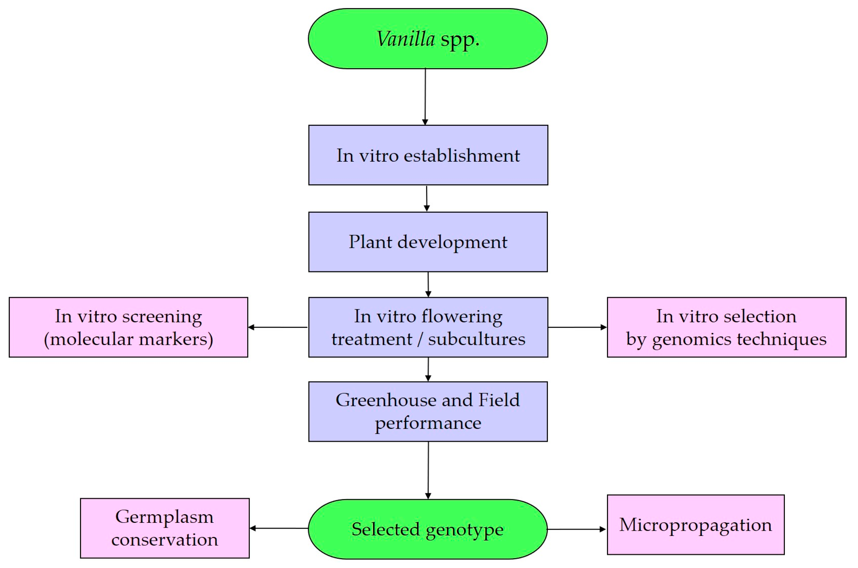
5. Orchid Biotechnology Approach

In the model plant, Arabidopsis, the molecular signaling network in response to several cues of in vitro floral induction and flowering has been thoroughly investigated [98]. Floral induction is controlled by the coordinated regulation of several genetic elements in various flowering pathways, such as photoperiod, vernalization, autonomy, and temperature [99]. However, in tropical orchids, the molecular mechanisms underlying the floral transition have not been elucidated. To date, the molecular cues underlying flower development are poorly understood in the Vanilla genus. Nonetheless, a variety of methods and genomic resources have been created for Vanilla to aid in describing the genetic variety among various vanilla species and accessions [99,100,101,102]. Among them, studies on genetic transformation, cytogenetics, and genome editing have been carried out.
Genetic transformation of tropical orchids. In orchids, the most widely used methods for gene transformation are Agrobacterium tumefaciens-mediated transformation and biolistics. Significant advancements have also been made in plant transformation techniques for the insertion of foreign DNA into plant genomes [103]. The first reports of successful orchid transformation were in Vanda tricolor [104] and Cymbidium niveo-marginatum [104,105] through microparticle bombardment, and biolistic-transformed orchids were developed. To date, transgenic technologies have been adapted in Oncidium spp. and Cymbidium spp., using protocorm-like bodies (PLBs) [103]. However, there have not been many efforts to genetically transform vanilla [99,106]; the lack of genetic transformation protocols for Vanilla is a critical barrier. In V. planifolia, Retheesh and Bhat [106] developed an Agrobacterium-mediated genetic transformation protocol for inducing high-frequency PLBs. In V. pompona, Edmon et al. [99] developed a tissue culture-based regeneration protocol and A. tumefaciens-mediated transformation systems with enhanced efficiency using selection agents like hygromycin and phosphinothricin. These works constitute the first attempts at using genetic engineering techniques in vanilla breeding. Despite successfully inserting reporter genes, further studies are needed to support their potential use in the genetic breeding of this crop. In addition, the legislation on genetically modified organisms in countries like Mexico, which is considered the center of origin and domestication of vanilla, should be reviewed.
Cytogenetics of tropical orchids. Research on in vitro seed germination, in vitro regeneration, and the study of meiosis and pollination to get past hybridization barriers has been conducted alongside large-scale orchid production since the 1980s [107]. In tropical orchids, wild and commercial species are selected as progenitors for the development of new orchid cultivars. However, high infertility is frequently the outcome of orchid hybrid breeding. Fertility can be restored by using antimitotic agents during PTC to double the number of chromosomes. Polyploid orchids, including autopolyploids, have desirable traits, including large flowers, round shape and coloration intensity, and thicker stems and leaves. Cytogenetic evidence is available for a limited number of tropical orchid species, including Cattleya tigrina Lind., Phalaepnosis amabilis, Paphiopedilum villosum, and Vanda “Miss Joaquin” [108]. In Vanilla, limited attempts have been focused on cytogenetic research [109,110,111].
Genomic resources of tropical orchids. In tropical orchids, the application of modern genetics and molecular biology expands, in a highly significant manner, the possibilities of plant breeders. The development of genomics techniques allows for editing of their own genes, which can significantly speed up and increase the success of the traditional selection process [90]. In recent years, significant genomic resources for vanilla have been developed [100,101]. Using a genotyping-by-sequencing approach, SNPs (single-nucleotide polymorphisms) markers for phylogenetic studies have been produced from a draft genome assembly based on V. planifolia [101]. The genome draft of V. planifolia was made possible through the availability of two reference genomes [102]. On the other hand, the molecular cues underlying flower development are poorly understood in V. planifolia. The type II MADS-box gene family is a transcription factor-encoding family involved in various developmental processes such as flowering. In V. planifolia, Himani et al. [112] identified 33 MADS-box transcripts that encode putative MADS-box proteins, and the spatiotemporal expression profile for VpMADS genes at different plant developmental stages showed high expression of these genes in specific floral tissues. Genome editing (GE) is a method that involves precisely inserting, deleting, or swapping out DNA bases to change a particular target DNA sequence of the genome [100]. To continue this endeavor, the CRISPR/Cas9 system has been developed as a new advancement of GE technologies. In P. equestris, the CRISPR/Cas9 tool has been successfully implemented by taking small insertion/deletion or reversal mutations into target genes [108]. In V. planifolia, Brym et al. [113], by using the CRISPR/Cas9 tool, performed gene editing of the phytoene desaturase gene. In this context, the CRISPR/Cas9 should be considered a potential tool for targeting flowering genes (e.g., Flowering Locus T-like gene and LEAFY gene) or stress-response genes (e.g., Fusarium oxysporum f sp. vanillae resistance). In addition, an optimized protocol should take into consideration intermediate steps, such as in vitro selection for abiotic and biotic stress factors by molecular markers or genomics tools, and have an optimal protocol for the acclimatization stage (Figure 4).

6. Issues to Consider in a Protocol for the In Vitro Flowering of Vanilla

Complex protocols have been developed for the in vitro floral induction and flowering of several tropical orchids. However, in Vanilla, attempts to generate a specific protocol are scarce. Therefore, establishing an in vitro protocol to regenerate, study inflorescence and flower morphogenesis, and induce early flowering in vanilla orchids is necessary. In this context, the protocol must consider different issues, including internal (macro and micro-nutrients, carbon source, vitamins, PGR, etc.) and external (photoperiod, light, temperature, humidity, PGR, explant types, and culture techniques) factors that could affect the in vitro flowering in vanilla (Table 2).
In this sense, we present the most important issues to consider for the establishment of a protocol for the in vitro induction and flowering of Vanilla. First issue: The incubation room and culture conditions must be controlled. Regarding the photoperiod, the in vitro flower induction is possible in light (12- and 16 h photoperiods) and darkness. For the in vitro culture of vanilla orchids, the temperature is a key factor to consider; in vitro floral induction has been carried out at a lower temperature regimen (18–24 °C) [7,26]. Second issue: the culture media composition is a key issue in the success of in vitro floral induction and flowering. The commonly used culture medium is based on modifications of the MS salts; however, the Knudson C medium is considered as well. The MS medium should be used from half-strength to full-strength [7,26]. Then, the micro- and macronutrient content, sucrose content, PGR, and additives are essential for plant growth and development. The mineral composition of the culture medium, particularly the nitrogen and phosphorus concentrations, needs to be established. The potassium could be used at a concentration of 1.25 times full-strength MS salts, while the nitrogen content was reduced to 0.25 times [7,26]. In addition, the flower formation in tropical orchids was enhanced by the potassium and calcium supply. The potassium and calcium may be used at a concentration ranging from 1.25 to 1.50 times in the full-strength MS medium [7,26]. For the in vitro culture of vanilla orchids, sucrose as a carbon source has been evaluated in different concentrations. Sucrose can be used at a concentration corresponding from 1.5% to 3% (w/v) [7,26]. However, in different tropical orchids, the consumption of glucose and fructose coincided with the development of flower buds. Regarding PGRs, a single PGR or combinations of PGRs could be used for the in vitro induction and flowering of vanilla orchids. The PGRs often used are 6-benzyladenine, N6-isopentenyladenine, thidiazuron, paclobutrazol, abscisic acid, kinetin, gibberellic acid, 1-naphthaleneacetic acid, 2,4-dichlorophenoxyacetic acid, and zeatin. Cytokinins, such as benzyladenine, can be used at a concentration ranging from 0.5 mg/L to 5 mg/L, while thidiazuron can be used at a concentration varying from 0.1 mg/L to 3 mg/L. Auxins, such as abscisic acid and 1-naphthaleneacetic acid, can be used at a concentration corresponding from 0.1 mg/L to 0.5 mg/L. Other PGRs, such as paclobutrazol, can be used at a concentration from 0.1 mg/L to 1.0 mg/L, while gibberellic acid can be used at a concentration corresponding from 0.5 mg/L to 2.0 mg/L [7,26]. Regarding additives, the addition of activated charcoal and CW could have beneficial effects but, at the same time, reduce the effective PGR concentration for in vitro flowering. The activated charcoal can be used at a concentration from 0.5 g/L to 2.0 g/L [7,26]. The CW can be used at a concentration from 50 to 200 mL/L; the use of CW is recommended as a pretreatment for in vitro flowering. Third issue: The in vitro conditions significantly reduce the juvenile period. Therefore, the semisolid culture is very effective in the in vitro floral induction and flowering stages, but considerations should be made for aspects like individualized explants in culture containers, providing efficient and inert support for the various types of explants. Consider dual-phase culture systems (e.g., liquid media for proliferation→solid media for floral induction).

7. Conclusions and Prospects

Flowering is one of the crucial processes for crop plants, particularly in an era of climate change. Research to induce early flowering in tropical orchids has been designed as a biotechnological tool for assisting orchid-breeding programs. To date, in vitro culture techniques have contributed to this research progress. Optimizing protocols for commercial scaling is a bottleneck and calls for exhaustive research. On the other hand, molecular working mechanisms have not been elucidated at all. In order to elucidate the mechanisms of the putative orchid-flowering genes and to offer a platform for the application of genetic resources to the orchid industry in Vanilla, genomics approaches are needed. In addition, gene-editing techniques and the integration of multi-omics to unravel Vanilla-specific flowering pathways are necessary. In vitro flowering can be used for further breeding of this tropical orchid and hybrid generation to obtain hybrids with greater yields, resistance to Fusarium oxysporum f. sp. vanillae, and tolerance to drought.

Author Contributions

O.B.-B. and J.L.S.-C. performed data analysis and manuscript preparation. O.B.-B. wrote the manuscript. J.L.S.-C. helped in preparing the manuscript. All authors have read and agreed to the published version of the manuscript.

Funding

This research received no external funding.

Data Availability Statement

The raw data supporting the conclusions of this article will be made available by the authors on request.

Conflicts of Interest

The authors declare no conflicts of interest.

References

  1. Kean-Galeno, T.; Lopez-Arredondo, D.; Herrera-Estrella, L. The shoot apical meristem: An evolutionary molding of higher plants. Int. J. Mol. Sci. 2024, 25, 1519. [Google Scholar] [CrossRef]
  2. Rehman, S.; Bahadur, S.; Xia, W. An overview of floral regulatory genes in annual and perennial plants. Gene 2023, 885, 147699. [Google Scholar] [CrossRef]
  3. Shen, C.; Liu, H.; Guan, Z.; Yan, J.; Zheng, T.; Yan, W.; Wu, C.; Zhang, Q.; Yin, P.; Xing, Y. Structural Insight into DNA Recognition by CCT/NF-YB/YC Complexes in Plant Photoperiodic Flowering. Plant Cell 2020, 32, 3469–3484. [Google Scholar] [CrossRef]
  4. Phillips, G.C. In vitro morphogenesis in plants-recent advances. In Vitro Cell Dev. Biol-Plant 2004, 40, 342–345. [Google Scholar] [CrossRef]
  5. Peer, L.A.; Bhat, M.Y.; Ahmad, N.; Mir, B.A. Floral induction pathways: Decision making and determination in plants to flower-A comprehensive review. J. Appl. Biol. Biotech. 2021, 9, 7–17. [Google Scholar] [CrossRef]
  6. Kaur, S. In vitro florigenesis with special reference to orchids—A Review. Recent. Pat. Biotechnol. 2022, 16, 311–318. [Google Scholar] [CrossRef]
  7. Teixeira da Silva, J.A.; Kerbauy, G.; Zeng, S.; Chen, Z.; Duan, J. In vitro flowering of orchids. Crit. Rev. Biotechnol. 2013, 34, 56–76. [Google Scholar] [CrossRef]
  8. Wang, S.L.; Viswanath, K.K.; Tong, C.G.; An, H.R.; Jang, S.; Chen, F.C. Floral induction and flower development of orchids. Front. Plant Sci. 2019, 10, 1258. [Google Scholar] [CrossRef]
  9. Pérez-Escobar, O.A.; Bogarín, D.; Przelomska, N.A.S.; Ackerman, J.D.; Balbuena, J.A.; Bellot, S.; Bühlmann, R.P.; Cabrera, B.; Cano, J.A.; Charitonidou, M.; et al. The origin and speciation of orchids. New Phytol. 2024, 242, 700–716. [Google Scholar] [CrossRef]
  10. Peakall, R. Speciation in the Orchidaceae: Confronting the challenges. Mol. Ecol. 2007, 16, 2834–2837. [Google Scholar] [CrossRef]
  11. Arditti, J. Fundamentals of Orchid Biology; John Wiley and Sons: Hoboken, NJ, USA, 1992. [Google Scholar]
  12. Sailo, N.; Rai, D.; De, L. Physiology of Temperate and Tropical Orchids—An Overview. Int. J. Sci. Res. 2019, 3, 3–8. [Google Scholar]
  13. Silvera, K.; Santiago, L.S.; Cushman, J.C.; Winter, K. Crassulacean acid metabolism and epiphytism linked to adaptive radiations in the Orchidaceae. Plant Physiol. 2009, 149, 1838–1847. [Google Scholar] [CrossRef]
  14. Hew, C.S.; Yong, J.W.H. The Physiology of Tropical Orchids in Relation to the Industry, 2nd ed.; World Scientific: Singapore, 2004. [Google Scholar]
  15. Koopowitz, H. Tropical Slipper Orchids: Paphiopedilum and Phragmipedium Species and Hybrids; Timber Press: Portland, OR, USA, 2008; p. 41. [Google Scholar] [CrossRef]
  16. Dirks-Mulder, A.; Butôt, R.; van Schaik, P.; Wijnands, J.W.P.M.; Berg, R.v.D.; Krol, L.; Doebar, S.; van Kooperen, K.; de Boer, H.; Kramer, E.M.; et al. Exploring the evolutionary origin of floral organs of Erycina pusilla, an emerging orchid model system. BMC Evol. Biol. 2017, 17, 89. [Google Scholar] [CrossRef]
  17. Freudenstein, J.V. Orchid phylogenetics and evolution: History, current status and prospects. Ann. Bot. 2025, 135, 805–822. [Google Scholar] [CrossRef]
  18. de Oliveira, R.T.; da Silva Oliveira, J.P.; Macedo, A.F. Vanilla beyond Vanilla planifolia and Vanilla × tahitensis: Taxonomy and historical notes, reproductive biology, and metabolites. Plants 2022, 11, 3311. [Google Scholar] [CrossRef]
  19. Spinoso-Castillo, J.L.; Chavez-Santoscoy, R.A.; Bogdanchikova, N.; Pérez-Sato, J.A.; Morales-Ramos, V.; Bello-Bello, J.J. Antimicrobial and hormetic effects of silver nanoparticles on in vitro regeneration of vanilla (Vanilla planifolia Jacks. ex Andrews) using a temporary immersion system. Plant Cell Tiss. Org. Cult. 2017, 129, 195–207. [Google Scholar] [CrossRef]
  20. Erel, R.; Dag, A.; Ben-Gal, A.; Schwartz, A.; Yermiyahu, U. Flowering and fruit set of olive trees in response to nitrogen, phosphorus, and potassium. J. Am. Soc. Hortic. Sci. 2008, 133, 639–647. [Google Scholar] [CrossRef]
  21. Anuradha, K.; Shyamala, B.N.; Naidu, M.M. Vanilla—Its Science of Cultivation, Curing, Chemistry, and Nutraceutical Properties. Crit. Rev. Food Sci. Nutr. 2013, 53, 1250–1276. [Google Scholar] [CrossRef]
  22. Gantait, S.; Sinniah, U.R. Rapid micropropagation of monopodial orchid hybrid (Aranda Wan Chark Kuan ‘Blue’ × Vanda coerulea Grifft. ex. Lindl.) through direct induction of protocorm-like bodies from leaf segments. Plant Growth Regul. 2012, 68, 129–140. [Google Scholar] [CrossRef]
  23. Yam, T.W.; Arditti, J.; Cameron, K.M. The orchids have been a splendid sport—An alternative look at Charles Darwin’s contribution to orchid biology. Am. J. Bot. 2009, 96, 2128–2154. [Google Scholar] [CrossRef]
  24. Duan, J.X.; Yazawa, S. Floral induction and development in Phalaenopsis in vitro. Plant Cell Tiss. Org. Cult. 1995, 43, 71–74. [Google Scholar] [CrossRef]
  25. Kerbauy, G. In vitro flowering of Oncidium varicosum mericlones (Orchidaceae). Plant Sci. Lett. 1984, 35, 73–75. [Google Scholar] [CrossRef]
  26. Teixeira da Silva, J.A.; Zeng, S.; Cardoso, J.C.; Dobránszki, J.; Kerbauy, G.B. In vitro flowering of Dendrobium. Plant Cell Tiss. Org. Cult. 2014, 119, 447–456. [Google Scholar] [CrossRef]
  27. Kostenyuk, I.; Oh, B.J.; So, I.S. Induction of early flowering in Cymbidium niveo-marginatum Mak in vitro. Plant Cell Rep. 1999, 19, 1–5. [Google Scholar] [CrossRef]
  28. Chang, C.; Chang, W.C. Cytokinins promotion of flowering in Cymbidium ensifolium var. misericors in vitro. Plant Growth Regul. 2003, 39, 217–221. [Google Scholar] [CrossRef]
  29. Wang, G.Y.; Liu, P.; Xu, Z.H.; Chua, N.H. Effect of ABA on the in vitro production of flower buds of Dendrobium candidum Wall. ex Lindl. Acta Bot. Sin. 1995, 37, 374–378. [Google Scholar]
  30. Wang, G.Y.; Xu, Z.H.; Chia, T.F.; Chua, N.H. In vitro flowering of Dendrobium candidum. Sci. China (Ser. C) 1997, 40, 35–42. [Google Scholar] [CrossRef]
  31. Wen, Y.F.; Lu, R.L.; Xie, Z.L. Rapid propagation and induction of floral buds of Dendrobium huoshanase. Plant Physiol. Commun. 1999, 35, 296–297. [Google Scholar]
  32. Bhadra, S.K.; Hossain, M.M. In vitro germination and micropropagation of Geodorum densiflorum (Lam.) Schltr., an endangered orchid species. Plant Cell Tiss. Org. Cult. 2003, 13, 165–171. [Google Scholar]
  33. Vaz, A.P.A.; Rita de Cássia, L.; Kerbauy, G.B. Photoperiod and temperature effects on in vitro growth and flowering of P. pusilla, an epiphytic orchid. Plant Physiol. Biochem. 2004, 42, 411–415. [Google Scholar] [CrossRef]
  34. Rojanawong, T.; Thepsithar, C.; Thongpukdee, A. Micropropagation of Phalaenopsis cygnus ‘Silky Moon’ from leaf segments. In Proceedings of the 32nd Congress on Science and Technology of Thailand, Bangkok, Thailand, 10–12 October 2006; pp. 10–12. [Google Scholar]
  35. Wang, Z.H.; Tu, H.Y.; Ye, Q.S. Rapid propagation and in vitro flowering of Dendrobium moniliforme (L.) Sw. Plant Physiol. Commun. 2006, 42, 1143–1144. [Google Scholar]
  36. Ferreira, W.M.; Kerbauy, G.B.; Kraus, J.E.; Pescador, R.; Suzuki, R.M. Thidiazuron influences the endogenous levels of cytokinins and IAA during flowering of isolated shoots of Dendrobium. J. Plant Physiol. 2006, 163, 1126–1134. [Google Scholar] [CrossRef]
  37. Sim, G.E.; Loh, C.S.; Goh, C.J. High frequency early in vitro flowering of Dendrobium Madame Thong-In (Orchidaceae). Plant Cell Rep. 2007, 26, 383–393. [Google Scholar] [CrossRef]
  38. Hee, K.H.; Loh, C.S.; Yeoh, H.H. Early in vitro flowering and seed production in culture in Dendrobium Chao Praya Smile (Orchidaceae). Plant Cell Rep. 2007, 26, 2055–2062. [Google Scholar] [CrossRef]
  39. Tee, C.S.; Maziah, M.; Tan, C.S. Induction of in vitro flowering in the orchid Dendrobium Sonia 17. Biol. Plant 2008, 52, 723–726. [Google Scholar] [CrossRef]
  40. Vaz, A.P.A.; Kerbauy, G.B. In vitro precocious orchid flowering: A strategy for basic research and commercial approaches. In Floriculture, Ornamental and Plant Biotechnology: Advances and Topical Issues, 1st ed.; Teixeira da Silva, J.A., Ed.; Global Science Books Ltd.: Isleworth, UK, 2008; Volume V, Chapter 45; pp. 421–426. [Google Scholar]
  41. Deb, C.R.; Sungkumlong, T. Rapid multiplication and induction of early in vitro flowering in Dendrobium primulinum Lindl. J. Plant Biochem. Biotechnol. 2009, 18, 241–244. [Google Scholar] [CrossRef]
  42. Guan, P.; Shi, J.M. Tissue culture of stem segment and induction of floral buds of Dendrobium denndanum. Lishizhen Med. Mater. Med. Res. 2009, 20, 205–206. [Google Scholar]
  43. Than, M.M.M.; Pal, A.; Jha, S. In vitro flowering and propagation of Bulbophyllum auricomum Lindl., the royal flower of myanmar. Acta Hortic. 2009, 829, 105–111. [Google Scholar] [CrossRef]
  44. Te-chato, S.; Nujeen, P.; Muangsorn, S. Paclobutrazol enhance budbreak and flowering of Friederick’s Dendrobium orchid in vitro. J. Agric. Technol. 2009, 5, 157–165. [Google Scholar]
  45. Wang, Z.H.; Wang, L.; Ye, Q.S. High frequency early flowering from in vitro seedlings of Dendrobium nobile. Sci. Hortic. 2009, 122, 328–331. [Google Scholar] [CrossRef]
  46. Cen, X.F.; Huang, C.H.; Wei, P.X. Effects of hormone factors on the in vitro culture flowering induction of Dendrobium officinate Kimura et Migo. Agric. Sci. Technol. 2010, 11, 75–79. [Google Scholar]
  47. Chen, J.T. Induction of petal-bearing embryos from root-derived callus of Oncidium ‘Gower Ramsey’. Acta Physiol. Plant 2012, 34, 1337–1343. [Google Scholar] [CrossRef]
  48. Hossain, M.M.; Sharma, M.; Pathak, P. In vitro propagation of Dendrobium aphyllum (Orchidaceae)—Seed germination to flowering. J. Plant Biochem. Biotechnol. 2013, 22, 157–167. [Google Scholar] [CrossRef]
  49. Zhao, Y.H. Study of tube flowering of Dendrobium candidum. Seed 2013, 32, 16–23. [Google Scholar]
  50. Ding, L.; Wang, Y.; Yu, H. Overexpression of DOSOC1, an ortholog of Arabidopsis SOC1, promotes flowering in the orchid Dendrobium Chao Parya Smile. Plant Cell Physiol. 2013, 54, 595–608. [Google Scholar] [CrossRef] [PubMed]
  51. Wu, G.J.; Lai, Z.X. Optimization of in vitro culture conditions and in vitro flowering in Dendrobium nobile. Chin. Trop. Crops 2013, 34, 451–458. [Google Scholar]
  52. Lee, P.L.; Chen, J.T. Plant regeneration via callus culture and subsequent in vitro flowering of Dendrobium huoshanense. Acta Physiol. Plant 2014, 36, 2619–2625. [Google Scholar] [CrossRef]
  53. Sawettalake, N.; Bunnag, S.; Wang, Y.; Shen, L.; Yu, H. DOAP1 Promotes Flowering in the Orchid Dendrobium Chao Praya Smile. Front. Plant Sci. 2017, 8, 400. [Google Scholar] [CrossRef]
  54. Chiu, Y.T.; Chang, C. In Vitro Flowering and Breeding of Erycina pusilla. In Orchid Propagation: From Laboratories to Greenhouses—Methods and Protocols. Springer Protocols Handbooks; Lee, Y.I., Yeung, E.T., Eds.; Humana Press: New York, NY, USA, 2018. [Google Scholar] [CrossRef]
  55. Pujari, I.; Babu, S.V. Precocious in vitro flowering in threatened ornamental orchid, Dendrobium ovatum—Decoding the causal factors. Curr. Plant Biol. 2022, 31, 100257. [Google Scholar] [CrossRef]
  56. Ahmad, S.; Chen, J.; Chen, G.; Huang, J.; Hao, Y.; Shi, X.; Liu, Y.; Tu, S.; Zhou, Y.; Zhao, K.; et al. Transcriptional Proposition for Uniquely Developed Protocorm Flowering in Three Orchid Species: Resources for Innovative Breeding. Front. Plant Sci. 2022, 13, 942591. [Google Scholar] [CrossRef]
  57. Nadal, M.C.; Andrade, G.V.S.; Flores, J.H.N.; Reis, M.V.D.; Rodrigues, V.A.; Pasqual, M. Dendrobium nobile in vitro flowering induction. Ornam. Hortic. 2023, 29, 135–142. [Google Scholar] [CrossRef]
  58. Ríos-Barreto, Y.; Arellano-Ostoa, G.; Fernández-Pavía, Y.L.; García-Villanueva, E.; Tejeda-Sartorius, O. Plant growth and early in vitro floral differentiation of vanilla (Vanilla planifolia Jacks. ex Andrews). Agro Product. 2023, 17, 127–135. [Google Scholar] [CrossRef]
  59. Wu, Z.; Liu, S.; An, B.; Zhang, H.; Wu, J.; Li, C.; Long, Y. Mutagenesis and flowering promotion through sodium azide in vitro culture of Cymbidium faberi Rolfe. Horticulturae 2024, 10, 889. [Google Scholar] [CrossRef]
  60. Bello-Bello, J.J.; García-García, G.G.; Iglesias-Andreu, L. Conservación de vainilla (Vanilla planifolia Jacks.) bajo condiciones de lento crecimiento in vitro. Rev. Fitotec. Mex. 2015, 38, 165–171. [Google Scholar] [CrossRef]
  61. Halim, R.; Akyol, B.; Güner, A. In vitro Regeneration of vanilla (Vanilla planifolia L.). J. Appl. Biol. Sci. 2017, 11, 5–10. [Google Scholar]
  62. Lee-Espinosa, H.E.; Murguía-González, J.; García-Rosas, B.; Córdova-Contreras, A.L.; Laguna-Cerda, A.; Mijangos-Cortés, J.O.; Santana-Buzzy, N. In vitro clonal propagation of vanilla (Vanilla planifolia ‘Andrews’). HortScience 2008, 43, 454–458. [Google Scholar] [CrossRef]
  63. Ramírez-Mosqueda, M.A.; Iglesias-Andreu, L.G. Indirect organogenesis and assessment of somaclonal variation in plantlets of Vanilla planifolia Jacks. Plant Cell Tiss. Org. Cult. 2015, 123, 657–664. [Google Scholar] [CrossRef]
  64. Sreelekshmi, R.; Siril, E.A. Investigation on in vitro bouquets and flower longevity of micropropagated Dianthus chinensis L. Sci. Hortic. 2021, 275, 109708. [Google Scholar] [CrossRef]
  65. Divakaran, M.; Babu, K.N. Micropropagation and In Vitro Conservation of Vanilla (Vanilla planifolia Andrews). In Protocols for In Vitro Cultures and Secondary Metabolite Analysis of Aromatic and Medicinal Plants. Methods in Molecular Biology; Jain, S.M., Saxena, P.K., Eds.; Humana Press: Totowa, NJ, USA, 2009; Volume 547. [Google Scholar] [CrossRef]
  66. Ramos-Castellá, A.; Iglesias-Andreu, L.G.; Bello-Bello, J.; Lee-Espinosa, H. Improved propagation of vanilla (Vanilla planifolia Jacks. ex Andrews) using a temporary immersion system. In Vitro Cell Dev. Biol.-Plant 2014, 50, 576–581. [Google Scholar] [CrossRef]
  67. Ramírez-Mosqueda, M.; Iglesias Andreu, L.; Luna-Sánchez, I.J. Light quality affects growth and development of in vitro plantlet of Vanilla planifolia Jacks. S. Afr. J. Bot. 2017, 109, 288–293. [Google Scholar] [CrossRef]
  68. Yan, A.; Chen, Z. The control of seed dormancy and germination by temperature, light and nitrate. Bot. Rev. 2020, 86, 39–75. [Google Scholar] [CrossRef]
  69. Prasongsom, S.; Thammasiri, K.; Pritchard, H.W. Seed dormancy concepts in orchids: Dendrobium cruentum as a model species. Seed Sci. Res. 2022, 32, 175–186. [Google Scholar] [CrossRef]
  70. Goh, C.J.; Arditti, J. Handbook of Flowering; CRC Press: Boca Raton, FL, USA, 1985. [Google Scholar]
  71. Arditti, J.; Ernst, R. Micropropagation of Orchids; John Willey and Sons Inc.: Hoboken, NJ, USA, 1993. [Google Scholar]
  72. Knudson, L. A new nutrient solution for the germination of orchid seed. Am. Orchid. Soc. Bull. 1946, 15, 214–217. [Google Scholar]
  73. Vacin, E.F.; Went, F.W. Some pH changes in nutrient solutions. Bot. Gaz. 1949, 110, 605–613. [Google Scholar] [CrossRef]
  74. Murashige, T.; Skoog, F. A revised medium for rapid growth and bio assays with tobacco tissue cultures. Physiol. Plant 1962, 15, 473–497. [Google Scholar] [CrossRef]
  75. Malmgren, S. Orchid propagation: Theory and practice. In North American Native Terrestrial Orchids: Propagation and Production, Proceedings of the North American Native Terrestrial Orchid Conference, Germantown, MD, USA, 16–17 March 1996; Allen, C., Ed.; National Arboretum: Washington, DC, USA, 1996; pp. 63–71. [Google Scholar]
  76. Gayatri, M.C.; Kavyashree, R. Influence of carbon sources on in vitro seed germination, protocorm and shoot formation in Vanilla planifolia. J. Curr. Sci. 2005, 7, 43–48. [Google Scholar]
  77. Sidek, N.; Anuar, N.S.M.; Naher, L.; Rahman, K.A.M.A. The effect of different nutrient media on in vitro shoot and root proliferation of Vanilla planifolia Jacks. ex Andrews. Afr. J. Biotechnol. 2018, 17, 1241–1246. [Google Scholar] [CrossRef]
  78. Davidonis, G.; Knorr, D. Callus formation and shoot regeneration in Vanilla planifolia. Food Biotechnol. 1991, 5, 59–66. [Google Scholar] [CrossRef]
  79. Gu, Z.; Arditti, J.; Nyman, L.P. Vanilla planifolia: Callus induction and plantlet production in vitro. Lindleyana 1987, 2, 48–52. [Google Scholar]
  80. Janarthanam, B.; Seshadri, S. Plantlet regeneration from leaf derived callus of Vanilla planifolia Andr. In Vitro Cell Dev. Biol. 2008, 44, 84–89. [Google Scholar] [CrossRef]
  81. Romagnoli, L.G.; Knorr, D. Effects of ferulic acid treatment on growth and flavor development of cultured Vanilla planifolia cells. Food Biotechnol. 1988, 2, 93–104. [Google Scholar] [CrossRef]
  82. Velankar, M.H.; Heble, M.R. Biotransformation of externally added vanillin related compounds by multiple shoot cultures of Vanilla planifolia L. J. Plant Biochem. Biotechnol. 2004, 13, 153–156. [Google Scholar] [CrossRef]
  83. Goh, C.J.; Yang, A.L. Effects of growth regulators and decapitation on flowering of Dendrobium orchid hybrids. Plant Sci. Lett. 1978, 12, 278–292. [Google Scholar] [CrossRef]
  84. Ziv, M.; Naor, V. Flowering of geophytes in vitro. Propag. Ornam. Plants 2006, 6, 3–16. [Google Scholar]
  85. Teixeira da Silva, J.A.; Yam, T.; Fukai, S.; Nayak, N.; Tanaka, M. Establishment of optimum nutrient media for in vitro propagation of Cymbidium Sw. (Orchidaceae) using protocorm-like body segments. Propag. Ornam. Plants 2005, 5, 129–136. [Google Scholar]
  86. Paiva-Neto, V.B.; Otoni, W.C. Carbon sources and their osmotic potential in plant tissue culture: Does it matter? Sci. Hortic. 2003, 97, 193–202. [Google Scholar] [CrossRef]
  87. Kane, M.E. Propagation by shoot culture. In Plant Tissue Culture, Development and Biotechnology; Trigiano, R.N., Gray, D.J., Eds.; CRC Press: Boca Raton, FL, USA, 2011; pp. 181–192. [Google Scholar]
  88. Liu, T.H.A.; Lin, J.J.; Wu, R.Y. The effects of using trehalose as a carbon source on the proliferation of Phalaenopsis and Doritaenopsis protocorm-like bodies. Plant Cell Tiss. Org. Cult. 2006, 86, 125–129. [Google Scholar] [CrossRef]
  89. Zha, X.Q.; Luo, J.; Jiang, P.S.T.; Wan, J.H. Enhancement of polysaccharide production in suspension cultures of protocorm-like bodies from Dendrobium huoshanense by optimization of medium compositions and feeding sucrose. Process Biochem. 2007, 42, 344–351. [Google Scholar] [CrossRef]
  90. Baker, F.S.; Miller, C.E.; Repik, A.J.; Tolles, E.D. (Eds.) Activated Carbon. In Kirk-Othmer Encyclopedia of Chemical Technology; John Wiley and Sons: Hoboken, NJ, USA, 2000. [Google Scholar] [CrossRef]
  91. Fridborg, G.; Pedersen, M.; Landstrom, L.E.; Eriksson, T. The effect of activated charcoal on tissue cultures: Adsorption of metabolites inhibiting morphogenesis. Physiol. Plant 1978, 43, 104–106. [Google Scholar] [CrossRef]
  92. Theander, O.; Nelson, D.A. Aqueous, high temperature transformation of carbohydrates relative to utilization of biomass. Adv. Carbohydr. Chem. Biochem. 1988, 46, 273–326. [Google Scholar]
  93. Thomas, T.D. The role of activated charcoal in plant tissue culture. Biotechnol. Adv. 2008, 26, 618–631. [Google Scholar] [CrossRef]
  94. Liu, M.S.C. Plant regeneration in cell suspension culture of sugarcane as affected by activated charcoal, medium composition and tissue culture. Taiwan. Sugar 1993, 40, 18–25. [Google Scholar]
  95. Teixeria, J.B.; Sondahl, M.R.; Kirby, E.G. Somatic embryogenesis from immature inflorescences of oil palm. Plant Cell Rep. 1994, 13, 247–250. [Google Scholar] [CrossRef]
  96. Owen, H.R.; Wengerd, D.; Miller, A.R. Culture medium pH is influenced by basal medium, carbohydrate source, gelling agent, activated charcoal, and medium storage method. Plant Cell Rep. 1991, 10, 583–586. [Google Scholar] [CrossRef]
  97. Dumas, E.; Monteuuis, O. In vitro rooting of micropropagated shoots from juvenile and mature Pinus pinaster explants—Influence of activated charcoal. Plant Cell Tiss. Org. Cult. 1995, 40, 231–235. [Google Scholar] [CrossRef]
  98. Andrés, F.; Coupland, G. The genetic basis of flowering responses to seasonal cues. Nat. Rev. Genet. 2012, 13, 627–639. [Google Scholar] [CrossRef]
  99. Edmond, V.; Moon, P.A.; Bremgartner, M.; Wu, X.; Bassil, E. Agrobacterium-mediated transformation, selection and regeneration of Vanilla pompona. Plant Cell Tiss. Org. Cult. 2024, 158, 41. [Google Scholar] [CrossRef]
  100. Gaj, T.; Gersbach, C.A.; Barbas, C.F. ZFN, TALEN, and CRISPR/Cas-based Methods for Genome Engineering. Trends Biotechnol. 2013, 31, 397–405. [Google Scholar] [CrossRef]
  101. Hu, Y.; Resende, M.F.R.; Bombarely, A.; Brym, M.; Bassil, E.; Chambers, A.H. Genomics-based diversity analysis of Vanilla species using a Vanilla planifolia draft genome and Genotyping-By-Sequencing. Sci. Rep. 2019, 9, 3416. [Google Scholar] [CrossRef]
  102. Hasing, T.; Tang, H.; Brym, M.; Khazi, F.; Huang, T.; Chambers, A.H. A phased Vanilla planifolia genome enables genetic improvement of flavour and production. Nat. Food 2020, 1, 811–819. [Google Scholar] [CrossRef]
  103. Ovcharenko, O.O.; Rudas, V.A. Modern approaches to Genetic Engineering in the Orchidaceae Family. Cytol. Genet. 2023, 57, 142–156. [Google Scholar] [CrossRef]
  104. Chia, T.F.; Chan, Y.S.; Chua, N.H. Genetic engineering of tolerance to Cymbidium Mosaic Virus. In Proceedings of the 13th World Orchid Conference; Kernohan, J., Bonham, N., Bonham, D., Cobb, L., Eds.; World Orchid Conference Trust: Auckland, New Zealand, 1990; p. 284. [Google Scholar]
  105. Nan, G.L.; Kuehnle, A.R. Factors affecting gene delivery by particle bombardment of Dendrobium orchids. In Vitro Cell Dev. Biol–Plant 1995, 31, 131–136. [Google Scholar] [CrossRef]
  106. Retheesh, S.T.; Bhat, A.I. Genetic transformation and regeneration of transgenic plants from protocorm—Like bodies of vanilla (Vanilla planifolia Andrews) using Agrobacterium tumefaciens. J. Plant Biochem. Biotechnol. 2011, 20, 262–269. [Google Scholar] [CrossRef]
  107. Felix, L.P.; Guerra, M. Chromosome Analysis in Psygmorchis pusilla (L.) Dodson & Dressier: The Smallest Chromosome Number Known in Orchidaceae. Caryologia 1999, 52, 165–168. [Google Scholar]
  108. Song, C.; Wang, Y.; Manzoor, M.A.; Mao, D.; Wei, P.; Cao, Y.; Zhu, F. Indepth analysis of genomes and functional genomics of orchid using cutting-edge high-throughput sequencing. Front. Plant Sci. 2022, 13, 1018029. [Google Scholar] [CrossRef]
  109. Bory, S.; Lubinsky, P.; Risterucci, A.M.; Noyer, J.L.; Grisoni, M.; Duval, M.F.; Besse, P. Patterns of introduction and diversification of Vanilla planifolia (Orchidaceae) in Reunion Island (Indian Ocean). Am. J. Bot. 2008, 95, 805–815. [Google Scholar] [CrossRef]
  110. Lepers-Andrzejewski, S.; Siljak-Yakovlev, S.; Brown, S.C.; Wong, M.; Dron, M. Diversity and dynamics of plant genome size: An example of polysomaty from a cytogenetic study of Tahitian vanilla (Vanilla × tahitensis, Orchidaceae). Am. J. Bot. 2011, 98, 986–997. [Google Scholar] [CrossRef]
  111. Piet, Q.; Droc, G.; Marande, W.; Sarah, G.; Bocs, S.; Klopp, C.; Bourge, M.; Siljak-Yakovlev, S.; Bouchez, O.; Lopez-Roques, C.; et al. A chromosome-level, haplotype-phased Vanilla planifolia genome highlights the challenge of partial endoreplication for accurate whole-genome assembly. Plant Comm. 2022, 3, 100330. [Google Scholar] [CrossRef]
  112. Himani; Sharma, A.; Ramkumar, T.R.; Sembi, J.K. Regulatory mechanisms underlying florigenesis in Vanilla planifolia Andrews: A study of MADS-box gene family. J. Hortic. Sci. Biotechnol. 2021, 96, 428–443. [Google Scholar] [CrossRef]
  113. Brym, M.; Brewer, S.; Wu, X.; Chambers, A.H. CRISPR/Cas9-mediated editing of the phytoene desaturase gene in Vanilla planifolia enabling targeted domestication. J. Hortic. Sci. Biotechnol. 2024, 99, 421–430. [Google Scholar] [CrossRef]
Figure 1. Structural changes associated with the floral transition and development of inflorescence of E. pusilla cultivated in vitro. (a) Apical view of a young developing inflorescence. (b) Apical view of a developing flower in an early developmental stage. (c,d) Developing adaxial petal (lip) with callus (boxed). (eh) Successive stages of the development of the gynostemium with the developing fertile stamen central and stelidia laterally. In (e), the scar of the removed abaxial sepal is visible. In (f,g), the two adaxial (lateral) carpels are visible (arrowed). In (h), the abaxial carpel is incorporated in the stigmatic cavity. (i) Apical view of an inflorescence axis with a removed developing flower, six vascular bundles are visible (arrowed). Abbreviations: Red asterisk = apical meristem; B = bract; F = flower (primordium); c = carpel; gm = gynostemium; pe = petal; se = sepal; s = fertile stamen; s (sl) = stelidium. Color codes: dark green = bract; red = petals; orange = gynostemium; yellow = androecium. Figure adapted from Figure 4 in Ref. Dirks-Mulder et al. [16].
Horticulturae 11 00829 g001
Figure 2. Type of inflorescence in Vanilla planifolia and drawing of possible conversion of vegetative shoot meristem to inflorescence meristem: (A) typical recemose-type architecture of the V. planifolia inflorescence and flower; (B) schematic drawing of shoot apical meristem and its conversion to inflorescence and flower meristems.
Horticulturae 11 00829 g002
Figure 3. In vitro growth of shoots (A,C,E,G) with fragments for anatomical cuts (white circles) and longitudinal sections of apical shoots (B,D,F,H) of V. planifolia subjected to treatments that induce flowering with PGRs (mg/L): (A,B): BA, 7 mg/L; (C,D): PBZ, 0.5 mg/L; (EH): AG3, 2 mg/L. (A,C,E,G) In vitro growth of shoots, (B) Vegetative apical meristem, (D,F) Apical meristem at the beginning of floral differentiation, and (H) Apical meristem at the beginning of floral differentiation where dome flattening is seen. Mb: meristem body; D: curve dome; Fd: flat dome; Lm: meristem length; Fm: fundamental meristem; Pc: procambium; Lp: leaf primordium; Ar: region of apical meristem lengthening; L1: tunic 1 or protodermis; L2: tunic 2. Figure adapted from Figure 3 in Ríos-Barreto et al. [58].
Horticulturae 11 00829 g003
Figure 4. Biotechnological roadmap for Vanilla improvement through in vitro flowering and genomics.
Horticulturae 11 00829 g004
Table 2. Internal and external issues to consider for the establishment of a protocol for the in vitro induction and flowering of Vanilla.
FactorsRecommended Ranges
Photoperiod12 to 16 h light
Temperature18 to 24 °C
Culture mediumKnudson C and MS
Nitrogen1/2 full-strength MS salts
Potassium1.25 to 1.50 times full-strength MS salts
Sucrose1.5 to 3% (w/v)
Benzyladenine0.5 to 5 mg/L
Thidiazuron0.1 to 3 mg/L
Naphthaleneacetic acid0.1 to 0.5 mg/L
Paclobutrazol0.1 to 1 mg/L
Gibberellic acid0.1 to 2 mg/L
Activated charcoal0.5 to 2.0 g/L
Coconut water50 to 200 mL/L
Disclaimer/Publisher’s Note: The statements, opinions and data contained in all publications are solely those of the individual author(s) and contributor(s) and not of MDPI and/or the editor(s). MDPI and/or the editor(s) disclaim responsibility for any injury to people or property resulting from any ideas, methods, instructions or products referred to in the content.

