Article

Optimized In Vitro Method for Conservation and Exchange of Zygotic Embryos of Makapuno Coconut (Cocos nucifera)

1
National Key Laboratory for Tropical Crop Breeding, School of Breeding and Multiplication, Hainan University, Sanya 572025, China
2
School of Breeding and Multiplication (Sanya Institute of Breeding and Multiplication), Hainan University, Sanya 572025, China
3
Growlab Company Limited, No. 40, Road 39, Van Phuc Residential Area, Hiep Binh Ward, Ho Chi Minh City 71300, Vietnam
4
Applied Biotechnology for Crop Development Research Unit, International University, Quarter 33, Linh Xuan Ward, Ho Chi Minh City 70000, Vietnam
5
Vietnam National University, Ho Chi Minh City 70000, Vietnam
6
Tissue Culture Division, Coconut Research Institute, Bandirippuwa Estate, Lunuwila 61150, Sri Lanka
7
Research Center for Infectious Diseases, International University, Quarter 33, Linh Xuan Ward, Ho Chi Minh City 70000, Vietnam
*
Author to whom correspondence should be addressed.
These authors contributed equally to this work.
Horticulturae 2025, 11(7), 816; https://doi.org/10.3390/horticulturae11070816
Submission received: 27 May 2025 / Revised: 20 June 2025 / Accepted: 30 June 2025 / Published: 9 July 2025
(This article belongs to the Special Issue Latest Advances and Prospects in Germplasm of Tropical Fruits)

Abstract

Coconut palm’s economic significance across the tropics, underpinning livelihoods and industries, is increasingly threatened by pests, diseases, genetic erosion, and natural disasters. This underscores the urgent need for efficient germplasm conservation strategies. In vitro culture of zygotic embryos provides a vital pathway for secure global conservation and exchange, particularly for elite varieties like Makapuno. However, standardized, practical protocols for the international exchange of fresh, non-cryopreserved embryos remain underdeveloped. To address this gap, this study refined a key protocol for fresh coconut embryo exchange by systematically optimizing critical parameters. The results demonstrated that an optimal culture medium containing low sucrose (10 g/L), activated charcoal (1 g/L), Gelrite (2.5 g/L), and 1 mL medium per cryotube significantly enhanced embryo size (40% increase; p < 0.05) compared to sucrose-free controls. While surface sterilization using AgNPs showed a marginal growth advantage over NaClO, rigorous transportation simulations confirmed that embryos retain high viability and regeneration potential only if delivered within seven days. These findings establish a robust, standardized framework for enhancing the global exchange and conservation of elite coconut germplasm, directly supporting genetic conservation and varietal improvement efforts.

1. Introduction

Coconut (Cocos nucifera L.) is one of the most important palm species worldwide [1]. It is an adaptable, versatile tropical fruit which is highly valued for its nutritious water, tasty flesh, and lipid-rich kernel [2]. Globally grown in coastal and tropical areas, coconut holds significant importance in culinary, industrial, and medicinal domains, acting as the primary income for thousands of farmers and businesses [3]. There are more than 100 coconut varieties worldwide, which are morphologically and physiologically classified into two major palm categories (i.e., Tall and Dwarf), distinguished by their height, trunk diameter, crown shape and size, as well as the timing of flower and fruit production [4]. These basic palm types have numerous variations found across their distribution regions [5].
The productivity of coconut plantations is notably restrained by various factors like pest infestations, diseases, natural calamities, urbanization, land degradation, and shifts in land [4]. Additionally, genetic erosion, which refers to the gradual reduction of a species’ gene pool in a specific area over time, is affecting coconut germplasm, leading to increased genetic vulnerability in these palms [6]. Therefore, it is crucial to urgently preserve coconut diversity to address genetic erosion [7]. Among the thousands of coconut cultivars globally, 338 are being conserved within the five international ex situ field genebanks, namely the International Coconut Genetic Resources Network (COGENT). Specifically, 269 of these conserved varieties are stored in just a single genebank. Twenty-five varieties have been preserved in two field genebanks, while 44 other varieties are conserved in three or more such genebanks [8]. Consequently, the world’s coconut genetic resources are facing severe jeopardy, and there is an urgent necessity to intensify the conservation efforts of coconut varieties to prevent further losses. The exchange of germplasm is highly significant for the preservation of existing individuals with elite characteristics as well as the creation of novel and enhanced coconut varieties that can achieve greater sustainability and cost-effectiveness [9]. However, no published protocols exist for the exchange of elite coconut varieties such as Makapuno. A practical and standardized protocol is therefore required to facilitate germplasm sharing among coconut breeders and researchers
Research institutes and governments are working together to preserve, define the traits, and plan the exchange of coconut genetic resources among main stakeholders [10]. Using plant material cultured in vitro is a secure method for exchanging germplasm globally. The exchange of coconut genetic resources along with tissue culture methods can contribute to enriching germplasm banks and open more opportunities for varietal improvement research [11]. This approach has been regularly conducted in tropical plantlets like cassava (Manihot esculenta, using in vitro plantlets), yam (Dioscorea spp., using in vitro plantlets, micro-tuberization, and encapsulated apices), coffee (Coffea arabica, using in vitro plantlets and seeds); oil palm (Elaeis guineensis, using somatic embryos and in vitro plantlets), and coconut (using zygotic embryos) [12,13].
A standardized protocol for the cryopreservation [14] and embryo culture in coconut palm has been established and adapted globally [15,16]. Zygotic embryos are pre-treated with cryoprotectants and dehydrated before being stored in liquid nitrogen. This ensures the embryos can be stored for extended periods and later regenerated with high survival rates [17]. In vitro culture combined with effective storage and transport techniques provides a reliable method for the international exchange of coconut zygotic embryos, facilitating germplasm conservation and genetic diversity. For years, the coconut embryo has been well known as the most suitable germplasm resource for international exchange due to the limitation of other forms of coconut tissues in multiple aspects. Whole coconut fruits have been banned from importation and exportation by several countries for various reasons, including biosecurity threads [18]. Also, naked coconut fruits with their exocarp removed are highly susceptible to decay, which can easily spread to the viable parts of the embryo [19]. Additionally, the rough surface of husked coconuts makes them prone to storing pests, invasive species, and other harmful organisms [20]. When pollen is used as the explant, the base of the pollen, after being stripped from the inflorescence, is highly prone to browning, making long-distance transportation and storage unfeasible [21]. Furthermore, whole inflorescences face similar issues to coconut fruits, as they can carry various pathogens, pests, or other harmful factors. Moreover, pollen culture techniques are not yet fully developed or widely implemented [22]. Therefore, sterilized coconut embryos are the best explant for international germplasm exchange. Exchanging coconut zygotic embryos internationally is crucial for preserving and sharing coconut germplasm, especially given the challenges posed by the large size and recalcitrant nature of coconut seeds [4]. In vitro culture techniques have been developed to facilitate the safe movement of these embryos across borders, in which zygotic embryos are excised and inoculated on a sterile, semi-solid medium treated with different supplements, such as sucrose and activated charcoal [23]. Various methods have been tested for transporting embryos, including storing them in plastic bags with KCl or on solidified agar medium. These protocols help maintain embryo viability during transport, even for extended periods under cold or room temperatures [11].
Overall, to date, very few published methods have been developed for coconut germplasm exchange, particularly utilizing non-cryopreserved or fresh embryos of elite coconut varieties, such as Makapuno, as explants. The goal of this study is to test the effects of sucrose concentration, activated charcoal, medium volume, and storage duration as related to in the vitro germplasm exchange method based on previous methods using fresh coconut embryos, as well as to examine the applicability of this protocol on the high-valued Makapuno coconut via a simulation that mimics the transportation conditions.

2. Materials and Methods

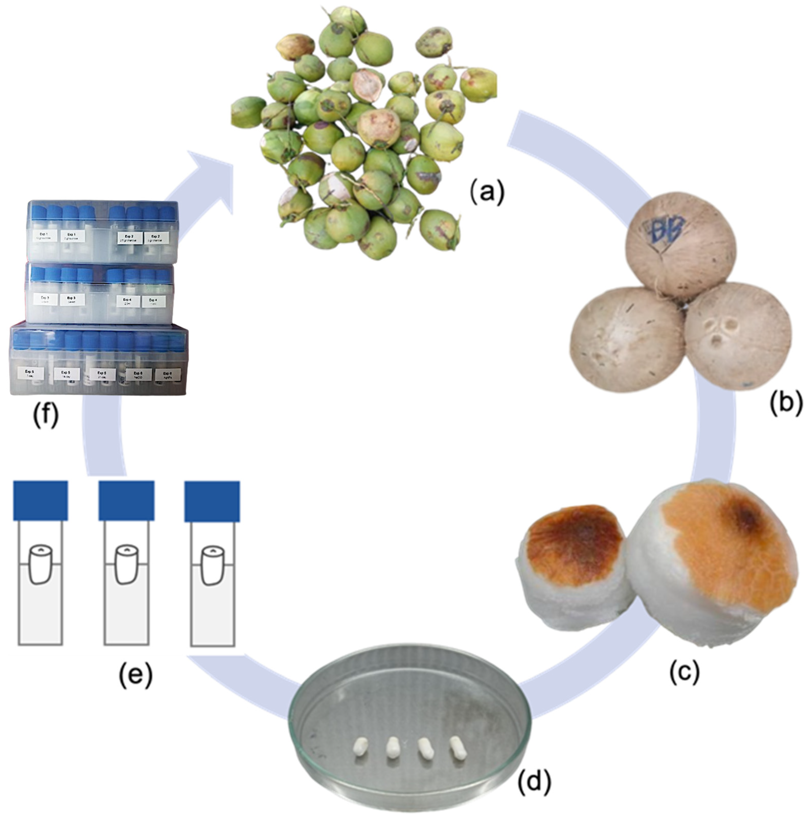
2.1. Plant Materials and Basal Medium Preparations

Makapuno coconut fruits aged between 10 to 12 months were collected in the Cau Ke district (9.92517° N, 106.05305° E, Tra Vinh province, Vietnam). Each fruit was carefully processed by dehusking and splitting with a machete in the field. The endosperm plugs containing embryos were then extracted using a sterilized cork borer, thoroughly washed with 70% ethanol (Sigma-Aldrich, St. Louis, MO, USA), and transferred to clean jars. All jars were transported to the tissue culture laboratory of Growlab Company Limited (Ho Chi Minh City, Vietnam) for further processing. At the laboratory, the plugs were placed in a sterile container inside a laminar airflow hood and rinsed with 70% ethanol. The embryos were isolated using forceps and a scalpel blade, then placed in sterile glass tubes. Surface sterilization was established with either sodium hypochlorite (NaOCl, 0.5% w/v; (Sigma-Aldrich, St. Louis, MO, USA)) or silver nanoparticles (AgNPs, 50 ppm; Sigma-Aldrich, St. Louis, MO, USA)) for 7 min with agitation. After three rinses with sterile water, the embryos were drained on autoclaved tissue paper sheets for 5 to 10 min and transferred to 2 mL cryotubes containing culture media tailored to different treatments. This method was described in detail in previous studies [24,25,26], and further improvements were made based on the existing protocol. The photos in this study were taken by a Nikon SMZ745T camera microscope (400× magnification; Nikon microscope, Melville, NY, USA), and lighting was provided by a Conviron CM8502 growth chamber (Conviron, Winnipeg, MB, Canada).
Culture media was prepared three days ahead of use, using tissue-culture-graded chemicals from Sigma-Aldrich (St. Louis, MO, USA) and Duchefa Biochemie (BH Haarlem, Netherlands). The basal medium for all experiments consists of Y3 macro- and micro-nutrients [27], MS vitamins [28], Gelrite (2 g/L), and activated charcoal (AC, 2.5 g/L, used only in one treatment). The formula underwent specific modifications based on the experimental design. Each medium was adjusted to pH 5.7 then autoclaved at 121 °C for 20 min.

2.2. Experiment Set-Up

The experiment was jointly designed by the Growlab Company Limited (GL), Ho Chi Minh City, Vietnam, and the School of Breeding and Multiplication (Sanya Institute of Breeding and Multiplication, abbreviated as SIBM), Hainan University, Sanya City, China. All experimental procedures were conducted in GL (Vietnam), with observations being recorded one month following the initiation of the experiments in the same place (refer to Figure 1); data processing and other procedures were conducted in SIBM (China). Coconut embryos were stored in 2 mL cryotubes and carried in compact containers (as depicted in Figure 1f). After several days of storage (based on the experiment) under dark conditions and 27 ± 2 °C to simulate the transportation processes, all coconut embryos were transferred to embryo culture medium as described by Biddle (2020) for recovery and further development [29]. A total of six experiments were undertaken, each employing distinct cultural conditions to establish an experimental protocol, as described in Table 1 below.

2.3. Assessment and Data Analysis
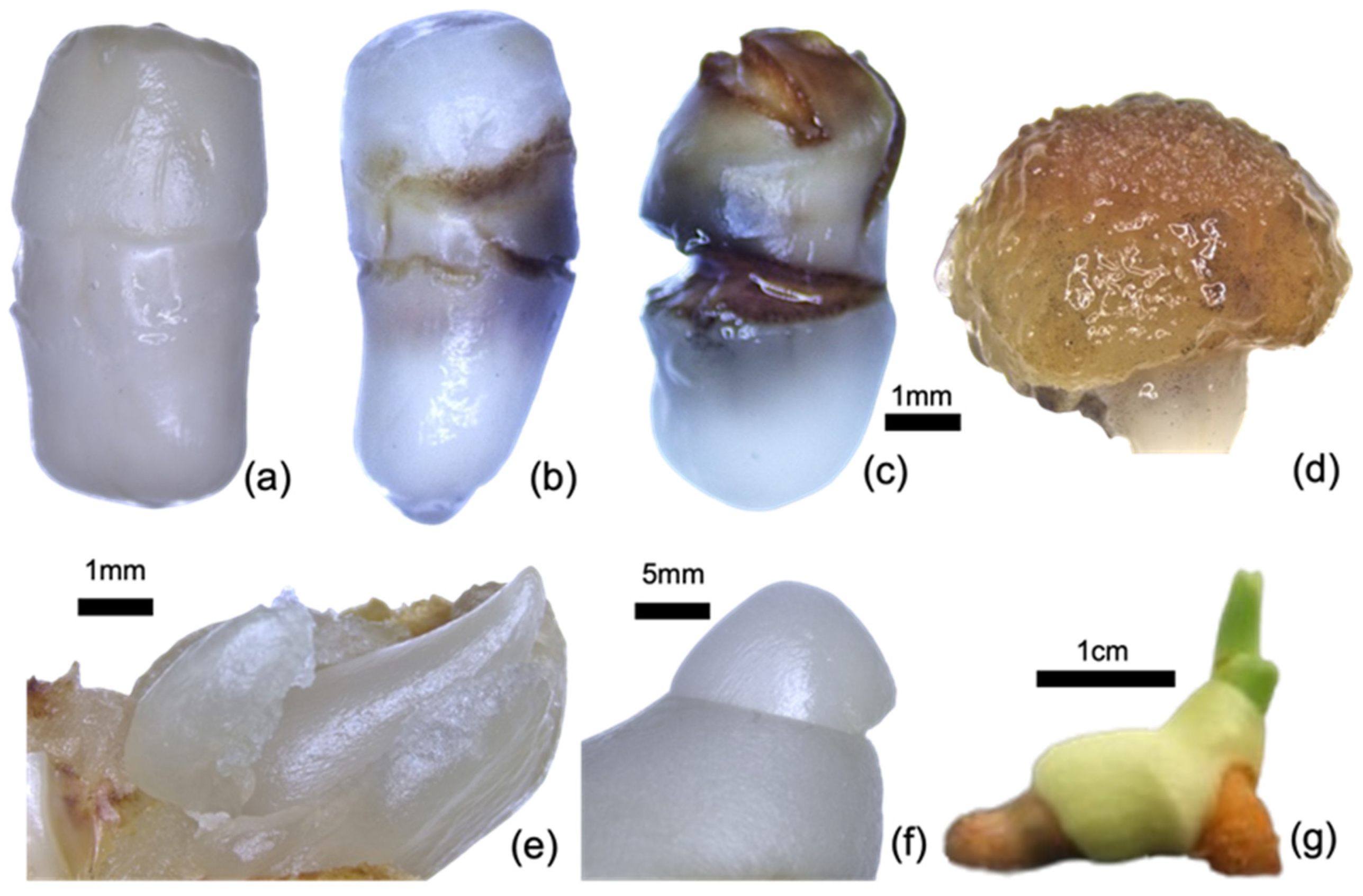
Two assessment processes were established for all experiments. The first observation was conducted at 7, 10, and 14 days after the end of the transportation stimulation and initiation of embryo culture process to evaluate the behaviors of coconut embryos, such as vigorous growth, contaminations, browning or necrosis, and vitrification (as shown in Figure 2a–c) during recovery. After 30 days of embryo culture, a second comprehensive assessment was carried out, focusing on the growth parameters of explants such as embryo germinations, embryo enlargement, and the formation of shoots and roots. The detailed formula of growth parameter calculation is indicated below, with statistical analysis performed using Student’s t-test and Analysis of Variance (ANOVA) by Microsoft Excel for Mac, version 16.78. The calculation of indexes is detailed in Mu (2024) [4]. The morphological observation of Makapuno embryos is detailed below.

3. Results

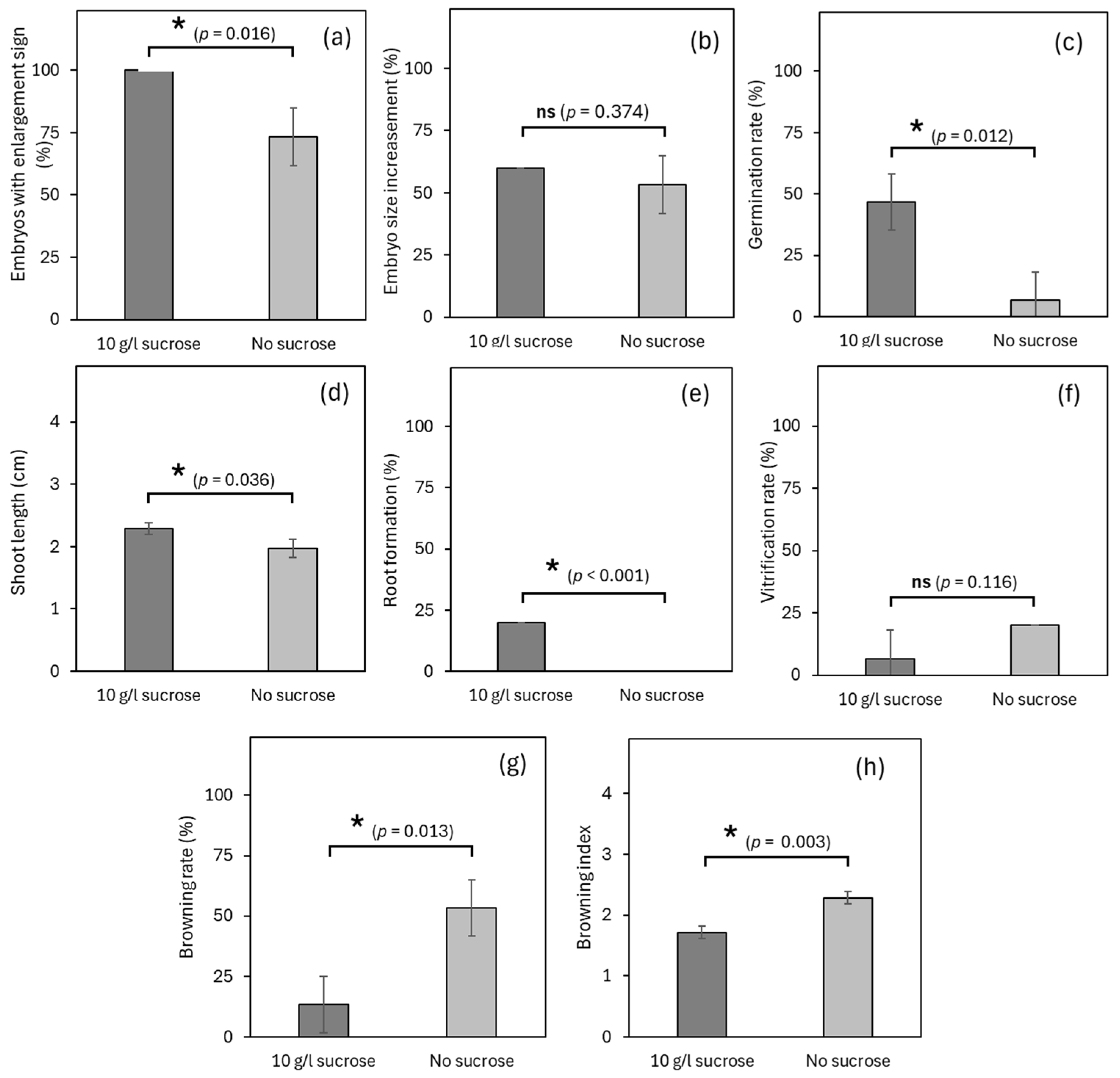
3.1. Experiment 1: Effects of Sucrose Concentration on Germplasm Exchange

To determine the necessity of exogenous sucrose during coconut germplasm exchange, Experiment 1 was conducted with a 7-day simulation of germplasm exchange, followed by the assessment processes as mentioned in Section 2.3. All embryos in the ST exhibited notable enlargement, being 60.0% longer compared to the original size after the simulation, whereas those from the NST only had a rate of 73.3%, being 53.3% longer compared to original size (Figure 3a,b). For the germination rate, ca. 46.7% of embryos in the sucrose treatment (ST) germinated in a month, significantly higher than those from the non-sucrose treatment (NST), which only saw ca. 6.7% of embryos germinated (Figure 3c). After 30 days of embryo culture, the length of germinated shoots from the ST reached 2.29 cm, which is significantly higher than the 1.98 cm for the NST (Figure 3d). Twenty percent of explants from the ST started to form primary root structures, while no root was found in any NST explants (Figure 3e). In terms of the physiological conditions of embryos, an average of 6.70% embryos experienced vitrification in the ST, while 20% of embryos were found to be vitrified from the NST; however, no statistical differences were recorded (Figure 3f). On the other hand, the NST resulted in a notable browning rate of 53.3% with a browning index of 2.28, while these parameters were significantly lower in the ST (only 13.3% and 1.72, respectively), as described in Figure 3g,h.

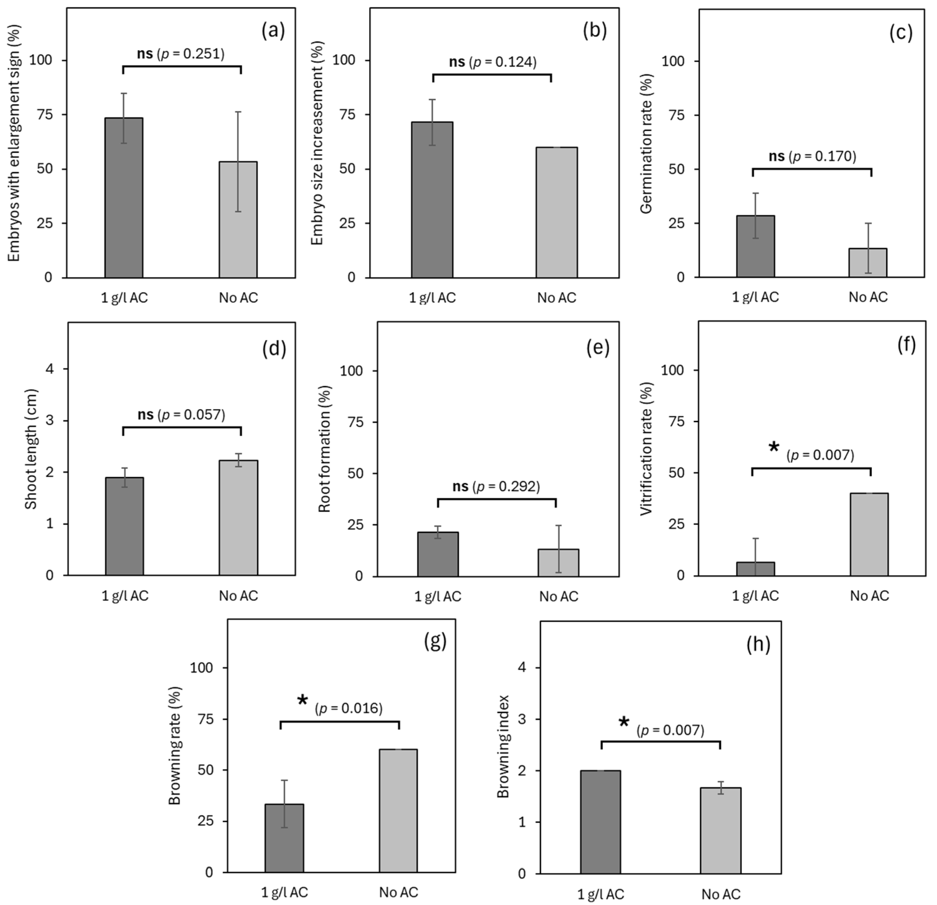
3.2. Experiment 2: Effects of Activated Charcoal on Germplasm Exchange

Next, an experiment was conducted to determine the need of activated charcoal during in vitro exchange of the coconut germplasm. All embryos were also maintained under a 7-day simulation of germplasm exchange, followed by the assessment processes as mentioned in Section 2.3. When using activated charcoal (AC) in the exchange medium, 73.3% of treated embryos showed enlargement responses, with a 71.4% increase in size compared to original explants. This is slightly higher than those in non-activated charcoal (NA) medium, with only 53.3% of embryos showing enlargement and a 60% increase in size (Figure 4a,b); however, the differences were not statistically significant. Similarly, a higher germination rate (28.6%) was found for AC treatment after 30-day recovery in embryo culture conditions, compared with a lower germination rate of 13.3% for NA treatment (Figure 4c). Considering post-germination development, both treatments resulted in no significant differences regarding the length of germinated shoots (1.90 cm for AC and 2.23 cm for NA) and the root formation rate (21.4% for AC and 13.3% for NA) (Figure 4d,e). Meanwhile, the presence of AC demonstrated a significant prevention of vitrification, in which only 6.7% of embryos were vitrified compared to 40.0% in those of NA (Figure 4f). Similar results were found for the explant browning rate, with 33.3% of embryos inoculated with AC showing notable browning, which was significantly lower than those from the NA group (60.0%). Interestingly, the intensity of browning was significantly higher for AC than for NA (Figure 4g,h).

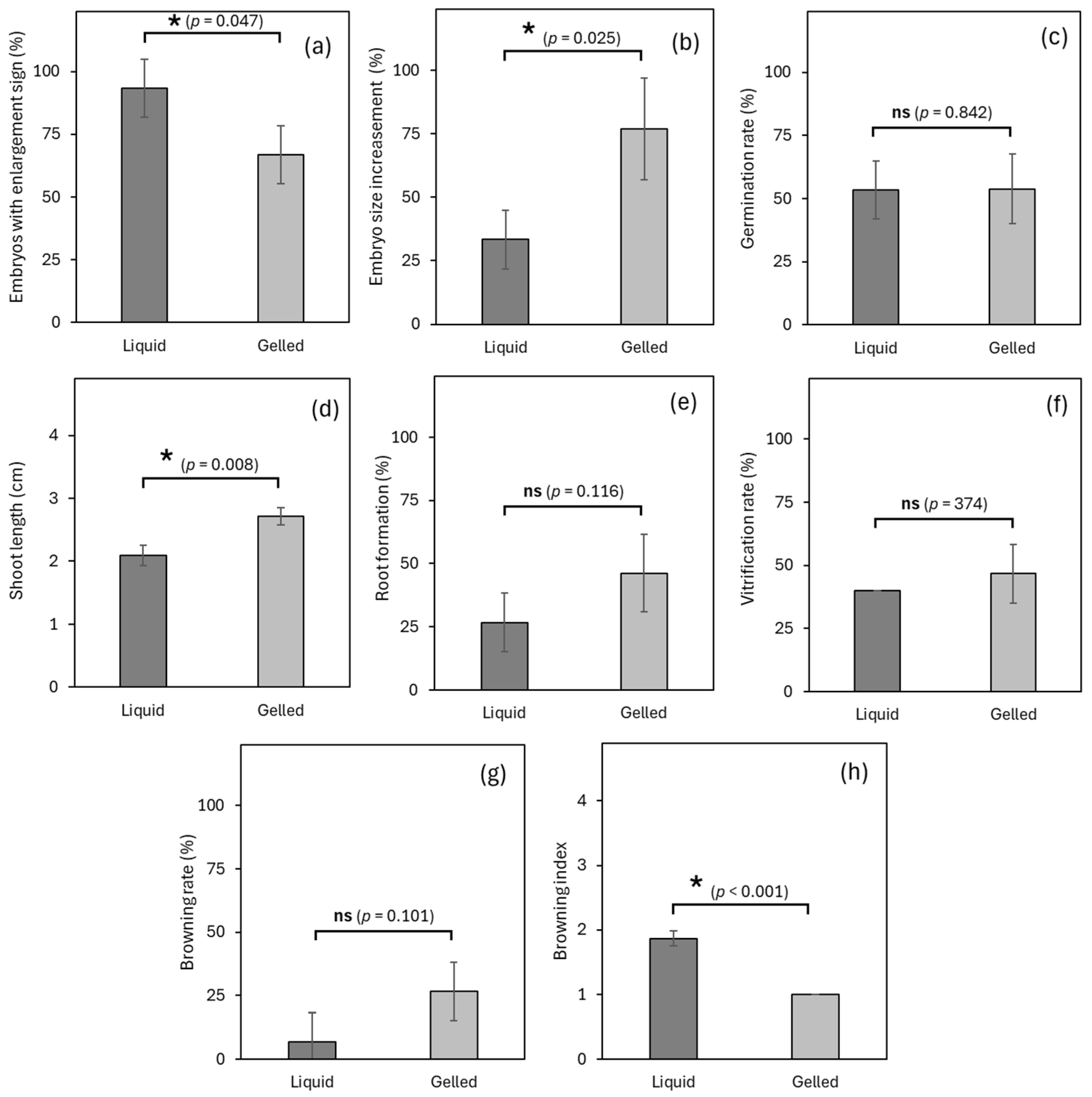
3.3. Experiment 3: Effects of Liquid Medium/Solid Medium on Germplasm Exchange

Experiment 3 illustrated comparative results between liquid and gelled media for coconut embryo germplasm exchange after a 7-day simulation. The results in Figure 5a show that the majority of embryos (93.3%) in the liquid medium (LM) exhibited a enlargement response, while a significantly lower percentage (66.7%) was recorded in gelled medium (GM). However, the size increase of embryos in GM (76.9%) significantly outperformed that of LM (33.3%), indicating notable reverse effects in embryo enlargement between the two treatments (Figure 5b). After 30 days of embryo culture, there was no significant difference in embryo germination rate between LM and GM (53.3% and 53.9%, respectively) (Figure 5c). Figure 5d represents shoot length after incubation, with significantly greater growth observed in GM (2.7 cm) than in LM (2.1 cm). A similar trend was also recorded for the vitrification phenomenon, with no statistical variation between the LM and GM (40.0% and 46.7%, respectively) after 30 days of culture (Figure 5e). Although embryos in GM appeared to have more browning (Figure 5g), their browning index was significantly lower compared to LM, at 1.0 vs. 1.9, respectively (Figure 5f). Regarding the browning aspect, Figure 5g,h presents the frequency and intensity of tissue browning between LM and GM, which shows a higher browning rate in GM (26.7%) than in LM (6.7%), although not statistically significant. However, the embryos had a significantly higher browning index in LM (1.9) than GM (1.0).

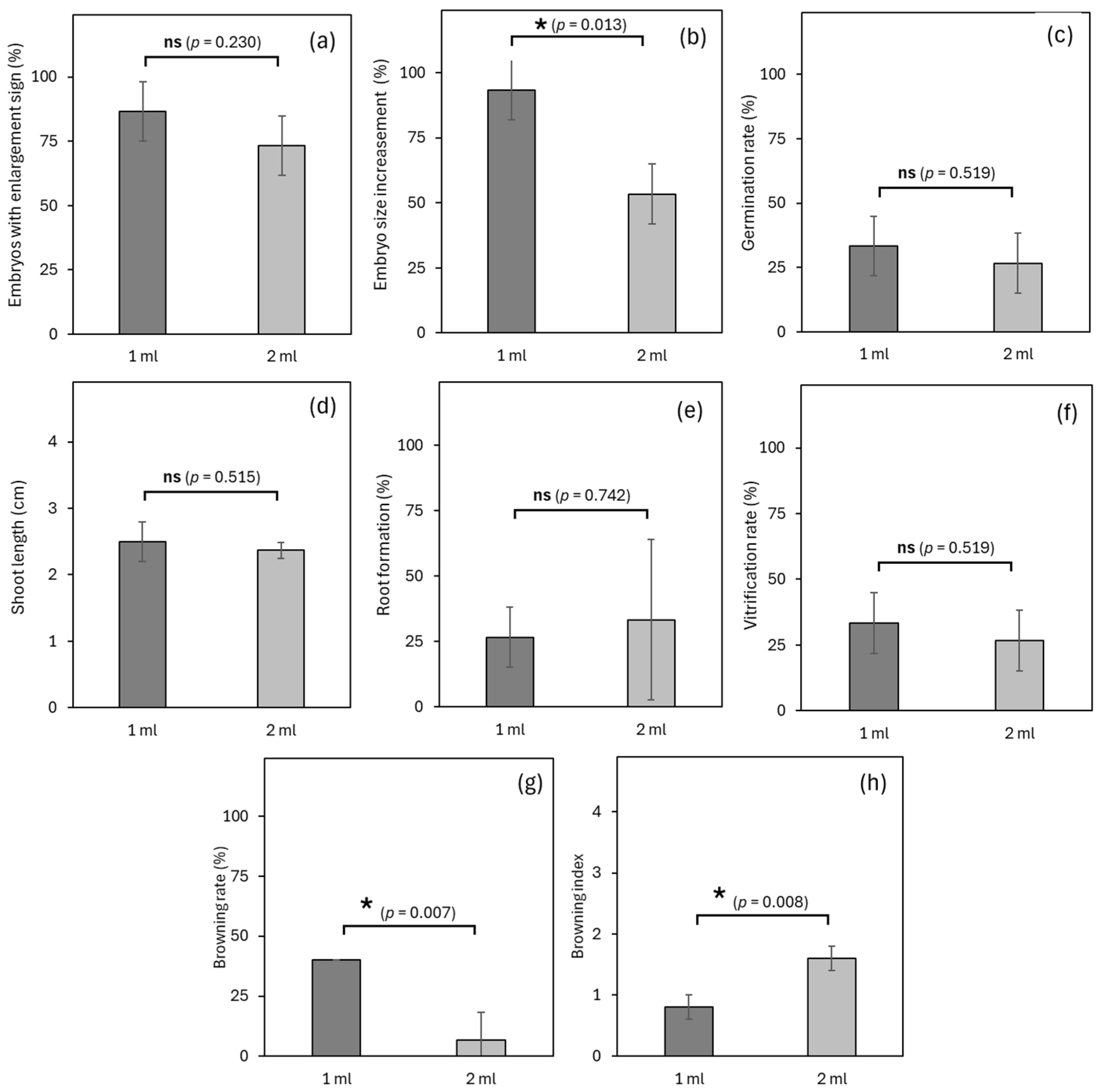
3.4. Experiment 4: Effects of Medium Volume on Germplasm Exchange

Experiment 4 was conducted to determine the suitable amount of culture medium needed for the efficient but cost-effective germplasm exchange of coconut embryos. The enlargement response of embryos after the simulation of germplasm exchange is highlighted in Figure 6b,c. The embryo size increase in T1 significantly outperformed T2 by 40% (93.3% and 53.3%, respectively). Similar results were recorded for shoot length, root formation rate, and vitrification rate, without any statistical significances despite the variation in mean values (Figure 6d–f). Figure 6g,h on the other hand, presents an interesting observation between the two treatments regarding the browning rate and index, in which T1 appeared to have significantly more browning explants (40.0%) than T2 (6.7%), while treated embryos in T2 conversely had a significantly higher browning index (1.6) compared to T1 (0.8).

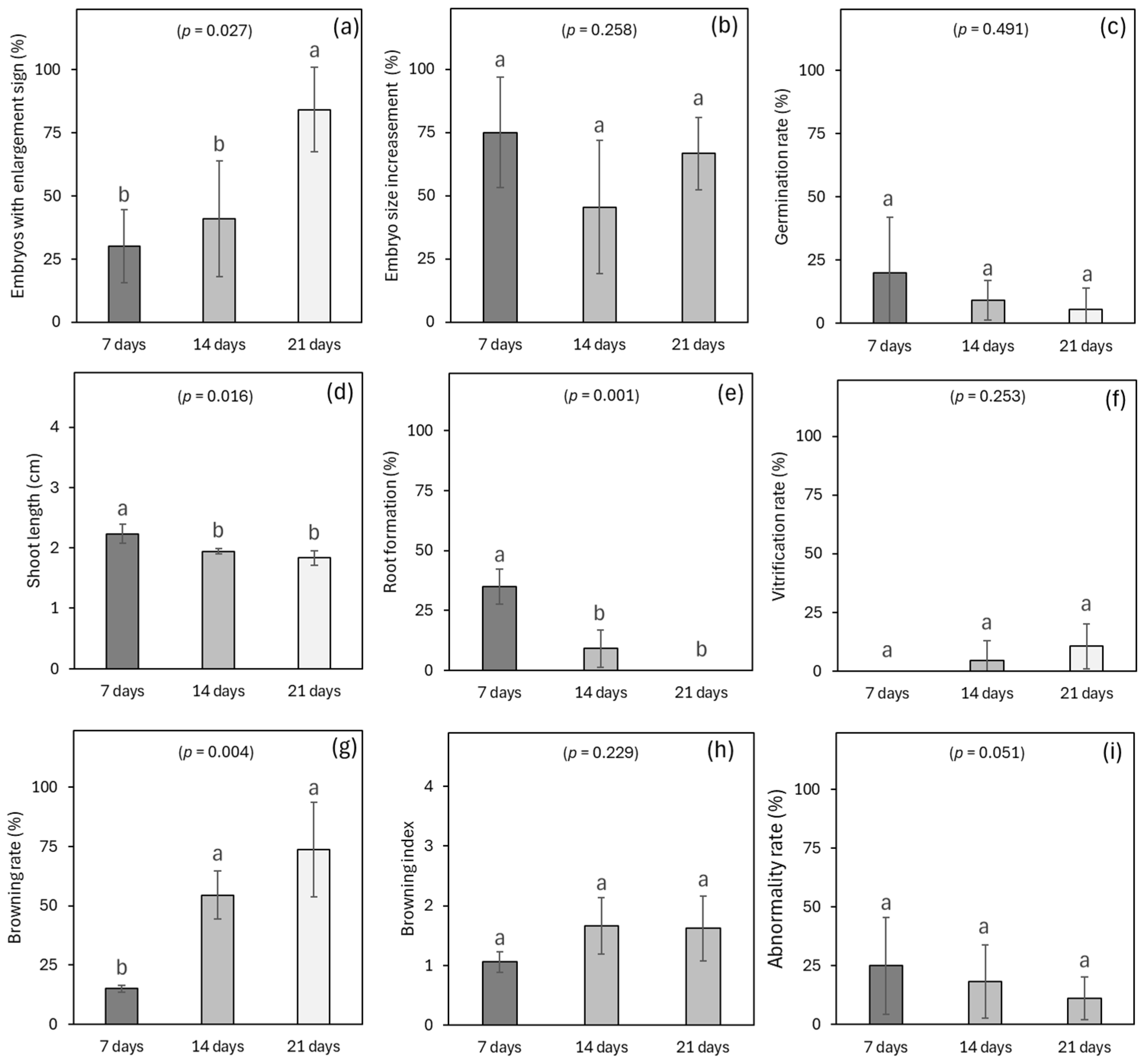
3.5. Experiment 5: Effects of Simulated Transportation Time on Germplasm Exchange

To evaluate the efficiency of the exchange protocol for various transportation routes and periods, different simulation durations, including 7 days (T7), 14 days (T14), and 21 days (T21), respectively represented the short, medium, and long routes, with the results presented in Figure 7. Figure 7a indicates that longer storage time during transportation, such as T21, showed a significantly greater percentage of embryos with enlargement signs (84.2%) compared to shorter durations such as T14 (40.9%) and T7 (30.0%) despite sharing a similar increase in enlarged embryos compared to the original explant. Considering germination capacity, the average germination rate of the coconut embryos gradually declined under longer transportation periods, from 20.0% T7 to 9.1% and 5.6% for T14 and T21, respectively. For the shoot development, the highest values of shoot length were recorded for T7 (2.2 cm), significantly greater than all longer transportation durations, such as T14 (1.94 cm) and T21 (1.83 cm) (Figure 7d,e). The same trend was noted in root development, with 35% of explants forming roots after 30 days of culture, which is significantly higher than T14 (9.1%), while there was no root observed in T21. The simulation of long transportation time also exhibited negative effects on the embryos by increasing the tissue browning rate, with significantly higher browning rate in T21 and T14 (73.7% and 54.6%, respectively) compared to T7 (15.0%), while a similar browning index was recorded among treatments, without any significant differences. (Figure 7g,h). Apart from that, the vitrification rate was also evaluated, as described in Figure 7f. It is worth noting that the average rate of embryo vitrification gradually increased as the transportation time became longer (10.5% for T21, 4.6% for T14), denoting the potential negative effects on the exchange of coconut embryos if transportation becomes longer than 21 days.

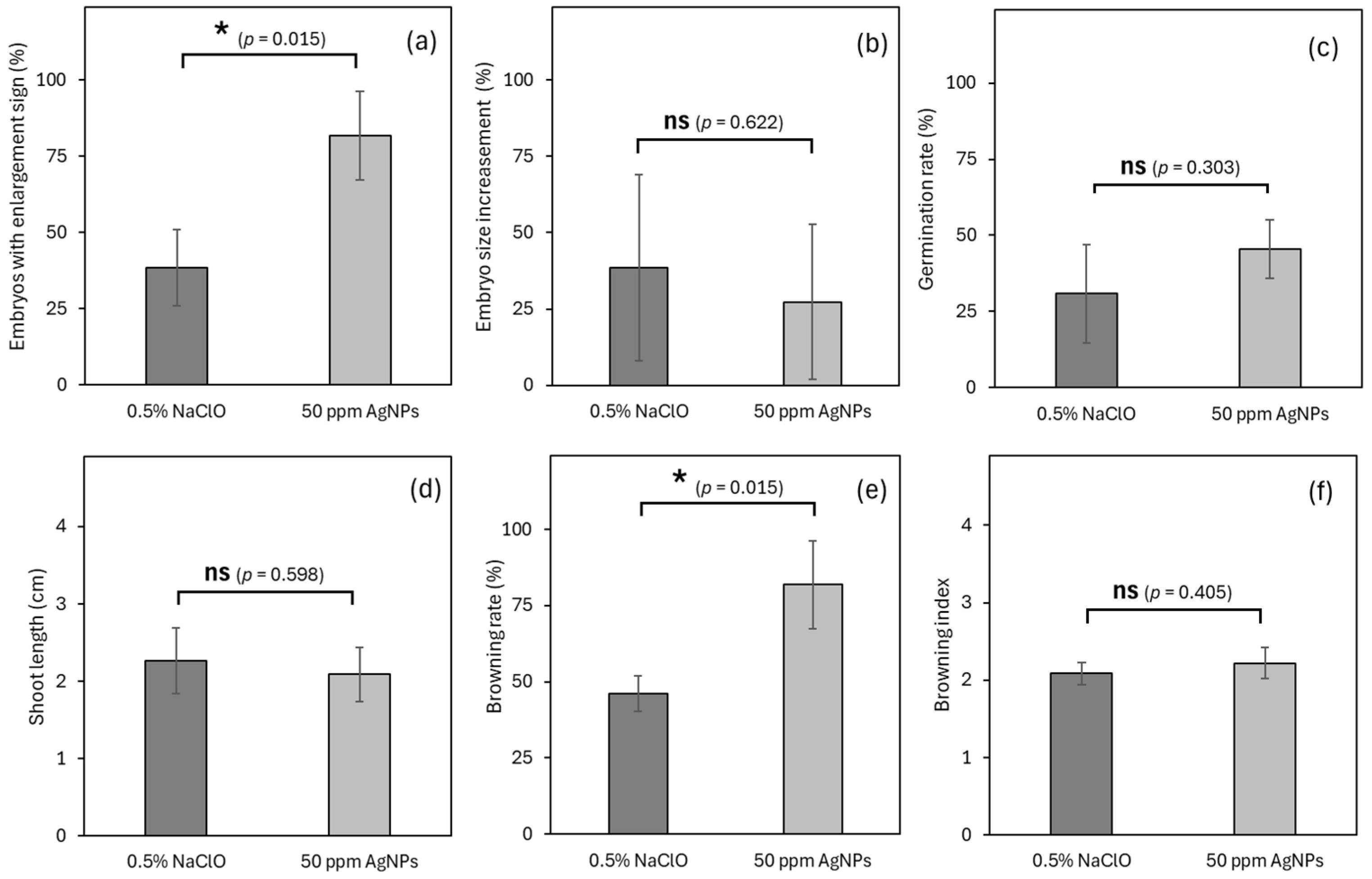
3.6. Experiment 6: Effects of Sterilant on Germplasm Exchange

To determine the most appropriate surface with regard to the efficiency of coconut embryo exchange, the two popular sterilants, i.e., NaOCl and AgNPs, were selected for this experiment. Both chemicals were effective in the surface sterilization of coconut embryos, since no contamination was found throughout the entire experiment., It was also observed that 81.8% of explants sterilized with AgNPs demonstrated enlargement after 30 days of culture, which is significantly higher than only 38.5% from NaOCl (Figure 8a). Meanwhile, NaOCl treatment showed a greater size increase in embryos, with 38.5% larger than their original size, while only 27.3% were enlarged in AgNPS treatment; still, no significant differences were indicated between treatments (Figure 8b). Similar observations were also noted for embryo germination rate (30.8% and 45.5%) and shoot length (2.3 and 2.1) between explants treated with NaOCl and AgNPs, respectively, lacking statistical differences (Figure 8c,d). In addition, browning was significantly lower in NaOCl (46.1%) than that in AgNPS (81.8%), but a similar browning index was observed in both NaOCl and AgNPS treatments (2.1 and 2.2, respectively), with no significant differences (Figure 8e,f). The vitrification and root formation rate for both treatments were very low (under 10% or even 0) and are not shown in Figure 8.

4. Discussion and Future Directions

Pathogen-free germplasm exchange is extremely important for the sharing and preservation of biodiversity around the world while maintaining critical biosecurity [13]. The utility of in vitro embryo culture is vital for international germplasm exchange, including the ability to bypass phytosanitary restrictions that typically accompany the transport of whole seeds or plants [30]. Hence, embryo culture was used as the standard procedure for the coconut germplasm in this study, while several related factors such as sucrose, activated charcoal, physical state and volume of culture medium, transportation duration, and chemical sterilants were evaluated to optimize the exchange procedure and evaluate the applicability of this procedure, particularly for the renowned Makapuno variety.
During tissue culture, exogenous sucrose is mainly supplemented as the major source of carbon and energy for plant tissues to grow and develop; however, this is not preferable for germplasm preservation and transfer due to the potential risk of microbial contamination. In coconut embryo culture, it is crucial to apply sucrose to culture media at high concentrations for the growth and development of coconut tissues [31]. Therefore, a complete depletion of sucrose may severely affect the survival and recovery of the coconut embryo during germplasm exchange, especially for long-distance routes. In this study, a comprehensive comparison in Experiment 1 between simulation conditions with or without the addition of sucrose indicated that the presence of 10 g/L sucrose notably improved the enlargement and germination of coconut embryos, facilitated post-germination shoot and root growth, and reduced browning and necrosis issues compared to the control sucrose-free procedure, with no contamination occurring. This result suggests that a small addition of sucrose into culture media during exchange could preserve the vigor of Makapuno embryos for subsequent recovery and regeneration as well as stimulate the embryo’s metabolism system to withstand oxidative stresses during this period.
Another exogenous supplement, activated charcoal (AC), is highly favorable during the process of embryo exchange due to its ability to absorb phenolic compounds and prevent oxidative stresses for plant tissues [29,32]. In Experiment 2 of this study, the use of AC in the culture medium for embryo exchange did not significantly affect any growth and development metrics of Makapuno zygotic embryos; however, the presence of AC significantly mitigated browning and necrosis issues in coconut embryos while resulting in a notably high vitrification phenomenon. As a result, it was clearly noticed that AC acted as an extremely beneficial substance to prevent oxidative stress during germplasm exchange. Still, the mechanism behind the rising vitrification response was yet unknown, and further investigation is needed to determine if this abnormality affects the final outcome of embryo exchange of coconut embryos.
The physical state of the culture medium is an important factor for maximizing the tissue growth. Depending on the species, types of tissues, and phase of culture, either liquid medium or gelled (or solidified) medium would be selected for optimal efficiency. In coconut, both liquid and gelled media are applied for in vitro culture of zygotic embryos; however, liquid media are often preferred due to higher contact between tissues and medium, evenly distributed nutrients, ease in preparation, and no need of a gelling agent [31]. For the current study, the liquid medium demonstrated a significantly higher rate of embryos showing an enlargement response after a 7-day simulation of germplasm exchange, indicating that the liquid medium could potentially facilitate water and nutrient uptake into the embryos and lead to more enlarged embryos. However, no further improvements regarding embryo germination and regeneration were recorded in the liquid medium compared to the standard gelled medium. Meanwhile, embryos maintained in culture medium gelled with Gelrite showed a significantly larger size increase, better shoot elongation, and lower browning index. Similar results were reported with high survival rates of coconut embryos during transport when cultured in a semi-solid medium, underlining the robustness of this method for long-distance germplasm exchange [7]. The probable explanation for these observations could be the stability of the embryo inoculation position during storage duration, since only the gelled medium was able to maintain the embryos at a fixed position in the cryotubes, which resulted in less mechanical damage to embryos during the storage or transportation process compared to liquid medium. Appropriate embryo orientation was also believed to significantly affect the growth and development of coconut embryos according to the findings [33].
Apart from medium supplementations, the amount (volume) of culture medium for each embryo/cryotube also affected the response of coconut embryos under germplasm exchange. The results from Experiment 4 of this study demonstrated no significant differences between the use of either 1 or 2 mL of culture medium for most of the assessment criteria. However, it was evident that the browning rate was significantly higher when using less culture medium (1 mL) compared to the other treatment (2 mL), while a reverse observation was recorded regarding the browning index of embryos from each treatment, suggesting the volume of culture medium may interfere with the browning process on coconut embryos under a different mechanism. Unfortunately, this remains unclear in this current study. Previous research suggested to use 5–10 mL of medium during coconut embryo preservation and transport to ensure proper gas exchange and embryo growth [33]. However, a higher volume of the medium will increase the size of the packaging and the cost for an excess amount of medium, thereby greatly reducing convenience and the efficiency of germplasm transportation. Therefore, in future research, we also need to find a balance between the culture medium volume and the storage/transportation time to obtain the optimal procedure of embryo exchange.
Different from other factors, transportation duration in embryo exchange heavily relies on origin and destination and is not always adjustable. Experiment 5 showed Makapuno embryo viability and regeneration capacity decline over time, especially after 21 days, with shorter shoot growth, no root formation, and rapid browning. Thus, delivery within less than 21 days is recommended. For surface sterilization, NaOCl and AgNPs both showed outstanding efficiency, but AgNPs promoted better embryo enlargement and slightly higher germination rate, though with higher browning. Further study on combining AgNPs with AC or antioxidants is suggested to address browning.
Previous research has demonstrated the importance and effectiveness of various techniques in the successful exchange and propagation of coconut germplasm [10,11,16]. Cryopreservation has emerged as a viable method for the long-term storage and exchange of coconut germplasm. Previous research demonstrated that coconut embryos can be successfully cryopreserved and later regenerated, preserving genetic diversity without significant loss of viability [34]. A case study documented the successful introduction of new coconut varieties in Brazil through embryo culture, which contributed to enhanced genetic diversity and improved resistance to local pests and diseases [35]. Despite these advances, challenges remain in the exchange of coconut germplasm. Browning and necrosis of coconut embryos during in vitro culture and transport are persistent issues. To mitigate these problems, the use of antioxidants and optimized culture media has been recommended. For instance, the application of ascorbic acid and polyvinylpyrrolidone (PVP) has been shown to reduce oxidative stress and improve the viability of cultured embryos [36].
Coconut holds significant importance for China, Vietnam, and other tropical countries, serving as a vital agricultural product and an essential component of local economies [37,38]. The growing coconut trade between China and Vietnam highlights the increasing economic interdependence of the two countries [39]. The exchange of germplasm resources is crucial in this trade, necessitating an efficient and reliable method for transporting coconut embryos. In response to this need, China and Vietnam have collaborated to develop an in vitro embryo transportation technique [40]. This innovative method ensures the safe and effective exchange of coconut germplasm, supporting the enhancement of coconut cultivation and trade between the two nations. Future research should focus on refining in vitro and cryopreservation techniques to further increase the efficiency and success rates of germplasm exchange. Moreover, the integration of molecular markers and genomic tools can enhance the selection and propagation of desirable traits in exchanged germplasm. Collaborative international efforts and the establishment of more germplasm exchange networks will be crucial in addressing the global challenges facing coconut cultivation [41].

5. Conclusions

In conclusion, an embryo culture protocol was successfully applied on Makapuno zygotic embryos for germplasm exchange under simulating conditions. A total of six factors were examined towards an efficient and cost-effective protocol for embryo exchange. The results showed that the suitable culture medium for embryo exchange was supplemented with small sucrose concentration (10 g/L) and activated charcoal (1 g/L), gelled with Gelrite (2.5 g/L), and at the volume of 1 mL for each cryotube (eq. one embryo). Both NaClO and AgNPs were effective for surface sterilization, though the difference was not statistically significant. For the effective duration of transportation, it was suggested to deliver all embryos in less than 7 days for maximal quality, as lower viability and regeneration would be achieved after longer duration. These findings would be the fundamental knowledge for the success of germplasm exchange for elite varieties, towards genetic conservation and varietal improvement of the coconut palm.

Author Contributions

Experiment designed by Z.M. and B.-M.T.; the two authors contributed equally. Experiments conducted by B.-M.T., Z.M., T.T.-T.P., M.-A.L., S.Y. and X.W. The first draft of the manuscript was written by Z.M. and S.Y., and all authors commented on previous versions of the manuscript. M.T.N.I., P.T.N. and J.L. contributed to the review and editing. All authors have read and agreed to the published version of the manuscript.

Funding

This work was supported by the Project of Sanya Yazhou Bay Science and Technology City, Grant No: SCKJ-JYRC-2024-35, Science and Technology Innovation Special Project of Sanya City (2022KJCX53), and “111” Project (No. D20024).

Data Availability Statement

The datasets generated during and/or analyzed during the current study are available from the corresponding author on reasonable request.

Acknowledgments

Thanks are extended to Growlab Company Limited and Sanya Kelaite Co., Ltd. for their support.

Conflicts of Interest

Authors Tran Binh-Minh, Pham Thi Thanh-Thuy and Minh-An Le were employed by the company Growlab Company Limited. The remaining authors declare that the research was conducted in the absence of any commercial or financial relationships that could be construed as a potential conflict of interest.

Abbreviations

The following abbreviations are used in this manuscript:
AgNPsSilver nanoparticles
NaOClSodium Hypochlorite
COGENTInternational Coconut Genetic Resources Network
KClPotassium Chloride
HCMCHo Chi Minh City
GLGrowlab Company Limited
SIBMSanya Institute of Breeding and Multiplication
ACActivated Charcoal
ANOVAAnalysis of Variance
STSucrose Treatment
NSTNo Sucrose treatment
NANon-Activated Charcoal
LMLiquid Medium
GMGelled Medium

References

  1. Yang, Z.; Liu, Z.; Xu, H.; Li, Y.; Huang, S.; Cao, G.; Shi, M.; Zhu, J.; Zhou, J.; Li, R.; et al. ArecaceaeMDB: A comprehensive multi-omics database for Arecaceae breeding and functional genomics studies. Plant Biotechnol. J. 2023, 21, 11. [Google Scholar]
  2. Mu, Z.; Tran, B.-M.; Xu, H.; Yang, Z.; Qamar, U.Z.; Wang, X.; Xiao, Y.; Luo, J. Exploring the potential application of coconut water in healthcare and biotechnology: A review. Beverage Plant Res. 2024, 4, 1–9. [Google Scholar]
  3. Sun, X.; Kaleri, G.A.; Mu, Z.; Feng, Y.; Yang, Z.; Zhong, Y.; Dou, Y.; Xu, H.; Zhou, J.; Luo, J.; et al. Comparative Transcriptome Analysis Provides Insights into the Effect of Epicuticular Wax Accumulation on Salt Stress in Coconuts. Plants 2024, 13, 141. [Google Scholar] [CrossRef]
  4. Mu, Z.; Yang, S.; Xu, H.; Yang, Z.; Haque, M.M.; Tran, B.-M.; Chen, J.; Wang, X.; Peng, H.; Luo, J. The Influence of Maturity, Storage, and Embryo Size on Coconut Callus Induction Success. Forests 2024, 15, 764. [Google Scholar] [CrossRef]
  5. Welewanni, I.; Bandupriya, D. Coconut cryopreservation: Present status and future prospects. Cord 2017, 33, 21. [Google Scholar]
  6. Salum, U.; Foale, M.; Biddle, J.; Bazrafshan, A.; Adkins, S. Towards the Sustainability of the “Tree of Life”: An Introduction. In Coconut Biotechnology: Towards the Sustainability of the ‘Tree of Life’; Springer: Berlin/Heidelberg, Germany, 2020; pp. 1–15. [Google Scholar]
  7. Karun, A.; Ramesh, S.V.; Rajesh, M.K.; Niral, V.; Sudha, R.; Muralikrishna, K.S. Conservation and Utilization of Genetic Diversity in Coconut (Cocos nucifera L.). In Cash Crops: Genetic Diversity, Erosion, Conservation and Utilization; Springer: Berlin/Heidelberg, Germany, 2022; pp. 197–250. [Google Scholar]
  8. Bazrafshan, A. Ex Situ Conservation of Coconut (Cocos nucifera L.) Germplasm Using Cryopreservation. Ph.D. Thesis, The University of Queensland, St Lucia, QLD, Austrilia, 2022. [Google Scholar]
  9. Sthapit, B.; Rao, V.R.; Jarvis, D. In situ conservation of coconut diversity. In Coconut Genetic Resources; IPGRI-APO: Serdang, Malaysia, 2005; p. 149. [Google Scholar]
  10. Arumugam, T.; Hatta, M.A.M. Improving coconut using modern breeding technologies: Challenges and opportunities. Plants 2022, 11, 3414. [Google Scholar] [CrossRef] [PubMed]
  11. Lédo, A.d.S.; Oliveira, L.A.; Machado, C.D.; Silva, A.V.; Ramos, S.R. Strategies for exchange of coconut germplasm in Brazil. Ciência Rural. 2017, 47, e20160391. [Google Scholar]
  12. Malaurie, B. Medium-and long-term conservation and safe international exchange of germplasm from food and cash tropical crops. In Proceedings of the IV International Symposium on In Vitro Culture and Horticultural Breeding, Tampere, Finland, 2–7 July 2000; p. 560. [Google Scholar]
  13. Yang, C.; Nguyen, V.A.; Nulu, N.P.C.; Kalaipandian, S.; Beveridge, F.C.; Biddle, J.; Young, A.; Adkins, S.W. Towards pathogen-free coconut germplasm exchange. Plants 2024, 13, 1809. [Google Scholar] [CrossRef]
  14. Sisunandar; Rival, A.; Turquay, P.; Samosir, Y.; Adkins, S.W. Cryopreservation of coconut (Cocos nucifera L.) zygotic embryos does not induce morphological, cytological or molecular changes in recovered seedlings. Planta 2010, 232, 435–447. [Google Scholar]
  15. Kong, E.Y.; Mu, Z.; Vidhanaarachchi, V.R.M.; Nguyen, Q.T.; Sisunandar, S.; Kalaipandian, S.; Panis, B. Chapter 10 Coconut Biotechnology. In The Coconut: Botany, Production and Uses; CABI Publishing: Wallingford, UK, 2024; p. 143. [Google Scholar]
  16. Samosir, Y.M.; Adkins, S. Improving acclimatization through the photoautotrophic culture of coconut (Cocos nucifera) seedlings: An in vitro system for the efficient exchange of germplasm. Vitr. Cell. Dev. Biol. Plant 2014, 50, 493–501. [Google Scholar]
  17. Sajini, K.; Karun, A.; Amamath, C.H.; Engelmann, F. Cryopreservation of coconut (Cocos nucifera L.) zygotic embryos by vitrification. CryoLetters 2011, 32, 317–328. [Google Scholar]
  18. McGregor, A. The export of horticultural and high-value agricultural products from the Pacific islands. Pac. Econ. Bull. 2007, 22, 81–99. [Google Scholar]
  19. Foale, M.; Julianne, B.; Amirhossein, B.; Steve, A. Biology, ecology, and evolution of coconut. In Coconut Biotechnology: Towards the Sustainability of the ‘Tree of Life’; Springer: Berlin/Heidelberg, Germany, 2020; pp. 17–27. [Google Scholar]
  20. Chan, E.; Elevitch, C.R. Cocos nucifera (coconut). Species Profiles Pac. Isl. Agrofor. 2006, 2, 1–27. [Google Scholar]
  21. Sharma, K.M.; Kumar, M.; Muralidharan, C.; Salomón-Torres, R. Date palm pollination management. Crop Prod. Sci. Hortic. 2023, 209–240. [Google Scholar]
  22. Karun, A.; Sajini, K.K.; Niral, V.; Amarnath, C.H.; Remya, P.; Rajesh, M.K.; Samsudeen, K.; Jerard, B.A.; Engelmann, F. Coconut (Cocos nucifera L.) pollen cryopreservation. CryoLetters 2014, 35, 407–417. [Google Scholar]
  23. Engelmann, F.; Malaurie, B.; N’Nan, O. In vitro culture of coconut (Cocos nucifera L.) zygotic embryos. In Plant Embryo Culture; Springer: Berlin/Heidelberg, Germany, 2011; pp. 63–72. [Google Scholar]
  24. Adkins, S.; Foale, M.; Bourdeix, R.; Nguyen, Q.; Biddle, J. Coconut Biotechnology: Towards the Sustainability of the ‘Tree of Life’; Springer: Berlin/Heidelberg, Germany, 2020. [Google Scholar]
  25. Mu, Z. Overcoming Bottlenecks in the Pathway of Clonal Propagation of Coconut (Cocos nucifera L.). Ph.D. Thesis, University of Queensland, St Lucia, QLD, Austrilia, 2022. [Google Scholar]
  26. Nguyen, T.Q. Clonal Propagation of Coconut (Cocos nucifera L.) for Elite Seedling Production and Germplasm Exchange. Ph.D. Thesis, University of Queensland, St Lucia, QLD, Austrilia, 2018. [Google Scholar]
  27. Eeuwens, C. Mineral requirements for growth and callus initiation of tissue explants excised from mature coconut palms (Cocos nucifera) and cultured in vitro. Physiol. Plant. 1976, 36, 23–28. [Google Scholar]
  28. Murashige, T.; Skoog, F. A revised medium for rapid growth and bio assays with tobacco tissue cultures. Physiol. Plant. 1962, 15, 473–497. [Google Scholar]
  29. Biddle, J.; Nguyen, Q.; Mu, Z.H.; Foale, M.; Adkins, S. Germplasm Reestablishment and Seedling Production: Embryo Culture. In Coconut Biotechnology: Towards the Sustainability of the ‘Tree of Life’; Springer: Berlin/Heidelberg, Germany, 2020; pp. 199–225. [Google Scholar]
  30. Ashburner, G.; Faure, M.G.; Tomlinson, D.R.; Thompson, W.K. A Guide to the Zygotic Embryo Culture of Coconut Palms (Cocos nucifera L.); Australian Centre for International Agricultural Research (ACIAR): Bruce, ACT, Australia, 1995.
  31. Nguyen, Q.T.; Bandupriya, H.D.; López-Villalobos, A.; Sisunandar, S.; Foale, M.; Adkins, S.W. Tissue culture and associated biotechnological interventions for the improvement of coconut (Cocos nucifera L.): A review. Planta 2015, 242, 1059–1076. [Google Scholar] [PubMed]
  32. Sáenz, L.; Herrera-Herrera, G.; Uicab-Ballote, F.; Chan, J.L.; Oropeza, C. Influence of form of activated charcoal on embryogenic callus formation in coconut (Cocos nucifera). Plant Cell Tissue Organ Cult. (PCTOC) 2010, 100, 301–308. [Google Scholar]
  33. Perera, P.; Hocher, V.; Verdeil, J.; Doulbeau, S.; Yakandawala, D.; Weerakoon, L. Unfertilized ovary: A novel explant for coconut (Cocos nucifera L.) somatic embryogenesis. Plant Cell Rep. 2007, 26, 21–28. [Google Scholar]
  34. Ake, A.P.; Souza, R.; Maust, B.; Santamaría, J.M.; Oropeza, C. Enhanced aerobic respiration improves in vitro coconut embryo germination and culture. Vitr. Cell. Dev. Biol. Plant 2004, 1, 90–94. [Google Scholar]
  35. Assy-Bah, B.; Engelmann, F. Cryopreservation of mature embryos of coconut (Cocos nucifera L.) and subsequent regeneration of plantlets. Cryoletters 1992, 13, 117–126. [Google Scholar]
  36. Lédo, A.d.S.; Ramos, S.; Diniz, L.; Konan, J.; George, M. Performance of coconut embryo culture accessions introduced at the International Coconut Genebank for Latin America and the Caribbean (ICG-LAC). In Proceedings of the XXVIII International Horticultural Congress on Science and Horticulture for People (IHC2010): III International Symposium on 918, Lisbon, Portugal, 22–27 August 2010. [Google Scholar]
  37. EL-Gioushy, S.; RLiu; Fan, H. A complete protocol to reduce browning during coconut (Cocos nucifera L.) tissue culture through shoot tips and inflorescence explants. Plant Arch. 2020, 20, 2196–2204. [Google Scholar]
  38. Bangkok Post PCL. Southeast Asian Coconuts Gain Popularity in China. 2022. Available online: https://www.bangkokpost.com/business/general/2424071/southeast-asian-coconuts-gain-popularity-in-china (accessed on 29 January 2024).
  39. Pratama, B.R.; Tooy, D.; Kim, J. Price Competition and Shifting Demand: The Relation between Palm and Coconut Oil Exports. Sustainability 2023, 16, 101. [Google Scholar] [CrossRef]
  40. Vietnam News Agency. Opportunity for Vietnam’s Coconut Exports to US, China. 2023. Available online: https://en.vietnamplus.vn/opportunity-for-vietnams-coconut-exports-to-us-china/271437.vnp (accessed on 29 January 2024).
  41. Mu, Z.; Peng, H.; Jayarathna, S.; Indrachapa, M.; Yang, C.; Yang, S.; Zhou, J.; Xu, W.; Jiang, Y.; Vidhanaarachchi, V.; et al. The coconut palm in China: Distribution, germplasm, and sustainable development through genomic and biotechnological approaches. Technol. Hortic. 2025, 5, e005. [Google Scholar]
Figure 1. The protocol applied for embryo transfer, demonstrated by timeline. (a) Mature Makapuno coconut nuts collected in Trà Vinh Province, Vietnam. (b) De-husked coconut fruits ready for plug extraction. (c) Endosperm plugs containing coconut embryos were extracted using a sterilized cork-borer. (d) Makapuno embryos after isolation and surface sterilization. (e,f) Embryos inoculated in cryotubes and compactly packed for the simulation of transportation.
Horticulturae 11 00816 g001
Figure 2. Responses of Makapuno embryos after the simulation of transportation. (a) A healthy embryo after one week of recovery. (b) A vitrified embryo showing the browning phenomenon, translucent peel, and softened tissue. (c) Brown/necrotic embryo facing severe oxidative stresses with abnormality occurrence. (d) An embryo that experienced simulated long-distance transportation (21 days), showing severe vitrification responses and abnormal enlargement in its upper part. (e) A severely vitrified shoot derived from tested embryos after 30 days of embryo culture. (f) A healthy-looking shoot from another tested embryo after 30 days of embryo culture. (g) A healthy germinated embryo after 2 months of embryo culture.
Horticulturae 11 00816 g002
Figure 3. The effects of exogenous sucrose on the growth of Makapuno embryos after the simulation of coconut (Cocos nucifera) germplasm exchange. * Indicates statistically significant differences between groups at p < 0.05, while “ns” indicates no statistically significant differences between groups at p ≥ 0.05. These data were measured: (a) Embryos with enlargement sign; (b) Embryo size increasement; (c) Germination ratel; (d) Shoot length; (e) Root formation; (f) Vitrification rate; (g) Browning rate; (h) Browning index.
Horticulturae 11 00816 g003
Figure 4. The effects of activated charcoal (AC) on the growth of Makapuno embryos after the simulation of coconut (Cocos nucifera) germplasm exchange. * Indicates statistically significant differences between groups at p < 0.05, while “ns” indicates no statistically significant differences between groups at p ≥ 0.05. These data were measured: (a) Embryos with enlargement sign; (b) Embryo size increasement; (c) Germination ratel; (d) Shoot length; (e) Root formation; (f) Vitrification rate; (g) Browning rate; (h) Browning index.
Horticulturae 11 00816 g004
Figure 5. The effects of liquid and gelled media on the growth of Makapuno embryos after the simulation of coconut (Cocos nucifera) embryo germplasm exchange. * Indicates statistically significant differences between groups at p < 0.05, while “ns” indicates no statistically significant differences between groups at p ≥ 0.05. These data were measured: (a) Embryos with enlargement sign; (b) Embryo size increasement; (c) Germination ratel; (d) Shoot length; (e) Root formation; (f) Vitrification rate; (g) Browning rate; (h) Browning index.
Horticulturae 11 00816 g005
Figure 6. The effects of different medium volumes on the growth of Makapuno embryos after the simulation of coconut (Cocos nucifera) embryo exchange. * Indicates statistically significant differences between groups at p < 0.05, while “ns” indicates no statistically significant differences between groups at p ≥ 0.05. These data were measured: (a) Embryos with enlargement sign; (b) Embryo size increasement; (c) Germination ratel; (d) Shoot length; (e) Root formation; (f) Vitrification rate; (g) Browning rate; (h) Browning index.
Horticulturae 11 00816 g006
Figure 7. The effects of different transportation time on the growth of Makapuno embryos after the simulation of coconut (Cocos nucifera) embryo exchange. Data denoted by different letters, abc, indicate statistically significant differences between groups at p < 0.05. These data were measured: (a) Embryos with enlargement sign; (b) Embryo size increasement; (c) Germination ratel; (d) Shoot length; (e) Root formation; (f) Vitrification rate; (g) Browning rate; (h) Browning index; (i) Abnormality rate.
Horticulturae 11 00816 g007
Figure 8. Effect of surface sterilant on the growth of Makapuno embryos after the simulation of coconut (Cocos nucifera) germplasm exchange. * Indicates statistically significant differences between groups at p < 0.05, while “ns” indicates no statistically significant differences between groups at p ≥ 0.05. These data were measured: (a) Embryos with enlargement sign; (b) Embryo size increasement; (c) Germination ratel; (d) Shoot length; (e) Browning rate; (f) Browning Index.
Horticulturae 11 00816 g008
Table 1. The set-up of six experiments conducted in this study.
Experiment 1: Effects of Sucrose Concentration on Germplasm Exchange
Culture mediumY3 + vitamins + sucrose * + no AC + 2 g/L Gelrite
TreatmentsWithout sucrose (0 g/L)With sucrose (10 g/L)
Replicates15 embryos 15 embryos
Experiment 2: Effects of Activated Charcoal on Germplasm Exchange
Culture mediumY3 + vitamins + no sucrose + AC * + 2 g/L Gelrite
TreatmentsWithout AC (0 g/L)With AC (2.5 g/L)
Replicates15 embryos15 embryos
Experiment 3: Effects of Liquid Medium/Solid Medium on Germplasm Exchange
Culture mediumY3 + vitamins + no sucrose + no AC + Gelrite *
TreatmentsGelled medium (2.0 g/L Gelrite)Liquid medium (0 g/L Gelrite)
Replicates15 embryos15 embryos
Experiment 4: Effects of Medium Volume on Germplasm Exchange
Culture mediumY3 + vitamins + no sucrose + no AC + 2 g/L Gelrite + medium volume *
Treatments1 mL medium2 mL medium
Replicates15 embryos15 embryos
Experiment 5: Effects of Transportation Time on Germplasm Exchange
Culture mediumY3 + vitamins + no sucrose + no AC + 2 g/L Gelrite + transportation period *
Treatments7 days14 days21 days
Replicates15 embryos15 embryos15 embryos
Experiment 6: Effects of Sterilant on Germplasm Exchange
Culture mediumY3 + vitamins + no sucrose + no AC + 2 g/L Gelrite + sterilizing agent *
TreatmentsNaOCl (0.5% w/v)AgNPs (50 ppm)
Replicates15 embryos15 embryos
* = variable; 15 explants were divided into 5 replicates; 3 groups in each treatment.
Disclaimer/Publisher’s Note: The statements, opinions and data contained in all publications are solely those of the individual author(s) and contributor(s) and not of MDPI and/or the editor(s). MDPI and/or the editor(s) disclaim responsibility for any injury to people or property resulting from any ideas, methods, instructions or products referred to in the content.

