Article

Integrated Transcriptomic and Functional Analyses Reveal the Role of the Plant–Pathogen Interaction Pathway in Fusarium solani Infection of Zingiber officinale

1
College of Architecture and Design, Yangtze University College of Arts and Sciences, Jingzhou 434020, China
2
Hubei Key Laboratory of Spices & Horticultural Plant Germplasm Innovation & Utilization, Yangtze University, Jingzhou 434025, China
3
College of Smart Agriculture, Chongqing University of Arts and Sciences, Chongqing 402160, China
*
Authors to whom correspondence should be addressed.
Horticulturae 2025, 11(7), 791; https://doi.org/10.3390/horticulturae11070791
Submission received: 29 April 2025 / Revised: 23 June 2025 / Accepted: 26 June 2025 / Published: 4 July 2025
(This article belongs to the Special Issue Biotic and Abiotic Stress Responses of Horticultural Plants)

Abstract

Fusarium wilt, caused by Fusarium solani, is a devastating disease that leads to significant losses in ginger (Zingiber officinale) crops worldwide. To explore the molecular mechanisms underlying F. solani infection and disease progression, we performed a comparative transcriptome analysis of ginger rhizomes during storage, comparing inoculated and non-inoculated samples. A total of 647 and 6398 DEGs were identified in the 1.5- and 2-day infection groups, respectively. KEGG analysis revealed that most DEGs were enriched in the plant–pathogen interaction pathway, with both PTI and ETI being activated. Six DEGs in this pathway were validated by qRT-PCR at two time points, showing a strong correlation with FPKM values from the transcriptome data. Furthermore, transient expression analysis in Nicotiana benthamiana leaves demonstrated that overexpressing ZoCEBiP1 helped scavenge excess ROS, thereby reducing disease severity. Transcriptional profiling of DEGs in the plant–pathogen interaction pathway revealed significant changes in genes involved in ROS and NO metabolism. In F. solani-infected ginger rhizomes, levels of H2O2 and O2 were elevated, along with increased activities of antioxidant enzymes (POD, CAT, SOD, and APX) and higher NO content and NOS activity. These findings elucidated the early defense response of ginger rhizomes to F. solani infection and provided insights for developing effective strategies to manage fungal diseases.

1. Introduction

Ginger (Zingiber officinale), a perennial plant in the Zingiberaceae family, is extensively grown for its fragrant rhizomes, which hold significant value in culinary, medicinal, and economic contexts [1,2]. Despite its economic significance, ginger faces considerable postharvest challenges, particularly fungal infections that result in severe spoilage and quality degradation during storage [3]. Factors such as mechanical damage, surface wounds caused by the division of rhizomes before germination, and the warm, humid conditions necessary for germination are primary contributors to pathogen infection [4]. Among these pathogens, Fusarium solani is one of the most prominent, causing Fusarium wilt of the rhizomes [5], thereby reducing both the marketability and shelf life of ginger. In addition to impacting ginger yield and seed quality, F. solani also poses a direct threat to human health, causing diseases such as tinea and fungal keratitis [6]. Furthermore, during its metabolic processes, F. solani produces a variety of toxic secondary metabolites, including mycotoxins, which are harmful to humans and exhibit carcinogenic properties [6]. Therefore, enhancing control over F. solani during the postharvest storage of ginger is crucial to ensuring the quality and safety of this agricultural product.
Up to now, research on ginger rhizome loss due to postharvest diseases has mainly focused on chemical treatments [7] and biological control methods [4,5] to enhance disease resistance and elucidate their underlying mechanisms. However, there has been limited research on the resistance of ginger to F. solani infection caused by Fusarium wilt, and the molecular mechanisms underlying ginger’s interaction with pathogenic fungi are not yet fully understood. Plant–pathogen interactions involve sophisticated and multi-layered biological processes that are dynamically regulated during infection [8]. Transcriptome sequencing offers enhanced insights into plant–pathogen interactions, aids in the discovery of novel disease resistance genes, and provides a more thorough understanding of plant immune responses [9]. At present, transcriptome technology has significantly advanced our understanding of disease resistance mechanisms in postharvest horticultural crops such as kiwifruit [9], mango fruit [10], Lanzhou lily [11], and button mushroom [12]. For example, Yang et al. [12] used RNA-Seq analysis to characterize ten differentially expressed genes (DEGs) that played crucial roles in the initial defense of Agaricus bisporus against Pseudomonas tolaasii. Furthermore, combining physiological and transcriptomic findings, they concluded that jasmonate acid (JA)-mediated defense mechanisms were fundamentally involved in the antifungal response. However, the transcriptomic characteristics of ginger rhizome in response to F. solani infection during postharvest storage remain unexplored.
Thus, transcriptomic and physiological analyses were employed to identify DEGs and characterize the associated physiological changes in ginger rhizomes during the early responses to F. solani inoculation, thereby providing a solid theoretical foundation and valuable insights for preserving ginger rhizome quality and developing targeted disease management strategies.

2. Materials and Methods

2.1. Plant Material and Fungal Pathogen

‘Zhugen’ ginger is a local cultivar widely cultivated in the Shandong and Sichuan provinces of China, known for its crisp and juicy texture, low fiber content, and rich ginger aroma. In our previous assessment of Fusarium wilt disease resistance, ‘Zhugen’ ginger was classified as having a light resistance level (data not published). Healthy ginger rhizomes were obtained from the experimental farm of Yangtze University and immediately transported to the laboratory. The selected rhizomes displayed uniform morphological features, with no visible signs of physical damage or disease.
The fungal pathogen Fusarium solani (strain D6), originally isolated from symptomatic ginger rhizomes in Enshi County, Hubei Province, was maintained on potato dextrose agar (PDA) slants at 4 °C in our microbial culture collection. For experimental purposes, the fungus was subcultured on PDA plates and incubated at 25 ± 1 °C in the dark for 7 d. Conidial suspensions were prepared by gently scraping the mycelial mat with sterile 0.05% Tween-80 solution, followed by filtration to remove the hyphae. The spore concentration was then adjusted to 1 × 10⁸ sporangia mL−1. All reagents were purchased from Beijing Solarbio Science & Technology Co., Ltd. (Beijing, China), unless otherwise specified.

2.2. Ginger Rhizome Treatment

The surface-sterilized ginger rhizomes were wounded at two opposite points using a sterile borer to create consistent inoculation sites (3 mm deep × 3 mm diameter). The rhizomes were then randomly assigned to two groups: the F. solani treatment group, which received 10 µL of spore suspension, and the control group, which was injected with 10 µL of sterile water. Following complete absorption of the inoculum, the ginger rhizomes were incubated in sterile containers at 28 °C with 85% relative humidity [13]. Tissue samples from a 1 cm radius around the inoculation sites were harvested in 1.5 and 2 d post-inoculation, immediately frozen in liquid nitrogen, and stored at −80 °C for subsequent analysis. Each group was replicated three times.

2.3. Determination of Disease Spot Diameter, Decay Rate, Water Loss, Hardness, and F. solani Biomass

To evaluate disease progression and physiological changes in ginger rhizomes following F. solani infection, the disease spot diameter, decay rate, water loss, and hardness were measured at 1.5 and 2 d post-inoculation. Lesion diameters were measured using digital calipers. Decay was assessed by observing visible mycelial growth on the rhizome surface. The disease incidence rate (%) and water loss rate (%) were calculated as follows:
Disease incidence rate (%) = [(Number of decaying ginger rhizomes)/(Total number of ginger rhizomes)] × 100
Water loss rate (%) = [(Initial weight of ginger rhizomes − Weight of stored ginger rhizomes)/(Initial weight of ginger rhizomes)] × 100
Ginger rhizome’s hardness was performed using a texture analyzer (TA.XT Plus, Stable Micro Systems, Godalming, UK) with a 5-mm cylindrical probe penetrating to 3 mm depth at 1 mm s−1, recording maximum force (N) as an indicator of tissue firmness.
The biomass of F. solani in ginger rhizomes was analyzed using Quantitative real-time PCR (qRT-PCR). Total RNA was isolated from ginger rhizome with the MagicPure Total RNA Kit (TransGen, Beijing, China). For qRT-PCR, cDNA was synthesized from the RNA with the Fast Quant RT Kit (Tiangen, Beijing, China). Reactions were run on a QuantStudio 5 Real-Time PCR System (Bio-Rad, Hercules, CA, USA) with ChamQ Universal SYBR qPCR Master Mix (Vazyme, Nanjing, China). The Fusarium solani EF-1α (FsEF-1α) (KX940968.1) primers (Table S1) were used, and expression levels were normalized to the Zingiber officinale RBP gene [14] using the 2−∆∆Ct method. Three technical replicates were performed per sample.

2.4. RNA-Seq Analysis

Total RNA was isolated from ginger rhizome with the MagicPure Total RNA Kit (TransGen, Beijing, China). For library preparation, polyadenylated mRNA was extracted from 1 μg of RNA with oligo (dT)-conjugated magnetic beads, then fragmentation and cDNA synthesis. The mRNA was purified, fragmented, and both the first- and second-strand cDNA were synthesized. The library fragments were purified with the AMPure XP system (Beckman Coulter, Brea, CA, USA) and assessed using the Agilent Bioanalyzer 2100 (Agilent Technologies, Santa Clara, CA, USA). The clean reads were then aligned to the Zingiber officinale reference genome [15] using HISAT2 (v2.2.1). RNA-seq data were generated using the Illumina NovaSeq 6000 platform to ensure reproducibility and have been deposited in the NCBI database (PRJNA1279401). Further analysis was performed using the BMKCloud bioinformatics platform (www.biocloud.net accessed on 15 March 2025). Gene expression levels were quantified by fragments per kilobase of transcript per million reads (FPKM), accounting for gene lengths and sequencing biases. DEGs were identified with a log2|fold change| ≥ 1 and false discovery rate (FDR) < 0.05. Three biological replicates were used in RNA-seq analysis.

2.5. qRT-PCR Validation

The cDNA samples used for qRT-PCR assays were prepared following in Section 2.4. The primer sequences are provided in Table S1, with the specific method referring to Section 2.3.

2.6. Transient Agroinfiltration Assays

Based on integrated analysis of RNA-Seq and qRT-PCR data, the critical DEG associated with ginger rhizome’s defense response against F. solani infection was identified. To investigate the biological function of the critical DEG, we performed transient expression analysis in Nicotiana benthamiana, followed by Phytophthora infestans inoculation assays.

2.6.1. Transient Transformation and Inoculation Treatment of N. benthamiana Leaves

The coding sequence of the target DEG was amplified using 2 × Phanta Max Master Mix (Vazyme, Nanjing, China) according to the manufacturer’s protocol. The purified PCR products were then directionally cloned into a linearized pBWA(V)BS-GFP vector (digested with BsaI/Eco31I) through homologous recombination using the ClonExpress II cloning system (Vazyme, Nanjing, China). The primer sequences are provided in Table S1. For transient expression assays, recombinant pBWA(V)BS-ZoCEBiP1-GFP and an empty vector control were introduced into Agrobacterium GV3101 competent cells. GV3101 carrying vectors were pressure-injected into the abaxial surface of N. benthamiana leaves. After 36 h of overexpression, detached N. benthamiana leaves were inoculated with P. infestans (strain 88,069) sporangial suspension (4 × 10⁴ sporangia mL−1), following the method described by Li et al. [16]. At 5 d of inoculation, lesion diameters were documented and visualized under a handheld long-wavelength UV light (Analytic Jena, Jena, Germany). The experiment was performed with three biological replicates, each containing 10 leaves.

2.6.2. DAB Histochemical Staining

To further assess the oxidative burst induced during the defense response, hydrogen peroxide (H2O2) accumulation was assessed using the 3,3-diaminobenzidine (DAB) staining method [5]. Detached N. benthamiana leaves, collected 2 d post-inoculation, were immersed in freshly prepared DAB solution and vacuum infiltrated for 15 min. The leaves were then incubated in the dark, shaking at 100 rpm for 4 h. Following staining, the leaves were boiled in 95% ethanol to decolorize, leaving the unstained areas nearly colorless. Afterward, the leaves were air-dried and photographed for documentation. DAB staining was performed with three biological replicates, each containing three leaves.

2.7. Determination of H2O2, O2 Content, Enzyme Activities, NO Content, and NOS Activity

The contents of hydrogen peroxide (H2O2) and superoxide anion (O2) were quantified using commercial assay kits (Solarbio, Beijing, China) following the manufacturer’s protocols; the absorbances were measured at 415 and 530 nm, respectively. Results were expressed as mmol g−1 for O2 and μmol g−1 FW for H2O2.
Frozen ginger tissues (3 g) were homogenized in 9 mL ice-cold 0.1 M sodium phosphate buffer (pH 7.0). The homogenate was centrifuged at 12,000× g for 10 min at 4 °C, and the resulting supernatant served as the crude enzyme extract for determining the activities of peroxidase (POD) [17], catalase (CAT) [18], superoxide dismutase (SOD) [19], and ascorbate peroxidase (APX) [20]. Results were expressed as U g−1 FW for those enzyme activities.
The endogenous nitric oxide (NO) content was determined by the Griess reagent method [21]. The nitric oxide synthase (NOS) activity was analyzed by nitric oxide synthase assay kit (Jiancheng, Nanjing, China) according to the manufacturer’s instructions.

2.8. Statistical Analysis

All results were obtained from a minimum of three independent biological replicates to account for biological variability. The statistical evaluation was performed using SPSS Statistics v27.0. Student’s t-test was employed to determine significant differences between mean values at the 0.05 level. Data visualization was created using GraphPad Prism v9.0.

3. Results

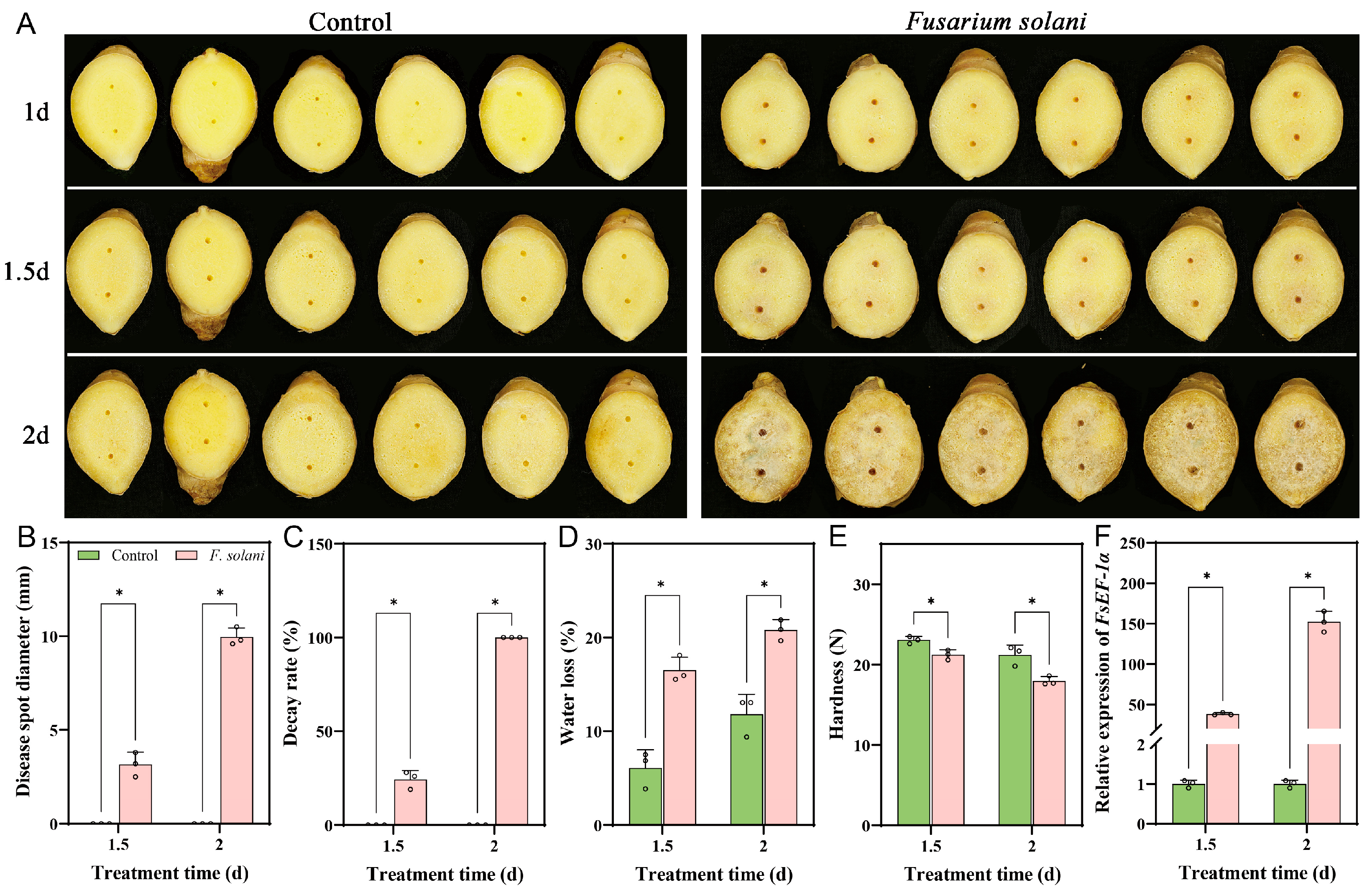
3.1. Symptoms of Ginger Rhizomes Inoculated with F. solani

As shown in Figure 1A, the initial invading hyphae began to form 1.5 d after infection. By 2 d post-inoculation, the hyphae had penetrated the ginger tissue and rapidly expanded on the wound surfaces, while the uninfected rhizome remained free of rot. Therefore, ginger rhizome samples were initially collected at 1.5 d after infection with F. solani to capture the early interaction between the ginger and the pathogen, during which no visible disease symptoms were present. A subsequent sampling at 2 d, when extensive infection had spread across the rhizome, was chosen to investigate disease progression during the rapid pathogenic expansion.
Moreover, the inoculation of F. solani caused Fusarium wilt disease, as evidenced by the disease spot diameter and decay rate. After 1.5 d of F. solani inoculation, the disease spot diameter and decay rate were 3.2 mm and 24.3%, respectively. After 2 d of inoculation, the disease spot diameter and decay rate reached up to 10.0 mm and 100%, respectively. The ginger rhizomes in the control group showed no Fusarium wilt symptoms (Figure 1B,C). Additionally, compared to the control, the water loss of ginger rhizomes in the F. solani treatment group increased by 171.7% after 1.5 d and 75.9% after 2 d (Figure 1D). The firmness of the F. solani-inoculated group decreased by 7.9% and 15.3%, respectively (Figure 1E). The biomass of F. solani was further analyzed to investigate Fusarium wilt progression after inoculation. The results showed that, compared to the control, the biomass of F. solani in ginger rhizomes increased 38.3-fold at 1.5 d after inoculation and 152.4-fold at 2 d after inoculation (Figure 1F). These results indicated that the F. solani strain could cause the typical characteristic Fusarium wilt symptoms, significantly increase weight loss, reduce rhizome hardness, and increase F. solani biomass of ginger rhizome.

3.2. Sequencing Data Quality Evaluation

In order to determine the genes responsive to F. solani infection and compare the gene expression patterns of infected ginger rhizome at 1.5 and 2 d post-inoculation, 12 ginger rhizome libraries were constructed. The RNA-Seq sequencing of 12 libraries produced a total of 40,341,026~49,445,898 raw bases (Table S2). The Q30 was 94.23~94.95%, and the GC content of 47.49~49.83%, respectively (Table S3). Quality assessment of the RNA-seq data (error rate distribution along reads and GC content distribution) revealed excellent sequencing performance (Figures S1 and S2). Alignment to the Zingiber officinale reference genome using the HISAT2 demonstrated consistently high mapping efficiencies ranging from 90.16% to 92.34% (Table S4).
The volcano plot provides a clear visualization of the overall distribution of DEGs (Figure 2). In total, 647 DEGs were identified, with 374 up-regulated and 374 down-regulated in the 1.5 d infection group (CK1.5 vs. FS1.5), and 6398 DEGs were identified, with 3108 up-regulated and 3290 down-regulated in the 2 d infection group (CK2 vs. FS2). Notably, a higher number of differential genes were regulated in the 2 d infection group.

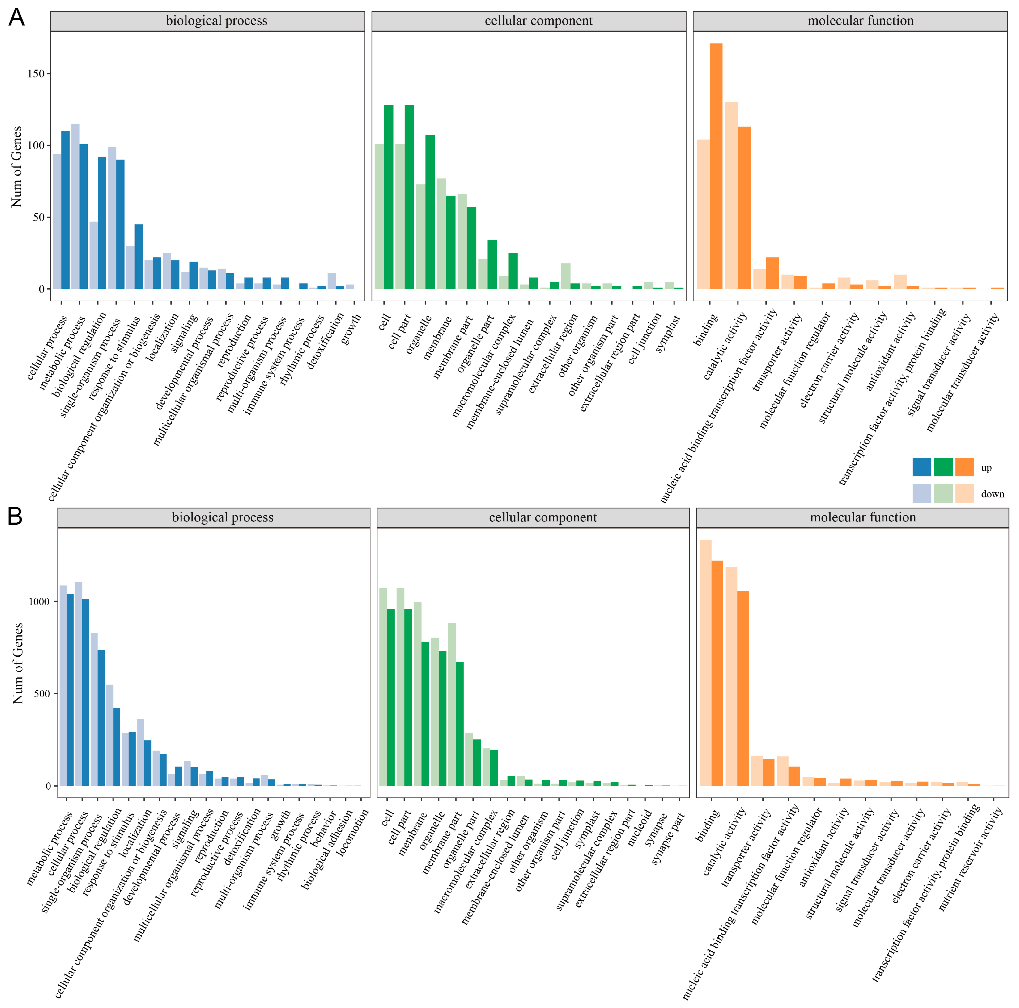
3.3. GO Function Annotation

GO functional annotation was performed on DEGs to explore their functions across three categories: Biological Process (BP), Cellular Component (CC), and Molecular Function (MF). At 1.5 d, 17 GO terms, such as ‘metabolic process’ and ‘single-organism process’, were enriched at the BP level. At the CC level, 15 GO terms, including ‘cell’ and ‘cell part’, were predominantly enriched. The MF level was predominantly associated with 11 GO terms, such as ‘binding’ and ‘catalytic activity’ (Figure 3A). At 2 d, 20 GO terms, such as ‘metabolic process’ and ‘cellular process’, were enriched at the BP level. In the CC level, 18 GO terms such as ‘cell’ and ‘cell part’ were mainly enriched. At the MF level, 12 GO terms, such as ‘binding’ and ‘catalytic activity’, were mostly enriched (Figure 3B).

3.4. KEGG Metabolic Pathway

To elucidate the functional roles and molecular networks of DEGs in the ginger rhizome transcriptome, KEGG pathway analysis was conducted. KEGG pathway annotation revealed that the DEGs were primarily associated with five major functional categories: Cellular Processes, Environmental Information Processing, Genetic Information Processing, Metabolism, and Organismal Systems. At 1.5 d, the DEGs were significantly enriched in pathways such as ‘Plant–pathogen interaction’ and ‘Plant hormone signal transduction’ (Figure 4A). At 2 d, the DEGs showed significant enrichment in pathways such as ‘Plant–pathogen interaction’, ‘Plant hormone signal transduction’, and ‘MAPK signaling pathway’ (Figure 4B). KEGG pathway enrichment analysis identified 217 and 2109 significantly enriched pathways for CK1.5 vs. FS1.5 and CK2 vs. FS2, respectively (Tables S5 and S6). To sum up, the infection of ginger rhizome tissue induces physiological responses, which may contribute to enhancing resistance to F. solani infection through the coordinated regulation of metabolic processes, cellular compartmentalization, and molecular interactions.

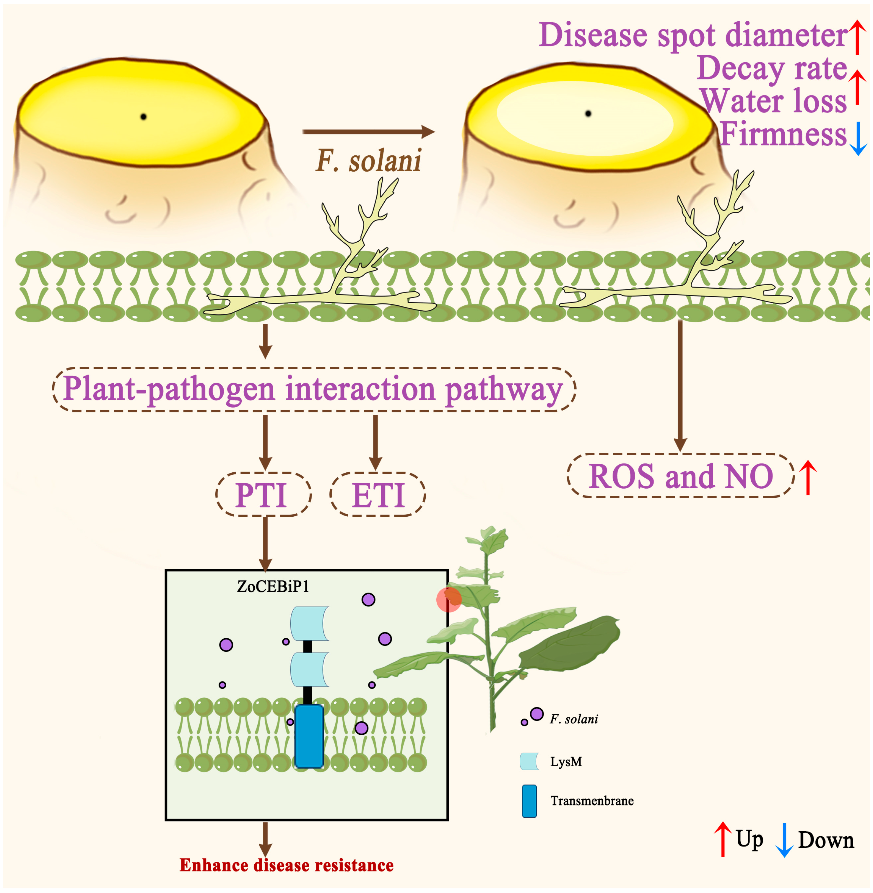
3.5. DEGs Related to Plant–Pathogen Interaction Pathway in Ginger Rhizome Against F. solani

KEGG enrichment analysis showed that the plant–pathogen interaction pathway (ko04626) was the most significantly enriched pathway in ginger rhizomes at both 1.5 and 2 d post-inoculation with F. solani (Tables S4 and S5). Based on this result, we conducted cluster analysis of the DEGs (|log2(FoldChange)| > 1.5 and FDR < 0.05) associated with this pathway and subsequently validated their expression patterns using qRT-PCR.
A total of 17 DEGs (|log2(FoldChange)| > 1.5 and FDR < 0.05) in the plant–pathogen interaction pathway were identified at 1.5 d post-inoculation with F. solani. Among these, fourteen genes showed significant up-regulation, including pattern recognition receptors (CEBiP and FLS2), resistance proteins (RPS2 and RRS1-R), signaling components (PBS1, CNGCs, and UPA20), transcription factors (WRKY22 and Pti5/1), and defense-related genes (FRK1 and EIX1/2). Conversely, three genes were down-regulated: one WKK4/5, one EIX1/2, and one Pti5/1 (Figure 5A). RNA-Seq analysis identified 106 significantly DEGs (|log2FoldChange| > 1.5, FDR < 0.05) in the plant–pathogen interaction pathway at 2 d post-inoculation with F. solani. Of these, 68 genes were significantly up-regulated, including pattern recognition receptors (two CEBiP, two CERK1, two FLS2, two EFR), resistance proteins (four RPM1, four RPS2, one RRS1-R), signaling components (seven PBS1, seven UPA20, seven CDPK, two CaMCML), transcription factors (three Pti5/6/1, one WRKY22), defense-related genes (two EIX1/2, three Pto, two PR1, five FRK1), and other components (four MKK4/5, one MEKK1, two CNGCs, one NOS). Conversely, thirty-eight genes showed significant down-regulation, comprising pattern recognition receptors (one CERK1, three FLS2, two EFR), signaling components (two RIN4, one PBS1, two UPA20, two CDPK), transcription factors (six WRKY33, two WRKY22, four Pti5/6/1), defense-related genes (one EIX1/2, two FRK1), and other components (three RRS1-R, one RPS2, one MEKK1, five Rboh) (Figure 5B).
In agreement with RNA-Seq analysis, qRT-PCR experiments further confirmed that Pti1 (Maker00011524.gene), CEBiP (Maker00045267.gene), RPS2 (Maker00011463.gene), UPA20 (Maker00077866.gene), and FLS2 (Maker00028416.gene) were up-regulated, and MKK4/5 (Maker00034618.gene) was down-regulated upon F. solani treatment 1.5 d, compared to the control (Figure 5(C1)). Moreover, CEBiP (Maker00045267.gene), CERK1 (Maker00028603.gene), UPA20 (Maker00004882.gene), and PR1 (Maker00071011.gene) were up-regulated, and EIX1/2 (Maker00069746.gene) and WRK33 (Maker00007282.gene) were down-regulated upon F. solani treatment 2 d, compared to the control (Figure 5(C2)). This consistency suggested the reliability of the RNA-Seq data. This comprehensive expression profiling reveals a complex regulatory network in ginger rhizomes during F. solani infection.
Moreover, among the DEGs, five genes encoding CEBiP (Maker00045267.gene), FLS2 (Maker00028416.gene), Pti5 (Zingiber_officinale_newGene_16346), Pti1 (Maker00011140.gene), and RPS1-R (Maker00037847.gene) were involved in defense response to F. solani against Fusarium wilt (Figure 5A,B). Especially, CEBiP (Maker00045267.gene), which was renamed as ZoCEBiP1, exhibited the most significant regulation, with expression levels more than 6.1 and 3.7 times higher than those of the control at 1.5 and 2 d post-inoculation, respectively (Figure 5(C1,C2)).

3.6. Functional Analysis of ZoCEBiP1

To investigate the biological function of ZoCEBiP1 in plant defense, we performed molecular cloning and transient expression analysis in N. benthamiana, followed by P. infestans inoculation assays.
The full-length coding sequence of ZoCEBiP1 (1248 bp) was successfully amplified, purified, and verified by sequencing (Figure 6A). Sequence alignment using DNAMAN demonstrated 100% identity with the predicted sequence in the ginger genome, confirming accurate gene annotation. Domain architecture analysis through SMART revealed that ZoCEBiP1 contains two characteristic LysM domains, showing conserved structural features with the known chitin receptor Oryza sativa LYP4 (OsLYP4) (Figure 6B).
For functional characterization, ZoCEBiP1 was transiently overexpressed in N. benthamiana leaves via Agrobacterium-mediated transformation. Subsequent inoculation with P. infestans showed that ZoCEBiP1 overexpression significantly enhanced disease resistance (Figure 6C), with lesion diameters reduced by 63.4% compared to GFP-expressing control leaves (Figure 6D). The DAB staining results showed that 72 h after P. infestans inoculation, the tobacco leaves of the WT and GFP control exhibited darker brown staining. In contrast, the leaves with transient overexpression of ZoCEBiP1 showed only a few light brown spots at the inoculation sites (Figure 6E). These results demonstrated that ZoCEBiP1 functions as a pathogen resistance gene.

3.7. Effects of ROS Accumulation, Antioxidant Enzyme Activities, NOS Activity, and NO Content of Ginger Rhizome After Infection with F. solani

Transcriptional profiling of DEGs in the plant–pathogen interaction pathway revealed that F. solani infection in ginger rhizomes caused significant changes in the expression of multiple genes related to ROS and NO metabolism. In the context of ROS metabolism, antioxidant enzymes like POD, CAT, SOD, and APX help modulate the levels of ROS such as H2O2 and O2 [22]. Meanwhile, NO, a crucial signaling molecule, is synthesized through the activity of NOS, with NOS activity directly regulating the production of NO [23,24]. To functionally validate these transcriptional changes, we subsequently quantified ROS accumulation, antioxidant enzyme activities (POD, CAT, SOD, and APX), NOS activity, and NO content in F. solani-infected ginger rhizomes to validate the functional consequences of these transcriptional changes.
As shown in Figure 7A,B, ginger rhizomes treated with the control showed an increase in ROS accumulation, while treatment with F. solani resulted in a decrease at 2 d. However, the H2O2 and O2 contents in the F. solani treatment group were significantly higher compared to the control group; the H2O2 content increased by 49.6% and 51.4%, while O2 levels increased by 20.7% and 33.0% at both time points, respectively. The antioxidant enzyme activities of ginger rhizomes were increased after 2 d of the control and F. solani treatments. Compared with the control, F. solani treatment enhanced the POD activity by 30.8% and 27.2% at 1.5 and 2 d, respectively (Figure 7C); increased CAT activity by 49.3% and 53.7%, respectively (Figure 7D); increased the SOD activity by 19.7% and 27.4%, respectively (Figure 7E); elevated the APX activity by 33.6% and 29.2%, respectively (Figure 7F). As shown in Figure 7G, F. solani treatment increased the NO content by 30.3% and 33.8% compared to the control at 1.5 and 2 d, respectively. Similarly, inoculation with F. solani treatment significantly increased the activity of NOS at 1.5 and 2 d, which were 41.4% and 49.3% higher than those of the control (Figure 7H). These data suggested that F. solani infection triggered an oxidative burst in ginger rhizomes, activating both ROS-scavenging systems and NO-mediated defense signaling. The temporal coordination of these responses indicated a dynamic interplay between oxidative stress and nitric oxide signaling during fungal infection.

4. Discussion

Fusarium wilt, caused by F. solani, is one of the most prevalent and damaging fungal diseases of ginger during postharvest storage, which is responsible for high economic loss globally [4]. In our previous research on biological control approaches for postharvest ginger diseases, we identified several promising alternatives, including eugenol [2], chitosan [5], silica nanoparticles [25], and hydrogen sulfide [7], for suppressing Fusarium wilt progression. However, despite these advances, conventional disease management strategies remain inadequate for controlling F. solani under commercial storage conditions. This highlights the urgent need for a deeper understanding of ginger–pathogen interactions at the molecular level to develop more effective, targeted control measures.
Recently, RNA-seq analysis has been widely utilized to investigate pathogen–plant interactions. For example, in the study by Jiang et al. [10], the analysis of DEGs in mango fruit treated with Bacillus siamensis demonstrated that the fruit’s response to the treatment was most prominent during the early days of storage. Xu et al. [26] identified several key mechanisms involved in the pear’s response to Penicillium expansum, as revealed by transcriptome analysis. In our study, compared with the control, 647 genes were significantly affected by F. solani at 1.5 d post-infection, and the expressions of 5751 DEGs were increased by treatment at 2 d post-infection, indicating that the early response of the ginger rhizome to F. solani infection was activated in 2 d. In addition, by 2 d post-infection, the number of enriched GO terms had increased, indicating a more complex and intensified defense response.
KEGG pathway enrichment analysis revealed that the plant–pathogen interaction was the most enriched at both 1.5 and 2 d post-treatment, indicating that this process played a central role in the ginger rhizome’s response to F. solani. In plants, the plant–pathogen interaction pathway is considered as a key defense mechanism during fungal pathogen invasion, involving both pathogen-associated molecular pattern (PAMP)-triggered immunity (PTI) and effector-triggered immunity (ETI) pathways [27]. PTI is the first layer of plant immune response, triggered by the recognition of PAMPs by pattern recognition receptors (PRRs) on the plant cell surface [28]. In this study, several PRRs, including CEBiP and FLS2, were significantly upregulated at both 1.5 and 2 d post-inoculation with F. solani. The upregulation of these PRRs suggested that ginger rhizomes were actively engaging PTI mechanisms to counteract the pathogen. ETI is a more specific and robust immune response activated by the recognition of pathogen effectors by intracellular nucleotide-binding leucine-rich repeat (NLR) proteins [29]. In our study, multiple NLR genes, such as RPS2 and RRS1-R, were significantly upregulated at both time points, indicating that the ginger rhizome was also mounting a specific ETI response to F. solani. Those results were consistent with the findings of Xiong et al. [30], who reported that several PRR genes, kinase genes, and genes involved in PTI and ETI signaling pathways were significantly induced in the Botrytis cinerea–strawberry interaction. Therefore, these results underscore the specific roles of these genes in plant defense, further supporting the idea that both PTI and ETI pathways were essential for successful pathogen defense in ginger.
It is worth noting that ZoCEBiP1 played an important role in the early defense response of F. solani against Fusarium wilt disease. Oryza sativa CEBiP (OsCEBiP) was the first identified polysaccharide receptor that binds fungal chitin elicitors, playing a crucial role in detecting chitin signals and initiating signal transduction [31,32]. It contains two LysM domains and one transmembrane domain. Knockout of the CEBiP gene severely impaired chitin-induced ROS bursts and the expression of chitin-responsive genes, leading to reduced disease resistance in rice plants [32]. Similarly, in this study, ZoCEBiP1 also contained two LysM domains, indicating that CEBiP was highly conserved across species, and transient overexpression experiments in tobacco demonstrated that ZoCEBiP1 could enhance the disease resistance of tobacco seedlings by scavenging ROS. These results demonstrated that ZoCEBiP1 functioned as a pathogen resistance gene, likely serving as a pattern recognition receptor in the plant immune system. However, its precise molecular function requires further investigation. The upregulation of signaling components (PBS1, CNGCs) and transcription factors (WRKY22, Pti5/1) in ginger rhizomes highlighted a sophisticated, multi-layered defense strategy against F. solani infection. These molecular players activate canonical immune pathways: PBS1 initiates MAPK cascades [33], while CNGCs mediate calcium signaling [34], representing conserved convergence points for both PTI and ETI responses. In the transcriptional response of Wolfberry to the infection by the endophytic fungus Fusarium nematophilum, the activation of FRK1 and EIX1/2 played a crucial role [35]. In our study, the strong induction of defense executors like FRK1 and EIX1/2 also demonstrated the activation of downstream antimicrobial mechanisms. Conversely, the downregulation of certain genes, such as WKK4/5 and specific isoforms of EIX1/2 and Pti5/1, may reflect a fine-tuning of the immune response. Studies have shown that certain transcription factors and signaling components could act as negative regulators to balance immune responses. For example, Arabidopsis thaliana WRKY50/51 (AtWRKY50/51) mediated low oleic acid- and SA-dependent repression of JA signaling, leading to increased susceptibility to Botrytis cinerea [36]. In addition, Gossypium hirsutum WRKY25 (GhWRKY25) negatively regulated B. cinerea infection [37]. This could involve negative regulatory mechanisms to prevent excessive immune activation, which can be detrimental to the plant.
The plant–pathogen interaction pathway involves a complex network of signaling cascades and defense responses that plants utilize. Activation of this pathway triggers downstream signaling events, including the production of ROS and NO, which are crucial components of the plant’s defense arsenal. In this study, the significant upregulation of genes associated with ROS and NO metabolism suggested that ROS and NO could be essential in the defense response of ginger rhizomes to F. solani infection. ROS are essential players in plant defense against pathogens; they serve both as signaling molecules and as direct antimicrobial agents. Rapid accumulation of ROS has been considered as one of the earliest events strongly associated with plant resistance to pathogens and involved in the development of disease resistance in ginger rhizomes [38], grapes [39], and kiwifruit [40] during postharvest storage. In the present study, H2O2 contents and H2O2 and O2 levels significantly increased in F. solani-infected ginger rhizomes, indicating a robust oxidative burst. However, ROS can also be detrimental to plant cells if not properly managed. The observed increase in antioxidant enzyme activities (POD, CAT, SOD, and APX) suggested that ginger rhizomes were actively balancing ROS production and scavenging to prevent oxidative damage. Similarly, H2S induced resistance in ginger rhizomes via increasing the capacity for antioxidant defense [7]. Therefore, the enhanced activities of these antioxidant enzymes indicated that the plant was effectively managing oxidative stress during infection. NO is another important signaling molecule involved in plant defense against pathogens. Studies have shown that NO treatment could enhance disease resistance in horticultural crops, such as kiwifruit [41] and tomato [23]. NOS was a key enzyme for the NO production [24]. For instance, melatonin has been reported to trigger NO accumulation by activating NOS, improving cold tolerance in postharvest litchi fruit [42]. Additionally, NO interacts with ROS signaling pathways, strengthening the overall defense response. For instance, NO application to Arabidopsis roots could rapidly activate protein kinases with MAPK properties [43]. In this study, the significant increase in NO content and NOS activity in F. solani-infected ginger rhizomes indicated the role of NO in mediating defense responses. Notably, a dynamic interplay between ROS and NO occurs during pathogenic fungal infection [39]. Future research can focus on unraveling the specific signaling pathways and molecular interactions between ROS and NO in ginger rhizomes.
In addition, our findings demonstrate that ginger rhizomes exhibit distinct phased immune responses during F. solani infection. In the early infection stage (1.5 d), although there were not many significantly upregulated defense-related genes in the plant–pathogen interaction pathway (Figure 5A), the recognition of PAMPs such as chitin through significantly upregulated pattern recognition receptors like ZoCEBiP1 (Figure 5(C1)) successfully activated early immune responses characterized by ROS burst [32]. This rapid accumulation of H2O2 and O2 (Figure 7A,B), consistent with typical pathogen recognition responses reported in postharvest ginger [5,7,13,25] and other crops [40,41], likely effectively restricted fungal spread before effector secretion [44], indicating that ginger rhizomes preferentially initiate PTI rather than comprehensive defense responses at this stage. As infection progressed to 2 d, we observed reduced activation of ZoCEBiP1, suggesting diminished pathogen recognition capacity. However, defense responses were significantly enhanced, manifested by increased enrichment of differentially expressed genes associated with the plant–pathogen interaction pathway, along with elevated antioxidant enzyme activities, NO content, and NOS activity. When the disease developed to the necrotic stage, F. solani likely secreted effectors to suppress PTI, thereby ensuring successful infection [44,45,46,47]. This dynamic pattern reveals the temporal characteristics of ginger’s defense strategy: early emphasis on pathogen recognition and rapid response, followed by transition to more comprehensive defense execution, which ultimately could be overcome by the pathogen’s virulence strategies.
While previous disease resistance evaluations confirmed that the ‘Zhugen’ ginger cultivar exhibits resistance to Fusarium wilt, this study further demonstrates its ability to activate defense responses upon F. solani inoculation. However, a comparison with susceptible varieties was not conducted. In subsequent experiments, we will systematically compare resistant and susceptible ginger varieties under pathogen challenge to identify and validate key regulators that positively correlate with resistance or negatively correlate with susceptibility [48].

5. Conclusions

This research provided comprehensive insights into the molecular defense mechanisms of ginger rhizomes against F. solani infection through transcriptomic and functional analyses. The results revealed a strong activation of the plant–pathogen interaction pathway, with significant upregulation of genes associated with both PTI and ETI, including PRRs like CEBiP and FLS2, and NLR proteins such as RPS2 and RRS1-R. The upregulation of signaling components (PBS1, CNGCs) and transcription factors (WRKY22, Pti5/1) highlighted a multi-layered defense strategy. Furthermore, the critical role of ZoCEBiP1 was identified in early defense through transient transformation of N. benthamiana leaves. The marked significance in ROS and NO metabolism genes, along with effective ROS management, coupled with elevated NO content and NOS activity, suggested that ROS and NO were essential players in the defense response (Figure 8). These findings not only advance our understanding of ginger immunity but also identify specific molecular targets for developing innovative control strategies against Fusarium wilt.

Supplementary Materials

The following supporting information can be downloaded at https://www.mdpi.com/article/10.3390/horticulturae11070791/s1, Figure S1. Error rate distribution along reads. Horizontal is position along reads, ordinate is error rate (%); Figure S2. GC content distribution. Horizontal is position along reads, ordinate is the proportion of single base (%), different colors represent different base types; Table S1: Primers’ sequence information used in this study; Table S2: List of quality of RNA-Seq data; Table S3: Quality control of the control/F. solani-treated; Table S4: The mapping efficiency percentage of reads mapped to the Zingiber officinale reference genome; Table S5: The KEGG pathways enrichment analysis of the control/F. solani-treated at 1.5 d; Table S6: The KEGG pathways enrichment analysis of the control/F. solani-treated at 2 d.

Author Contributions

L.Z.: writing—original draft and data curation. Q.J.: methodology, writing—review and editing, and funding acquisition. L.L.: project administration and funding acquisition. Y.L.: conceptualization, funding acquisition, and resources. All authors have read and agreed to the published version of the manuscript.

Funding

This work was funded by the Natural Science Foundation of Hubei Province (2023AFB1001).

Data Availability Statement

The RNA-seq data were deposited in the NCBI database (PRJNA1090657).

Conflicts of Interest

The authors declare no conflicts of interest.

References

  1. Srinivasan, K. Ginger rhizomes (Zingiber officinale): A spice with multiple health beneficial potentials. PharmaNutrition 2017, 5, 18–28. [Google Scholar] [CrossRef]
  2. Zhou, X.; Ma, H.H.; Xiong, S.J.; Zhang, L.L.; Zhu, X.D.; Zhu, Y.X.; Zhou, L.R. Evaluation of the inhibitory efficacy of eugenol against the pathogen of Fusarium wilt in ginger seedlings. Horticulturae 2023, 9, 1024. [Google Scholar] [CrossRef]
  3. Choi, J.H.; Nah, J.Y.; Lee, M.J.; Yim, S.B.; Jang, J.Y.; Lee, T.; Kim, J. Fungal diversity in ginger and effect of storage conditions on occurrence of Fusarium and its mycotoxins. Food Control 2024, 165, 110631. [Google Scholar] [CrossRef]
  4. Meng, X.L.; Wu, W.J.; Niu, B.; Liu, R.L.; Chen, H.Z.; Gao, H.Y.; Chen, H.J. Inhibitory effect and action mechanism of perillaldehyde on the Fusarium graminearum in postharvest fresh ginger. Postharvest Biol. Technol. 2024, 209, 112674. [Google Scholar] [CrossRef]
  5. Zhang, L.L.; Fang, S.Y.; Sun, C.; Liang, H.R.; Ma, J.W.; Jia, Q.; Yin, J.L.; Zhu, Y.X.; Liu, Y.Q. Chitosan boosts ginger disease resistance: Insights from transcriptomic and metabolomic analyses. LWT 2024, 205, 116478. [Google Scholar] [CrossRef]
  6. Ferreira da Cunha Neto, J.; da Silva Rocha, W.P.; Makris, G.; Sandoval-Denis, M.; Hagen, F.; Crous, P.W.; Chaves, G.M. Fusarioid keratitis and other superficial infections: A 10-years prospective study from Northeastern Brazil. PLOS Neglected Trop. Dis. 2024, 18, e0012247. [Google Scholar] [CrossRef]
  7. Zhang, L.L.; Wu, X.Q.; Zhong, Y.; Yang, Y.; Wei, S.W.; Sun, C.; Wei, L.J.; Liu, Y.Q. Hydrogen sulfide enhances the disease resistance of ginger to rhizome rot during postharvest storage through modulation of antioxidant response and nitric oxide-mediated S-nitrosylaion. Postharvest Biol. Technol. 2025, 220, 113321. [Google Scholar] [CrossRef]
  8. Abdullah, A.S.; Moffat, C.S.; Lopez-Ruiz, F.J.; Gibberd, M.R.; Hamblin, J.; Zerihun, A. Host-multi-pathogen warfare: Pathogen interactions in co-infected plants. Front. Plant Sci. 2017, 8, 1806. [Google Scholar] [CrossRef]
  9. Zhao, L.N.; Zhou, Y.L.; Quan, S.H.; Qiu, J.E.; Dhanasekaran, S.; Li, B.; Gu, X.Y.; Zhang, H.Y. Transcriptome analysis reveals mechanisms of the disease resistance in postharvest kiwifruit induced by Meyerozyma caribbica. Sci. Hortic. 2023, 322, 112452. [Google Scholar] [CrossRef]
  10. Jiang, Z.C.; Li, R.; Tang, Y.; Cheng, Z.Y.; Qian, M.J.; Li, W.; Shao, Y.Z. Transcriptome analysis reveals the inducing effect of Bacillus siamensis on disease resistance in postharvest mango fruit. Foods 2022, 11, 107. [Google Scholar] [CrossRef]
  11. Li, X.; Zhang, C.; Wang, X.Y.; Liu, X.Q.; Zhu, X.L.; Zhang, J. Integration of metabolome and transcriptome profiling reveals the effect of modified atmosphere packaging (MAP) on the browning of fresh-cut Lanzhou lily (Lilium davidii var. unicolor) bulbs during storage. Foods 2023, 12, 1335. [Google Scholar] [CrossRef]
  12. Yang, X.M.; Yang, K.X.; Wang, X.H.; Wang, Y.T.; Zhao, Z.Y.; Meng, D.M. Transcriptomic analysis reveals the mechanism of bacterial disease resistance of postharvest button mushroom (Agaricus bisporus). Physiol. Mol. Plant Pathol. 2022, 122, 101903. [Google Scholar] [CrossRef]
  13. Zhang, L.L.; Yang, Y.; Zhu, Y.X.; Hu, H.J.; Jia, Q.; Sun, C.; Zhu, X.D.; Liu, Y. Antifungal activity and mechanism of chitosan against Fusarium solani caused ginger soft rot during postharvest storage. Postharvest Biol. Technol. 2024, 208, 112680. [Google Scholar] [CrossRef]
  14. Li, H.L.; Wu, L.; Dong, Z.M.; Jiang, Y.S.; Jiang, S.J.; Xing, H.T.; Li, Q.; Liu, G.C.; Tian, S.M.; Wu, Z.Y.; et al. Haplotype-resolved genome of diploid ginger (Zingiber officinale) and its unique gingerol biosynthetic pathway. Hortic. Res. 2021, 8, 189. [Google Scholar] [CrossRef]
  15. Li, G.; Ma, J.W.; Yin, J.L.; Guo, F.L.; Xi, K.Y.; Yang, P.H.; Cai, X.D.; Jia, Q.; Li, L.; Liu, Y.Q.; et al. Identification of reference genes for reverse transcription-quantitative PCR analysis of ginger under abiotic stress and for postharvest biology studies. Front. Plant Sci. 2022, 13, 893495. [Google Scholar] [CrossRef] [PubMed]
  16. Li, Y.T.; Liu, X.; Xiao, Y.; Wen, Y.; Li, K.K.; Ma, Z.L.; Yin, J.L.; Yang, L.J.; Zhu, Y.X. Genome-wide characterization and function analysis uncovered roles of wheat LIMs in responding to adverse stresses and TaLIM8-4D function as a susceptible gene. Plant Genome 2022, 15, e20246. [Google Scholar] [CrossRef] [PubMed]
  17. Trinder, P. Determination of blood glucose using an oxidase-peroxidase system with a non-carcinogenic chromogen. J. Clin. Pathol. 1969, 22, 158–161. [Google Scholar] [CrossRef]
  18. Bergmeyer, H.U. Methods of Enzymatic Analysis; Academic Press: New York, NY, USA, 2012. [Google Scholar]
  19. Sun, Y.I.; Oberley, L.W.; Li, Y. A simple method for clinical assay of superoxide dismutase. Clin. Chem. 1988, 34, 497–500. [Google Scholar] [CrossRef]
  20. Kırgeç, Y.; Batı-Ay, E.; Açıkgöz, M.A. The effects of foliar salicylic acid and zinc treatments on proline, carotenoid, and chlorophyll content and anti-oxidant enzyme activity in Galanthus elwesii Hook. Horticulturae 2023, 9, 1041. [Google Scholar] [CrossRef]
  21. Wei, L.J.; Zhang, J.; Wei, S.H.; Hu, D.L.; Liu, Y.Y.; Feng, L.; Li, C.X.; Wang, C.L.; Liao, W.B. Nitric oxide enhanced salt stress tolerance in tomato seedlings, involving phytohormone equilibrium and photosynthesis. Int. J. Mol. Sci. 2022, 23, 4539. [Google Scholar] [CrossRef]
  22. Lei, S.; Rossi, S.; Huang, B. Metabolic and physiological regulation of aspartic acid-mediated enhancement of heat stress tolerance in Perennial Ryegrass. Plants 2022, 11, 199. [Google Scholar] [CrossRef] [PubMed]
  23. Shu, P.; Li, Y.J.; Wang, X.Y.; Yao, L.; Sheng, J.P.; Shen, L. Exogenous ferulic acid treatment increases resistance against Botrytis cinerea in tomato fruit by regulating nitric oxide signaling pathway. Postharvest Bio. Technol. 2021, 182, 111678. [Google Scholar] [CrossRef]
  24. Sharma, A.; Soares, C.; Sousa, B.; Martins, M.; Kumar, V.; Shahzad, B.; Sidhu, G.P.; Bali, A.S.; Asgher, M.; Bhardwaj, R.; et al. Nitric oxide-mediated regulation of oxidative stress in plants under metal stress: A review on molecular and biochemical aspects. Physiol. Plant. 2020, 168, 318–344. [Google Scholar] [CrossRef]
  25. Zhou, J.; Liu, X.L.; Sun, C.; Li, G.; Yang, P.M.; Jia, Q.; Cai, X.D.; Zhu, Y.X.; Yin, J.L.; Liu, Y.Q. Silica nanoparticles enhance the disease resistance of ginger to rhizome rot during postharvest storage. Nanomaterials 2022, 12, 1418. [Google Scholar] [CrossRef]
  26. Xu, M.Q.; Zhang, X.Y.; Dhanasekaran, S.; Godana, E.A.; Yang, Q.Y.; Zhao, L.N.; Zhang, H.Y. Transcriptome analysis of postharvest pear (Pyrus pyrifolia Nakai) in response to Penicillium expansum infection. Sci. Hortic. 2021, 288, 110361. [Google Scholar] [CrossRef]
  27. Ortiz-Morea, F.A.; Liu, J.; Shan, L.B.; He, P. Malectin-like receptor kinases as protector deities in plant immunity. Nat. Plants 2022, 8, 27–37. [Google Scholar] [CrossRef] [PubMed]
  28. Zipfel, C. Early molecular events in PAMP-triggered immunity. Curr. Opin. Plant Biol. 2009, 12, 414–420. [Google Scholar] [CrossRef]
  29. Yuan, M.H.; Jiang, Z.Y.; Bi, G.Z.; Nomura, K.; Liu, M.H.; Wang, Y.P.; Cai, B.Y.; Zhou, J.M.; He, S.Y.; Xin, X.F. Pattern-recognition receptors are required for NLR-mediated plant immunity. Nature 2021, 592, 105–109. [Google Scholar] [CrossRef]
  30. Xiong, J.S.; Zhu, H.Y.; Bai, Y.B.; Liu, H.; Cheng, Z.M. RNA sequencing-based transcriptome analysis of mature strawberry fruit infected by necrotrophic fungal pathogen Botrytis cinerea. Physiol. Mol. Plant Pathol. 2018, 104, 77–85. [Google Scholar] [CrossRef]
  31. Akamatsu, A.; Wong, H.L.; Fujiwara, M.; Okuda, J.; Nishide, K.; Uno, K.; Imai, K.; Umemura, K.; Kawasaki, T.; Kawano, Y.; et al. An OsCEBiP/OsCERK1-OsRacGEF1-OsRac1 module is an essential early component of chitin-induced rice immunity. Cell Host Microbe 2013, 13, 465–476. [Google Scholar] [CrossRef]
  32. Kaku, H.; Nishizawa, Y.; Ishii-Minami, N.; Akimoto-Tomiyama, C.; Dohmae, N.; Takio, K.; Minami, E.; Shibuya, N. Plant cells recognize chitin fragments for defense signaling through a plasma membrane receptor. Proc. Natl. Acad. Sci. USA 2006, 103, 11086–11091. [Google Scholar] [CrossRef] [PubMed]
  33. Haider, M.S.; Kurjogi, M.M.; Khalil-Ur-Rehman, M.; Fiaz, M.; Pervaiz, T.; Jiu, S.T.; Jia, H.F.; Chen, W.; Fang, J.G. Grapevine immune signaling network in response to drought stress as revealed by transcriptomic analysis. Plant Physiol. Biochem. 2017, 121, 187–195. [Google Scholar] [CrossRef]
  34. DeFalco, T.A.; Moeder, W.; Yoshioka, K. Opening the gates: Insights into cyclic nucleotide-gated channel-mediated signaling. Trends Plant Sci. 2016, 21, 903–906. [Google Scholar] [CrossRef] [PubMed]
  35. Yan, S.Y.; Li, J.; Zhang, Q.C.; Jia, S.X.; Zhang, Q.Q.; Wang, R.T.; Ju, M.X.; Gu, P.W. Transcriptional response of wolfberry to infestation with the endophytic Fusarium nematophilum strain NQ8GII4. Plant Dis. 2024, 108, 1514–1525. [Google Scholar] [CrossRef]
  36. Gao, Q.M.; Venugopal, S.; Navarre, D.; Kachroo, A. Low oleic acid-derived repression of jasmonic acid-inducible defense responses requires the WRKY50 and WRKY51 proteins. Plant Physiol. 2011, 155, 464–476. [Google Scholar] [CrossRef]
  37. Liu, X.F.; Song, Y.Z.; Xing, F.Y.; Wang, N.; Wen, F.J.; Zhu, C.X. GhWRKY25, a group I WRKY gene from cotton, confers differential tolerance to abiotic and biotic stresses in transgenic Nicotiana benthamiana. Protoplasma 2016, 253, 1265–1281. [Google Scholar] [CrossRef]
  38. Peng, H.M.; Hu, H.J.; Xi, K.Y.; Zhu, X.M.; Zhou, J.; Yin, J.L.; Guo, F.L.; Liu, Y.Q.; Zhu, Y.X. Silicon nanoparticles enhance ginger rhizomes tolerance to postharvest deterioration and resistance to Fusarium solani. Front. Plant Sci. 2022, 13, 816143. [Google Scholar] [CrossRef]
  39. Ding, L.N.; Li, Y.T.; Wu, Y.Z.; Li, T.; Geng, R.; Cao, J.; Zhang, W.; Tan, X.L. Plant disease resistance-related signaling pathways: Recent progress and future prospects. Int. J. Mol. Sci. 2022, 23, 16200. [Google Scholar] [CrossRef]
  40. Pan, L.Y.; Zhao, X.Y.; Chen, M.; Fu, Y.Q.; Xiang, M.L.; Chen, J.Y. Effect of exogenous methyl jasmonate treatment on disease resistance of postharvest kiwifruit. Food Chem. 2020, 305, 125483. [Google Scholar] [CrossRef]
  41. Yang, R.; Wang, J.; Cai, Z.P.; Shen, Y.G.; Gan, Z.Y.; Duan, B.; Yuan, J.; Huang, T.H.; Zhang, W.; Du, H.Y.; et al. Transcriptome profiling to elucidate mechanisms of the enhancement of the resistance to Botryosphaeria dothidea by nitric oxide in postharvest kiwifruit during storage. LWT 2022, 159, 113187. [Google Scholar] [CrossRef]
  42. Liu, J.L.; Zhang, W.L.; Hu, M.J.; Pan, Y.G.; Jiang, Y.M.; Zhang, Z.K.; Jiang, G.X. Nitric oxide is involved in melatonin-induced cold tolerance in postharvest litchi fruit. Postharvest Bio. Technol. 2023, 196, 112157. [Google Scholar] [CrossRef]
  43. Capone, R.; Tiwari, B.S.; Levine, A. Rapid transmission of oxidative and nitrosative stress signals from roots to shoots in Arabidopsis. Plant. Physiol. Biochem. 2004, 42, 425–428. [Google Scholar] [CrossRef] [PubMed]
  44. Weralupitiya, C.; Eccersall, S.; Meisrimler, C.N. Shared signals, different fates: Calcium and ROS in plant PRR and NLR immunity. Cell Rep. 2024, 43, 114910. [Google Scholar] [CrossRef]
  45. He, Q.; McLellan, H.; Hughes, R.K.; Boevink, P.C.; Armstrong, M.; Lu, Y.; Banfield, M.J.; Tian, Z.; Birch, P.R.J. Phytophthora infestans effector SFI3 targets potato UBK to suppress early immune transcriptional responses. New Phytol. 2019, 222, 438–454. [Google Scholar] [CrossRef] [PubMed]
  46. Kong, L.; Ma, X.Y.; Zhang, C.; Kim, S.I.; Li, B.; Xie, Y.P.; Yeo, I.C.; Thapa, H.; Chen, S.; Devarenne, T.P.; et al. Dual phosphorylation of DGK5-mediated PA burst regulates ROS in plant immunity. Cell 2024, 187, 609–623. [Google Scholar] [CrossRef]
  47. Zhu, Y.T.; Zhao, M.; Li, T.T.; Wang, L.Z.; Liao, C.L.; Liu, D.X.; Zhang, H.M.; Zhao, Y.P.; Liu, L.S.; Ge, X.Y.; et al. Interactions between Verticillium dahliae and cotton: Pathogenic mechanism and cotton resistance mechanism to Verticillium wilt. Front Plant Sci. 2023, 14, 1174281. [Google Scholar] [CrossRef]
  48. Chakraborty, N.; Basak, J. Comparative transcriptome profiling of a resistant vs. susceptible Vigna mungo cultivar in response to Mungbean yellow mosaic India virus infection reveals new insight into MYMIV resistance. Curr. Plant Biol. 2018, 15, 8–24. [Google Scholar] [CrossRef]
Figure 1. Effects of F. solani inoculation on the development of Fusarium wilt disease in ginger rhizomes. (A) Time series of F. solani infection on ginger rhizome at 1, 1.5, and 2 d. (BE) Effects of F. solani inoculation on the disease spot diameter (B), decay rate (C), water loss (D), hardness (E), and F. solani biomass (F) of ginger rhizomes after 1.5 and 2 d. Results represent the mean ± standard deviation (SD). * indicated p < 0.05.
Horticulturae 11 00791 g001
Figure 2. Differential gene volcano plot. Red means up-regulation genes, blue means down-regulation genes, and grey means no significant genes: (A) CK1.5 vs. FS1.5 and (B) CK2 vs. FS2.
Horticulturae 11 00791 g002
Figure 3. GO classification of DEGs: (A) CK1.5 vs. FS1.5 and (B) CK2 vs. FS2.
Horticulturae 11 00791 g003
Figure 4. KEGG classification of DEGs: (A) CK1.5 vs. FS1.5 and (B) CK2 vs. FS2.
Horticulturae 11 00791 g004
Figure 5. Transcriptional changes in DEGs involved in the plant–pathogen interaction pathway response to F. solani in ginger rhizomes. (A) KEGG mapping and heatmap of DEGs involved in the plant–pathogen interaction pathway at 1.5 d post-inoculation with F. solani. (B) KEGG mapping and heatmap of DEGs involved in the plant–pathogen interaction pathway at 2 d post-inoculation with F. solani. (C1,C2) qRT-PCR confirmation. EIX1/2, ethylene-inducing xylanase receptor 1/2; CEBiP, chitin elicitor binding protein; CERK1, chitin elicitor receptor kinase 1; RIN4, RPM1-interacting protein 4; PRM1, resistance to Pseudomonas maculicola 1; RPS2, resistance to Pseudomonas syringae 2; PBS1, AvrPphB susceptible 1; UPA20, ubiquitin-associated protein 20; RRS1-R, resistance to Ralstonia solanacearum 1-required; EFR, EF-Tu receptor, Pto, Pseudomonas syringae pv. tomato resistance protein; Pti5/6/1, Pto-interacting protein 5/6/1; FLS2, flagellin-sensitive 2; MEKK1, mitogen-activated protein kinase kinase kinase 1; MKK4/5, mitogen-activated protein kinase kinase 4/5; WRKY33, WRKY transcription factor 33; WRKY22, WRKY transcription factor 22; FRK1, flagellin-induced receptor kinase 1; PR1, pathogenesis-related protein 1; CNGCs, cyclic nucleotide-gated ion channels; CDPK, calcium-dependent protein kinase; Rboh, respiratory burst oxidase homolog; CaMCML, calmodulin-like protein; NOS, nitric oxide synthase. * indicated p < 0.05.
Horticulturae 11 00791 g005
Figure 6. Gene cloning, sequence characteristics, and function analysis of ZoCEBiP1. (A) PCR amplification result. (B) Protein sequence alignment of ZoCEBiP1 and OsLYP4, similarities are highlighted in red, while the green box indicated the position of the LysM (Lysin-motif) domain. (C) Effects of transient overexpression of ZoCEBiP1 and GFP in transient overexpression N. benthamiana leaves on disease progression, (D) disease spot diameter, and (E) H2O2 accumulation. * indicated p < 0.05.
Horticulturae 11 00791 g006
Figure 7. Effects of F. solani treatment on the contents of (A) H2O2 (B) O2, the activities of (C) POD, (D) CAT, (E) SOD, (F) APX, (G) NO content, and (H) NOS activity in ginger rhizomes. Results represent the mean ± standard deviation (SD). * indicated p < 0.05.
Horticulturae 11 00791 g007
Figure 8. Molecular network of interaction between saffron and F. solani.
Horticulturae 11 00791 g008
Disclaimer/Publisher’s Note: The statements, opinions and data contained in all publications are solely those of the individual author(s) and contributor(s) and not of MDPI and/or the editor(s). MDPI and/or the editor(s) disclaim responsibility for any injury to people or property resulting from any ideas, methods, instructions or products referred to in the content.

