Article

Identification and Morphophysiological Characterization of Oryzalin-Induced Polyploids and Variants in Lysimachia xiangxiensis

1
School of Design, Shanghai Jiao Tong University, Shanghai 200240, China
2
Shanghai Shangfang Horticulture Co., Shanghai 201114, China
*
Author to whom correspondence should be addressed.
Horticulturae 2025, 11(6), 654; https://doi.org/10.3390/horticulturae11060654
Submission received: 9 May 2025 / Revised: 3 June 2025 / Accepted: 6 June 2025 / Published: 9 June 2025
(This article belongs to the Section Propagation and Seeds)

Abstract

This study investigated the effects of oryzalin treatments on the induction of polyploids and variants, as well as their subsequent morphological and physiological characteristics, in Lysimachia xiangxiensis, a perennial herbaceous plant belonging to the Primulaceae family that is known for its ornamental value. A total of 52 of the 162 treated stem segments survived after treatments and further developed into plantlets, and significant morphological changes in leaf color and growth status were observed. Using flow cytometry and chromosome counting, plants are categorized into the three variant types (VT1, VT2, and VT3), that is, VT1 and VT2 were diploid aneuploids, while VT3 was triploid. The optimized polyploid induction scheme involved treatment with 0.001% oryzalin for 4 days, resulting in an induction rate of up to 100%. Higher concentrations and longer exposure durations resulted in lower survival and polyploid induction rates of all stem segments during the above-mentioned processing. Observation of morphological features indicated that triploid VT3 vines were longer, with larger and thicker leaves and more guard cells, but lower stomatal density, compared with diploid aneuploids or the wild type. Polyploids outperformed other types in terms of chlorophyll content, net photosynthesis rate, stomatal conductance, and intercellular CO2 concentration, but had a lower flavonoid content. The results demonstrate that oryzalin can effectively induce polyploidy and variants in L. xiangxiensis, resulting in beneficial changes in morphology and physiological characteristics; this should provide valuable insight into the improvement of excellent varieties in plants.

1. Introduction

Lysimachia xiangxiensis D.G.Zhang & C.Mou, Y.Wu, commonly known as the creeping Jenny, is a perennial herbaceous plant belonging to the Primulaceae family [1]. It is widely appreciated for its ornamental value, characterized by its lush foliage and bright yellow flowers, making it a popular choice for gardens and landscapes [2]. This species can propagate both sexually through seeds and asexually through various methods, including tissue culture and cuttings [3]. However, despite these diverse propagation strategies, L. xiangxiensis plants generally exhibit low seed set rates and limited natural dispersal capabilities [4]. This biological characteristic, combined with a relatively narrow genetic base and observed phenotypic uniformity in cultivated populations, restricts these plants’ adaptability to environmental stresses and poses challenges for traditional breeding efforts. Therefore, enhancing genetic diversity through plant breeding is crucial for this species, not only to improve its resilience to environmental stresses, but also to offer a broader range of esthetically diverse varieties, thereby providing consumers with more choices for ornamental use. However, breeding attempts for L. xiangxiensis have been noticeably few. To create new genotypes with improved features, there are quicker and less expensive methods to produce polyploid genotypes of an existing one, a process which is gaining popularity among researchers for its numerous advantages. Antimitotic agents such as colchicine and oryzalin are commonly employed to induce polyploidy [5]. Colchicine disrupts spindle microtubule formation during cell division, preventing chromosome segregation and resulting in genome duplication, but higher toxicity limits its widespread use [6,7]. Oryzalin, on the other hand, offers a preferable alternative due to its lower toxicity and higher affinity for microtubules at lower concentrations [8], which has led to its increasing adoption for polyploid induction in various plant species [9,10].
The effectiveness of the anti-mitotic agents in vitro is largely dependent on the types of explant, concentration used, treatment period, and compound penetration [11]. Among these factors, stem segments are commonly used for inducing polyploidy in plants, due to the presence of active dividing cells that promote genome duplication. Under sterile conditions, they can effectively absorb anti-mitotic agents such as oryzalin, thereby improving the efficiency of polyploidy induction [12]. Previous research has indicated that 2.5 μM [11] to 150 μM [13] oryzalin are most effective for generating polyploidy in various species.
Polyploidization, the process of genome duplication resulting in organisms with multiple sets of chromosomes, has been extensively utilized to induce desirable traits in plant breeding [14]. For instance, polyploids in various plant species often exhibit increased size, color, improved abiotic stress tolerance, superior photosynthetic performance, and a superior nitrogen balance index (NBI), and have demonstrated broader environmental adaptability [15,16,17]. Consequently, polyploidization serves as a crucial strategy for genetic improvement in horticultural crops, including ornamental plants [18].
In vitro polyploidization techniques have gained prominence due to their higher efficiency, controlled conditions, and shorter time requirements compared to traditional breeding methods [5]. Various explant types, such as shoot tips, leaves, stem segments, and callus cultures, have been successfully used for polyploid induction [19,20]. The optimization of antimitotic agent concentrations and exposure times is crucial to maximize survival rates and polyploidization efficiency across different explant types [21].
This study aimed to develop novel polyploid variants of L. xiangxiensis with improved morphological, physiological, and environmental adaptability traits. Thus, stem segments of this species were selected as explants and then treated with different concentrations of oryzalin for different durations. Flow ploidy analysis, chromosome counting, morphological comparison, stomatal analysis, and photosynthesis comparison were performed to characterize the regenerated morphological variants, in order to develop an efficient in vitro polyploidization technique for future Lysimachia breeding.

2. Materials and Methods

2.1. Plant Material Preparation

According to the IUCN Red List (https://www.iucnredlist.org/es accessed on 9 June 2025), the Convention on international trade in endangered species of sild fauna and flora (2023), the list of National Key Protected Wild Plants, the List of Hunan Provincial Key Protected Wild Plants, and relevant laws and regulations, Lysimachia xiangxiensis is not included in any tier of protected wild plant lists at the national or provincial level. Therefore, its collection complies with legal and regulatory requirements. Stem segments of L. xiangxiensis were obtained from non-conservation areas (28°31′–28°35′ N, 110°32′–110°36′ E) in Furong Town, Yongshun County, Hunan Province, China, which has been confirmed not to fall within any legally protected areas. Legal plant collection activities are permitted in this area. Professional technicians identified the collected plant as Lysimachia xiangxiensis based on Flora of China and relevant taxonomic standards. A formal identification certificate from Hunan Botanical Garden was obtained and is available upon request. The L. xiangxiensis seedlings were kept in Shanghai Jiao Tong University’s nursery (labeled as GLH1), and the plant materials were prepared according to Zheng et al. [22]. The Shanghai Shangfang Garden Plant Research Institute Co., Ltd. (Shanghai, China), provided in vitro plantlets of L. xiangxiensis, which were subcultured four times a year on MS basal medium, and the shoots that had developed after three weeks were used for further research.

2.2. Oryzalin Treatments and Experimental Design

The stem segments of L. xiangxiensis were treated with oryzalin to induce polyploidy. The experiment was conducted in a completely randomized design with a 3 × 3 factorial arrangement, comprising three oryzalin concentrations (0.001%, 0.002%, and 0.003% w/v) and three exposure durations (2, 4, and 6 days). Each treatment combination, including a control group (wild type, WT) cultivated in liquid MS medium for 6 days, was tested with six biological replicates, with each replicate corresponding to one experimental unit. An experimental unit for the initial treatment phase consisted of a 50 mL Eppendorf conical tube containing three stem segments and 30 mL of solution. Oryzalin (Shanghai Yuanye Bio-Technology Co., Ltd., Shanghai, China) at different concentrations was prepared by dissolving in 2% (v/v) dimethylsulfoxide (DMSO), and then incorporated into a specified volume of fresh MS medium and sterilized through 0.22 μm membrane filters. All tubes were placed in a shaker at 100 rpm and 22–25 °C in the dark for their designated durations (2, 4, or 6 days).

2.3. Plant Establishment and Visual Screening

When the above-mentioned oryzalin treatments were completed, the stem segments were rinsed with sterile water five times and placed on a solidified MS medium supplemented with 3% (w/v) sucrose and 0.65% (w/v) agar (pH 5.8) for plantlet regeneration. For each experimental unit (initially 3 stem segments from one Eppendorf tube), these segments were incubated in a single glass jar containing approximately 30 mL of MS medium. Subcultures were performed monthly on fresh medium. These glass jars were placed in a culture room at 22–25 °C under cool-white fluorescent lamps (1200 lx, 14 h light: 10 h dark cycle) for 3 months before ex vitro planting. Regenerated plantlets were transplanted individually into cell trays (6 × 6 × 11 cm/cell) filled with a commercial potting mix (NurserySoil Mixture, Yantai Jimi Yangguang Environment Technology Co., Ltd., Yantai, China) and grown in an artificial illumination incubator at 21–27 °C. After 7 months, the surviving plantlets were then transferred into plastic containers (top inside diameter: 10.0 cm, bottom diameter: 6.0 cm, height: 12.5 cm) filled with the same potting mix, and were cultured in a growth room at 21–27 °C, under cool-white fluorescent lamps (1200 lx, 14 h: 10 h light: dark cycle), for further developments. The overall survival rate (%) for each treatment was calculated as follows:
Plant   survival   rate   ( % ) = Reference   ploidy   ×   N u m b e r   o f   T r e a t e d   S u r v i v a l s N u m b e r   o f   T r e a t e d   S a m p l e s × 100 %
Following an additional 10 months of culture, regenerated plants were closely monitored weekly for any distinct morphological differences compared to the wild type. Visual observations and comparisons of shape, the color of leaves, and stable morphological variants were used for subsequent study.

2.4. Flow Cytometry Analysis

After 7 months of growth in the cell trays, all the treated samples were examined using the flow cytometric analyses described by Doležel [23]. Briefly, 50–100 mg of fresh leaf from each survival plantlet of L. xiangxiensis treated was placed in a Φ 60 mm Petri dish in a pre-cooled metal bath. A 1.5 mL volume of Galbraith’s buffer was added to release nuclei, and the tissue sample was finely chopped using a sharp razor blade. The homogenate was filtered through a 30 μm nylon mesh and about 500 μL of the filtered homogenate was transferred to a separate tube, then 2.5 μL of RNase A solution (10 mg·mL−1) was added, followed by incubation on ice for 15 min. Propidium iodide (PI) was added to achieve a final concentration of 50 μg·mL−1. The samples were analyzed using a Cytek Aurora flow cytometer (Cytek Biosciences, Fremont, CA, USA) with the B4 fluorescence channel. The fluorescence signals were analyzed using FlowJo 10.9 software (FlowJo LLC, Ashland, OR, USA). Three runs were performed for each sample according to the manufacturer’s instructions, and at least 1000 nuclei were analyzed in each run. Ploidy levels were calculated using the following formula:
Sample   ploidy   ( integer ) = Reference   ploidy   ×   M e a n   p o s i t i o n   o f   G 0   r e f e r e n c e   p e a k M e a n   p o s i t i o n   o f   t h e   G 1   s a m p l e   p e a k

2.5. Chromosome Counting

Based on morphological observation of the regenerated plants grown in plastic containers for 10 months and the results of flow cytometry analysis, chromosome counting experiments were conducted on all suspected variants, following a previously published method [14] with slight adjustments. Fresh root tips were pre-treated with 0.1% colchicine solution at 4 °C for three hours. Subsequently, they were fixed in an ethanol–acetic acid (3:1, v/v) solution at room temperature for 24 h. For chromosome visualization, root tips underwent hydrolysis in 1M HCl at 60 °C for 10 min, followed by staining with carbomer fuchsin dye (Solarbio, Beijing, China) at room temperature for 30 min.
The samples were covered with a slide and gently squeezed to disperse the stained cells, and then observed and photographed using a bright field microscope (Panthera L, Motic, Xiamen, China) with a magnification of 100×. By means of chromosome counting, flow analysis, and morphological observation, the treated plants were classified and labeled for further study. The variation rate of polyploids was calculated according to the following formulas:
Total   mutation - induced   plant   rate   ( % ) =   N u m b e r   o f   t h e   d i p l o i d   a n e u p l o i d s   a n d   p o l y p l o i d s N u m b e r   o f   t h e   s u r v i v a l   s a m p l e s × 100 %
Polyploid   plant   induction   rate   ( % ) = N u m b e r   o f   t h e   p o l y p l o i d s N u m b e r   o f   t h e   s u r v i v a l   s a m p l e s × 100 %

2.6. Stomatal Analysis

According to the method described by Cai et al. [24], leaf samples of the WT and the mutant strains VT1, VT2, and VT3 were selected and placed on glass slides. The length, width, and density of stomata were measured under a 40× magnification optical microscope (Panthera L, Motic, Xiamen, China). The actual area of the bright field of view at 40× magnification was determined to be 0.219 mm2. Stomatal density was calculated by counting the number of stomata within this defined field of view. For stomatal density analysis, ten random fields of view were examined for each plant type. Additionally, in each field of view, 3 representative stomata were selected to measure the length and width of their guard cells. The average values were then used to compare and analyze the differences in leaf stomatal size and density between WT and mutant strains. Stomatal density was calculated according to the following formula:
Stomatal   density = Reference   ploidy   ×   N u m b e r   o f   s t o m a t a   i n   e a c h   f i e l d   o f   v i e w A c t u a l   a r e a   o f   t h e   f i e l d   o f   v i e w

2.7. Morphological Characterization Observations

A completely randomized experimental design was employed for morphological characterization, involving four types (WT, VT1, VT2, and VT3) with two biological replicates (individual plants) for each type. From each replicate plant, five fully expanded, mature leaves were randomly selected for leaf-specific measurements. The morphological characteristics analyzed included stolon length, stolon diameter, leaf length, leaf width, and leaf thickness. All measurements were taken using an electronic digital caliper (Guanglu Instruments Co., Ltd., Guilin, China). Specifically, stolon length was measured from its origin at the main stem base to its tip. Stolon diameter was measured as the diameter of the stolon (cylindrical stem). Leaf length was measured from the base to the tip (excluding the petiole), and leaf width was measured at the widest part of the leaf, perpendicular to the leaf length. Leaf thickness was measured at the central part of the leaf blade.

2.8. Physiological Characteristics Analysis

Physiological characteristics analysis was performed following a previously published method [15] with slight adjustments. A completely randomized experimental design was employed, with four treatments (WT, VT1, VT2, and VT3) and six biological replicates for each. Each mature leaf served as an individual experimental unit. The nitrogen balance index (NBI) and the contents of total chlorophyll, flavonoids, and anthocyanins in the interveinal areas of the mature leaves were non-invasively estimated by a portable Dualex 4 sensor (ForceA, Orsay, France), following the manufacturer’s instructions. The carotenoid content was non-invasively estimated by the LSA-2050 leaf-state analyzer (Heinz Walz GmbH, Effeltrich, Germany).

2.9. Gas Exchange Measurements

A completely randomized experimental design was employed for gas exchange measurements, with four treatments (WT, VT1, VT2, and VT3) and five biological replicates. Each mature leaf served as an individual experimental unit. Using the LI-6400 portable photosynthesis system (Li-Cor, Lincoln, NE, USA), following the instructions of the manufacturer, four photosynthesis parameters, including the net photosynthetic rate, intercellular carbon dioxide concentration, stomatal conductance, and transpiration rate, were measured in the WT, VT1, VT2, and VT3 plants at 9:30 on a sunny morning.

2.10. Statistical Analysis

The plant survival rate, total mutation-induced plant rate, and polyploid plant induction rate were analyzed using Generalized Linear Models (GLMs). The effects of the treatment concentration, duration, and their interaction for these indicators were assessed using Wald Chi-Square tests. For all other parameters, data were analyzed statistically using one-way ANOVA in SPSS statistical software (version 23 for Windows, IBM, Armonk, NY, USA), and expressed as the mean value ± standard deviation. Significant differences between average values of the data were evaluated using Duncan’s multiple-range test at p < 0.05. All graphs were prepared using Microsoft Excel 2021 and Origin 2022 (OriginLab Corporation, Northampton, MA, USA).

3. Results

3.1. Plant Establishment and Identification of Three Variant Types

Out of the 162 stem segments of L. xiangxiensis treated with different concentrations of oryzalin, 52 of them survived and further developed into plantlets after 7 months. A small number of leaf buds surrounding the incision of the stem segments was observed on the MS basal medium after culture for 15 days. After 80 days of culture, the stem segments began to show signs of bud differentiation, and a large number of leaves appeared. Once these stem segments with adventitious buds were transferred to cell trays for further growth, they showed healthy root formation and developed differences in leaf color after another 45 days (total in 125 days), such as darker-green hues or mottled red patterns. Finally, after growing in an artificial climate chamber for 10 months, the regenerated plantlets showed significant growth potential, longer vines, and larger leaf areas in addition to the aforementioned differences in leaf color.
After 18 months of cultivation on the substrate, 34 surviving stem segments developed into regenerated plants. Based on the morphological characteristics of their leaves, flow cytometry, and chromosome counting analysis, nine mutants of the variation types (VTs) that showed significant differences in morphology and cytology from the wild type (WT) were identified and further classified as four of VT1, three of VT2, and two of VT3 (Figure 1). The mutant strains VT1, VT2 and VT3 had slightly curled leaves and a slighter green stolon than the WT. The leaves of VT2 had deep-purple spots which were not clear, and a thicker light-green stolon, while the leaves of VT3 had reddish-purple spots with different shapes.

3.2. Effects of Oryzalin on the Survival and Polyploid Induction Rates of Regenerated Plants

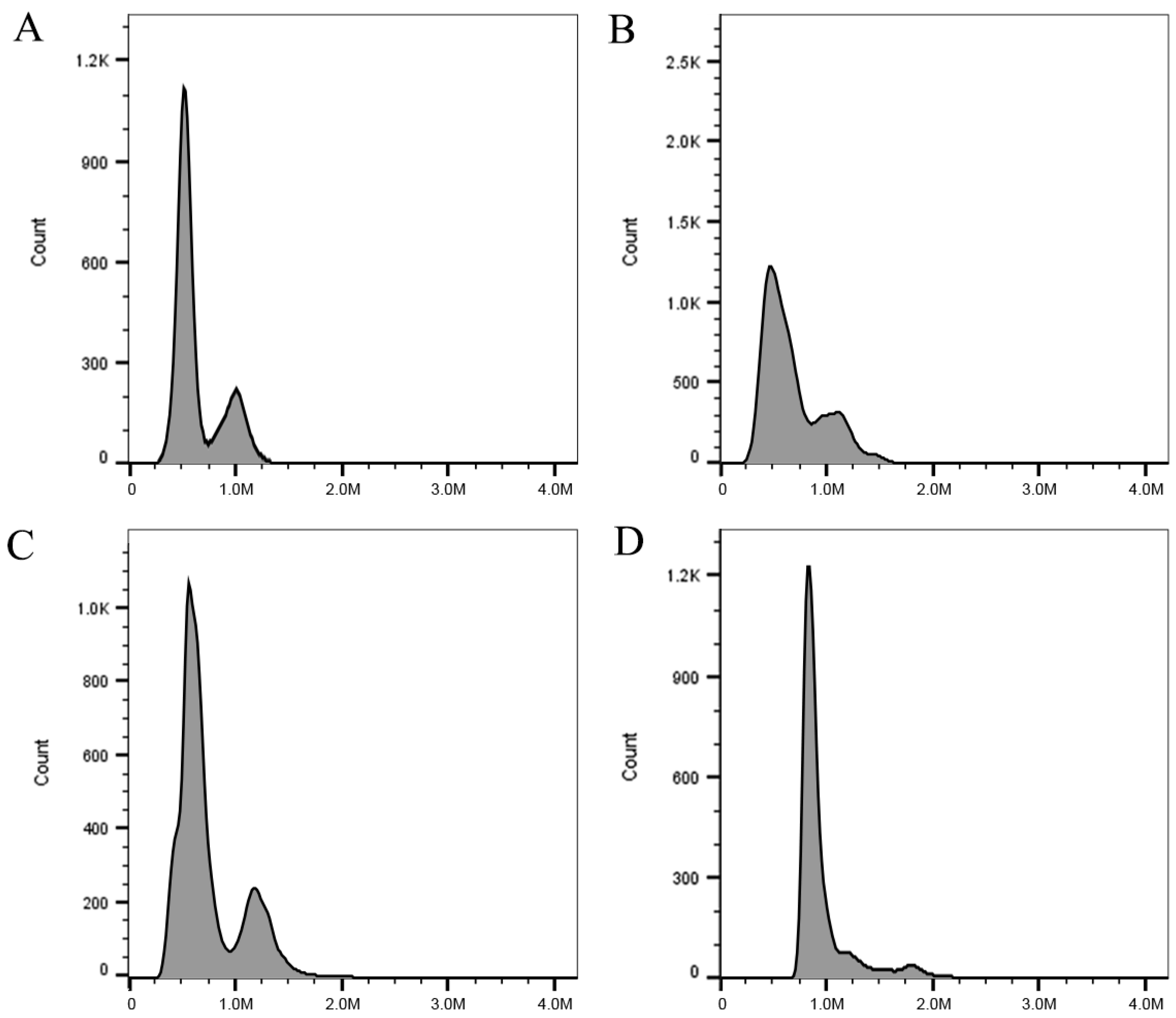
As depicted in the ploidy analysis of Figure 2, All three variants and the WT of L. xiangxiensis exhibited a main peak of relative fluorescence intensity, indicating that they were not chimeric or mixed ploidy. The sample ploidy was calculated according to the average peak positions of G0 and G1, and the results confirmed that the WT was diploid, VT1 and VT2 were diploid aneuploid, and VT3 was triploid, respectively.
To ensure appropriate statistical analysis for the response variables (plant survival rate, total mutation-induced plant rate, and polyploid plant induction rate), which are all proportional data, a preliminary data analysis was conducted. This analysis revealed that the data did not meet the assumptions of normality and homogeneity of variance required for traditional Analysis of Variance (ANOVA), even after arcsine transformation.
Consequently, the study employed Generalized Linear Models (GLMs), a more robust statistical approach suitable for non-normally distributed proportional data, to analyze the effects of oryzalin treatment concentration and duration on plant survival rate, total mutation-induced plant rate, and polyploid plant induction rate. The model fit and overall effect test results for each indicator are summarized in Table 1.
As shown in Table 1, the Generalized Linear Models for the plant survival rate and total mutation-induced plant rate both demonstrated goodness of fit (Deviance/df_res and Pearson χ2/df_res values close to 1). Furthermore, the overall models were significantly better than the null models (p < 0.05, based on Likelihood Ratio χ2 test), indicating a significant effect of the treatment factors on these two indicators.
For the polyploid plant induction rate, the model fit appeared to be perfect (Deviance and Pearson χ2 values of 0), but its results should be interpreted cautiously in conjunction with the table notes.
The Wald Chi-Square test results for the effects of treatment concentration, treatment duration, and their interaction on plant survival rate, total mutation-induced plant rate, and polyploid plant induction rate are presented in Table 2.
For plant survival rate, the main effect of the oryzalin treatment concentration was highly significant (Wald χ2 = 108,207.811, df = 3, p < 0.001). However, the main effect of the treatment duration (Wald χ2 = 2.756, df = 2, p = 0.252) and the interaction effect between the treatment concentration and duration (Wald χ2 = 4.376, df = 4, p = 0.358) did not reach statistical significance. The survival rate of CK was 100%, significantly higher than that of all the oryzalin treatment groups. In the treatment groups, the plant survival rate showed a decreasing trend as the treatment concentration increased from 0.001% to 0.003% (Table 3), with the 0.002% concentration group exhibiting a significantly higher survival rate than the 0.003% concentration group (p = 0.043). The treatment duration did not significantly affect the survival rate.
For the total mutation-induced plant rate, despite the overall model being significant (Likelihood Ratio χ2 test p = 0.033), the Wald Chi-Square tests for individual-factor effects showed that the effects of the treatment concentration, treatment duration, and their interaction did not reach statistical significance. This could be attributed to complete separation and data sparseness in the mutation induction rate data, leading to unstable Wald Chi-Square estimations. Trend-wise (Table 3), the total mutation induction rate for CK was 0%. Among the treatment groups, the 0.001% concentration for 4 days of treatment resulted in a 100% total mutation induction rate, significantly higher than that for the other treatment combinations. Other concentrations (0.002%, 0.003%) showed relatively low induction rates at different time points, e.g., the 0.002% concentration had induction rates of 33.33%, 20.00%, and 33.33% for 2, 4, and 6 days, respectively. The 0.003% concentration group consistently showed a 0% total mutation induction rate across all time points.
For the polyploid plant induction rate, the model effect test results showed that the effects of the treatment concentration, treatment duration, and their interaction did not reach statistical significance. This is primarily due to severe complete-separation phenomena in the polyploid induction rate data, which led to extremely unstable and unreliable model parameter estimations and Wald test results. Among all the oryzalin treatment combinations, only the 0.001% concentration for 4 days of treatment showed a 100% polyploid induction rate. All other treatment concentrations and duration combinations resulted in 0% polyploid induction rate. This suggests that polyploid induction is highly specific and condition-dependent, possibly occurring only under a particular combination of concentration and treatment duration.
The estimated marginal means of plant survival rate, total mutation-induced plant rate, and polyploid plant induction rate for the different oryzalin treatment concentrations and durations are presented in Table 3.

3.3. Changes in Ploidy Level and Chromosome Numbers

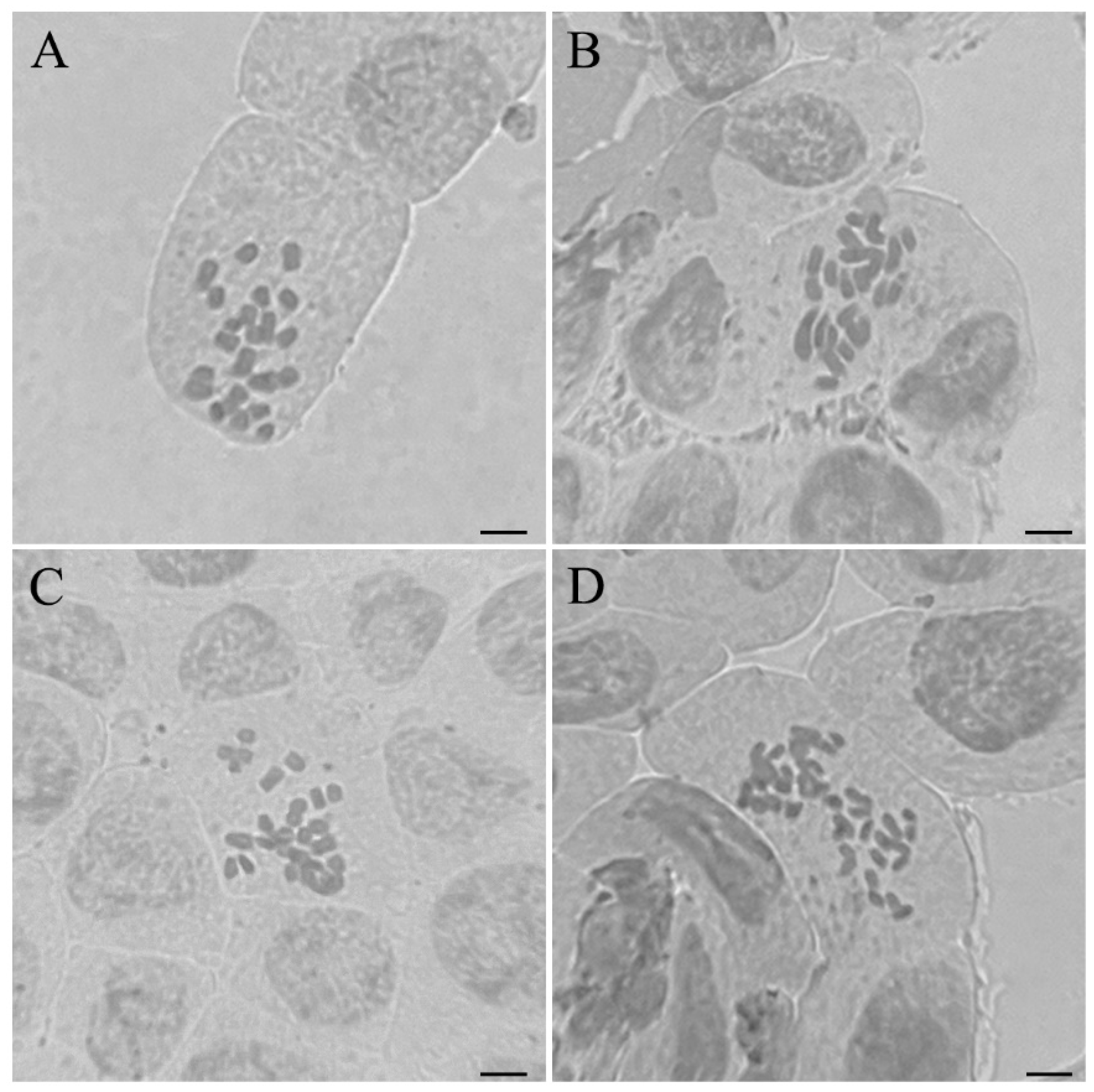
As evidenced by Figure 3 and Table 4, there were significant differences in chromosome numbers and ploidy levels between the wild type and all mutants, which included three variant types (VT1, VT2, VT3), of the regenerated plants grown in plastic containers for 10 months.
The wild-type control sample exhibited a ploidy level of approximately 2, indicating that it was diploid with 24 chromosomes (Figure 3A, 2n = 2x = 24). However, VT1 showed diploid aneuploidy with a ploidy level of 1.76 (Figure 3B, 2n = 2x − 2 = 22) due to the loss of two chromosomes. The ploidy level of VT2 rose to 2.40, reflecting diploid aneuploidy (2n = 2x + 2 = 26) with two extra chromosomes (Figure 3C). In contrast, VT3 attained a ploidy of 3.33, confirming triploidy with 36 chromosomes (3n = 3x = 36, Figure 3D). Chromosome analysis further confirmed the rationality of changes in the ploidy level of the three variants of L. xiangxiensis, that is, the decrease in the number of VT1 chromosomes was consistent with the decrease in its ploidy level; similarly, the increase in the number of VT2 and VT3 chromosomes was accompanied by a synchronous increase in their ploidy levels.

3.4. Differences in Morphological and Stomatal Characteristics

As depicted in Table 5, significant morphological differences were observed in stolon length, stolon diameter, and leaf dimensions among the variants compared with the WT, though no significant difference was observed in their leaf length-to-width ratio. The stolon length of the polyploid (VT3) was significantly longer, reaching 7.33 cm, compared to the wild-type and the diploid aneuploids (VT1 and VT2). Similarly, VT3 exhibited the longest leaf length, at 4.98 cm, a 52.76% increase over the wild type, while VT2 showed the shortest leaf length, at 2.38 cm. The maximum leaf width was also observed in the polyploid VT3 at 2.75 cm, with no significant difference in leaf width between the WT and the diploid aneuplids. Furthermore, polyploid VT3 possessed significantly thicker leaves and larger stolon diameters than the wild type, VT1, and VT2.
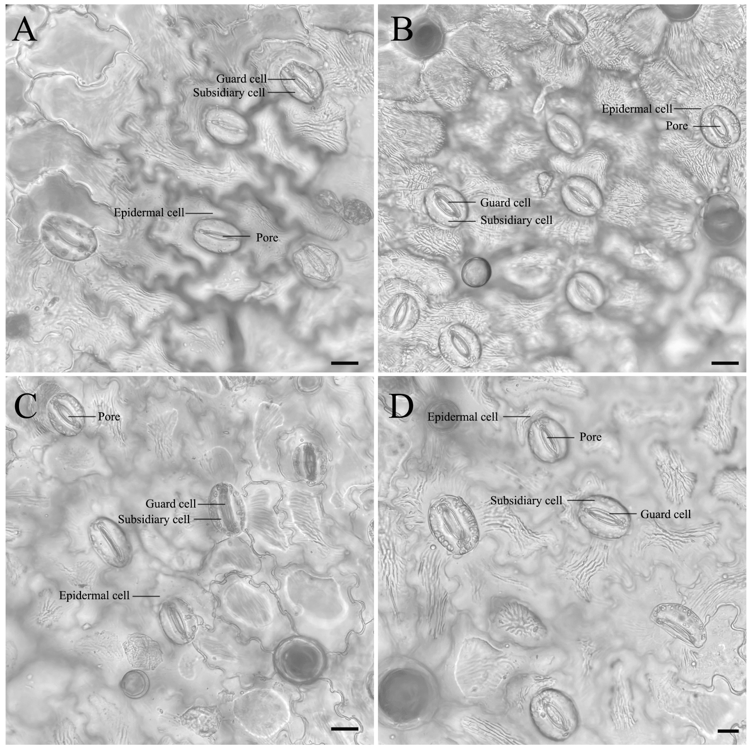
As presented in Table 5 and Figure 4, the number of stomata was highest in the VT1 leaves, at 14.52, and lowest in the polyploid, at 6.12, with the WT and VT2 showing intermediate values. The polyploid had the longest stomatal guard cells, measuring 82.43 μm, significantly longer than that of the WT, at 76.18 μm, VT2, at 75.86 μm, and VT1, at 60.08 μm. Additionally, the widest stomatal guard cells were in the polyploid VT3 at 67.46 μm, followed by the WT at 55.49 μm, with VT1 (54.81 µm) and VT2 (53.1 µm) having narrower guard cells.
The stomatal density of VT1 was the highest, at 63.93 cells μm−2, while that of polyploids was the lowest, at 27.4 cells μm−2, with moderate levels in the WT and VT2. These results reveal significant differences in leaf morphology and stomatal characteristics among the WT, the diploid aneuploids (VT1 and VT2), and the polyploid (VT3). In summary, the polyploid exhibited a greater stolon length, leaf length, width and thickness, and petiole diameter, compared to the WT and the diploid aneuploids. Additionally, the polyploid had the longest and widest stomatal guard cells, but the lowest number and density of stomata in the leaves.

3.5. Analysis of Changes in Physiological Characteristics

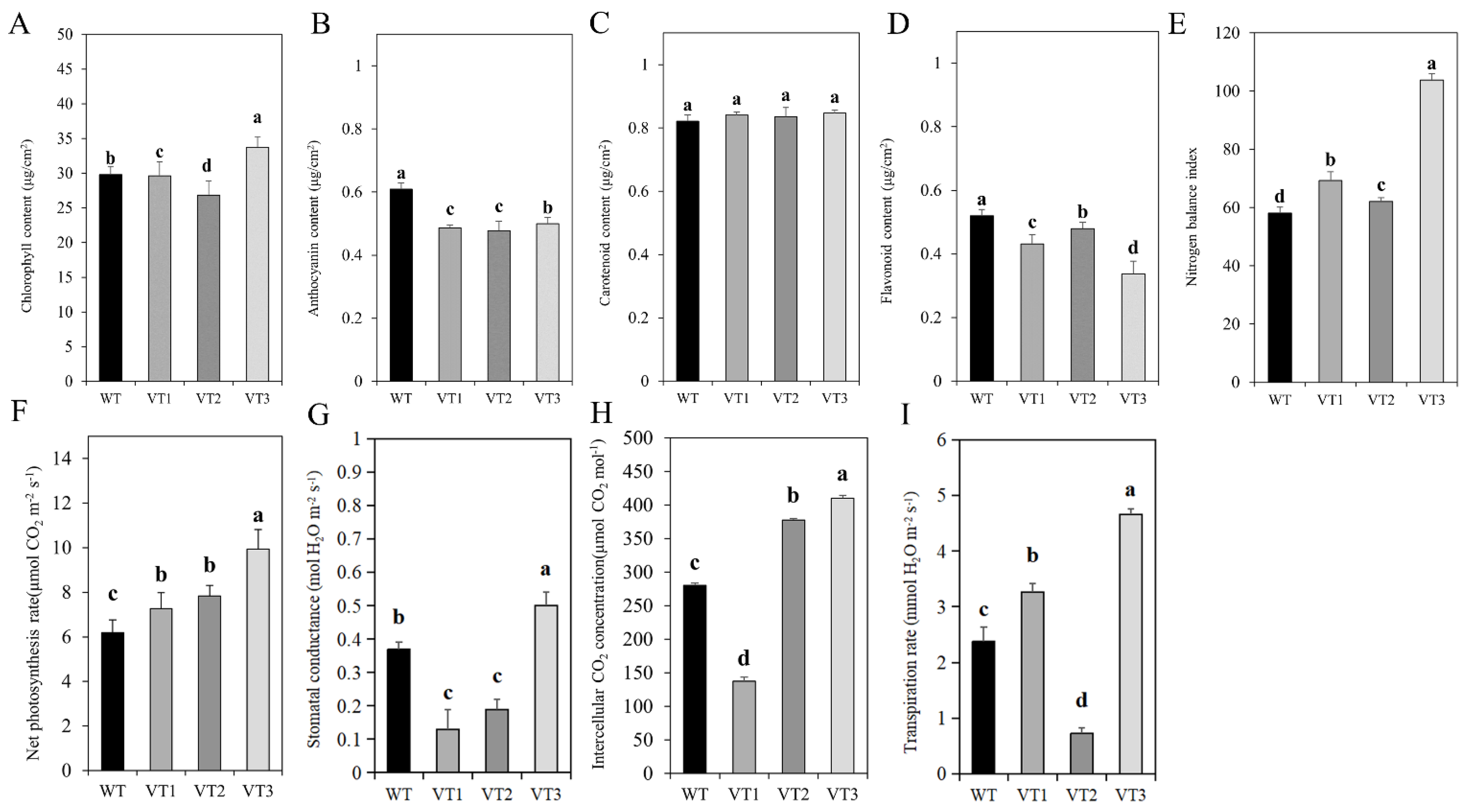
The chlorophyll content of the polyploid (VT3) was the highest, at 33.71 μg·cm−2, significantly higher than that of the WT, VT1, and VT2. Similarly, the NBI of the polyploid (VT3) was the highest, at 103.80, significantly higher than that of the WT, VT1, and VT2. Among these, the NBI values of VT1 and VT2 were higher than that of the WT, as illustrated in Figure 5A,B. As shown in Figure 5C–E, the flavonoid content was the highest in the WT, at 0.52 μg cm−2, with moderate levels in VT1 (0.43 μg·cm−2) and VT2 (0.48 μg·cm−2), while the lowest content was in the polyploid VT3 (0.34 μg·cm−2).
The WT had a slightly higher anthocyanin content, of 0.61 μg·cm−2, compared with VT1 (0.49 μg·cm−2), VT2 (0.48 μg·cm−2), and the polyploid (VT3, 0.50 μg·cm−2). The carotenoid content of the three variants remained largely similar, ranging from 0.82 μg·cm−2 in the WT to 0.85 μg·cm−2 in the polyploid. Overall, the analysis revealed notable variations in photosynthetic pigment and nitrogen balance indices among the WT, the diploid aneuploids (VT1 and VT2), and the polyploid (VT3), consistent with the detailed findings presented above.
Changes in leaf morphology, stomatal quantity, and stomatal density of the three variants would inevitably affect their photosynthetic efficiency. Compared with the wild type, the polyploid (VT3) exhibited the highest net photosynthetic rate (9.93 μmol CO2 m−2 s−1), stomatal conductance (0.5 mol H2O m−2 s−1), and intercellular CO2 concentration (410.11 μmol CO2), all of which were also higher than those of VT1 and VT2. However, the changes in these physiological indicators in VT1 and VT2 were inconsistent, as illustrated in Figure 5F–I.

4. Discussion

It is known that oryzalin can cause genome duplication by disrupting microtubules in cells, and it is widely used for polyploid induction in plants [25]. The present study indicated that continuous treatment of stem segments of L. xiangxiensis with 0.001% oryzalin for 4 days resulted in a polyploidization induction rate of up to 100%, which is consistent with previous reports that low concentrations of antimitotic agents with shorter treatment times were most effective in inducing polyploidization of Solanum spp. without causing excessive cytotoxicity [26]. Similar research results have also been obtained for several other species. For example, it has been reported that treatment of nodes of triploid Populus with 0.0005% oryzalin for 3 days resulted in a 100% induction duplication rate [27], and the highest polyploidy induction rate of 88.89% was obtained in shoots of Tectona grandis treated with 0.001% oryzalin for 5 days [28]. When micro-propagated nodal segments of the aromatic plant Mentha spicata were treated with 0.006% oryzalin for 48 h, a polyploid induction rate of 8% was achieved, with a survival rate below 30% [9].
The Generalized Linear Model analysis of this study (Table 2) showed that the oryzalin treatment concentration had an extremely significant effect on the plant survival rate (p < 0.001), with a significant decrease in survival rate as the concentration increased (Table 3). This indicates that cytotoxicity is a critical factor that cannot be ignored in polyploid induction. Although the main effect of treatment duration (p = 0.252) and the interaction effect between treatment concentration and duration (p = 0.358) did not reach statistical significance, we observed that in certain specific combinations, such as the 4-day treatment at 0.001% concentration, the survival rate (11.11%) was lower than that for the 6-day treatment (38.89%). This might reflect potential individual variations or sensitivities when a certain treatment duration is combined with a specific concentration.
For the polyploid plant induction rate, the Generalized Linear Model analysis in this study did not identify statistically significant effects of the treatment concentration, treatment duration, or their interaction (Table 2). Although the polyploid induction rate based on surviving plants was as high as 100% for the 0.001% oryzalin treatment for 4 days (Table 3), it is crucial to emphasize that the model’s goodness-of-fit tests indicated the presence of complete separation in the data (Table 1 note), leading to instability in model parameter estimation and Wald test results. This implies that under certain treatment conditions (e.g., 0.001% concentration for 4 days), all surviving plants underwent polyploidization, whereas in many other treatment combinations, the polyploid induction rate was zero. This extreme data distribution limited the model’s ability to statistically identify treatment effects.
Indeed, when the oryzalin concentration increased to 0.002% and 0.003%, no polyploidization of L. xiangxiensis was observed with any of the tested treatment durations (2, 4, and 6 days) (Table 3). This further corroborates the limiting effect of oryzalin’s cytotoxicity on polyploid induction efficiency. Higher oryzalin concentrations and/or excessively long treatment durations often lead to severe cellular damage or even mortality, thereby rendering polyploid plant production impossible. Notably, previous studies have also indicated that higher concentrations of oryzalins (0.0021–0.028%) and longer treatment times (2–4 days) significantly reduce the survival rate of polyploids, indicating that the cytotoxicity of oryzalin greatly limits its effectiveness in plant polyploid induction [14,29,30]. For example, In Thymus vulgaris, treatment of shoot segments with 0.028% oryzalin for 48 h only achieved an induction rate of 3.5% and a survival rate of 12% [29].
Therefore, in the process of plant polyploid induction, finding the optimal treatment regimen requires balancing the polyploid induction rate with the plant survival rate. Although the induction percentage based on surviving plants might be high, if the absolute number of initial explants or surviving plants is too low, the actual quantity of desired polyploids obtained will be very limited. The results of this study emphasize that for L. xiangxiensis polyploid induction, the concentration and treatment duration of oryzalin must be carefully balanced to achieve effective polyploid induction while ensuring a reasonable survival rate. Successfully induced polyploid plants often exhibit significant morphological changes in their phenotype, which are among the most direct and easily recognizable characteristics for identifying polyploid variants.
Polyploid plants often exhibit a series of morphological changes, with gigantism being one of the most prominent phenotypic traits, making it one of the most direct and easily recognizable methods for identifying polyploid variants, which are often characterized by an increased leaf size, thicker stems, and changes in pigmentation [17].
In this study, the induced variants (VT1, VT2, and VT3) exhibited distinct changes in morphological traits compared to the WT, such as in their leaf size, stolon length and thickness, and color patterns, which were consistent with previous reports highlighting the impact of polyploidy on plant morphology [31,32]. In an oryzalin-induced polyploid of the Escallonia genus, notable changes, such as larger leaves and altered pigmentation, were observed in three important evergreen woody ornamental species within this genus, and it was concluded that the effect of polyploidization on Escallonia is highly variable and species-dependent [31]. Similarly, polyploidy led to larger leaf sizes and more robust stem structures in Satureja khuzistanica compared to diploid counterparts [32].
Although morphological features such as gigantism offer an accessible means of identifying polyploids, they are not always conclusive, due to the variability of polyploid effects across different species and the influence of environmental factors. Therefore, cytological methods to supplement phenotype observation are crucial for more accurate polyploid identification. Flow cytometry and chromosome counting are two powerful cytological techniques that provide a precise pathway for identifying polyploidy by determining the chromosome number and ploidy level of the plant [33], thereby minimizing the subjectivity associated with phenotypic observations.
Previous results of flow cytometry analysis of Cuminum cyminum [34] and herbaceous peony [35] were closely related to chromosome counting, further verifying its application in polyploids. However, there are only reports on the chromosome count of two new species, Lysimachia tianmaensis and L. qimenensis, in this genus [36].
The chromosome number of the L. xiangxiensis wild type (2n = 2x = 24) in our study was consistent with previous reports, which also strongly proved that VT1 (2n = 2x − 2) and VT2 (2n = 2x + 2) are diploid aneuploids, while VT3 (3n = 3x = 36) is triploid. The identification of these variants (diploid aneuploids VT1 and VT2, and triploid VT3) highlights the diverse genetic outcomes from oryzalin treatment. For the aneuploids, their formation is likely attributed to the effects of oryzalin as a microtubule inhibitor. Oryzalin disrupts spindle formation during mitosis, which can lead to various chromosomal segregation errors, such as non-disjunction of chromosomes or chromosome loss/gain. This disturbance in the mitotic apparatus results in cells with an abnormal number of chromosomes, manifesting as aneuploidy [15]. In contrast, the formation of the triploid is primarily a result of genome duplication.
In this study, chromosome counting confirmed that the ploidy of regenerated plants was diploid aneuploid and triploid, without any tetraploids detected, which contradicts the typical dominance of tetraploids in polyploid induction. The possible reasons or mechanisms behind this are as follows: Firstly, the concentration or duration of oryzalin treatment might have been insufficient to sustain complete endoreduplication. Microtubule inhibitors require persistent action during mitosis to block spindle formation and achieve genome duplication [25]. The emergence of triploids suggests a non-canonical genome duplication pathway deviating from the typical 2n to 4n route. Secondly, one plausible explanation is failed cytokinesis after endoreduplication: oryzalin treatment might have triggered DNA replication without subsequent mitotic division, allowing cells to enter a new cycle directly as 3n. A similar phenomenon was reported in Crassostrea giga [37] and triploid bananas [38]. Similarly, treatment with a 0.3% colchicine solution for 4 h using cotton leaching and the injection method was found to be effective for triploid generation in Ziziphus jujuba [39]. Thirdly, the heterogeneity of stem-segment cells (e.g., varying differentiation status or cell-cycle stages) might have influenced drug responsiveness [40]. For instance, cells in the G1-phase may resume a new cell cycle post-treatment, potentially generating populations with different ploidy levels [41]. Additionally, triploid cells might have been selectively retained during regeneration due to metabolic or proliferative advantages, further explaining their stable presence [42]. In summary, the induction of triploids rather than tetraploids by oryzalin in stem segments of this species highlights the species-specificity of polyploidization mechanisms and their sensitivity to treatment conditions.
Polyploid plants often exhibit enhanced chlorophyll contents and photosynthetic rates due to the upregulation of chlorophyll synthesis and photosynthetic genes [43,44]. In this study, the diploid aneuploids (VT1 and VT2) and the polyploid (VT3) of L. xiangxiensis displayed a significantly higher chlorophyll content, intercellular carbon dioxide concentration, stomatal conductance, and NBI compared to the WT, which suggests enhanced photosynthetic capacity, nitrogen assimilation efficiency, and environmental adaptability in the polyploid variants. However, the decrease in anthocyanin and carotenoid content in the polyploids, especially the reduction in flavonoid compounds that are crucial for UV protection and stress tolerance [45], indicates that the redistribution of these metabolites shifts from stress defense to growth processes, resulting in significant growth in the leaf size, stem length, and thickness, which is consistent with Arabidopsis thaliana, Chamomilla recutita, and Solanum commersonii polyploids [46,47]. Stomatal conductance refers to the parameter of gas flow rate per unit area and unit time, which characterizes leaf stomatal opening, water transpiration, and carbon dioxide absorption. The polyploidization of L. xiangxiensis results in a higher stomatal conductance, intercellular carbon dioxide concentration, net photosynthetic rate, and transpiration rate. The most important and fundamental change is the increase in leaf stomatal guard cell size and the decrease in stomatal density, leading to an increase in stomatal conductance, which, in turn, promotes gas exchange, increases the photosynthesis rate [27,48] and biomass accumulation [49,50], and enhances stress resistance through compensatory mechanisms to increase the transpiration rate and reduce water loss [50].
The NBI, which is the ratio of chlorophyll to flavonoids, is an essential stress fluorescence parameter used to evaluate nitrogen nutritional status and plant growth potential. In this study, the polyploid (VT3) exhibited the highest NBI, with a value of 103.80, significantly higher than that of the diploid aneuploids VT1 and VT2 and the WT, indicating improved nitrogen utilization efficiency and enhanced growth and development. Similar trends have been observed in tetraploid Daylily plants, which showed an increased NBI under drought conditions, reflecting better nitrogen utilization and improved growth potential compared to diploids [51]. These results align with previous studies showing that polyploidy enhances photosynthetic capacity through both structural and functional leaf modifications, promoting higher growth rates and drought tolerance in citrus, wheat, and Hemerocallis spp. [52,53].
In the future, more emphasis should be placed on elucidating the physiological and molecular mechanisms related to polyploid induction, as well as thoroughly characterizing the reproductive and flowering traits of the induced polyploids, which are critical for their agricultural and ornamental potential. The compelling positive morphological changes, such as increased stolon length, larger and thicker leaves, along with the enhanced physiological characteristics, including photosynthetic capacity and nitrogen utilization efficiency, observed in the induced L. xiangxiensis polyploids indicate promising potential for trait improvement through polyploidization. Therefore, the insights gained from this study, particularly concerning the delicate balance between oryzalin concentration/duration and plant survival, provide a valuable reference. These findings serve as a foundation for exploring the applicability of similar polyploidization strategies to other economically important or ornamental horticultural species. By optimizing induction protocols tailored to specific species, this biotechnological approach can contribute to the breeding of novel varieties exhibiting improved vigor, enhanced esthetic attributes, and increased adaptability to diverse environmental conditions for broader horticultural applications.

5. Conclusions

In this study, polyploid plants of L. xiangxiensis were successfully obtained by treating stem segments with 0.001% oryzalin for 4 days and further developing the plantlets in vitro. Compared with the wild type, genome duplication and aneuploidy induction caused some significant morphological and physiological changes in the polyploids and diploid aneuploids, including changes in leaf size, color, and photosynthetic efficiency. Among them, the triploid VT3 showed the highest chlorophyll content and nitrogen balance index and a lower flavonoid content, maintaining a higher competitive advantage in terms of photosynthetic capacity, nitrogen use efficiency, and growth status. The acquisition of these variants provides important experimental materials and enormous potential for the genetic improvement and chromosome engineering of L. xiangxiensis. Subsequent field experiments will continue to evaluate the phenotypic characteristics and genetic stability of these variants under natural conditions and further reveal the phenotypic, physiological, and molecular mechanisms of polyploid and diploid aneuploid plants using genomics, transcriptomics, and metabolomics, facilitating the development of robust and high-ornamental-value plants.

Author Contributions

Conceptualization, Y.Z.; funding acquisition, R.S., Q.L., and X.S.; investigation, R.S.; methodology, Y.Z.; project administration, G.C., R.S., Q.L., and X.S.; resources, G.C. and R.S.; software, Y.Z.; supervision, G.C., R.S., Q.L., and X.S.; visualization, Y.Z.; writing—original draft, Y.Z.; writing—review and editing, G.C. and X.S. All authors have read and agreed to the published version of the manuscript.

Funding

This research was funded by the Shanghai Science and Technology Innovation Action Plan, grant number 22Z530201080.

Data Availability Statement

The original contributions presented in the study are included in the article; further inquiries can be directed to the corresponding author.

Conflicts of Interest

Authors Ruixue Shen and Qiujing Li were employed by the company Shanghai Shangfang Horticulture Co. The remaining authors declare that the research was conducted in the absence of any commercial or financial relationships that could be construed as a potential conflict of interest.

References

  1. Mou, C.; Wu, Y.; Xiang, L.; Xiang, X.-M.; Zhang, D.-G. Lysimachia xiangxiensis (Primulaceae), a New Species from Limestone Area in Hunan Province, Central China. PhytoKeys 2020, 140, 23–32. [Google Scholar] [CrossRef] [PubMed]
  2. Oh, I.-C.; Anderberg, A.-L.; Schönenberger, J.; Anderberg, A.A. Comparative Seed Morphology and Character Evolution in the Genus lysimachia (Myrsinaceae) and Related Taxa. Plant Syst. Evol. 2008, 271, 177–197. [Google Scholar] [CrossRef]
  3. Hu, X. Physiological Responses of Lysimachia alfredii to Cadmium Stress. Master’s Thesis, Hunan Agricultural University, Changsha, China, 2023. [Google Scholar]
  4. Zheng, W. Studies on Germplasm Characteristics and Enhancement of Four Lysimachia Landscape Species. Ph.D. Thesis, Huazhong Agricultural University, Wuhan, China, 2015. [Google Scholar]
  5. Eng, W.-H.; Ho, W.-S. Polyploidization Using Colchicine in Horticultural Plants: A Review. Sci. Hortic. 2019, 246, 604–617. [Google Scholar] [CrossRef]
  6. Thao, N.T.P.; Ureshino, K.; Miyajima, I.; Ozaki, Y.; Okubo, H. Induction of Tetraploids in Ornamental Alocasia through Colchicine and Oryzalin Treatments. Plant Cell Tiss Organ Cult. 2003, 72, 19–25. [Google Scholar] [CrossRef]
  7. Zhou, X.; Zhao, P.; Zeng, F.; Geng, X.; Zhou, J.; Sun, J. Induction and Identification of Polyploids in Four Rhododendron Species. Plant Cell Tiss Organ Cult. 2024, 158, 11. [Google Scholar] [CrossRef]
  8. Winkelmann, T.; Braun, P.; Dhooghe, E.; Huylenbroeck, J. van Advances in Conventional Breeding Techniques for Ornamentals. In Achieving Sustainable Cultivation of Ornamental Plants; Burleigh Dodds Science Publishing: Sawston, UK, 2020; ISBN 978-1-003-04776-6. [Google Scholar]
  9. Bharati, R.; Gupta, A.; Novy, P.; Severová, L.; Šrédl, K.; Žiarovská, J.; Fernández-Cusimamani, E. Synthetic Polyploid Induction Influences Morphological, Physiological, and Photosynthetic Characteristics in Melissa officinalis L. Front. Plant Sci. 2023, 14, 1332428. [Google Scholar] [CrossRef]
  10. Fakhrzad, F.; Jowkar, A.; Shekafandeh, A.; Kermani, M.J.; Moghadam, A. Tetraploidy Induction Enhances Morphological, Physiological and Biochemical Characteristics of Wallflower (Erysimum cheiri (L.) Crantz). Sci. Hortic. 2023, 308, 111596. [Google Scholar] [CrossRef]
  11. Allum, J.F.; Bringloe, D.H.; Roberts, A.V. Chromosome Doubling in a Rosa Rugosa Thunb. Hybrid by Exposure of in Vitro Nodes to Oryzalin: The Effects of Node Length, Oryzalin Concentration and Exposure Time. Plant Cell Rep. 2007, 26, 1977–1984. [Google Scholar] [CrossRef] [PubMed]
  12. Wu, H.; Jiang, L.; Li, J.; Lu, M.; An, H. Polyploid Induction and Identification of Rosa roxburghii f. eseiosa. Plants 2023, 12, 2194. [Google Scholar] [CrossRef]
  13. Contreras, R.N.; Ranney, T.G.; Tallury, S.P. Reproductive Behavior of Diploid and Allotetraploid rhododendron L. ‘Fragrant Affinity’. HortScience 2007, 42, 31–34. [Google Scholar] [CrossRef]
  14. Zhang, Y.-S.; Chen, J.-J.; Cao, Y.-M.; Duan, J.-X.; Cai, X.-D. Induction of Tetraploids in ‘Red Flash’ Caladium Using Colchicine and Oryzalin: Morphological, Cytological, Photosynthetic and Chilling Tolerance Analysis. Sci. Hortic. 2020, 272, 109524. [Google Scholar] [CrossRef]
  15. Chen, J.-J.; Zhang, Y.-S.; Duan, J.-X.; Cao, Y.-M.; Cai, X.-D. Morphological, Cytological, and Pigment Analysis of Leaf Color Variants Regenerated from Long-Term Subcultured Caladium Callus. In Vitro Cell. Dev. Biol.-Plant 2021, 57, 60–71. [Google Scholar] [CrossRef]
  16. Li, S.; Lin, Y.; Pei, H.; Zhang, J.; Zhang, J.; Luo, J. Variations in Colchicine-Induced Autotetraploid Plants of Lilium davidii var. unicolor. Plant Cell Tiss Organ Cult. 2020, 141, 479–488. [Google Scholar] [CrossRef]
  17. Wang, L.-J.; Cao, Q.-Z.; Zhang, X.-Q.; Jia, G.-X. Effects of Polyploidization on Photosynthetic Characteristics in Three Lilium Species. Sci. Hortic. 2021, 284, 110098. [Google Scholar] [CrossRef]
  18. Miri, S.M. Artificial Polyploidy in the Improvement of Horticultural Crops. J. Plant Physiol. Breed. 2020, 10, 1–28. [Google Scholar] [CrossRef]
  19. Kermani, M.J.; Sarasan, V.; Roberts, A.V.; Yokoya, K.; Wentworth, J.; Sieber, V.K. Oryzalin-Induced Chromosome Doubling in Rosa and Its Effect on Plant Morphology and Pollen Viability. Theor. Appl. Genet. 2003, 107, 1195–1200. [Google Scholar] [CrossRef]
  20. Podwyszyńska, M.; Sowik, I.; Machlańska, A.; Kruczyńska, D.; Dyki, B. In Vitro Tetraploid Induction of Malus × Domestica Borkh. Using Leaf or Shoot Explants. Sci. Hortic. 2017, 226, 379–388. [Google Scholar] [CrossRef]
  21. Kirchner, S.; Pianowski, Z. Photopharmacology of Antimitotic Agents. Int. J. Mol. Sci. 2022, 23, 5657. [Google Scholar] [CrossRef]
  22. Zheng, W.; Xu, X.-D.; Dai, H.; Chen, L.-Q. Direct Regeneration of Plants Derived from in Vitro Cultured Shoot Tips and Leaves of Three Lysimachia Species. Sci. Hortic. 2009, 122, 138–141. [Google Scholar] [CrossRef]
  23. Doležel, J.; Greilhuber, J.; Suda, J. Estimation of Nuclear DNA Content in Plants Using Flow Cytometry. Nat. Protoc. 2007, 2, 2233–2244. [Google Scholar] [CrossRef]
  24. Cai, X.; Cao, Z.; Xu, S.; Deng, Z. Induction, Regeneration and Characterization of Tetraploids and Variants in ‘Tapestry’ Caladium. Plant Cell Tissue Organ Cult. 2015, 120, 689–700. [Google Scholar] [CrossRef]
  25. Dhooghe, E.; Van Laere, K.; Eeckhaut, T.; Leus, L.; Van Huylenbroeck, J. Mitotic Chromosome Doubling of Plant Tissues in Vitro. Plant Cell Tiss Organ Cult. 2011, 104, 359–373. [Google Scholar] [CrossRef]
  26. Chauvin, J.E.; Souchet, C.; Dantec, J.P.; Ellissèche, D. Chromosome Doubling of 2x Solanum Species by Oryzalin: Method Development and Comparison with Spontaneous Chromosome Doubling in Vitro. Plant Cell Tissue Organ Cult. 2003, 73, 65–73. [Google Scholar] [CrossRef]
  27. Zeng, Q.; Liu, Z.; Du, K.; Kang, X. Oryzalin-Induced Chromosome Doubling in Triploid Populus and Its Effect on Plant Morphology and Anatomy. Plant Cell Tiss Organ Cult. 2019, 138, 571–581. [Google Scholar] [CrossRef]
  28. Fauzan, Y.S.A.; Supriyanto; Mulyono, J.; Tajuddin, T. The Effectiveness of Colchicine and Oryzalin on Polyploidy Induction in Teak (Tectona grandis Linn. f.) in vitro. Indones. J. For. Res. 2024, 11, 1–15. [Google Scholar] [CrossRef]
  29. Shmeit, Y.H. Autopolyploidy Effect on Morphological Variation and Essential Oil Content in Thymus vulgaris L. Sci. Hortic. 2020, 263, 109095. [Google Scholar] [CrossRef]
  30. Bharati, R.; Fernández-Cusimamani, E.; Gupta, A.; Novy, P.; Moses, O.; Severová, L.; Svoboda, R.; Šrédl, K. Oryzalin Induces Polyploids with Superior Morphology and Increased Levels of Essential Oil Production in Mentha spicata L. Ind. Crops Prod. 2023, 198, 116683. [Google Scholar] [CrossRef]
  31. Denaeghel, H.E.R.; Van Laere, K.; Leus, L.; Lootens, P.; Van Huylenbroeck, J.; Van Labeke, M.-C. The Variable Effect of Polyploidization on the Phenotype in Escallonia. Front. Plant Sci. 2018, 9, 354. [Google Scholar] [CrossRef]
  32. Shariat, A.; Sefidkon, F. Tetraploid Induction in Savory (Satureja khuzistanica): Cytological, Morphological, Phytochemical and Physiological Changes. Plant Cell Tiss Organ Cult. 2021, 146, 137–148. [Google Scholar] [CrossRef]
  33. Roux, N.; Toloza, A.; Radecki, Z.; Zapata-Arias, F.J.; Dolezel, J. Rapid Detection of Aneuploidy in Musa Using Flow Cytometry. Plant Cell Rep. 2003, 21, 483–490. [Google Scholar] [CrossRef]
  34. Sanaei-Hoveida, Z.; Mortazavian, S.M.M.; Norouzi, M.; Sadat-Noori, S.A. Exploring the Potential of Polyploidization as a Breeding Tool for Medicinal Plants: A Case Study on Cumin (Cuminum cyminum L.). Plant Cell Tiss Organ Cult. 2023, 156, 23. [Google Scholar] [CrossRef]
  35. Zhu, W.; Dong, Z.; Chen, X.; Cao, J.; Zhang, W.; Sun, R.; Teixeira Da Silva, J.A.; Yu, X. Induction of 2n Pollen by Colchicine during Microsporogenesis to Produce Polyploids in Herbaceous Peony (Paeonia lactiflora Pall.). Sci. Hortic. 2022, 304, 111264. [Google Scholar] [CrossRef]
  36. Liu, K.; Hong, X.; Zhou, S.-B.; Cheng, Y.-S.; Tang, C.-F.; Xu, H.-J. A New Species of Lysimachia (Myrsinaceae) from Dabieshan Mountain, China. Plant Syst. Evol. 2014, 300, 1615–1620. [Google Scholar] [CrossRef]
  37. Gerard, A.; Naciri, Y.; Peignon, J.-M.; Ledu, C.; Phelipot, P. Optimization of Triploid Induction by the Use of 6-DMAP for the Oyster Crassostrea Gigas (Thunberg). Aquac. Res. 1994, 25, 709–719. [Google Scholar] [CrossRef]
  38. Ramakrishnan, R.; Banadka, A.; Dubey, S.; Al-Khayri, J.M.; Nagella, P. Advances in Triploid Plant Production: Techniques, Benefits, and Applications. Plant Cell Tiss Organ Cult. 2025, 160, 70. [Google Scholar] [CrossRef]
  39. Liu, S.; Zhang, C.; Yang, W.; Li, X.; Hou, L.; Li, M.; Pang, X.; Li, Y. Hybrid Triploid Induced by Megaspore Chromosome Doubling in Jujube (Ziziphus Jujuba Mill.) ‘Maya’ and Its Characteristics. Forests 2021, 12, 112. [Google Scholar] [CrossRef]
  40. Hong, L.; Dumond, M.; Zhu, M.; Tsugawa, S.; Li, C.-B.; Boudaoud, A.; Hamant, O.; Roeder, A.H.K. Heterogeneity and Robustness in Plant Morphogenesis: From Cells to Organs. Annu. Rev. Plant Biol. 2018, 69, 469–495. [Google Scholar] [CrossRef]
  41. Zhang, F.; Rathod, B.; Jones, J.B.; Wang, Q.; Bernhard, E.; Godyn, J.; Studzinski, G.P. Increased Stringency of the 1,25-dihydroxyvitamin D3-induced G1 to S Phase Block in Polyploid HL60 Cells. J. Cell. Physiol. 1996, 168, 18–25. [Google Scholar] [CrossRef]
  42. Trojak-Goluch, A.; Kawka-Lipińska, M.; Wielgusz, K.; Praczyk, M. Polyploidy in Industrial Crops: Applications and Perspectives in Plant Breeding. Agronomy 2021, 11, 2574. [Google Scholar] [CrossRef]
  43. Cao, Q.; Zhang, X.; Gao, X.; Wang, L.; Jia, G. Effects of Ploidy Level on the Cellular, Photochemical and Photosynthetic Characteristics in Lilium FO Hybrids. Plant Physiol. Biochem. 2018, 133, 50–56. [Google Scholar] [CrossRef]
  44. Rahmawati, K.; Yunianta, Y.; Risjani, Y. Polyploid Induction Enhance Pigment Production in Nannochoropsis Oculata: A Study Using a Mutagenesis Agent [Preprint]. Res. Sq. 2023. [Google Scholar] [CrossRef]
  45. Tossi, V.E.; Martínez Tosar, L.J.; Laino, L.E.; Iannicelli, J.; Regalado, J.J.; Escandón, A.S.; Baroli, I.; Causin, H.F.; Pitta-Álvarez, S.I. Impact of Polyploidy on Plant Tolerance to Abiotic and Biotic Stresses. Front. Plant Sci. 2022, 13, 869423. [Google Scholar] [CrossRef]
  46. Fasano, C.; Diretto, G.; Aversano, R.; D’Agostino, N.; Di Matteo, A.; Frusciante, L.; Giuliano, G.; Carputo, D. Transcriptome and Metabolome of Synthetic Solanum Autotetraploids Reveal Key Genomic Stress Events Following Polyploidization. New Phytol. 2016, 210, 1382–1394. [Google Scholar] [CrossRef]
  47. Madani, H.; Escrich, A.; Hosseini, B.; Sanchez-Muñoz, R.; Khojasteh, A.; Palazon, J. Effect of Polyploidy Induction on Natural Metabolite Production in Medicinal Plants. Biomolecules 2021, 11, 899. [Google Scholar] [CrossRef]
  48. Baker, R.L.; Brock, G.L.; Newsome, E.L.; Zhao, M. Polyploidy and the Evolution of Phenotypic Integration: Network Analysis Reveals Relationships among Anatomy, Morphology, and Physiology. Appl. Plant Sci. 2024, 12, e11605. [Google Scholar] [CrossRef]
  49. Yao, P.-Q.; Chen, J.-H.; Ma, P.-F.; Xie, L.-H.; Cheng, S.-P. Stomata Variation in the Process of Polyploidization in Chinese Chive (Allium tuberosum). BMC Plant Biol. 2023, 23, 595. [Google Scholar] [CrossRef]
  50. Dunn, J.; Hunt, L.; Afsharinafar, M.; Meselmani, M.A.; Mitchell, A.; Howells, R.; Wallington, E.; Fleming, A.J.; Gray, J.E. Reduced Stomatal Density in Bread Wheat Leads to Increased Water-Use Efficiency. J. Exp. Bot. 2019, 70, 4737–4748. [Google Scholar] [CrossRef]
  51. Lourkisti, R.; Oustric, J.; Quilichini, Y.; Froelicher, Y.; Herbette, S.; Morillon, R.; Berti, L.; Santini, J. Improved Response of Triploid Citrus Varieties to Water Deficit Is Related to Anatomical and Cytological Properties. Plant Physiol. Biochem. 2021, 162, 762–775. [Google Scholar] [CrossRef]
  52. Misiukevičius, E.; Mažeikienė, I.; Stanys, V. Ploidy’s Role in Daylily Plant Resilience to Drought Stress Challenges. Biology 2024, 13, 289. [Google Scholar] [CrossRef]
  53. Khazaei, H.; Monneveux, P.; Hongbo, S.; Mohammady, S. Variation for Stomatal Characteristics and Water Use Efficiency among Diploid, Tetraploid and Hexaploid Iranian Wheat Landraces. Genet. Resour. Crop Evol. 2010, 57, 307–314. [Google Scholar] [CrossRef]
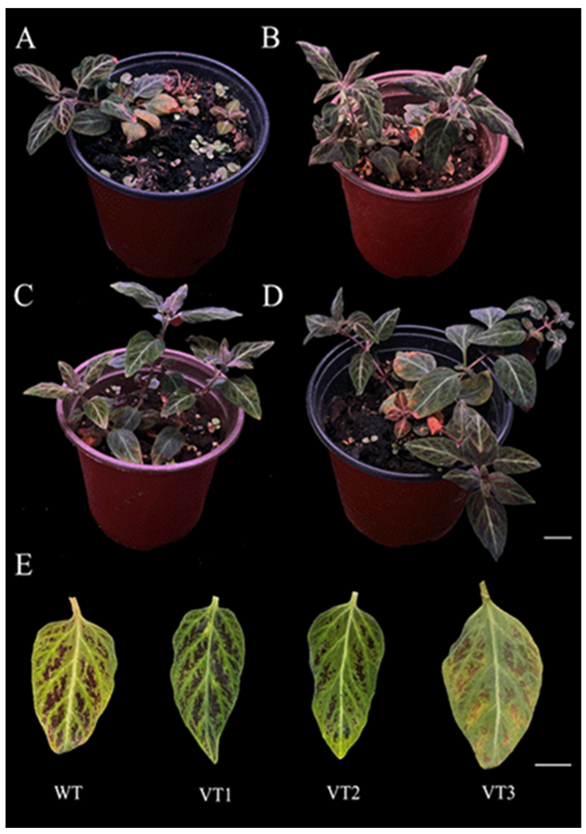
Figure 1. The growth status of L. xiangxiensis plantlets transplanted into pots for 18 months ((A): WT, (BD): VT1-VT3), and differences in the typical characteristics of leaves (E) of the three variation types (VT1, VT2, and VT3) compared with the wild type (WT). (AD): bar = 2 cm; (E): bar = 1 cm.
Horticulturae 11 00654 g001
Figure 2. Ploidy analysis of the wild type (A) and the three variation types ((BD): VT1–VT3) of L. xiangxiensis by flow cytometry analysis.
Horticulturae 11 00654 g002
Figure 3. Micrographs of somatic chromosome numbers in the root tips of the wild type and three variation types of L. xiangxiensis. (A) WT (2n = 2x = 24); (B) VT1 (2n = 2x − 2 = 22); (C) VT2 (2n = 2x + 2 = 26); (D) VT3 (2n = 3x = 36), bars =5 μm.
Horticulturae 11 00654 g003
Figure 4. Comparison of stomata size and density among different plant types and the wild type of L. xiangxiensis. (A) Wild type (diploid, 2n = 2x = 24); (B) diploid aneuploid (VT1, 2n = 2x − 2 = 22); (C) diploid aneuploid (VT2, 2n = 2x + 2 = 26); (D) triploid plant (VT3, 3n = 3x = 36). Images were captured under 40× magnification. Scale bars = 20 μm.
Horticulturae 11 00654 g004
Figure 5. The total chlorophyll content (A), nitrogen balance index (NBI, (B)), flavonoid content (C), anthocyanin content (D), carotenoid content (E) in the interveinal areas, net photosynthetic rate (F), stomatal conductance (G), intercellular carbon dioxide concentration (H), and transpiration rate (I) of the mature leaves for the wild type and three variation types of L. xiangxiensis. The different letters indicate significant differences at 5% level by Duncan’s range test.
Horticulturae 11 00654 g005
Table 1. Generalized Linear Model fit and overall effect tests for each indicator.
IndicatorPlant Survival RateTotal Mutation-Induced Plant RatePolyploid Plant Induction Rate
Error distributionBinomialBinomialBinomial
Link functionComplementary Log-logComplementary Log-logLogit
Deviance18.5527.8350
Residual df (df_res) 1201313
Deviance/df-res0.9280.6030
Pearson χ215.7075.9920
Pearson χ2/df0.7850.4610
Likelihood Ratio χ261.99518.19616.875
Model df (df_model) 2999
p-value<0.0010.0330.051
Model FitGoodGoodApparently good
Overall significanceSignificantSignificantBorderline significant
1 df_res (residual degrees of freedom): corresponds to the Deviance and Pearson Chi-Square goodness-of-fit tests. 2 df_model (model degrees of freedom): corresponds to the Likelihood Ratio Chi-Square test, representing the number of parameters added by the current model compared to a null model.
Table 2. Model effect test results (Wald Chi-Square).
SourcePlant Survival RateTotal Mutation-Induced Plant RatePolyploid Plant Induction Rate
χ2 (df), p-valueχ2 (df), p-valueχ2 (df), p-value
Treatment Concentration108,207.811 (3), <0.0010.000 (3), 1.0000.000 (3), 1.000
Treatment Duration2.756 (2), 0.2520.003 (2), 0.9980.000 (2), 1.000
Treatment Conc. * Duration4.376 (4), 0.3580.008 (3), 1.0000.000 (2), 1.000
* Indicates an interaction effect between Treatment Concentration and Treatment Duration.
Table 3. Survival, variation, and polyploid rates for the regenerated stem segments of L. xiangxiensis treated with three concentrations and three durations of oryzalin.
Treatment Conc. (%)Treatment Duration (d)Plant Survival Rate (%) Estimated Marginal Mean (±SE)Total Mutation-Induced Plant Rate (%) Estimated Marginal Mean (±SE)Polyploid Plant Induction Rate (%) Estimated Marginal Mean (±SE)Total Number of Plants TreatedNumber of Survival PlantsNumber of Mutation-Induced Plants
0 (CK)6100.00 ± 0.00--1818-
Conc. mean100.00 ± 0.00 A-----
0.001227.78 ± 9.801.92 ± 22.930.00 ± 0.001851
411.11 ± 7.40100.00 ± 0.00100.00 ± 0.001822
638.89 ± 11.5028.57 ± 17.080.00 ± 0.001872
Conc. mean25.00 ± 6.00 B76.00 ± 15.800.00 ± 0.00
0.002216.67 ± 8.8033.33 ± 27.220.00 ± 0.001831
427.78 ± 10.6020.00 ± 17.900.00 ± 0.001851
633.33 ± 11.1033.33 ± 19.200.00 ± 0.001862
Conc. mean26.00 ± 6.00 B28.00 ± 12.600.00 ± 0.00
0.00325.56 ± 5.400.00 ± 0.000.00 ± 0.001810
416.67 ± 8.800.00 ± 0.000.00 ± 0.001830
611.11 ± 7.400.00 ± 0.000.00 ± 0.001820
Conc. mean11.00 ± 4.30 C0.00 ± 0.000.00 ± 0.00---
A–C: Different capital letters indicate significant differences at the 0.05 level (based on multiple comparisons using Fisher’s LSD test).
Table 4. Polyploidization and chromosome counting of the wild type and the 3 variant types of L. xiangxiensis grown in plastic pots for 10 months.
Plant Types Estimated Ploidy LevelChromosome Number (2n)Change in Chromosome Number
WT2.05 ± 0.08 c242x
VT11.76 ± 0.03 d222x − 2
VT22.40 ± 0.17 b262x + 2
VT33.33 ± 0.05 a363x
a/b/c/d Means (±standard deviation) within rows followed by different letters are significantly different at 5% level according to Duncan’s multiple-range test.
Table 5. Morphological and stomatal characteristics of L. xiangxiensis wild type, polyploid (VT3), and diploid aneuploids (VT1 and VT2) in this study.
Phenotypic CharacteristicsWild Type (2n = 2x)Diploid Aneuploid (VT1, 2n = 2x − 2)Diploid Aneuploid (VT2, 2n = 2x + 2)Polyploid (VT3, 2n = 3x)
Leaf edgeSlightly wavy, not curledSlightly wavy, slightly curledSlightly wavy, not curledSlightly wavy, not curled
Stolon characteristicsLight-greenLight-green, thinner veins Light-green, thinner veins Light-green, thicker veins
Leaf colorGreenDark-greenLight-greenMuted-green with red-purple patches
Spot colorDark-purpleDark-purpleDark-purpleRed-purple
Spot shapeIrregular, scatteredIrregular, more prominentIrregular, less definedIrregular, varied patches
Stolon length (cm) 5.65 ± 0.26 bc6.33 ± 0.6 ab4.90 ± 1.08 c7.33 ± 1.30 a
Leaf length (cm) 3.26 ± 0.35 b3.13 ± 0.50 b2.38 ± 0.23 c4.98 ± 0.22 a
Leaf width (cm) 1.64 ± 0.32 b1.60 ± 0.08 b1.45 ± 0.23 b2.75 ± 0.17 a
Leaf length/width ratio 2.04 ± 0.35 a1.95 ± 0.22 a1.68 ± 0.26 a1.81 ± 0.12 a
Leaf thickness (cm) 0.04 ± 0.03 ab0.05 ± 0.02 ab0.03 ± 0.01 b0.06 ± 0.02 a
Stolon diameter (cm) 0.16 ± 0.03 b0.17 ± 0.01 ab0.15 ± 0.03 b0.20 ± 0.01 a
Stomatal number9.39 ± 0.2 b14.52 ± 0.32 a8.22 ± 0.24 c6.12 ± 0.09 d
Stomatal guard cell length (μm) 76.18 ± 0.03 b60.08 ± 0.2 d75.86 ± 0.1 c82.43 ± 0.35 a
Stomatal guard cell width (μm) 55.49 ± 0.41 b54.81 ± 0.2 c53.1 ± 0.3 d67.46 ± 0.1 a
Stomatal density (no./mm2)41.1 ± 0.02 b63.93 ± 0.3 a36.53 ± 0.24 c27.4 ± 0.31 d
a/b/c/d Means (±standard deviation) within rows followed by different letters are significantly different at 5% level according to Duncan’s multiple-range test.
Disclaimer/Publisher’s Note: The statements, opinions and data contained in all publications are solely those of the individual author(s) and contributor(s) and not of MDPI and/or the editor(s). MDPI and/or the editor(s) disclaim responsibility for any injury to people or property resulting from any ideas, methods, instructions or products referred to in the content.