Article

Characterization of Walls Are Thin 1 Family in Cucumis sativus and Functional Identification of CsWAT1-20 in Response to Podosphaera xanthii

1
College of Bioscience and Biotechnology, Shenyang Agricultural University, Shenyang 110866, China
2
Key Laboratory of Protected Horticulture of Ministry of Education, Shenyang Agricultural University, Shenyang 110866, China
*
Authors to whom correspondence should be addressed.
These authors contributed equally to this work.
Horticulturae 2025, 11(6), 620; https://doi.org/10.3390/horticulturae11060620
Submission received: 20 April 2025 / Revised: 17 May 2025 / Accepted: 21 May 2025 / Published: 1 June 2025
(This article belongs to the Special Issue New Insights into Protected Horticulture Stress)

Abstract

:
Cucumber (Cucumis sativus) is an economically important vegetable but powdery mildew (caused by Podosphaera xanthii) limits cucumber production. The WALLS ARE THIN1 (WAT1) gene is crucial for regulating secondary cell wall thickness and is pivotal in plant immune responses. However, the role of WAT1 in cucumber defense against P. xanthii remains poorly characterized. In this study, we identified 47 CsWAT1 genes in the C. sativus genome and classified them into five clusters. Comprehensive analyses of the chromosome location, gene structure, and protein motifs revealed both conserved evolutionary and functional characteristics across plant species, as well as novel features specific to cucumber. Promoter analysis suggested that nine CsWAT1 genes may participate in the cucumber response to P. xanthii stress. Further expression profiling and functional analysis indicated that CsWAT1-20 positively regulates cucumber defense against P. xanthii stress. Our results provide fundamental insights into the characterization of CsWAT1 genes and the function of CsWAT1-20 in P. xanthii defense, laying the groundwork for further studies on the roles of the CsWAT1 gene family in cucumber plants.

1. Introduction

Cucumber (Cucumis sativus L., 2n = 2x = 14, diploid) is an economically significant vegetable crop, cultivated on approximately 2.3 million hectares across more than 140 countries. However, cucumber yields have been severely compromised due to stress from various factors, including bacterial, viral, and fungal infections [1]. Among these challenges, powdery mildew (PM), an airborne disease primarily caused by Podosphaera xanthii, poses a particularly severe threat to Cucurbitaceae crops, particularly melon [2] and cucumber [3]. P. xanthii is characterized by its rapid infestation process and is notoriously difficult to control in the field [4,5].
P. xanthii initiates the formation of bud tubes during the conidial stage and invades the epidermal cells of the host. After penetrating the plant cell wall, the pathogen absorbs nutrients from within the host cell. The plant cell wall is composed of an intercellular layer, primary wall, and secondary wall, which comprise varying proportions of cellulose, hemicellulose, lignin, pectin, and some wall proteins [6]. When pathogenic aerial fungus invades plant cells, the deposition and thickening of the cell wall can serve as a physical defense barrier for the plant, restricting the entry of cell wall-degrading enzymes and aerial fungus into the host cell. This prevents the transfer of nutrients from the host cell to the pathogenic bacteria, thereby inhibiting their proliferation. Thus, the plant cell wall plays a crucial role as the first line of defense against pathogen stress [7,8].
Numerous studies have demonstrated a clear link between cell wall biosynthesis and growth factors. For instance, the plant hormone IAA (indole-3-acetic acid) plays a pivotal role in regulating the expression of cell wall relaxation factors. It remodels the network structure of the cell wall, thereby enabling rapid cell wall expansion. Additionally, IAA promotes the expression of expansion proteins and cell wall relaxation proteins through polar transport mechanisms, which in turn facilitates cell wall expansion and genesis [9]. Moreover, IAA enhances both cell wall expansion and genesis by stimulating phenylpropanoid biosynthesis and inhibiting ARF2 (Auxin Response Factor 2). This inhibition of ARF2 promotes lignin deposition, thereby strengthening the cell wall and enhancing plant stress tolerance [10,11]. Furthermore, growth hormones, such as IAA, also influence the distribution and assembly of cellulose. Studies have shown that the expression of cellulose biosynthesis-related genes is significantly upregulated following exogenous IAA treatment [12,13].
The WALLS ARE THIN1 (WAT1) gene family, which is essential for secondary cell wall formation and auxin export, is unique to plants [14]. Initially, the WAT1 gene family was identified as being highly expressed in Rhizobium-induced nodules of Medicago truncatula [15]. Subsequently, 46 WAT1 genes were characterized in Arabidopsis. Functional studies of the Arabidopsis WAT1 gene (At1g75500) revealed its crucial role in regulating stem elongation under short-day conditions. The wat1 mutant exhibited significant reductions in secondary cell wall thickness, auxin content, and the expression of auxin-related genes [16]. Furthermore, WAT1 has been identified as a vacuolar auxin transport facilitator, playing a key role in maintaining auxin homeostasis [17]. Notably, WAT1 family members are also closely associated with plant immune responses. For example, inactivation of WAT1 in Arabidopsis leads to altered levels of salicylic acid (SA) and auxin, thereby conferring broad-spectrum resistance to vascular pathogens [18]. In cotton (Gossypium hirsutum), WAT1, WAT2, and WAT3 have been shown to suppress SA biosynthesis and lignin deposition, and they function in defense against Verticillium dahlia [19]. Similarly, in tomato (Solanum lycopersicum), loss-of-function mutations in WAT1 result in enhanced resistance to Clavibacter michiganensis, Verticillium dahliae, V. albo-atrum, and Fusarium oxysporum f. sp. lycopersici [20,21].
Although the pivotal roles of the WAT1 gene family have been identified in several plants, their functions remain relatively underexplored. In our previous study, we found that WAT1 genes are linked to resistance against Podosphaera xanthii in cucumber (unpublished). Building on this, we identified and characterized 47 members of the CsWAT1 gene family from the cucumber database. Using in silico analysis and expression profiling, we pinpointed a key gene, CsWAT1-20, which likely plays a role in cucumber’s response to P. xanthii stress. Using virus-induced gene silencing (VIGS), we confirmed the role of CsWAT1-20 in cucumber under P. xanthii attack. Our findings not only further elucidate the specific functions of the WAT1 gene family in PM resistance in cucumber but also provide novel interpretations into how these genes interact with other disease resistance genes and signaling molecules. This knowledge could offer new strategies and molecular markers for disease resistance breeding in cucumber.

2. Materials and Methods

2.1. Plant Materials, Fungal Inoculation, and Growth Conditions

In our experiment, three cucumber varieties—B21-a-2-1-2(resistant), B21-a-2-2-2(susceptible), and Xintaimici—were cultivated in pots with soil in a greenhouse maintained at 25 °C with a photoperiod of 16 h of light and 8 h of darkness. Each treated group contains 48 plants for experiments. The two lines B21-a-2-1-2 and B21-a-2-2-2, derived from four generations of selfing of a South Korean cultivar and part of a segregated population, exhibit differences in powdery mildew (PM) resistance. However, they share similarities in plant type, commodity characteristics, tolerance to other stresses, among other aspects. Our previous studies [3] have validated that B21-a-2-1-2 line will resist P. xanthii infection, contrarily, B21-a-2-2-2 line is more susceptible to P. xanthii infestation. Xintaimici is an excellent variety selected from China; as a traditional variety, it has a clear genetic background and can be used for crossbreeding or molecular marker-assisted breeding. P. xanthii was isolated from previously infected cucumber leaves in field cultivation, which mirrors real-world infections where multiple strains coexist. When the plants reached the two-leaf-one-heart stage, the leaves of the experimental plants were sprayed with a P. xanthii spore suspension. P. xanthii was harvested from diseased cucumber leaves and propagated on XinTaiMiCi transfer in the laboratory. P. xanthii (105 conidia mL−1) was uniformly applied to cucumber plants. Cucumber leaves were collected at 0, 6, 12, 24, 48, 72, 96, and 144 h post inoculation (hpi). The collected samples were immediately flash frozen in liquid nitrogen and stored at −80 °C for subsequent gene expression analysis. Each sampling time point included three biological replicates.

2.2. Identification and Characterization of WAT1s in Cucumber

We obtained the protein sequences of the WALLS ARE THIN1 (WAT1) family and the genome annotation files of cucumber from the CuGenDB database (http://www.cucurbitgenomics.org, accessed on 1 November 2023.). To predict the basic physical and chemical properties of these proteins, including the number of amino acids, molecular weight, isoelectric points, hydrophilicity, and the Grand Average of Hydropathy (GRAVY) index, the sequences were submitted to the ProtParam tool on the ExPASy website (https://web.expasy.org/protparam/, accessed on 13 November 2023.). Additionally, to identify potential signal peptides in the WAT1 proteins, the sequences were analyzed using the SignalP-5.0 online tool (https://services.healthtech.dtu.dk/service.php?SignalP-5.0, accessed on 22 November 2023.).

2.3. Analysis of Structure and Motif and Phylogenetic of CsWAT1s

We extracted information on the untranslated regions, coding sequences, introns, exons, and motif distributions of the CsWAT1 genes. Gene structures of CsWAT1s were determined using the TBtools software(2.152 version). To identify conserved motifs within the protein sequences, we employed the online tool MEME (available at https://meme-suite.org/meme/tools/meme, accessed on 21 November 2023). The results were subsequently visualized using TBtools (2.152 version). For phylogenetic analysis, we used the neighbor-joining (NJ) method to construct a phylogenetic tree based on the whole protein sequences. The NJ tree was generated using MEGA11, with 1000 bootstrap replicates sampled to assess the robustness of the tree topology.

2.4. Chromosome Localization and Collinearity Analysis of CsWAT1s

The chromosomal positions and lengths of the CsWAT1 genes were determined using the cucumber genome sequence and its corresponding annotation file. The chromosomal locations of these genes were visualized using the Advanced Circles tool in TBtools (2.152 version). The genome sequences and annotation files for Arabidopsis thaliana and Cucumis melo were obtained from the TAIR (https://www.arabidopsis.org, accessed on 12 November 2023) and CuGenDB databases, respectively. To assess the interspecific collinearity of WAT1 genes, a Dual Synteny Plot was generated using the McscanX tool in TBtools (2.152 version).

2.5. Cis-Acting Elements Analysis of CsWAT1s Promoter

We obtained the promoter sequences upstream of the transcription start sites of CsWAT1 genes by extracting 2000 bp from the annotated cucumber genome file. The extracted sequences were then entered into the PlantCARE database (available at http://bioinformatics.psb.ugent.be/webtools/plantcare/html/, accessed on 21 November 2023) for analysis to identify cis-acting elements within the WAT1 promoters. The resulting data were subsequently imported into the TBtools software for visualization and further analysis.

2.6. RT-qPCR Validation

In this study, RNA was extracted from cucumber plants using an RNA extraction kit (AG21019, Accurate Biotechnology, Co. Ltd., Changsha, China), and cDNA was synthesized using the Evo M-MLV Mix Kit (AG11706, Accurate Biotechnology, Co. Ltd., Hunan, China). The RT-qPCR was performed using the SYBR Green method on a Roche Light Cycler 480 real-time PCR instrument. CsActin was used as an internal reference gene. The relative expression levels of the genes were analyzed using the 2−ΔΔCT method. Primers for RT-qPCR were designed using Primer 5.0 and are listed in Table S1. The significance of differences in expression levels was assessed using the least significant difference (LSD) multiple comparison tests (p ≤ 0.05 or p ≤ 0.01) via GraphPad Prism 9 software.

2.7. Virus-Induced Gene Silence Test

In this study, the CDS-specific fragment of CsWAT1-20 was amplified via PCR using the primers pTRV2-CsWAT1-20-F and pTRV2-CsWAT1-20-R (Table S1) and the 2 × Es Taq MasterMix (Dye) (CW biotech). The target sequence of CsWAT1-20 was cloned from the cucumber whole genome and then homologously recombined into the linear pTRV2 vector to construct the pTRV2-CsWAT1-20 recombinant plasmid. The pTRV1, pTRV2, and pTRV2-CsWAT1-20 plasmids were transformed into Agrobacterium tumefaciens strain GV3101 using the freeze-thaw method. The transformed bacteria were cultured overnight at 28 °C in YEP liquid medium containing 50 µg·mL−1 rifampicin and 50 µg·mL−1 kanamycin. The bacterial cells were collected by centrifugation and resuspended in an infiltration solution containing 10 mM MgCl2, 10 mM MES, and 10 mM acetosyringone. The pTRV1 and pTRV2 solutions were mixed in equal volumes to form the TRV::00 solution, and the pTRV1 and pTRV2-CsWAT1-20 solutions were mixed in equal volumes to form the TRV::CsWAT1-20 solution. The OD600 value of the infiltration solution was adjusted to 0.4–0.6 and stored in a dark at room temperature for 3 h. The infiltration solution was then injected into the cotyledons of 2-week-old cucumber seedlings using a needleless syringe. Seven days after injection, leaves were collected for gene expression analysis, and P. xanthii was inoculated to further detect and observe fungal biomass

2.8. Disease Index Investigation and P. xanthii Mycelia Staining

The disease index of cucumber plants inoculated with P. xanthii was evaluated based on the proportion of lesion area [22]. Cotyledons of cucumber plants inoculated with P. xanthii were immersed in a decolorizing solution and subjected to a water bath at 70 °C for 30 min until the leaves were completely decolorized. The decolorizing solution used in this step was a 0.225 g trichloroacetic acid, 150 mL anhydrous ethanol, and 50 mL trichloromethane mix which was applied to remove chlorophyll from leaf tissues and highlight specific staining signals of fungal hyphae to enhance the visibility of fungal hyphae under microscopy. Subsequently, the cotyledons were soaked in Coomassie brilliant blue staining solution for 1 min. After rinsing off the stain with distilled water, the leaves were examined and photographed under an optical microscope.

3. Results

3.1. Identification and Phylogeny Analysis of WAT1s in Cucumis sativus

A comprehensive search within the CuGenDB database identified a total of 47 members of the WALLS ARE THIN1 (WAT1) family in cucumber (Table 1). Biochemical analyses of the proteins encoded by CsWAT1s revealed that the number of amino acids ranged from 273 (CsWAT1-46) to 584 (CsWAT1-5). Their molecular weights varied from 30,516.62 Da (CsWAT1-46) to 65,291.84 Da (CsWAT1-5). The isoelectric points of CsWAT1 proteins ranged from 6.93 (CsWAT1-16) to 9.57 (CsWAT1-42), with the majority being basic proteins. Only one member (CsWAT1-40) exhibited a negative grand average of hydropathy (GRAVY) value, indicating that the remaining members are hydrophobic proteins. Instability index analysis showed that only six members (CsWAT1-38, CsWAT1-7, CsWAT1-42, CsWAT1-6, CsWAT1-3, and CsWAT1-40) had instability indices above 40, with values of 40, 42.31, 42.34, 42.45, 42.95, and 56.11, respectively. The remaining family members exhibited instability indices ranging from 24.44 to 39.66, suggesting that only the six aforementioned proteins are unstable. Additionally, only CsWAT1-10 was found to contain a signal peptide, while all other members lacked this feature.
To elucidate the evolutionary relationships among members of the WAT1 family, we constructed a phylogenetic analysis using the neighbor-joining method (Figure 1). Our analysis included 47 CsWAT1s and 46 AtWAT1s. The WAT1 family members from cucumber and A. thaliana were classified into five distinct clades. The CsWAT1 members were unevenly distributed across these clades, with clade WAT1-D containing the highest number of genes (29 in total) and clade WAT1-A including the most CsWAT1s (16 members), while clade WAT1-C had the fewest genes (10 in total) and the least number of CsWAT1s (4 members).

3.2. Chromosome Localization and Collinearity Analysis of CsWAT1s

The chromosomal distribution of the 47 CsWAT1 genes in cucumber was visualized using TBtools (Figure 2a). The results showed that these genes are unevenly distributed across six chromosomes, with chromosome six harboring the highest number of CsWAT1 genes (12 genes), while chromosomes one and four have the fewest (four genes each). Chromosome seven was found to lack any CsWAT1 genes.
To further explore the evolutionary history of CsWAT1 genes, collinearity analysis was conducted among the genomes of cucumber, A. thaliana, and Cucumis melo (Figure 2b). The analysis revealed that there were more collinear pairs of CsWAT1 genes between cucumber and A. thaliana (31 pairs) than between cucumber and C. melo (28 pairs). The number of collinear gene pairs did not perfectly match the total number of CsWAT1 genes, as a single CsWAT1 gene could potentially pair with multiple genes within the same species.

3.3. Gene Structure and Motif Distribution of CsWAT1s

The variation in gene structure and the conservation of protein motifs are crucial for gene functions [23]. As shown in Figure S1, the CsWAT1 genes exhibit significant structural variability. The number of UTRs ranges from 0 to 3, CDSs from 4 to 13, and introns from 3 to 14. Specifically, CsWAT1-12, CsWAT1-16, CsWAT1-17, CsWAT1-18, CsWAT1-31, and CsWAT1-47 all contain 0 UTRs, 6 CDSs, and 5 introns. In addition, CsWAT1-9, CsWAT1-11, CsWAT1-33, and CsWAT1-36 display a similar structural pattern, comprising 2 UTRs, 7 CDSs, and 6 introns. CsWAT1-37 and CsWAT1-14 each have 2 UTRs, 7 CDSs, and 7 introns. CsWAT1-38, CsWAT1-27, CsWAT1-13, CsWAT1-15, and CsWAT1-22 possess 2 UTRs, 6 CDSs, and 7 introns. CsWAT1-1, CsWAT1-23, CsWAT1-25, CsWAT1-30, CsWAT1-34, CsWAT1-39, CsWAT1-41, and CsWAT1-45 all contain 2 UTRs, 7 CDSs, and 7 introns. CsWAT1-44, CsWAT1-6, CsWAT1-19, CsWAT1-3, CsWAT1-20, CsWAT1-21, CsWAT1-28, and CsWAT1-32 each have 2 UTRs, 7 CDSs, and 8 introns. The highest numbers of UTRs, CDSs, and introns were observed in CsWAT1-5, with 0 UTRs, 13 CDSs, and 14 introns. In contrast, the lowest numbers were found in CsWAT1-29 (1 UTR, 4 CDSs, and 3 introns) and CsWAT1-46 (0 UTRs, 5 CDSs, and 3 introns). This structural diversity suggests that different CsWAT1 genes may have evolved to perform specialized functions within the cucumber genome.
A total of 10 conserved motifs were identified within the CsWAT1 proteins and were designated as motifs 1 to 10 (Figure 3). The number of conserved motifs varied among the proteins, ranging from 0 (in CsWAT1-40) to 10. To infer functional domains, MEME was used to identify conserved motifs in CsWAT1s proteins. We first uploaded the genome annotation file and protein sequences; MEME can combine these two files to analysis conserved motifs. Motif 5, which is crucial for maintaining structural integrity and functional activity—especially in processes such as auxin transport and signaling—was found to be highly conserved among the WAT1 proteins, present in all proteins except CsWAT1-40. Thus, it can be considered a specific conserved motif for the WAT1 family. Several CsWAT1 proteins shared similar sets of conserved motifs: CsWAT1-3, CsWAT1-19, CsWAT1-20, and CsWAT1-21 shared the same set of conserved motifs, including motifs 1, 2, 3, 4, 5, 6, 8, 9, and 10; CsWAT1-2, CsWAT1-5, CsWAT1-14, and CsWAT1-28 shared another set of conserved motifs, comprising motifs 1, 2, 3, 4, 5, 6, 7, 9, and 10; CsWAT1-7 and CsWAT1-34 shared yet another set of conserved motifs, including motifs 1, 2, 3, 4, 5, 6, 7, 8, and 9. This pattern of motif conservation suggests functional and structural similarities among these proteins, potentially indicating shared roles in biological processes.

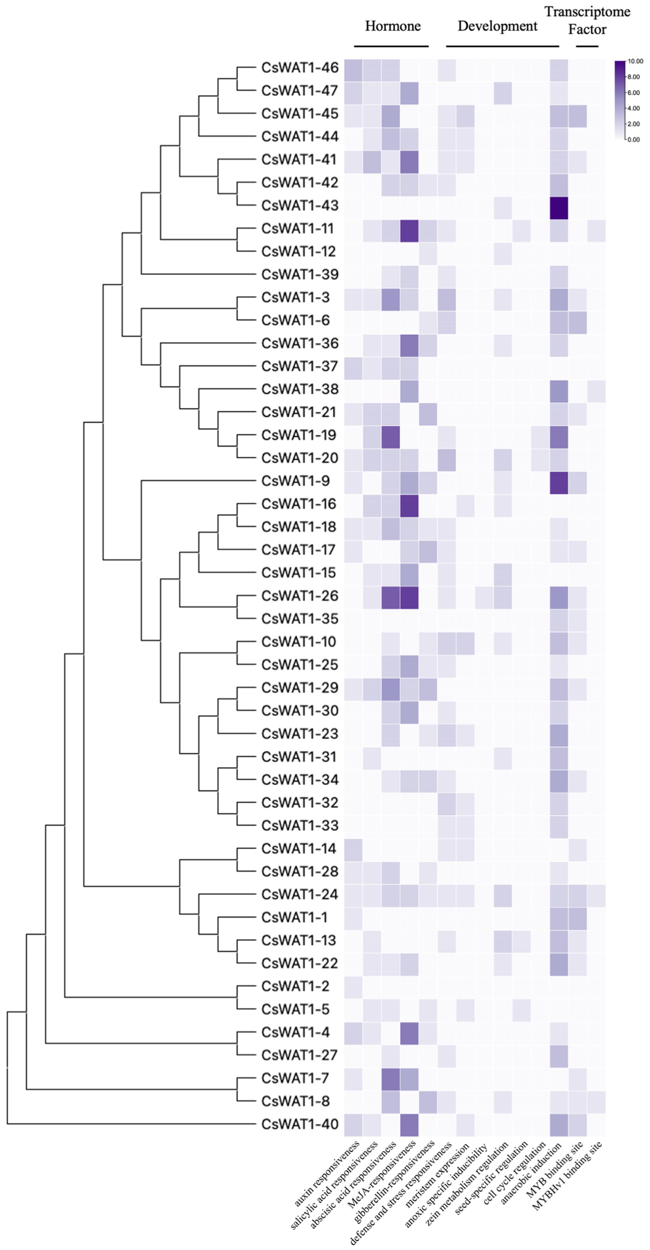
3.4. Cis-Acting Elements Analysis of CsWAT1s Promoters

Cis-acting elements in gene promoters play an essential role in regulating gene expression, thereby influencing plant growth and responses to various stresses [24]. To elucidate the potential biological functions and regulatory characteristics of the CsWAT1 genes, we analyzed the 2000 bp upstream promoter sequences of CsWAT1s using the PlantCARE database (Figure 4). Our analysis identified several key cis-acting elements associated with hormone action, stress response, growth and development regulation, and transcription factor binding. Specifically, the hormone-responsive elements included salicylic acid (SA)-responsive elements, methyl jasmonate (MeJA)-responsive elements, abscisic acid (ABA)-responsive elements, auxin (IAA)-responsive elements, and gibberellin (GA)-responsive elements. Stress-responsive elements included defense and stress-responsive elements, which are crucial for the plant’s ability to cope with biotic and abiotic stresses. Growth and development-responsive elements encompassed meristem expression-responsive elements, cell cycle regulation-responsive elements, seed-specific regulation-responsive elements, and anaerobic induction-responsive elements. Transcription factor binding elements included MYBHV1 binding elements and MYB binding elements. These findings suggest that the CsWAT1 genes are likely regulated by a complex network of hormonal, stress, and developmental signals, mediated through these cis-acting elements. This regulatory complexity may contribute to the diverse functions of CsWAT1 genes in cucumber.
Among the CsWAT1 genes, CsWAT1-2 contains the fewest number of cis-acting elements, with only one IAA-responsive element. In contrast, CsWAT1-26 has the highest number of cis-acting elements, including eight MeJA-responsive elements, seven ABA-responsive elements, five anaerobic induction elements, two zein metabolism regulation-responsive elements, one defense and stress-responsive element, one MYB binding site, one SA-responsive element, and one hypoxia-specific induction element. The most widely distributed element is the anaerobic induction element, present in 83% of the family members. The second most widespread is the ABA-responsive element, found in 68% of the family members. The defense and stress-responsive element is present in 57% of the family members, while the MeJA-responsive element is found in 55% of the family members. The SA-responsive element is present in 53% of the family members. Both the IAA-responsive element and MYB binding site are found in 43% of the family members. The GA-responsive element is present in 40% of the family members, and the zein metabolism regulation-responsive element is found in 34% of the family members. The meristem expression-responsive element is present in 26% of the family members. The hypoxia-specific induction element is the least widely distributed, being found only in CsWAT1-26. The cell cycle regulation element is the second least widespread, present in CsWAT1-19 and CsWAT1-24. The seed-specific regulation element is found in CsWAT1-11, CsWAT1-13, and CsWAT1-5. The MYBHv1 binding site is present in CsWAT1-11, CsWAT1-24, CsWAT1-8, and CsWAT1-38. Numerous studies have reported that SA-responsive elements and MeJA-responsive elements are tightly connected to responding to pathogen stress. When pathogens affect cells, the SA and MeJA signaling pathways are activated to mount a defense response. Therefore, we predict that the following nine CsWAT1 members, which contain both SA-responsive and MeJA-responsive elements, are strongly correlated with defense against pathogen stress: CsWAT1-3, CsWAT1-15, CsWAT1-18, CsWAT1-20, CsWAT1-24, CsWAT1-26, CsWAT1-29, CsWAT1-40, and CsWAT1-44.

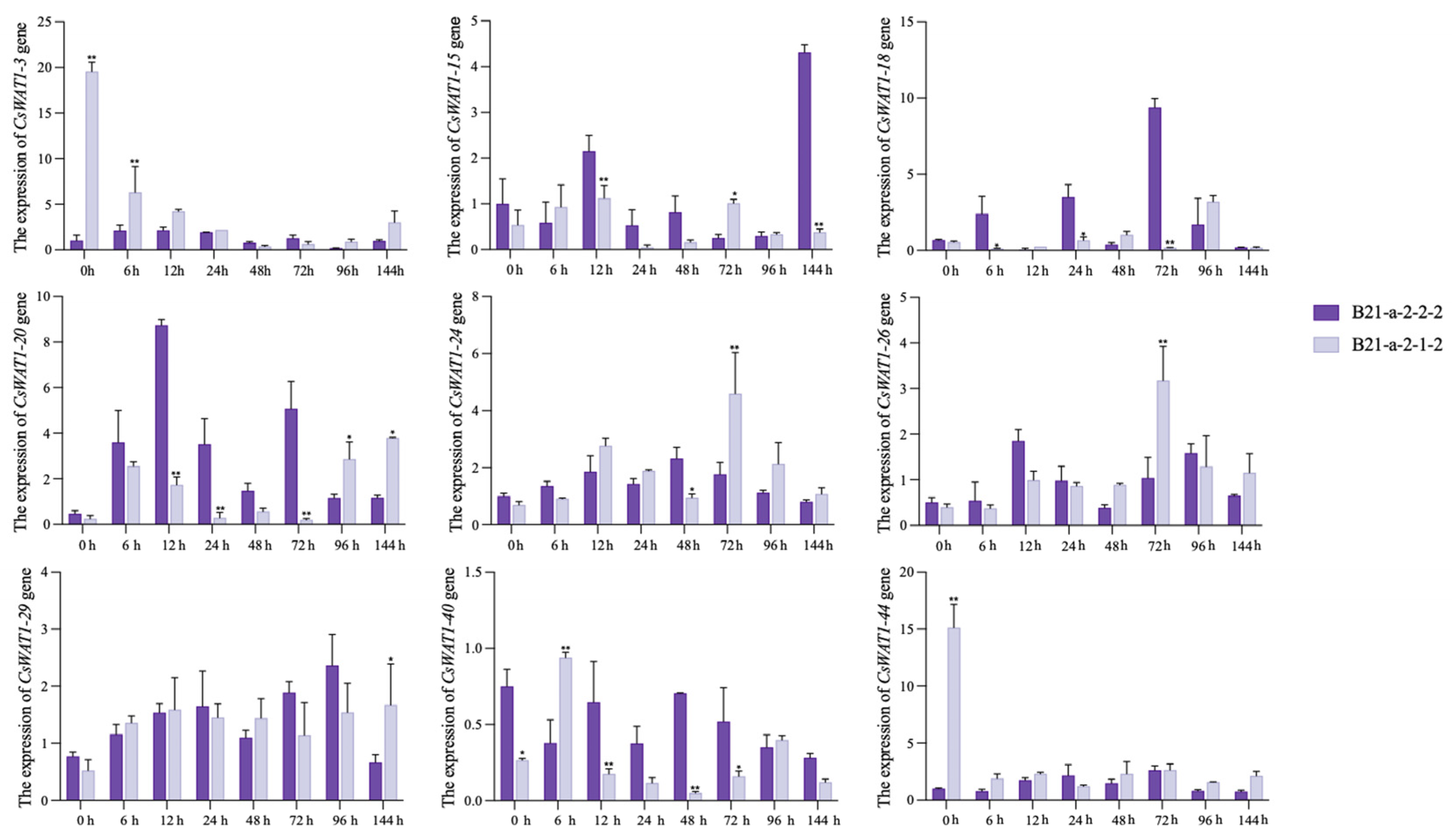
3.5. Expression Profiles of CsWAT1s in Response to P. xanthii Stress

We analyzed the cis-acting elements of the promoter regions of all members of this gene family. A large number of previous studies have shown that SA elements are closely related to plant resistance to P. xanthii stress; therefore, we initially selected nine members with SA elements in the promoter regions for the validation of CsWAT1s expression in different periods after P. xanthii infestation suggested the nine CsWAT1 genes that might be involved in cucumber’s response to P. xanthii stress. Differential expression patterns of genes can reflect their functions in stress response. To investigate whether and which CsWAT1 genes are involved in the defense response of cucumber against P. xanthii, we examined the expression patterns of these nine genes at 0, 6, 12, 24, 48, 72, 96, and 144 hpi in two cucumber lines: the highly PM-resistant line B21-a-2-1-2 and the highly PM-susceptible line B21-a-2-2-2 (Figure 5). Under P. xanthii attack: CsWAT1-3 and CsWAT1-44 showed significantly higher transcript levels in the resistant line B21-a-2-1-2 compared to the susceptible line B21-a-2-2-2; CsWAT1-15, CsWAT1-18, and CsWAT1-20 exhibited higher transcript levels in the susceptible line B21-a-2-2-2 than in the resistant line B21-a-2-1-2, indicating that these genes were downregulated following P. xanthii infection. Notably, starting from 72 hpi, CsWAT1-26 showed higher expression levels in the resistant line B21-a-2-1-2 compared to the susceptible line B21-a-2-2-2, suggesting that CsWAT1-26 was upregulated in response to P. xanthii infection. Based on these results, we speculate that the distinct expression patterns of CsWAT1 genes play opposing regulatory roles in cucumber’s response to PM stress. The expression patterns of CsWAT1-29, CsWAT1-40, and CsWAT1-44 revealed fluctuating transcript levels throughout the sampling period following P. xanthii infection. Additionally, we observed a notable contrast in the expression level of CsWAT1-20 at 96 hpi compared to pre-inoculation levels, suggesting that CsWAT1-20 may regulate plant defense against P. xanthii throughout the entire infection process.

3.6. Silencing of CsWAT1-20 in Cucumber Seedlings Contributes to P. xanthii Stress Tolerance

Through the method of RT-qPCR gene expression pattern analysis, we found that the expression of CsWAT1-20 changed at various periods after P. xanthii infestation; therefore, we selected CsWAT1-20 for detailed investigation. To elucidate the role of CsWAT1-20 in mediating cucumber tolerance to P. xanthii, we employed a tobacco rattle virus (TRV)-based virus-induced gene silencing (VIGS) approach to silence the expression of the CsWAT1-20 gene in cucumber. Subsequently, we examined the impact of CsWAT1-20 silencing on disease resistance in cucumber plants (Figure 6). In this experiment, a 268 bp fragment of CsWAT1-20 CDS region—which has high sequence specificity and is significantly different from other gene sequences in the plant genome, thereby avoiding cross-silencing effects on other non-target genes and allowing us to more precisely analyze the single function of the CsWAT1-20 gene without interference from other genes—was cloned into the pTRV2 vector, resulting in the recombinant vector pTRV2-CsWAT1-20. Quantitative analysis revealed that CsWAT1-20 expression was significantly downregulated in plants transformed with TRV::CsWAT1-20 compared to those transformed with the control TRV::00, indicating the successful generation of CsWAT1-20-silenced cucumber plants (Figure S2).
To assess the susceptibility of CsWAT1-20-silenced and control cucumber plants to PM disease, the leaves of both TRV::CsWAT1-20 and TRV::00 plants were inoculated with P. xanthii. PM symptoms were observed in both TRV::CsWAT1-20 and TRV::00 plants at 10 dpi. However, the symptoms were notably more severe in TRV::CsWAT1-20 plants compared to TRV::00 plants. Microscopic examination of P. xanthii hyphae stained with Coomassie Brilliant Blue (CBB) revealed a significantly higher hyphal density in TRV::CsWAT1-20 plants. These results suggest that the silencing of CsWAT1-20 enhances the growth and colonization of P. xanthii hyphae. Additionally, at least 48 TRV::CsWAT1-20 and TRV::00 plants infected with P. xanthii were used to calculate the disease index (DI). The DI of TRV::CsWAT1-20 plants was significantly higher than that of TRV::00 plants in response to P. xanthii infections. Collectively, these results demonstrate that the silencing of CsWAT1-20 in cucumber plants compromises their resistance to P. xanthii, highlighting the essential role of CsWAT1-20 in mediating defense against these pathogens.

4. Discussion

The WALLS ARE THIN1 (WAT1) family is plant-specific and primarily involved in secondary cell wall formation and plant hormone regulation. In A. thaliana, the AtWAT1 family comprises 46 members, with only AtWAT1 (At1g75500) being extensively studied. The wat1 mutant exhibits a dwarfed phenotype, reduced secondary cell wall thickness, and lower auxin content [16]. Notably, inactivation of WAT1 enhances plant defense against pathogens, such as R. solanacearum and V. dahliae [18]. Thus, WAT1 plays a crucial role in both plant growth and disease resistance. However, research on WAT1 family members in plants remains limited.
In this study, we conducted a comprehensive genome-wide analysis to identify CsWAT1 family members in cucumber. Based on the cucumber reference genome, we identified 47 CsWAT1 genes, which is more than the 46 members reported in A. thaliana [25]. This suggests interspecific divergence in the size of the WAT1 family. These CsWAT1 genes are unevenly distributed across six of the seven cucumber chromosomes. Bioinformatic analysis of CsWAT1 protein physicochemical properties revealed that most family members are alkaline and stable proteins, with all but CsWAT1-40 exhibiting hydrophobic characteristics. Notably, no signal peptides were detected in any CsWAT1 proteins. Gene structure analysis demonstrated variability in exon-intron organization among CsWAT1 members, coupled with divergent distribution patterns of conserved motifs. Intriguingly, motif 5 was present in all proteins except CsWAT1-40. This finding contrasts with conventional understanding of this gene family and highlights the structural diversity and functional complexity within the CsWAT1 family. These results suggest that while WAT1 genes exhibit high homology and evolutionary conservation within cucumber, lineage-specific expansion and structural diversification may contribute to functional specialization in plant growth and stress responses.
Analysis of cis-acting elements within the promoter region of genes is crucial for understanding plant stress responses [26]. Our bioinformatics investigation identified three major regulatory element categories in cucumber WAT1 family members: hormone-responsive elements, growth-associated elements, and transcription factor binding sites. These elements suggest potential involvement in developmental regulation and stress adaptation. Previous studies have highlighted the essential roles of SA-responsiveness elements, MeJA-responsiveness elements, and defense-/stress-related elements in plant defense against pathogen infection. These cis-elements mediate pathogen resistance through transcriptional regulation. Specifically, SA signaling activation following pathogen challenge induces pathogenesis-related (PR) gene expression and enhances antioxidant enzyme activities. MeJA-responsive elements coordinate defense responses through interactions with bZIP and MYB transcription factors. Within the CsWAT1s, we identified nine notable members (CsWAT1-3, CsWAT1-15, CsWAT1-18, CsWAT1-20, CsWAT1-24, CsWAT1-26, CsWAT1-29, CsWAT1-40, CsWAT1-44) that possess one or two SA-responsiveness elements, two to eight MeJA-responsiveness elements, and zero to three defense and stress-responsiveness elements. Based on these characteristics, we hypothesize that these nine genes may play crucial roles in plant resistance to stress.
To preliminarily determine whether the nine specifically noted members of the CsWAT1 gene family are involved in the biological processes underlying cucumber resistance to P. xanthii stress, we examined the expression levels of these nine genes in cucumber. At 0-72 hpi, the expression level of CsWAT1-20 in B21-a-2-1-2 was significantly lower than that in B21-a-2-2-2. However, from 96 phi, the expression level in B21-a-2-1-2 was significantly higher than that in B21-a-2-2-2. This indicates that infection with P. xanthii for a certain period induces the expression of CsWAT1-20, which in turn enhances the resistance of plants to P. xanthii. Further silencing of CsWAT1-20 expression in cucumber indicated that CsWAT1-20 positively regulates cucumber defense against P. xanthii. However, our results are not consistent with the function of WAT1s in disease resistance in other plants. There are two reasons that explain this result. Firstly, WAT1s have been shown to be negative regulators of plant disease resistance, mostly against vascular pathogens. However, our study was conducted on P. xanthii, which is a viable nutrient fungus, suggesting that it is different from vascular pathogens. Therefore, even the same gene may initiate different mechanisms to protect against different types of pathogen stresses. Secondly, the number of WAT1 family members is large, indicating that they may function similarly or completely differently. The current reports on WAT1s members against pathogen stress are insufficient to suggest that all WAT1s negatively regulate disease resistance.
WAT1s are crucial regulators of secondary wall synthesis. Numerous evidence uncovered that plant cell wall serves as a key responder to pathogen stress through two distinct mechanisms. First, as a physical barrier, increased cell wall thickness enhances disease resistance in plants. For instance, the OsWAK gene enhances rice resistance to the blast fungus Magnaporthe oryzae by modulating cell wall integrity, which in turn activates the expression of downstream defense-related genes and strengthens the overall immune response of the plant [27]. Similarly, in A. thaliana, overexpression of AtWAK1 enhances resistance to pathogens. AtWAK1 functions by sensing changes in cell wall integrity, which subsequently activates downstream pattern recognition receptors (PRRs) and triggers pattern-triggered immunity (PTI) responses [28]. Second, the cell wall can act as a signaling component. Downregulation of cell wall-associated genes negatively regulates disease resistance. The OsPMEs family in rice plays crucial roles in maintaining cell wall integrity. Downregulation of these genes compromises the cell wall’s physical barrier function against pathogen invasion, thereby reducing rice resistance to the blast fungus M. oryzae [29]. Similarly, downregulation of the AtPMEI17 gene leads to a loosening of the cell wall structure, which is detrimental to the plant’s defense response and consequently reduces resistance to Botrytis cinerea [30]. The relationship between cell wall and plant disease resistance depends on which mechanism is activated. We found that CsWAT1-20 positively regulates disease resistance. However, in Arabidopsis and cotton, the WAT1 gene negatively regulates plant disease resistance through the SA signaling pathway. These findings suggest that different WAT1 genes in various species can modulate plant immune responses by activating different resistance mechanisms. Therefore, studies on the specific mechanisms by which CsWAT1-20 regulate P. xanthii resistance in cucumber are necessary.

5. Conclusions

In conclusion, we have conducted a comprehensive bioinformatics analysis to elucidate the structural features and potential evolutionary relationships of the CsWAT1 gene family members. Additionally, we analyzed the expression patterns to infer the potential functions of these genes and employed virus-induced gene silencing (VIGS) to demonstrate that CsWAT1-20 positively regulates cucumber defense against P. xanthii stress. Our results not only deepen the core comprehension of how to develop new disease-resistant cucumber varieties but also offer important insights for future research on WAT1-mediated immunity in other plants.

Supplementary Materials

The following supporting information can be downloaded at: https://www.mdpi.com/article/10.3390/horticulturae11060620/s1, Figure S1: Structure of CsWAT1 gene family. The green box represents untranslated regions (UTRs), and the yellow box represents coding sequences (CDSs); Table S1: List of primers used in this study title; Figure S2: Silencing of CsWAT1-20 attenuated the resistance of cucumber to P. xanthii. The expression level of CsWAT1-20 was determined in pTRV::00 and pTRV::CsWAT1-20 cucumbers by RT-qPCR. Data are means ± SD of three biological replicates per treatment. Significant differences were detected by LSD multiple comparison tests (* p ≤ 0.05 and ** p ≤ 0.01).

Author Contributions

X.M. and H.F. conceived and designed the manuscript. J.H. and H.Z. conducted experiments. Y.Y. (Youmei Yuan), Y.Y. (Yang Yu), J.W., N.C. and H.F. revised the manuscript. J.H. and X.M. wrote the manuscript. All authors have read and agreed to the published version of the manuscript.

Funding

This study was funded by the Natural Science Foundation of Liaoning Province of China (2024-MSLH-418).

Data Availability Statement

The datasets generated during and/or analyzed during the current study are available from the corresponding author on reasonable request.

Conflicts of Interest

The authors have no conflicts of interest to declare that are relevant to the content of this article.

References

  1. Zhang, P.; Zhu, Y.; Zhou, S. Comparative analysis of powdery mildew resistant and susceptible. cultivated cucumber (Cucumis sativus L.) varieties to reveal the metabolic responses to Sphaerotheca fuliginea infection. BMC Plant Biol. 2021, 21, 24. [Google Scholar]
  2. Chikh-Rouhou, H.; Garcés-Claver, A.; Kienbaum, L.; Ben Belgacem, A.; Gómez-Guillamón, M.L. Resistance of Tunisian Melon Landraces to Podosphaera xanthii. Horticulturae 2022, 8, 1172. [Google Scholar] [CrossRef]
  3. Meng, X.; Yu, Y.; Song, T.; Yu, Y.; Cui, N.; Ma, Z.; Chen, L.; Fan, H. Transcriptome Sequence Analysis of the Defense Responses of Resistant and Susceptible Cucumber Strains to Podosphaera xanthii. Front. Plant Sci. 2022, 13, 872218. [Google Scholar] [CrossRef]
  4. Li, X.; Liu, Y.; He, Q.; Li, S.; Liu, W.; Lin, C.; Miao, W. A Candidate Secreted Effector Protein of Rubber Tree Powdery Mildew Fungus Contributes to Infection by Regulating Plant ABA Biosynthesis. Front. Microbiol. 2020, 11, 591387. [Google Scholar] [CrossRef]
  5. Zhang, D.; Wu, S.; Li, N.; Gao, J.; Liu, S.; Zhu, S.; Li, Z.; Ren, G.; Kuai, B. Chemical induction of leaf senescence and powdery mildew resistance involves ethylene-mediated chlorophyll degradation and ROS metabolism in cucumber. Hortic. Res. 2022, 9, uhac101. [Google Scholar] [CrossRef]
  6. Li, J.; Gu, C.; Yuan, Y.; Gao, Z.; Qin, Z.; Xin, M. Comparative transcriptome analysis revealed that auxin and cell wall biosynthesis play important roles in the formation of hollow hearts in cucumber. BMC Genom. 2024, 25, 36. [Google Scholar] [CrossRef]
  7. Dora, S.; Terrett, O.M.; Clara, S.R. Plant-microbe interactions in the apoplast: Communication at the plant cell wall. Plant Cell 2022, 34, 1532–1550. [Google Scholar] [CrossRef]
  8. Yu, Y.; Yu, Y.; Cui, N.; Ma, L.; Tao, R.; Ma, Z.; Meng, X.; Fan, H. Lignin biosynthesis regulated by CsCSE1 is required for Cucumis sativus defence to Podosphaera xanthii. Plant Physiol. Biochem. 2022, 186, 88–98. [Google Scholar] [CrossRef]
  9. Miedes, E.; Zarra, I.; Hoson, T.; Herbers, K.; Sonnewald, U.; Lorences, E.P. Xyloglucan endotransglucosylase and cell wall extensibility. J. Plant Physiol. 2011, 168, 196–203. [Google Scholar] [CrossRef]
  10. Fu, Y.; Win, P.; Zhang, H.; Li, C.; Shen, Y.; He, F.; Luo, K. PtrARF2.1 Is Involved in Regulation of Leaf Development and Lignin Biosynthesis in Poplar Trees. Int. J. Mol. Sci. 2019, 20, 4141. [Google Scholar] [CrossRef]
  11. Jiang, S.; Tian, X.; Huang, X.; Xin, J.; Yan, H. Physcomitrium patens CAD1 has distinct roles in growth and resistance to biotic stress. BMC Plant Biol. 2022, 22, 518. [Google Scholar] [CrossRef] [PubMed]
  12. Awwad, F.; Bertrand, G.; Grandbois, M.; Beaudoin, N. Auxin protects Arabidopsis thaliana cell suspension cultures from programmed cell death induced by the cellulose biosynthesis inhibitors thaxtomin A and isoxaben. BMC Plant Biol. 2019, 19, 512. [Google Scholar] [CrossRef] [PubMed]
  13. Lehman, T.A.; Sanguinet, K.A. Auxin and Cell Wall Crosstalk as Revealed by the Arabidopsis thaliana Cellulose Synthase Mutant Radially Swollen 1. Plant Cell Physiol. 2019, 60, 1487–1503. [Google Scholar] [CrossRef]
  14. Ranocha, P.; Dima, O.; Felten, J.; Freydier, A.; Hoffmann, L.; Ljung, K.; Lacombe, B.; Corratgé, C.; Thibaud, J.B.; Sundberg, B.; et al. WAT1 (WALLS ARE THIN1) defines a novel auxin transporter in plants and integrates auxin signaling in secondary wall formation in Arabidopsis fibers. BMC Proc. 2011, 5 (Suppl. S7), O24. [Google Scholar] [CrossRef]
  15. Gamas, P.; Niebel, F.d.C.; Lescure, N.; Cullimore, J. Use of a subtractive hybridization approach to identify new Medicago truncatula genes induced during root nodule development. Mol. Plant Microbe Interact. 1996, 9, 233–242. [Google Scholar] [CrossRef]
  16. Ranocha, P.; Denancé, N.; Vanholme, R.; Freydier, A.; Martinez, Y.; Hoffmann, L.; Köhler, L.; Pouzet, C.; Renou, J.P.; Sundberg, B.; et al. Walls are thin1 (WAT1), an Arabidopsis homolog of Medicago truncatula NODULIN21, is a tonoplast-localized protein required for secondary wall formation in fibers. Plant J. 2010, 63, 469–483. [Google Scholar] [CrossRef]
  17. Ranocha, P.; Dima, O.; Nagy, R.; Felten, J.; Corratgé-Faillie, C.; Novák, O.; Morreel, K.; Lacombe, B.; Martinez, Y.; Pfrunder, S.; et al. Arabidopsis WAT1 is a vacuolar auxin transport facilitator required for auxin homoeostasis. Nat. Commun. 2013, 4, 2625. [Google Scholar] [CrossRef]
  18. Denancé, N.; Sánchez-Vallet, A.; Goffner, D.; Molina, A. Disease resistance or growth: The role of plant hormones in balancing immune responses and fitness costs. Front. Plant Sci. 2013, 4, 155. [Google Scholar] [CrossRef]
  19. Tang, Y.; Zhang, Z.; Lei, Y.; Hu, G.; Liu, J.; Hao, M.; Chen, A.; Peng, Q.; Wu, J. Cotton WATs modulate SA biosynthesis and local lignin deposition participating in plant resistance against Verticillium dahlia. Front. Plant Sci. 2019, 10, 526. [Google Scholar] [CrossRef]
  20. Hanika, K.; Schipper, D.; Chinnappa, S.; Oortwijn, M.; Schouten, H.J.; Thomma, B.P.H.J.; Bai, Y. Impairment of Tomato WAT1 Enhances Resistance to Vascular Wilt Fungi Despite Severe Growth Defects. Front. Plant Sci. 2021, 12, 721674. [Google Scholar] [CrossRef]
  21. Koseoglou, E.; Hanika, K.; MohdNadzir, M.M.; Kohlen, W.; vanderWolf, J.M.; Visser, R.G.F.; Bai, Y. Inactivation of tomato WAT1 leads to reduced susceptibility to Clavibacter michiganensis through downregulation of bacterial virulence factors. Front. Plant Sci. 2023, 14, 1082094. [Google Scholar] [CrossRef] [PubMed]
  22. Liu, L.; Ma, L.; Yu, Y.; Ma, Z.; Yin, Y.; Zhou, S.; Yu, Y.; Cui, N.; Meng, X.; Fan, H. Cucumis sativus CsbZIP90 suppresses Podosphaera xanthii resistance by modulating reactive oxygen species. Plant Sci. 2024, 339, 111945. [Google Scholar] [CrossRef] [PubMed]
  23. Ao, B.; Han, Y.; Wang, S.; Wu, F.; Zhang, J. Genome-Wide Analysis and Profile of UDP-Glycosyltransferases Family in Alfalfa (Medicago sativa L.) under Drought Stress. Int. J. Mol. Sci. 2022, 23, 7243. [Google Scholar] [CrossRef]
  24. Liao, G.; Li, Y.; Wang, H.; Liu, Q.; Zhong, M.; Jia, D.; Huang, C.; Xu, X. Genome-wide identification and expression profiling analysis of sucrose synthase (SUS) and sucrose phosphate synthase (SPS) genes family in Actinidia chinensis and A. eriantha. BMC Plant Biol. 2022, 22, 215. [Google Scholar] [CrossRef]
  25. Swarbreck, D.; Wilks, C.; Lamesch, P.; Berardini, T.Z.; Garcia-Hernandez, M.; Foerster, H.; Li, D.; Meyer, T.; Muller, R.; Ploetz, L.; et al. The Arabidopsis Information Resource (TAIR): Gene structure and function annotation. Nucleic Acids Res. 2008, 36, D1009–D1014. [Google Scholar] [CrossRef]
  26. Liu, Z.; Zhang, Y.; Altaf, M.A.; Hao, Y.; Zhou, G.; Li, X.; Zhu, J.; Ma, W.; Wang, Z.; Bao, W. Genome-wide identification of myeloblastosis gene family and its response to cadmium stress in Ipomoea aquatica. Front. Plant Sci. 2022, 13, 979988. [Google Scholar] [CrossRef]
  27. Baez, L.A.; Tichá, T.; Hamann, T. Cell wall integrity regulation across plant species. Plant Mol. Biol. 2022, 109, 483–504. [Google Scholar] [CrossRef]
  28. Bacete, L.; Mélida, H.; Miedes, E.; Molina, A. Plant cell wall-mediated immunity: Cell wall changes trigger disease resistance responses. Plant J. 2018, 93, 614–636. [Google Scholar] [CrossRef]
  29. Molina, A.; Jordá, L.; Torres, M.Á.; Martín-Dacal, M.; Berlanga, D.J.; Fernández-Calvo, P.; Gómez-Rubio, E.; Martín-Santamaría, S. Plant cell wall-mediated disease resistance: Current understanding and future perspectives. Mol. Plant 2024, 17, 699–724. [Google Scholar] [CrossRef]
  30. Del Corpo, D.; Fullone, M.R.; Miele, R.; Lafond, M.; Pontiggia, D.; Grisel, S.; Kieffer-Jaquinod, S.; Giardina, T.; Bellincampi, D.; Lionetti, V. AtPME17 is a functional Arabidopsis thaliana pectin methylesterase regulated by its PRO region that triggers PME activity in the resistance to Botrytis cinerea. Mol. Plant Pathol. 2020, 21, 1620–1633. [Google Scholar] [CrossRef]
Figure 1. Phylogenetic relationships of WAT1 proteins among C. sativus and A. thaliana. The unrooted phylogenetic tree was constructed by MEGA11 by neighbor-joining method with 1000 bootstrap replicates. The WAT1s are divided into five major subfamilies. A different subfamily represents A to E. The different colors of circles represent different species. The purple circles represent cucumber and yellow circles represent Arabidopsis.
Horticulturae 11 00620 g001
Figure 2. Chromosome localization and collinearity analysis of CsWAT1s. (a) The distribution of the CsWAT1 genes located on the chromosomes in cucumber plants. The genetic distance of seven chromosomes were represented by the scale in megabases (Mb) on the left. The CsWAT1 genes are displayed using nomenclature for genome version 3 of Chinese Long Cucumber. Purple lines represent the location of the gene on each chromosome. (b) Collinearity analysis of the WAT1 genes among cucumber and other species. Gray lines in the background represent collinear blocks in cucumber plants and other genomes. The collinear gene pairs with WAT1 genes between different species were highlighted by the red lines.
Horticulturae 11 00620 g002
Figure 3. Motif distribution of CsWAT1s. Protein conserved motifs were tested by the MEME online tool, numbers 1 to 10 represent different motifs, each displayed in different color boxes.
Horticulturae 11 00620 g003
Figure 4. Cis-acting elements of CsWAT1s promoters. Cis-acting elements can be divided into hormone group, development group and transcriptome factor group. The number of cis-acting elements of CsWAT1s promoters were shown on a heatmap using a number value, and 0 to 10 was artificially set with the color scale limits according to the normalized value. The color scale showed increasing expression levels from white to purple.
Horticulturae 11 00620 g004
Figure 5. Expression patterns of CsWAT1 genes in cucumber varieties B21-a-2-2-2 (susceptible) and B21-a-2-1-2 (resistant) against P. xanthii. The leaves were measured at 0, 6, 12, 24, 48, 72, 96 and 144 hpi. Data are means ± SD of three biological replicates per treatment. Significant differences were detected by LSD multiple comparison tests (* p ≤ 0.05 and ** p ≤ 0.01).
Horticulturae 11 00620 g005
Figure 6. Silencing of CsWAT1-20 attenuated the resistance of cucumber to P. xanthii. Disease resistance of CsWAT1-20-silenced cucumber leaves at 10 days post inoculation (dpi) with P. xanthii was assessed based on phenotypic observations (a), Coomassie brilliant blue (CBB) staining (b) and disease index (DI) (c).
Horticulturae 11 00620 g006
Table 1. The physicochemical properties of CsWAT1 family member proteins.
Gene ID Rename Number of Amino Acids Molecular Weight (kDa)Theoretical
pI
GravyInstability IndexSingle Peptide
CsaV3_1G004090CsWAT1-136240,240.517.580.55936.93No
CsaV3_1G015750CsWAT1-236740,023.029.340.40233.84No
CsaV3_1G029400CsWAT1-336940,737.248.860.61542.95No
CsaV3_1G037450CsWAT1-435439,043.529.320.67032.07No
CsaV3_2G007260CsWAT1-558465,291.848.840.45131.17No
CsaV3_2G007860CsWAT1-636940,123.758.210.79142.45No
CsaV3_2G011920CsWAT1-739443,412.449.200.56142.31No
CsaV3_2G011970CsWAT1-838643,122.369.400.31433.06No
CsaV3_2G012900CsWAT1-936040,729.159.160.54439.5No
CsaV3_2G013970CsWAT1-1038642,020.279.580.74233.34No
CsaV3_2G018030CsWAT1-1137441,491.288.900.36733.12No
CsaV3_2G018040CsWAT1-1231434,780.279.070.59636.94No
CsaV3_2G029170CsWAT1-1339642,845.518.950.59135.73No
CsaV3_3G002650CsWAT1-1434237,246.739.030.84239.66No
CsaV3_3G039950CsWAT1-1539743,687.789.130.53429.42No
CsaV3_3G039960CsWAT1-1630533,692.496.930.37232.50No
CsaV3_3G039970CsWAT1-1738742,729.219.370.66936.12No
CsaV3_3G039980CsWAT1-1838142,023.389.360.72833.98No
CsaV3_3G042150CsWAT1-1934937,839.848.890.72431.54No
CsaV3_3G042160CsWAT1-2035638,697.678.350.65033.29No
CsaV3_3G042170CsWAT1-2136139,111.18.340.67237.53No
CsaV3_3G045560CsWAT1-2239442,528.029.230.64033.71No
CsaV3_4G024710CsWAT1-2337541,352.429.230.54732.18No
CsaV3_4G027990CsWAT1-2437440,087.278.500.69930.19No
CsaV3_4G028340CsWAT1-2537440,756.599.300.67027.13No
CsaV3_4G029290CsWAT1-2644149,378.128.940.35035.64No
CsaV3_5G001460CsWAT1-2736239,127.528.770.70230.82No
CsaV3_5G004250CsWAT1-2836038,969.859.020.85137.27No
CsaV3_5G006960CsWAT1-2930834,086.169.660.30834.56No
CsaV3_5G007970CsWAT1-3037741,435.338.990.62736.52No
CsaV3_5G026330CsWAT1-3137640,929.429.380.52226.25No
CsaV3_5G026340CsWAT1-3235539,086.419.430.62224.44No
CsaV3_5G026350CsWAT1-3335739,279.639.510.6530.04No
CsaV3_5G026360CsWAT1-3437440,691.159.560.47130.09No
CsaV3_5G029570CsWAT1-3540645,391.569.350.36532.63No
CsaV3_5G031760CsWAT1-3637441,277.688.620.60134.41No
CsaV3_5G037340CsWAT1-3734838,209.479.160.77833.58No
CsaV3_5G037350CsWAT1-3837741,006.998.250.49240.00No
CsaV3_6G005240CsWAT1-3936541,024.89.570.50225.22No
CsaV3_6G013640CsWAT1-4033237,085.68.98-0.71456.11No
CsaV3_6G041910CsWAT1-4141746,071.849.480.46331.34No
CsaV3_6G041920CsWAT1-4227731,060.889.750.44242.34No
CsaV3_6G041930CsWAT1-4328831,413.179.460.63930.95No
CsaV3_6G041950CsWAT1-4436039,766.279.450.60631.37No
CsaV3_6G041960CsWAT1-4542547,743.049.590.50530.60No
CsaV3_6G041970CsWAT1-4627330,516.629.260.60630.32No
CsaV3_6G041980CsWAT1-4736239,794.298.960.58131.84No
Disclaimer/Publisher’s Note: The statements, opinions and data contained in all publications are solely those of the individual author(s) and contributor(s) and not of MDPI and/or the editor(s). MDPI and/or the editor(s) disclaim responsibility for any injury to people or property resulting from any ideas, methods, instructions or products referred to in the content.

