Article

Identification and Expression Profiles Reveal Key Myelocytomatosis (MYC) Involved in Drought, Chilling, and Salt Tolerance in Solanum lycopersicum

1
College of Bioscience and Biotechnology, Shenyang Agricultural University, Shenyang 110866, China
2
Key Laboratory of Protected Horticulture of Ministry of Education, Shenyang Agricultural University, Shenyang 110866, China
3
Institute of Animal Science, Chinese Academy of Agriculture Sciences, Beijing 100193, China
4
Vegetable Research Institute, Liaoning Academy of Agricultural Sciences, Shenyang 110866, China
*
Authors to whom correspondence should be addressed.
These authors contribute equally to this work.
Horticulturae 2025, 11(6), 693; https://doi.org/10.3390/horticulturae11060693
Submission received: 28 April 2025 / Revised: 5 June 2025 / Accepted: 12 June 2025 / Published: 16 June 2025
(This article belongs to the Special Issue New Insights into Protected Horticulture Stress)

Abstract

:
Tomato (Solanum lycopersicum) is a vital crop in China, yet its growth and quality are compromised by environmental stresses. This study investigated the role of myelocytomatosis (MYC) transcription factors (SlMYCs) in tomato stress tolerance. We identified 23 potential SlMYC genes and analyzed their physicochemical properties, evolutionary relationships, gene structures, conserved domains, expression profiles, interaction networks, promoter sequences, and 3D models using bioinformatics. Phylogenetic analysis classified the SlMYCs into three groups with similar structural characteristics. Protein interaction networks revealed significant connections between SlMYCs and proteins involved in drought, chilling, and salt tolerance, particularly emphasizing the jasmonic acid signaling pathway. Experimental treatments with methyl jasmonate (MeJA) and simulated stress conditions showed that several SlMYC genes were responsive to these stimuli, with SlMYC1 and SlMYC2 demonstrating consistent expression patterns across various tissues. Further network analyses and molecular docking studies indicated potential binding interactions for these two genes. The findings confirmed that SlMYC1 and SlMYC2 contributed to tomato’s abiotic stress tolerance, highlighting their potential for breeding programs aimed at improving stress resilience in tomato varieties. This research laid the groundwork for enhancing tomato varieties under environmental stressors.

1. Introduction

Tomato (Solanum lycopersicum) is a widely grown vegetable crop worldwide. It is sensitive to cold temperatures and thrives in sunlight [1,2], but faces challenges such as diseases, salt, drought, and heavy metal stress [3,4,5], affecting its yield and quality. Understanding stress tolerance genes and regulatory mechanisms in tomato is crucial for overcoming environmental challenges. It also paves the way for developing high-quality, high-yield tomato varieties through selective breeding efforts. Tomato’s resilience to abiotic stress is influenced by various hormones, with jasmonic acid (JA) being a significant endogenous plant growth regulator. The application of methyl jasmonate (MeJA) externally can affect the growth and development processes of tomato [6], such as inducing stomatal opening, inhibiting seed germination, hypocotyl elongation, root growth, stamen development, flowering, and promoting leaf senescence [7,8,9,10,11].
JA serves as a central regulator crucial for plant responses to biotic and abiotic stresses [12,13,14,15]. Its activation is essential for plant defense against biological stresses like herbivorous insects [16] and microbial pathogens [17,18,19], as well as abiotic stresses including salt stress [20], low and high temperatures [21,22], and drought [23]. JA is recognized as a vital stress-resistant hormone in higher plants [24,25], acting not only as a signaling molecule but also inducing the expressions of specific genes to help plants cope with environmental stress [18,26]. It functions within a complex signaling network involving other plant hormones, with MYC transcription factors playing a pivotal role in JA signaling [27,28]. While SlMYC1 and SlMYC2 in the MYC IIIe subfamily have been extensively studied in tomato regarding their involvement in JA signaling, the precise mechanisms of their actions are still unclear. Limited research has been conducted on other genes within the SlMYC family, highlighting the need for further exploration in this area [18].
Plant MYC transcription factors are characterized by conserved bHLH domains and are part of the bHLH family of transcription factors [29]. The bHLH domain includes a basic domain responsible for DNA binding and an HLH domain that facilitates the dimerization of bHLH proteins to form homologous or heterodimeric complexes [30]. The MYC family of transcription factors plays a pivotal role in plant growth and development, secondary metabolite synthesis, stress responses, plant hormone signaling, and crosstalk between different hormone pathways [31,32,33,34,35]. Among the MYC family members, MYC2 is a prominent regulator in the JA signaling pathway [36,37,38]. Numerous studies have highlighted MYC2 involvement in regulating plant growth and development [33,34,39], including seedling growth and hypocotyl elongation in Arabidopsis thaliana [40,41,42,43], JA-mediated anthocyanin biosynthesis [44,45], and leaf senescence induction [46,47]. OsMYC2 regulates disease defense and senescence in rice [48,49]. In tobacco, NtMYC2 is involved in nicotine biosynthesis regulation [50,51], while MdMYC2 in apple controls ethylene biosynthesis and aluminum stress tolerance during fruit ripening by binding to specific promoters [52,53]. In Taxus chinensis, TcJAMYC1/2/4 was found to negatively regulate the expression of taxol biosynthesis genes [54]. Additionally, SlMYC2 activates the JA signaling pathway to combat and manage gray mold disease efficiently in tomato [55]. In Ginkgo biloba, the transcription factor MYC2 positively regulates the biosynthesis of trilactone terpenes by activating the expression of GbGGPPS [56].
The MYC transcription factors play crucial regulatory roles in response to various abiotic and biological stresses in plants. In A. thaliana, MYC and MYB function together as regulatory sites to bind to ABA-mediated genes, reducing plant damage caused by water scarcity [57]. And MYC2 and MYB43 transcription factors synergistically inhibit the expression of HMA2 and HMA4 and alter cadmium tolerance in Arabidopsis [58]. In japonica rice (Zoysia japonica), MYC acts as a cis-acting element and binds to stress-responsive ZjICE1 to enhance tolerance to low-temperature stress [59]. In tomato, the non-structural protein NSs of the tomato provirus binds to MYC2/3/4, disrupting MYC functions and weakening host defenses against spotted blight [60]. This highlights the collaborative nature of MYCs with other proteins in responding to stress.
The comprehensive analysis of the SlMYC gene family in this study included investigations into physical and chemical properties, phylogenetic relationships, gene structures, conserved domains, expression patterns, interaction networks, promoter regions, 3D homology modeling, molecular docking predictions, and expression levels analyzed through qRT-PCR. Understanding the identification and functions of the tomato MYC family is essential for unraveling JA signal transduction mechanisms, enhancing crop stress tolerance, and laying a foundation for genomic organization and evolutionary dynamics analysis of the tomato MYC family. This research highlighted the potential roles of SlMYCs in enhancing tomato tolerance, particularly emphasizing their involvement in key signaling pathways such as JA-mediated responses, with a focus on the functions of SlMYC1 and SlMYC2. The findings offer valuable knowledge for breeding high-quality, high-yielding, and resilient tomato varieties, ultimately improving tomato cultivation standards.

2. Materials and Methods

2.1. Genome-Wide Identification of MYC Genes in Tomato

The Ensembl Plants website (https://plants.ensembl.org/index.html, accessed on 3 January 2023) was utilized to download the genome and genome annotation files for tomato and Arabidopsis thaliana. The sequence information for the AtMYC gene family was obtained based on existing reports and the TAIR database (https://www.arabidopsis.org, accessed on 3 January 2023). The tomato genes and sequences were extracted and screened using TBtools-II v2.142 from the tomato genome database to identify 23 genes within the SlMYC family.

2.2. Prediction and Analysis of Physical and Chemical Properties

We utilized ProtParam from ExPASy (https://web.expasy.org/protparam/, accessed on 4 January 2023) to verify the sequence analysis of the SlMYC protein. Subsequently, we obtained gene length, instability index (II), half-life, isoelectric point (pI), grand average of hydropathicity (GRAVY), protein size, and molecular weight (Mw). Further, the protein sequences were submitted to an online website (http://cello.life.nctu.edu.tw/, accessed on 4 January 2023) for subcellular localization prediction [61,62,63]. In addition, we employed an online tool (http://mg2c.iask.in/mg2c_v2.0/, accessed on 4 January 2023) to process the obtained tomato SlMYC genome data, formatting it into a list that illustrated the physical location of genes on each tomato chromosome. The length of the tomato chromosomes was obtained from the Genome Browser available (https://www.genome.ucsc.edu/, accessed on 4 January 2023).

2.3. Phylogenetic Relationship, Gene Structure, and Conserved Motif Analysis

The evolutionary relationship of the tomato SlMYC genes was determined through the retrieval of the tomato SlMYC protein using a multiple sequence comparison (NCBI BLAST) (https://blast.ncbi.nlm.nih.gov/Blast.cgi, accessed on 3 January 2023). Subsequently, the MYC evolutionary tree of tomato was constructed in comparison with Arabidopsis using the MEGA7.0 software Neighbor-Joining (NJ) method [63]. The exon–intron structure was visualized using the Gene Structure Display Server (GSDS2.0) online tool (http://gsds.cbi.pku.edu.cn/, accessed on 3 January 2023) [64]. To analyze the structural domains of the SlMYC genes, the MEME software was utilized (https://meme-suite.org/meme/tools/meme, accessed on 3 January 2023) to investigate the sequence location and configuration [65]. Subsequently, the evolutionary tree, domain, and conserved motif analysis results were integrated and visualized using TBtools for a comprehensive analysis presentation.

2.4. Promoter Analysis

The 2000 base pair promoter sequences were extracted from 23 SlMYC genes identified using TBtools. Subsequently, the promoters were analyzed using Plant CARE (http://bioinformatics.psb.ugent.be/webtools/plantcare/html/, accessed on 5 January 2023) to identify important cis-acting elements. The results were visualized and exported using TBtools for further examination.

2.5. Spatiotemporal Differential Expression, Interaction Network Analysis, and Signal Pathway Enrichment Analysis

The expression levels of SlMYCs in five distinct tissues (root, leaf, bud, flower, and fruit) were obtained from the BAR database (http://bar.utoronto.ca/, accessed on 6 January 2023) as documented [66]. The data were utilized to unveil the spatiotemporal expression patterns of SlMYCs. Heat maps illustrating these patterns were generated using Sangerbox 3.0 (http://vip.sangerbox.com/home.html, accessed on 6 January 2023), completing the analysis of the expression levels of SlMYCs in different tissues. Furthermore, interaction data among these SlMYC proteins were retrieved from the STRING database to create an interaction network visualized through Cytoscape 3.10.3 software. By importing the protein data into the KOBAS online tool (http://bioinfo.org/kobas, accessed on 6 January 2023), an analysis of signaling pathways enriched with SlMYCs was conducted. The results were exported as visual figures using online tools to predict the functional mechanisms of SlMYCs.

2.6. Homology Modeling and Molecular Docking of SlMYC 3D Structure

Understanding the 3D structure of a protein is crucial for unraveling its function, as it is through interactions that proteins carry out their roles. Utilizing the AlphaFold Colab platform (https://colab.research.google.com/github/deepmind/alphafold/blob/main/notebooks/AlphaFold.ipynb#scrollTo=woIxeCPygT7K, accessed on 7 January 2023) [67], the complete three-dimensional structures of SlMYCs were simulated. Subsequently, molecular docking analysis was conducted (https://zdock.umassmed.edu/, accessed on 7 January 2023) to provide an initial forecast of SlMYC interactions. The ligand was SlMYC1. Water molecules and unnecessary ligands were deleted using PyMOl-2.4.1 software. This approach aided in gaining insights into the functional mechanisms of SlMYCs through their structural conformation and molecular interactions.

2.7. qRT-PCR

The CDS sequences of the entire SlMYC gene family were downloaded from the ePlant website. Subsequently, a BLAST search was conducted through NCBI to identify specific fragments of each gene. Using the obtained specific sequences, primers were designed with the help of Primer5 software, ensuring that the primers met the criteria of a target fragment length of 250–350 bp, a melting temperature (Tm) range of 55–65 °C, and a primer length limitation of 25–30 bp (Supplementary Materials, Table S1). The primer specificity was tested by Primer-Blast (https://blast.ncbi.nlm.nih.gov/Blast.cgi, accessed on 8 January 2023). The synthesized primers were then stored at −80 °C for future experiments.
Next, 200 seeds of Micro-TOM were selected and subjected to 55 °C water for 5 min with continuous stirring. The seeds were washed twice, transferred to a Petri dish with filter paper, and maintained at a constant temperature of 28 °C to facilitate germination. Upon germination, the seedlings were transplanted into seedling trays, and plants with similar growth were selected and transplanted into nutrient bowls for further cultivation until the two leaves reached the same growth phase. Subsequently, the plants were treated with 100 mmol·L−1 MeJA for 24 h, 15% PEG for 5 d, 100 mmol·L−1 NaCl for 24 h, and 6 °C for 24 h. The samples were then taken and homogenized. Three biological replicates were performed for each treatment.
RNA extraction from the samples was carried out in accordance with the ultra-pure RNA extraction kit of Cwbio (Taizhou, China). cDNA synthesis was carried out using the FastKing cDNA First Strand Synthesis Kit from Tiangen (Beijing, China). For specific operation methods, please refer to the manual. For gene expression analysis, quantitative real-time polymerase chain reaction (qRT-PCR) was conducted using the SuperReal fluorescence quantitative Premix Enhanced version (FP205) reagent from Tiangen (Beijing, China) in a Bole CFX96 Touch fluorescence quantitative PCR instrument. The calculation formula was 2−△△Ct. The reference gene was Solyc03g078400 (SlActin). This enabled the precise measurement of gene expression levels in response to different treatments applied to the plants.

2.8. LCI

We engineered recombinant plasmids carrying the genes 35S::nLUC-SlMYC1 and 35S::cLUC-SlMYC2 (35S was the CamV35S promoter and LUC, which was the luciferase gene). These plasmids were introduced into EHA105 bacterial cells through a standard transformation protocol involving incubation on ice and subsequent heat treatment. The transformed cells were grown in YEP liquid medium supplemented with 100 mg·L−1 Rifampicin for selection. Following growth, the bacterial cells were plated on solid YEP medium containing 100 mg·L−1 Rifampicin and 50 mg·L−1 Kanamycin to isolate colonies expressing the recombinant genes. Positive clones were identified through PCR analysis. The confirmed Agrobacterium strains were preserved at −80 °C in a glycerol solution (plasmid profiles are shown in the Supplementary Materials, Figure S1).
The recombinant plasmids were further utilized to investigate gene expression in tobacco leaves. Various combinations of nluc/cluc, nluc-SlMYC1/cluc, nluc/cluc-SlMYC2, and nluc-SlMYC1/cluc-SlMYC2 were prepared for Agrobacterium-mediated infiltration into tobacco leaves. A sterile fine-point needle was used to make a small incision on the abaxial surface of the 4th or 5th true leaf. Subsequently, a syringe was employed to slowly inject the bacterial suspension through the incision, ensuring that the entire leaf was infiltrated until it was visibly saturated with the solution. After 72 h, the surface of tobacco leaves was sprayed with 0.2 mmol·L−1 fluorescein solution, and the fluorescence intensity of LUC was detected by the NightSHADE LB 985 plant imaging system after 5 min of light avoidance reaction. Data analysis revealed insights into the gene expression patterns in tobacco plants [68].
The negative controls were nluc/cluc, nluc-SlMYC1/cluc, and nluc/cluc-SlMYC2, and the recombinant plasmid nluc-SlMYC1/cluc-SlMYC2 was injected into tobacco leaves.

2.9. Statistical Analysis

Experimental data included at least three replicates of 5–10 plants, and data were expressed as mean  ±  standard deviation (SD). The experimental data were analyzed and visualized using Microsoft Office Excel 2010 and GraphPad 8 Prism software. Statistical analyses were conducted using Two-way ANOVA to evaluate the significance of differences with a threshold of p < 0.05. The mean test was the Tukey test.

3. Results

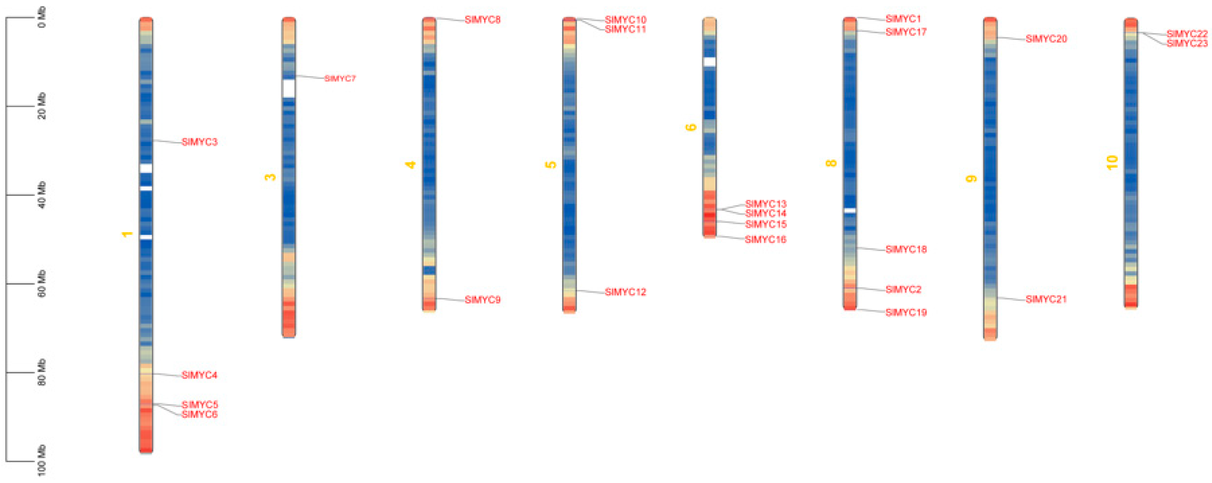
3.1. Analysis of Whole Genome and Basic Physicochemical Properties of the SlMYC Family in Tomato

The physical and chemical properties of the 23 SlMYC proteins were predicted and analyzed. Notably, the protein lengths ranged from 209 to 1160 amino acids with molecular weights varying between 22,589.12 and 130,949.79 Da. The hydrophobicity analysis indicated that all proteins were hydrophilic with isoelectric points between 5.00 and 8.56. Furthermore, the subcellular localization of the SlMYC family protein was compared between the predictions from online software and experimental results. It was observed that SlMYC9 localized to the chloroplast, while the remaining 22 SlMYCs were predominantly located in the nucleus. Additionally, the proteins exhibited a half-life of 30 h and instability coefficients ranging from 37.55 to 72.82 (Table 1).
The positional information of the 23 genes on tomato chromosomes, including their starting and ending positions obtained from NCBI, was formatted as required by the website. Analysis of the chromosome lengths of all 12 chromosomes in tomato was conducted. Notably, the SlMYC genes were found to be distributed across 8 out of the 12 chromosomes, albeit unevenly. Chromosome 8 harbored five SlMYC genes, which were SlMYC1, SlMYC17, SlMYC18, SlMYC2, and SlMYC19. Chromosomes 1 and 6 contained four SlMYC genes, which were SlMYC3, SlMYC4, SlMYC5, and SlMYC6 on chromosome 1, and SlMYC13, SlMYC14, SlMYC15, and SlMYC16 on chromosome 6. Chromosomes 4, 5, 9, and 10 hosted two SlMYC genes, which were SlMYC8 and SlMYC9 on chromosome 4, while SlMYC10, SlMYC11, and SlMYC12 were hosted on chromosome 5, SlMYC20 and SlMYC21 were hosted on chromosome 9, and SlMYC22 and SlMYC23 were hosted on chromosome 10. Moreover, chromosome 3 housed a single gene, SlMYC7. However, chromosomes 2, 11, and 12 did not exhibit the presence of any SlMYC genes.
Except for SlMYC1 and SlMYC2, the remaining genes were denoted as SlMYC3 through SlMYC23 based on their respective chromosomal locations (Figure 1).

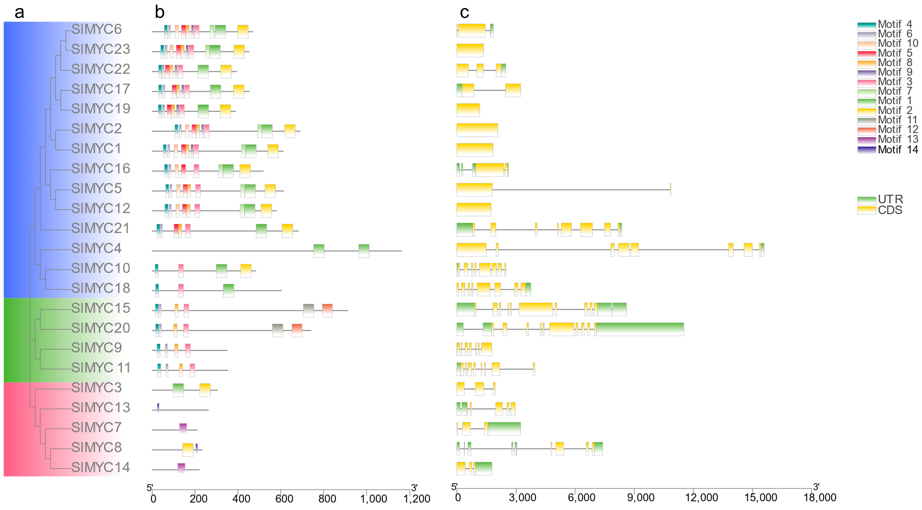
3.2. Phylogeny, Conserved Motif, and Structure Analysis of the SlMYC Family in Tomato

To investigate the evolutionary relationships and patterns of MYC genes, we created a neighborhood junction tree (NJ) using the full-length protein sequences of 23 identified SlMYCs for phylogenetic analysis (Figure 2a). Additionally, we conducted an exon–intron analysis on these genes (Figure 2b). The evolutionary tree revealed that the SlMYC family can be categorized into three subfamilies. Subfamily I comprised 14 genes (SlMYC1, SlMYC2, SlMYC4, SlMYC5, SlMYC6, SlMYC10, SlMYC12, SlMYC16, SlMYC17, SlMYC18, SlMYC19, SlMYC21, SlMYC22, and SlMYC23). Subfamily II consisted of four genes (SlMYC9, SlMYC11, SlMYC15, and SlMYC20) and subfamily III included five genes (SlMYC3, SlMYC7, SlMYC8, SlMYC13, and SlMYC14). We identified several conserved motifs (motif3, motif4, motif6, and motif8) within the SlMYC family. The motif analysis within each subfamily demonstrated high similarity. In subfamily I, most genes displayed a motif arrangement consisting of motif4, motif6, motif10, motif5, motif8, motif9, motif3, motif7, motif1, and motif2, with a few exceptions such as SlMYC21 and SlMYC4. Among the genes in subfamily II, the orders of the first four motifs aligned with those in subfamily I. Notably, SlMYC15 and SlMYC20 had motif11 and motif12 as their last two motifs, while SlMYC9 and SlMYC11 exhibited different motif patterns. In subfamily III, only SlMYC3 and SlMYC8 contained two motifs each. Below are the tagged sequences of the conserved motifs of tomato SlMYCs (Supplementary Materials, Figure S2).
The exon–intron structure played a vital role in understanding gene evolution and served as a significant evolutionary indicator (Figure 2c). Notably, only a subset of genes in subfamily I (SlMYC1 and SlMYC2, and SlMYC12, SlMYC19, and SlMYC23) lack introns entirely, highlighting a unique genetic feature within this subfamily. Furthermore, the majority of genes across the subfamilies exhibited highly similar genetic structures.

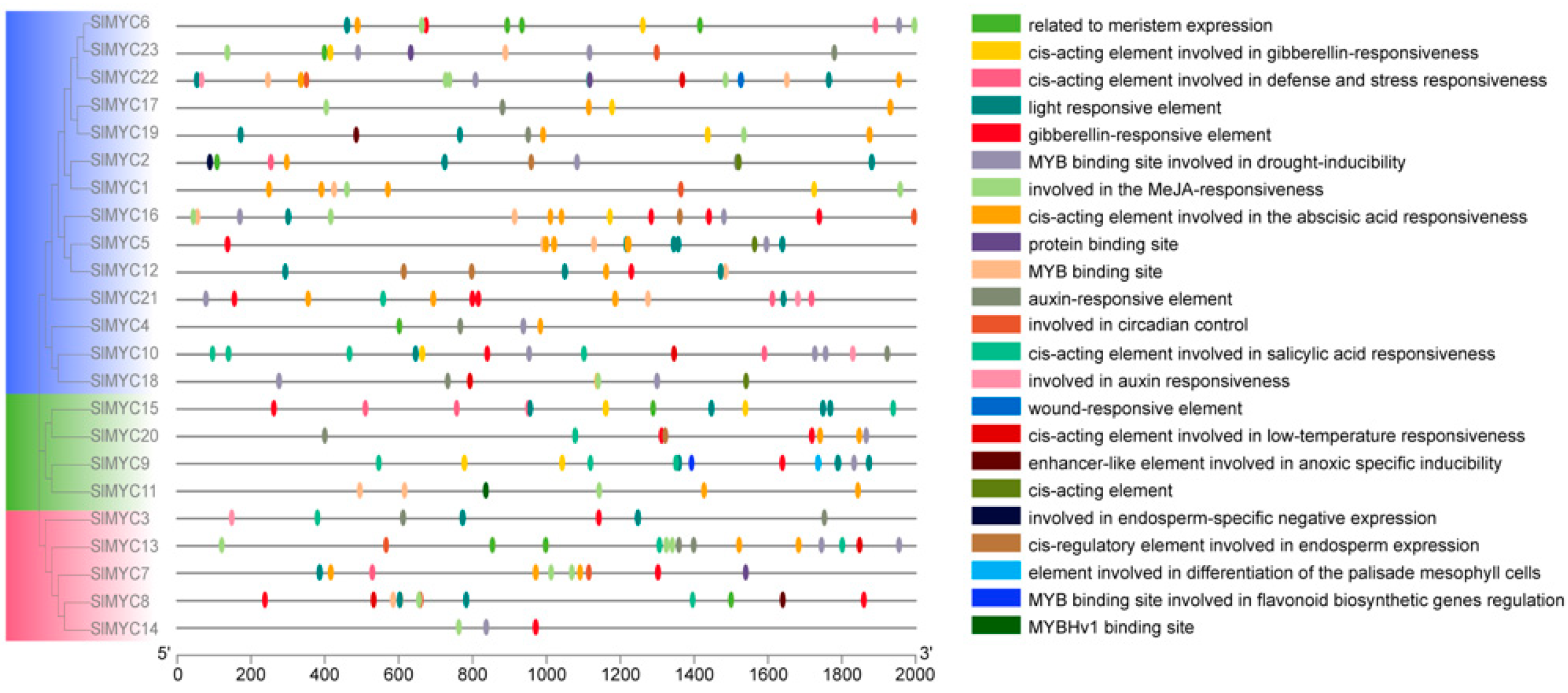
3.3. Promoter Analysis of the SlMYC Family

The promoter analysis of the SlMYC family genes revealed the presence of 23 functional elements, providing insights into their potential functions. These elements encompassed a wide range of hormone-related factors such as auxin, gibberellin, and salicylic acid, as well as those associated with plant growth and development such as meristem expression, palisade mesophyll cell differentiation, and endosperm expression. Additionally, stress tolerance elements including those involved in drought induction, low-temperature response, defense, and stress response were also identified. Most of the genes contained three or more hormone-related elements, indicating their importance in the hormone-regulatory network of tomato. Specific genes within different subfamilies displayed elements related to MeJA response (JA signaling pathway), drought induction via MYB binding sites, and low-temperature responses. For example, the genes SlMYC6, SlMYC16, SlMYC1, SlMYC17, SlMYC18, SlMYC19, SlMYC22, SlMYC23, and SlMYC6 in branch I, as well as SlMYC11 in clade II, and SlMYC13, SlMYC14, SlMYC7, and SlMYC8 in clade III, exhibited elements associated with MeJA response. But the genes SlMYC10, SlMYC18, and SlMYC22 in clade I, and SlMYC8 and SlMYC13 in clade III, contained cis-acting elements involved in low-temperature response (Figure 3).

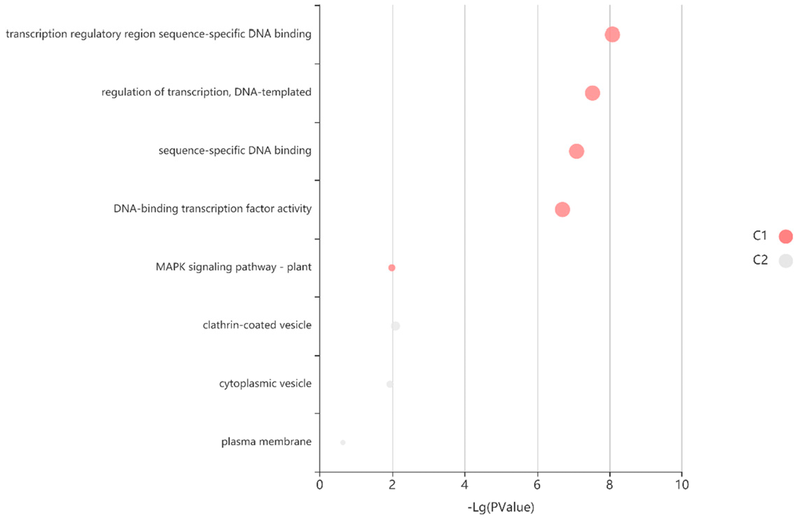
3.4. Expression and Pathway Enrichment of the SlMYC Family in Tomato Tissues

The expression profile analysis of SlMYCs in various tomato tissues highlighted significant insights (Figure 4). Among the SlMYC family members, SlMYC1 and SlMYC2 emerged as notably expressed genes across various stages of tomato growth. Both genes exhibited prominent expression in flowers, fruits, leaves, and roots, with expression levels surpassing those of other genes, showcasing tissue specificity. The expression pattern of SlMYC1 primarily appeared concentrated in the fruit stages, suggesting a potential close association with the reproductive growth of tomato. In contrast, SlMYC2 showed prevalent expression in flowers, leaves, fruits, and rhizomes, indicating a potential link to both vegetative growth and reproductive development in tomato. A comparison of the heat map data for SlMYC1 and SlMYC2 revealed similar expression patterns across different tissues and fruit growth stages in tomato. Notably, both genes displayed increasing expression levels during the flowering stage with fluctuating expressions observed during the fruiting stage, hinting at possible functional redundancy between them. These findings underlined the significant roles of SlMYC1 and SlMYC2 in tomato growth and development, with indications of functional redundancy. This observation aligned with the evolutionary tree analysis of the tomato SlMYC gene family. However, further experimental validation was necessary to elucidate the specific mechanisms underlying the actions of SlMYC1/2 in tomato development.
The expression analysis of various SlMYCs in different tissues and growth stages of tomato provided valuable insights into their potential roles in plant development. Several genes, including SlMYC3, SlMYC4, SlMYC10, SlMYC12, SlMYC17, SlMYC19, SlMYC21, and SlMYC22, showed negligible expression levels across all stages, suggesting a limited role in tomato growth and development. On the other hand, some genes, like SlMYC5, exhibited specific expression patterns, such as being expressed in flower buds with reduced expression post-flowering, indicating a role in the flowering process. SlMYC6 displayed higher expression in leaves, roots, and fruits, potentially implicating roles in photosynthesis and ripening. SlMYC7, although expressed at lower levels compared with SlMYC1/SlMYC2, showed increasing expression during flowering and fruit stages, suggesting involvement in reproductive growth. SlMYC8 and SlMYC16 demonstrated stable expressions in various tissues, with SlMYC8 showing increased expression during fruit ripening, while SlMYC16 displayed decreasing expression. Notably, genes like SlMYC9 and SlMYC13 exhibited dynamic expression patterns correlating with flower and fruit development stages, indicating potential roles in these processes. SlMYC15 showed prominent expression in roots, suggesting a role in root development, while genes like SlMYC18 and SlMYC20 displayed significant expressions in specific tissues such as flower buds, leaves, and roots, implying potential roles in flowering and root growth regulation, respectively. Furthermore, the expression of SlMYC23 fluctuated with fruit development stages, indicating potential involvement in fruit ripening processes. The detailed mechanisms underlying the functions of these SlMYC genes in tomato growth and development warranted further investigation.
The path enrichment analysis of the SlMYC gene family revealed enrichment in the MAPK pathway, which was crucial for plant responses to various stresses (Figure 5). Previous studies have indicated that the JA signaling pathway triggers a MAPK cascade reaction, highlighting the biological significance of JA in plant stress tolerance as a key plant hormone. Given the enrichment of 23 SlMYCs in the MAPK pathway, it suggested their potential role in tomato response to abiotic stress. To further investigate the involvement of SlMYCs in stress response, the study subjected tomato plants to treatments including low temperature, salt stress, and simulated drought in the laboratory. By examining the expressions of SlMYCs under these different stress conditions, the study aimed to elucidate the mechanisms by which the SlMYC family contributed to tomato tolerance to abiotic stress. This research not only laid a foundation for understanding the functional roles of SlMYCs in stress responses but also provided valuable insights for developing tomato varieties with enhancing tolerance to various environmental stresses. By elucidating the molecular mechanisms underlying stress responses mediated by the SlMYC gene family, it offered a theoretical basis for breeding stress-tolerant tomato cultivars.
The expression levels of SlMYC1 and SlMYC2 were consistently higher than those of other MYC family members across all tomato tissues and organs. This made SlMYC1 and SlMYC2 particularly interesting for further research. Signal pathway enrichment analysis suggested that SlMYC2 was closely involved in the JA-mediated MAPK cascade pathway, but the exact mechanisms of this involvement required further investigation.

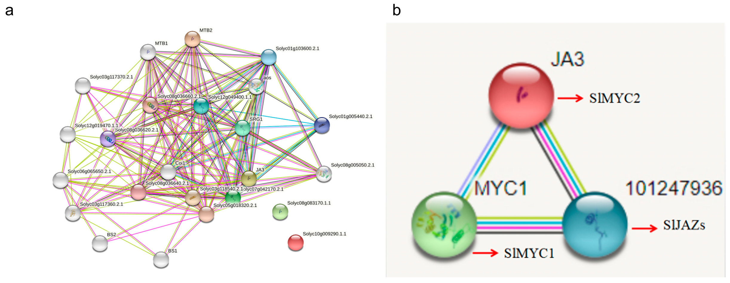
3.5. Interaction Network Analysis of the SlMYC Family

In order to better understand the biological functions and protein regulatory networks of the SlMYCs, the 23 obtained SlMYC protein sequences were imported into the STRING database to predict the protein–protein interactions associated with SlMYCs (Figure 6a). In the interaction network obtained, in addition to the normally labeled SlMYC family members, JA3 (SlMYC2) and MTB2 (SlMYC12) were also members of the tomato MYC family, and the rest were proteins that interacted with SlMYCs. The function of the proteins was analyzed, respectively. For example, the transcription factors (MTB1/2) that negatively regulated JA signaling, jasmonate zim domain protein 3 (Solyc01g005440), zinc jasmonate domain protein/salt-reactive protein 1 (SRG1), SlJAZ, and MPK2, were all proteins related to JA signaling and stress tolerance. So, the prediction of SlMYCs played an important role in responding to tomato stress.
The prediction of the interaction network in this study provided the direction for further research on the role of SlMYC proteins. Combining the two figures, it could be seen that SlMYCs were closely related to proteins that were related to stress tolerance, such as SRG1, Coil1, MTB1, Solyc01g005440, and aos, especially those related to JA signaling, which more intuitively reflects the relationship between SlMYC1/2 and JAZ (Figure 6b). SlMYC1/2 participated in the regulation of the tomato aging process, stress defense response, and secondary metabolite biosynthesis by responding to JAZ in the JA signaling pathway.

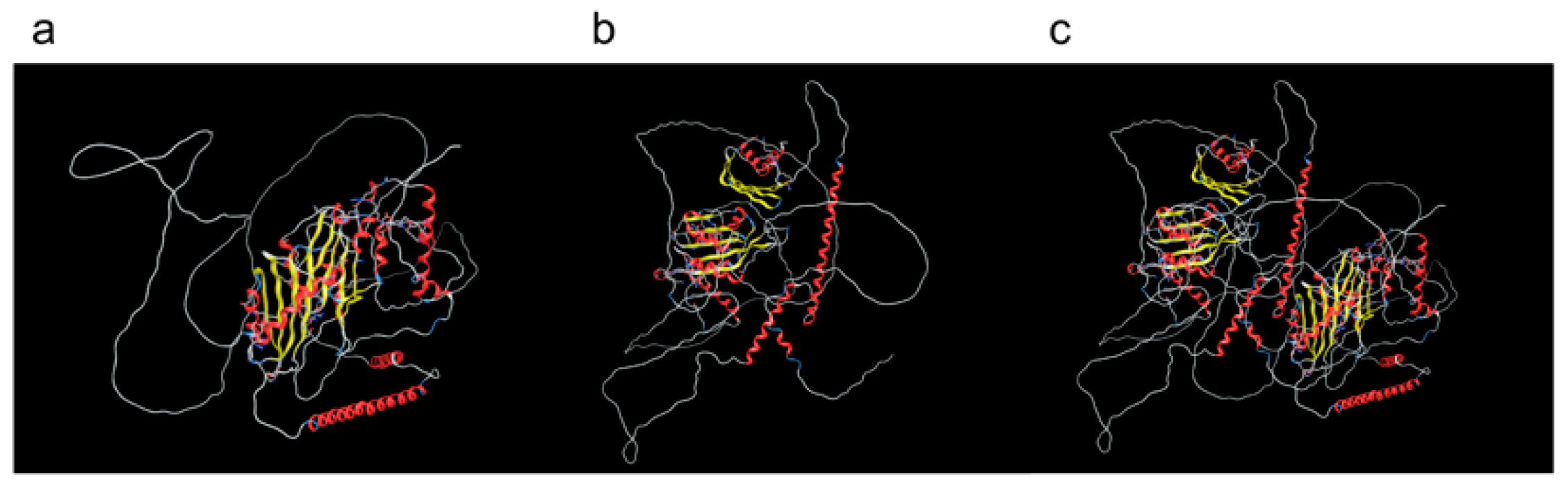
3.6. Protein 3D Models Predicted Docking with Protein Molecules

The obtained protein sequences were imported into the Swiss model, and homology modeling was used to further predict the three-dimensional structure of proteins. The optimal template was selected according to the three dimensions of score, similarity, and ligand to construct the three-dimensional structural homology model of the SlMYC1 and SlMYC2 proteins in the SlMYC family (Figure 7a,b). The results showed that the protein models of the same evolutionary branch were of high similarity, with α-helix wrapped β-folding structure. But the protein sequence of the other branch was relatively simple, as just the α-helix was used in 3D modeling. When screening in this study, the required model score of Seq Identity was higher than 20%. After the export, the models of SlMYC1 and SlMYC2 were constructed by using big data modeling as a template for molecular docking. The results showed that there was a protein interaction relationship between SlMYC1 and SlMYC2 (Figure 7c). Therefore, SlMYC1 and SlMYC2 might jointly participate in the JA signaling pathway by combining to form dimers and play a role in tomato abiotic stress regulation.

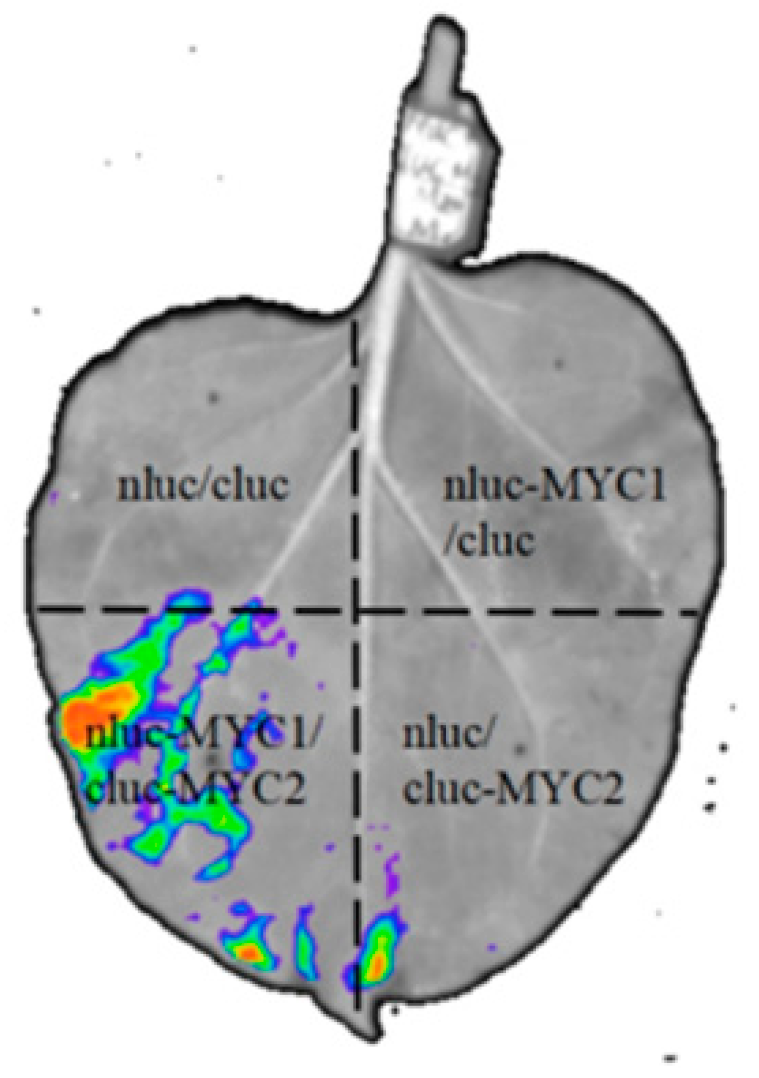
3.7. Interaction of SlMYC1 and SlMYC2

To further demonstrate the protein interaction relationship between SlMYC1 and SlMYC2, LCI experiments were employed. The negative controls were nluc/cluc, nluc-SlMYC1/cluc, and nluc/cluc-SlMYC2, and the recombinant plasmid nluc-SlMYC1/cluc-SlMYC2 was injected into tobacco leaves (Figure 8). It was found that there was no fluorescence signal except for nluc-SlMYC1/cluc-SlMYC2, indicating that there was interaction between nluc-SlMYC1 and cluc-SlMYC2. This result provided a basis for further research on the role of SlMYC1 and SlMYC2 interaction in regulating the abiotic stress of tomato.

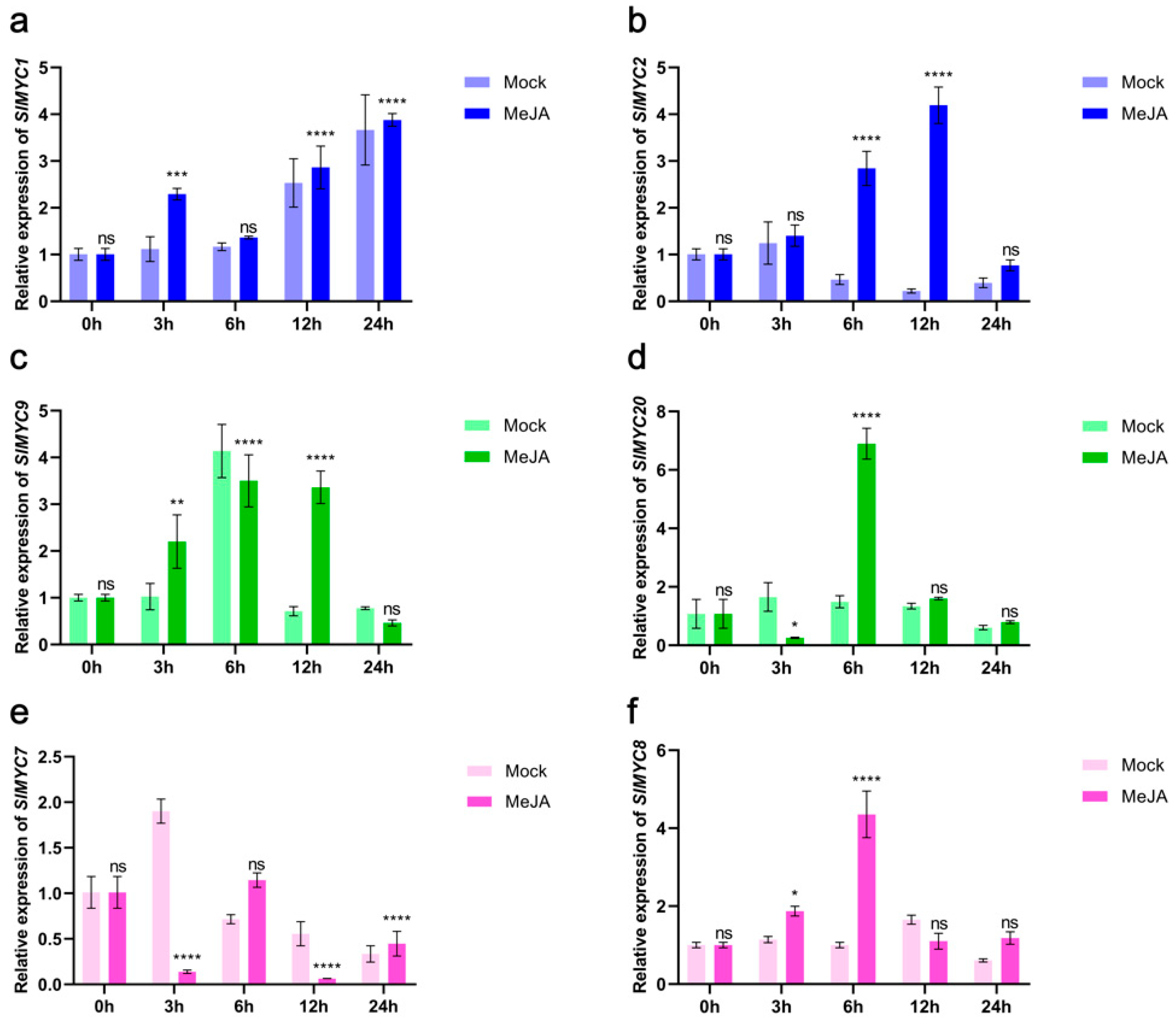
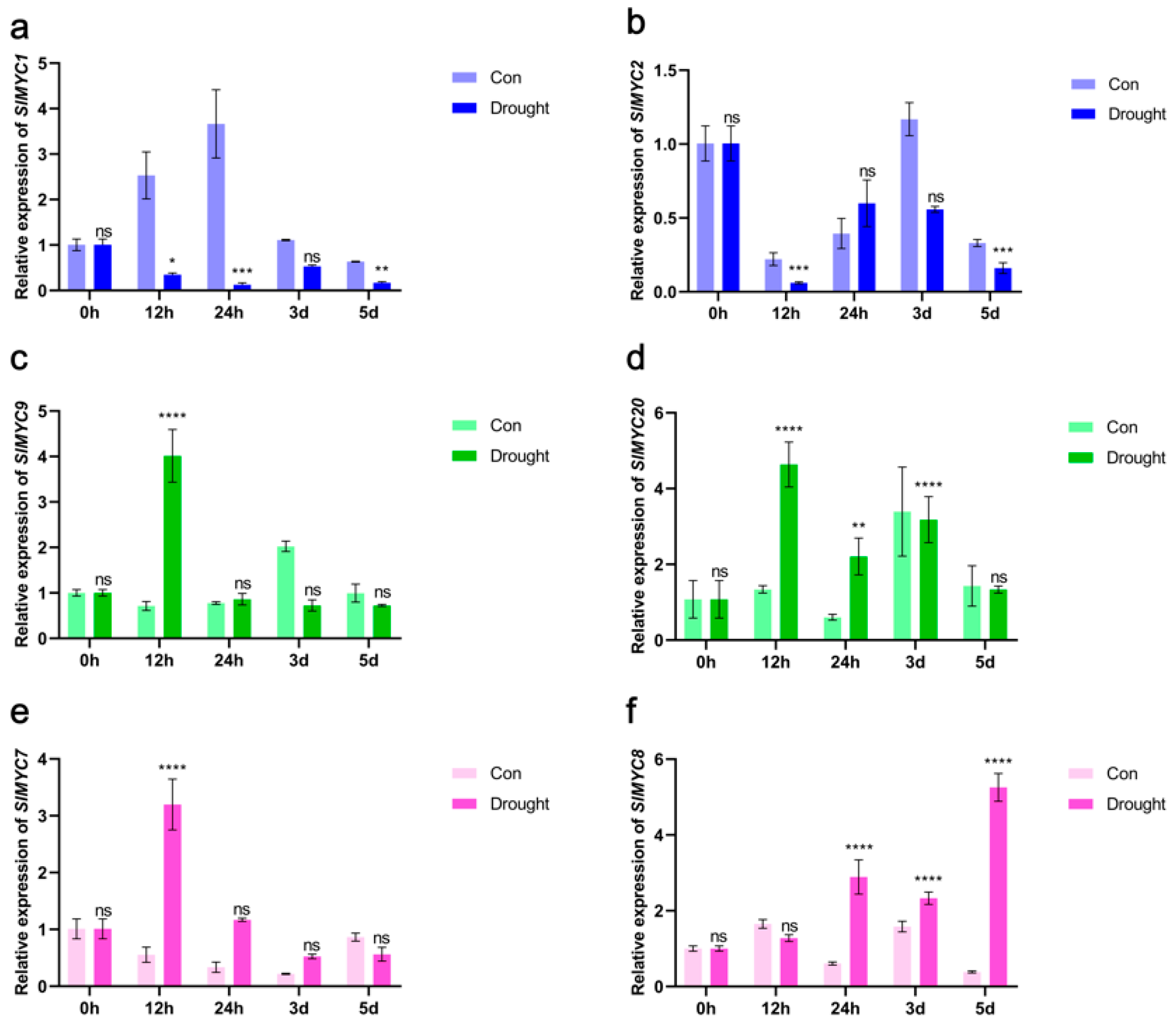
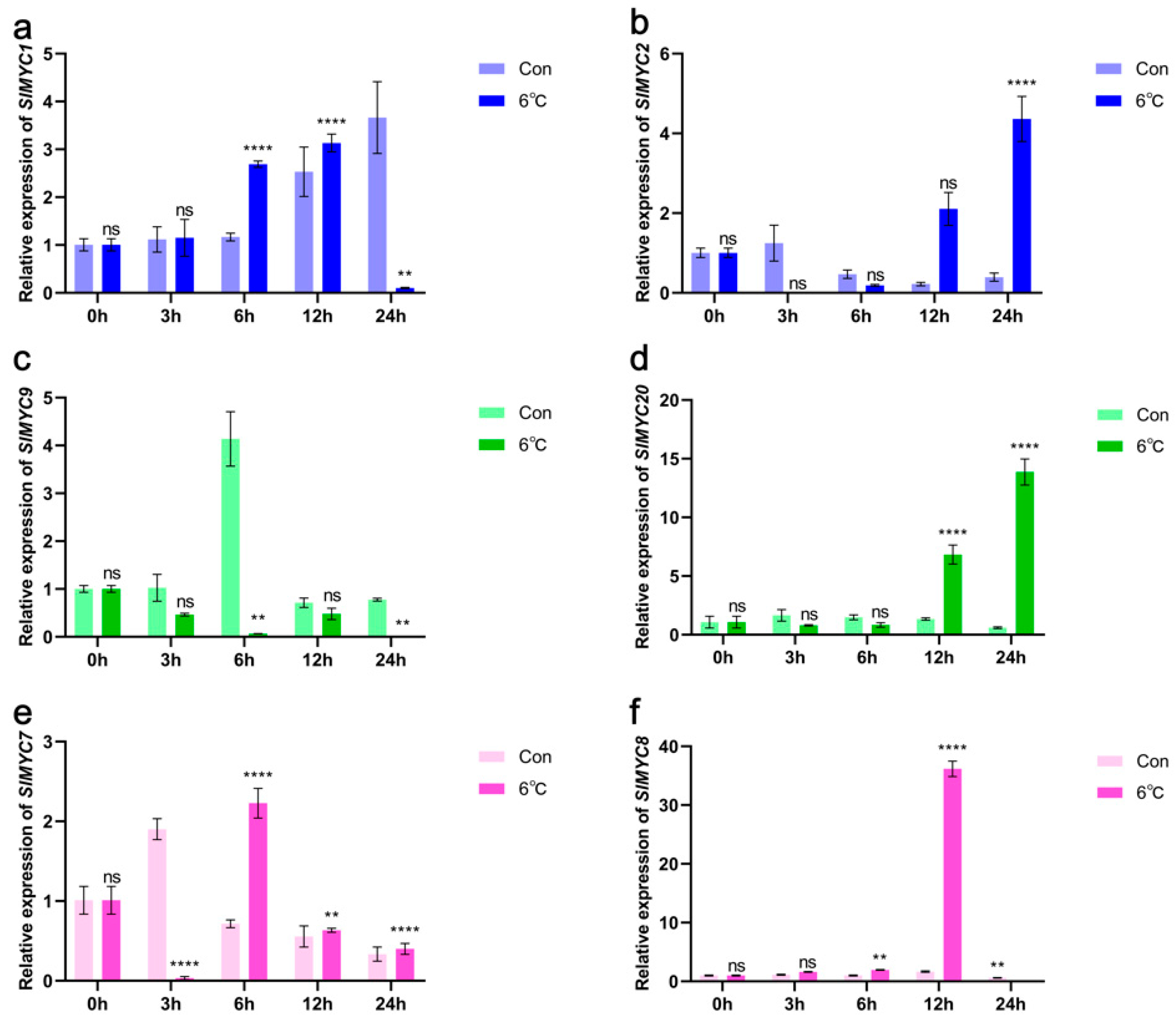
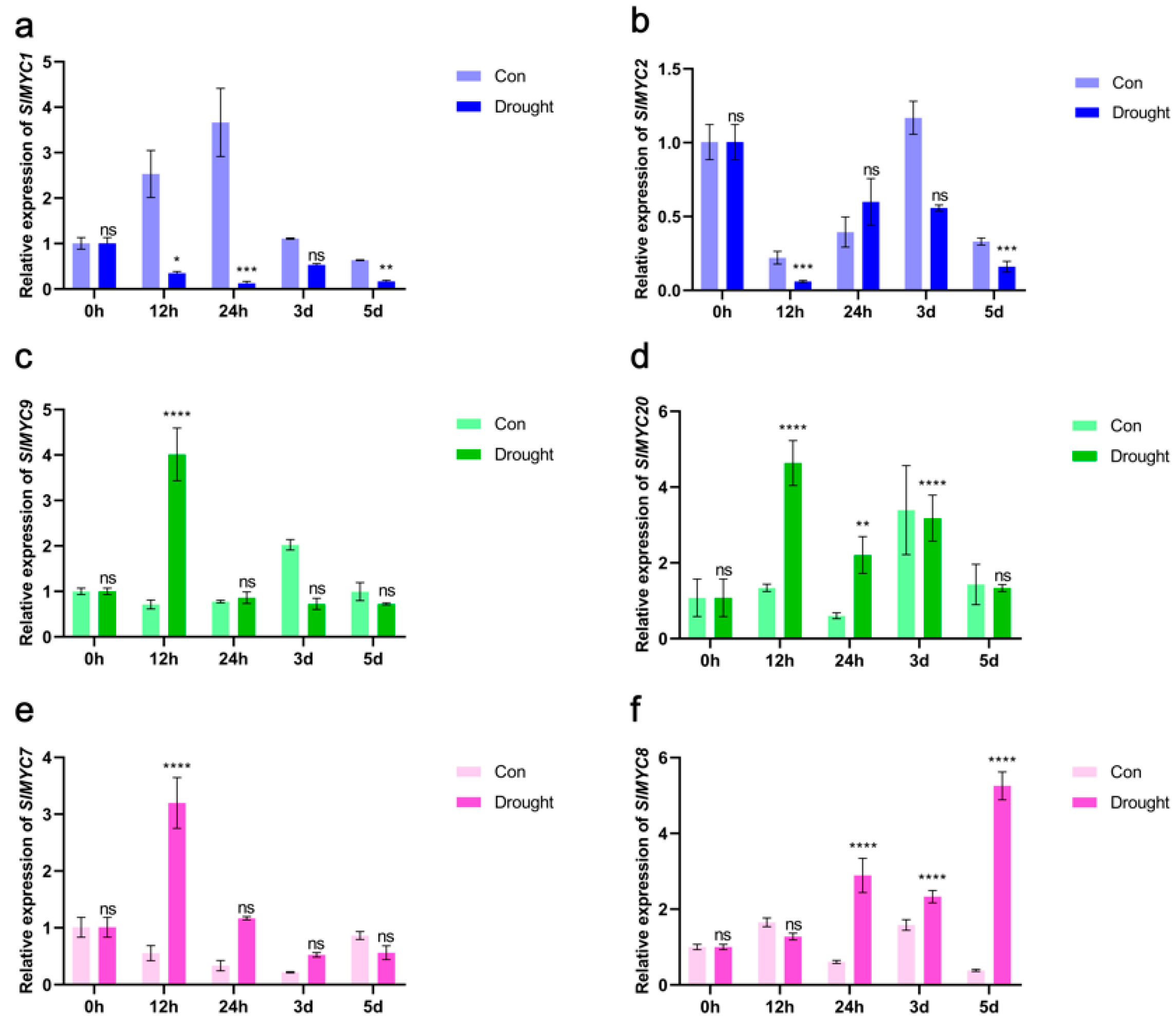
3.8. Expression of SlMYCs in Tomato

A tissue-specific expression analysis was conducted in this study. The expression levels of six genes, SlMYC1, SlMYC2, SlMYC7, SlMYC8, SlMYC9, and SlMYC20, were higher than those of other genes. They belonged to three homologous groups. SlMYC1 and SlMYC2 belonged to the first homologous group. SlMYC8 and SlMYC20 belonged to the second homologous group, but SlMYC7 and SlMYC9 belonged to homologous group III. Since the MYC family was closely related to the JA signaling pathway, we performed MeJA spray treatment and examined the changes in SlMYC gene expression under this treatment (Figure 9). Since samples of the hormone were involved at different times of the day, the genes were first tested for whether they were regulated by MeJA. At the same time, since JA signaling and the MYC family of key transcription factors on its signaling pathway were involved in the regulation of abiotic stress, this study examined the expression levels of these six genes under three abiotic stress conditions, namely drought (Figure 10), low temperature (Figure 11), and salt stress (Figure 12).
The expression levels of the six genes, SlMYC1, SlMYC2, SlMYC7, SlMYC8, SlMYC9, and SlMYC20, were detected in tomato seedlings treated with MeJA by real-time fluorescence quantitative PCR (Figure 9). The results showed that the expression level of the SlMYC1 gene in homologous group I was significantly higher than that in the control group at 3 h after MeJA treatment, and the expression level of the SlMYC2 gene was significantly higher than that in the control group at 3–24 h, but the expression level was the highest at 12 h. The expression levels of SlMYC8 and SlMYC20 genes in homologous group II were significantly higher than those in the control group at 6 h after treatment. The expression of SlMYC7 in homologous group III fluctuated significantly, but the SlMYC9 gene was higher than that in the control group at 3 h and 12 h. These results indicated that the expressions of the SlMYC1, SlMYC2, SlMYC7, SlMYC8, SlMYC9, and SlMYC20 genes were all affected by MeJA, which were consistent with the gene function predicted by promoter cis-acting elements.
The expression levels of six genes, SlMYC1, SlMYC2, SlMYC7, SlMYC8, SlMYC9, and SlMYC20, showed (Figure 10) that the expression levels of the SlMYC1 and SlMYC2 genes in homologous group I were down-regulated under drought conditions. The expression levels of SlMYC8 and SlMYC20 in homologous group II and SlMYC7 and SlMYC9 in homologous group III were significantly higher than those of the control group at the 12th hour after treatment, while SlMYC8 remained highly expressed 5 d after treatment, indicating that the expressions of these six genes were affected by drought stress and might be related to the regulation of drought stress.
The expression of SlMYC1 was significantly higher at 3–6 h than the control group, but the expression of SlMYC2 was highly expressed at 6–24 h in low-temperature stress. The expression level of SlMYC8 was extremely significantly expressed at 12 h, but the expression of SlMYC20 was significantly higher than the control group after 12 h. The expression of SlMYC7 was highly expressed at 6 h; however, SlMYC9 was lower than the control group during the whole treatment process (Figure 11), indicating that these six genes might play a regulatory role in low-temperature stress.
The results of salt stress treatment showed that the expressions of the SlMYC1 and SlMYC9 genes were significantly higher than those in the control group at 3 h after treatment, SlMYC8 was higher than that of the control group at 3 h after treatment, SlMYC20 was significantly higher than that of the control group at 12 h after treatment, but SlMYC7 was highly expressed at 24 h. However, SlMYC2 decreased first and then increased in the treated group, and was higher than the control group at 12 h (Figure 12).

4. Discussion

The tomato, being a light-loving and warm-loving vegetable, is often vulnerable to various stresses such as pests, diseases, low temperatures, and drought during production and cultivation. These stress factors can significantly reduce both the yield and quality of tomatoes. Jasmonic acid (JA) stands out as a crucial stress-resistant hormone in higher plants [24,25], playing a key role in plant growth and development [69,70,71]. It is involved in seed and root development and regulates plant responses to diverse biological and abiotic stresses [18,19]. The MYC gene family, presented in numerous plant species including Arabidopsis thaliana [72], tobacco [73], cucumber [74], and poplar [30], is essential in activating the JA signaling pathway and is vital for plant stress tolerance. Previous research has highlighted the significant regulatory role of JA signaling in tomato growth, development, and stress responses [75,76]. The MYC family, as a key transcription factor in the JA signaling pathway, plays a critical role in stress tolerance mechanisms. By conducting functional verification and analysis of the MYC family, researchers can gain deeper insights into the regulatory functions of JA signaling in stress tolerance through the MYC transcription factor family. This research could pave the way for developing superior tomato varieties with enhanced tolerance to biotic and abiotic stresses.
The study identified a total of 23 tomato SlMYC genes through a database retrieval method. These genes were unevenly distributed across the 12 chromosomes of the tomato, with a notable concentration on chromosome 8, especially at both ends, indicating a potential susceptibility to evolutionary variations. Gene cluster and tandem repeat analyses revealed the presence of gene clusters on chromosomes 1, 5, 6, and 10, with two genes in each cluster forming tandem repeats. This indicated that tandem repeats were crucial for gene expansion and cluster formation mechanisms [77]. Subcellular localization prediction revealed that except for SlMYC9, which was located in chloroplasts, the remaining SlMYCs were all located in the nucleus. This localization suggested that the primary function of SlMYC9 might involve participation in tomato photosynthesis. The evolutionary relationships among these genes categorized them into three groups in the homologous evolutionary tree. Members of clade I clustered together into a larger branch, suggesting a common ancestral origin and formation through frequent gene replication [78].
By using the homology method, interactions among tomato SlMYC proteins and other proteins were predicted, and SlMYC protein pathway enrichment analysis was conducted. This study found that SlMYC proteins interacted with proteins such as SlJAZ, MPK2, and MTB1, which were all related to the JA signaling pathway, with a prominent interaction observed between SlMYC1/2 and JAZ. JAZ negatively regulates the JA signaling pathway by inhibiting MYC2 transcription. Furthermore, studies have shown that JAZ can bind to the homologous transcription factors MYC2, MYC3, and MYC4, which cooperatively regulate the JA signaling pathway [79]. Moreover, MTB1, identified as a transcription factor, negatively regulates the JA signaling pathway [80]. The pathway enrichment analysis revealed that MYC2 is linked to the MAPK pathway, which is known to be triggered by JA signals and is essential in plant stress tolerance mechanisms [81]. Therefore, there was a preliminary prediction that the SlMYC family plays a significant role in stress response based on these findings.
The research revealed that SlMYC1 and SlMYC2 belong to MYC IIIe [29]. Moreover, MYC IIIe can regulate downstream target genes by forming dimers or heterodimers. This study focused on the three-dimensional structure of SlMYC1/2, as it was closely related to JA signaling. The conservation of the three-dimensional structure of the identified SlMYC1/2 protein was found to be consistent with phylogenetic, gene structure, and conserved domain analyses. Recent research on tomato has highlighted the synergistic regulation of transcriptional activation of SGAs by JA signaling through SlMYC1 and SlMYC2, indicating functional redundancy between the two proteins [82]. Based on this understanding, molecular docking analysis of the protein model of SlMYC1/2 was conducted using molecular docking software (PyMOl-2.4.1). The results revealed an interactive relationship between SlMYC1 and SlMYC2 proteins, in line with the findings from the interacting protein network analysis. Further validation through LCI experiments confirmed the binding interaction between the two proteins. The regulatory relationship identified through string analysis and molecular docking offers valuable insights into unraveling the SlMYC1/2-mediated JA signaling network. This comprehensive approach aided in further exploring the roles of SlMYCs in the JA signaling pathway and provided important clues to better understand the biological functions of the tomato SlMYC gene family. The findings of this study contribute to advancing our knowledge of the molecular mechanisms underlying stress responses in tomato and could potentially lead to the development of improved tomato varieties with enhanced stress tolerance.
The comprehensive study of the expression patterns of the SlMYC gene family in tomato revealed interesting insights into their spatiotemporal dynamics and potential functional roles. The expressions of SlMYC genes across various tissues such as seeds, roots, leaves, flowers, and fruits indicate their widespread presence and likely diverse functions in different parts of the plant [66]. The expression pattern of SlMYC1 primarily appeared to concentrate in the fruit stages, suggesting a potential close association with the reproductive growth of tomato. In contrast, SlMYC2 showed prevalent expression in flowers, leaves, fruits, and rhizomes, indicating a potential link to both vegetative growth and reproductive development in tomato. SlMYC1 regulates trichome formation and terpene biosynthesis [83]. SlMYC2 regulates growth and fruit quality [84]. Among the family members, SlMYC1/2 demonstrated high expression levels across all stages of tomato development, suggesting a fundamental role in the plant. Specific members like SlMYC5, SlMYC7, SlMYC8, and SlMYC16 showed significant expressions in fruits and flowers, pointing towards their involvement in reproductive growth processes in tomato. Conversely, SlMYC15 and SlMYC20 exhibited high expressions in roots, indicating a potential regulatory role in root development. The correlation between SlMYC genes and the JA signaling pathway in stress tolerance has been underlined in previous studies using model plants like Arabidopsis and tobacco [72,73]. The example of AtMYC2 in Arabidopsis thaliana binding to the promoter of its target gene AtERD1 to trigger the JA-induced dehydration stress response underscores the crucial role of MYC transcription factors in mediating plant stress responses [85]. In this study, we investigated the response of the tomato SlMYC gene family to MeJA treatment by applying 100 mmol·L−1 MeJA to tomato plants. Our results revealed that several SlMYC genes, including SlMYC1, SlMYC2, SlMYC7, SlMYC8, SlMYC9, and SlMYC20, exhibited changes in gene expression levels in response to MeJA treatment. These findings were consistent with predictions based on the promoter cis-acting elements, indicating that the tomato SlMYC family was influenced by JA signaling. The diverse responses of different MYC genes to hormonal and abiotic stressors highlight the complex regulatory network governing plant responses to environmental stimuli. Moreover, our study also identified that specific MYC genes in tomato plants respond to a variety of stimuli, including auxin, cytokinin, salt, drought, jasmonic acid, low temperature, and JA signaling. For instance, The genes of PtrMYC12a, PtbHLH14b, and PtrMYC2b are found to be responsive to multiple hormonal and abiotic stress factors [30].
The qRT-PCR results demonstrated that most MYC genes could be induced by low temperature, NaCl, and drought stress, while also responding to JA signaling. In Arabidopsis, MeJA treatment activates MYC2 [86]. In our study, most of the SlMYC genes were activated after MeJA treatment. SlMYC1 reduced the stress tolerance of NaCl and drought [87]. In our study, the expressions of SlMYC1 and SlMYC2 were lower than the others of the MYC family, and all the others were raised after NaCl and drought treatment. Low-temperature stress and JA treatment induced the expression of TaMYC2 [88]. In our study, SlMYC1 was increased, while SlMYC2 was first decreased and then increased. The implication is that the MYC gene family plays a role in plant growth, development, and responses to abiotic stress. Different members of the MYC gene family may be involved in distinct biological processes. However, further investigation is necessary to fully elucidate the specific regulatory mechanisms governing the functions of these genes in plants.
This study delved into the potential of the SlMYC gene family in tomato, investigating their roles in regulating the JA signaling pathway under abiotic stress conditions. The research also examined how the SlMYC gene family responded to stress, offering insights into the possible members and functions within this gene family. These findings serve as a valuable reference for gaining a deeper understanding of the SlMYC gene family and its involvement in the JA signaling pathway under abiotic stress. Additionally, the study highlighted the potential of SlMYCs to enhance tomato tolerance, setting the stage for further exploration of how the JA signaling pathway could be leveraged through SlMYC1/2 to bolster tomato resilience.

5. Conclusions

In summary, the results of this study suggest that the SlMYC gene family is associated with the JA signaling pathway and likely exerts a regulatory influence on the plant response to abiotic stress. Specifically, it was found that SlMYC1/2 might interact with each other to regulate target genes involved in stress responses. This insight into the potential regulatory roles of the SlMYC family in mediating plant stress responses underscored the importance of further research into MYC transcription factors and their impact on plant growth, development, and resilience in challenging environmental conditions.

Supplementary Materials

The following supporting information can be downloaded at https://www.mdpi.com/article/10.3390/horticulturae11060693/s1: Figure S1: Marker sequences of tomato SlMYC conserved motifs; Table S1: List of the primers used in this study for gene expression analysis via qPCR; Figure S2: Marker sequences of tomato SlMYC conserved motifs.

Author Contributions

Conceptualization, C.K., B.Z., and N.C.; methodology, C.K. and B.Z.; software, C.K.; validation, M.S. and B.Q.; formal analysis, C.K. and Q.Z.; investigation, C.K., B.Z., Q.Z., S.B., Z.W., and Y.Z.; resources, N.C., M.S., and B.Q.; data curation, C.K.; writing—original draft preparation, C.K.; writing—review and editing, N.C.; visualization, C.K.; supervision, N.C.; project administration, N.C., M.S., and B.Q.; funding acquisition, N.C., M.S., and B.Q. All authors have read and agreed to the published version of the manuscript.

Funding

This research was funded by the National Key R&D Program (2023YFC2604500), the Project of Shenyang Science and Technology (24-215-2-11), and the Project of Shenyang Science and Technology (24-215-2-10).

Data Availability Statement

The datasets generated and/or analyzed during the current study are available from the corresponding author upon reasonable request.

Acknowledgments

We would like to express our gratitude to the Plant Stress and Adaptation Team of Shenyang Agricultural University for providing experimental equipment and data support.

Conflicts of Interest

The authors declare no conflicts of interest.

References

  1. Xiong, Y.; Sheen, J. The role of target of rapamycin signaling networks in plant growth and metabolism. Plant Physiol. 2014, 164, 499–512. [Google Scholar] [CrossRef] [PubMed]
  2. Harris, T.E.; Lawrence, J.C., Jr. TOR signaling. Sci. Signal. 2003, 2003, re15. [Google Scholar] [CrossRef] [PubMed]
  3. Ouziad, F.; Hildebrandt, U.; Schmelzer, E.; Bothe, H. Differential gene expressions in arbuscular mycorrhizal-colonized tomato grown under heavy metal stress. J. Plant Physiol. 2005, 162, 634–649. [Google Scholar] [CrossRef]
  4. Tanveer, K.; Gilani, S.; Hussain, Z.; Ishaq, R.; Adeel, M.; Ilyas, N. Effect of salt stress on tomato plant and the role of calcium. J. Plant Nutr. 2020, 43, 28–35. [Google Scholar] [CrossRef]
  5. Zhou, R.; Yu, X.; Ottosen, C.O.; Rosenqvist, E.; Zhao, L.; Wang, Y.; Yu, W.; Zhao, T.; Wu, Z. Drought stress had a predominant effect over heat stress on three tomato cultivars subjected to combined stress. BMC Plant Biol. 2017, 17, 24. [Google Scholar] [CrossRef]
  6. Wang, Y.; Mostafa, S.; Zeng, W.; Jin, B. Function and mechanism of jasmonic acid in plant responses to abiotic and biotic stresses. Int. J. Mol. Sci. 2021, 22, 8568. [Google Scholar] [CrossRef]
  7. Ruan, J.; Zhou, Y.; Zhou, M.; Yan, J.; Khurshid, M.; Weng, W.; Cheng, J.; Zhang, K. Jasmonic acid signaling pathway in plants. Int. J. Mol. Sci. 2019, 20, 2479. [Google Scholar] [CrossRef]
  8. Huang, H.; Liu, B.; Liu, L.; Song, S. Jasmonate action in plant growth and development. J. Exp. Bot. 2017, 68, 1349–1359. [Google Scholar] [CrossRef] [PubMed]
  9. Kim, J.; Chang, C.; Tucker, M.L. To grow old: Regulatory role of ethylene and jasmonic acid in senescence. Front. Plant Sci. 2015, 6, 20. [Google Scholar] [CrossRef]
  10. Song, S.; Qi, T.; Wasternack, C.; Xie, D. Jasmonate signaling and crosstalk with gibberellin and ethylene. Curr. Opin. Plant Biol. 2014, 21, 112–119. [Google Scholar] [CrossRef]
  11. Wasternack, C.; Hause, B. Jasmonates: Biosynthesis, perception, signal transduction and action in plant stress response, growth and development. An update to the 2007 review in Annals of Botany. Ann. Bot. 2013, 111, 1021–1058. [Google Scholar] [CrossRef] [PubMed]
  12. Li, C.; Xu, M.; Cai, X.; Han, Z.; Si, J.; Chen, D. Jasmonate signaling pathway modulates plant defense, growth, and their trade-offs. Int. J. Mol. Sci. 2022, 23, 3945. [Google Scholar] [CrossRef]
  13. Li, L.; Zhang, H.; Chai, X.; Lv, J.; Hu, L.; Wang, J.; Li, Z.; Yu, J.; Liu, Z. Genome-wide identification and expression analysis of the MYC transcription factor family and its response to sulfur stress in cabbage (Brassica oleracea L.). Gene 2022, 814, 146116. [Google Scholar] [CrossRef]
  14. Goossens, J.; Fernández-Calvo, P.; Schweizer, F.; Goossens, A. Jasmonates: Signal transduction components and their roles in environmental stress responses. Plant Mol. Biol. 2016, 91, 673–689. [Google Scholar] [CrossRef]
  15. Yan, Y.; Stolz, S.P.; Chételat, A.; Reymond, P.; Pagni, M.; Dubugnon, L.; Farmer, E.E. A downstream mediator in the growth repression limb of the jasmonate pathway. Plant Cell 2007, 19, 2470–2483. [Google Scholar] [CrossRef] [PubMed]
  16. TariqJaveed, M.; Farooq, T.; Al-Hazmi, A.S.; Hussain, M.D.; Rehman, A.U. Role of Trichoderma as a biocontrol agent (BCA) of phytoparasitic nematodes and plant growth inducer. J. Invertebr. Pathol. 2021, 183, 107626. [Google Scholar] [CrossRef]
  17. Yan, L.; Zhai, Q.; Wei, J.; Li, S.; Wang, B.; Huang, T.; Du, M.; Sun, J.; Kang, L.; Li, C.; et al. Role of tomato lipoxygenase D in wound-induced jasmonate biosynthesis and plant immunity to insect herbivores. PLoS Genet. 2013, 9, e1003964. [Google Scholar] [CrossRef] [PubMed]
  18. Wang, J.; Song, L.; Gong, X.; Xu, J.; Li, M. Functions of jasmonic acid in plant regulation and response to abiotic stress. Int. J. Mol. Sci. 2020, 21, 1446. [Google Scholar] [CrossRef]
  19. Creelman, R.A.; Mullet, J.E. Jasmonic acid distribution and action in plants: Regulation during development and response to biotic and abiotic stress. Proc. Natl. Acad. Sci. USA 1995, 92, 4114–4119. [Google Scholar] [CrossRef]
  20. Delgado, C.; Mora-Poblete, F.; Ahmar, S.; Chen, J.-T.; Figueroa, C.R. Jasmonates and plant salt stress: Molecular players, physiological effects, and improving tolerance by using genome-associated tools. Int. J. Mol. Sci. 2021, 22, 3082. [Google Scholar] [CrossRef]
  21. Paes, B.; Carpinetti, P.d.A.; Fraga, O.T.; Rodrigues-Silva, P.L.; Fioresi, V.S.; de Camargos, L.F.; Ferreira, M.F.d.S. Abiotic stresses in plants and their markers: A practice view of plant stress responses and programmed cell death mechanisms. Plants 2022, 11, 1100. [Google Scholar] [CrossRef] [PubMed]
  22. Mostofa, M.G.; Rahman, M.M.; Ansary, M.M.U.; Keya, S.S.; Abdelrahman, M.; Miah, M.G.; Phan Tran, L.-S. Silicon in mitigation of abiotic stress-induced oxidative damage in plants. Crit. Rev. Biotechnol. 2021, 41, 918–934. [Google Scholar] [CrossRef]
  23. Ilyas, M.; Nisar, M.; Khan, N.; Hazrat, A.; Khan, A.H.; Hayat, K.; Fahad, S.; Khan, A.; Ullah, A. drought tolerance strategies in plants: A mechanistic approach. J. Plant Growth Regul. 2021, 40, 926–944. [Google Scholar] [CrossRef]
  24. Campos, M.L.; Kang, J.-H.; Howe, G.A. Jasmonate-triggered plant immunity. J. Chem. Ecol. 2014, 40, 657–675. [Google Scholar] [CrossRef] [PubMed]
  25. Llanes, A.S.; Andrade, A.M.; Alemano, S.G.; Luna, M.V. Alterations of endogenous hormonal levels in plants under drought and salinity. Am. J. Plant Sci. 2016, 7, 1357–1371. [Google Scholar] [CrossRef]
  26. Gupta, A.; Hisano, H.; Hojo, Y.; Matsuura, T.; Ikeda, Y.; Mori, I.C.; Senthil-Kumar, M. Global profiling of phytohormone dynamics during combined drought and pathogen stress in Arabidopsis thaliana reveals ABA and JA as major regulators. Sci. Rep. 2017, 7, 4017. [Google Scholar] [CrossRef] [PubMed]
  27. Aerts, N.; Pereira Mendes, M.; Van Wees, S.C. Multiple levels of crosstalk in hormone networks regulating plant defense. Plant J. 2021, 105, 489–504. [Google Scholar] [CrossRef]
  28. Kudla, J.; Becker, D.; Grill, E.; Hedrich, R.; Hippler, M.; Kummer, U.; Parniske, M.; Romeis, T.; Schumacher, K.J.N.P. Advances and current challenges in calcium signaling. New Phytol. 2018, 218, 414–431. [Google Scholar] [CrossRef]
  29. Heim, M.A.; Jakoby, M.; Werber, M.; Martin, C.; Weisshaar, B.; Bailey, P.C. The basic helix–loop–helix transcription factor family in plants: A genome-wide study of protein structure and functional diversity. Mol. Biol. Evol. 2003, 20, 735–747. [Google Scholar] [CrossRef]
  30. Hu, M.; Song, X.; Liu, Y.; Zhao, S. Analysis of expression patterns of MYC family members in Populus. For. Res. 2023, 36, 32–40. [Google Scholar]
  31. Song, C.; Cao, Y.; Dai, J.; Li, G.; Manzoor, M.A.; Chen, C.; Deng, H. The multifaceted roles of MYC2 in plants: Toward transcriptional reprogramming and stress tolerance by jasmonate signaling. Front. Plant Sci. 2022, 13, 868874. [Google Scholar] [CrossRef] [PubMed]
  32. Lorenzo, O.; Chico, J.M.; Saénchez-Serrano, J.J.; Solano, R. Jasmonate-insensitive1 encodes a MYC transcription factor essential to discriminate between different jasmonate-regulated defense responses in Arabidopsis. Plant Cell 2004, 16, 1938–1950. [Google Scholar] [CrossRef] [PubMed]
  33. Dombrecht, B.; Xue, G.P.; Sprague, S.J.; Kirkegaard, J.A.; Ross, J.J.; Reid, J.B.; Fitt, G.P.; Sewelam, N.; Schenk, P.M.; Manners, J.M.; et al. MYC2 differentially modulates diverse jasmonate-dependent functions in Arabidopsis. Plant Cell 2007, 19, 2225–2245. [Google Scholar] [CrossRef]
  34. Li, R.; Weldegergis, B.T.; Li, J.; Jung, C.; Qu, J.; Sun, Y.; Qian, H.; Tee, C.; van Loon, J.J.; Dicke, M.; et al. Virulence factors of geminivirus interact with MYC2 to subvert plant resistance and promote vector performance. Plant Cell 2014, 26, 4991–5008. [Google Scholar] [CrossRef]
  35. Hua, B.; Chang, J.; Wu, M.; Xu, Z.; Zhang, F.; Yang, M.; Xu, H.; Wang, L.; Chen, X.; Wu, S. Mediation of JA signalling in glandular trichomes by the woolly/SlMYC1 regulatory module improves pest resistance in tomato. Plant Biol. J. 2021, 19, 375–393. [Google Scholar] [CrossRef]
  36. Zhai, Q.; Deng, L.; Li, C. Mediator subunit MED25: At the nexus of jasmonate signaling. Curr. Opin. Plant Biol. 2020, 57, 78–86. [Google Scholar] [CrossRef]
  37. Ming, R.; Zhang, Y.; Wang, Y.; Khan, M.; Dahro, B.; Liu, J. The JA-responsive MYC2-BADH-like transcriptional regulatory module in Poncirus trifoliata contributes to cold tolerance by modulation of glycine betaine biosynthesis. New Phytol. 2021, 229, 2730–2750. [Google Scholar] [CrossRef]
  38. Xu, J.; van Herwijnen, Z.O.; Dräger, D.B.; Sui, C.; Haring, M.A.; Schuurink, R.C. SlMYC1 Regulates Type VI Glandular Trichome Formation and Terpene Biosynthesis in Tomato Glandular Cells. Plant Cell 2018, 30, 2988–3005. [Google Scholar] [CrossRef] [PubMed]
  39. Reymond, P.; Bodenhausen, N.; Van Poecke, R.M.; Krishnamurthy, V.; Dicke, M.; Farmer, E.E. A conserved transcript pattern in response to a specialist and a generalist herbivore. Plant Cell 2004, 16, 3132–3147. [Google Scholar] [CrossRef]
  40. Yadav, V.; Mallappa, C.; Gangappa, S.N.; Bhatia, S.; Chattopadhyay, S. A basic helix-loop-helix transcription factor in Arabidopsis, MYC2, acts as a repressor of blue light–mediated photomorphogenic growth. Plant Cell 2005, 17, 1953–1966. [Google Scholar] [CrossRef]
  41. Chen, Q.; Sun, J.; Zhai, Q.; Zhou, W.; Qi, L.; Xu, L.; Wang, B.; Chen, R.; Jiang, H.; Qi, J.; et al. The basic helix-loop-helix transcription factor MYC2 directly represses plethora expression during jasmonate-mediated modulation of the root stem cell niche in Arabidopsis. Plant Cell 2011, 23, 3335–3352. [Google Scholar] [CrossRef] [PubMed]
  42. Wang, H.; Li, Y.; Pan, J.; Lou, D.; Hu, Y.; Yu, D. The bHLH transcription factors MYC2, MYC3, and MYC4 are required for jasmonate-mediated inhibition of flowering in Arabidopsis. Mol. Plant 2017, 10, 1461–1464. [Google Scholar] [CrossRef] [PubMed]
  43. Li, Y.; Cheng, Y.; Wei, F.; Liu, Y.; Zhu, R.; Zhao, P.; Zhang, J.; Xiang, C.; Kang, E.; Shang, Z. Arabidopsis thaliana MYC2 and MYC3 are involved in ethylene-regulated hypocotyl growth as negative regulators. Int. J. Mol. Sci. 2024, 25, 8022. [Google Scholar] [CrossRef] [PubMed]
  44. Qi, T.; Song, S.; Ren, Q.; Wu, D.; Huang, H.; Chen, Y.; Fan, M.; Peng, W.; Ren, C.; Xie, D. The Jasmonate-ZIM-domain proteins interact with the WD-Repeat/bHLH/MYB complexes to regulate Jasmonate-mediated anthocyanin accumulation and trichome initiation in Arabidopsis thaliana. Plant Cell 2011, 23, 1795–1814. [Google Scholar] [CrossRef]
  45. Shan, X.; Zhang, Y.; Peng, W.; Wang, Z.; Xie, D. Molecular mechanism for jasmonate-induction of anthocyanin accumulation in Arabidopsis. J. Exp. Bot. 2009, 60, 3849–3860. [Google Scholar] [CrossRef]
  46. Qi, T.; Wang, J.; Huang, H.; Liu, B.; Gao, H.; Liu, Y.; Song, S.; Xie, D. Regulation of jasmonate-induced leaf senescence by antagonism between bHLH subgroup IIIe and IIId factors in Arabidopsis. Plant Cell 2015, 27, 1634–1649. [Google Scholar] [CrossRef]
  47. Zhu, X.; Chen, J.; Xie, Z.; Gao, J.; Ren, G.; Gao, S.; Zhou, X.; Kuai, B. Jasmonic acid promotes degreening via MYC2/3/4-and ANAC019/055/072-mediated regulation of major chlorophyll catabolic genes. Plant J. 2015, 84, 597–610. [Google Scholar] [CrossRef]
  48. Uji, Y.; Akimitsu, K.; Gomi, K. Identification of OsMYC2-regulated senescence-associated genes in rice. Planta 2017, 245, 1241–1246. [Google Scholar] [CrossRef]
  49. Uji, Y.; Taniguchi, S.; Tamaoki, D.; Shishido, H.; Akimitsu, K.; Gomi, K. Overexpression of OsMYC2 Results in the up-regulation of early JA-responsive genes and bacterial blight resistance in rice. Plant Cell Physiol. 2016, 57, 1814–1827. [Google Scholar] [CrossRef]
  50. Shoji, T.; Hashimoto, T.J.P.; Physiology, C. Tobacco MYC2 regulates jasmonate-inducible nicotine biosynthesis genes directly and by way of the NIC2-locus ERF genes. Plant Cell Physiol. 2011, 52, 1117–1130. [Google Scholar] [CrossRef]
  51. Zhang, H.; Bokowiec, M.T.; Rushton, P.J.; Han, S.; Timko, M.P. Tobacco Transcription Factors NtMYC2a and NtMYC2b Form Nuclear Complexes with the NtJAZ1 Repressor and Regulate Multiple Jasmonate-Inducible Steps in Nicotine Biosynthesis. Mol. Plant 2012, 5, 73–84. [Google Scholar] [CrossRef]
  52. An, J.; Wang, X.; Yao, J.; Ren, Y.; You, C.; Wang, X.; Hao, Y. Apple MdMYC2 reduces aluminum stress tolerance by directly regulating MdERF3 gene. Plant Soil 2017, 418, 255–266. [Google Scholar] [CrossRef]
  53. Li, T.; Xu, Y.; Zhang, L.; Ji, Y.; Tan, D.; Yuan, H.; Wang, A. The jasmonate-activated transcription factor MdMYC2 regulates ETHYLENE RESPONSE FACTOR and ethylene biosynthetic genes to promote ethylene biosynthesis during apple fruit ripening. Plant Cell 2017, 29, 1316–1334. [Google Scholar] [CrossRef]
  54. Lenka, S.K.; Nims, N.E.; Vongpaseuth, K.; Boshar, R.A.; Roberts, S.C.; Walker, E.L. Jasmonate-responsive expression of paclitaxel biosynthesis genes in Taxus cuspidata cultured cells is negatively regulated by the bHLH transcription factors TcJAMYC1, TcJAMYC2, and TcJAMYC4. Front. Plant Sci. 2015, 6, 115. [Google Scholar] [CrossRef] [PubMed]
  55. Zheng, L.; Gu, X.; Xiao, Y.; Wang, S.; Liu, L.; Pan, H.; Zhang, H. Antifungal activity of Bacillus mojavensis D50 against Botrytis cinerea causing postharvest gray mold of tomato. Sci. Hortic. 2023, 312, 111841. [Google Scholar] [CrossRef]
  56. Zheng, J.; Liao, Y.; Ye, J.; Xu, F.; Zhang, W.; Zhou, X.; Wang, L.; He, X.; Cao, Z.; Yi, Y.; et al. The transcription factor MYC2 positively regulates terpene trilactone biosynthesis through activating GbGGPPS expression in Ginkgo biloba. Hortic. Res. 2024, 11, uhae228. [Google Scholar] [CrossRef] [PubMed]
  57. Uimari, A.; Strommer, J. Myb26: A MYB-like protein of pea flowers with affinity for promoters of phenylpropanoid genes. Plant J. 1997, 12, 1273–1284. [Google Scholar] [CrossRef] [PubMed]
  58. Cao, L.; Liu, L.; Zhang, C.; Ren, W.; Zheng, J.; Tao, C.; Zhu, W.; Xiang, M.; Wang, L.; Liu, Y.; et al. The MYC2 and MYB43 transcription factors cooperate to repress HMA2 and HMA4 expression, altering cadmium tolerance in Arabidopsis thaliana. J. Hazard. Mater. 2024, 479, 135703. [Google Scholar] [CrossRef]
  59. Zuo, Z.; Kang, H.; Park, M.; Jeong, H.; Sun, H.; Song, P.; Lee, H. Zoysia japonica MYC type transcription factor ZjICE1 regulates cold tolerance in transgenic Arabidopsis. Plant Sci. 2019, 289, 110254. [Google Scholar] [CrossRef]
  60. Wu, X.; Xu, S.; Zhao, P.; Zhang, X.; Yao, X.; Sun, Y.; Fang, R.; Ye, J.J.P.P. The Orthotospovirus nonstructural protein NSs suppresses plant MYC-regulated jasmonate signaling leading to enhanced vector attraction and performance. PLoS Pathog. 2019, 15, e1007897. [Google Scholar] [CrossRef]
  61. Yu, C.; Lin, C.; Hwang, J. Predicting subcellular localization of proteins for gram-negative bacteria by support vector machines based on n-peptide compositions. Protein Sci. 2004, 13, 1402–1406. [Google Scholar] [CrossRef] [PubMed]
  62. Gillani, M.; Pollastri, G. Protein subcellular localization prediction tools. Comput. Struct. Biotechnol. J. 2024, 23, 1796–1807. [Google Scholar] [CrossRef] [PubMed]
  63. Felsenstein, J. Confidence limits on phylogenies: An approach using the bootstrap. Evolution 1985, 39, 783–791. [Google Scholar] [CrossRef]
  64. Hu, B.; Jin, J.; Guo, A.-Y.; Zhang, H.; Luo, J.; Gao, G. GSDS 2.0: An upgraded gene feature visualization server. Bioinformatics 2015, 31, 1296–1297. [Google Scholar] [CrossRef] [PubMed]
  65. Bailey, T.L.; Boden, M.; Buske, F.A.; Frith, M.; Grant, C.E.; Clementi, L.; Ren, J.; Li, W.W.; Noble, W.S. MEME SUITE: Tools for motif discovery and searching. Nucleic Acids Res. 2009, 37, W202–W208. [Google Scholar] [CrossRef]
  66. Consortium, T.G. The tomato genome sequence provides insights into fleshy fruit evolution. Nature 2012, 485, 635. [Google Scholar] [CrossRef]
  67. Jumper, J.; Evans, R.; Pritzel, A.; Green, T.; Figurnov, M.; Ronneberger, O.; Tunyasuvunakool, K.; Bates, R.; Žídek, A.; Potapenko, A.; et al. Highly accurate protein structure prediction with AlphaFold. Nature 2021, 596, 583–589. [Google Scholar] [CrossRef]
  68. Chen, Q.; Zhou, S.; Qu, M.; Yang, Y.; Chen, Q.; Meng, X.; Fan, H. Cucumber (Cucumis sativus L.) translationally controlled tumor protein interacts with CsRab11A and promotes activation of target of rapamycin in response to Podosphaera xanthii. Plant J. 2024, 119, 332–347. [Google Scholar] [CrossRef]
  69. Chini, A.; Gimenez-Ibanez, S.; Goossens, A.; Solano, R. Redundancy and specificity in jasmonate signalling. Curr. Opin. Plant Biol. 2016, 33, 147–156. [Google Scholar] [CrossRef]
  70. Gasperini, D.; Chételat, A.; Acosta, I.F.; Goossens, J.; Pauwels, L.; Goossens, A.; Dreos, R.; Alfonso, E.; Farmer, E.E. Multilayered organization of jasmonate signalling in the regulation of root growth. PLoS Genet. 2015, 11, e1005300. [Google Scholar] [CrossRef]
  71. Havko, N.E.; Major, I.T.; Jewell, J.B.; Attaran, E.; Browse, J.; Howe, G.A. Control of carbon assimilation and partitioning by jasmonate: An accounting of growth–defense tradeoffs. Plants 2016, 5, 7. [Google Scholar] [CrossRef] [PubMed]
  72. Ramirez-Prado, J.S.; Latrasse, D.; Rodriguez-Granados, N.Y.; Huang, Y.; Manza-Mianza, D.; Brik-Chaouche, R.; Jaouannet, M.; Citerne, S.; Bendahmane, A.; Hirt, H.; et al. The Polycomb protein LHP 1 regulates Arabidopsis thaliana stress responses through the repression of the MYC 2-dependent branch of immunity. Plant J. 2019, 100, 1118–1131. [Google Scholar] [CrossRef] [PubMed]
  73. Feng, H.; Ma, N.; Meng, X.; Zhang, S.; Wang, J.; Chai, S.; Meng, Q. A novel tomato MYC-type ICE1-like transcription factor, SlICE1a, confers cold, osmotic and salt tolerance in transgenic tobacco. Plant Physiol. Biol. 2013, 73, 309–320. [Google Scholar] [CrossRef]
  74. Shimomura, K.; Ohm, M.; San Thein, M. Collaborative exploration of Cucurbitaceae vegetable genetic resources in Myanmar in 2019. Annu. Rep. Explor. Introd. Plant Genet. Resour. 2020, 36, 148–158. [Google Scholar]
  75. Liu, Y.; Du, M.; Deng, L.; Shen, J.; Fang, M.; Chen, Q.; Lu, Y.; Wang, Q.; Li, C.; Zhai, Q. MYC2 regulates the termination of jasmonate signaling via an autoregulatory negative feedback loop. Plant Cell 2019, 31, 106–127. [Google Scholar] [CrossRef]
  76. Ali, M.S.; Baek, K.-H. Jasmonic acid signaling pathway in response to abiotic stresses in plants. Int. J. Mol. Sci. 2020, 21, 621. [Google Scholar] [CrossRef]
  77. Yang, Z.; Zhou, Y.; Wang, X.; Gu, S.; Yu, J.; Liang, G.; Yan, C.; Xu, C. Genomewide comparative phylogenetic and molecular evolutionary analysis of tubby-like protein family in Arabidopsis, rice, and poplar. Genomics 2008, 92, 246–253. [Google Scholar] [CrossRef]
  78. Han, Y.; Cheng, H.; Jiang, Y.; Wang, X.; Liu, X.; Zhang, D.; Wu, J.; Liu, L.; Yan, M.; Zhou, D. Identification and characterization of the BnFAR1/FHY3 gene family and expression analysis under shading and low-temperature responses in Brassica napus L. Agronomy 2024, 14, 202. Agronomy 2024, 14, 202. [Google Scholar] [CrossRef]
  79. Goossens, J.; Mertens, J.; Goossens, A. Role and functioning of bHLH transcription factors in jasmonate signalling. J. Exp. Bot. 2017, 68, 1333–1347. [Google Scholar] [CrossRef]
  80. Wasternack, C. Termination in jasmonate signaling by MYC2 and MTBs. Trends Plant Sci. 2019, 24, 667–669. [Google Scholar] [CrossRef]
  81. Meldau, S.; Ullman-Zeunert, L.; Govind, G.; Bartram, S.; Baldwin, I.T. MAPK-dependent JA and SA signalling in Nicotiana attenuata affects plant growth and fitness during competition with conspecifics. BMC Plant Biol. 2012, 12, 213. [Google Scholar] [CrossRef]
  82. Swinnen, G.; De Meyer, M.; Pollier, J.; Molina-Hidalgo, F.J.; Ceulemans, E.; De Clercq, R.; Bossche, R.V.; Fernández-Calvo, P.; Ron, M.; Pauwels, L.; et al. Constitutive steroidal glycoalkaloid biosynthesis in tomato is regulated by the clade IIIe basic helix-loop-helix transcription factors MYC1 and MYC2. BioRxiv 2020. [Google Scholar] [CrossRef]
  83. Yu, L.; Zhang, Y.; Ding, Q.; Wang, H.; Meng, X.; Fan, H.; Yu, Y.; Cui, N. The SlMYC1-TOR module regulates trichome formation and terpene biosynthesis in tomatoes (Solanum lycopersicum L.). J. Plant Growth Regul. 2024, 43, 3282–3294. [Google Scholar] [CrossRef]
  84. Zhang, Y.; Xing, H.; Wang, H.; Yu, L.; Yang, Z.; Meng, X.; Hu, P.; Fan, H.; Yu, Y.; Cui, N. SlMYC2 interacted with the SlTOR promoter and mediated JA signaling to regulate growth and fruit quality in tomato. Front Plant Sci. 2022, 13, 1013445. [Google Scholar] [CrossRef]
  85. Li, Y.; Yang, X.; Li, X. Role of jasmonate signaling pathway in resistance to dehydration stress in Arabidopsis. Acta Physiol. Plant. 2019, 41, 100. [Google Scholar] [CrossRef]
  86. Luo, L.; Wang, Y.; Qiu, L.; Han, X.; Zhu, Y.; Liu, L.; Man, M.; Li, F.; Ren, M.; Xing, Y. MYC2: A master switch for plant physiological processes and specialized metabolite synthesis. Int. J. Mol. Sci. 2023, 24, 3511. [Google Scholar] [CrossRef]
  87. Feng, Y.; Zeng, S.; Yan, J.; Li, K.; Xu, H. Genome-Wide analysis and expression of MYC family genes in tomato and the functional identification of SlMYC1 in response to salt and drought stress. Agronomy 2023, 13, 757. [Google Scholar] [CrossRef]
  88. Wang, R.; Yu, M.; Xia, J.; Xing, J.; Fan, X.; Xu, Q.; Cang, J.; Zhang, D. Overexpression of TaMYC2 confers freeze tolerance by ICE-CBF-COR module in Arabidopsis thaliana. Front Plant Sci. 2022, 13, 1042889. [Google Scholar] [CrossRef]
Figure 1. Distribution of 23 identified SlMYC genes in the tomato genome. MB: the length of the chromosome by online website UCSC (https://www.genome.ucsc.edu/ (accessed on 4 January 2023)). After the geometric drawing, SlMYC positioning data were imported to obtain a gene chromosome location map. All tomato chromosomes were drawn according to the actual physical length.
Horticulturae 11 00693 g001
Figure 2. Evolutionary tree, conserved motifs, and structural analysis of tomato SlMYCs. (a) Tomato SlMYC evolutionary tree; (b) analysis of the conservative motif of tomato SlMYCs; and (c) gene structure analysis of tomato SlMYCs.
Horticulturae 11 00693 g002
Figure 3. Analysis of promoter elements of SlMYC genes in tomato.
Horticulturae 11 00693 g003
Figure 4. Heat map of tissue-specific expressions of SlMYCs at different developmental stages in tomato.
Horticulturae 11 00693 g004
Figure 5. Results of pathway enrichment analysis. C1: SlMYC1; C2: SlMYC2.
Horticulturae 11 00693 g005
Figure 6. Prediction of SlMYC protein interaction. (a) String SlMYC protein interaction map, in addition to the normally labeled SlMYC family members, JA3 (Solyc08g076930) and MTB2 (Solyc05g050560) were also tomato MYC family members. The rest were proteins that interact with SlMYCs. The functions of other proteins were analyzed separately. (b) SlMYC1/2 and JA interconnection prediction.
Horticulturae 11 00693 g006
Figure 7. Construction of 3D structural homology models of the SlMYC1 and SlMYC2 proteins and docking of proteins. (a) The 3D model of SlMYC1. (b) The 3D model of SlMYC2. (c) Interconnection prediction of SlMYC1/2. The red part represents the α-helix, and the yellow part represents the β-sheet.
Horticulturae 11 00693 g007
Figure 8. LCI interaction analysis of SlMYC1/2.
Horticulturae 11 00693 g008
Figure 9. Expression analysis of 6 genes in 3 homologous groups after MeJA treatment. Changes in the expressions of the six genes from three homologous groups were identified by qRT-PCR after 100 mmol·L−1 MeJA treatment in tomato. Mock: 0.5% Twain 20. (a) Relative expression of the SLMYC1 gene. (b) Relative expression of the SLMYC2 gene. (c) Relative expression of the SLMYC9 gene. (d) Relative expression of the SLMYC20 gene. (e) Relative expression of the SLMYC7 gene. (f) Relative expression of the SLMYC8 gene. (* p ≤ 0.05,** p ≤ 0.01,*** p ≤ 0.001, and **** p ≤ 0.0001).
Horticulturae 11 00693 g009
Figure 10. Expression analysis of 6 genes in 3 homologous groups under drought conditions. Changes in the expression levels of the six genes from three homologous groups were identified by qRT-PCR using a 15% PEG solution to simulate drought conditions. Con: Con was ddH2O. (a) Relative expression of the SLMYC1 gene. (b) Relative expression of the SLMYC2 gene. (c) Relative expression of the SLMYC9 gene. (d) Relative expression of the SLMYC20 gene. (e) Relative expression of the SLMYC7 gene. (f) Relative expression of the SLMYC8 gene. (* p ≤ 0.05, ** p ≤ 0.01, *** p ≤ 0.001, and **** p ≤ 0.0001).
Horticulturae 11 00693 g010
Figure 11. Expression analysis of 6 genes in 3 homologous groups after low-temperature treatment. After low-temperature treatment at 6 °C, the expression changes in the six genes from three homologous groups were identified by qRT-PCR. Con: Con was 25 °C. (a) Relative expression of the SLMYC1 gene. (b) Relative expression of the SLMYC2 gene. (c) Relative expression of the SLMYC9 gene. (d) Relative expression of the SLMYC20 gene. (e) Relative expression of the SLMYC7 gene. (f) Relative expression of the SLMYC8 gene. (** p ≤ 0.01 and **** p ≤ 0.0001).
Horticulturae 11 00693 g011
Figure 12. Expression analysis of 6 genes in 3 homologous groups under salt stress. After treatment with 100 mmol·L−1 NaCl solution, qRT-PCR was used to identify the expression changes in the six genes from three homologous groups. Con: Con was ddH2O. (a) Relative expression of the SLMYC1 gene. (b) Relative expression of the SLMYC2 gene. (c) Relative expression of the SLMYC9 gene. (d) Relative expression of the SLMYC20 gene. (e) Relative expression of the SLMYC7 gene. (f) Relative expression of the SLMYC8 gene. (* p ≤ 0.05,** p ≤ 0.01 and **** p ≤ 0.0001).
Horticulturae 11 00693 g012
Table 1. Basic physicochemical properties of tomato SlMYCs.
Gene NameEnsemblPlants IDpIInstability IndexEstimated Half-Life (h)Deduced Protein Size (aa)Molecular Weight (Da)GravySubcellular Location
SlMYC1Solyc08g005050.3.15.9849.193061066,980.97−0.545Nucleus
SlMYC2Solyc08g076930.1.15.5151.33068975,041.11−0.598Nucleus
SlMYC3Solyc01g020170.1.17.1131.3030434,387.89−0.645Nucleus
SlMYC4Solyc01g081110.3.16.5939.71301160130,949.9−0.366Nucleus
SlMYC5Solyc01g096050.3.17.6042.613061368,254.22−0.506Nucleus
SlMYC6Solyc01g096370.3.15.2937.553046851,573.11−0.348Nucleus
SlMYC7Solyc03g046570.3.15.0053.293020922,589.12−0.425Nucleus
SlMYC8Solyc04g005280.3.15.5948.423023225,362.96−0.750Nucleus
SlMYC9Solyc04g078520.3.16.2745.273034839,536.46−0.484Chloroplast
SlMYC10Solyc05g005300.2.15.8440.753048254,013.27−0.394Nucleus
SlMYC11Solyc05g005810.3.16.3649.153035240,109.79−0.593Nucleus
SlMYC12Solyc05g050560.1.17.5940.543057964,371.00−0.425Nucleus
SlMYC13Solyc06g069370.3.15.6067.193026128,572.41−0.764Nucleus
SlMYC14Solyc06g069375.1.18.5648.603021923,507.12−0.525Nucleus
SlMYC15Solyc06g074110.3.15.7139.9130911100,314.2−0.404Nucleus
SlMYC16Solyc06g083980.2.17.6036.753051757,731.58−0.383Nucleus
SlMYC17Solyc08g008600.3.16.4740.103045251,296.03−0.431Nucleus
SlMYC18Solyc08g062780.2.15.6148.523060268,432.78−0.874Nucleus
SlMYC19Solyc08g083170.1.17.7438.993038743,819.96−0.283Nucleus
SlMYC20Solyc09g011170.3.15.4750.743074081,944.16−0.316Nucleus
SlMYC21Solyc09g065100.2.15.0755.563068175,138.60−0.407Nucleus
SlMYC22Solyc10g009270.3.15.8550.453039345,112.04−0.454Nucleus
SlMYC23Solyc10g009290.1.15.9840.213045150,463.80−0.435Nucleus
Disclaimer/Publisher’s Note: The statements, opinions and data contained in all publications are solely those of the individual author(s) and contributor(s) and not of MDPI and/or the editor(s). MDPI and/or the editor(s) disclaim responsibility for any injury to people or property resulting from any ideas, methods, instructions or products referred to in the content.

