Article

Chitin Oligomers from Alternaria alternata Induce Activation of Signal Transduction Pathways by Ethylene, Jasmonic Acid, and Salicylic Acid in Solanum lycopersicum Fruits

by
Orlando Reyes-Zamora
1,
Martín Ernesto Tiznado-Hernández
1,
María Elena Báez-Flores
2,
Agustín Rascón-Chu
1 and
Rosalba Troncoso-Rojas
1,*
1
Coordinación de Tecnología en Alimentos de Origen Vegetal, Centro de Investigación en Alimentación y Desarrollo, A.C. (CIAD), Carretera Gustavo Enrique Astiazarán Rosas, No. 46, Col. La Victoria, Hermosillo C.P. 83304, Sonora, Mexico
2
Facultad de Ciencias Químico Biológicas, Universidad Autónoma de Sinaloa, Calle de las Américas y Josefa Ortiz de Domínguez, Culiacán C.P. 80013, Sinaloa, Mexico
*
Author to whom correspondence should be addressed.
Horticulturae 2025, 11(6), 565; https://doi.org/10.3390/horticulturae11060565
Submission received: 18 April 2025 / Revised: 14 May 2025 / Accepted: 19 May 2025 / Published: 22 May 2025

Abstract

:
Tomato is among the most widely traded and consumed vegetables throughout the world; however, it is highly vulnerable to infection by the fungus Alternaria alternata. Fungal elicitors such as chitin oligomers have been shown to trigger the plant’s immune response, protecting the plant against pathogen attacks. Signaling molecules such as ethylene (Et), jasmonic acid (JA), and salicylic acid (SA) are key players in this immune response; however, it is unknown whether fungal chitin oligomers induce the production of these molecules. This study aimed to assess the effect of chitin oligomers isolated from the biomass of the A. alternata on the production of Et, JA, and SA in tomato fruits, as well as the expression of genes encoding transcription factors related with the signaling of Et (SlERF1), JA (SlMYC2), and SA (SlWRKY31). Low-molecular weight chitin oligomers were obtained from A. alternata. The results showed that SlMYC2 involved in JA signaling and production was the first gene induced by chitin oligomers 0.5 h post treatment. Furthermore, after 6 h, a second increase in gene expression was observed. However, SlERF1 involved in Et signaling increased 1 h post treatment and was highly correlated with high expression levels of the SlMYC2 gene, suggesting a strong relationship between Et and JA signaling. The most significant increase in gene expression was observed in SlWRKY31 involved in SA signaling 6 h post treatment with chitin oligomers, which showed a high correlation with Et production. It is concluded that the chitin oligomers of A. alternata elicit an early response in the production of Et, JA, and SA in tomato fruit, which play an important role as signaling molecules in the activation of plant defense mechanisms.

Graphical Abstract

1. Introduction

Tomato (Solanum lycopersicum) is an economically important fruit with a high demand worldwide [1]. In addition, the fruit is important for consumer health because it is rich in vitamins and minerals, bioactive compounds, and high antioxidant capacity, providing protection against certain diseases. Because of its physiological traits and high-water content, this fruit has a short shelf life and is highly susceptible to pathogen attack [2,3]. The postharvest quality of tomato is affected by the necrotrophic fungi, among which Alternaria alternata stands out [4,5], which causes substantial economic losses [6] [7]. For its control, synthetic fungicides are applied to control fungal rots in fruits and vegetables. Since these agrochemicals can be harmful to human health and induces the development of resistant phytopathogen strains [8,9], it is crucial to enhance the postharvest safety and quality of tomato fruit by inducing its defense mechanisms. These defense mechanisms can be induced through biological elicitors, such as chitin, a polysaccharide that constitutes part of the fungal cell wall, and in the hard outer shells of crustaceans and insects [10,11]. Chitin is an amino polysaccharide made up of N-acetylglucosamine (GlcNAc) monomers connected by β-1,4 bonds [10]. According to their biological activity, chitin and its oligosaccharides belong to the compounds known as pathogen-associated molecular patterns (PAMPs), which can elicit the defense mechanism across a broad spectrum of plant species, such as Arabidopsis, cabbage [12,13], pepper [14], wheat [15], tobacco [16], and tomato [17,18,19], among others. This suggests that a conserved mechanism exists for the perception of these molecules.
The plant defense mechanisms are induced when chitin oligomers (PAMPs) with a low degree of polymerization (5–9 GlcNAc monomers) are detected by receptors (PRRs: pattern recognition receptors) embedded in the plasma membrane [17,18,19,20,21]. These PRRs along with other molecules start the signal transduction, triggering defense responses, commonly referred as PAMP-triggered immunity (PTI) [19,22,23,24]. Within this complex network of molecular events, it is suggested that plant hormones, such as abscisic acid, auxins, brassinosteroids, cytokinins, gibberellins, ethylene (ET), jasmonic acid (JA), and salicylic acid (SA), [25,26,27], are the central regulators of plant innate immunity, which activates the gene expression related with the plant immune response [28]. According to Li et al. [25], Et and JA-mediated defense responses are crucial for combating necrotrophic pathogens, including Botrytis cinerea in Arabidopsis, A. alternata in tobacco, and Pectobacterium carotovorum in Chinese cabbage [29,30], which invade and kill host tissues. On the other hand, the infection of Arabidopsis with biotrophic pathogens, including P. syringae, triggers the SA-mediated defense response [31].
Ethylene is a plant hormone involved in regulating multiple metabolic activities in plants, including fruit ripening phenomena and resistance to pathogen stresses. Its effect depends largely on the sensitivity of the plant and its concentration in the cell, and under biotic stress conditions ethylene engages in crosstalk with other signaling molecules such as JA and SA [32]. Several studies have shown that ethylene production increases in response to biotic stress, and its presence has been correlated with apoptotic processes [33,34]. In addition, ethylene participates in signaling processes associated with plant resistance against necrotrophic pathogenic fungi [35,36]. The effect of ethylene on signaling is regulated by several transcription factors such as CTR1 (CONSTITUTIVE TRIPLE RESPONSE 1), EIN2 (ETHYLENE INSENSITIVE 2), and ERF1 (ETHYLENE RESPONSE FACTOR 1), which are the most reported transcription factors [37,38]. ERFs are a subfamily of transcription factors classified under the APETALA2 (AP2)/ERF family in Arabidopsis, although their presence has also been reported in other plants, such as grapes and soybean [35,39]. ERF1 is a key regulator within the ERF branch of the JA/ET signaling pathway and influences plant defense responses by promoting the expression of pathogenesis-related (PR) genes like chitinases and defensin (PDF 1.2), and it increases the resistance of Arabidopsis to necrotrophic fungi B. cinerea and Plectosphaerella cucumerina, as well as in peach to Lasiodiplodia theobromae [40]. Another study reported that overexpressing the SlERF1 gene dramatically increased the resistance of tomato fruit to Rhizopus nigricans [41].
Other phytohormones considered to be components of the signal transduction pathway engaged in the defense mechanisms of plants are JA and its derivatives known as jasmonates. Increased endogenous levels of JA have been reported in plants under biotic stress [42]. Overall, JA predominantly activates defense mechanisms in plants in response to necrotrophic pathogens and herbivorous insects and induces systemic resistance (ISR) [42,43]. In contrast, when using a biological elicitor such as chitosan (a deacetylated chitin molecule), it was reported that its application in grape and strawberry fruits infected with B. cinerea induced an increase in JA [44]. It has been reported that JA-treated rice plants exhibit increased resistance to the necrotrophic fungus Rhizoctonia solani [45]. The enhanced resistance to the necrotrophic fungus R. solani in rice plants that constitutively expressed the pathogen-inducible WRKY30 gene is linked to increased expression of JA biosynthesis genes [46,47]. A relevant aspect in this complex signaling network results from the synergistic action between Et and JA, which have been associated with the attack of insects and necrotrophic fungi such as A. alternata. It had been reported that both EIN3 (ethylene insensitive 3) and EIL1 (ethylene insensitive like 1), transcription factors for Et production, as well as MYC2 (the transcription factor that regulate the JA signaling pathway and other physiological processes) promote the plant defense responses in the mutant phenotypes of Arabidopsis thaliana [48,49]. MYC2 is a transcription factor classified as part of a family of proteins that form loops (basic helix–loop–helix, bHLH), and it interacts at the N-terminal end with JAZ 1, and at the C-terminal end, it interacts with the promoter region of genes in response to JA [50]. MYC2 can induce JA biosynthesis, adapt to oxidative stress and injury response, and suppress tryptophan metabolism and defense responses against fungal pathogens regulated by JA. MYC2 is often over-expressed in plant responses to the necrotrophic fungi A. alternata, particularly during the early stages of infection [51]. A similar behavior was observed in apples susceptible to A. alternata [52], and cucumbers infected with B. cinerea [53]. Based on these studies, the synergistic effect between Et and JA that protects plants against necrotrophic pathogens is clear; however, there is little information about the behavior of these two phytohormones in the plant or fruit responses to fungal chitin oligomers.
Another important phytohormone that regulates plant defense responses is salicylic acid. Salicylic acid (SA) is a phenolic acid associated with the activation of the defense responses, mainly against biotrophic fungi [32,54]. Furthermore, in the presence of a pathogen, SA induces the expression of genes that encode pathogenesis-related (PR) proteins and enhances pathogenic resistance across a wide range of plant species [25,55]. The impact of SA on signaling processes is mediated by several transcription factors, including the family of transcription factors called WRKYs. It had been reported in several crops, such as rice, soybean, and tomato, among others [56,57]. Many WRKY genes were identified from wild plants that are activated in response to pathogens, and some of them were demonstrated to contribute to disease resistance [58]. The transcription factor SlWRKY3 was identified in tomato and has been implicated in signaling pathways as well as in resistance of tomato to B. cinerea [59]. In grapes (Vitis quinquangularis), the overexpression of VqWRKY31 conferred resistance to Erysiphe necator by activating salicylic acid signaling and stimulating the production of specific metabolites, including flavonoids, stilbenes, and proanthocyanidins [58]. Depending on the pathogen and host, SA-regulated signaling pathways may act individually, synergistically, or antagonistically [60]. Based on the above mentioned, and according to other authors [61], the interplay between SA and other phytohormones, including Et, JA, and abscisic acid (ABA) may give rise to a complex signaling network, the characteristics of which can vary depending on the nature of inducer, the hormone concentration, the plant species, and tissue type involved.
The results of the aforementioned studies show that Et, JA, and SA in plants are stimulated by the presence of pathogens or by biological elicitors such as chitosan, and these molecules are related to the induction of immune response in plants. However, it is unknown whether fungal chitin oligomers stimulate the production of these phytohormones or whether they promote the expression of genes related to these signaling molecules in fruits. Based on the above facts, the objective of this study was to evaluate the effect of the chitin oligomers from the necrotrophic fungus A. alternata on the induction of Et, JA, and SA in tomato fruits. We also analyzed the effect of chitin oligomers on the expression levels of the genes encoding transcription factors involved in Et, JA, and SA signaling pathways in tomatoes.

2. Materials and Methods

2.1. Extraction and Characterization of Chitin Oligomers from Alternaria alternata

Chitin oligomers were extracted from the fungus A. alternata following the method published by Henry et al. [62]. For this purpose, this study used a strain of A. alternata previously isolated from infected tomato fruit and maintained at 4 °C at the Plant Biotechnology and Postharvest Laboratory of CIAD. The strain’s micro- and macromorphological features matched the descriptions reported by other authors [63,64] for A. alternata. DNA sequencing of the ITS (internal transcribed spacer) regions revealed 99% identity with A. alternata sequences available in GenBank (accession number: AF347031.1). Fungal biomass was obtained from A. alternata that was cultured in PDB media (Difco Laboratories, Detroit, MI, USA), and shaken in an orbital shaker (Environ shaker lab line 3527, Melrose Park, IL, USA) at 120 rpm, 27 °C for 9 days, with light/dark cycles of 8 h/16 h. After 9 days, the submerged culture was filtered, and the biomass was collected. Commercial proteases (20 U/mL) and glucanases (0.5 U/mL) (Sigma-Aldrich Corp., St. Louis, MO, USA) were appended to the material obtained from the previous step according to protocol reported previously [62]. Then, the pellet was resuspended in Milli-Q water and was sonicated (Branson Ultrasonicator, model 2510) at 250 W and 50 °C for 4 h, and subsequently, the suspension was filtered in an AMICON stirred cell reservoir (EMD Millipore Co., Billerica, MA, USA), using filtration membranes (regenerated cellulose) with an NMWL (nominal molecular weight limit) of 1 and 10 kDa (Sigma-Aldrich Corp., St. Louis, MO, USA). The permeate (liquid) with chitin oligomers of 1 kDa or less (≤1 kDa) were collected and kept at 4 °C until characterization.
For the partial characterization of chitin oligomers, the acetylation degree, the N-acetyl glucosamine content (GlcNAc), and the protein content were analyzed following the method described by Henry et al. [62], with slight modifications. The acetylation degree was determined with conductometric titration using a potentiometer (Model 215; Denver Instrument Co., CO, USA) according to the method reported previously [62,65]. Briefly, a sample of the liquid permeate was titrated by slowly adding 0.1 N HCl. Conductance and pH were recorded in relation to the volume of 0.1 N HCl added to the sample, and the results were plotted. The volume (mL) of HCl required to fully protonate the NH2 groups on chitin was determined based on where the linear portions of the conductivity and pH plots intersected (or breakpoint). The percentage of acetylated groups was calculated with the following equation:
D A % = M o l e c H C l × M W   G l c N A c × 100 C h i t i n   M W
where DA (%) is the degree of acetylation; Molec (HCl) is the number of titrants HCl molecules; MW GlcNAc is the molecular weight of N-acetyl glucosamine; and Chitin MW is the molecular weight of chitin.
The N-acetyl glucosamine content was determined by applying the Lambert–Beer Law through spectrophotometric determination (DR5000 UV-Vis Spectrophotometer Hach Germany) of this compound by subjecting the sample to a 0.1 N HCl treatment and after that, a strongly alkaline medium provided by concentrated NaOH [62]. Triplicate analyses were carried out, and GlcNAc content in the permeate was expressed as a percentage. The total protein concentration was analyzed with the Bradford method, using BSA (bovine serum albumin; Bio-Rad Laboratories, Inc, USA) as a standard [66]. The determinations were carried out in triplicate, and the proteins content were reported as µg protein/mL.

2.2. Determination of the Biological Activity of Chitin Oligomers on Tomato Fruits

2.2.1. Plant Material

Round-type tomatoes cv. ‘Torero’ in a pink stage of maturity (identified as color number four based on the USDA color reference chart) were used. They were selected based on uniform size and color and were free from visual damage. They were disinfected with chlorinated water (150 µg/mL) for 3 min, rinsed in sterile distilled water, and left to dry at room temperature.

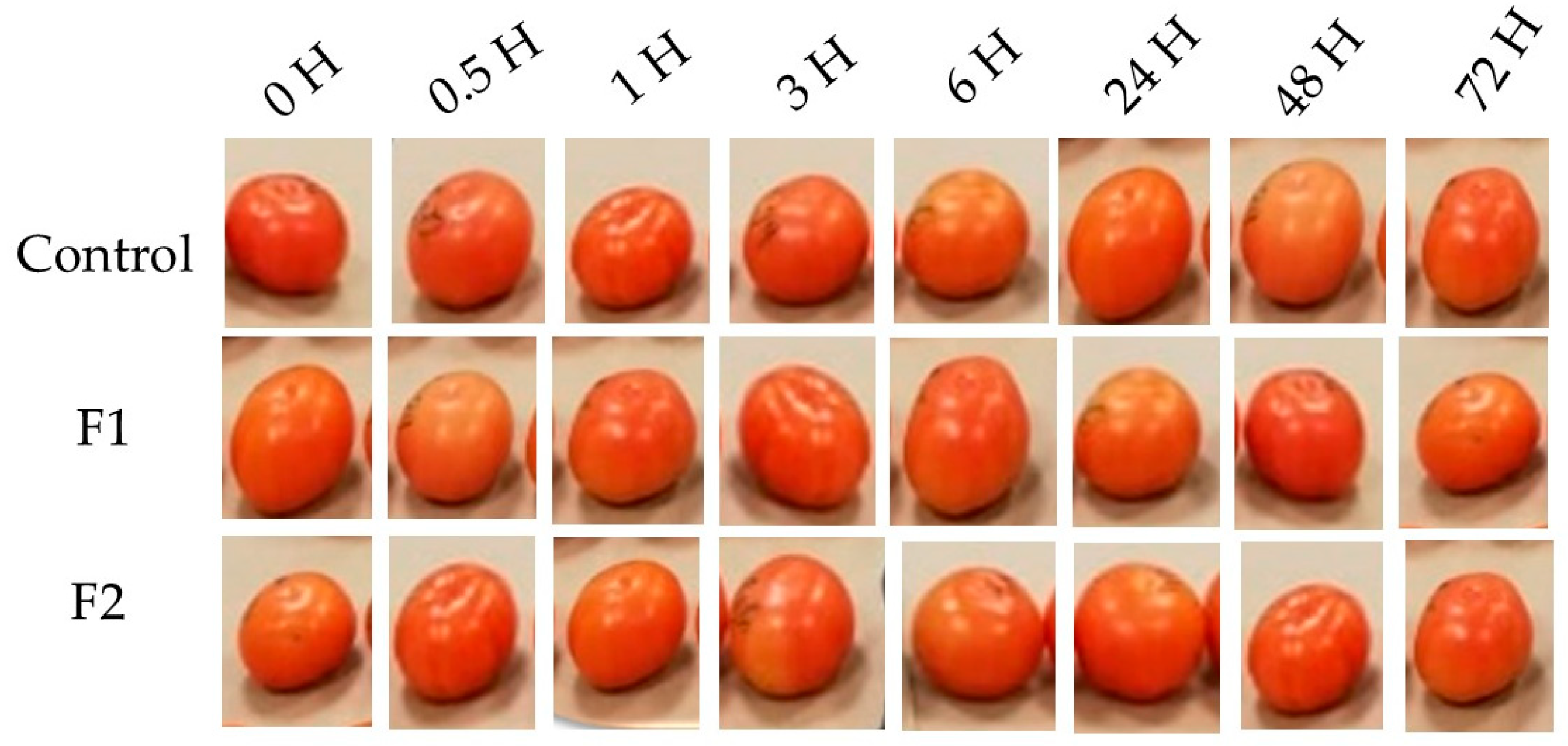
2.2.2. Postharvest Treatment of Tomato Fruits with Chitin Oligomers from Alternaria

Tomato fruits were split into 3 groups of 30 fruits each. Two groups of tomatoes were immersed for 30 s in chitin oligomers solutions at concentrations of 50 µg/mL (F1) and 100 µg/mL (F2). MilliQ water was applied to the third group and was considered as a control. Three fruits per treatment were sampled for the ethylene production assay. The treated and control fruits were kept at room temperature (24 °C) and a relative humidity of approximately 60% for 72 h. Furthermore, three fruits per treatment were sampled at different times (0, 0.5, 1, 3, 6, 24, 48, and 72 h) after the oligomer’s exposure. The fruit mesocarp was removed, and the pericarp was cut, flash-frozen with liquid nitrogen and maintained at −80 °C until the analysis of JA, SA, and gene expression were performed. The bioassay was repeated twice.

2.3. Quantification of Ethylene Production Using Gas Chromatography

The whole fruits treated with the chitin oligomers and the control were weighed using a balance. One fruit (approximately 220 g in weight) was placed in each glass container with capacity of 0.5 L, and it was kept at 20 °C. Three replicates were included per treatment. The flasks were covered with a lid adapted with silicone septum for gas sampling and incubated for 30 min. After this time, 1 mL of headspace gas was extracted using a hypodermic syringe and introduced into a Gas Chromatograph (Varian 3400 cx, Agilent Technologies, Santa Clara, CA, USA) fitted with a HayaSep N column (2 m × 3.17 mm internal diameter, Supelco (Supelco Analytical, Inc., Bellefonte, PA, USA) and a flame ionization detector. The chromatographic parameters were as follows: injector temperature of 100 °C, and detector temperature of 120 °C. The carrier gas, nitrogen, was delivered at a rate of 25 mL/min [67]. Four samples were injected for each replicate, and the ethylene production was calculated using the following equation:
C 2 H 4 µ L / K g h = ( S p a × S t c × h s ) S t p a × w × t
where Spa is the sample area, Stc is the standard concentration (10.2 µL/L), hs is the headspace volume, Stpa is the area of the standard, w is fruit weight in kilograms, and t is the incubation time in hours.

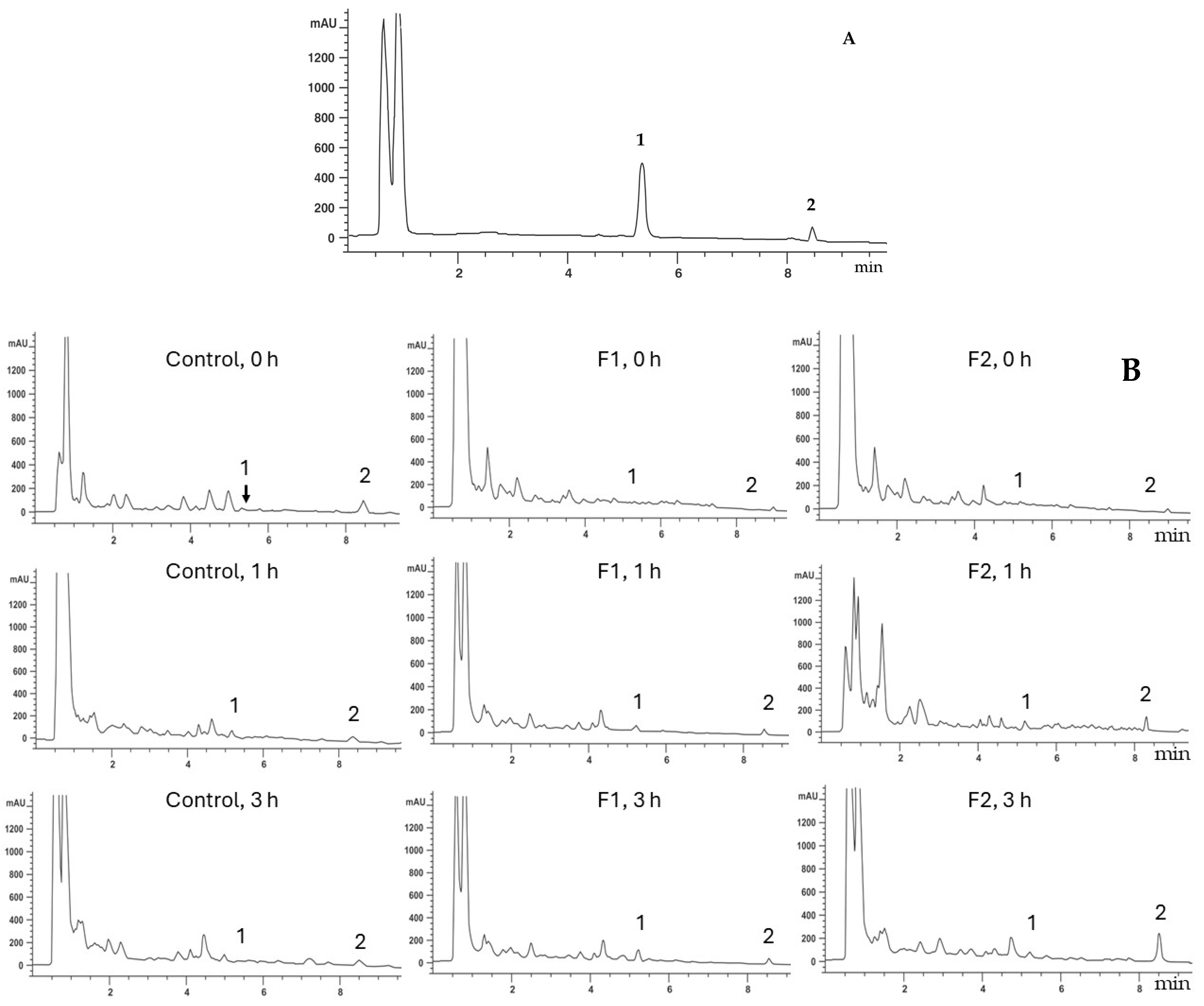
2.4. Jasmonic Acid and Salicylic Acid Quantification Using HPLC

A simultaneous determination of JA and SA was performed following to Durgbanshi et al. [68]. Briefly, the tomato pericarp sample that was previously cut and stored at −80 °C, was freeze-dried in Yamato Freeze Dryer DC801 (Yamato Scientific, Co., Ltd., Japan). A 0.5 g dry tissue sample was mixed with 5 mL of a methanol/water (80:20) mixture. Subsequently, the homogenate was spun at 5000 g for 10 min, and the supernatant was collected. The pH of the supernatant was brought to 2.8 using acetic acid (CH3COOH), followed by the addition of diethyl ether. The mixture was placed in a separating funnel, stirred manually, and left to rest for phase separation. The aqueous layer was removed, and the organic layer was evaporated using a dry bath at 37 °C. The resulting pellet was dissolved in 1 mL of a water/methanol mixture (90:10) and passed through a 0.22 µm cellulose acetate filter. An aliquot (20 µL) of the filtered solution was introduced into the high-performance liquid chromatograph (HPLC).
A Liquid Chromatograph (Agilent 1290 Infinity, Wall Creek, CA, USA) equipped with a C18 column (ZORBAX SB, 2.5 × 50 mm i.d., 3.5 µm) was used. JA and SA were eluted with a 0.1% formic acid/acetonitrile gradient, starting with a 90:10 (v/v) ratio and gradually shifting 60:40 (v/v) in 10 min. The gradient was then adjusted to 80:20 (v/v) and was maintained for the last minute of the analysis. Baseline conditions were reinstated and stabilized for 5 min, resulting in a total runtime of 20 min per sample. The flow rate was set to 0.3 mL/min, and the system was operated under pressures in the range of 70–100 bar [68]. The concentrations of JA and SA were calculated according to the standard curve of JA and SA (Sigma-Aldrich Corp., St. Louis, MO, USA).

2.5. RNA Extraction, First-Strand DNA Synthesis, and Quantification of Gene Expression

RNA was extracted following the LiCl precipitation method [69]. Samples of tomato fruits exposed to different concentrations of chitin oligomers that were previously cut, processed with liquid nitrogen, and stored at −80 °C were used for RNA extraction. To eliminate DNA contamination, the extracted RNA was treated with DNAse RQ1 (Promega- Co., Madison, WI, USA) following the manufacturer’s protocol. The concentration of RNA was estimated in an ultra-low volume spectrophotometer at 260 nm (Nanodrop 2000, Thermo Scientific, Waltham, MA, USA). The RNA integrity was evaluated by gel electrophoresis on a 1% agarose gel. Subsequently, first-strand DNA was synthesized from 500 ng of RNA, and then the retrotranscription reaction was carried out by SuperScript® III Reverse Transcriptase kit (Invitrogen; Thermo Fisher Scientific Inc., USA), following the manufacturer’s protocol with slight modifications reported by Henry et al., [19] and Feng et al., [70].
To quantify gene expression levels, quantitative real-time PCR was performed using StepOne Real-Time PCR System (Applied Biosystems Inc., USA). SYBR Green Master Mix (BIO-RAD), 20 ng of cDNA template, 250 nM of both forward and reverse primers, and nuclease free H2O were used for the experimental reaction. Glyceraldehyde-3-phosphate-dehydrogenase (GAPDH) was included as a reference gene. A dynamic range analysis was performed using a fivefold serial dilution. Primers specific for the target genes were designed using the Primer 3.0 program [71] (Table 1). Gene expression was assessed using three biological replicates and two technical replicates per reaction, under the following conditions: 5 min at 95 °C for denaturing and 35 cycles of 2 min at 95 °C, and 30 s at 60 °C. Relative gene expression levels were calculated with the 2−∆∆Ct methodology [72].

2.6. Statistical Analysis

The experiments were conducted using a completely randomized design with factorial arrangement, where factor A was the treatments (two concentrations of chitin oligomers and the control), and factor B was the sampling times (0, 0.5, 1, 3, 6, 24, 48, and 72 h). A two-way analysis of variance (ANOVA) was applied, and differences among means were evaluated using the Tukey–Kramer multiple range test at a 95% confidence level. NCSS 2021 version 21.0.8 (Kaysville, UT, USA) was used for statistical analysis. Pearson correlation analysis and matrix construction were performed in Excel 2108 (Microsoft Office LTSC 2021), and Python 3.13.2 program (phyton.org).

3. Results

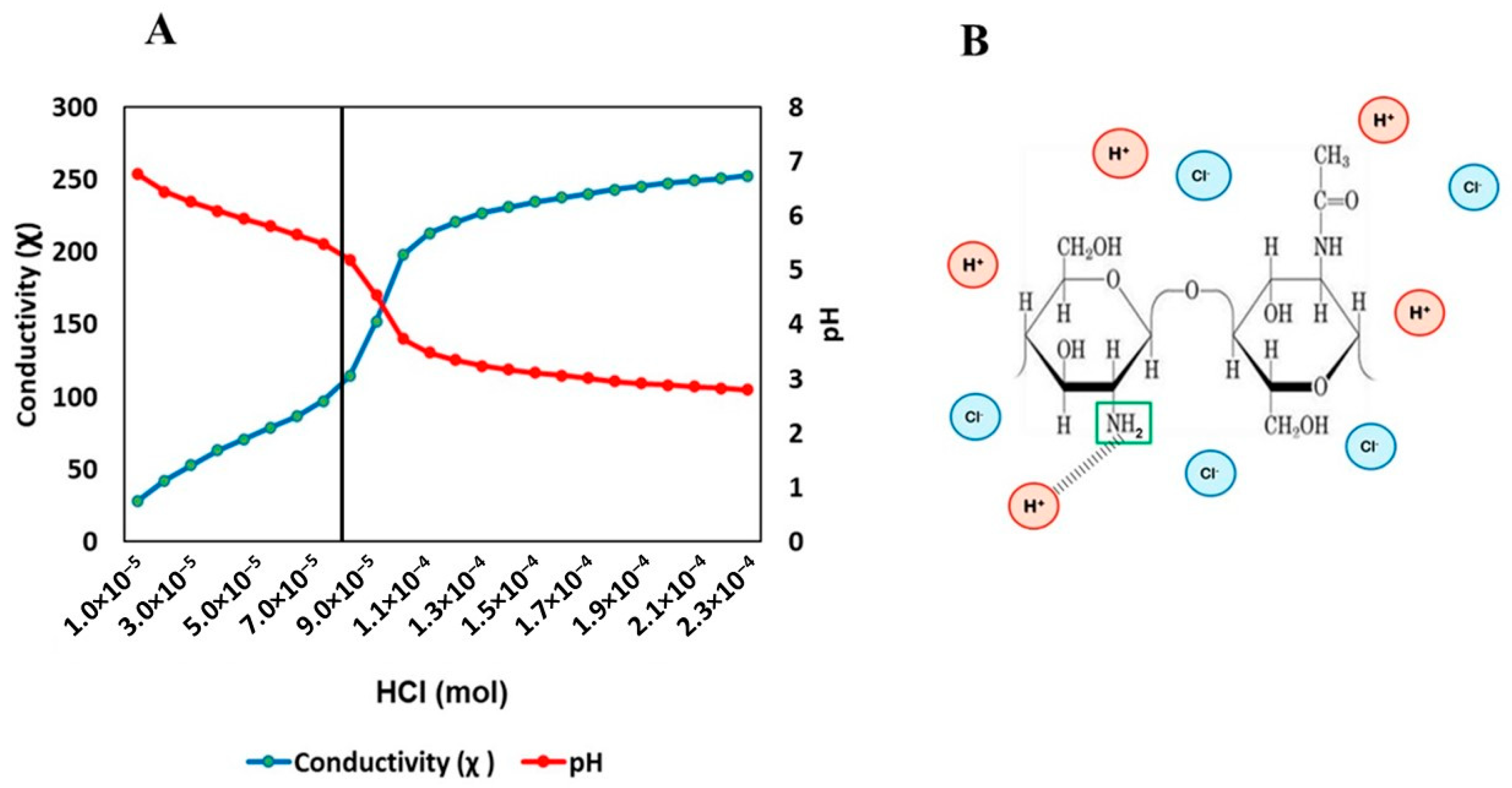
3.1. Chemical Characteristics of Chitin Oligomers from A. alternata

Chitin oligomers (≤1 kDa) were obtained from A. alternata using enzymatic treatment combined with ultrasonication and ultrafiltration processes. A clear, transparent, and colorless solution containing the chitin oligomers was obtained. Figure 1 shows the degree of acetylation of these oligomers. The plot shows the conductivity (χ) and pH values vs. the volume of HCl consumed. As HCl is added, NH2 groups are protonated by binding to H+ ions, while CL anions increase conductivity. Based on the volume of HCl consumed when the pH begins to decrease and the conductivity begins to increase, the degree of acetylation of 77% was obtained, suggesting that the molecule in the permeate was chitin. The permeate contained 100 µg/mL of chitin oligomers, equivalent to 3.8 mg of chitin oligomers/g of dry weight, with a residual protein content of 0.025 µg/mL. The polymerization degree of these oligomers was estimated as ≤5 monomers, considering the molecular weight of the chitin.

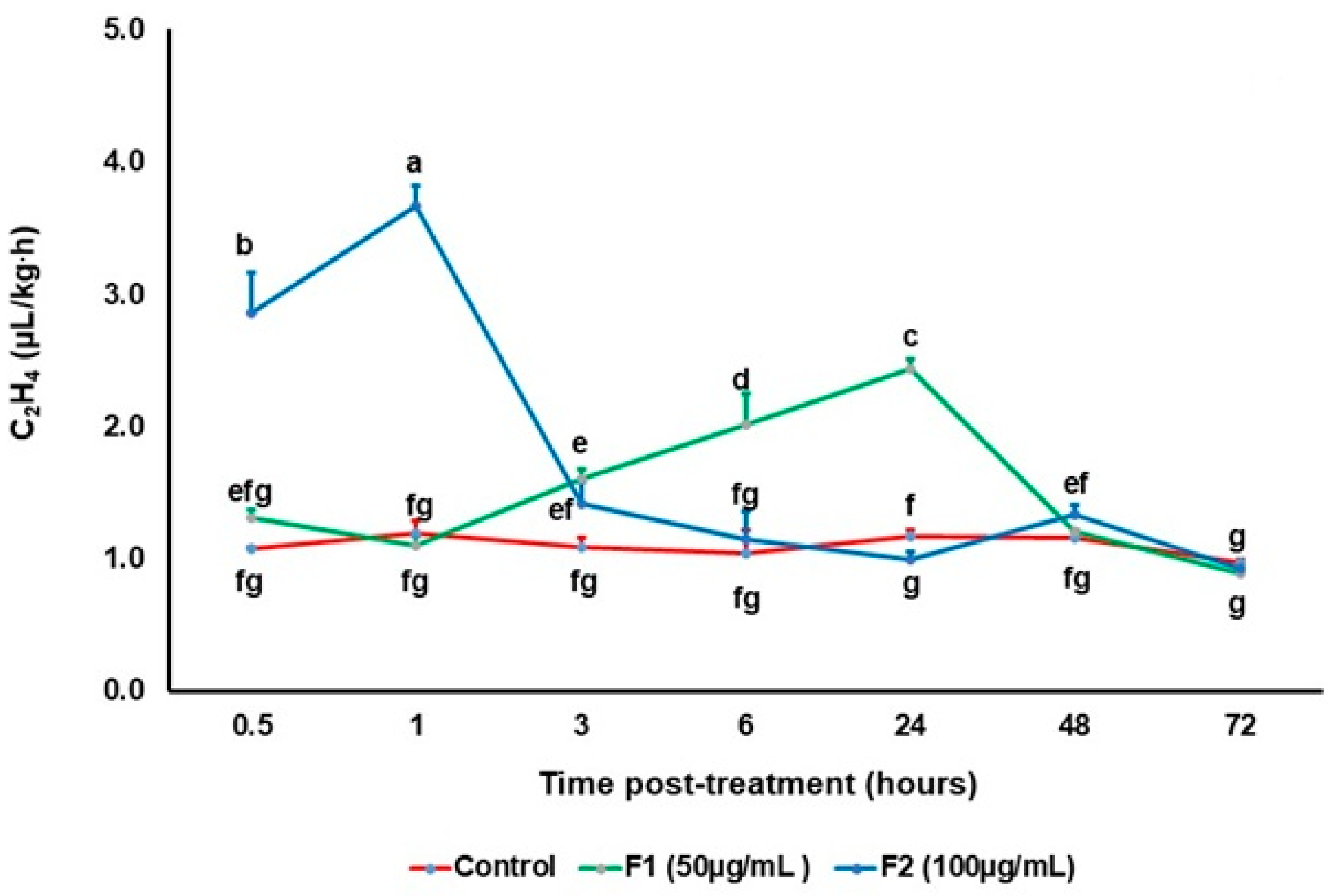
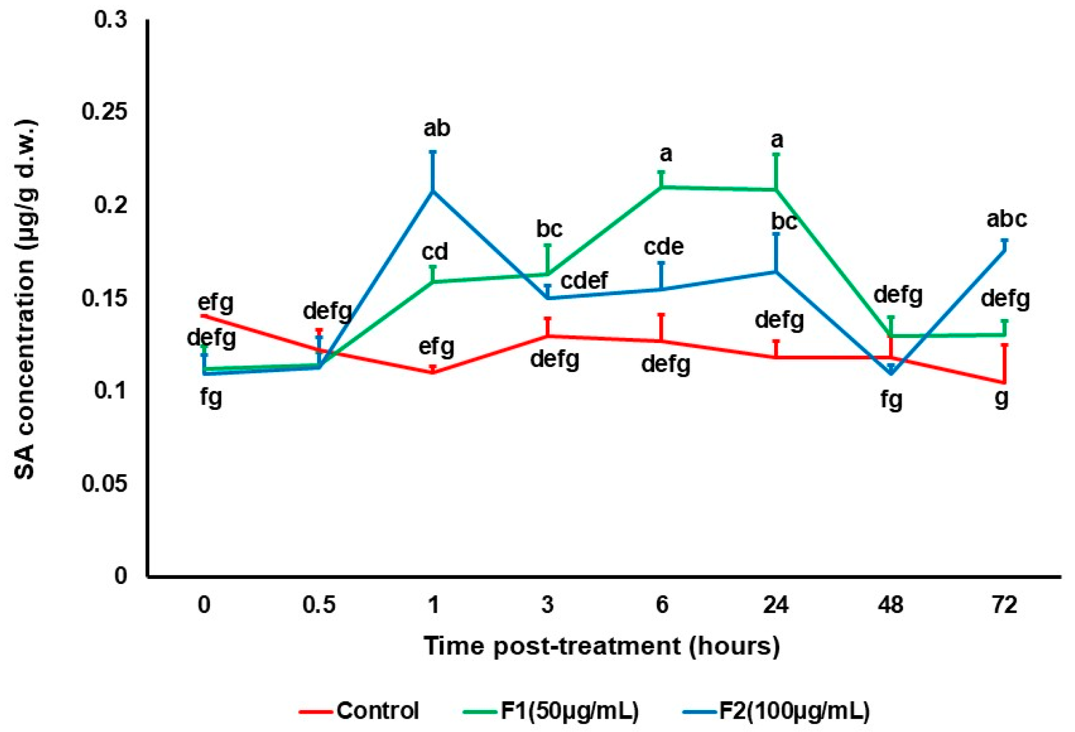
3.2. Effect of Chitin Oligomers on the Production of Signaling Molecules

Chitin oligomers of A. alternata induced changes in the amount of Et, JA, and SA in tomato fruits, without affecting their postharvest quality for 72 h after treatment (Figure 2). Figure 3 shows the results of Et production in tomato fruit exposed to chitin oligomers from A. alternata. Significant differences are observed among the treatments. From Figure 3, it is clear that an early response in Et production was observed in response to the highest concentration of chitin oligomers (F2, 100 µg/mL) at 0.5 h and 1 h post-treatment, exhibiting a 2.8-fold increase and 3.6-fold increase in Et production compared to the control, respectively. After 3 h post treatment, the production of this phytohormone decreased over time. In contrast, the F1 treatment (50 µg/mL) induced a significant increase in the Et production at 6 and 24 h post treatment with respect to the control and the F2 treatment, but this production was lower than that observed for the F2 treatment at 0.5 and 1 h post-treatment.
In the present study, JA was detected by HPLC with a retention time of 8.4 min. The best chromatographic resolution was obtained at a wavelength of 235 nm, observing a clearly defined peak (peak number 2) (Figure 4A). Based on these chromatographic conditions, JA was detected in all the treatments. Figure 4B presents the HPLC chromatograms of tomato fruits exposed to fungal chitin oligomers. In control fruits at time 0, a peak corresponding to JA was detected at 8.4 min; while in fruits exposed to chitin oligomers at the same time, no JA was detected. After 1 h of exposure to chitin oligomers, JA was detected in both the control and F2 treatments. Three hours after exposure, treatment F2 presented a peak at 8.4 min, which was higher than that of the control and F1, suggesting that F2 had a higher concentration of JA at this time point.
Figure 5 shows the results of JA production in tomato fruits exposed to chitin oligomers from A. alternata. Significant differences (p ≤ 0.05) were observed among the treatments and control. At the beginning of the experiment, higher JA production was observed in control fruits, while fruits exposed to F1 and F2 showed the lowest JA production. After 3 h of treatment, F2 induced a significant increase in the JA production compared to the control and F1 treatment. The same observation was made at 6 and 24 h post treatment. These results indicate that chitin oligomers stimulate JA production in tomato fruits but only after 3, 6, and 24 h of exposure to the oligomers.
SA was detected simultaneously with JA by HPLC with a retention time of 5.2–5.3 min. The best chromatographic resolution was obtained at the same wavelength of JA (235 nm), observing a clearly defined peak (Figure 4A). Based on these chromatographic conditions, SA was determined in all the treatments. Figure 4B shows the HPLC chromatograms of SA in tomato fruits exposed to chitin oligomers from A. alternata. The results of SA production in tomato fruits exposed to A. alternata chitin oligomers are shown in Figure 6. Significant differences (p < 0.05) were observed among the chitin treatments and control. In this Figure, it can be observed that the application of the chitin oligomers caused a significant increase in the SA production, after 1 h, 6 h, and 24 h post treatment. After 1 h, F2 treatment was higher (p < 0.05), and after 6 and 24h, F1 treatment induced a higher amount of SA (p < 0.05).

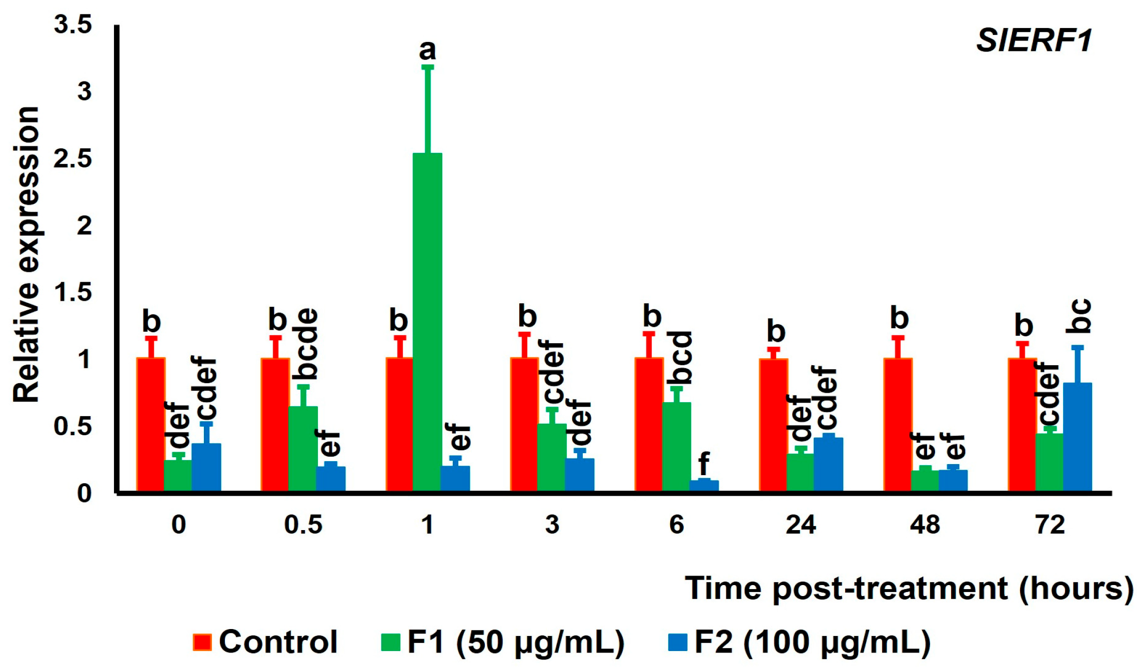
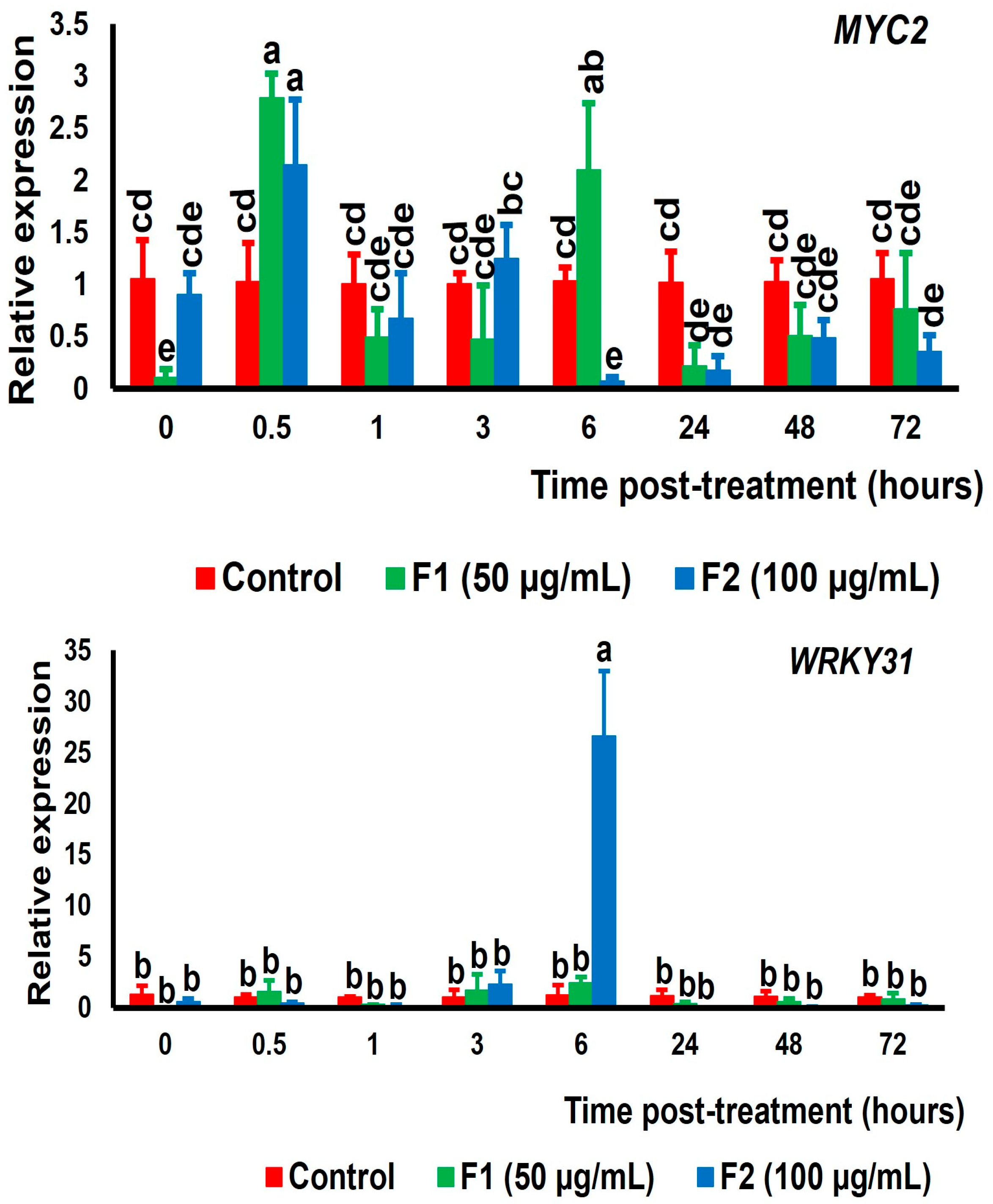
3.3. Expression of Genes Encoding Transcription Factors Related with Et, JA, and SA Signaling Pathways in Tomato Fruit Exposed to Chitin Oligomers

The results of the chitin oligomers from A. alternata on the changes in expression of the transcription factors genes SlERF1, SlMYC2, and SlWRKY31, which plays a role in the regulation of signaling hormones, such as ethylene, jasmonic acid and salicylic acid, respectively, are included in Figure 7.
The expression results of the SlERF1 gene related to ethylene signaling are shown in Figure 7. One hour post treatment, the maximum gene expression was observed for the F1 treatment, which was 1.5 times higher than that of the control. Lower gene expression levels of SlERF1 were observed in the F2 treatment over time, except at 72 h, where a slight increase was observed, but these levels were not statistically different (p > 0.05) compared to those of the control and F1 treatment. These results suggest that the expression of the SlERF1 gene is induced in the early stages following exposure to low concentrations of chitin oligomers.
Figure 7 shows the expression results of the gene encoding transcription factor SlMYC2 in tomato fruits treated with chitin oligomers from A. alternata. A higher gene expression (p < 0.05) was observed in the F1 treatment (1.8 times) after 0.5 h post treatment and a second increase in the gene expression level was observed after 6 h post treatment. Similarly, the F2 treatment showed a significant increase (1.1 times with respect to the control) in gene expression after 0.5 h post treatment, and then, the gene expression decreased over time. These results could indicate an increase in jasmonic acid production mainly after 3 and 6 h of exposure to chitin oligomers.
Figure 7 shows the results of relative expression of the transcription factor WRKY31 gene involved in the salicylic acid signaling, in tomato fruits treated with fungal chitin oligomers. Chitin oligomers induced significant changes in the levels of relative expression of the WRKY31 gene after 6 h post treatment only. Furthermore, this increase was 24 times greater than the control. The relative gene expression was rather low in the other treatments and time points measured and was not significantly different.

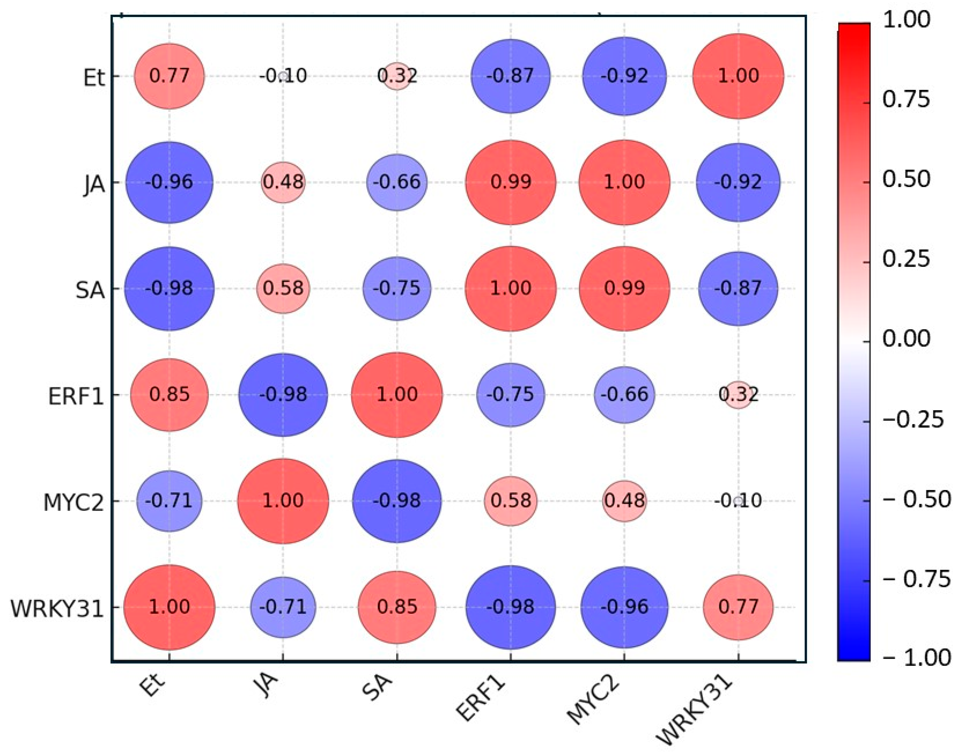
3.4. Correlation Analysis of Signaling Hormones
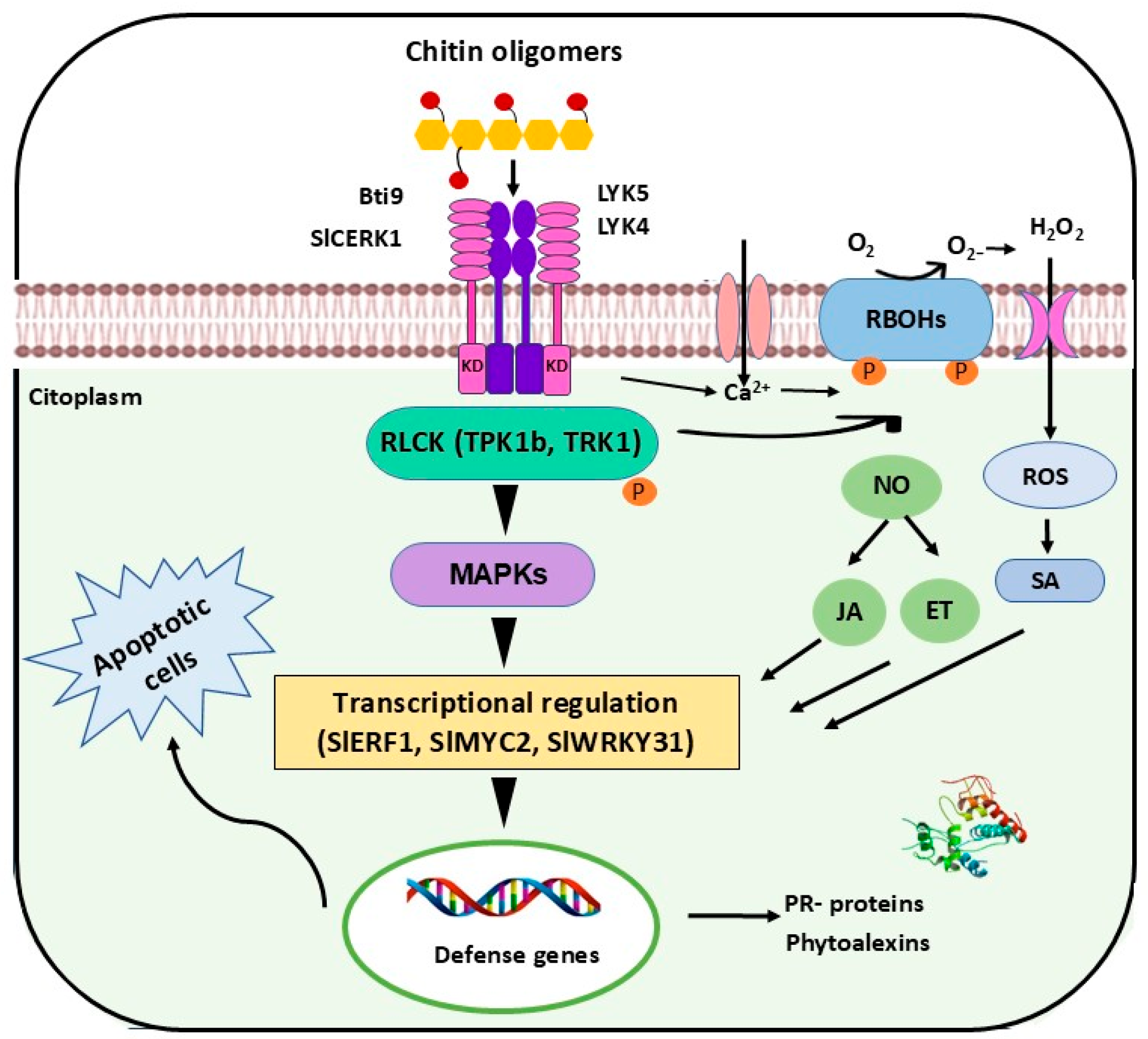
In Figure 8, the results of the correlation analysis carried out with the data of the changes in ethylene, salicylic acid, and jasmonic acid phytohormones content and the changes in gene expression of SlERF1, SlMYC2, and SlWRKY31 are presented. Correlation analysis showed a significant correlation among all the signaling hormones either positive or negative (Table S1). It was found that the transcription factor SlERF1 gene showed the highest correlation with the transcription factor SlMYC2 gene with a correlation coefficient of 0.99, suggesting the activation of both signal transductions related with ethylene and jasmonic acid. In addition, it was observed that Et was positively correlated with SA and the SlWRKY31 gene, with a correlation coefficient of 0.85 and 0.77, respectively. In the case of JA, this hormone showed a positive correlation with SlERF1 and SlMYC2, with a correlation coefficient of 0.58 and 0.48, respectively. In contrast, a negative and significant correlation was observed between Et and SlMYC2, JA and SA, and SlWRKY31 and SlMYC2, with a correlation coefficient of −0.96, −0.98, and −0.92, respectively. It is interesting that the correlation data agree between JA and SA and the gene expression of SlWRKY31 and SlMYC2, which are part of the signal transduction of these phytohormones.

4. Discussion

In this study, chitin oligomers of molecular weight ≤ 1 kDa were extracted from A. alternata, a necrotrophic fungus that provoke postharvest decay in tomato fruits. The chitin oligomers showed a GlcNAc content in the soluble fraction 100 µg/mL (38 mg/g) of chitin oligomers, 0.025 µg/mL protein content, an acetylation degree of 77%, and a polymerization degree estimated in ≤5. In the present study, enzymatic process was used as an alternative treatment to extract chitin from A. alternata, without affecting the release of acetyl groups of the polysaccharide, as occurs with traditional chemical treatment [11,73]. Some authors reported that chitin molecules contain more than 50–60% acetylated units, whereas chitosan contains less than 50% of acetylated units. The percentage of acetylation degree obtained in this study (77%) coincide with the results reported for chitin extracted from other fungi, such as Aspergillus niger with 76.53% acetylation degree [74]. In addition, the use of protease treatment resulted in a low protein concentration, which coincides with the results reported by other authors [75]. In previous studies performed in our laboratory, the presence of chitin in the fungal extract was confirmed by FT-IR analysis [18,62], in which the absorbance bands recorded in the region between 1420 and 1320 cm−1 suggested the presence of chitin. These results obtained in the present study agree with those reported by other authors [18,62,74,76]. The characteristics of the chitin oligomers obtained from A. alternata, such as a high acetylation degree, and the estimated polymerization degree of ≤5 monomers, strongly suggest that these molecules can be recognized by plant membrane receptors such as RLK and RLP, activating the plant’s defense mechanism, as reported by some authors [19,77,78]. Previous work carried out in our laboratory, showed that tomato fruits treated with A. alternata chitin oligomers with an acetylation degree of 94% and a polymerization degree of ≤5 monomers, exhibited a higher increase in the level of enzymatic activity of chitinase and glucanase, and the rot caused by the fungus A. alternata was inhibited by 78% [18]. Recently, it was reported that tomato genes encoding SlLYK4 and SlCERK1 plasma membrane receptors were induced in tomato fruit by chitin oligomers from A. alternata. According to these results and those reported by other authors [79], chitin can induce a potent defense response in plants.
The plant defense responses are complex networks of molecular events, where the phytohormones ethylene (Et), jasmonic acid (JA), and salicylic acid (SA) are considered the central regulators of plant immunity. In this study, the fungal chitin oligomers were able to induce changes in the expression levels of genes involved in the signaling hormones (Et, JA, and SA) in tomato fruits, after 30 min of the treatment. These findings are consistent with previous reports indicating that the initial molecular interactions triggering biochemical responses occur around 5 min after the plant encounters the invading agent. [80]. Although the early stages of interaction are not yet fully characterized, it is well established that plants initiate defense mechanisms shortly after encountering an elicitor. In agreement with this, Valle-Sotelo et al., [18] found that tomato fruits exposed with fungal chitin oligomers showed a higher increase in the enzymatic activity of chitinase and glucanase 30 min after treatment. In another study, the transcriptional analysis performed in tomato fruits treated with fungal chitin oligomers by 30 min revealed an increase in the expression of genes encoding to chitin receptor-like kinases (RLK; SlLYK4 and SlCERK1) and in the level of gene expression encoding to PR proteins, among other genes [19]. Thus, the findings of this study are especially significant, as they clearly demonstrate that chitin-triggered defense response in the fruits initiated within a few minutes after contact with the fungal elicitor.
Some genes that encode transcription factors related with Et (SlERF1), JA (MYC2) and SA (WRKY31) signaling pathways in tomato fruit, as well as phytohormones production, were evaluated. We observed that both SlERF1 expression and ethylene production increased significantly in the early stages of exposure to chitin oligomers. Previous studies have linked the gene expression of ERFs in fruits with their responses to pathogens attacks [35,81]. It was observed in grapes (Vitis vinifera) that the overexpression of the VvERF1 gene was related to resistance to B. cinerea infection [82], and in tomato (Solanum lycopersicum), the expression of SlERF1 was upregulated after B. cinerea infection [83]. Furthermore, in bananas (Musa acuminata), the overexpression of MaERF1 was associated with resistance to Colletotrichum musae [84]. In agreement, the induction of the gene expression related to ethylene production in tomato plants exposed to chitosan (deacetylated chitin) has been reported [85], which indicates the activity of this elicitor as an inducer of plant defense responses. The data generated in the present study clearly suggest that the increase in the expression of the ERFs gene observed in the studies mentioned above can be ascribed to the chitin molecule present in the wall of the necrotrophic fungus Alternaria alternata.
The activity of phytohormones is generally the result of synergistic or antagonistic action, rather than independent action. In this study, in addition to the changes observed in Et, significant changes in JA were also observed in response to chitin oligomers. In the present study, the expression of the transcription factor SlMYC2 gene was recorded (related to the regulation of the JA signaling pathway and its production) in response to the treatment with chitin oligomers in the early stages (0.5 h) post treatment, which coincides with the early response of SlERF1 gene observed in this study. In addition, a high correlation was observed between the expressions of SlERF1 and SlMYC2 (r = 0.99) independent of the applied treatment, suggesting a strong relationship between Et and JA signaling observed in response to chitin oligomers from necrotrophic fungi. These results agree with those reported by other authors who mentioned the synergistic effect of Et and JA in plants infected with necrotrophic fungi [42,86]. Within the jasmonic acid (JA) signaling pathway, MYC2 serves as the primary downstream effector, whereas JAZ proteins negatively regulate the pathway by interacting with MYC2 and suppressing its activity as a transcription factor [59]. It was observed in Arabidopsis that MYC2 is an essential transcription factor in the regulation of genes related with the biosynthesis of JA [87]. In the work just mentioned, the addition of methyl jasmonate (MeJA) activates MYC2, a negative regulator of ERF1 that is also an activator of PDF 1.2, modulating the expression of both genes, which is essential in the regulation of jasmonic acid signaling. Other studies have linked the presence of MYC2, MYC3, and MYC4 in the regulation of jasmonic acid-inducible genes with their interaction with bHLH-type transcription factors such as JAM 1 (JASMONATE-ASSOCIATED MYC2-LIKE1) [88], as well as their association with YUCCA8 and YUCCA9 transcription factors for binding to the G-box element present in the gene promoter in the presence of biotic stress [89]. Although the transcription factor MYC2 has been widely studied in vegetative tissues, there is little information about the function of MYC2 in fruits. For instance, it was found that JA and MYC2 increase the expression of ACO (1-aminocyclopropane-1-carboxylic acid oxidase) and ACS (1-aminocyclopropane-1-carboxylic acid synthase) genes and ethylene synthesis to start apple fruit ripening [90]. In another study, MYC2 expression was upregulated in loquat fruits applied with recombinant serine protease, leading to the enhanced expression of genes encoding PAL (phenylalanine ammonia lyase), PPO (polyphenol oxidase), and other fungi resistance-related genes, helping to effectively delay of fruit rot [91].
In contrast, a significant increase in JA concentration in tomato fruits exposed to high concentration of chitin oligomers (F2), for 3 to 24 h post treatment was observed, and this response was registered a few moments after the peak of Et production (Figure 3). Altogether, these data suggest that after the exposure of tomato fruit to chitin oligomers, slightly elevated JA levels allow the fruit’s defense response to remain active; however, further studies are required to verify this hypothesis. A positive correlation was also observed between SlMYC2 expression and jasmonic acid production (0.48), suggesting the involvement of SlMYC2 in the JA biosynthesis. Previous studies observed that the treatment with chitosan of the grape varieties Kyoho and Shine Muscat resulted in a significant increase in JA production [44]. In agreement with these results, previous studies performed on rice cells showed that chitin treatment induced a transient increase in JA production [92]. It had been proposed that the activation of defensive genes by oligosaccharides could occur by the octadecanoid pathway [93]. In this pathway, linolenic acid is released from membrane phospholipids by the enzyme phospholipase and is converted to 12-oxophytodienoic acid and JA, resulting in the transcriptional activation of defensive genes [94].
An interesting result was observed in SA. Numerous studies have demonstrated that endogenous signaling molecules such as SA play a crucial role in signaling processes for activating defense responses, mainly against biotrophic fungi [32]. In response to biotic stress, SA showed a synergistic relationship with other phytohormones. In agreement with this statement, in this study it was observed that chitin oligomers induced high levels of SA in tomato fruits, mainly after inducing high levels of Et and JA. The transcription factor SlWRKY31 gene, related to the regulation of SA, shows high expression levels in tomato fruits exposed to F2, after 6 h post treatment with a 24-fold increase with respect to the control. This observation had not been reported before, because SA has not been associated with the presence of necrotrophic fungi such as A. alternata. Although in this study the tomato fruits were not inoculated with this fungus but only the chitin oligomers present in the fungi cell wall were applied to tomato fruits. Furthermore, it was found that SA production (Figure 6) increased from 1 h to 72 h which correlated with the increase in the expression level of the SlWRKY31 gene (0.32) in response to chitin oligomers. Contrarily, a higher correlation between SA and Et (0.85) and between Et and WRKY31 (0.76) was observed, suggesting a strong relationship between ethylene and SA signaling observed in response to chitin oligomers from necrotrophic fungi. It had been reported that systemic defense responses are based mainly on phytohormones such as Et and SA, promoting the expression of pathogenesis-related proteins such as chitinases, and preventing infection upon biotrophic pathogen attacks [85]. In the study performed by Valle-Sotelo et al., [18], chitin oligomers from A. alternata with similar characteristics of the ones used in this study were applied to tomato fruits, and the authors reported a significant increase in the activity of the pathogenesis-related proteins such as chitinase and glucanase 1 h post treatment. Furthermore, Henry et al., [19] performed a transcriptomic analysis on tomato fruits exposed to chitin oligomers from A. alternata with an estimated polymerization degree of ≤5 monomers, and an acetylation degree of 76%. These authors reported an increase in the expression of genes encoding pathogenesis-related proteins such as chitinase, glucanase, peroxidase, PR5, PR10, defensins, and PAL, as well as the transcription factor SlERF1 gene, 30 min post treatment. The increase in enzymatic activity and the expression levels of genes encoding PR proteins and SlERF1 transcription factor coincides with the higher production of Et and the higher expression of SlERF1 observed in this study. Altogether, these data suggest that Et could be involved in the early response to the presence of the chitin oligomers. However, further studies are needed to confirm this hypothesis. Based on our results and taking into account the results reported by Valle-Sotelo et al., [18] and Henry et al., [19], we can suggest that chitin oligomers from A. alternata induce hormone signaling (Figure 9), where Et and JA stimulate the SA production, and these phytohormones promote the systemic defense responses, providing protection to fruits against the attacks of necrotrophic fungi such as A. alternata.

5. Conclusions

The rapid response of tomato fruits to A. alternata chitin oligomers suggests that this molecule elicits the signaling phytohormones such as Et, JA, and SA. The exposure of chitin oligomers from A. alternata to tomato fruits stimulated ethylene synthesis suggesting its participation in the activation of the plant’s natural defense mechanism. Furthermore, the concentration of JA was found to be associated with the last exposure time, reaching its maximum peak at 3 h. Meanwhile, SA was an antagonist of ethylene and JA production. At the same time, we were able to establish that chitin oligomers induced an increase in the expression level of genes encoding transcription factors (SlERF1, SlMYC2, and SlWRKY31), which, in turn, regulated genes related to the production of the signaling molecules ethylene, jasmonic acid, and salicylic acid. These data suggest that under conditions of biotic stress, ethylene is a signaling molecule that, in some ways, responds to the presence of bioelicitors such as chitin oligomers and is related to the synthesis of JA. Furthermore, both molecules (Et and JA) are essential in plant defense responses against necrotrophic fungi, while SA acts as an antagonist to immune response regulation in fruits.
However, further studies in horticultural crops are needed to fully understand the phenomenon of chitin oligomer recognition and the molecular mechanisms that respond to pathogen attack. A deeper insight into how chitin and its oligosaccharides are perceived and how they activate signaling cascades and defense responses in horticultural plants could offer meaningful benefits for agriculture. This knowledge could be of great help in the development of horticultural varieties with durable and strong resistance to fungal pathogens. Furthermore, the knowledge generated may support the development of more effective biocontrol strategies aimed at managing fungal diseases in horticultural crops.

Supplementary Materials

The following supporting information can be downloaded at: https://www.mdpi.com/article/10.3390/horticulturae11060565/s1, Table S1. Correlation matrix.

Author Contributions

O.R.-Z. and R.T.-R., conceived and designed the idea and wrote the manuscript; M.E.B.-F., M.E.T.-H. and A.R.-C. offered technical assistance, scientific correction, and language revision for the final manuscript version. All authors have read and agreed to the published version of the manuscript.

Funding

This research received financial support from the Sectoral Research Fund for the Education, of the National Council for Humanities, Science and Technology (SECIHTI) from Mexico (CB 2016-01, Grant No. 287254).

Data Availability Statement

The data used to support the findings of this study are available from the corresponding author upon request.

Acknowledgments

Financial support from the SECIHTI (Mexico) is fully appreciated. The author O.R.Z. gives thanks to SECIHTI for the PhD scholarship assigned. Thanks is also given to the Research Center of Food and Development (CIAD, AC) for all the equipment facilities.

Conflicts of Interest

The authors declare no conflicts of interest.

References

  1. FAOSTAT. Food and Agriculture Organization of the United Nations. Available online: https://www.fao.org/faostat/ (accessed on 11 December 2024).
  2. Prusky, D.; Alkan, N.; Mengiste, T.; Fluhr, R. Quiescent and necrotrophic lifestyle choice during postharvest disease development. Annu. Rev. Phytopathol. 2013, 51, 155–176. [Google Scholar] [CrossRef] [PubMed]
  3. Quinet, M.; Angosto, T.; Yuste-Lisbona, F.J.; Blanchard-Gros, R.; Bigot, S.; Martinez, J.P.; Lutts, S. Tomato Fruit Development and Metabolism. Front. Plant Sci. 2019, 10, 1554. [Google Scholar] [CrossRef] [PubMed]
  4. Troncoso-Rojas, R.; Tiznado-Hernández, M.E. Chapter 5—Alternaria alternata (Black Rot, Black Spot). In Postharvest Decay; Bautista-Baños, S., Ed.; Academic Press: San Diego, CA, USA, 2014; pp. 147–187. [Google Scholar]
  5. Takao, K.; Akagi, Y.; Tsuge, T.; Kodama, M. Functional characterization of putative G protein-coupled receptors in the tomato pathotype of Alternaria alternata. J. Gen. Plant Pathol. 2016, 82, 82–88. [Google Scholar] [CrossRef]
  6. Pane, C.; Fratianni, F.; Parisi, M.; Nazzaro, F.; Zaccardelli, M. Control of Alternaria post-harvest infections on cherry tomato fruits by wild pepper phenolic-rich extracts. Crop Prot. 2016, 84, 81–87. [Google Scholar] [CrossRef]
  7. Kader, K. Small-Scale Postharvest Handling Practices: A Manual for Horticultural Crops; University of California, Davis, Postharvest Technology Research and Information Center: Berkeley, CA, USA, 2015. [Google Scholar]
  8. Islam, T.; Danishuddin; Tamanna, N.T.; Matin, M.N.; Barai, H.R.; Haque, M.A. Resistance Mechanisms of Plant Pathogenic Fungi to Fungicide, Environmental Impacts of Fungicides, and Sustainable Solutions. Plants 2024, 13, 2737. [Google Scholar] [CrossRef] [PubMed]
  9. Pathak, V.M.; Verma, V.K.; Rawat, B.S.; Kaur, B.; Babu, N.; Sharma, A.; Dewali, S.; Yadav, M.; Kumari, R.; Singh, S.; et al. Current status of pesticide effects on environment, human health and it’s eco-friendly management as bioremediation: A comprehensive review. Front. Microbiol. 2022, 13, 962619. [Google Scholar] [CrossRef]
  10. Singh, R.; Upadhyay, S.K.; Singh, M.; Sharma, I.; Sharma, P.; Kamboj, P.; Saini, A.; Voraha, R.; Sharma, A.K.; Upadhyay, T.K.; et al. Chitin, Chitinases and Chitin Derivatives in Biopharmaceutical, Agricultural and Environmental Perspective. Biointerface Res. Appl. Chem. 2021, 11, 9985–10005. [Google Scholar] [CrossRef]
  11. Malerba, M.; Cerana, R. Recent Applications of Chitin- and Chitosan-Based Polymers in Plants. Polymers 2019, 11, 839. [Google Scholar] [CrossRef]
  12. Parada, R.Y.; Egusa, M.; Aklog, Y.F.; Miura, C.; Ifuku, S.; Kaminaka, H. Optimization of nanofibrillation degree of chitin for induction of plant disease resistance: Elicitor activity and systemic resistance induced by chitin nanofiber in cabbage and strawberry. Int. J. Biol. Macromol. 2018, 118, 2185–2192. [Google Scholar] [CrossRef]
  13. Zhu, M.; Lu, S.; Zhuang, M.; Zhang, Y.; Lv, H.; Ji, J.; Hou, X.; Fang, Z.; Wang, Y.; Yang, L. Genome-wide identification and expression analysis of the Brassica oleracea L. chitin-binding genes and response to pathogens infections. Planta 2021, 253, 80. [Google Scholar] [CrossRef]
  14. Um, E.A.; Nisar, N.; Tsuzuki, T.; Lowe, A.; Rossiter, J.T.; Javaid, A.; Powell, G.; Waseem, R.; Al-Mijalli, S.H.; Iqbal, M. Chitin nanofibers trigger membrane bound defense signaling and induce elicitor activity in plants. Int. J. Biol. Macromol. 2021, 178, 253–262. [Google Scholar] [CrossRef]
  15. Hao, G.; Tiley, H.; McCormick, S. Chitin Triggers Tissue-Specific Immunity in Wheat Associated with Fusarium Head Blight. Front. Plant Sci. 2022, 13, 832502. [Google Scholar] [CrossRef]
  16. Zhou, Y.; Jiang, S.; Jiao, Y.; Wang, H. Synergistic effects of nanochitin on inhibition of tobacco root rot disease. Int. J. Biol. Macromol. 2017, 99, 205–212. [Google Scholar] [CrossRef]
  17. Sun, C.; Fu, D.; Jin, L.; Chen, M.; Zheng, X.; Yu, T. Chitin isolated from yeast cell wall induces the resistance of tomato fruit to Botrytis cinerea. Carbohydr. Polym. 2018, 199, 341–352. [Google Scholar] [CrossRef] [PubMed]
  18. Valle-Sotelo, E.; Troncoso-Rojas, R.; Tiznado-Hernández, M.; Carvajal-Millan, E.; Estrada, A.; García, Y. Bioefficacy of fungal chitin oligomers in the control of postharvest decay in tomato fruit. Int. Food Res. J. 2022, 29, 1131–1142. [Google Scholar] [CrossRef]
  19. Henry García, Y.; Troncoso-Rojas, R.; Báez-Flores, M.E.; Hernández-Oñate, M.Á.; Tiznado-Hernández, M.E. RNA-Seq of Tomato Fruit-Alternaria Chitin Oligomer Interaction Reveals Genes Encoding Chitin Membrane Receptors and the Activation of the Defense Response. Horticulturae 2023, 9, 1064. [Google Scholar] [CrossRef]
  20. Iizasa, E.; Mitsutomi, M.; Nagano, Y. Direct binding of a plant LysM receptor-like kinase, LysM RLK1/CERK1, to chitin in vitro. J. Biol. Chem. 2010, 285, 2996–3004. [Google Scholar] [CrossRef] [PubMed]
  21. Brulé, D.; Villano, C.; Davies, L.J.; Trdá, L.; Claverie, J.; Héloir, M.C.; Chiltz, A.; Adrian, M.; Darblade, B.; Tornero, P.; et al. The grapevine (Vitis vinifera) LysM receptor kinases VvLYK1-1 and VvLYK1-2 mediate chitooligosaccharide-triggered immunity. Plant Biotechnol. J. 2019, 17, 812–825. [Google Scholar] [CrossRef]
  22. Jaiswal, N.; Liao, C.J.; Mengesha, B.; Han, H.; Lee, S.; Sharon, A.; Zhou, Y.; Mengiste, T. Regulation of plant immunity and growth by tomato receptor-like cytoplasmic kinase TRK1. New Phytol. 2022, 233, 458–478. [Google Scholar] [CrossRef]
  23. Abdul Malik, N.A.; Kumar, I.S.; Nadarajah, K. Elicitor and Receptor Molecules: Orchestrators of Plant Defense and Immunity. Int. J. Mol. Sci. 2020, 21, 963. [Google Scholar] [CrossRef]
  24. Ai, Y.; Li, Q.; Li, C.; Wang, R.; Sun, X.; Chen, S.; Cai, X.Z.; Qi, X.; Liang, Y. Tomato LysM receptor kinase 4 mediates chitin-elicited fungal resistance in both leaves and fruit. Hortic. Res. 2023, 10, uhad082. [Google Scholar] [CrossRef] [PubMed]
  25. Li, N.; Han, X.; Feng, D.; Yuan, D.; Huang, L.-J. Signaling Crosstalk between Salicylic Acid and Ethylene/Jasmonate in Plant Defense: Do We Understand What They Are Whispering? Int. J. Mol. Sci. 2019, 20, 671. [Google Scholar] [CrossRef] [PubMed]
  26. Takeuchi, J.; Fukui, K.; Seto, Y.; Takaoka, Y.; Okamoto, M. Ligand-receptor interactions in plant hormone signaling. Plant J. For. Cell Mol. Biol. 2021, 105, 290–306. [Google Scholar] [CrossRef]
  27. Xu, X.; Chen, Y.; Li, B.; Zhang, Z.; Qin, G.; Chen, T.; Tian, S. Molecular mechanisms underlying multi-level defense responses of horticultural crops to fungal pathogens. Hortic. Res. 2022, 9, uhac066. [Google Scholar] [CrossRef] [PubMed]
  28. Reyes Zamora, O.; Troncoso-Rojas, R.; Báez-Flores, M.E.; Tiznado-Hernández, M.E.; Rascón-Chu, A. Signaling of Plant Defense Mediated by Receptor-like Kinases, Receptor-like Cytoplasmic Protein Kinases and MAPKs Triggered by Fungal Chitin in Horticultural Crops. Horticulturae 2024, 10, 361. [Google Scholar] [CrossRef]
  29. Sun, H.; Song, N.; Ma, L.; Li, J.; Ma, L.; Wu, J.; Wu, J. Ethylene signalling is essential for the resistance of Nicotiana attenuata against Alternaria alternata and phytoalexin scopoletin biosynthesis. Plant Pathol. 2017, 66, 277–284. [Google Scholar] [CrossRef]
  30. Chen, C.-l.; Yuan, F.; Li, X.-y.; Ma, R.-c.; Xie, H. Jasmonic acid and ethylene signaling pathways participate in the defense response of Chinese cabbage to Pectobacterium carotovorum infection. J. Integr. Agric. 2021, 20, 1314–1326. [Google Scholar] [CrossRef]
  31. Yu, X.; Feng, B.; He, P.; Shan, L. From Chaos to Harmony: Responses and Signaling upon Microbial Pattern Recognition. Annu. Rev. Phytopathol. 2017, 55, 109–137. [Google Scholar] [CrossRef]
  32. Aerts, N.; Pereira Mendes, M.; Van Wees, S.C.M. Multiple levels of crosstalk in hormone networks regulating plant defense. Plant J. 2021, 105, 489–504. [Google Scholar] [CrossRef]
  33. Park, Y.S.; Borrego, E.J.; Gao, X.; Christensen, S.A.; Schmelz, E.; Lanubile, A.; Drab, D.A.; Cody, W.; Yan, H.; Shim, W.B.; et al. Fusarium verticillioides Induces Maize-Derived Ethylene to Promote Virulence by Engaging Fungal G-Protein Signaling. Mol. Plant-Microbe Interact 2021, 34, 1157–1166. [Google Scholar] [CrossRef]
  34. Regmi, R.; Newman, T.E.; Kamphuis, L.G.; Derbyshire, M.C. Identification of B. napus small RNAs responsive to infection by a necrotrophic pathogen. BMC Plant Biol. 2021, 21, 366. [Google Scholar] [CrossRef]
  35. Li, S.; Wu, P.; Yu, X.; Cao, J.; Chen, X.; Gao, L.; Chen, K.; Grierson, D. Contrasting Roles of Ethylene Response Factors in Pathogen Response and Ripening in Fleshy Fruit. Cells 2022, 11, 2484. [Google Scholar] [CrossRef]
  36. Gu, S.; Xie, L.; Guan, Q.; Sheng, X.; Fang, Y.; Wang, X. Effect of ethylene production by four pathogenic fungi on the postharvest diseases of green pepper (Capsicum annuum L.). Int. J. Food Microbiol. 2024, 418, 110729. [Google Scholar] [CrossRef]
  37. Wang, Q. Study on the expression regulation of the CTR1 gene in the ethylene signaling pathway. Biochem. Biophys. Res. Commun. 2024, 739, 150590. [Google Scholar] [CrossRef]
  38. Wang, X.; Wen, H.; Suprun, A.; Zhu, H. Ethylene Signaling in Regulating Plant Growth, Development, and Stress Responses. Plants 2025, 14, 309. [Google Scholar] [CrossRef]
  39. Wang, X.; Meng, H.; Tang, Y.; Zhang, Y.; He, Y.; Zhou, J.; Meng, X. Phosphorylation of an ethylene response factor by MPK3/MPK6 mediates negative feedback regulation of pathogen-induced ethylene biosynthesis in Arabidopsis. J. Genet. Genom. Yi Chuan Xue Bao 2022, 49, 810–822. [Google Scholar] [CrossRef]
  40. Zhang, D.; Zhu, K.; Shen, X.; Meng, J.; Huang, X.; Tan, Y.; Cardinale, F.; Liu, J.; Li, G.; Liu, J. Two interacting ethylene response factors negatively regulate peach resistance to Lasiodiplodia theobromae. Plant Physiol. 2023, 192, 3134–3151. [Google Scholar] [CrossRef]
  41. Pan, X.; Zhu, B.; Luo, Y.; Fu, D. Unraveling the protein network of tomato fruit in response to necrotrophic phytopathogenic Rhizopus nigricans. PLoS ONE 2013, 8, e73034. [Google Scholar] [CrossRef]
  42. Macioszek, V.K.; Jęcz, T.; Ciereszko, I.; Kononowicz, A.K. Jasmonic Acid as a Mediator in Plant Response to Necrotrophic Fungi. Cells 2023, 12, 1027. [Google Scholar] [CrossRef]
  43. Pandey, D.; Rajendran, S.R.C.K.; Gaur, M.; Sajeesh, P.K.; Kumar, A. Plant Defense Signaling and Responses Against Necrotrophic Fungal Pathogens. J. Plant Growth Regul. 2016, 35, 1159–1174. [Google Scholar] [CrossRef]
  44. Peian, Z.; Haifeng, J.; Peijie, G.; Sadeghnezhad, E.; Qianqian, P.; Tianyu, D.; Teng, L.; Huanchun, J.; Jinggui, F. Chitosan induces jasmonic acid production leading to resistance of ripened fruit against Botrytis cinerea infection. Food Chem. 2021, 337, 127772. [Google Scholar] [CrossRef] [PubMed]
  45. Taheri, P.; Tarighi, S. Riboflavin induces resistance in rice against Rhizoctonia solani via jasmonate-mediated priming of phenylpropanoid pathway. J. Plant Physiol. 2010, 167, 201–208. [Google Scholar] [CrossRef] [PubMed]
  46. Peng, X.; Hu, Y.; Tang, X.; Zhou, P.; Deng, X.; Wang, H.; Guo, Z. Constitutive expression of rice WRKY30 gene increases the endogenous jasmonic acid accumulation, PR gene expression and resistance to fungal pathogens in rice. Planta 2012, 236, 1485–1498. [Google Scholar] [CrossRef] [PubMed]
  47. Trang Nguyen, H.; Thi Mai To, H.; Lebrun, M.; Bellafiore, S.; Champion, A. Jasmonates—The Master Regulator of Rice Development, Adaptation and Defense. Plants 2019, 8, 339. [Google Scholar] [CrossRef]
  48. Song, S.; Huang, H.; Gao, H.; Wang, J.; Wu, D.; Liu, X.; Yang, S.; Zhai, Q.; Li, C.; Qi, T.; et al. Interaction between MYC2 and ETHYLENE INSENSITIVE3 modulates antagonism between jasmonate and ethylene signaling in Arabidopsis. Plant Cell 2014, 26, 263–279. [Google Scholar] [CrossRef]
  49. Gautam, J.K.; Giri, M.K.; Singh, D.; Chattopadhyay, S.; Nandi, A.K. MYC2 influences salicylic acid biosynthesis and defense against bacterial pathogens in Arabidopsis thaliana. Physiol. Plant. 2021, 173, 2248–2261. [Google Scholar] [CrossRef]
  50. Luo, L.; Wang, Y.; Qiu, L.; Han, X.; Zhu, Y.; Liu, L.; Man, M.; Li, F.; Ren, M.; Xing, Y. MYC2: A Master Switch for Plant Physiological Processes and Specialized Metabolite Synthesis. Int. J. Mol. Sci. 2023, 24, 3511. [Google Scholar] [CrossRef]
  51. Huang, Y.; Ma, H.; Yue, Y.; Zhou, T.; Zhu, Z.; Wang, C. Integrated transcriptomic and transgenic analyses reveal potential mechanisms of poplar resistance to Alternaria alternata infection. BMC Plant Biol. 2022, 22, 413. [Google Scholar] [CrossRef]
  52. Zhu, L.; Ni, W.; Liu, S.; Cai, B.; Xing, H.; Wang, S. Transcriptomics Analysis of Apple Leaves in Response to Alternaria alternata Apple Pathotype Infection. Front. Plant Sci. 2017, 8, 22. [Google Scholar] [CrossRef]
  53. Yang, Y.; Wang, X.; Chen, P.; Zhou, K.; Xue, W.; Abid, K.; Chen, S. Redox Status, JA and ET Signaling Pathway Regulating Responses to Botrytis cinerea Infection Between the Resistant Cucumber Genotype and Its Susceptible Mutant. Front. Plant Sci. 2020, 11, 559070. [Google Scholar] [CrossRef]
  54. Ullah, C.; Chen, Y.H.; Ortega, M.A.; Tsai, C.J. The diversity of salicylic acid biosynthesis and defense signaling in plants: Knowledge gaps and future opportunities. Curr. Opin. Plant Biol. 2023, 72, 102349. [Google Scholar] [CrossRef] [PubMed]
  55. Vlot, A.C.; Dempsey, D.A.; Klessig, D.F. Salicylic Acid, a multifaceted hormone to combat disease. Annu. Rev. Phytopathol. 2009, 47, 177–206. [Google Scholar] [CrossRef] [PubMed]
  56. Wang, W.; Cao, H.; Wang, J.; Zhang, H. Recent advances in functional assays of WRKY transcription factors in plant immunity against pathogens. Front. Plant Sci. 2024, 15, 1517595. [Google Scholar] [CrossRef]
  57. Javed, T.; Gao, S.J. WRKY transcription factors in plant defense. Trends Genet. 2023, 39, 787–801. [Google Scholar] [CrossRef]
  58. Yin, W.; Wang, X.; Liu, H.; Wang, Y.; Nocker, S.; Tu, M.; Fang, J.; Guo, J.; Li, Z.; Wang, X. Overexpression of VqWRKY31 enhances powdery mildew resistance in grapevine by promoting salicylic acid signaling and specific metabolite synthesis. Hortic. Res. 2022, 9, uhab064. [Google Scholar] [CrossRef]
  59. Luo, D.; Cai, J.; Sun, W.; Yang, Q.; Hu, G.; Wang, T. Tomato SlWRKY3 Negatively Regulates Botrytis cinerea Resistance via TPK1b. Plants 2024, 13, 1597. [Google Scholar] [CrossRef]
  60. Kaya, C.; Ugurlar, F.; Ashraf, M.; Ahmad, P. Salicylic acid interacts with other plant growth regulators and signal molecules in response to stressful environments in plants. Plant Physiol. Biochem. 2023, 196, 431–443. [Google Scholar] [CrossRef]
  61. Mishra, S.; Roychowdhury, R.; Ray, S.; Hada, A.; Kumar, A.; Sarker, U.; Aftab, T.; Das, R. Salicylic acid (SA)-mediated plant immunity against biotic stresses: An insight on molecular components and signaling mechanism. Plant Stress 2024, 11, 100427. [Google Scholar] [CrossRef]
  62. Henry García, Y.; Troncoso-Rojas, R.; Tiznado-Hernández, M.E.; Báez-Flores, M.E.; Carvajal-Millan, E.; Rascón-Chu, A.; Lizardi-Mendoza, J.; Martínez-Robinson, K.G. Enzymatic treatments as alternative to produce chitin fragments of low molecular weight from Alternaria alternata. J. Appl. Polym. Sci. 2019, 136, 47339. [Google Scholar] [CrossRef]
  63. Pryor, B.M.; Michailides, T.J. Morphological, pathogenic, and molecular characterization of Alternaria isolates associated with Alternaria late blight of pistachio. Phytopathology 2002, 92, 406–416. [Google Scholar] [CrossRef]
  64. Lawrence, D.P.; Gannibal, P.B.; Peever, T.L.; Pryor, B.M. The sections of Alternaria: Formalizing species-group concepts. Mycologia 2013, 105, 530–546. [Google Scholar] [CrossRef] [PubMed]
  65. Farris, S.; Mora, L.; Capretti, G.; Piergiovanni, L. Charge Density Quantification of Polyelectrolyte Polysaccharides by Conductometric Titration: An Analytical Chemistry Experiment. J. Chem. Educ. 2012, 89, 121–124. [Google Scholar] [CrossRef]
  66. Bradford, M.M. A rapid and sensitive method for the quantitation of microgram quantities of protein utilizing the principle of protein-dye binding. Anal. Biochem. 1976, 72, 248–254. [Google Scholar] [CrossRef] [PubMed]
  67. Troncoso-Rojas, R.; Sánchez-Estrada, A.; Ruelas, C.; García, H.S.; Tiznado-Hernández, M. Effect of benzyl isothiocyanate on tomato fruit infection development by Alternaria alternata. J. Sci. Food Agric. 2005, 85, 1427–1434. [Google Scholar] [CrossRef]
  68. Durgbanshi, A.; Arbona, V.; Pozo, O.; Miersch, O.; Sancho, J.V.; Gómez-Cadenas, A. Simultaneous determination of multiple phytohormones in plant extracts by liquid chromatography-electrospray tandem mass spectrometry. J. Agric. Food Chem. 2005, 53, 8437–8442. [Google Scholar] [CrossRef]
  69. López-Gómez, R.; Gómez-Lim, M.A. A Method for Extracting Intact RNA from Fruits Rich in Polysaccharides using Ripe Mango Mesocarp. HortScience 1992, 27, 440–442. [Google Scholar] [CrossRef]
  70. Feng, L.; Lintula, S.; Ho, T.H.; Anastasina, M.; Paju, A.; Haglund, C.; Stenman, U.H.; Hotakainen, K.; Orpana, A.; Kainov, D.; et al. Technique for strand-specific gene-expression analysis and monitoring of primer-independent cDNA synthesis in reverse transcription. BioTechniques 2012, 52, 263–270. [Google Scholar] [CrossRef]
  71. Kõressaar, T.; Lepamets, M.; Kaplinski, L.; Raime, K.; Andreson, R.; Remm, M. Primer3_masker: Integrating masking of template sequence with primer design software. Bioinformatics 2018, 34, 1937–1938. [Google Scholar] [CrossRef]
  72. Schmittgen, T.D.; Livak, K.J. Analyzing real-time PCR data by the comparative C(T) method. Nat. Protoc. 2008, 3, 1101–1108. [Google Scholar] [CrossRef]
  73. Izadi, H.; Asadi, H.; Bemani, M. Chitin: A comparison between its main sources. Front. Mater. 2025, 12, 1537067. [Google Scholar] [CrossRef]
  74. Wu, T.; Zivanovic, S.; Draughon, F.A.; Conway, W.S.; Sams, C.E. Physicochemical properties and bioactivity of fungal chitin and chitosan. J. Agric. Food Chem. 2005, 53, 3888–3894. [Google Scholar] [CrossRef] [PubMed]
  75. Percot, A.; Viton, C.; Domard, A. Optimization of chitin extraction from shrimp shells. Biomacromolecules 2003, 4, 12–18. [Google Scholar] [CrossRef] [PubMed]
  76. Hongkulsup, C.; Khutoryanskiy, V.V.; Niranjan, K. Enzyme assisted extraction of chitin from shrimp shells (Litopenaeus vannamei). J. Chem. Technol. Biotechnol. 2016, 91, 1250–1256. [Google Scholar] [CrossRef]
  77. Liao, C.J.; Hailemariam, S.; Sharon, A.; Mengiste, T. Pathogenic strategies and immune mechanisms to necrotrophs: Differences and similarities to biotrophs and hemibiotrophs. Curr. Opin. Plant Biol. 2022, 69, 102291. [Google Scholar] [CrossRef]
  78. Gong, B.Q.; Wang, F.Z.; Li, J.F. Hide-and-Seek: Chitin-Triggered Plant Immunity and Fungal Counterstrategies. Trends Plant Sci. 2020, 25, 805–816. [Google Scholar] [CrossRef]
  79. Saberi Riseh, R.; Gholizadeh Vazvani, M.; Vatankhah, M.; Kennedy, J.F. Chitin-induced disease resistance in plants: A review. Int. J. Biol. Macromol. 2024, 266, 131105. [Google Scholar] [CrossRef]
  80. Shen, Y.; Liu, N.; Li, C.; Wang, X.; Xu, X.; Chen, W.; Xing, G.; Zheng, W. The early response during the interaction of fungal phytopathogen and host plant. Open Biol. 2017, 7, 170057. [Google Scholar] [CrossRef]
  81. Yu, W.; Zhao, R.; Sheng, J.; Shen, L. SlERF2 Is Associated with Methyl Jasmonate-Mediated Defense Response against Botrytis cinerea in Tomato Fruit. J. Agric. Food Chem. 2018, 66, 9923–9932. [Google Scholar] [CrossRef]
  82. Dong, T.; Zheng, T.; Fu, W.; Guan, L.; Jia, H.; Fang, J. The Effect of Ethylene on the Color Change and Resistance to Botrytis cinerea Infection in ‘Kyoho’ Grape Fruits. Foods 2020, 9, 892. [Google Scholar] [CrossRef]
  83. Blanco-Ulate, B.; Vincenti, E.; Powell, A.L.; Cantu, D. Tomato transcriptome and mutant analyses suggest a role for plant stress hormones in the interaction between fruit and Botrytis cinerea. Front. Plant Sci. 2013, 4, 142. [Google Scholar] [CrossRef]
  84. Zhu, X.; Wang, A.; Zhu, S.; Zhang, L. Expression of ACO1, ERS1 and ERF1 genes in harvested bananas in relation to heat-induced defense against Colletotrichum musae. J. Plant Physiol. 2011, 168, 1634–1640. [Google Scholar] [CrossRef]
  85. Czékus, Z.; Iqbal, N.; Pollák, B.; Martics, A.; Ördög, A.; Poór, P. Role of ethylene and light in chitosan-induced local and systemic defence responses of tomato plants. J. Plant Physiol. 2021, 263, 153461. [Google Scholar] [CrossRef]
  86. Veselova, S.; Nuzhnaya, T.; Maksimov, I. The Role of Salicylic, Jasmonic Acid and Ethylene in the Development of the Resistance/Susceptibility of Wheat to the SnTox1-Producing Isolate of the Pathogenic Fungus Stagonospora nodorum (Berk.). Plants 2024, 13, 2546. [Google Scholar] [CrossRef]
  87. Dombrecht, B.; Xue, G.P.; Sprague, S.J.; Kirkegaard, J.A.; Ross, J.J.; Reid, J.B.; Fitt, G.P.; Sewelam, N.; Schenk, P.M.; Manners, J.M.; et al. MYC2 differentially modulates diverse jasmonate-dependent functions in Arabidopsis. Plant Cell 2007, 19, 2225–2245. [Google Scholar] [CrossRef] [PubMed]
  88. Hickman, R.; Van Verk, M.C.; Van Dijken, A.J.H.; Mendes, M.P.; Vroegop-Vos, I.A.; Caarls, L.; Steenbergen, M.; Van der Nagel, I.; Wesselink, G.J.; Jironkin, A.; et al. Architecture and Dynamics of the Jasmonic Acid Gene Regulatory Network. Plant Cell 2017, 29, 2086–2105. [Google Scholar] [CrossRef]
  89. Pérez-Alonso, M.M.; Sánchez-Parra, B.; Ortiz-García, P.; Santamaría, M.E.; Díaz, I.; Pollmann, S. Jasmonic Acid-Dependent MYC Transcription Factors Bind to a Tandem G-Box Motif in the YUCCA8 and YUCCA9 Promoters to Regulate Biotic Stress Responses. Int. J. Mol. Sci. 2021, 22, 9768. [Google Scholar] [CrossRef] [PubMed]
  90. Aune, D.; Giovannucci, E.; Boffetta, P.; Fadnes, L.T.; Keum, N.; Norat, T.; Greenwood, D.C.; Riboli, E.; Vatten, L.J.; Tonstad, S. Fruit and vegetable intake and the risk of cardiovascular disease, total cancer and all-cause mortality-a systematic review and dose-response meta-analysis of prospective studies. Int. J. Epidemiol. 2017, 46, 1029–1056. [Google Scholar] [CrossRef] [PubMed]
  91. Yan, F.; Cai, T.; Wu, Y.; Chen, S.; Chen, J. Physiological and transcriptomics analysis of the effect of recombinant serine protease on the preservation of loquat. Genomics 2021, 113, 3750–3761. [Google Scholar] [CrossRef]
  92. Nojiri, H.; Sugimori, M.; Yamane, H.; Nishimura, Y.; Yamada, A.; Shibuya, N.; Kodama, O.; Murofushi, N.; Omori, T. Involvement of Jasmonic Acid in Elicitor-Induced Phytoalexin Production in Suspension-Cultured Rice Cells. Plant Physiol. 1996, 110, 387–392. [Google Scholar] [CrossRef]
  93. Doares, S.H.; Syrovets, T.; Weiler, E.W.; Ryan, C.A. Oligogalacturonides and chitosan activate plant defensive genes through the octadecanoid pathway. Proc. Natl. Acad. Sci. USA 1995, 92, 4095–4098. [Google Scholar] [CrossRef]
  94. Zhang, B.; Ramonell, K.; Somerville, S.; Stacey, G. Characterization of early, chitin-induced gene expression in Arabidopsis. Mol. Plant Microbe Interact 2002, 15, 963–970. [Google Scholar] [CrossRef] [PubMed]
Figure 1. Conductometric titration with 0.1N HCl (A). The black line shows the point at which the complete protonation of the amino groups occurs, where the beginning of both the decrease in pH decrease and the increase in conductivity is observed. (B) The schematic representation of protonation of free amino groups (-NH2) in chitin oligomers induced by HCl.
Horticulturae 11 00565 g001
Figure 2. Postharvest quality of tomato fruits exposed to chitin oligomers from A. alternata during the 72 h after treatment.
Horticulturae 11 00565 g002
Figure 3. Ethylene production in tomatoes exposed to A. alternata chitin oligomers for different times of exposure. (control: tomato fruits exposed to milliQ water; F1: tomato fruits exposed to 50 µg/mL of chitin oligomers; and F2: tomato fruits exposed to 100 µg/mL of chitin oligomers). The bars represent the standard deviation (n = 3). The letters above the bars indicate significant differences between the treatments (p ≤ 0.05).
Horticulturae 11 00565 g003
Figure 4. HPLC chromatograms of the salicylic acid (peak 1) and jasmonic acid (peak 2) standards recorded at 235 nm (A), and HPLC chromatograms (B) showing the peaks of SA and JA detected in tomato fruits exposed to chitin oligomers from A. alternata, at different times after treatment. (Control: tomato fruits exposed to milliQ water; F1: tomato fruits exposed to 50 µg/mL of chitin oligomers; and F2: tomato fruits exposed to 100 µg/mL of chitin oligomers).
Horticulturae 11 00565 g004
Figure 5. Jasmonic acid production in tomatoes exposed to A. alternata chitin oligomers for different times of exposure. (control: tomato fruits exposed to milliQ water; F1: tomato fruits exposed to 50 µg/mL of chitin oligomers; and F2: tomato fruits exposed to 100 µg/mL of chitin oligomers). The bars represent the standard deviation (n = 3). The letters above the bars indicate significant differences between the treatments (p ≤ 0.05).
Horticulturae 11 00565 g005
Figure 6. Salicylic acid production in tomatoes exposed to A. alternata chitin oligomers for different times of exposure. (control: tomato fruits exposed to milliQ water; F1: tomato fruits exposed to 50 µg/mL of chitin oligomers; and F2: tomato fruits exposed to 100 µg/mL of chitin oligomers). The bars represent the standard deviation (n = 3). The letters above the bars indicate significant differences between the treatments (p ≤ 0.05).
Horticulturae 11 00565 g006
Figure 7. Relative expression level of transcription factors SlERF1, SlMYC2, and SlWRKY31 genes in tomato fruits treated with chitin oligomers from A. alternata. (Control: tomato fruits exposed to milliQ water; F1: tomato fruits exposed to 50 µg/mL of chitin oligomers; and F2: tomato fruits exposed to 100 µg/mL of chitin oligomers). The columns represent the mean, and the bars represent the standard deviation (n = 24). Three biological replicates were used for each sample point (in duplicate). The literal assignment indicates significant differences between the treatments (p ≤ 0.05).
Horticulturae 11 00565 g007aHorticulturae 11 00565 g007b
Figure 8. Results of the correlation analysis carried out with the data of the changes in ethylene, salicylic acid, and jasmonic acid phytohormones content and the changes in expression of SlERF1, SlMYC2, and SlWRKY31 genes. The size of the circles represents the magnitude of the correlation, while the color indicates whether the correlation is positive (red) or negative (blue).
Horticulturae 11 00565 g008
Figure 9. Representative scheme of the plant immune response activated by the recognition of chitin oligomers. After exposure to chitin oligomers, the RLKs are activated by the union of chitin oligomers on their extracellular side. In turn, the RCLKs are activated, and these become phosphorylated and can activate the synthesis of the signaling molecules ethylene (ET), jasmonic acid (JA), and salicylic acid (SA), as well as the MAP kinase cascade. These events cause the activation of transcription factors that promote the increase in the expression of defense genes.
Horticulturae 11 00565 g009
Table 1. Primers sequences used for gene expression analysis via qRT–PCR.
PrimersSequenceSize (bp)Tm (°C)
ERF1Fw-GACAAGGCGGCTTTGAGAAT
Rv-GATCCTCCTCCATGCTTCTCA
20
21
55.8
56.2
MYC2Fw-TACTTCCAGGGGAAGCAATG
Rv-GACGTGATTCAATGGCTCCT
20
20
54.6
55.0
WRKY31Fw-AATTGATCAGGGGCAGCAAG
Rv-TCTGCCCGTATTTCCTCCAA
20
20
55.7
56.1
GAPDHFw-GTGGCTGTTAACGATCCCTT
Rv-GTGACTGGCTTCTCATCGAA
20
20
55.0
54.6
The primers presented 45–55% G-C and did not form secondary structures.
Disclaimer/Publisher’s Note: The statements, opinions and data contained in all publications are solely those of the individual author(s) and contributor(s) and not of MDPI and/or the editor(s). MDPI and/or the editor(s) disclaim responsibility for any injury to people or property resulting from any ideas, methods, instructions or products referred to in the content.

