Article

Integration of Physiological and Transcriptomic Analyses Provides Insights into the Regulatory Mechanisms of Adventitious Root Formation in Phoebe bournei Cuttings

1
Co-Innovation Center for Sustainable Forestry in Southern China, Nanjing Forestry University, Nanjing 210037, China
2
Camphor Engineering and Technology Research Centre of National Forestry and Grassland Administration, Jiangxi Academy of Forestry, Nanchang 330032, China
*
Authors to whom correspondence should be addressed.
These authors contributed equally to this work.
Horticulturae 2025, 11(10), 1238; https://doi.org/10.3390/horticulturae11101238
Submission received: 1 September 2025 / Revised: 4 October 2025 / Accepted: 11 October 2025 / Published: 13 October 2025
(This article belongs to the Section Propagation and Seeds)

Abstract

Phoebe bournei is an important economic tree species in China, its large-scale propagation is limited by the difficulty of adventitious root (AR) formation in cuttings. In this study, morphological, physiological, and transcriptomic analyses were conducted to investigate the process of AR formation in P. bournei. The results showed that ARs mainly originated from callus tissue. During AR formation, soluble sugar and soluble protein contents changed significantly. Malondialdehyde (MDA) and oxygen free radicals (OFRs) peaked at first sampling stage (PB0), while the activities of polyphenol oxidase (PPO) and indoleacetic acid oxidase (IAAO) exhibited similar patterns. Lignin content increased during callus induction stage, whereas phenolic content continuously declined throughout rooting. Endogenous hormone levels also changed markedly, and Orthogonal partial least squares discriminant analysis (OPLS-DA) analysis indicated that indole-3-acetic acid (IAA) and abscisic acid (ABA) played dominant roles in this process. KEGG enrichment analysis revealed significant enrichment of the phenylpropanoid biosynthesis pathway in all three comparison groups. A total of 48 differentially expressed genes (DEGs) were enriched in plant hormone signal transduction pathways, with 22 and 14 genes associated with IAA and ABA signaling, respectively. Weighted gene co-expression network analysis (WGCNA) further identified two hub modules related to IAA and ABA contents, including eight hub genes such as D6PKL1 and ISTL1. Correlation analysis revealed that the hub genes D6PKL1 and HSP were significantly positively correlated with IAA4 in the IAA signaling pathway. Overall, this study provides new insights into the mechanisms underlying AR formation in P. bournei cuttings and offers a theoretical basis for optimizing its clonal propagation system.

1. Introduction

Phoebe bournei is an evergreen broad-leaved tree that is widely distributed in the subtropical regions of southern China. It is endemic to China, and is valued as an important timber and ecological species, and is currently designated as a Class II nationally protected plant [1]. The wood of P. bournei is characterized by its dense structure, visually appealing grain patterns, and natural luster, making it highly valuable for high-quality furniture production, architectural applications, and artisanal products, thus indicating significant economic potential [2]. However, conventional sexual propagation methods are constrained by prolonged reproductive cycles, low seed germination rates, and significant trait segregation, which collectively limit mass seedling production and rapid propagation of superior genotypes [3]. Consequently, vegetative propagation has become the primary technique for the breeding and promotion of P. bournei, with stem cuttings being the most practical technique due to operational simplicity, cost-effectiveness, and scalability compared to tissue culture, grafting, or air layering [4,5]. Nevertheless, the practical application of cutting propagation in P. bournei is significantly hampered by extended rooting periods and low rooting efficiency, posing major bottlenecks for commercial seedling production and forest industry development [6]. Therefore, elucidating the physiological and molecular mechanisms governing adventitious root (AR) formation is imperative for establishing a reliable and efficient propagation system.
AR formation, a pivotal developmental process in cutting propagation, is coordinately regulated by multiple physiological factors, including phytohormones, nutritional status, and enzymatic activities [7]. Among various hormones, indole-3-acetic acid (IAA) may act as a major regulator of AR initiation, where its localized accumulation at rooting sites triggers the developmental program [8,9]. In Arabidopsis thaliana, studies have demonstrated that TAR2 and ASA1, along with auxin signaling components such as GH3, SAUR, and AUX/IAA gene families, collectively modulate both primary root elongation and lateral root density [10]. The spatial–temporal distribution of IAA, mediated by polar auxin transport through PIN-FORMED (PIN) and ATP-binding cassette subfamily B (ABCB) transporters, appears to be important for establishing auxin gradients necessary for AR induction [11,12]. In contrast, abscisic acid (ABA) primarily functions in stress signaling and redox homeostasis maintenance, though its role in AR formation remains contentious with both promotive and inhibitory effects reported across species [13,14,15]. The antagonistic interaction between ABA and IAA is documented, with ABA suppressing auxin transport to inhibit root primordium formation as evidenced in Arachis hypogaea [16]. Exogenous ABA application may alter IAA homeostasis by increasing conjugated auxin levels, thereby indirectly modulating AR development [17]. The dynamic equilibrium between IAA and ABA, quantified by their concentration ratios, may represent an important factor influencing root processes. Furthermore, indole-3-butyric acid (IBA) serves as another potent rooting-inducing phytohormone, demonstrating particularly remarkable efficacy in woody plants [18]. Zeatin riboside (ZR) primarily functions in regulating cell division processes [19], while gibberellins (GAs) play modulatory roles in cell elongation and differentiation across various developmental stages [20].
Concurrent with hormonal regulation, substantial metabolic reprogramming occurs during AR formation, particularly in nutrient mobilization and redox homeostasis. Soluble sugars and proteins serve dual roles as energy substrates and biosynthetic precursors, with their dynamic fluctuations closely correlating with cellular proliferation rates [21]. Malondialdehyde (MDA), a product of membrane lipid peroxidation, serves as an indicator of cellular oxidative stress levels. Meanwhile, various redox enzymes may contribute to AR formation by scavenging reactive oxygen species (ROS) and regulating intracellular redox homeostasis [22,23]. Furthermore, phenylpropanoid pathway derivatives, including flavonoids, lignin, and phenolic compounds are likely involved in root morphogenesis. While lignin and phenolic compounds regulate cell wall rigidity and differentiation patterns, excessive lignification may inhibit root formation [24,25]. Flavonoids, beyond their antioxidant capacity, can modulate auxin transport through established inhibitory mechanisms in mature tissues [26]. These metabolic processes are closely associated with antioxidant enzyme activities, collectively ensuring proper cell differentiation and root tissue maturation.
The advent of high-throughput RNA sequencing (RNA-Seq) has revolutionized systematic identification of genetic regulators underlying AR formation across temporal phases [27]. Weighted gene co-expression network analysis (WGCNA) further enables deconvolution of complex transcriptional networks by identifying functionally correlated gene modules and hub regulators [28]. Successful applications in model species such as tree peony and Cinnamomum bodinieri have established WGCNA as a powerful tool for deciphering transcriptional networks controlling AR development [29,30]. At present, research on AR formation in P. bournei remains limited.
Although previous studies have advanced our understanding of AR formation and its regulation, stage-specific mechanisms remain poorly defined. We hypothesize that: (i) in the early stage of cutting propagation, environmental stress might induce oxidative stress, such that ROS and MDA may accumulate at PB0 before subsequently declining; (ii) endogenous IAA and ABA levels could undergo notable changes during PB0–PB1, with corresponding signal transduction pathways likely to be activated; (iii) lignin and phenolic compounds derived from phenylpropanoid metabolism may show a decreasing trend during the rooting process (PB0–PB3).

2. Materials and Methods

2.1. Plant Materials

Cuttings were collected from five-year-old P. bournei seedlings of the same clone, grown in the germplasm resource repository of the Jiangxi Academy of Forestry (27°53′ N, 116°47′ E). Dormant branches that were healthy and free from pests and diseases were selected. These branches were cut into segments approximately 10 cm long. The base of each segment was then trimmed into a slanting cut, after which the cuttings were immersed in water for about 30 min. Sampling was conducted at four time points: 0 (PB0), 15 (PB1), 30 (PB2), and 50 (PB3) days after cutting. Upon collection, samples were immediately flash-frozen in liquid nitrogen. For each biological replicate, the basal cortex tissues from 10 to 15 cuttings were pooled and stored at −80 °C for subsequent physiological measurements and transcriptome sequencing.

2.2. Morphological and Anatomical Observations

Morphological alterations in the cuttings were analyzed via the conventional paraffin sectioning method [31]. A 5 mm segment of callus tissue from the basal stem of each cutting was dissected, washed, and fixed in 70% formalin—acetic acid—alcohol (FAA) for 48 h. Samples were subjected to dehydration in a graded ethanol series, cleared with xylene, infiltrated with soft paraffin, and subsequently embedded and sectioned. Sections were treated with safranin and fast green stains, affixed with neutral balsam, and dried at 37 °C for 12 h in an oven. Key morphological features were examined and documented using a Leica optical microscope (Leica Microsystems, Wetzlar, Germany).

2.3. Determination of Physiological Parameters

Soluble sugar and soluble protein contents were determined using the anthrone colorimetric method and the Coomassie brilliant blue method, respectively. Peroxidase (POD), indole-3-acetic acid oxidase (IAAO), superoxide dismutase (SOD), and polyphenol oxidase (PPO) activities were assessed through colorimetric assays [32,33], and malondialdehyde (MDA) content was analyzed using the thiobarbituric acid (TBA) method. Phenolics, lignin, and oxygen free radicals (OFRs) contents were determined using an enzymatic assay, while endogenous hormone levels, including indole-3-acetic acid (IAA), indole-3-butyric acid (IBA), gibberellins (GA), abscisic acid (ABA), and zeatin riboside (ZR), were quantified using high-performance liquid chromatography–mass spectrometry (HPLC-MS). For hormone analysis, samples were ground in liquid nitrogen and extracted with an isopropanol–water–HCl mixture, spiked with internal standard solution, followed by dichloromethane partitioning, nitrogen evaporation in the dark, and re-dissolution in methanol (0.1% formic acid). Standard solutions of hormones were prepared in methanol (0.1% formic acid) at concentrations ranging from 0.1 to 200 ng/mL, with 20 ng/mL of internal standard added to each, and calibration curves showed R2 > 0.99. For method validation, the limit of detection (LOD) was defined at a signal-to-noise ratio (S/N) of 3, and the limit of quantification (LOQ) was defined at an S/N of 10. All measurements were conducted with six biological replicates.

2.4. Transcriptome Sequencing and Data Analysis

Samples from four developmental rooting stages of P. bournei cuttings were subjected to RNA-seq at Bena Technology Co., Ltd. (Wuhan, China), with three biological replicates per stage. mRNA was isolated with oligo(dT) magnetic beads and subsequently sheared into fragments. First-strand and second-strand cDNA were generated from the fragmented mRNA, followed by end repair, A-tailing, adapter ligation, purification, polymerase chain reaction (PCR) amplification, and circularization to complete library construction. Following quality validation, sequencing was conducted on the BGI DNBSEQ platform (BGI, Shenzhen, China). Raw sequencing reads were preprocessed with FASTP (version 0.21.0; Shenzhen, China), and differentially expressed genes (DEGs) were determined using DESeq2 (version 1.26.0; Bioconductor, Seattle, WA, USA) with the thresholds of padj < 0.05 and |log2FC| > 1. Functional annotation of DEGs was performed using gene ontology (GO) and kyoto encyclopedia of genes and genomes (KEGGs) analyses.

2.5. Weighted Gene Co-Expression Network Analysis (WGCNA) Analysis

WGCNA was performed to establish a co-expression network of DEGs. Gene correlations were computed using the scale-free topology criterion, with strongly correlated genes clustered into distinct modules via a dynamic tree-cutting algorithm. Pearson correlation analysis was employed to evaluate associations between gene modules and endogenous hormone levels, and key hub modules were identified. Genes in hub modules were screened based on their weight using the WGCNA package in R (version4.4.1; R Foundation for Statistical Computing, Vienna, Austria), with co-expression networks of key genes depicted using Cytoscape (version 3.10.2; Cytoscape Consortium, San Diego, CA, USA)

2.6. Quantitative Real-Time Polymerase Chain Reaction (qRT-PCR) Validation

The expression of DEGs associated with adventitious root (AR) formation was validated by qRT-PCR. Gene-specific primers were designed using Primer Premier 5 and synthesized by Qingke Biotechnology Co., Ltd. (Table S1). (Beijing, China). Total RNA was extracted using the M5 Plant RNeasy Plus Mini Kit (Mei5 Biotechnology Co., Ltd., Beijing, China). A total of 500 ng of RNA from each sample was reverse-transcribed into cDNA. qRT-PCR assays were performed using a 20 μL reaction mixture containing 2 μL of diluted cDNA, 10 μL of 2× Real-time PCR SuperMix (Mei5 Biotechnology Co., Ltd., Beijing, China), 0.5 μL of each primer, and 7 μL of nuclease-free water. The cycling conditions were as follows: 95 °C for 60 s, followed by 40 cycles of 95 °C for 15 s, 60 °C for 15 s, and 72 °C for 60 s. EF1α served as the reference gene, and the relative expression levels were calculated using the 2−ΔΔCT method [34], normalized to the expression of each corresponding gene at stage PB0. Each sample was assessed using three technical replicates and three independent biological replicates to ensure data reliability.

2.7. Statistical Analysis

Statistical analyses were performed using SPSS (version 27.0.1; IBM, Armonk, NY, USA). One-way analysis of variance (ANOVA) was applied, followed by post hoc multiple comparisons using the least significant difference (LSD) test. A significance threshold was set at p < 0.05. Orthogonal partial least squares discriminant analysis (OPLS-DA) analysis was conducted with SIMCA (version 14.1; Sartorius Stedim Data Analytics AB, Umea, Sweden), and data visualization and graphing were carried out using GraphPad Prism (version 9.0; GraphPad Software, San Diego, CA, USA).

3. Results

3.1. Morphological and Anatomical Changes During AR Formation

Morphological observations of AR formation in P. bournei cuttings revealed that, following insertion, small protrusions develop at the stem base, which subsequently enlarge and form root primordia, ultimately giving rise to AR (Figure 1A–D). Anatomical analysis indicated no latent root primordia at the PB0 stage, confirming that root primordia in P. bournei are induced (Figure 1a). At the PB1 stage, cortical cells undergo significant differentiation, forming clustered callus tissue (Figure 1b). Specific cells within the callus tissue differentiate further to form root primordia (Figure 1c), which proliferate and elongate to produce AR (Figure 1d). Based on these morphological and anatomical characteristics, the AR formation process in P. bournei is categorized into three stages: (1) Callus induction stage (PB0–PB1), characterized by the development of clustered callus tissue; (2) Root primordium formation stage (PB1–PB2), defined by the differentiation of callus tissue into root primordia; and (3) AR formation stage (PB2–PB3), marked by the emergence of AR penetrating the cortical cells.

3.2. Changes in Physiological Parameters During AR Formation in P. bournei Cuttings

Significant changes in soluble sugar, soluble protein, and MDA content were observed during AR formation in P. bournei cuttings (p < 0.05). Soluble sugar content peaked at the PB1 stage and subsequently declined, whereas soluble protein content exhibited an opposite trend. MDA content decreased significantly from PB1 to PB2 stage and increased again from PB2 to PB3 stage. Concurrently, antioxidant enzyme activities varied significantly across different stages. SOD activity showed a pattern of initial decline, followed by an increase and subsequent decrease. POD activity displayed an “N”-shaped trend, characterized by an increase, a decline, and a subsequent increase. IAAO and PPO activities followed a similar pattern, both exhibiting an initial decline followed by an increase. Lignin content increased initially and then decreased, whereas Phenolics and OFR contents declined throughout the rooting process (Figure 2A–J).

3.3. Changes in Endogenous Hormone Levels During AR Formation

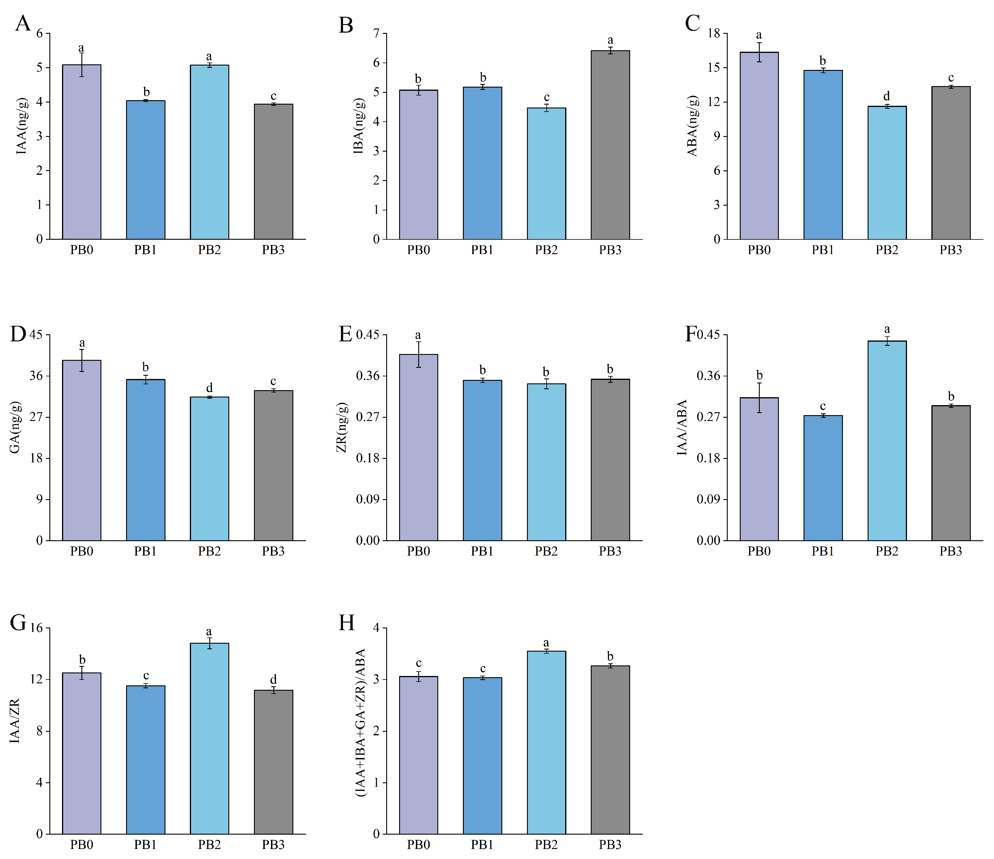
Significant changes in endogenous hormone levels were observed during AR formation in P. bournei. IAA content exhibited a decrease–increase–decrease trend, while IBA content significantly decreased from PB2 to PB3. ABA and GA showed similar trends, continuously decreasing from PB0 to PB2 and increasing from PB2 to PB3. ZR content declined during the callus formation stage, then stabilized. The ratios of IAA/ZR and IAA/ABA followed a decrease–increase–decrease trend, and the (IAA + IBA + GA + ZR)/ABA ratio increased from PB1 to PB2 before decreasing (Figure 3A–H).
OPLS-DA was performed on five endogenous hormones during AR formation. All data points for the five hormones fell within the 95% confidence interval, with biological replicates from the same stage showing clear clustering, indicating high sample reproducibility (Figure S1A). Model diagnostics further confirmed the robustness of the OPLS-DA model, with R2X = 0.954, R2Y = 0.982, and Q2 = 0.977 (p < 0.005), while permutation tests further verified that the model was not overfitted or driven by random variation (Figure S1B,C). Based on the highest variable importance in projection (VIP) values, key hormones influencing each stage of rooting were identified: IAA was the primary endogenous hormone during the Callus induction stage (PB0–PB1); IAA and ABA were the primary endogenous hormones during the root primordium formation stage (PB1–PB2); and IAA and ABA were the primary endogenous hormones during the AR formation stage (PB2–PB3) (Table S2). These results suggest that IAA and ABA may play critical regulatory roles in AR formation during P. bournei cutting propagation.

3.4. Transcriptomic Analysis of AR Formation

Transcriptome sequencing was performed on samples from 12 stages of AR formation in P. bournei cuttings, yielding 520,384,364 clean reads after quality control and filtering. The Guanine-Cytosine (GC) content of each sample exceeded 45%, with Q20 values above 98% and Q30 values above 94.5%, indicating high sequencing quality (Table S3). Principal component analysis (PCA) revealed that samples from different stages were distributed in distinct regions, while the three biological replicates within each stage clustered together, confirming high sample reproducibility and biological significance (Figure S2A). Fifteen root formation-related genes were selected for qRT-PCR validation, and their relative expression levels showed trends consistent with the Fragments Per Kilobase of transcript per Million mapped reads (FPKM) values from RNA-seq, indicating that the transcriptome data are reliable and can be used for subsequent analyses (Figure S2B).
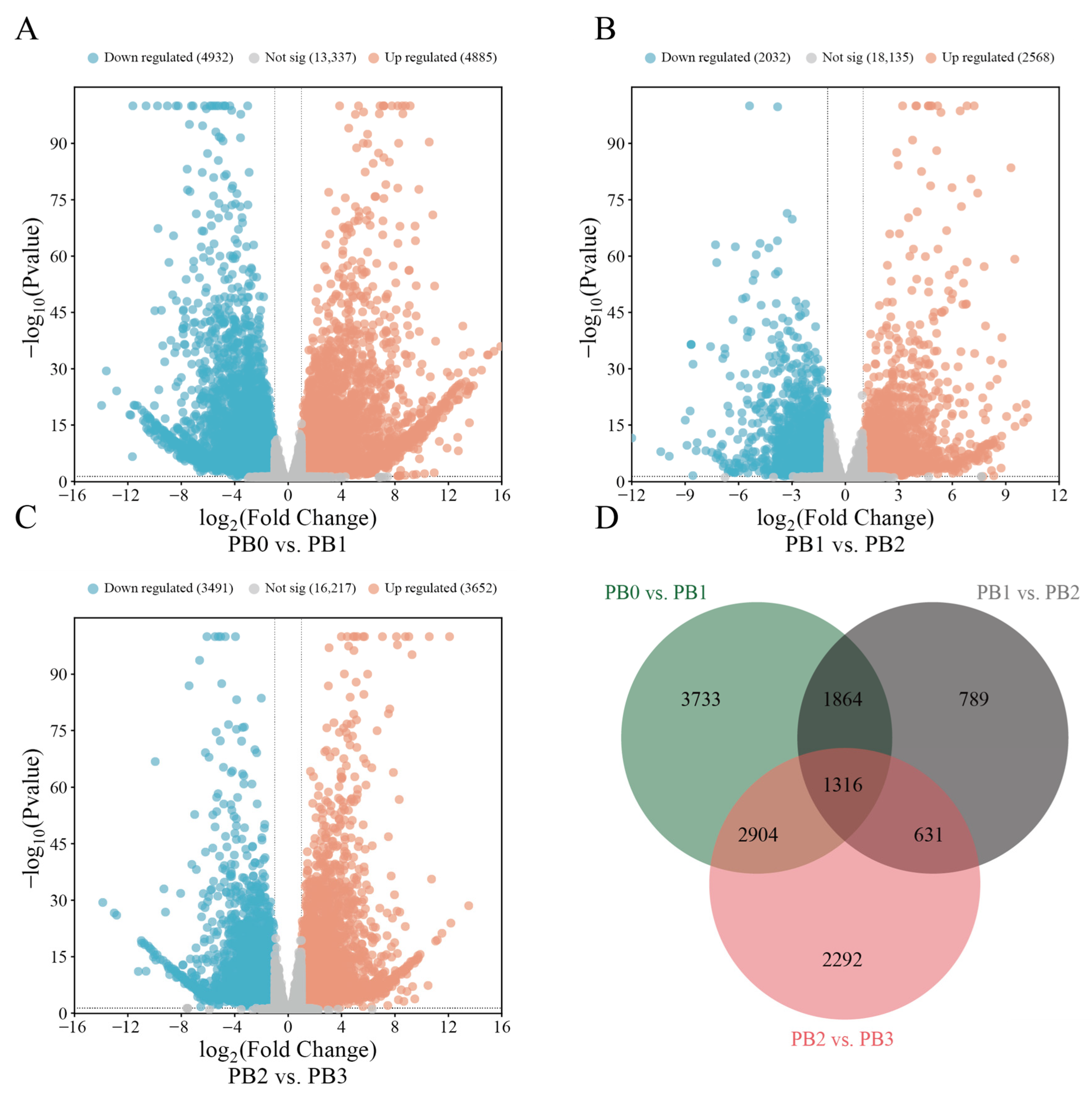
Gene expression levels across different stages were quantified, and differential expression analysis was conducted using DESeq2 software. The screening criteria were set at padj < 0.05 and |log2FoldChange| > 1. The highest number of DEGs was identified in the PB0 vs. PB1 comparison, with 9817 DEGs, including 4885 upregulated and 4932 downregulated genes. In the PB1 vs. PB2 comparison, 4600 DEGs were identified, with 2568 upregulated and 2032 downregulated genes. In the PB2 vs. PB3 comparison, 7143 DEGs were identified, with 3652 upregulated and 3491 downregulated genes (Figure 4A–C). A Venn diagram of DEGs revealed 1316 genes commonly differentially expressed across the three comparison groups (Figure 4D).

3.5. Functional Annotation and Enrichment Analysis of DEGs

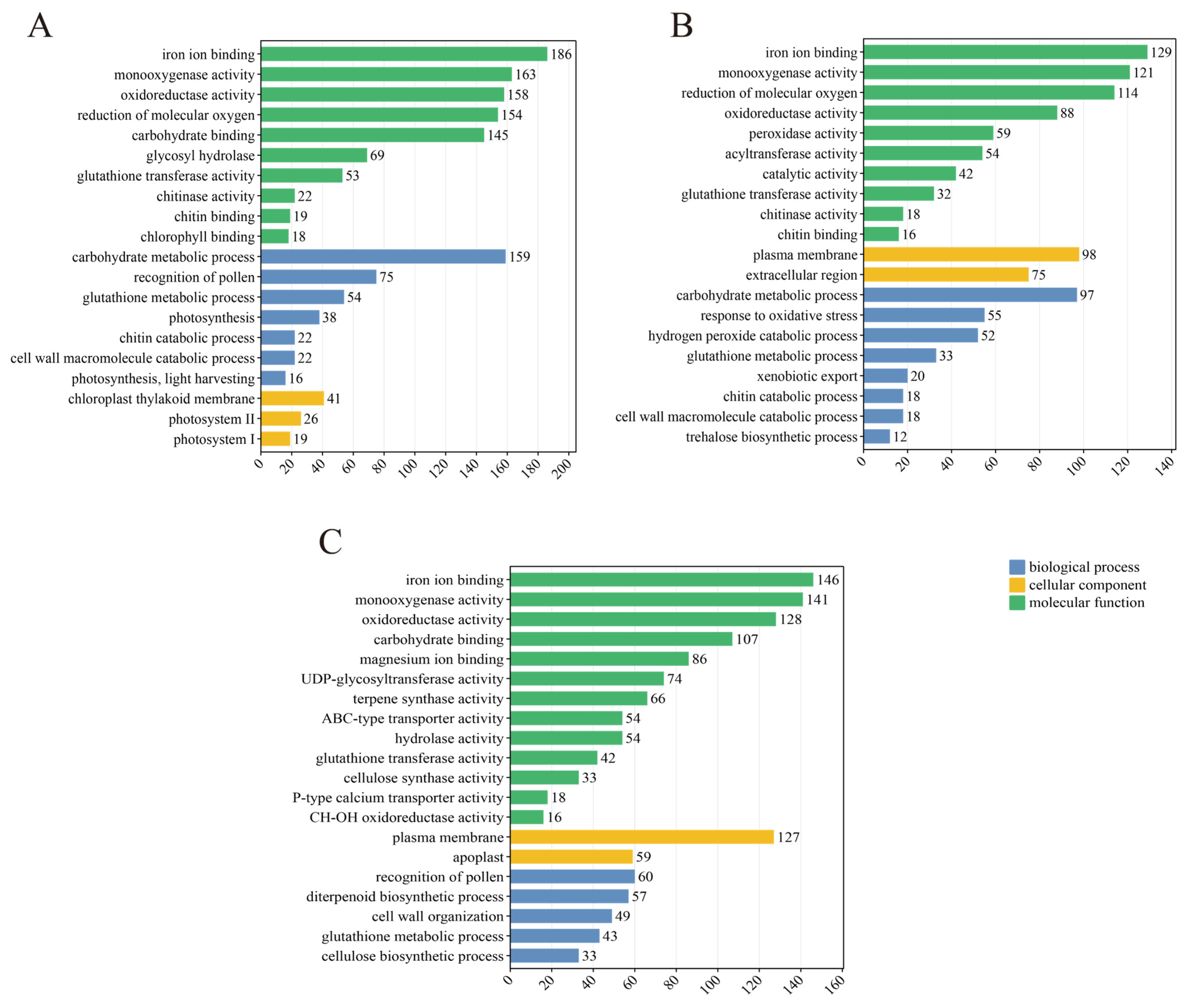
GO enrichment analysis was conducted to explore the biological roles of DEGs during AR formation in P. bournei cuttings (Figure 5). The top 20 significantly enriched GO terms, according to p-values rankings, were chosen for graphical representation. In the PB0 vs. PB1 comparison, the dominant enriched term in the molecular function category was iron ion binding, encompassing 186 DEGs, whereas the most enriched term in the biological process category was carbohydrate metabolic process, involving 158 DEGs. In the PB1 vs. PB2 comparison, the leading enriched terms within the molecular function category included iron ion binding and monooxygenase activity, comprising 129 and 121 DEGs, respectively. In the PB2 vs. PB3 comparison, the Predominant enriched terms in the molecular function category were iron ion binding and monooxygenase activity, while the primary enriched term in the cellular component category was cytoplasmic membrane, encompassing 127 DEGs.
KEGG pathway annotation was performed to clarify the metabolic roles of DEGs, with the top 10 significantly enriched pathways chosen for visualization (Figure 6). For the PB0 vs. PB1 comparison, the carbon metabolism pathway was predominantly enriched, comprising 139 DEGs, followed by the phenylpropanoid biosynthesis pathway with 101 DEGs. For the PB1 vs. PB2 comparison, the phenylpropanoid biosynthesis pathway was the most enriched, including 85 DEGs, followed by the protein processing in endoplasmic reticulum pathway and the starch and sucrose metabolism pathway comprising 65 and 58 DEGs, respectively. For the PB2 vs. PB3 comparison, the phenylpropanoid biosynthesis pathway and the glutathione metabolism pathway were predominantly enriched, including 61 and 56 DEGs, respectively. These findings indicate that the phenylpropanoid biosynthesis pathway may have a pivotal function in AR formation in P. bournei cuttings.

3.6. Phenylpropanoid Biosynthesis and Plant Hormone Signal Transduction in AR Formation

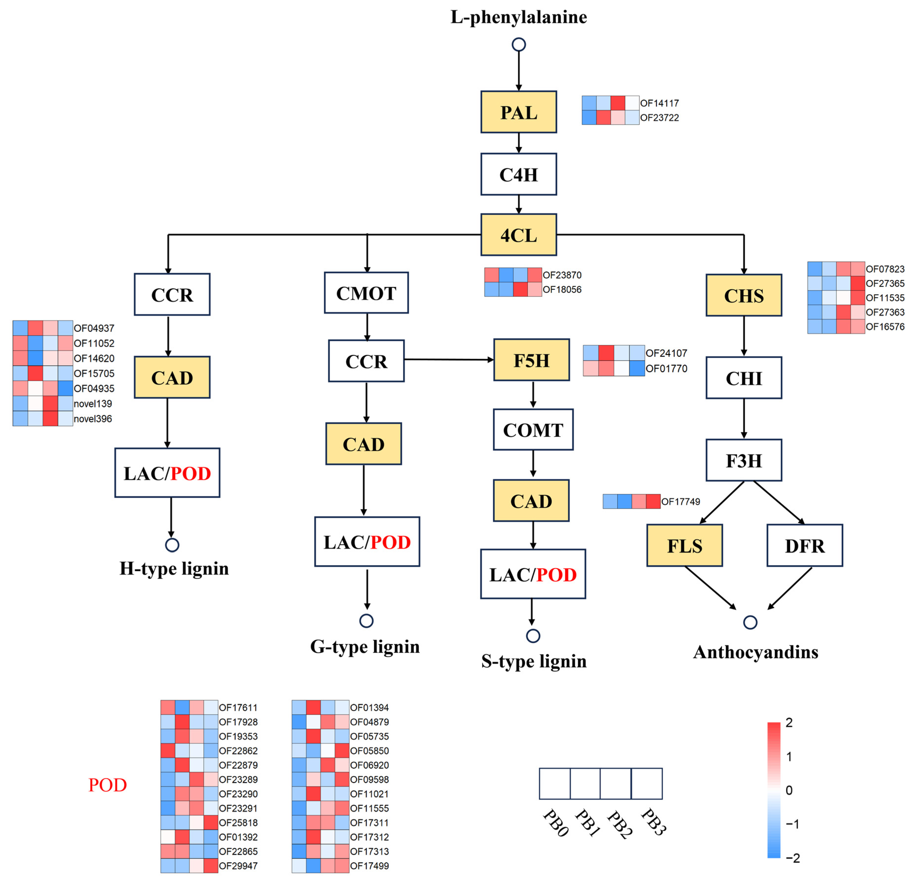
The phenylpropanoid biosynthesis pathway is closely associated with AR formation. Phenylpropanoid-derived compounds, such as lignin and flavonoids, play critical roles in root formation and development [24,35,36]. To further explore the role of the phenylpropanoid biosynthesis pathway in AR formation in P. bournei cuttings, key genes involved in this pathway were investigated (Figure 7). DEGs encoding phenylalanine ammonia-lyase (PAL) were upregulated during the callus induction stage and downregulated during the AR formation stage. Genes encoding chalcone synthase (CHS) were upregulated in the later stages of cutting propagation. Genes encoding ferulate 5-hydroxylase (F5H) were upregulated during the callus induction stage and subsequently downregulated. Genes encop89 expression pattern. Genes encoding flavonol synthase (FLS) were consistently upregulated following callus formation. The largest number of DEGs encoded peroxidase (POD), with most being upregulated during the callus induction stage.
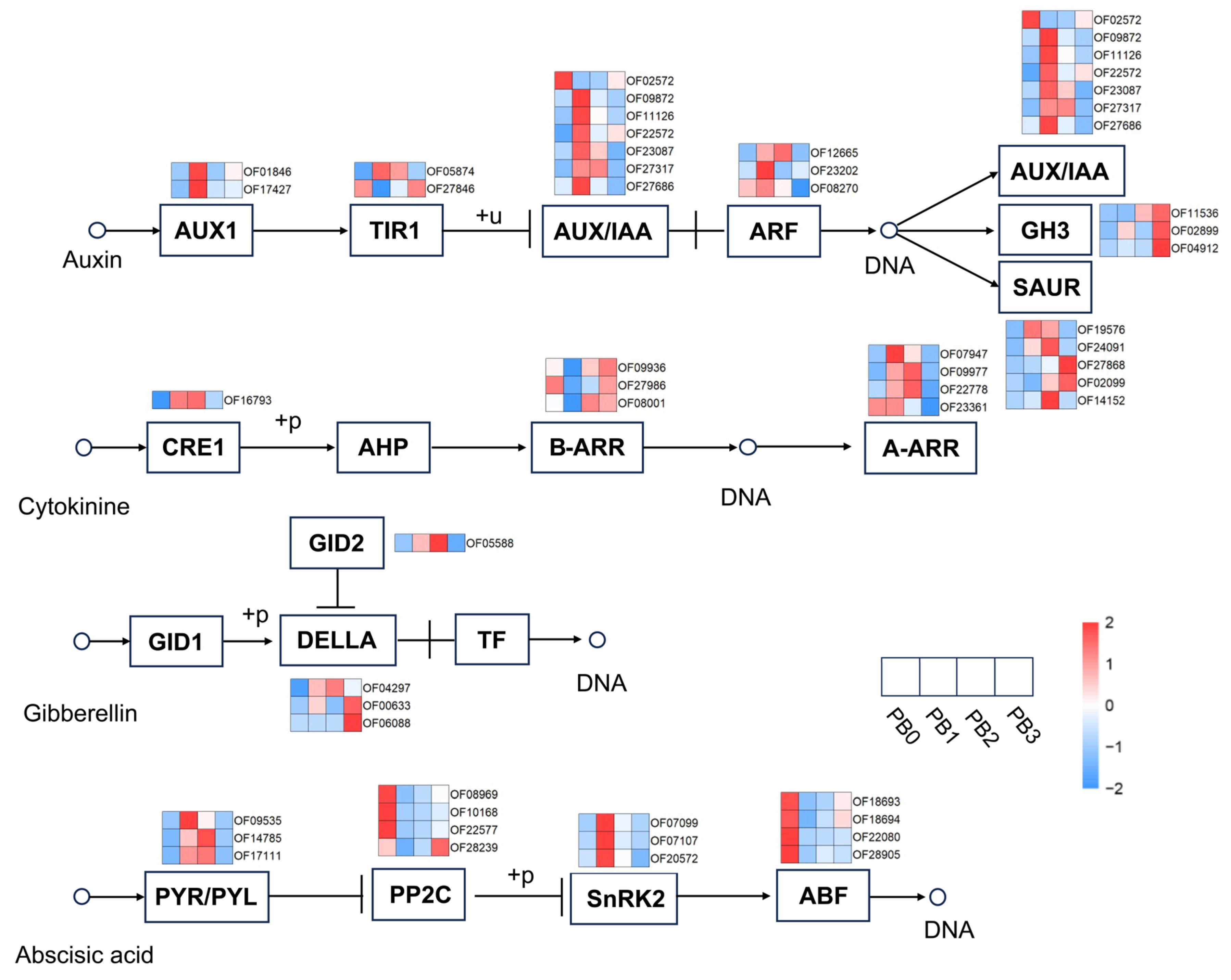
Endogenous hormones are critical for AR formation, as demonstrated in multiple species [29,30,37,38]. Based on this, DEGs involved in plant hormone signal transduction pathways were analyzed (Figure 8). A total of 48 DEGs were identified, enriched in the IAA, ABA, GA, and CTK signaling pathways, with 22, 14, 4, and 8 DEGs, respectively. The largest number of genes were enriched in pathways associated with IAA and ABA, consistent with the results of OPLS-DA, suggesting that these two hormones may play critical roles in AR formation in P. bournei cuttings. In the auxin signaling pathway, genes encoding AUX/IAA were upregulated during the callus induction stage and subsequently downregulated, while genes encoding GH3 were upregulated during the AR formation stage. In the cytokinin signaling pathway, genes encoding B-ARR were upregulated during the root primordium formation stage, whereas genes encoding A-ARR were downregulated during the AR formation stage. In the gibberellin signaling pathway, genes encoding DELLA were upregulated during the AR formation stage. In the abscisic acid signaling pathway, genes encoding PYR/PYL were upregulated during the callus induction stage and downregulated during the AR formation stage. Genes encoding PP2C were downregulated during the callus induction stage, while genes encoding ABF showed an opposite trend. Genes encoding SnRK2 were upregulated during the callus induction stage and downregulated during the root primordium formation stage.

3.7. WGCNA of DEGs and Hormone Traits in AR Formation

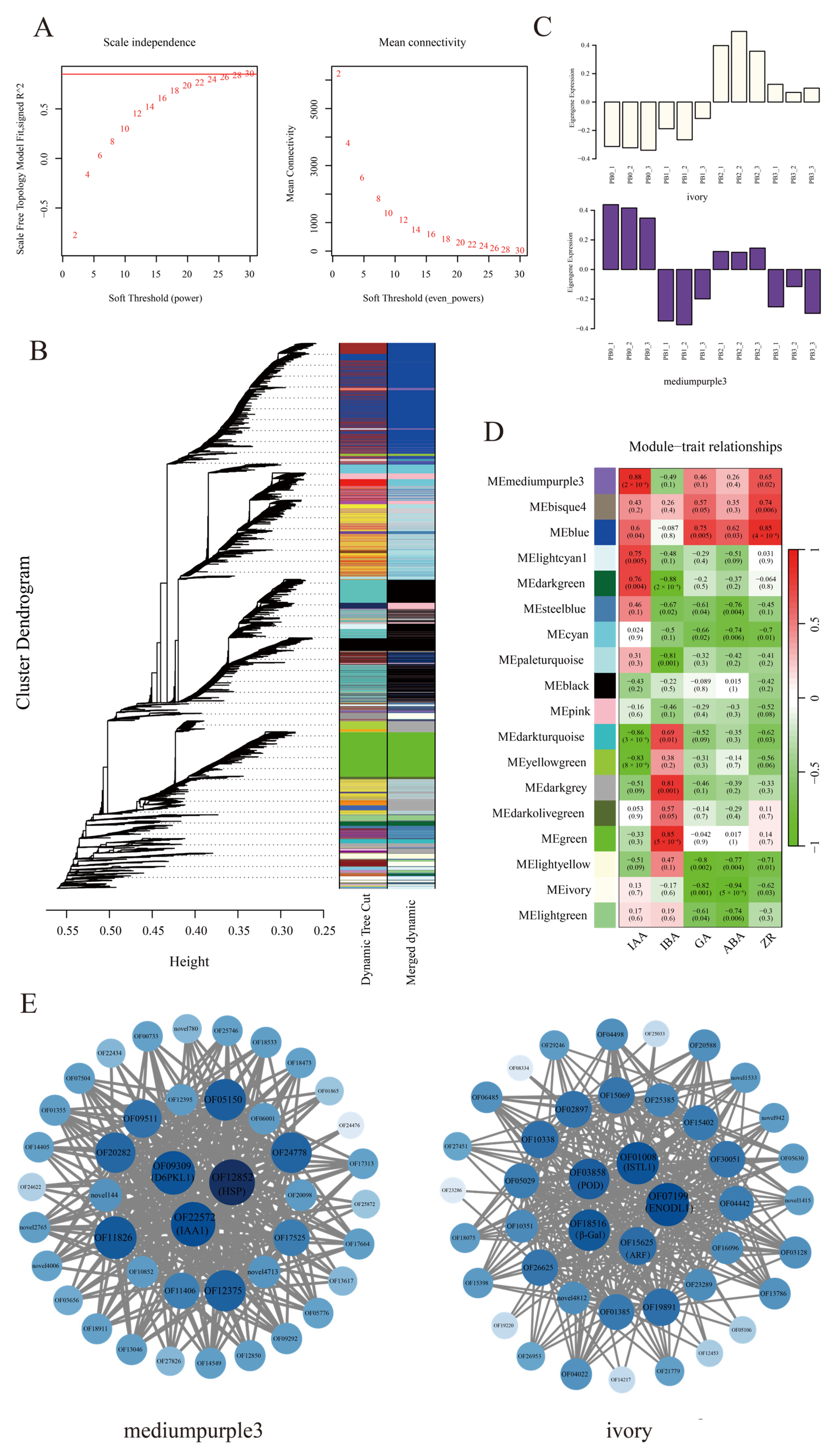
WGCNA was conducted using the WGCNA package in R to determine key genes involved in IAA and ABA regulation during AR formation in P. bournei cuttings, based on DEGs [39,40]. Utilizing a soft threshold of 26, a weighted adjacency matrix was constructed to ensure the gene co-expression network adhered to a scale-free distribution. Module clustering was conducted using the dynamic tree-cutting method, with highly similar modules consolidated to yield 18 modules (Figure 9A,B). The mediumpurple3 and ivory modules exhibited strong correlations with IAA and ABA, respectively, and were identified as hub modules. Genes in the ivory module displayed an initial upregulation followed by a downregulation in expression, whereas those in the mediumpurple3 module demonstrated a trend of initial downregulation, followed by an upregulation and later downregulation (Figure 9C,D). The TA gene co-expression network was established to determine key genes within hub modules, using the top 10% of genes based on module weight values (Figure 9E). Eight hub genes were pinpointed, including three within the mediumpurple3 module: OF09309 (PbD6PKL1), OF12852 (PbHSP), and OF22572 (PbIAA1). The ivory module comprised five hub genes: OF01008 (PbISTL1), OF03858 (PbPOD), OF18156 (Pbβ-Gal), OF07199 (PbENODL1), and OF15625 (PbARF). These hub genes may serve as putative regulators within the IAA and ABA signaling pathways.

3.8. Correlation Analysis of DEGs and Hub Genes in ABA and IAA Signal Transduction Pathways

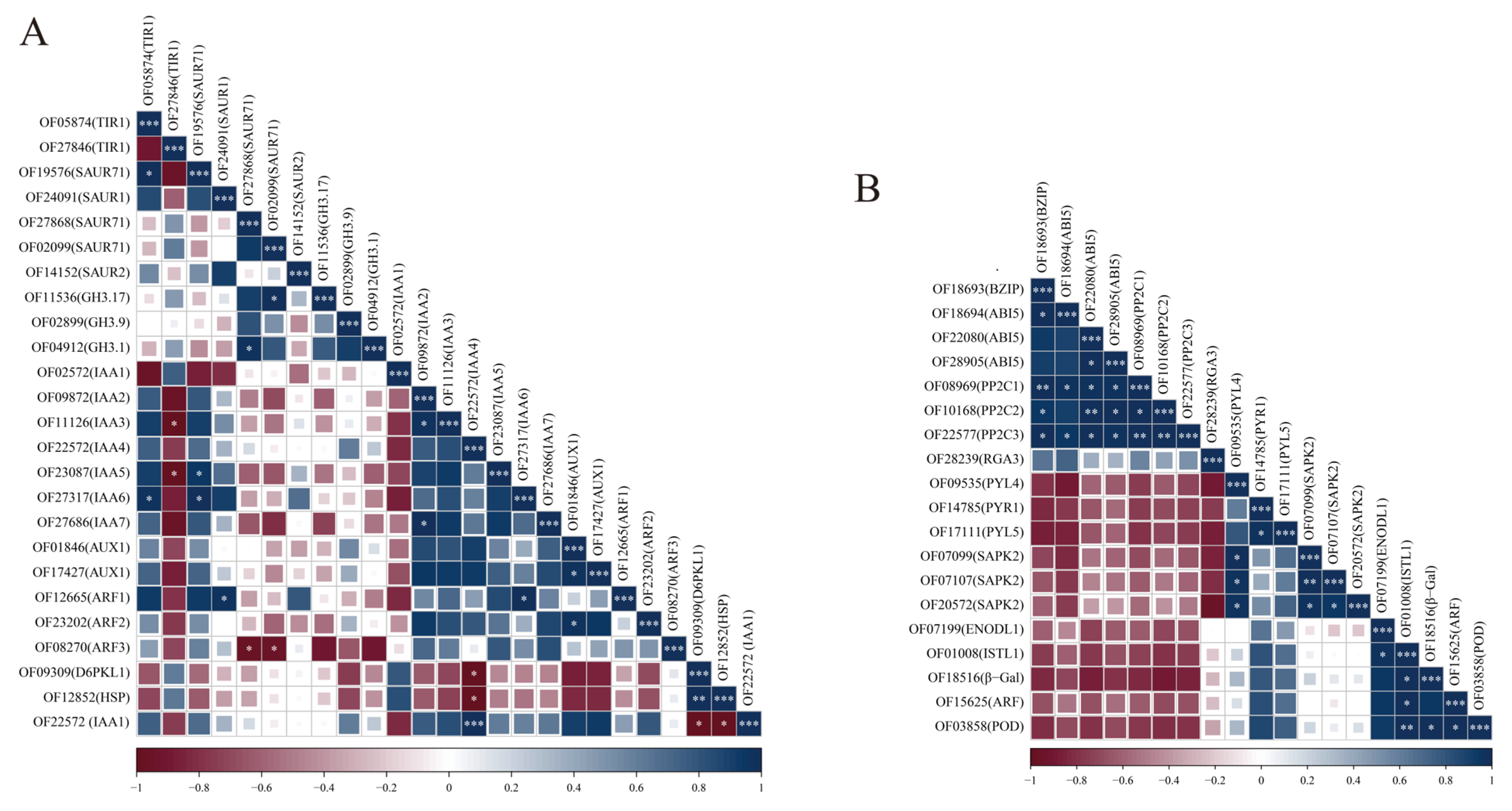
Correlation analysis was conducted on 36 DEGs in the IAA and ABA signal transduction pathways, along with eight hub genes from the mediumpurple3 and ivory modules, based on their correlations with IAA and ABA (Figure 10). In the IAA signal transduction pathway, hub genes D6PKL1 and HSP from the mediumpurple3 module exhibited significant positive correlations with IAA4, whereas IAA1 showed a significant negative correlation with IAA4 (Figure 10A). In the ABA signal transduction pathway, SAPK2 displayed a significant negative correlation with PYL4, while no significant correlations were observed between hub genes from the ivory module and DEGs in the ABA signal transduction pathway (Figure 10B).

4. Discussion

4.1. AR Formation in P. bournei Cuttings

AR formation is a critical component of cutting propagation and plays a decisive role in its success [4,5]. The initiation of root primordia represents the key step in AR development, and depending on their origin, they can be classified into latent and induced types. Anatomical observations in P. bournei revealed no presence of latent primordia, indicating that its AR formation follows the induced pathway. This pattern is consistent with other Lauraceae species such as C. bodinieri and Cinnamomum parthenoxylon [30,38]. Based on their tissue origin, ARs are generally categorized as pericyclic, callus-derived, or mixed. In P. bournei, ARs predominantly originate from callus tissue. Previous studies have reported that callus is often negatively correlated with rooting ability [41], largely because callus-derived ARs frequently fail to establish functional vascular connections with the stem, leading to impaired water and nutrient transport. This may represent a physiological barrier to effective rooting in P. bournei. Moreover, ARs derived from callus tissue are structurally fragile and prone to breakage, which further reduces transplant survival in later stages.

4.2. Physiological Responses During AR Formation

The development of ARs in cuttings is typically accompanied by intensive consumption of metabolic substances to support cellular dedifferentiation, division, and tissue differentiation. In this study, soluble sugar content increased significantly during callus induction, suggesting that sugars not only serve as a primary energy source but also provide carbon skeletons for cell wall and structural biosynthesis, thereby sustaining rapid cell division and tissue reorganization [42]. Concurrently, soluble protein levels increased steadily from PB1 to PB3, reflecting the demand for functional and structural proteins, such as those involved in cytoskeletal assembly, which are essential for the establishment of root morphology and physiological function [26].
Following excision from the mother plant, cuttings experience an abrupt disruption of water and nutrient supply, transitioning from a stable environment to an autonomous state. This transition results in osmotic imbalance, nutrient deprivation, wound exposure, and metabolic disturbance, all of which constitute a typical stress response [43]. Consequently, the cellular redox balance is disrupted, and abnormal respiratory and photosynthetic electron transport leads to rapid accumulation of ROS. Excess ROS triggers lipid peroxidation, generating large amounts of MDA, thereby increasing membrane permeability and causing structural damage [44,45]. This is consistent with our observation of elevated ROS and MDA levels during the early stages of cutting, suggesting that membrane lipid peroxidation is a key feature of the initial stress response and may restrict AR induction efficiency. POD, PPO, IAAO, and SOD contribute to AR formation by regulating cellular redox homeostasis [46]. In particular, PPO and IAAO are involved in IAA oxidation and degradation, thereby affecting auxin signaling. In this study, PPO and IAAO activities increased during AR formation, coinciding with decreased IAA levels, indicating their role in hormone homeostasis during AR development. SOD, a critical antioxidant enzyme, converts excess O2 into O2 and H2O2, which is further decomposed by POD and related enzymes, alleviating oxidative damage to cellular components and structures [47,48]. In P. bournei cuttings, SOD activity peaked at PB0 and declined thereafter, reaching its lowest at PB3. This suggests that stress conditions immediately after excision stimulated antioxidant defense, whereas subsequent AR development restored absorptive function and alleviated stress, resulting in decreased SOD activity.

4.3. Regulatory Roles of Endogenous IAA and ABA Signaling

Endogenous phytohormones integrate environmental cues and nutrient status to regulate root system architecture, including primary root elongation, lateral root initiation, and AR induction [49,50,51]. Hormones interact through intricate signaling networks that influence biosynthesis, degradation, perception, and signal transduction, thereby orchestrating precise control of root growth and differentiation. Among them, IAA and ABA play central roles, with IAA promoting cell division and elongation, and ABA modulating root maturation, lignification, and stress responses [7,52]. OPLS-DA analysis of phytohormone profiles revealed that IAA and ABA are the key regulators of AR formation in P. bournei. KEGG enrichment further supported this conclusion, as most DEGs were mapped to IAA and ABA signaling pathways.
IAA and ABA interact through concentration-dependent and environment-dependent regulatory networks [52]. In this study, IAA levels decreased from PB0 to PB1, accompanied by upregulation of AUX1, AUX/IAA, and ARF genes in the auxin signaling pathway. This suggests that reduced IAA concentrations may activate auxin signaling and facilitate callus formation at early stages [30]. AUX1, which mediates polar auxin transport, regulates downstream genes involved in cell expansion and wall loosening, thereby promoting growth [53]. Its upregulation indicates enhanced auxin transport activity during callus formation, which is crucial for auxin homeostasis. Downstream auxin-responsive genes such as GH3 and SAUR were upregulated at later rooting stages, potentially increasing cellular auxin availability. In A. thaliana, SAUR genes enhance hypocotyl elongation by promoting cell expansion [54]. In P. bournei, SAUR upregulation may similarly facilitate cell division and elongation during AR formation. WGCNA identified two hub genes, D6PKL1 and HSP, in the auxin-related mediumpurple3 module. D6PKL1 likely regulates AR development via auxin transport, as reported in A. thaliana hypocotyl bending [55]. Interestingly, HSP, although typically associated with stress responses and ABA signaling, showed a strong correlation with IAA levels, indicating that it may participate in the crosstalk between IAA and ABA during AR formation [56]. Future studies could perform overexpression of these two hub genes to investigate changes in the IAA signaling pathway and their effects on AR formation.
ABA primarily functions in stress adaptation but also influences root development, with low ABA concentrations generally favoring AR induction [52,57]. In this study, ABA peaked at PB0, declined during PB0–PB2, and increased again during AR formation. The initial rise may reflect stress-induced ABA accumulation following excision, whereas the subsequent decline coincided with PYR/PYL upregulation, indicating activation of ABA signaling despite lower ABA levels [58]. Binding of ABA to PYR/PYL receptors triggers the phosphorylation of ABFs/AREBs, enhancing stress resistance during rooting [59]. Hub genes in the ABA-related ivory module included POD, which mediates oxidative stress response [46]. ISTL1, which indirectly regulates auxin transport via PIN proteins [60]; ENODL1, involved in pathogen defense [61]. β-Gal, which hydrolyzes hemicellulose and pectin to promote cell wall loosening [62]. and ARF, a key transcriptional regulator in auxin signaling [63]. Collectively, these findings suggest that ABA signaling is activated in the early cutting stage, with POD and ENODL1 potentially acting as downstream responsive genes to enhance stress tolerance. ARF and ISTL1 may be regulated by ABA signaling while also influencing the IAA signaling pathway, potentially serving as key regulators in the ABA–IAA crosstalk network. Future studies could explore the roles of these hub genes by treating cuttings with ABA receptor antagonists. The resulting changes in the expression of the five hub genes and in AR development could then be observed to validate their functions.

4.4. Dual Regulatory Role of Phenylpropanoid Biosynthesis in AR Development

The phenylpropanoid pathway plays a crucial role in root development, as its derivatives, such as lignin and phenolics, contribute to both cell wall architecture and regulation of cell division, elongation, and differentiation [25,64]. Elevated lignin accumulation in callus tissue enhances tissue lignification, increases wall rigidity, and suppresses cell proliferation and elongation, thereby inhibiting AR formation [24]. Thus, phenylpropanoid metabolism is essential for both stress responses and root primordium initiation [23]. In Vigna radiata, lignin biosynthesis-related genes such as CHS and POD are upregulated under stress conditions. In P. bournei, CHS was upregulated during PB1 to PB2, potentially promoting root primordium initiation [65], whereas PAL, F5H and POD upregulation was most evident during callus induction, consistent with concurrent increases in lignin content. This may result from nutrient and water deficits after cutting excision, which activate lignin biosynthesis and enhance lignification at the stem base, improving stress tolerance but may delay AR emergence [24]. This may explain why AR formation in P. bournei is generally slower compared with other Lauraceae species. Conversely, phenolic compounds are closely linked to rooting ability, with lower levels favoring AR induction, while higher levels inhibit cell division and elongation [66]. In this study, phenolic content decreased continuously during rooting, potentially promoting wall loosening and primordium differentiation, thereby accelerating AR development. Overall, phenylpropanoid metabolism exerts a dual regulatory effect in P. bournei: lignin accumulation imposes inhibitory effects, whereas phenolic reduction facilitates root initiation, and their dynamic balance largely determines the progress of AR formation (Figure S3).

5. Conclusions

In this study, morphological and anatomical observations revealed that AR in P. bournei originated from callus tissue, and the root primordium belonged to the induced type. AR formation was accompanied by the consumption of nutrients, significant changes in enzyme activities, and fluctuations in endogenous hormones. Soluble protein increased continuously from PB1 to PB3, reflecting the elevated demand for functional and structural proteins such as cytoskeletal components during this stage. OFR and MDA peaked at PB0, indicating that membrane lipid peroxidation is a key feature of the early stress response in cuttings. The activities of IAAO and PPO exhibited similar trends, suggesting that both may regulate AR formation by mediating IAA oxidation and degradation. OPLS-DA analysis suggested that IAA and ABA are the key endogenous hormones during AR formation, which was further supported by the enrichment of the largest number of DEGs in their signaling pathways. Notably, hub genes such as ARF and ISTL1 may serve as central nodes mediating the crosstalk between IAA and ABA. In addition, the phenylpropanoid pathway was significantly enriched during AR formation Lignin accumulation during callus induction may accelerate tissue lignification and delay AR initiation, whereas the decrease in phenolic compounds may favor root primordium differentiation and AR development. Collectively, this study integrates morphological, physiological, and molecular evidence to elucidate the regulatory mechanisms of AR formation in P. bournei cuttings, providing a theoretical basis for understanding the regulatory mechanisms of AR formation and optimizing clonal propagation systems.

Supplementary Materials

The following supporting information can be downloaded at: https://www.mdpi.com/article/10.3390/horticulturae11101238/s1, Figure S1. OPLS-DA analysis of endogenous hormone profiles at different stages of AR formation in P. bournei cuttings. Figure S2. PCA and qRT-PCR Validation. Figure S3. Simplified schematic model of lignin and phenolic regulation during AR formation in P. bournei; Table S1: Primers used in qRT-PCR in this study. Table S2. VIP values of endogenous hormone contents during AR formation in P. bournei. Table S3. Statistical summary of sequencing data volume.

Author Contributions

Conceptualization, Y.L. and H.X.; methodology, Y.L.; software, H.X.; validation, Y.Z. (Yongjie Zheng) and C.L.; formal analysis, Y.Z. (Yueting Zhang); investigation, Y.L.; resources, Y.L.; data curation, H.X.; writing—original draft preparation, Y.L.; writing—review and editing, X.L.; visualization, Y.W.; supervision, Y.W.; project administration, X.L.; funding acquisition, Y.W. All authors have read and agreed to the published version of the manuscript.

Funding

This research was funded by National Key Research and Development Program of China (No.2024YFD2200302); Jiangxi Forestry Science and Technology Innovation Special Project (No. [2021]15); Graduate Research and Practice Innovation Program of Jiangsu Province (No. SJCX25_0420); The Basic Research Project of Jiangxi Academy of Forestry (No. 2023522703 and No. 2025522703).

Data Availability Statement

The raw sequence data reported in this paper have been deposited in the Genome Sequence Archive (Genomics, Proteomics & Bioinformatics 2021) in National Genomics Data Center (Nucleic Acids Res 2025), China National Center for Bioinformation/Beijing Institute of Genomics, Chinese Academy of Sciences (GSA: subCRA047698), which are publicly accessible at https://ngdc.cncb.ac.cn/gsa (accessed on 1 September 2025).

Acknowledgments

We thank Chao Fu from the Jiangxi Academy of Forestry for collecting the plant materials used in this manuscript.

Conflicts of Interest

The authors declare no conflicts of interest.

References

  1. Han, X.; Zhang, J.; Han, S.; Chong, S.L.; Meng, G.; Song, M.; Wang, Y.; Zhou, S.; Liu, C.; Lou, L. The chromosome-scale genome of Phoebe bournei reveals contrasting fates of terpene synthase (TPS)-a and TPS-b subfamilies. Plant Commun. 2022, 3, 100410. [Google Scholar] [CrossRef] [PubMed]
  2. Li, Y.; Liu, X.; Ma, J.; Zhang, X.; Xu, L. Phenotypic variation in Phoebe bournei populations preserved in the primary distribution area. J. For. Res. 2018, 29, 35–44. [Google Scholar] [CrossRef]
  3. Xu, W.T.; Zhang, M.; Wang, C.; Lou, X.Z.; Han, X.; Zhang, J.L.; Zhang, Y.T.; Tong, Z.K. Somatic embryo induction and Agrobacterium-mediated transformation of embryonic callus tissue in Phoebe bournei, an endangered woody species in Lauraceae. Not. Bot. Horti Agrobot. Cluj-Napoca 2020, 48, 572–587. [Google Scholar] [CrossRef]
  4. Wetzstein, H.Y.; Porter, J.A.; Janick, J.; Ferreira, J.F.S.; Mutui, T.M. Selection and clonal propagation of high artemisinin genotypes of Artemisia annua. Front. Plant Sci. 2018, 9, 358. [Google Scholar] [CrossRef]
  5. Tate, H.T.; Page, T. Cutting propagation of Santalum austrocaledonicum: The effect of genotype, cutting source, cutting size, propagation medium, IBA and irradiance. New For. 2018, 49, 551–570. [Google Scholar] [CrossRef]
  6. Li, T.; Min, X. Dormancy characteristics and germination requirements of Phoebe bournei seed. Sci. Hortic. 2020, 260, 108903. [Google Scholar] [CrossRef]
  7. Gonin, M.; Bergougnoux, V.; Nguyen, T.D.; Gantet, P.; Champion, A. What makes adventitious roots? Plants 2019, 8, 240. [Google Scholar] [CrossRef]
  8. Liu, G.; Zhao, J.; Liao, T.; Wang, Y.; Guo, L.; Yao, Y.; Cao, J. Histological dissection of cutting-inducible adventitious rooting in Platycladus orientalis reveals developmental endogenous hormonal homeostasis. Ind. Crops Prod. 2021, 170, 113817. [Google Scholar] [CrossRef]
  9. Wang, S.T.; Sun, G.D.; Luo, Y.; Qian, W.J.; Fan, K.; Ding, Z.T.; Hu, J.H. Role of IAA and Primary Metabolites in Two Rounds of Adventitious Root Formation in Softwood Cuttings of Camellia sinensis (L.). Agronomy 2022, 12, 2486. [Google Scholar] [CrossRef]
  10. Ma, W.Y.; Li, J.J.; Qu, B.Y.; He, X.; Zhao, X.Q.; Li, B.; Fu, X.D.; Tong, Y.P. Auxin biosynthetic gene TAR2 is involved in low nitrogen-mediated reprogramming of root architecture in Arabidopsis. Plant J. 2014, 78, 70–79. [Google Scholar] [CrossRef] [PubMed]
  11. Habets, M.E.J.; Offringa, R. PIN-driven polar auxin transport in plant developmental plasticity: A key target for environmental and endogenous signals. New Phytol. 2014, 203, 362–377. [Google Scholar] [CrossRef]
  12. Ugartechea-Chirino, Y.; Swarup, R.; Swarup, K.; Péret, B.; Whitworth, M.; Bennett, M.; Bougourd, S. The AUX1 LAX family of auxin influx carriers is required for the establishment of embryonic root cell organization in Arabidopsis thaliana. Ann. Bot. 2009, 105, 277–289. [Google Scholar] [CrossRef]
  13. Tahir, M.M.; Mao, J.P.; Li, S.H.; Li, K.; Liu, Y.; Shao, Y.; Zhang, D.; Zhang, X.Y. Insights into factors controlling adventitious root formation in apples. Horticulturae 2022, 8, 276. [Google Scholar] [CrossRef]
  14. Meng, X.Y.; Wang, Z.; He, S.L.; Shi, L.Y.; Song, Y.L.; Lou, X.Y.; He, D. Endogenous hormone levels and activities of IAA-modifying enzymes during adventitious rooting of tree peony cuttings and grafted scions. Hortic. Environ. Biotechnol. 2019, 60, 187–197. [Google Scholar] [CrossRef]
  15. Cisse, E.H.M.; Zhang, J.; Li, D.-D.; Miao, L.F.; Yin, L.Y.; Yang, F. Exogenous ABA and IAA modulate physiological and hormonal adaptation strategies in Cleistocalyx operculatus and Syzygium jambos under long-term waterlogging conditions. BMC Plant Biol. 2022, 22, 523. [Google Scholar] [CrossRef] [PubMed]
  16. Guo, D.; Liang, J.; Qiao, Y.; Yan, Y.; Li, L.; Dai, Y. Involvement of G1-to-S transition and AhAUX-dependent auxin transport in abscisic acid-induced inhibition of lateral root primodia initiation in Arachis hypogaea L. J. Plant Physiol. 2012, 169, 1102–1111. [Google Scholar] [CrossRef]
  17. Tartoura, K.A.H. Effect of abscisic acid on endogenous IAA, auxin protector levels and peroxidase activity during adventitious root initiation in Vigna radiata cuttings. Acta Physiol. Plant. 2001, 23, 149–156. [Google Scholar] [CrossRef]
  18. El-Banna, M.F.; Farag, N.B.B.; Massoud, H.Y.; Kasem, M.M. Exogenous IBA stimulated adventitious root formation of Zanthoxylum beecheyanum K. Koch stem cutting: Histo-physiological and phytohormonal investigation. Plant Physiol. Biochem. 2023, 197, 107639. [Google Scholar] [CrossRef]
  19. Kuroha, T.; Kato, H.; Asami, T.; Yoshida, S.; Kamada, H.; Satoh, S. A trans-zeatin riboside in root xylem sap negatively regulates adventitious root formation on cucumber hypocotyls. J. Exp. Bot. 2002, 53, 2193–2200. [Google Scholar] [CrossRef]
  20. Steffens, B.; Wang, J.; Sauter, M. Interactions between ethylene, gibberellin and abscisic acid regulate emergence and growth rate of adventitious roots in deepwater rice. Planta 2006, 223, 604–612. [Google Scholar] [CrossRef] [PubMed]
  21. Roychoudhry, S.; Kepinski, S. Auxin in root development. Cold Spring Harb. Perspect. Biol. 2022, 14, 039933. [Google Scholar] [CrossRef] [PubMed]
  22. Li, X.P.; Xu, Q.Q.; Liao, W.B.; Ma, Z.J.; Xu, X.T.; Wang, M.; Ren, P.J.; Niu, L.J.; Jin, X.; Zhu, Y.C. Hydrogen peroxide is involved in abscisic acid-induced adventitious rooting in cucumber (Cucumis sativus L.) under drought stress. J. Plant Biol. 2016, 59, 536–548. [Google Scholar] [CrossRef]
  23. Li, S.W.; Shi, R.F.; Leng, Y.; Zhou, Y. Transcriptomic analysis reveals the gene expression profile that specifically responds to IBA during adventitious rooting in mung bean seedlings. BMC Genom. 2016, 17, 43. [Google Scholar] [CrossRef]
  24. Chang, E.M.; Guo, W.; Xie, Y.H.; Jiang, Z.P.; Dong, Y.; Jia, Z.R.; Zhao, X.L.; Liu, J.F.; Zhang, J. Changes of lignified-callus and wound-induced adventitious rooting in ancient Platycladus orientalis cuttings as affected by tree age. Ind. Crops Prod. 2023, 203, 117183. [Google Scholar] [CrossRef]
  25. Palama, T.L.; Menard, P.; Fock, I.; Choi, Y.H.; Bourdon, E.; Govinden-Soulange, J.; Bahut, M.; Payet, B.; Verpoorte, R.; Kodja, H. Shoot differentiation from protocorm callus cultures of Vanilla planifolia (Orchidaceae): Proteomic and metabolic responses at early stage. BMC Plant Biol. 2010, 10, 82. [Google Scholar] [CrossRef]
  26. Aumond, M.L., Jr.; de Araujo, A.T., Jr.; de Oliveira Junkes, C.F.; de Almeida, M.R.; Matsuura, H.N.; de Costa, F.; Fett-Neto, A.G. Events associated with early age-related decline in adventitious rooting competence of Eucalyptus globulus Labill. Front. Plant Sci. 2017, 8, 1734. [Google Scholar] [CrossRef] [PubMed]
  27. Lowe, R.; Shirley, N.; Bleackley, M.; Dolan, S.; Shafee, T. Transcriptomics technologies. PLoS Comput. Biol. 2017, 13, e1005457. [Google Scholar] [CrossRef]
  28. Langfelder, P.; Horvath, S. WGCNA: An R package for weighted correlation network analysis. BMC Bioinform. 2008, 9, 559. [Google Scholar] [CrossRef]
  29. Wen, S.S.; Miao, D.P.; Cui, H.Y.; Li, S.H.; Gu, Y.N.; Jia, R.R.; Leng, Y.F. Physiology and transcriptomic analysis of endogenous hormones regulating in vitro adventitious root formation in tree peony. Sci. Hortic. 2023, 318, 112122. [Google Scholar] [CrossRef]
  30. Yu, W.W.; Li, Y.H.; Zhang, Y.T.; Luo, C.L.; Zheng, Y.J.; Zhang, T.; Fu, C.; Liu, X.L. Integrated Physiological and Transcriptomic Analyses Reveal Mechanisms Regulating Endogenous Phytohormones in Adventitious Root Formation During Cinnamomum bodinieri Cutting Propagation. Forests 2025, 16, 509. [Google Scholar] [CrossRef]
  31. Moreno-Sanz, P.; D’Amato, E.; Nebish, A.; Costantini, L.; Grando, M. An optimized histological proceeding to study the female gametophyte development in grapevine. Plant Methods 2020, 16, 61. [Google Scholar] [CrossRef] [PubMed]
  32. Liu, Z.Y.; Ji, Z.Y.; Han, Y.Y.; Sun, Y. The mitigation effects of exogenous dopamine treatment on continuous cropping obstacles in watermelon. J. Soil Sci. Plant Nutr. 2023, 23, 4233–4249. [Google Scholar] [CrossRef]
  33. Zhou, Y.H.; Feng, Y.Q.; Zhou, Q.; Jiang, T. Chamaecyparis lawsoniana with different cutting mediums and growth regulators. J. West China For. Sci. 2017, 46, 32–37. (In Chinese) [Google Scholar]
  34. Livak, K.J.; Schmittgen, T.D. Analysis of relative gene expression data using real-time quantitative PCR and the 2−ΔΔCT method. Methods 2001, 25, 402–408. [Google Scholar] [CrossRef]
  35. Cheng, L.B.; Zhao, C.; Zhao, M.R.; Han, Y.Y.; Li, S.Y. Lignin Synthesis, Affected by Sucrose in Lotus (Nelumbo nucifera) Seedlings, Was Involved in Regulation of Root Formation in the Arabidopsis thanliana. Int. J. Mol. Sci. 2022, 23, 2250. [Google Scholar] [CrossRef]
  36. Sharma, S.; Singh, H.P.; Batish, D.R.; Kohli, R.K. Nitric oxide induced modulations in adventitious root growth, lignin content and lignin synthesizing enzymes in the hypocotyls of Vigna radiata. Plant Physiol. Biochem. 2019, 141, 225–230. [Google Scholar] [CrossRef]
  37. Luo, C.L.; Zhang, T.; Dai, X.Y.; Zhang, Y.T.; Zheng, Y.J.; Liu, X.L.; Zhang, X.H. Physiological characteristics and transcriptomic analysis of young stems differentiation in adventitious bud and root formation in Cinnamomum parthenoxylon. Forests 2025, 16, 1049. [Google Scholar] [CrossRef]
  38. Luo, C.L.; Liu, X.L.; Zheng, Y.J.; Dai, X.Y.; Tang, X.L.; Zhang, T.; Zhang, X.H. Physiological and transcriptomic analysis reveal the regulation of adventitious root formation in Cinnamomum parthenoxylon cuttings. BMC Plant Biol. 2024, 24, 1217. [Google Scholar] [CrossRef]
  39. Bao, S.H.; Li, J.N.; Wang, J.Q.; Lan, T.; Wei, M.Y.; Sun, X.Y.; Fang, Y.L.; Ma, T.T. How Does Nature Create the Painting “Gradient Coloration of ‘Manicure Finger’ Grape”? Integrated Omics Unveil the Pigments Basis and Metabolism Networks of Its Formation. Food Front. 2025, 6, 921–939. [Google Scholar]
  40. Ni, L.J.; Wang, J.F.; Zhou, F.W.; Chen, Z.M. Integrated multi-omics reveals Li-miR828z-LiMYB114 regulatory module controlling anthocyanin biosynthesis during flower color development in Lagerstroemia indica. Ind. Crops Prod. 2025, 234, 121524. [Google Scholar]
  41. Martins, M.; Gomes, A.F.G.; da Silva, É.M.; da Silva, D.F.; Peche, P.M.; Magalhães, T.A.; Pio, R. Effects of anatomical structures and phenolic compound deposition on the rooting of olive cuttings. Rhizosphere 2022, 23, 100557. [Google Scholar] [CrossRef]
  42. Ahkami, A.H.; Lischewski, S.; Haensch, K.T.; Porfirova, S.; Hofmann, J.; Rolletschek, H.; Melzer, M.; Franken, P.; Hause, B.; Druege, U. Molecular physiology of adventitious root formation in Petunia hybrida cuttings: Involvement of wound response and primary metabolism. New Phytol. 2009, 181, 613–625. [Google Scholar]
  43. Zhang, W.X.; Fan, J.J.; Tan, Q.Q.; Zhao, M.M.; Zhou, T.; Cao, F.L. The effects of exogenous hormones on rooting process and the activities of key enzymes of Malus hupehensis stem cuttings. PLoS ONE 2017, 12, e0172320. [Google Scholar]
  44. Roussos, P.A. Adventitious root formation in plants: The implication of hydrogen peroxide and nitric oxide. Antioxidants 2023, 12, 862. [Google Scholar] [CrossRef]
  45. Kora, D.; Bhattacharjee, S. Redox gateway associated with adventitious root formation under stress and hormonal signalling in plants. Curr. Sci. 2020, 119, 462–472. [Google Scholar] [CrossRef]
  46. Ilczuk, A.; Jacygrad, E. The effect of IBA on anatomical changes and antioxidant enzyme activity during the in vitro rooting of smoke tree (Cotinus coggygria Scop.). Sci. Hortic. 2016, 210, 268–276. [Google Scholar] [CrossRef]
  47. Ueda, Y.; Uehara, N.; Sasaki, H.; Kobayashi, K.; Yamakawa, T. Impacts of acute ozone stress on superoxide dismutase (SOD) expression and reactive oxygen species (ROS) formation in rice leaves. Plant Physiol. Biochem. 2013, 70, 396–402. [Google Scholar] [CrossRef] [PubMed]
  48. Bowler, C.; Montagu, M.v.; Inze, D. Superoxide dismutase and stress tolerance. Annu. Rev. Plant Physiol. Plant Mol. Biol. 1992, 43, 83–116. [Google Scholar]
  49. Druege, U.; Hilo, A.; Pérez-Pérez, J.M.; Klopotek, Y.; Acosta, M.; Shahinnia, F.; Zerche, S.; Franken, P.; Hajirezaei, M.R. Molecular and physiological control of adventitious rooting in cuttings: Phytohormone action meets resource allocation. Ann. Bot. 2019, 123, 929–949. [Google Scholar] [CrossRef]
  50. Blázquez, M.A.; Nelson, D.C.; Weijers, D. Evolution of plant hormone response pathways. Annu. Rev. Plant Biol. 2020, 71, 327–353. [Google Scholar] [CrossRef]
  51. Xiao, G.; Zhang, Y. Adaptive growth: Shaping auxin-mediated root system architecture. Trends Plant Sci. 2020, 25, 121–123. [Google Scholar] [CrossRef]
  52. Zhang, Q.; Yuan, W.; Wang, Q.W.; Cao, Y.Y.; Xu, F.Y.; Dodd, I.C.; Xu, W.F. ABA regulation of root growth during soil drying and recovery can involve auxin response. Plant Cell Environ. 2022, 45, 871–883. [Google Scholar] [CrossRef]
  53. Cosgrove, D.J. Loosening of plant cell walls by expansins. Nature 2000, 407, 321–326. [Google Scholar] [CrossRef]
  54. Spartz, A.K.; Lee, S.H.; Wenger, J.P.; Gonzalez, N.; Itoh, H.; Inzé, D.; Peer, W.A.; Murphy, A.S.; Overvoorde, P.J.; Gray, W.M. The SAUR19 subfamily of SMALL AUXIN UP RNA genes promote cell expansion. Plant J. 2012, 70, 978–990. [Google Scholar] [CrossRef]
  55. Stanislas, T.; Hüser, A.; Barbosa, I.C.R.; Kiefer, C.S.; Brackmann, K.; Pietra, S.; Gustavsson, A.; Zourelidou, M.; Schwechheimer, C.; Grebe, M. Arabidopsis D6PK is a lipid domain-dependent mediator of root epidermal planar polarity. Nat. Plants 2015, 1, 15162. [Google Scholar] [CrossRef] [PubMed]
  56. Tiwari, M.; Kumar, R.; Min, D.; Jagadish, S.V.K. Genetic and molecular mechanisms underlying root architecture and function under heat stress—A hidden story. Plant Cell Environ. 2022, 45, 771–788. [Google Scholar] [CrossRef] [PubMed]
  57. Miao, R.; Yuan, W.; Wang, Y.; Garcia-Maquilon, I.; Dang, X.L.; Li, Y.; Zhang, J.H.; Zhu, Y.Y.; Rodriguez, P.L.; Xu, W.F. Low ABA concentration promotes root growth and hydrotropism through relief of ABA INSENSITIVE 1-mediated inhibition of plasma membrane H+-ATPase 2. Sci. Adv. 2021, 7, 12. [Google Scholar] [CrossRef]
  58. Zeng, Y.; Verstraeten, I.; Trinh, H.K.; Heugebaert, T.; Stevens, C.V.; Garcia-Maquilon, I.; Rodriguez, P.L.; Vanneste, S.; Geelen, D. Arabidopsis hypocotyl adventitious root formation is suppressed by ABA signaling. Genes 2021, 12, 1141. [Google Scholar] [CrossRef]
  59. Fidler, J.; Graska, J.; Gietler, M.; Nykiel, M.; Prabucka, B.; Rybarczyk-Płońska, A.; Muszyńska, E.; Morkunas, I.; Labudda, M. PYR/PYL/RCAR receptors play a vital role in the abscisic-acid-dependent responses of plants to external or internal stimuli. Cells 2022, 11, 1352. [Google Scholar] [CrossRef]
  60. Buono, R.A.; Paez-Valencia, J.; Miller, N.D.; Goodman, K.; Spitzer, C.; Spalding, E.P.; Otegui, M.S. Role of SKD1 regulators LIP5 and IST1-LIKE1 in endosomal sorting and plant development. Plant Physiol. 2016, 171, 251–264. [Google Scholar] [CrossRef] [PubMed]
  61. Ntui, V.O.; Tripathi, J.N.; Shah, T.; Tripathi, L. Targeted knockout of early nodulin-like 3 (MusaENODL3) gene in banana reveals its function in resistance to Xanthomonas wilt disease. Plant Biotechnol. J. 2024, 22, 1101–1112. [Google Scholar] [CrossRef]
  62. Geshi, N.; Johansen, J.N.; Dilokpimol, A.; Rolland, A.; Belcram, K.; Verger, S.; Kotake, T.; Tsumuraya, Y.; Kaneko, S.; Tryfona, T. A galactosyltransferase acting on arabinogalactan protein glycans is essential for embryo development in Arabidopsis. Plant J. 2013, 76, 128–137. [Google Scholar] [CrossRef]
  63. Dubey, S.M.; Han, S.; Stutzman, N.; Prigge, M.J.; Medvecká, E.; Platre, M.P.; Busch, W.; Fendrych, M.; Estelle, M. The AFB1 auxin receptor controls the cytoplasmic auxin response pathway in Arabidopsis thaliana. Mol. Plant 2023, 16, 1120–1130. [Google Scholar] [CrossRef]
  64. Booker, F.L.; Miller, J.E. Phenylpropanoid metabolism and phenolic composition of soybean [Glycine max (L.) Merr.] leaves following exposure to ozone. J. Exp. Bot. 1998, 49, 1191–1202. [Google Scholar] [CrossRef]
  65. Stevens, M.E.; Woeste, K.E.; Pijut, P.M. Localized gene expression changes during adventitious root formation in black walnut (Juglans nigra L.). Tree Physiol. 2018, 38, 877–894. [Google Scholar] [CrossRef] [PubMed]
  66. Fu, Z.Z.; Xu, P.P.; He, S.L.; da Silva, J.A.T.; Tanaka, M. Dynamic changes in enzyme activities and phenolic content during in vitro rooting of tree peony (Paeonia suffruticosa Andr.) plantlets. Maejo Int. J. Sci. Technol. 2011, 5, 252. [Google Scholar]
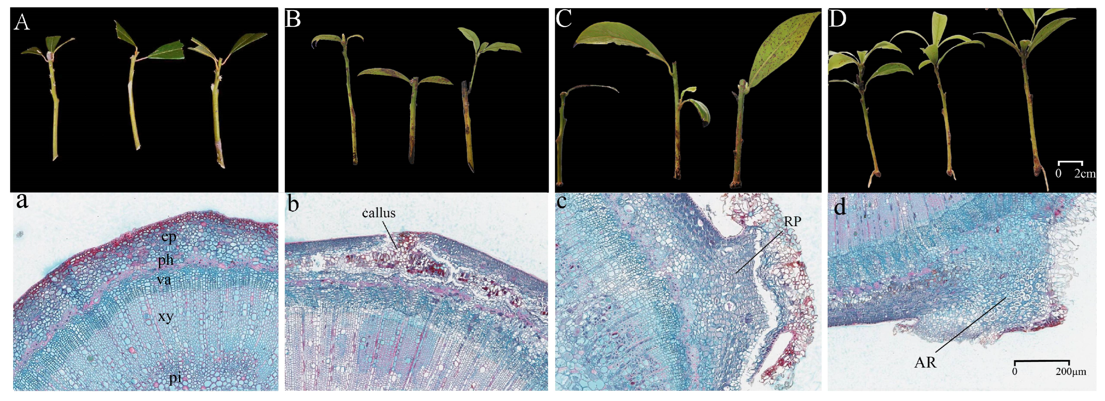
Figure 1. Morphological and anatomical changes during AR formation in P. bournei cuttings. Note: (A,a) PB0. (B,b) PB1. (C,c) PB2. (D,d) PB3. RP, root primordia; AR, adventitious root; ep, epidermis; ph, phloem; va, vascular; xy, xylem; pi, pith. PB0, PB1, PB2, and PB3 represent 0, 15, 30, and 50 days after cutting, respectively.
Horticulturae 11 01238 g001
Figure 2. Changes in physiological indices during AR formation in P. bournei. (A,B) Change in key nutritional indicators. (CG) Changes in enzymatic activities. (HJ) Changes in stress-responsive metabolite levels. Different lowercase letters indicate significant differences (p < 0.05), and this applies hereafter. PB0, PB1, PB2, and PB3 represent 0, 15, 30, and 50 days after cutting, respectively.
Horticulturae 11 01238 g002
Figure 3. Changes in endogenous hormone levels during AR Formation. (AE) Change in the content of five endogenous hormones. (FH) Change in hormonal ratios. PB0, PB1, PB2, and PB3 represent 0, 15, 30, and 50 days after cutting, respectively.
Horticulturae 11 01238 g003
Figure 4. Volcano plot and Venn diagram of DEGs. In the volcano plot, each point corresponds to a unigene. The dashed lines in the volcano plot represent the significance thresholds for −log10(p-value) and the fold change magnitude, used to identify genes that are significantly up-regulated or down-regulated. (AC) Volcano plot of three comparison groups. (D) Venn diagram of the three comparison groups. The x-axis represents the log2 fold change, indicating a logarithmic measure of the expression level difference for a unigene between two samples. The y-axis displays the negative log10-transformed p-value or adjusted p-value (padj), reflecting statistical significance. PB0, PB1, PB2, and PB3 represent 0, 15, 30, and 50 days after cutting, respectively.
Horticulturae 11 01238 g004
Figure 5. GO enrichment analysis of differentially expressed genes across comparison groups. (A) PB0 vs. PB1; (B) PB1 vs. PB2; (C) PB2 vs. PB3. The x-axis represents the number of DEGs, and the y-axis represents the GO terms. PB0, PB1, PB2, and PB3 represent 0, 15, 30, and 50 days after cutting, respectively.
Horticulturae 11 01238 g005
Figure 6. KEGG enrichment analysis of differentially expressed genes across comparison groups. (A) PB0 vs. PB1; (B) PB1 vs. PB2; (C) PB2 vs. PB3. The x-axis represents the gene ratio, the y-axis represents the KEGG pathways, and the color of the dots indicates the adjusted p-value (p.adjust). PB0, PB1, PB2, and PB3 represent 0, 15, 30, and 50 days after cutting, respectively.
Horticulturae 11 01238 g006
Figure 7. Expression analysis of DEGs in the phenylpropanoid biosynthesis pathway. The heatmap illustrates changes in gene expression levels across different stages, the color gradient from blue to red indicates expression levels ranging from −2 to 2, and this applies here after. PB0, PB1, PB2, and PB3 represent 0, 15, 30, and 50 days after cutting, respectively.
Horticulturae 11 01238 g007
Figure 8. Expression analysis of DEGs in the plant hormone signal transduction pathway. PB0, PB1, PB2, and PB3 represent 0, 15, 30, and 50 days after cutting, respectively.
Horticulturae 11 01238 g008
Figure 9. Module construction and hub gene identification based on WGCNA. (A) The appropriate soft threshold was selected to construct the weighted co-expression network model. (B) Modules identified using the dynamic tree cut method, with each color corresponding to a distinct module. (C) Expression patterns of hub module characteristic genes. (D) Heatmap of correlations between each module and hormonal traits, with values within each cell representing the correlation coefficient. (E) Construction of the gene co-expression network for hub modules.
Horticulturae 11 01238 g009
Figure 10. Correlation analysis of genes associated with endogenous hormones. (A) Correlation analysis of genes associated with IAA; (B) Correlation analysis of genes associated with ABA. * p < 0.05, ** p < 0.01, *** p < 0.001.
Horticulturae 11 01238 g010
Disclaimer/Publisher’s Note: The statements, opinions and data contained in all publications are solely those of the individual author(s) and contributor(s) and not of MDPI and/or the editor(s). MDPI and/or the editor(s) disclaim responsibility for any injury to people or property resulting from any ideas, methods, instructions or products referred to in the content.