Article

Olive Escudete (Dalmatian Disease) Caused by Botryosphaeria dothidea as a Result of Fly–Midge–Fungus Interaction

by
Hani K. Aldebis
1,
Antonio Santos-Rufo
1,2,*,
Ibrahim Eldesouki-Arafat
3,
Enrique Vargas-Osuna
1,
Juan Moral
1,
Antonio Trapero
1 and
Francisco Javier López-Escudero
1
1
Excellence Unit ‘María de Maeztu’ 2020–23, Department of Agronomy, Campus de Rabanales, University of Cordoba, 14071 Cordoba, Spain
2
Department of Agroforestry Sciences, ETSI University of Huelva, 21007 Huelva, Spain
3
Horticulture Research Institute (HRI), Agricultural Research Center (ARC), Giza 12619, Egypt
*
Author to whom correspondence should be addressed.
Horticulturae 2024, 10(4), 321; https://doi.org/10.3390/horticulturae10040321
Submission received: 10 January 2024 / Revised: 8 February 2024 / Accepted: 10 February 2024 / Published: 26 March 2024

Abstract

:
Escudete, which is caused by Botryosphaeria dothidea, is a disease that is widely distributed in the Mediterranean basin, but is of little general importance. Nevertheless, serious attacks have been observed on occasion, which have caused a considerable reduction in the quality of table olives. The incidence of the pathogen has been associated with damage caused by the olive fly (Bactrocera oleae) and the presence of a possible vector agent, i.e., the midge Prolasioptera berlesiana, whose larvae can feed on fly eggs (although the role the midge may play in the spread of this disease is not well known). Therefore, it is necessary to clarify these interactions to adopt appropriate disease control measures. Studies were conducted in olive orchards planted with the Gordal Sevillana, Picudo, and Hojiblanca olive cultivars. Field surveys were carried out in order to sample their fruits for laboratory analysis, and several bioassays were also performed. Moreover, the population of B. oleae adults was monitored using traps that were baited with food attractants. The results indicated that the three agents developed and evolved in parallel under field conditions. Thus, the midges were attracted by the oviposition punctures caused in fruits by olive fruit flies, regardless of whether the punctures contained eggs. All the investigated olive fruits in which midges were present inside punctures created by olive fruit flies exhibited typical symptoms of escudete, which is necessary for the development of this disease. Forty-eight hours after fly punctures were artificially simulated in the olive fruits, 48.0% of them contained a midge, whereas no midges appeared in the artificially created shapeless wounds in the fruits. This indicates that an olive fly egg is not required for the development of midges; however, they do prefer punctures made by B. oleae. Moreover, when the olive fruits were incubated in a humid chamber, the B. dothidea fungus only appeared in those fruits that contained midges, thus indicating a close relationship between these two agents. Additionally, the midges were able to complete their entire development from egg to adult under controlled conditions, and they fed on the pure cultures of the B. dothidea fungus. Furthermore, although no pathogens were present in the immature midges, some of the pathogens could have been isolated from the inner tissues of the adult female midges. The fact that mycangia is present in the abdomen of P. berlesiana supports the hypothesis that their relationship with B. dothidea may be mutualistic and that they may act as a vector for the fungus.

1. Introduction

More than 98% of all olive trees grow around the Mediterranean basin, with Spain being the leader in olive production [1,2]. The region of Andalucía is the most important olive-growing area [3]. Many parasites (including pests and diseases) compromise the sanitary status of this crop. Dalmatian disease—which is also better known as “escudete” (i.e., small shield in Spanish and Portuguese)—is caused by Botryosphaeria dothidea (Moug.) (see Trapero and Blanco (2010) [4] for details), and it is widespread in olive groves in the Mediterranean area [5,6,7]. In general, this disease is of little importance except for its influence on the quality of table olive fruits. However, Salgues (1937) [8] indicated that olive fruits that are affected by Dalmatian disease (hereafter referred to as escudete disease) can reduce oil production by 3%. In a study carried out by González et al. (2006) [9], it was shown that there is not much difference in the quality of the oil extracted from the samples of healthy olives compared to others that are infected by escudete. The current average data on the disease in Andalucía indicate that 4.1%, 2.1%, 1.2%, 0.5%, and 0.7% of fruits have symptoms (these were estimated on different dates) in the provinces of Cádiz, Jaen, Granada, Seville, and Huelva, respectively [10].
The infection of olive fruits by B. dothidea is greatly favored by the presence of wounds in the fruits, and it has been positively correlated with attacks by the main pest of olive flies: Bactrocera oleae (Gmelin) (Diptera: Tephritidae). In addition, with the incidence of Prolasioptera berlesiana (Diptera: Cecidomyiidae), which is cited as a predator of olive flies’ eggs, it has also been suggested that the midge may act as a pathogen vector [11,12,13,14,15,16,17,18,19,20]. Although most authors agree that B. dothidea develops in olives that have been previously punctured by olive fruit flies, there are different approaches regarding the role of P. berlesiana in the transmission of the pathogen. Several authors have attributed transmission to the ectoparasitic activity of olive fruit fly eggs and the possible role of a vector of the pathogen [12,17,21,22,23,24,25,26], whereas some consider P. berlesiana to be the main species responsible for the spread of the pathogen [18]. However, others have argued that it is instead a mycophagus whose females take advantage of the stings of B. oleae to oviposit, with larvae feeding on the invading fungi (which are most commonly B. dothidea) as well as (most likely) on decaying plant tissues [27,28]. Additionally, Harpaz and Gerson [27] maintain that, in most of the infections that occur in the field, the pathogen enters through the oviposition or exit holes created by B. oleae. However, they also advised that it can utilize any puncture in olive fruits in order to infect them and does not depend on the presence of oviposition holes created by olive fruit flies or on midges as vectors. Moreover, they noted that P. berlesiana only feeds on fungi and therefore cannot be considered a predator of olive fruit fly eggs.
The supposed relationship between the fungus B. dothidea and the midge P. berlesiana in olive groves poses a big question about the role that midges play in the spread of this disease. Their characterization as predators of the eggs and young larvae of olive fruit flies or olive tree pests, thereby generating escudete disease, is not easy to confirm. In terms of its practical importance, the productive orientation of an olive grove must be considered. The negative effects of these midges on table olives are evident; however, when the final product is olive oil, their presence may be considered positive due to the significant reduction in the reproductive potential of olive fruit fly populations, especially in the summer months.
Despite efforts to unravel the interactions between the three agents (i.e., B. dothidea, B. oleae, and P. berlesiana), their nature is not well known; therefore, it is necessary to clarify these interactions to adopt appropriate disease control measures, which is the aim of this study.

2. Materials and Methods

2.1. Experimental Field

An experimental field (37°30′41″ N, 4°25′58″ W; “La Mina”; Andalusian Institute for Research and Formation in Agriculture and Fishery “Cabra” Centre; IFAPA in Spanish) located in the foothills of the Subbetic region of Córdoba (southern Spain; 73 km southeast of the capital and 488 m high) was selected for sampling. This is a fly endemic area that, in previous years, has presented a high incidence of escudete.
In this field, there were 12 olive cultivars (from olive trees planted in 1987), which were replicated 12 times in a complete block design, with one tree from each cultivar per block. From these 12 varieties, the Gordal Sevillana, Hojiblanca, and Picudo table olive cultivars were selected.

2.2. Temporal Evolutions of Agents

A total of 10 olive trees were randomly selected for each cultivar in order to determine the dynamics of the three agents in terms of their involvement in disease development under field conditions. Four branches were marked per olive tree (one per cardinal orientation), with 20–25 fruits each. Evaluations were conducted weekly from summer to fall (July to November 2011). Olive fruits showing fly punctures and/or escudete symptoms were harvested and transported to the laboratory to be examined under a stereoscopic microscope in order to check for the presence of P. berlesiana. The olives collected had been injured by olive flies and/or had escudete symptoms. In total, 913, 1225, and 188 olive fruits were collected from the Gordal Sevillana, Hojiblanca, and Picudo cultivars, respectively, which were then divided into three groups: those with escudete symptoms only, those with midges only, and those with both agents. Due to the low number of injured olives (i.e., those injured by olive flies) in the Hojiblanca and Picudo cultivars, further studies were carried out with the “Gordal Sevillana” cultivars only.
The putative role of the olive fruit fly in the direct propagation of the B. dothidea fungus and the development of escudete disease in the absence of P. berlesiana was also studied. To achieve this, field observations were made weekly (from July to November) of 4 of the branches out of the 10 “Gordal Sevillana” olive trees (where each branch possessed 20 to 25 fruits), which were randomly selected. All the fruits that were punctured by olive flies or that possessed escudete symptoms were collected and examined under a stereoscopic microscope to prove the presence of midges. In addition, 2475 olives from the 3 cultivars were collected and examined under a stereoscopic microscope in order to check for the presence of punctures made by olive flies without signs of midges. Completely healthy olives (1938) were also collected and placed among punctured fruits that did not appear to contain midges, and they were then placed in a humid chamber for one week to be examined for fungal infection.
The monitoring of olive fruit flies in the study plot was carried out using McPhail traps, which were baited with 300 mL of an aqueous solution of 4% diammonium phosphate (NH4)2HPO4. The meteorological data (i.e., the daily mean temperature and relative humidity) were recorded by the meteorological station of the IFAPA “Cabra” Centre near the study area.

2.3. Bioassays

2.3.1. Live and Non-Live Olive Fly Oviposition Stings

The incidence of P. berlesiana and B. dothidea was verified in the olive fruits from “Gordal Sevillana”, which were collected and classified according to the presence or absence of B. oleae oviposition stings. Olives with the presence of eggs, larvae in different stages, pupae, and empty hallways of the emerged imago were classified as live stings. In addition, olives without eggs, which could also include oviposition punctures by P. berlesiana with predated eggs, were classified as non-live stings.

2.3.2. Artificial Wounds

The incidence of P. berlesiana and B. dothidea was also verified in two of the types of artificial wounds, which were performed in the field on the olive fruits of the Gordal Sevillana cultivar (where the cultivars that presented greater susceptibility were selected).
For the first type, the field experiments were carried out in two periods to cover the flight of the flies for a longer time: late July (Experiment A) and late August (Experiment B). For each period, 10 trees were randomly selected from each of the 4 tree branches with 20–25 completely healthy olives on the north and south sides (2 branches per side). Fruits on two branches (one on each side) were pricked once with a needle, simulating olive fly oviposition punctures (SFOPs) (1 mm deep, 0.5 mm wide, and an approximately 45° inclination); the fruits on the other two branches were left unpricked as controls. Weekly observations were made until October, and the olives with escudete symptoms were harvested and examined in the laboratory under a stereoscopic microscope to detect the presence of midges.
The second type of wound was carried out with the main objective of verifying if P. berlesiana only selects oviposition punctures made by olive flies or if they simply select any oviposition wounds present in olives. For this, two types of wounds were made, one with SFOP and another 3–4 mm in size with a sterile scalpel without a determined shape, which we call shapeless wounds (SWs). For this, four of the trees of the Gordal Sevillana cultivar were randomly selected. Six branches were marked on each tree (where each branch had an average of 43 olives) and two types of artificial punctures were made in the four-branched fruits: SFOPs in two branches and SWs in two branches; two other branches were used as controls (i.e., left unpricked). The samples were collected in two periods: 24 and 48 h after puncture. In the laboratory, the fruits were examined to detect P. berlesiana. Subsequently, all the olives were placed in a humid chamber to check the development of B. dothidea.
To evaluate the behavior of the midges as well as the development of escudete disease in the shapeless wounds (SWs) that were in place for a longer time after the punctures were made, six branches per tree were marked in the five trees belonging to the same cultivar (where each contained 20–25 olives). Artificial SWs were performed on four branches of fruit, and two branches were used as controls without wounds. Weekly observations were made for a month. Fruits with typical escudete symptoms were taken and examined under a stereoscopic microscope to detect the possible presence of midges.
To study the development of B. dothidea in the two types of artificial wounds (SFOP and SW), the olive fruits of “Gordal Sevillana” were harvested 48 h after the punctures were made. These were then classified into two groups according to the type of wound. A third group of olives without lesions (i.e., completely healthy) was used as a control. Within each group, two subgroups were formed based on the presence or absence of midges. Isolations from all the groups were immediately performed on potato dextrose agar (PDA) by taking small pieces of olivaceous tissue from inside the wound.

2.3.3. Food Behavior of Midge Larvae

The feeding behavior of midge larvae was evaluated in two ways: by rearing them in B. dothidea cultures and by rearing them in olive fruits.
For the former, eggs or newly hatched larvae of P. berlesiana that were extracted from olives with artificial wounds via SFOP were placed in a pure culture of B. dothidea that was grown on PDA and kept in an environmental chamber programmed at 26 ± 2 °C, 60 ± 5% RH, with a photoperiod of L16:D8 (1500 lux).
For the breeding on olive fruits, newly hatched midge larvae that were obtained in the same way as in the previous method were transferred to healthy olives. A total of 64 larvae were used; half of them were transferred to the olives previously disinfected with 15% sodium hypochlorite for 40 s and the other half were transferred to olives without disinfection. Before transferring the larvae, the olives were artificially pricked via SFOP. The olives with the transferred larvae were placed in transparent plastic cylindrical boxes in groups of eight larvae per box, which were then kept under the same conditions as mentioned above.

2.4. Isolation of B. dothidea

External and internal isolations were performed in a PDA acidified with lactic acid (APDA; 2.5% (vol/vol) at 2.5 mL/L of medium) to study the presence of B. dothidea in the immature stages (i.e., the egg, larvae, and pupae stages) and in the adult olive fly and midge females. The B. oleae adults used were captured using INVAGINATED EOSTRAP®-type pheromone traps with a diffuser (DACUSNEX 4) that attracted females and males (Sanidad Agrícola Econex, S.L., Murcia, Spain). B. oleae that were in the immature stages were extracted from olives that had been infested by olive flies, while P. berlesiana in the adult and immature stages were obtained from olives that had been artificially or naturally punctured by olive flies.
For external isolation, 15 individuals of each species and stage were transferred to a PDA medium (5 individuals per plate) contained in Petri dishes. For the isolation of the internal tissues, 20 individuals of each species and stage were superficially sterilized with 15% sodium hypochlorite for 40 s and then rinsed in autoclaved water. After drying with sterile filter paper, these were immersed in 5 mL of sterile distilled water and ground with an electric homogenizer. From the initial maceration, dilutions of 0.1, 0.01, and 0.001 were made. Next, 300 μL of each one and of the initial maceration were deposited in PDA plates (5 plates per case). The identity of the fungus was confirmed by molecular analysis based on “nested PCR” according to the methodology of Moral et al. [7].
In addition, newly emerged P. berlesiana females were immersed in a 5% KOH solution for 24 h; after washing with sterile water, they were observed with a NIKON (Tokyo, Japan) light microscope to detect the B. dothidea inside the midge body. For this, the entirety of the females or only their abdomens were spread on a slide with a drop of sterile water and observed at 200×.

2.5. Statistical Analysis

The incidence of the agents involved (B. oleae, P. berlesiana, and B. dothidea; 0–100%) was analyzed via a chi-squared test (χ2) at p = 0.05 to check whether the frequency values obtained were significantly different from the theoretical ones.
The analysis was applied to the presence of P. berlesiana in relation to the state of the sting of B. oleae (live and non-live) in the olives and the incidence of B. dothidea in relation to the damage caused by B. oleae [9].
All data in this study were analyzed using Statistix 10 (Analytical Software, Tallahassee, FL, USA).

3. Results

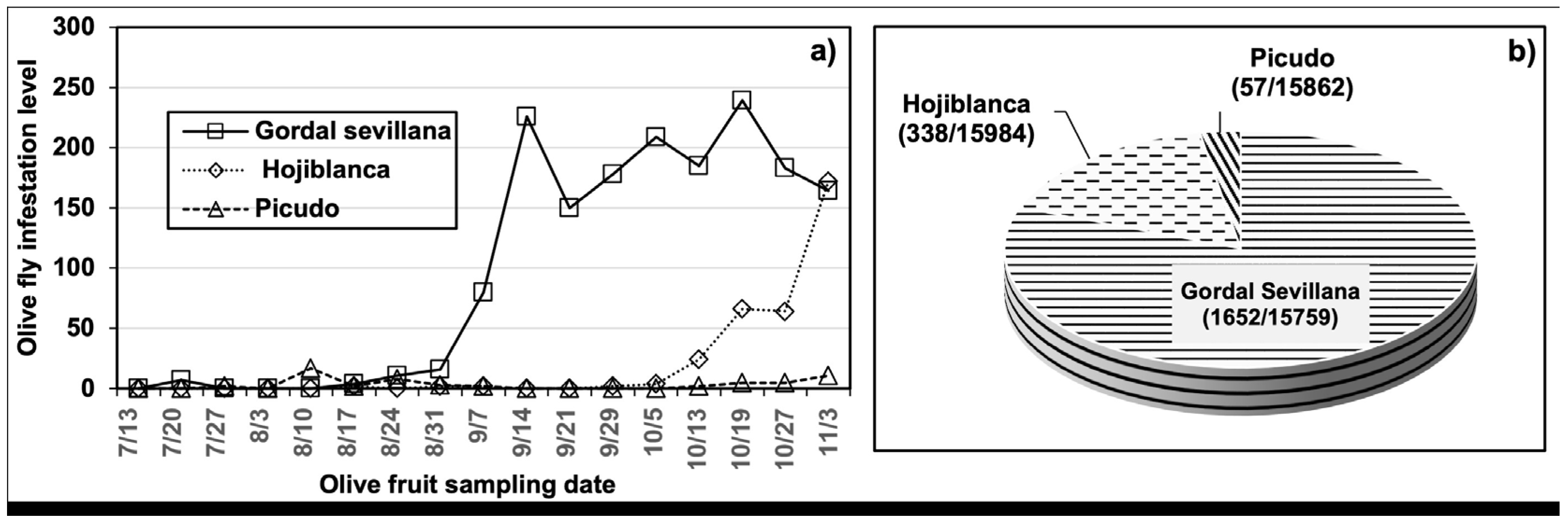
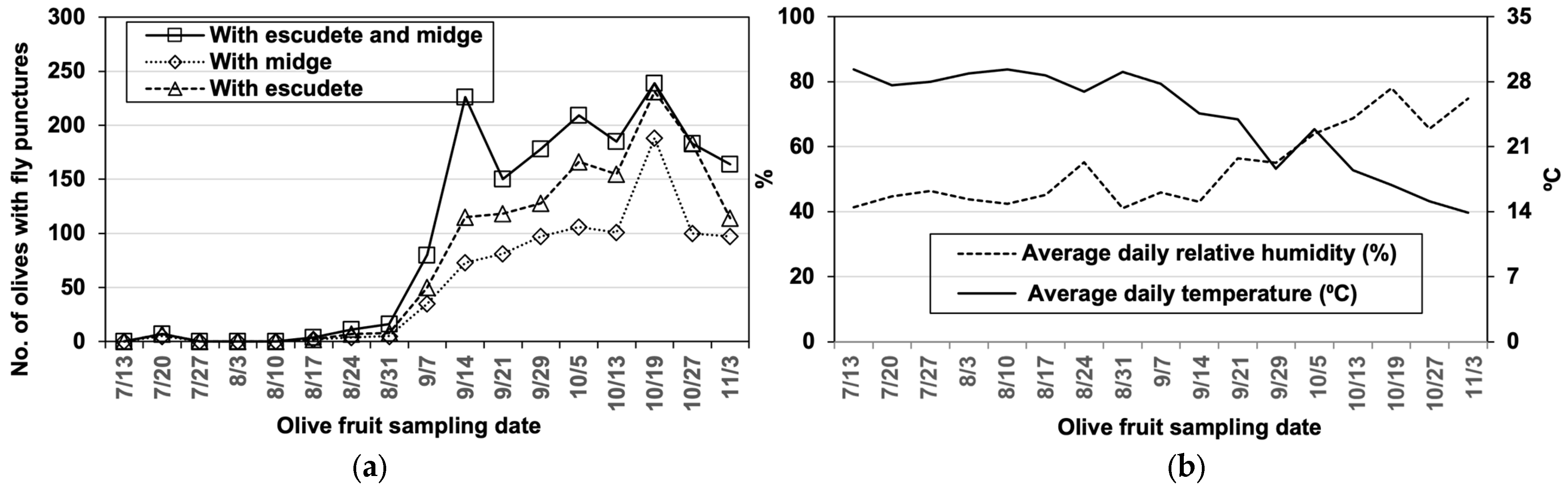
3.1. The Dynamics of B. oleae, P. berlesiana, and B. dothidea

In general, the attack of the olive flies throughout the campaign was not high in the three cultivars studied (Figure 1a). The Gordal Sevillana cultivar was the most affected, with 10.0% of the olives damaged, while the damage did not exceed 2.1% and 0.4% for the Hojiblanca and Picudo cultivars, respectively (Figure 1b). The first punctures of the olive flies were detected in the second week of July; then, the damage to the flies began to increase progressively, becoming more visible from September to November (Figure 1a).
Weekly field observations indicated that the time course of the three agents developed in parallel. The presence of P. berlesiana was observed one week after the first fly punctures were detected, while escudete symptoms appeared one week later. At the same time, the midge and disease symptoms increased as the fly attacks increased from September to November. Although the three cultivars had the same tendency, this was more evident in the Gordal Sevillana cultivar (Figure 1a and Figure 2a). In the Hojiblanca and Picudo cultivars, however, the low incidence of the fly punctures did not make them as marked (Figure 1). The climatic conditions during the study period are shown in Figure 2b.
The symptoms of midges and escudete were only found in the oviposition punctures and the exit holes of olive flies. In most of the fly punctures, a midge egg or larva was detected. However, two of them were occasionally observed in the same puncture. The pupae were always found outside the B. oleae puncture hole or very close to it. As expected, clear differences can be observed between the flies, the fungi, and the midges in the Venn diagram: the midges always appeared in fruits with escudete symptoms and the latter always appeared in fruits with fly wounds (Figure 3).
Pearson’s correlation analysis indicated a positive correlation (r > 0.9; p = 0.05) between the three agents. It should be noted that this correlation was excellent in the case of the cultivar Gordal Sevillana. Thus, a strong positive correlation was found between the damage produced by the olive fly and the midge population and escudete disease, with correlation coefficient values of 0.94 and 0.96, respectively (where the value was even higher between the midge population and escudete disease (r = 0.997)).
In addition, laboratory observation of olives with B. oleae oviposition punctures collected from the three cultivars showed midge and disease symptoms. The incidence of the latter agents was higher in the Gordal Sevillana cultivar; 22.7% of fly stings showed the presence of the cecidomyiid and 41.2% showed symptoms of the disease (Table 1) (all punctures with midges had disease symptoms simultaneously). The percentage of olive fruits with escudete symptoms was significantly higher than those with midges in the three cultivars studied (χ2 = 72.0, 156.8, and 17.9; p < 0.0001). It should be noted that this result likely did not reflect the real presence of midges in olives with disease symptoms.

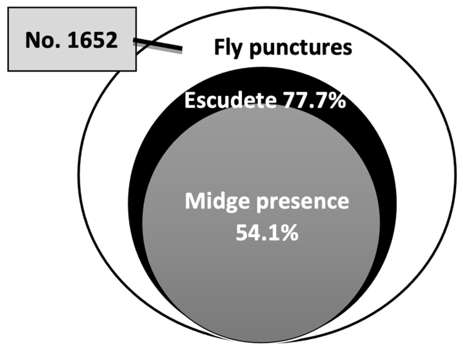
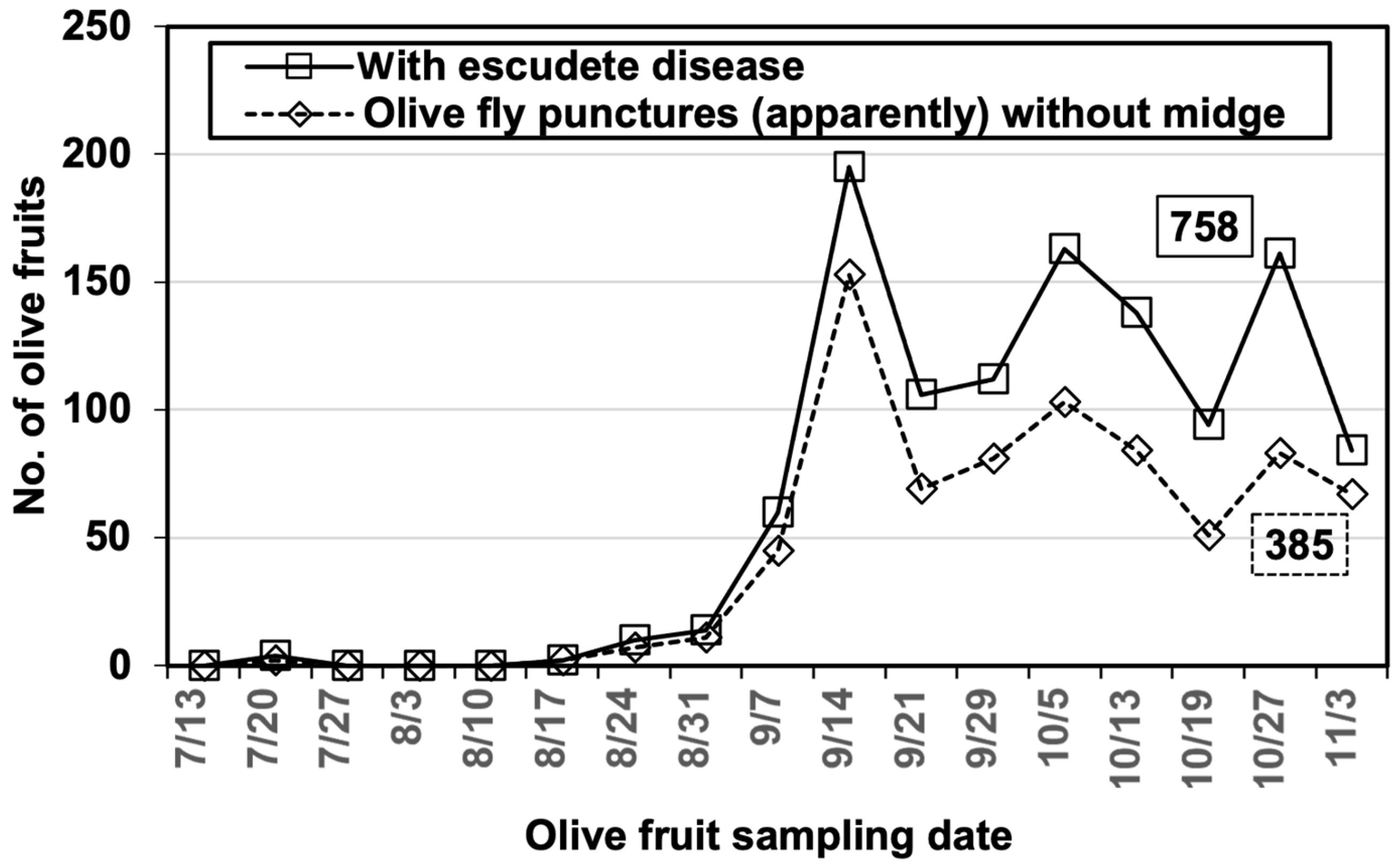
3.2. Role of B. olea in Disease Development

The main objective of this experiment was to verify the role of the olive fly in the propagation of B. dothidea spores and its ability to produce infections that led to the development of escudete disease in oviposition punctures without the presence of midges. The field observations carried out weekly on the Gordal Sevillana cultivar indicated that 25.4–94.0% (50.8% in total) of the stings without the potential presence of P. berlesiana showed signs of escudete caused by B. dothidea (Figure 4).
Likewise, under controlled conditions, when the olive fruits injured by the flies—which did not present visible signs of P. berlesiana—were placed in a humid chamber, they presented the development of B. dothidea (χ2 = 163.6; p < 0.0001). Likewise, the development of fungi was also observed in a low percentage of completely healthy olives (Table 2).
The isolations of B. dothidea in the adult and immature stages of B. oleae showed that the fungus was only detected on the fly bodies. Thus, about 72.0% (n = 255) of the adult fly females carried this fungus on their bodies’ external surfaces. The identity of the fungus was confirmed via molecular analysis based on the “nested-PCR” technique, which was conducted in accordance with the methodology of Moral et al. [7].

3.3. Incidence of P. berlesiana and B. dothidea in the Live and Non-Live Olive Fly Stings

The results indicated that a high presence of B. dothidea and P. berlesiana was found in the non-live stings. The χ2 (chi-squared) test showed that the incidence of both agents in the non-live stings was significantly higher compared to the live stings (χ2 = 37.5 and 26.7, respectively; p < 0.0001). In all cases, the incidence of B. dothidea was higher than that of the cecidomyiids (Table 3). The fly punctures with midges always, simultaneously, had symptoms of disease; however, it should be noted that it would be necessary to know if this also occurs in artificial wounds.

3.4. Incidence of P. berlesiana and B. dothidea in Artificial Wounds

Regarding the artificial wounds as determined by SFOP, the data from the field experiments that were conducted in late July (A) and late August (B) indicated that P. berlesiana was oviposited in 38.2% (n = 1043) of the artificial wounds without eggs or fly larvae. It was also shown that the growth of B. dothidea in artificial SFOP wounds followed the same trend as when the wounds were produced naturally by flies. The pathogen appeared in 47.4% of the wounds and its presence was four times higher when they were occupied by P. berlesiana (χ2 = 374.15, p < 0.0001) than when midges were not in the wounds (Table 4).
Likewise, in the branches with unpricked olives that were assigned as controls, a small number of fruits with B. oleae fly punctures were found, with P. berlesiana being present in 74.2% of them (n = 93).
The other assay—where the incidence of P. berlesiana and the development of B. dothidea in the SFOP and SW artificial wounds were compared—indicated that there were cecidomyiid eggs or larvae in 40.0% of the olives with SFOPs 48 h after the punctures were made; none were found in SWs. When all the olives were incubated in a humid chamber and observed three weeks later, the B. dothidea fungus had only developed in olives with SFOPs, of which a small percentage (4.3%) did not contain P. berlesiana (Table 5).
However, when olive fruits with SWs were observed on a weekly basis in the field for a long period of time (i.e., 4 weeks), some cecidomyiids and fungal development were recorded in 6.4% of these wounds. It should be noted that, in most cases, the midges fed gregariously (3–6 larvae) on the mycelium of the fungus that grew outside the wounds. Likewise, the development of B. dothidea was observed with an incidence of 14.5% in these wounds. Both agents were detected in the second week after the punctures were made, but the highest incidence was recorded in the third week.
The isolations from the olive tissue within the two types of artificial wounds 48 h after puncture (see above) showed the development of B. dothidea in 100.0% of the wounds that contained cecidomyiids. Likewise, these isolations showed the development of B. dothidea, but without the presence of cecidomyiids, in 15.0% of the olives. In both cases, the development of B. dothidea came from olives with SFOPs, while SWs and the control (olives without lesions) did not present fungal development (Table 6).

3.5. Food Behavior of Midge Larvae

In this study, 70% of the eggs or larvae reared on a pure culture of B. dothidea mycelium were able to complete their development to the adult state using the fungi’s mycelia as a food source. However, the larvae that were transferred to the olives previously disinfected and artificially punctured, i.e., the simulated B. oleae oviposition punctures, did not develop and, thus, all of them died within two days.

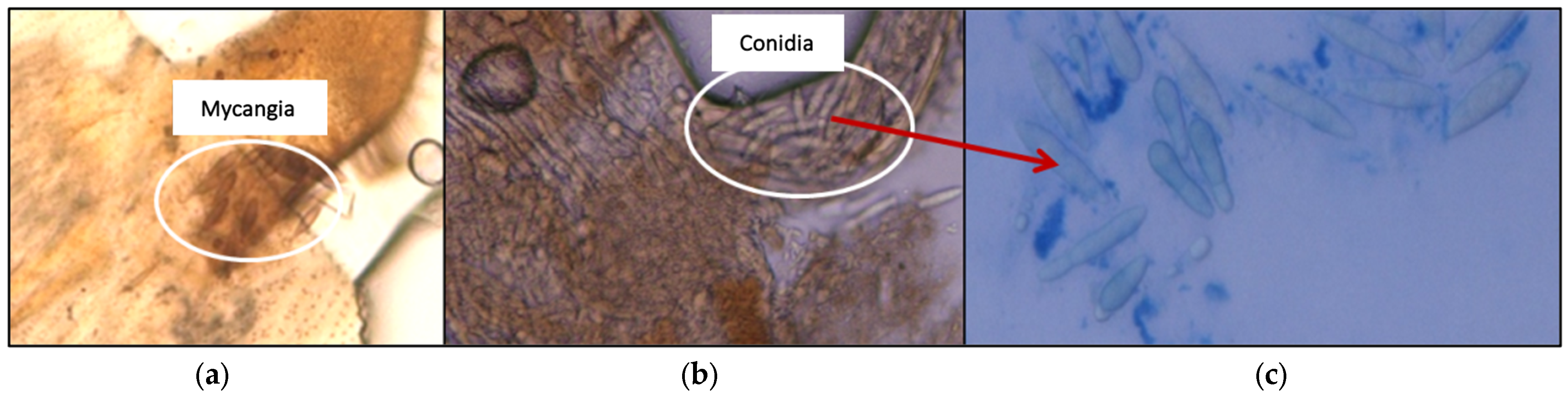
3.6. Verification of the Presence of B. dothidea in P. berlesiana

The isolations from P. berlesiana, both on the external surface and inside the immature stage and adult female bodies, were only found on the fungus inside the female bodies. Likewise, microscopic observations of the female abdomens showed the existence of mycangia, which were associated with the terminal segments of the abdomen close to the ovipositor; the conidia of B. dothidea were observed within this mycangia (Figure 5).

4. Discussion

Olive escudete, which is caused by B. dothidea, is a disease that exclusively affects olive fruit and has been reported in several Mediterranean countries [4]. Several investigations have related the development of escudete disease to the presence of olive fly punctures [11,12,13,14,15,20,21,22,23,25,27,29,30,31,32]. However, certain authors have suggested that the fungus of B. dothidea (i.e., the causative agent) grows in the wounds of olives punctured by B. oleae after being transmitted by the P. berlesiana cecidomyiid. In this context, the different ways in which mosquitoes participate in this relationship have been indicated.
In the present study, weekly field observations from mid-July showed different types of damage, as well as their associated agents that evolved in parallel. First, the flies’ oviposition punctures appeared, then the midges; later, the fungi that caused the disease appeared, along with an increase in the development of the three agents, which were observed from September to November. Similar observations were reported by González [33] in Sevilla (Spain), Solinas [13] and Sasso and Viggiani [28] in southern Italy, and Basher et al. [30] in Syria. One P. berlesiana egg or larva was detected in most of the spawning holes. Occasionally, two eggs or larvae were observed, which is in agreement with the findings of De Laurentiis [34], who observed the presence of two to three midge eggs in the same type of fly wound. The pupae were always found outside or especially close to the B. oleae puncture hole.
The incidence of the three agents was different between the cultivars. Thus, in the Gordal Sevillana cultivar, there was a greater presence of the agents involved. The same tendency was also observed in the Hojiblanca and Picudo cultivars, but the low incidence of escudete disease was not as marked due to the fly punctures. Several authors have advised that olive flies prefer table olive cultivars, which include “Gordal Sevillana” [6,35,36,37,38]. This cultivar is also susceptible to escudete disease attack [18,20,34].
The fly punctures with midges always simultaneously showed symptoms of the disease. However, some of the olives affected by the disease were found to be free of midges. This was evident in the Gordal Sevillana cultivar, where 18.0% of the olives showed disease symptoms without the apparent presence of midges. This percentage could be greatly reduced if, in many of the samples, the cecidomyiids had left the fruit after it had completed its development without leaving any detectable trace behind. As can be seen in the Venn diagram, there was an apparent symbiotic interaction between the three agents as follows: midges were found to always appear on fruits showing escudete symptoms, and escudete always appeared on fruits with fly wounds. Furthermore, when we conducted a Pearson correlation analysis, a positive correlation between the three agents was indicated, as has been previously reported [29,30]. On the one hand, a strong positive correlation was found between the injury caused by the olive flies and the presence of both P. berlesiana and B. dothidea (where the correlation coefficient values were 0.94 and 0.96, respectively). On the other hand, an even stronger positive correlation was found between the cecidomyiids and the fungi (a correlation coefficient of >0.99).
It is evident that the emergence of escudete disease is related to the damage caused by olive flies. However, its role in the direct spread of the B. dothidea fungus and its ability to develop the disease without the presence of P. berlesiana are not well understood. A considerable percentage of the olives with fly punctures showed signs of escudete disease in the absence of midges. However, this high percentage of olives infected by B. dothidea cannot be solely attributed to the olive flies as they are not directly responsible for the spread of the fungus and the development of the disease in oviposition punctures. This is most likely due to the fact that mosquitoes leave holes after completing their development, and these punctures cannot be detected via weekly or long-term observations. Likewise, both the flies and midges may not be propagating agents of the fungus spores; instead, the fungus may have been transmitted through the air, as Harpaz and Gerson [27] have advised. Other authors also agree that olive flies should not be considered vectors of B. dothidea [16,17]. In addition, under controlled conditions, 22.9% of the olive fruits injured by flies (which did not present P. berlesiana or symptoms of the disease) developed B. dothidea three weeks after being placed in a humid chamber. However, only 4.6% presented a development of B. dothidea three weeks later when completely healthy olives were placed under the same conditions. The growth of the fungus in the olives injured by the flies may not have been due to the direct transmission of the fungus by the flies, but rather mosquitoes may have landed on the fly bite without leaving a trace. Regarding the second group (i.e., the completely healthy olives), the growth of the fungus manifested itself in only a small percentage of olives; moreover, it was also induced in a humid chamber, meaning that it could not occur under field conditions. However, Latinović [39]—who conducted a study in Montenegro—stated that, of the olives collected directly from the field without any type of damage that were not placed in a humid chamber, only 23.0% presented escudete symptoms. Mateo-Sagasta [14] highlighted that the fungus can act as an independent pathogen “by itself”, although the severity of the disease is always more significant in fruits previously damaged in any form. These statements are not in full agreement with the opinions of several authors due to the fact that the disease develops in olives that have been previously punctured by flies or via other injuries [13,16,27,28]. Even Solinas [13] believes that the role of the fly is not exclusively limited to the opening of wounds in olive fruit or to the possible first food supply of midge larvae with their own eggs. The authors of this study maintain that the role of flies, whose presence creates favorable—and sometimes necessary—conditions for the development of escudete disease, must also be taken into account in the actions carried out by flies. Acid PDA culture isolates have shown that B. dothidea is only detected on the outer surface in 74.0% of adult female B. oleae, while none have been detected in the immature stages. The identity of the fungi was confirmed by means of PCR technique procedures. Harpaz and Gerson [27] experimentally demonstrated that B. oleae can transport B. dothidea spores over short distances on their body surfaces. However, there is no evidence that they do so in nature, and the main role of the fly is apparently to provide an entry point for B. dothidea spores into the impenetrable skin of the olive.
The presence of P. berlesiana and B. dothidea was found to be significantly higher in non-live stings. P. berlesiana larvae were only found in the oviposition punctures of flies, with no traces of fly eggs or signs of larval tunnels observed. Therefore, this suggests that mosquitoes play a predatory role with respect to olive fly eggs and larvae, thus masking the proportion of olives with live stings, which may help explain this huge difference between live and non-live stings. Other investigations have indicated a greater presence of P. berlesiana in the non-live stings of flies, which has also been thought to be due to the predatory behavior of mosquitoes [6,15,16,34].
Although the high presence of P. berlesiana in olives that have non-live stings of B. oleae can be attributed to the predatory behavior of the cecidomyiid, it cannot be ruled out that many of these stings were not live, as demonstrated by fly oviposition tests. P. berlesiana was found in 38.2% of the field experiments, in which artificial wounds were made to simulate fly oviposition punctures (SFOPs) at the end of July (Experiment A) and late August (Experiment B). In addition, its presence was found to be greatly reduced in Experiment A (24.9%) compared to B (50.0%). This indicated an increase in the population of P. berlesiana in parallel with the increase in the population of B. oleae that was observed in September. An increase in the fly population at this time, on the same farm, has been previously observed by Aldebis et al. [40], Aldebis and Vargas-Osuna [41], and Sabariego-Sánchez [42]. Other authors have stated that the presence of B. oleae eggs is not strictly necessary for the development of P. berlesiana, and the latter can only evolve at the expense of the fungus [13,27,28]. On the other hand, it has also been shown that the development of B. dothidea in artificial SFOP experiments has followed the same trend as when the stings were produced by the fly naturally. According to Arambourg [16], although P. berlesiana is a predator of olive fly eggs, escudete disease can appear even when olive flies produce sterile punctures since midges can transmit the inoculum of the pathogenic fungus. B. dothidea appeared in 47.4% of the artificial wounds, but its presence was four times higher when the wounds were occupied by P. berlesiana (80.8%) than when midges were not present in the wounds (19.3%). In field experiments, Harpaz and Gerson [27] showed that up to 96.0% of olives that were pricked three times with a needle and observed 2–4 weeks later showed the development of B. dothidea. The authors attributed this infection to the airborne transmission of the fungus. With respect to the limitations of considering biological relationships as valid in absolute terms, other hypotheses could be centered on the fact that a large part of this infection was due to the previous presence of midges in the wounds. This is because, at 4 weeks, the fungus may have completed its development and subsequently abandoned the fruit. Likewise, Solinas [13] argued that the usual concomitance of the development of B. dothidea and P. berlesiana, as well as the ordinary transmission of the fungus itself together with the eggs of cecidomyiid, greatly reduce the importance of the eventual airborne transmission of the fungus.
Only the olive fly oviposition punctures or any other wound on the olives were selected in the experiment to check if P. berlesiana oviposits. Our results showed that midges were found in 40.2% of the olives that were pricked by SFOPs 48 h after the punctures were made, and only one egg or larva was observed per wound. All the olives with P. berlesiana developed B. dothidea 3 weeks after placing them in a humid chamber, and only 4.0% did not contain midges. None of the olives pricked without a certain shape (i.e., shapeless wounds (SWs)) contained midges, nor had there been any development of the fungus in a humid chamber. Likewise, the PDA isolates from olive tissue within the two types of artificial wounds 48 h after the punctures were made and from healthy olives corroborated the results. B. dothidea developed in 100.0% of the wounds that had cecidomyiids, as well as in 15.0% of the olives without their presence; in both cases, they came from olives with SFOPs. No development of fungi was observed in the isolations of the SWs and the control (i.e., the olives without lesions). Thus, the isolation of the fungus from the SFOP wounds only confirmed the close relationship between the three agents. This fact also increased the probability that the development of the fungus in wounds in which midges were detected was due to its previous presence in the fly punctures. When the olives with SWs were observed weekly in the field for one month, the presence of some of the cecidomyiids was recorded in 6.4% of these wounds. Likewise, the development of B. dothidea was observed in 14.5% of the SWs 4 weeks after pricking. It is worth mentioning that, in most cases, the midges fed gregariously (3–6 larvae) on the mycelia of the fungi that grew outside the wounds. The late appearance of the fungi was probably due to the oviposition of the midges in these wounds 48 h after the isolations were made. Therefore, although P. berlesiana did not require the live laying of flies, it did prefer to carry out its laying on wound types such as fly punctures as opposed to other types of wounds. However, according to Sasso and Viggiani [28], during the summer and autumn, P. berlesiana is able to oviposit in any wound found in the olive tree. Likewise, Sasso and Viggiani [28] and Viggiani and Sasso [43] advised that the larvae of P. berlesiana have opportunistic, mycophagic, or saprophytic behavior and feed on fungi that take advantage of wounds of a different nature in olives. These wounds are mainly caused by flies, and the fungus is B. dothidea.
The rearing experiment of P. berlesiana in a pure PDA culture of B. dothidea indicated that 70.0% of the eggs or neonate larvae transferred to this culture managed to complete their adult development. These results indicated that the main food of P. berlesiana is the fungus and that the larvae do not need to feed on the olive tissue, which is in agreement with the finding of Harpaz and Gerson [27]; however, it remains to be verified in future experiments whether midges also feed on the other fungal species distant from B. dothidea, as was indicated by these authors. However, the midge larvae that were transferred to previously disinfected and artificially punctured olives simulating B. oleae oviposition punctures failed to develop; thus, they all died two days later. Likewise, the adults of P. berlesiana that had emerged from olives with escudete symptoms and were confined to the healthy olives previously pricked with SFOP wounds failed to transmit the B. dothidea fungus and cause infections in olives. Shun et al. [44] were unable to isolate B. dothidea from the mycangias of Illiciomyia yukawai adults that had emerged from galls that were confined in plastic bags. These authors concluded that the recently emerged adults did not incorporate the fungus into the mycangia directly from the galls where they had developed.
The PDA isolates showed that B. dothidea was only detected within the adult female bodies of P. berlesiana. Likewise, microscopic observations of the female abdomen showed the existence of a bag (mycangia) associated with the terminal segments of the abdomen next to the ovipositor; moreover, B. dothidea conidia were observed in these bags. The scientific literature indicates that many species of cecidomyiids carry this specialized structure (mycangia) where the female incorporates the conidia or mycelia of the mutualistic fungi [44,45,46,47]. In the Lasiopterini tribe, this structure is found in the eighth abdominal segment [48,49]. The fact that certain species of midges, in the relationship between these insects and associated fungi, carry mycangia on their abdomen or ovipositor reinforces the concept of mutualism rather than opportunism [45,46,48,50]. In accordance with this concept, the results of the present study suggest that the relationship between B. dothidea and P. berlesiana could be mutualistic, with the cecidomyiid acting as a vector of the pathogen. On the contrary, Viggiani and Sasso [43] insisted that P. berlesiana is opportunistic and has no direct association with B. dothidea, nor is its incidence in olive groves related to the population of B. oleae. According to the hypotheses of Solinas [13], the symbiosis between the three agents (fly, midge, and fungus) can be considered valid in the strict sense of the word because P. berlesiana requires the fungus for its nutrition; however, it also feeds on eggs, young fly larvae, and even those of their own species. However, we think that these biological relationships cannot be accepted as valid in an absolute sense, i.e., as in the typical obligatory symbiosis, since this symbiosis is limited only to the microenvironmental and edaphic conditions that occur in fly stings.

5. Conclusions

This study investigated the nature of the interaction between the three agents (fly—B. oleae, midge—P. berlesiana, and fungus—B. dothidea) involved in olive escudete disease, a disease that is widely distributed in the Mediterranean basin. Our results support the view that there is a close relationship between the three agents involved in the development of escudete disease. On the one hand, our results showed that midges are attracted by the oviposition punctures caused in fruits by olive fruit flies, regardless of whether the punctures have eggs. On the other hand, it was also shown that the punctures were necessary for the development of the disease, but not for the olive fly. The fact that mycangias were detected in the abdomen of P. berlesiana, together with the fact that all of the fruits with midges (eggs or larvae) also showed escudete symptoms, reinforces the assumption that its relationship with B. dothidea may be mutualistic and act as a vector of the fungus. In this relationship, B. dothidea would be the fungus causing the disease, P. berlesiana would be the main factor in the spread of the pathogen, and B. oleae would be the link between the two. This opinion coincides with certain authors but contradicts others regarding the degree of association between the agents, i.e., the way in which the pathogen spreads and the degree of involvement of each agent in the development of the disease.

Author Contributions

Conceptualization, H.K.A., E.V.-O. and F.J.L.-E.; data curation, H.K.A., I.E.-A., E.V.-O. and F.J.L.-E.; formal analysis, H.K.A., A.S.-R. and I.E.-A.; funding acquisition, A.T. and F.J.L.-E.; investigation, H.K.A., I.E.-A., E.V.-O. and F.J.L.-E.; methodology, H.K.A., I.E.-A., E.V.-O., J.M., A.T. and F.J.L.-E.; project administration, A.T. and F.J.L.-E.; resources, H.K.A., E.V.-O., J.M., A.T. and F.J.L.-E.; supervision, H.K.A. and F.J.L.-E.; writing—original draft, H.K.A., I.E.-A., E.V.-O., J.M. and F.J.L.-E.; writing—review and editing, A.S.-R. and F.J.L.-E. All authors have read and agreed to the published version of the manuscript.

Funding

This research was supported by Project P08-AGR-03635 from the Ministry of Science and Innovation, Government of Andalusia (Spain).

Data Availability Statement

The data are contained within the article.

Acknowledgments

The authors wish to thank the team of the Department of Agronomy at the University of Cordoba, Spain, for its support.

Conflicts of Interest

The authors declare no conflicts of interest.

References

  1. International Olive Council (IOOC). Olivae; International Olive Council: Madrid, Spain, 2010; Volume 113, Available online: www.internationalolivecouncil.org (accessed on 28 December 2022).
  2. Anonymous. Anuario de Estadística Agraria. Parte Segunda, Superficies y Producciones de Cultivos. Olivar. In Plataforma de Conocimiento Para el Medio Rural y Pesquero. Artículos de Revista; Ministerio de Agricultura, Alimentación y Medio Ambiente: Madrid, España, 2014. Available online: https://www.mapa.gob.es/es/estadistica/temas/publicaciones/anuario-de-estadistica/2014/default.aspx?parte=3&capitulo=13&grupo=12&seccion=1 (accessed on 28 December 2022).
  3. Junta de Andalucía. Boletín Oficial de La Junta de Andalucía—Histórico Del BOJA. Boletín Número 199. 2019. Available online: https://www.juntadeandalucia.es/boja/2019/199/2 (accessed on 28 December 2022).
  4. Trapero, A.; Blanco, M.A. Diseases. In Olive Growing; Barranco, D., Fernández-Escobar, R., Rallo, L., Eds.; Junta de Andalucía/Mundi-Prensa/RIRDC/AOA: Pendle Hill, NSW, Australia, 2010; pp. 521–578. [Google Scholar]
  5. Zachos, D.G.; Tzavella-Klonari, K. Recherches Sur Les Causes Des Infections Localisées Ou Géneéralisées Des Olives Attaquées Par Le Champignon Camarosporium dalmatica. I. Influence de L’humidité, de La Pression Osmotique et Du PH Des Fruits. Ann. Inst. Phytopathol. Benaki 1983, 14, 1–9. [Google Scholar]
  6. Iannotta, N.; Noce, M.E.; Ripa, V.; Scalercio, S.; Vizzarri, V. Assessment of Susceptibility of Olive Cultivars to the Bactrocera oleae (Gmelin, 1790) and Camarosporium dalmaticum (Thüm.) Zachos & Tzav.-Klon. Attacks in Calabria (Southern Italy). J. Environ. Sci. Health B 2007, 42, 789–793. [Google Scholar] [CrossRef]
  7. Moral, J.; Muñoz-Díez, C.; González, N.; Trapero, A.; Michailides, T.J. Characterization and Pathogenicity of Botryosphaeriaceae Species Collected from Olive and Other Hosts in Spain and California. Phytopathology 2010, 100, 1340–1351. [Google Scholar] [CrossRef] [PubMed]
  8. Salgues, R. Affections Parasitaires Des Olives et Modification Physico-Chimiques de L’huile Extraite. Comptes Rendus Seances Soc. Biol. Fil. 1937, 124, 817–819. [Google Scholar]
  9. Gonzalez, N.; Vargas-Osuna, E.; Trapero, A. EI Escudete de La Aceituna I: Biología y Daños En Olivares de La Provincia de Sevilla. Bol. San. Veg. Plagas 2006, 32, 709–722. [Google Scholar]
  10. Red de Alerta e Información Fitosanitaria (RAIF): Consejería de Agricultura, Pesca, Agua y Desarrollo Rural (Junta de Andalucía) Incidencia de Escudete Durante La Campaña Del Olivar En Andalucía; Sevilla. 2022. Available online: https://www.juntadeandalucia.es/agriculturapescaaguaydesarrollorural/raif/incidencia-de-escudete-durante-la-campana-del-olivar-en-andalucia/ (accessed on 5 February 2024).
  11. Verona, O. Notizie Sopra Una Dannosa Micosi Delle Olive. Boll. Tec. Lst. Pat. Ueg. Pisa 1952, 5, 8. [Google Scholar]
  12. Ayoutantis, A.J.; Pélécassis, E.D.; Argyriou, L.C.; Mourikis, P.A.; Tsacas, L.E. Rapport Sur Le Travaux Experimentaux de Lutte Contre Le Dacus a Rovies (Eubée) Pendant l’année 1953. Ann. L’institut Phytopathol. Benaki 1954, 8, 3–75. [Google Scholar]
  13. Solinas, M. Osservazioni Biologiche Condotte in Puglia Sulla Prolasioptera berlesiana (Paoli), Noc Particolare Riferimento Au Rapporti Simbioti Ci Col Dacus oleae (Gmelin) a Noc La Sphaeropsis Dalmatica (Thüm.) Gigante. Entomologica 1967, 3, 129–176. [Google Scholar]
  14. Mateo-Sagasta, E. Daños y Enfermedades Del Olivo. In Olivicultura Moderna; FAO-INA; Agrícola Española: Madrid, Spain, 1976; pp. 213–234. [Google Scholar]
  15. Neuenschwander, P.; Bigler, F.; Delucchi, V.; Michelakis, S. Natural Enemies of Preimaginal Stages of Dacus oleae Gmel. (Dipt., Tephritidae) in Western Crete. I. Bionomics and Phenologies. Boll. Lab. Entomol. Agrar. “Filippo Silvestri” 1983, 40, 3–32. [Google Scholar]
  16. Arambourg, Y. Traité d’Entomologie Oleicole; Conseil Oleicole Internacional: Madrid, Spain, 1986; Available online: https://datos.bne.es/edicion/bimo0000004667.html (accessed on 28 December 2022).
  17. De Andrés Cantero, F. Enfermedades y Plagas Del Olivo; Riquelme y Vargas Ediciones, S.L.: Jaén, Spain, 2001; Available online: https://books.google.es/books/about/Enfermedades_y_plagas_del_olivo.html?id=ghVIAAAAYAAJ&redir_esc=y (accessed on 28 December 2022).
  18. La Greca, L.; Vrenna, G. Damages by Sphaeropsis Dalmatica in Calabria (South Italy) [Olea europaea]. Inf. Fitopatol. 1995, 45, 32–33. [Google Scholar]
  19. Fraval, A. Olive Fruit Midge; National Research Institute for Agriculture, Food and the Environment: Paris, France, 1997; Available online: www.inra.fr/Internet/Produits/HYPPZ (accessed on 15 November 2023).
  20. Longo, O.; Cavallo, C.; D’Agnano, G.; Schiavone, D.; Porcelli, E. Inusuale Cascola Di Olive per Azione Combinata Di Tre Parassiti. Inf. Agrar. 2004, 22, 57–59. [Google Scholar]
  21. Koronéos, J. Les Insectes de l’olivier Dans Le Pélion; Taraussopoulos Ed.: Athenas, Greece, 1946; Available online: https://icgf.myspecies.info/node/7942 (accessed on 28 December 2022).
  22. Silvestri, F. Contribution à La Biologie de La Petite Cécidomyie Des Olives (Prolasioptera berlesiana Paoli) En Italie. Monit. Eur. Int. Prot. Plantes 1945, 19, 73–76. [Google Scholar]
  23. Tominic, A. Prilog Izucavanju Maslinovih Cecidomia Lasioptera berlesiana (Paoli). Zast. Bilja 1966, 17, 221–228. (In Serbocroatian) [Google Scholar]
  24. Neuenschwander, P.; Michelakis, S.; Holloway, P.; Berchtol, W. Factors Affecting the Susceptibility of Fruits of Different Olive Varieties to Attack by Dacus oleae (Gmelin) (Dipt., Tephritidae). Z. Angew. Entomol. 1985, 100, 174–188. [Google Scholar] [CrossRef]
  25. Civantos, M.; Sánchez, M. Control Integrado En El Olivar Español y Su Influencia En La Calidad. Agricultura 1993, 735, 854–858. [Google Scholar]
  26. Goidànich, G. La Difesa Delle Piante Da Frutto, 5th ed.; Edagricole: Bologna, Italy, 1990; Available online: https://www.edagricole.it/wp-content/uploads/2020/03/5540-La-difesa-delle-piante-da-frutto-SFOGLIA.pdf (accessed on 28 December 2022).
  27. Harpaz, I.; Gerson, U. The Biocomplex of the Olive Fruit Fly Dacus Oleae (Gmelin), the Olive Fruit Midge Prolasioptera Berlesiana, and the Fungus Macrophoma dalmatica. Berl. y Vogl. in Olive Fruits in the Mediterranean Basin. Scr. Hierosolymitana 1966, 18, 81–126. [Google Scholar]
  28. Sasso, R.; Viggiani, G. Preliminary Notes on the Gall Midges (Diptera: Cecidomyiidae) Associated with the Olive Fly, Bactrocera Oleae (Gmelin) (Diptera: Tephritidae). Integr. Prot. Olive Crops IOBC/Wprs Bull. 2007, 30, 43–46. [Google Scholar]
  29. Iannotta, N.; Belfiore, T.; Noce, M.E.; Scalercio, S.; Vizzarri, V. Correlation between Bactrocera Oleae Infestation and Camarosporium Dalmaticum Infection in an Olive Area of Southern Italy. Acta Hortic. 2012, 949, 309–316. [Google Scholar] [CrossRef]
  30. Basher, A.; Abdelrazak, F.; Saleh, A. The Relationship between the Olive Fruit Fly Bactrocera oleae Rossi and the Predatory Fly Prolasioptera berlesiana Paoli at an Olive Orchard in Quneitra Governorate, Syria. Arab. J. Plant Prot. 2019, 37, 232–239. [Google Scholar] [CrossRef]
  31. Silvestri, F. Un Piccolo Insetto Amico Degli Olivicoltori. Olearia 1949, 1, 3–10. [Google Scholar]
  32. Silvestri, F. Nuove Notizie Sulla Cecidomia Delle Olive (Prolasioptera berlesiana Paoli). Rend. Accad. Naz. Lincei 1947, 8, 750–752. [Google Scholar]
  33. González, N. Aspectos Biológicos y Epidemiológicos Del Escudete de La Aceituna Causado Por Camarosporium Dalmaticum. Master’s Thesis, ETSIAM, Universidad de Córdoba, Cordoba, Spain, 2005. [Google Scholar]
  34. De Laurentiis, G. Attacchi Di Prolasioptera Berlesiana Sulle Olive in Abruzzo. Inf. Agrar. 1993, 49, 49–50. [Google Scholar]
  35. Dominici, M.; Pucci, C.; Montanan, G.E. Dacus oleae (Gmel.) Ovipositing in Olive Drupes (Diptera, Tephrytidae). J. Appl. Entomol. 1986, 101, 111–120. [Google Scholar] [CrossRef]
  36. Rizzo, R.; Caleca, V. Resistance to the Attack of Bactrocera oleae (Gmelin) of Some Sicilian Olive Cultivars. In Proceedings of the Olivebioteq, Mazara del Vallo, Italy, 5–10 November 2006; pp. 5–10. [Google Scholar]
  37. Pucci, C.; Ambrosi, G. Ovideposizione Del Dacus Oleae (Gmelin) e Dimensioni Delle Drupe. Frustala Entomol. 1981, 4, 181–194. [Google Scholar]
  38. Alvarado, M.; Civantos, M.; Durán, J.M. Pests. In Olive Growing; Barranco, D., Fernández-Escobar, R., Rallo, L., Eds.; Junta de Andalucía/Mundi-Prensa/RIRDC/AOA: Pendle Hill, NSW, Australia, 2010; p. 756. [Google Scholar]
  39. Latinović, J.; Hrnčić, S.; Perović, T.; Nedeljko Latinović, N. Botryosphaeria dothidea Causal Agent of Olive Fruit Rot, Pathogen of Wounds or Not? Integr. Prot. Olive Crops IOBC-WPRS Bull. 2014, 108, 35–38. [Google Scholar]
  40. Aldebis, H.K.; Trapero-Casas, A. Influencia de La Variedad y Técnicas de Cultivo Sobre Las Principales Enfermedades Del Olivar. In Proceedings of the Jornadas de Investigación y Transferencia de Tecnología al Sector Oleícola, Junta de Andalucía, Cordoba, Spain, 20–21 November 2002; pp. 178–182. [Google Scholar]
  41. Aldebis, H.K.; Vargas-Osuna, E. La Mosca Del Olivo, Daños y Métodos de Lucha. Vida Rural 2003, 176, 42–46. [Google Scholar]
  42. Sabariego-Sánchez, E.M. Evaluación de Las Poblaciones de Bactrocera Oleae (Gmelin) y Niveles de Daños En Distintas Técnicas de Cultivo, Trabajo Profesional de Fin de Carrera; Universidad de Córdoba: Cordoba, Spain, 2007. [Google Scholar]
  43. Viggiani, G.; Sasso, R. Lasioptera berlesiana Not a “Danger” to Olives. Inf. Agrar. 2008, 64, 54–56. [Google Scholar]
  44. Kobune, S.; Kajimura, H.; Masuya, H.; Kubono, T. Symbiotic Fungal Flora in Leaf Galls Induced by Illiciomyia yukawai (Diptera: Cecidomyiidae) and in Its Mycangia. Microb. Ecol. 2012, 63, 619–627. [Google Scholar] [CrossRef]
  45. Rohfritsch, O. Plants, Gall Midges, and Fungi: A Three-Component System. Entomol. Exp. Appl. 2008, 128, 208–216. [Google Scholar] [CrossRef]
  46. Heath, J.J.; Stireman, J.O. Dissecting the Association between a Gall Midge, Asteromyia carbonifera, and Its Symbiotic Fungus, Botryosphaeria dothidea. Entomol. Exp. Appl. 2010, 137, 36–49. [Google Scholar] [CrossRef]
  47. Lebel, T.; Peele, C.; Veenstra, A. Fungi Associated with Asphondylia (Diptera: Cecidomyiidae) Galls on Sarcocornia quinqueflora and Tecticornia arbuscula (Chenopodiaceae). Fungal Divers. 2012, 55, 143–154. [Google Scholar] [CrossRef]
  48. Borkent, A.; Bissett, J. Gall Midges (Diptera: Cecidomyiidae) Are Vectors for Their Fungal Symbionts. Symbiosis 1985, 1, 185–194. [Google Scholar]
  49. Roskam, J.C. Phylogeny of Gall Midges (Cecidomyiidae). In Biology, Ecology, and Evolution of Gall-Inducing Arthropods; Raman, A., Schaefer, C.W., Withers, T.M., Eds.; Science Publishers: Enfield, NH, USA, 2005; pp. 305–319. [Google Scholar]
  50. Adair, R.J.; Burgess, T.; Serdani, M.; Barber, P. Fungal Associations in Asphondylia (Diptera: Cecidomyiidae) Galls from Australia and South Africa: Implications for Biological Control of Invasive Acacias. Fungal Ecol. 2009, 2, 121–134. [Google Scholar] [CrossRef]
Figure 1. Timeline for the evolution (a) and cumulative number (b) of the Bactrocera oleae fruit infestations in the three olive cultivars representing different susceptibility levels during the five study months: “Gordal Sevillana” was highly susceptible; “Hojiblanca” was susceptible; and “Picudo” had low susceptibility. The infestation level was based on the presence of oviposition punctures.
Horticulturae 10 00321 g001
Figure 2. Temporal evolution of the number of olives with fly punctures (Bactrocera oleae) in relation to the number of olives with escudete (Botryosphaeria dothidea) as well as the cecidomyiid (Prolasioptera berlesiana) in the Gordal Sevillana cultivar (a). The weekly sample included ≈930 olives. The average daily relative humidity and temperature (b).
Horticulturae 10 00321 g002
Figure 3. Venn diagram showing the interaction between the agents involved in escudete disease development in the Gordal Sevillana cultivar: Bactrocera oleae (fly), Prolasioptera berlesiana (midge), and Botryosphaeria dothidea (escudete).
Horticulturae 10 00321 g003
Figure 4. The incidence of escudete disease in Bactrocera oleae punctures in the olives of the Gordal Sevillana cultivar without the apparent presence of Prolasioptera berlesiana.
Horticulturae 10 00321 g004
Figure 5. Terminal segments of female Prolasioptera berlesiana abdomens showing mycangial fungi: (a) the conidia of Botryosphaeria dothidea inside the female ovipositor and (b) the conidia extracted from it (c).
Horticulturae 10 00321 g005
Table 1. Incidence of Prolasioptera berlesiana and escudete symptoms in olives with Bactrocera oleae oviposition punctures.
VarietyOlives with Olive
Fly Punctures
Punctures with MidgesPunctures with EscudeteWith both Agents
N%N%N%
Gordal Sevillana91320722.7 a 137641.2 b207100
Hojiblanca1255987.8 a33526.7 b98100
Picudo188136.9 a4222.3 b13100
1 Values with different letters in each line are significantly different. χ2 test (p = 0.05).
Table 2. The incidence of Botryosphaeria dothidea in olives with fly punctures that did not present an apparent presence of Prolasioptera berlesiana, as well as those in healthy fruits after being incubated in a humid chamber.
TotalNo. of Fruits
Observed 1
Punctured
Fruits (N)
With B. dothidea 2
N%
Punctured fruits247544610222.8 a
Healthy fruits1932-894.6 b
1 Fruits of the Gordal Sevillana, Hojiblanca, and Picudo cultivars. 2 Values with different letters in each line are significantly different. χ2 test (p = 0.05).
Table 3. The incidence of the Prolasioptera berlesiana and Botryosphaeria dothidea observed in the olives of the Gordal Sevillana cultivar according to the type of fly stings (i.e., live or non-live) in laboratory conditions.
StingsWith Fly Stings and B. dothidea
(728 Fruits)
With Fly Stings and P. berlesiana
(600 Fruits)
N%N%
Live9913.6 a 1141.9 a
Non-live44574.2 b26844.7 b
1 The values with different letters in each column are significantly different. χ2 test (p = 0.05).
Table 4. The incidence of Botryosphaeria dothidea in the Prolasioptera berlesiana that were present in the artificial wounds when simulating fly oviposition punctures (SFOPs) in the Gordal Sevillana cultivar.
Artificial SFOP Wounds (No.)Olives with B. dothidea in Artificial SFOP Wounds
Total%With P. berlesianaWithout P. berlesiana
N%N%
Exp. 1 A (489)16433.512274.44225.6
Exp. B (554)33059.827783.95316.0
Exp. A + B (1043)49447.439980.8 a 29519.2 b
1 Experiment dates: 27 July (A) and 31 August (B). The data present the total pricked olives of both experiments. 2 Values with different letters in each line are significantly different. χ2 test (p = 0.05).
Table 5. The incidence of Prolasioptera berlesiana and the development of Botryosphaeria dothidea in the two types of artificial wounds made in olives of the Gordal Sevillana cultivar collected 48 h after pricking in humid chamber conditions.
Artificial Wound TypesPricked OlivesOlives with P. berlesianaOlives with B. dothidea 3
NN%N%
SFOP 140016140.2517844.50
SW 240600.0000.00
1 Wounds simulating oviposition fly punctures. 2 Shapeless wounds. 3 The results of olives with B. dothidea three weeks after they were incubated in a humid chamber.
Table 6. Isolation frequency of Botryosphaeria dothidea according to the presence of Prolasioptera berlesiana in olives of the Gordal Sevillana cultivar with two types of artificial wounds 48 h after pricking.
TreatmentTotal
Observed
With P. berlesianaWith B. dothidea
N%
Control
(Olives without lesions)
60-00.0
SFOP 160+60100.0
60-915.0
SW 260-00.0
1 Wound simulating fly punctures. 2 Shapeless wounds.
Disclaimer/Publisher’s Note: The statements, opinions and data contained in all publications are solely those of the individual author(s) and contributor(s) and not of MDPI and/or the editor(s). MDPI and/or the editor(s) disclaim responsibility for any injury to people or property resulting from any ideas, methods, instructions or products referred to in the content.

