Communication

Molecular Characteristics and Biological Properties of Bean Yellow Mosaic Virus Isolates from Slovakia

1
Department of Biology, Faculty of Natural Sciences, Institute of Biology and Biotechnology, University of Ss. Cyril and Methodius in Trnava, Námestie J. Herdu 2, 917 01 Trnava, Slovakia
2
Institute of Virology, Biomedical Research Center of Slovak Academy of Sciences, Dúbravská cesta 9, 845 05 Bratislava, Slovakia
3
Department of Microbiology and Virology, Faculty of Natural Sciences, Comenius University in Bratislava, Ilkovičova 6, 842 15 Bratislava, Slovakia
*
Authors to whom correspondence should be addressed.
Horticulturae 2024, 10(3), 262; https://doi.org/10.3390/horticulturae10030262
Submission received: 8 February 2024 / Revised: 5 March 2024 / Accepted: 6 March 2024 / Published: 10 March 2024

Abstract

:
Analysis of the viromes of three symptomatic Fabaceae plants, i.e., red clover (Trifolium pratense L.), pea (Pisum sativum L.), and common bean (Phaseolus vulgaris L.), using high-throughput sequencing revealed complex infections and enabled the acquisition of complete genomes of a potyvirus, bean yellow mosaic virus (BYMV). Based on phylogenetic analysis, the Slovak BYMV isolates belong to two distinct molecular groups, i.e., VI (isolate FA40) and XI (isolates DAT, PS2). Five commercial pea genotypes (Alderman, Ambrosia, Gloriosa, Herkules, Senator) were successfully infected with the BYMV-PS2 inoculum and displayed similar systemic chlorotic mottling symptoms. Relative comparison of optical density values using semi-quantitative DAS-ELISA revealed significant differences among virus titers in one of the infected pea genotypes (Ambrosia) when upper fully developed leaves were tested. Immunoblot analysis of systemically infected Alderman plants showed rather uneven virus accumulation in different plant parts. The lowest virus accumulation was repeatedly detected in the roots, while the highest was in the upper part of the plant stem.

1. Introduction

Cultivated or wildly grown legumes, i.e., plants belonging to the Fabaceae family, are known to naturally host a wide range of viruses [1,2]. Some of these pathogens cause severe symptoms, decreasing yields and the quality of legumes [3]. This results in subsequent economic losses, as legumes represent an important source of protein in human and livestock nutrition [4].
Potyviruses belong to the largest genus in the Potyviridae family, comprising more than 200 species (https://ictv.global/taxonomy, accessed on 22 January 2024). Potyviruses are characterized by a positive-sense single-stranded RNA genome of approximately 10 kb encapsidated in flexuous rod-shaped filaments [5,6] (Supplementary Figure S1). Bean yellow mosaic virus (BYMV) is one of the most important legume-infecting potyviruses, although its wide host range also includes various monocots and dicots within both domesticated and wild plant species [7,8]. Based on studies of BYMV molecular variability, at least nine molecular groups have been identified [9,10,11].
Over the past decade, high-throughput sequencing (HTS) technologies have revolutionized plant virome research by providing an unbiased approach to the identification and characterization of viruses present [12,13]. The application of HTS has proven to be very successful for virus discovery, resolving disease etiology in many agricultural crops, and has enabled the description of new viral pathogens also in various legumes [14,15,16]. In addition to the discovery of new virus species, the use of HTS has enabled the characterization of divergent forms of known and well-established viruses, thus expanding knowledge of their global molecular variability [17,18].
As for other RNA viruses, mutations and recombination contribute to host adaptation, host-dependent pathogenicity, vector transmissibility, and/or viral accumulation in different hosts [19,20,21]. Therefore, understanding intra-species diversity may provide essential information to adopt tailored control and preventive measures, e.g., through polyvalent detection or planting of less susceptible plant genotypes [22,23].
Molecular and phylogenetic analyses based on partial genomic sequencing or single genes can provide biased results and misleading interpretations due to unequal evolutionary rates of individual genes or possible recombination event(s) in untargeted parts of the genome. In this work, we obtained three complete genomes of BYMV isolates from Slovakia by HTS-based virome analysis of Fabaceae environmental samples and performed their molecular and phylogenetic analyses. To contribute to a better understanding of host/virus interactions, we further evaluated the susceptibility of commercial pea genotypes to BYMV infection and compared the relative accumulation of viral proteins in different plant parts.

2. Materials and Methods

2.1. Determination of Complete BYMV Genomes Using HTS

HTS analysis of the Fabaceae samples was performed, as described previously [24]. Briefly, total RNAs were extracted from the leaves of original host plants using a SpectrumTM Plant Total RNA Kit (Sigma-Aldrich, St. Louis, MO, USA). To enrich the viral fraction, ribosomal RNA was depleted using the Zymo-Seq RiboFree Universal cDNA Kit (Zymo Research, Irvine, CA, USA). Ribosome-depleted RNA preparations were used for double-stranded cDNA synthesis using the SuperScript II kit (Thermo Fisher Scientific, Waltham, MA, USA), and the samples were processed with the transposon-based chemistry library preparation kit (Nextera XT, Illumina, San Diego, CA, USA), followed by HTS on an Illumina MiSeq platform (2 × 150 bp paired reads paired-end sequencing, Illumina, San Diego, CA, USA).
High-quality trimmed reads were used for de novo assembly and contigs were aligned to the viral genomes database (ftp://ftp.ncbi.nih.gov/genomes/Viruses/all.fna.tar.gz, accessed on 4 November 2023) using CLC Genomic Workbench and Geneious v.8.1.9 software. Subsequently, the reads were remapped against the BYMV full-length sequence NC_003492 retrieved from GenBank (https://www.ncbi.nlm.nih.gov, accessed on 4 November 2023) and against the genomes of additional viruses identified in the previous step to complement and validate the obtained genomic sequences.
The presence of BYMV in the samples analyzed using HTS was confirmed by Sanger sequencing of RT-PCR products encompassing the CP gene, amplified using specific primers designed from the HTS-based sequence (BY_8548F 5′-AGAGAAGCTCAATGCTGGTG-3′, forward/BY_9322R 5′-GACATCTCCTGCTGTGTGTC-3′, reverse). Moreover, double-antibody sandwich (DAS)-ELISA, using commercial BYMV-specific antibodies (DSMZ No. RT-0717, [25]), or western blot analysis, using BYMV-specific polyclonal antibodies [26], were used for initial immunological detection.

2.2. Biological Experiments

The PS2 isolate was mechanically transmitted to Nicotiana benthamiana from the original pea plant (Figure 1). Fourteen days p.i., the systemically infected leaves were harvested, sliced, and frozen at −80 °C in order to be used as a homogenous source of BYMV for subsequent biological experiments.
Five commercial pea varieties, i.e., Alderman, Ambrosia, Gloriosa, Senator (all supplied by Osiva Moravia Ltd., Olomouc, Czech Republic), and Herkules (supplied by Zelseed Ltd., Horná Potôň, Slovakia) were sown in a sterilized garden soil in classic plastic rooting containers and cultivated under controlled conditions in a growth chamber with a 16 h photoperiod (16 h of light/8 h of darkness), a light intensity of 152 μmol m−2 s−1 FAR, and a temperature of 22 ± 2 °C.
Pea plants were then mechanically inoculated at the stage of one to two fully developed leaves. The infectious juice was obtained by grinding PS2-infected N. benthamiana leaves from the previous step. The development of symptoms was observed visually, and the presence of BYMV was tested by DAS-ELISA 14 days p.i.

2.3. Estimation of Virus Accumulation in Plants

Two-week-old plants of P. sativum from five BYMV-susceptible genotypes (Alderman, Ambrosia, Gloriosa, Herkules, Senator) were mechanically inoculated with BYMV-PS2-containing sap (1/15 dilution in Norit buffer, https://www.dsmz.de/fileadmin/_migrated/content_uploads/Inoculation_01.pdf, accessed on 22 April 2020). Overall, homogeneous groups of five to eight plants of each genotype, grown in individual pots, were successfully inoculated. The plants were tested individually by DAS-ELISA [25] 21 days p.i. using the polyclonal antibody (DSMZ set No. RT-0717) to compare relatively the virus antigen accumulation in systemically infected plants. Five leaf discs of ca. 0.15 g were taken from the top fully developed leaf of each inoculated plant and uninfected control pea plants using the bottom of a pipette tip to standardize the amount of tested sample for each plant. The discs were homogenized in PBS (1/25 w/v) containing 0.05% Tween-20 and 2% polyvinylpyrrolidone 40. Each plant sample (5–8 per genotype) was applied in duplicate to a single ELISA plate, so that absorbance values measured at 405 nm for four genotypes could be compared. Statistical analysis was conducted in R version 4.3.2 [27], using the “stats” package.
Whole proteins were extracted from six different parts of the Alderman pea plants (pod, upper stem, upper leaf, bottom stem, bottom leaf, root) using the PierceTM Plant Total Protein Extraction Kit (Thermo Fisher Scientific, Waltham, MA, USA). A standard curve prepared using bovine serum albumin was applied for the calculation of the protein concentrations, determined using the Pierce TM BCA Protein Assay Kit (Thermo Fisher Scientific, Waltham, MA, USA). For each sample, the same amount of proteins (50 μg) was mixed 1:1 with Laemmli sample buffer, subjected to SDS-PAGE in 12% gel, transferred to a PVDF membrane by a semidry blotting apparatus, and detected using a polyclonal anti-BYMV antibody (DSMZ No. AS-0717)/goat anti-rabbit IgG antibody conjugated with alkaline phosphatase (Sigma, St. Louis, MO, USA) and NBT/BCIP as substrate (described in Nováková et al. [28]). The membrane was recorded using a camera, and densitometry analysis of specific bands was performed using the Image Studio Lite Quantification Software v. 5.2 (LI-COR Biosciences, Lincoln, NE, USA).

3. Results and Discussion

In an effort to unravel the virome of three symptomatic Fabaceae plants, i.e., red clover (T. pratense), pea (P. sativum), and common bean (P. vulgaris), leaf samples were subjected to HTS analysis.
For two samples (DAT, FA40), HTS analysis revealed multiple viral infections. While the DAT sample was infected with BYMV, soybean dwarf virus (SDV, genus Luteovirus), and blackgrass cryptic virus-2 (unclassified), the infection of FA40 involved BYMV and cucumber mosaic virus (CMV, genus Cucumovirus). On the contrary, only a single BYMV infection was found in the PS2 sample (Table 1). Currently, it is largely proven through virome analyses that mixed infections of plants are frequently found in nature, complicating the elucidation of disease etiology [24,29,30]. The presence of multiple viruses in both DAT and FA40 plants made it difficult to decipher the role of any single virus in causing the observed symptoms (Figure 1).

3.1. Genome Characterization of Slovak BYMV Isolates
Although BYMV is a worldwide pathogen infecting a huge range of hosts, information on virus molecular variability from Slovakia was lacking. Three complete BYMV genomes have been assembled from HTS data (Table 2), further confirming the robustness and applicability of new sequencing technologies for plant virome studies [12,13]. Although the 5′ and 3′ extremities of the Slovak BYMV genomes were not properly confirmed by RACE, the high coverage of reads in both untranslated regions enabled their proper determination in silico. The complete genomic BYMV sequences obtained have been deposited in the GenBank database under the accession numbers OR791742 (DAT), OR791741 (PS2) and OR791743 (FA40).
For all three isolates, a single variant BYMV genome was identified, and no indication of intra-isolate heterogeneity was obvious in the individual datasets. However, mixed infection of a plant with different genetic variants belonging to the same virus species has been previously reported for some viruses [31,32,33].
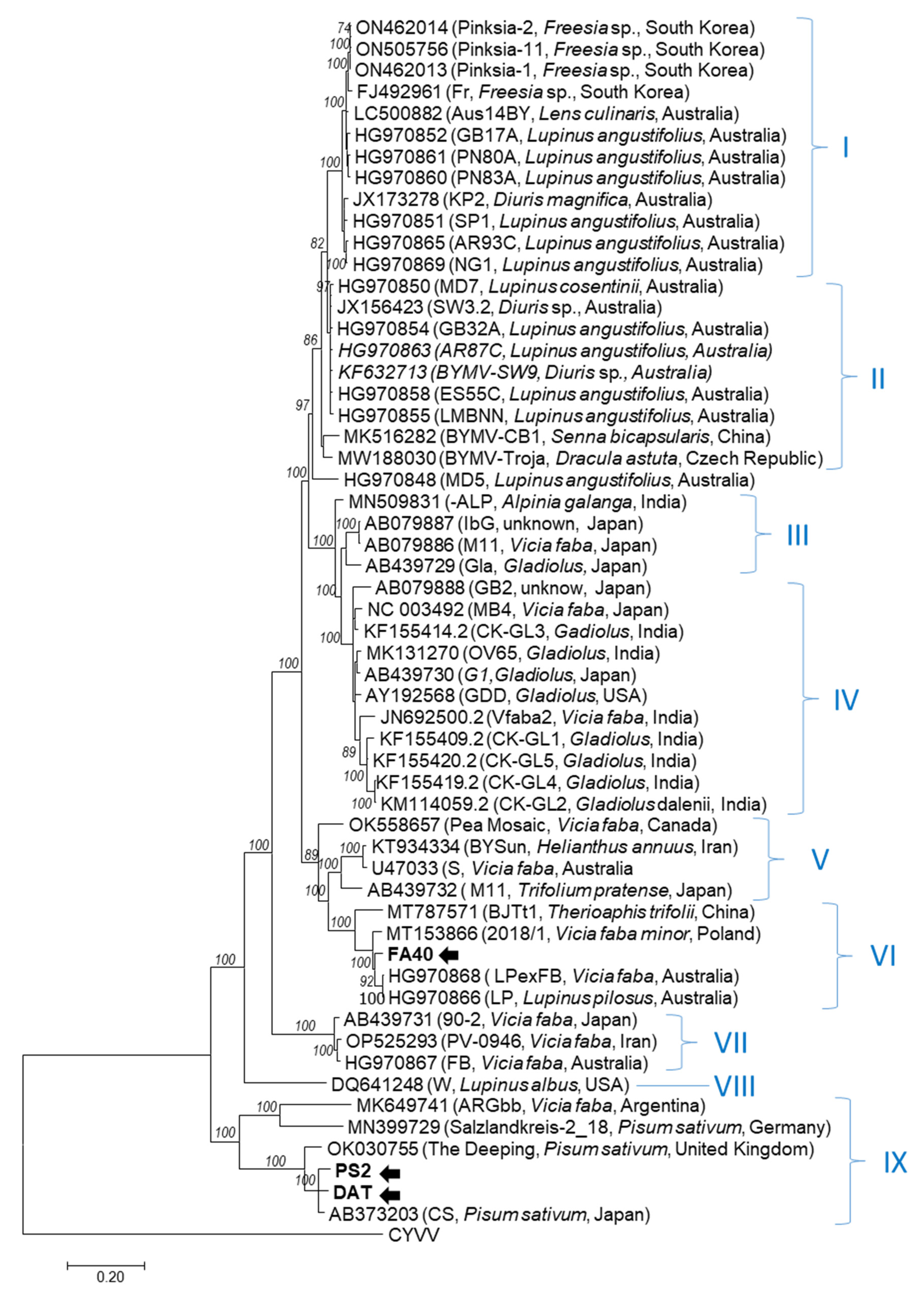
Previous works have revealed the broad intra-species molecular diversity of BYMV [20], resulting in their classification into nine molecular groups [9]. Phylogenetic analysis generated from complete BYMV genomes revealed the similar grouping of DAT and PS2 isolates into molecular group IX, together with pea and broad bean isolates from different continents (Figure 2). The closest sequence found by the BLAST search belonged to a Japanese pea isolate (AB373203). On the contrary, the FA40 isolate clustered in group VI contains geographically distant isolates from lupine and broad bean. Interestingly, the closest relative of FA40 is the HG970866 isolate from Australia [9]. Therefore, the phylogenetic analysis showed the absence of geographical- or host-based clustering of BYMV, indicating the long evolutionary history of the virus and its effective dissemination within regions facilitated by hardly controlled seed transmission [34,35].
The complete coding sequences of the three Slovak BYMV isolates were collinear and determined to be 9168 nucleotides (nt) in length, putatively encoding a polyprotein of 3056 aa (347.3–347.7 kDa). Computer analysis showed that the potyviral motifs were conserved in the BYMV DAT, PS2, and FA40 polyproteins, confirming the functionality of the obtained genomes. Specifically, three separate motifs associated with aphid transmission were conserved in the HC-Pro, i.e., RITC (aa position 335–339, appearing generally as KITC in potyviruses) [36], CCC (aa 575–577), and PTK (aa 593–595). The metal-binding motif FRNK-X12-CDNQLD, affecting symptom expression [37], was found at aa position 464–485.
The CI protein, thought to function as an RNA helicase in genome replication, contained the expected NTP-binding motif GAVGSGKST (aa 1227–1235) as well as other motifs characteristic of helicase proteins: DECH (aa 1316–1319), KVSAT (aa 1343–1347), LVYV (aa 1394–1397), VATNIIENGVTL (aa 1445–1456), and GERIQRLGRVGR (aa 1489–1500), while the VLLLEPTRPL motif appeared as VLM(I/V)ESTRPL (aa 1247–1256) [38,39,40].
Typical conserved motifs of potyviral polymerases QPSTVVDN and GDD, found in the NIb protein at aa positions 2577–2584 and 2615–2617, respectively, correspond to a broader motif SG-(X)3-T-(X)3-NT-(X)30-GDD (aa 2575–2617), proposed as the active site of RNA-dependent RNA polymerases [41]. Other conserved motifs, including SLKAEL (aa 2435–2440, RNA polymerase activity), CVDDFN (aa 2468–2473), CHADGS (as CDADGS, aa 2510–2515, and RNA-dependent polymerase activity [42], were present.
Similarly, CP of all Slovak isolates contained several highly conserved potyvirus motifs. The DAG motif, associated with aphid transmission, appeared as NAG (aa 2790–2792), similar to other BYMV [20,43]. Other conserved motifs found included MVWCIEN (2906–2912), AFDF (2989–2992), QMKAAA (3009–3014), and ENTERH 3034–3039 [44,45].
Figure 2. Phylogenetic tree generated from the complete genome nucleotide sequences of bean yellow mosaic (BYMV) isolates. Phylogenetic analysis was inferred using maximum likelihood (ML) based on the General Time Reversible (GTR) model, selected as the best fitting nucleotide substitution model based on the Bayesian Information Criterion (BIC) implemented in MEGA X [46]. Isolates are identified by their name, GenBank accession number, and country of origin. Slovak BYMV isolates sequenced in this study are highlighted in bold and marked with an arrow. Bootstrap values higher than 70% (500 bootstrap resamplings) are indicated. A closely related potyvirus, clover yellow vein virus (CYVV, Genbank accession number NC_003536), was used as an outgroup. Scale bars indicate genetic distances of 0.2.
Horticulturae 10 00262 g002

3.2. Experimental Infection of Pea Genotypes and Analysis of Virus Accumulation in Plants

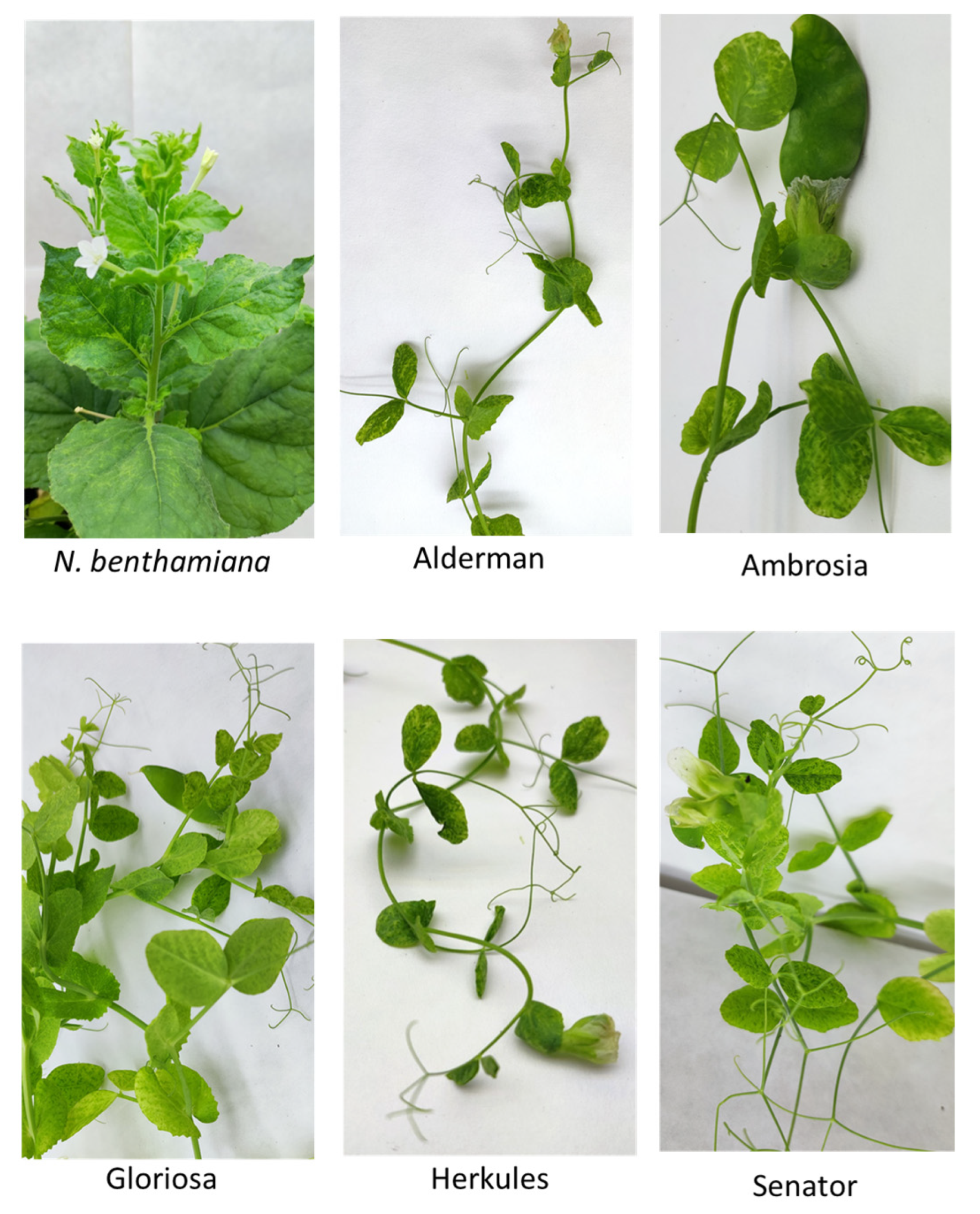
In recent years, mixed viral infections and complex viromes have been frequently detected within a single plant [47,48], which can complicate the biological characterization of respective viruses or plant/virus interaction studies. In our work, HTS analysis revealed a single BYMV infection in only one of the three analyzed samples (pea PS2, Table 1). Therefore, this sample was used as a source of inoculum to establish systemic infection in experimental N. benthamiana plants by mechanical inoculation. Twenty days p.i., infected tobacco plants displayed mottling and mosaic symptoms (Figure 3) and tested positive in DAS-ELISA and RT-PCR. Verification of the CP sequence from N. benthamiana by Sanger sequencing (primed byBY_8548F/BY_9322R) did not reveal any nucleotide changes compared to the original HTS-derived sequence.
As an important part of virus disease management, attention has been given to breeding for resistance in order to provide growers with plant genotypes that are resistant or suffer less from infection [3,49]. In order to evaluate host susceptibility to BYMV-PS2 infection, homogenous lots of five commercially available pea genotypes were mechanically inoculated and tested (cvs. Alderman, Ambrosia, Gloriosa, Herkules, Senator). Based on repeated experiments, all five genotypes were repeatedly infected by BYMV, as determined by symptom evaluation (Figure 3), ELISA, and western blot analysis. The infection rate was 62.5% (Gloriosa) or 100% (Alderman, Ambrosia, Herkules, Senator). Previous reports have revealed different degrees of susceptibility of pea to experimental BYMV infection, including resistant genotypes [50,51,52]. It has been reported that the recessive resistance gene mo controls resistance to BYMV and wlv confers specific resistance to BYMV-W [50,53]. As these reports may be biased, continued biological experiments with molecularly different isolates are needed to confirm potential resistance of pea genotypes to BYMV due to the possible emergence of resistance-breaking BYMV variants.
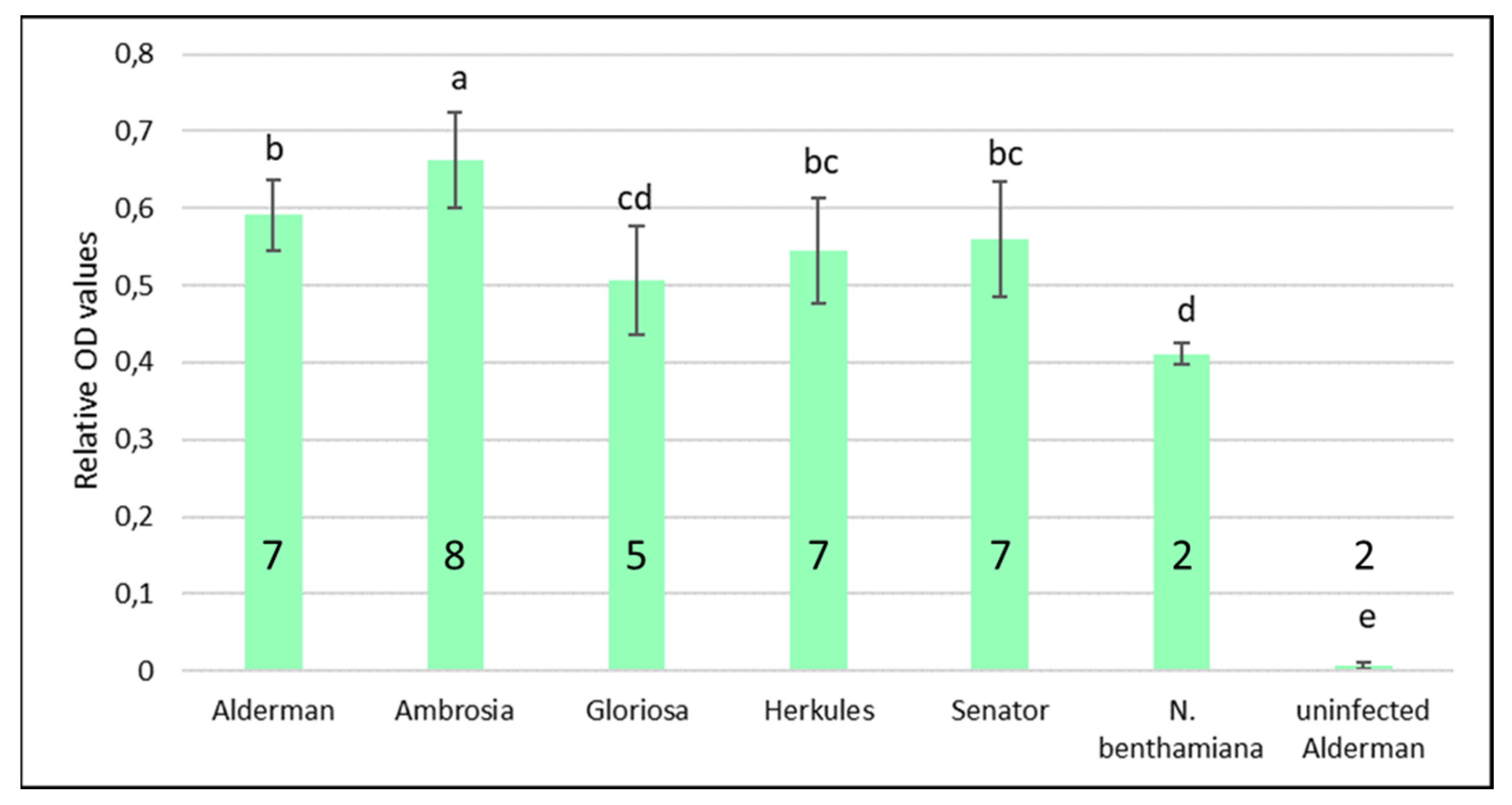
In our experiments, all five BYMV-PS2-infected pea genotypes displayed similar symptoms, consisting of chlorotic mottling and mosaics on leaves, while the growth of plants was not affected compared to non-inoculated controls. In order to comparatively assess the accumulation of virus in the top leaves of different pea genotypes, a semi-quantitative ELISA was performed. One-way ANOVA revealed significant differences between OD values of different plants [F(6, 69) = 65.49, p < 0.001)]. Differences between individual pea genotypes were further addressed by a post hoc TukeyHSD test (Figure 4), which revealed no significant difference between Herkules, Senator, and Alderman cultivars but a significant difference between Ambrosia and all other genotypes (Figure 4).
The Alderman genotype was selected for subsequent experiments aimed at evaluating the accumulation of virus in different parts of the pea plant, as tested by immunoblot analysis. Separately, for each of the four Alderman plants tested, a relative comparison was made between different standardized samples consisting of equal amounts of protein extracted from the root, stems, and leaves from the bottom and upper parts of the plant and from the immature pod (Figure 5A). Immunoblot analyses showed that the level of virus accumulation was specific for each tissue. The results from the four replicates, although with broad variability and no statistical significance, suggested a higher virus accumulation in the actively growing parts of the plant. The relative virus accumulation (compared between different samples as the optical density of the corresponding specific band) was indeed 2.2–13.1-fold higher in the upper stem compared to the root (Figure 5B).
Although it is well-documented that potyviruses systemically infect a wide range of plant species, information on the distribution of the virus within a single plant is still limited. Uneven distribution of potyviruses in different hosts has been reported [54,55,56]. Rajamäki and Valkonen [57] noted differences in the accumulation of potato virus A in the roots and systemic leaves of Solanum commersonii between different isolates of the virus.
Analysis of the correlation between virus titer and symptom severity yielded conflicting results depending on the particular plant/potyvirus model. Furthermore, the results suggest that viral load may or may not correlate (TuMV, [58]; PVY, [59]) with symptom severity. Overall, knowledge of virus distribution in the plant is critical for disease management (although it appears to be virus- and host-specific) and may also have practical implications, e.g., for efficient sampling before testing.

Supplementary Materials

The following supporting information can be downloaded at: https://www.mdpi.com/article/10.3390/horticulturae10030262/s1, Figure S1: Schematic representation of the BYMV genome.

Author Contributions

Conceptualization, M.M. and M.G.; methodology, M.M., K.Š. and M.G.; validation, J.K., A.A. and L.P.; formal analysis, M.M., L.P., K.Š., Z.Š. and M.G.; investigation, M.M., J.K., A.A., P.A., L.P., K.Š., Z.Š. and M.G.; resources, M.M., Z.Š. and M.G., data curation, M.M., L.P., K.Š. and M.G.; writing—original draft preparation, M.M. and M.G.; writing—review and editing, M.M., A.A., K.Š., Z.Š. and M.G.; supervision, M.G.; funding acquisition, M.G. All authors have read and agreed to the published version of the manuscript.

Funding

This research was supported by grant VEGA 2/0036/24 from the Scientific Grant Agency of the Ministry of Education and the Slovak Academy of Sciences and partially by APVV-20-0015 from the Slovak Research and Development Agency.

Data Availability Statement

The nucleotide sequences reported in this paper are deposited in the GenBank database (www.ncbi.nlm.nih.gov) under the accession numbers listed in the text.

Acknowledgments

J.K. acknowledges the support from the Research Support Fund at the University of Ss. Cyril and Methodius in Trnava (FPPV-07-2024). The authors thank Dr Richard Hančinský for his help with the statistical analyses.

Conflicts of Interest

The authors declare no conflicts of interest. The funders had no role in the design of the study; in the collection, analyses, or interpretation of data; in the writing of the manuscript; or in the decision to publish the results.

References

  1. Rashed, A.; Feng, X.; Prager, S.M.; Porter, L.D.; Knodel, J.J.; Karasev, A.; Eigenbrode, S.D. Vector-Borne Viruses of Pulse Crops, With a Particular Emphasis on North American Cropping System. Ann. Entomol. Soc. Am. 2018, 111, 205–227. [Google Scholar] [CrossRef]
  2. Chatzivassiliou, E.K. An Annotated List of Legume-Infecting Viruses in the Light of Metagenomics. Plants 2021, 10, 1413. [Google Scholar] [CrossRef] [PubMed]
  3. Jha, U.C.; Nayyar, H.; Chattopadhyay, A.; Beena, R.; Lone, A.A.; Naik, Y.D.; Thudi, M.; Prasad, P.V.V.; Gupta, S.; Dixit, G.P.; et al. Major viral diseases in grain legumes: Designing disease resistant legumes from plant breeding and OMICS integration. Front. Plant Sci. 2023, 14, 1183505. [Google Scholar] [CrossRef]
  4. Affrifah, N.S.; Uebersax, M.A.; Amin, S. Nutritional significance, value-added applications, and consumer perceptions of food legumes: A review. Legume Sci. 2023, 5, e192. [Google Scholar] [CrossRef]
  5. Wylie, S.J.; Adams, M.; Chalam, C.; Kreuze, J.; López-Moya, J.J.; Ohshima, K.; Praveen, S.; Rabenstein, F.; Stenger, D.; Wang, A.; et al. ICTV Virus Taxonomy Profile: Potyviridae. J. Gen. Virol. 2017, 98, 352–354. [Google Scholar] [CrossRef]
  6. Yang, X.; Li, Y.; Wang, A. Research Advances in Potyviruses: From the Laboratory Bench to the Field. Annu. Rev. Phytopathol. 2021, 59, 1–29. [Google Scholar] [CrossRef] [PubMed]
  7. Wylie, S.J.; Coutts, B.A.; Jones, M.G.K.; Jones, R.A.C. Phylogenetic analysis of Bean yellow mosaic virus isolates from four continents: Relationship between the seven groups found and their hosts and origins. Plant Dis. 2008, 92, 1596–1603. [Google Scholar] [CrossRef]
  8. Kehoe, M.A.; Coutts, B.A.; Buirchell, B.J.; Jones, R.A. Split personality of a Potyvirus: To specialize or not to specialize? PLoS ONE 2014, 9, e105770. [Google Scholar] [CrossRef]
  9. Kehoe, M.A.; Coutts, B.A.; Buirchell, B.J.; Jones, R.A. Plant virology and next generation sequencing: Experiences with a Potyvirus. PLoS ONE 2014, 9, e104580. [Google Scholar] [CrossRef]
  10. Maina, S.; Zheng, L.; King, S.; Aftab, M.; Nancarrow, N.; Trębicki, P.; Rodoni, B. Genome Sequence and Phylogeny of a Bean Yellow Mosaic Virus Isolate Obtained from a 14-Year-Old Australian Lentil Sample. Microbiol. Resour. Announc. 2020, 9, e01437-19. [Google Scholar] [CrossRef]
  11. Baradar, A.; Hosseini, A.; Ratti, C.; Hosseini, S. Phylogenetic analysis of a Bean yellow mosaic virus isolate from Iran and selecting the phylogenetic marker by comparing the individual genes and complete genome trees of BYMV isolates. Physiol. Mol. Plant Pathol. 2021, 114, 101632. [Google Scholar] [CrossRef]
  12. Villamor, D.E.V.; Ho, T.; Al Rwahnih, M.; Martin, R.R.; Tzanetakis, I.E. High Throughput Sequencing For Plant Virus Detection and Discovery. Phytopathology 2019, 109, 716–725. [Google Scholar] [CrossRef]
  13. Maclot, F.; Candresse, T.; Filloux, D.; Malmstrom, C.M.; Roumagnac, P.; van der Vlugt, R.; Massart, S. Illuminating an Ecological Blackbox: Using High Throughput Sequencing to Characterize the Plant Virome Across Scales. Front. Microbiol. 2020, 11, 578064. [Google Scholar] [CrossRef]
  14. Bejerman, N.; Giolitti, F.; Trucco, V.; de Breuil, S.; Dietzgen, R.G.; Lenardon, S. Complete genome sequence of a new enamovirus from Argentina infecting alfalfa plants showing dwarfism symptoms. Arch. Virol. 2016, 161, 2029–2032. [Google Scholar] [CrossRef]
  15. Filardo, F.F.; Thomas, J.E.; Webb, M.; Sharman, M. Faba bean polerovirus 1 (FBPV-1); a new polerovirus infecting legume crops in Australia. Arch. Virol. 2019, 164, 1915–1921. [Google Scholar] [CrossRef]
  16. Naito, F.Y.; Melo, F.L.; Fonseca, M.E.; Santos, C.A.; Chanes, C.R.; Ribeiro, B.M.; Gilbertson, R.L.; Boiteux, L.S.; de Cássia Pereira-Carvalho, R. Nanopore sequencing of a novel bipartite new world begomovirus infecting cowpea. Arch. Virol. 2019, 164, 1907–1910. [Google Scholar] [CrossRef] [PubMed]
  17. Glasa, M.; Šoltys, K.; Predajňa, L.; Sihelská, N.; Budiš, J.; Mrkvová, M.; Kraic, J.; Mihálik, D.; Ruiz- Garcia, A.B. High-throughput sequencing of Potato virus M from tomato in Slovakia reveals a divergent variant of the virus. Plant Protect. Sci. 2019, 55, 159–166. [Google Scholar] [CrossRef]
  18. Elmore, M.G.; Groves, C.L.; Hajimorad, M.R.; Stewart, T.P.; Gaskill, M.A.; Wise, K.A.; Sikora, E.; Kleczewski, N.M.; Smith, D.L.; Mueller, D.S.; et al. Detection and discovery of plant viruses in soybean by metagenomic sequencing. Virol. J. 2022, 19, 149. [Google Scholar] [CrossRef]
  19. Moury, B.; Simon, V. dN/dS-Based methods detect positive selection linked to trade-offs between different fitness traits in the coat protein of potato virus Y. Mol. Biol. Evol. 2011, 28, 2707–2717. [Google Scholar] [CrossRef]
  20. Nigam, D.; LaTourrette, K.; Souza, P.F.N.; Garcia-Ruiz, H. Genome-Wide Variation in Potyviruses. Front. Plant Sci. 2019, 10, 1439. [Google Scholar] [CrossRef]
  21. Stobbe, A.; Roossinck, M.J. Plant Virus Diversity and Evolution. Curr. Res. Top. Plant Virol. 2016, 22, 197–215. [Google Scholar] [CrossRef]
  22. Rubio, L.; Galipienso, L.; Ferriol, I. Detection of Plant Viruses and Disease Management: Relevance of Genetic Diversity and Evolution. Front Plant Sci. 2020, 11, 1092. [Google Scholar] [CrossRef] [PubMed]
  23. Majumdar, A.; Sharma, A.; Belludi, R. Natural and Engineered Resistance Mechanisms in Plants against Phytoviruses. Pathogens 2023, 12, 619. [Google Scholar] [CrossRef] [PubMed]
  24. Mrkvová, M.; Hančinský, R.; Predajňa, L.; Alaxin, P.; Achs, A.; Tomašechová, J.; Šoltys, K.; Mihálik, D.; Olmos, A.; Ruiz-García, A.B.; et al. High-Throughput Sequencing Discloses the Cucumber Mosaic Virus (CMV) Diversity in Slovakia and Reveals New Hosts of CMV from the Papaveraceae Family. Plants 2022, 11, 1665. [Google Scholar] [CrossRef]
  25. Clark, M.F.; Adams, A.N. Characteristics of the microplate method of enzyme-linked immunosorbent assay for the detection of plant viruses. J. Gen. Virol. 1977, 34, 475–483. [Google Scholar] [CrossRef]
  26. Šubr, Z.; Matisová, J. Preparation of diagnostic monoclonal antibodies against two potyviruses. Acta Virol. 1999, 43, 255–257. [Google Scholar]
  27. R Core Team. R: A Language and Environment for Statistical Computing; R Foundation for Statistical Computing: Vienna, Austria, 2018; Available online: https://www.R-project.org/ (accessed on 18 February 2023).
  28. Nováková, S.; Klaudiny, J.; Kollerová, E.; Šubr, Z.W. Expression of a part of the Potato virus A non-structural protein P3 in Escherichia coli for the purpose of antibody preparation and P3 immunodetection in plant material. J. Virol. Methods 2006, 137, 229–235. [Google Scholar] [CrossRef] [PubMed]
  29. Moreno, A.B.; López-Moya, J.J. When Viruses Play Team Sports: Mixed Infections in Plants. Phytopathology 2020, 110, 29–48. [Google Scholar] [CrossRef]
  30. Tomašechová, J.; Hančinský, R.; Predajňa, L.; Kraic, J.; Mihálik, D.; Šoltys, K.; Vávrová, S.; Böhmer, M.; Sabanadzovic, S.; Glasa, M. High-Throughput Sequencing Reveals Bell Pepper Endornavirus Infection in Pepper (Capsicum annum) in Slovakia and Enables Its Further Molecular Characterization. Plants 2020, 9, 41. [Google Scholar] [CrossRef]
  31. Maliogka, V.I.; Salvador, B.; Carbonell, A.; Sáenz, P.; León, D.S.; Oliveros, J.C.; Delgadillo, M.O.; García, J.A.; Simón-Mateo, C. Virus variants with differences in the P1 protein coexist in a Plum pox virus population and display particular host-dependent pathogenicity features. Mol. Plant Pathol. 2012, 13, 877–886. [Google Scholar] [CrossRef]
  32. Della Bartola, M.; Byrne, S.; Mullins, E. Characterization of Potato virus Y Isolates and Assessment of Nanopore Sequencing to Detect and Genotype Potato Viruses. Viruses 2020, 12, 478. [Google Scholar] [CrossRef] [PubMed]
  33. Glasa, M.; Hančinský, R.; Šoltys, K.; Predajňa, L.; Tomašechová, J.; Hauptvogel, P.; Mrkvová, M.; Mihálik, D.; Candresse, T. Molecular Characterization of Potato Virus Y (PVY) Using High-Throughput Sequencing: Constraints on Full Genome Reconstructions Imposed by Mixed Infection Involving Recombinant PVY Strains. Plants 2021, 10, 753. [Google Scholar] [CrossRef] [PubMed]
  34. Sasaya, T.; Iwasaki, M.; Yamamoto, T. Seed Transmission of Bean Yellow Mosaic Virus in Broad Bean (Vicia faba). Ann. Phytopath. Soc. Jpn. 1993, 59, 559–562. [Google Scholar] [CrossRef]
  35. Mali, V.R.; Šubr, Z.; Kúdela, O. Seed Transmission of Como and Potyviruses in Fababean and Vetch Genotypes Introduced into Slovakia. Acta Phytopathol. Entomol. Hung. 2003, 38, 87–97. [Google Scholar] [CrossRef]
  36. Atreya, C.D.; Pirone, T.P. Mutational analysis of the helper component-proteinase gene of a potyvirus: Effects of amino acid substitutions, deletions, and gene replacement on virulence and aphid transmissibility. Proc. Natl. Acad. Sci. USA 1993, 90, 11919–11923. [Google Scholar] [CrossRef] [PubMed]
  37. Shiboleth, Y.M.; Haronsky, E.; Leibman, D.; Arazi, T.; Wassenegger, M.; Whitham, S.A.; Gaba, V.; Gal-On, A. The conserved FRNK box in HC-Pro, a plant viral suppressor of gene silencing, is required for small RNA binding and mediates symptom development. J. Virol. 2007, 81, 13135–13148. [Google Scholar] [CrossRef]
  38. Kadaré, G.; Haenni, A. Virus-encoded RNA helicases. J. Virol. 1997, 71, 2583–2590. [Google Scholar] [CrossRef]
  39. Deng, P.; Wu, Z.; Wang, A. The multifunctional protein CI of potyviruses plays interlinked and distinct roles in viral genome replication and intercellular movement. Virol. J. 2015, 12, 141. [Google Scholar] [CrossRef]
  40. Li, Y.; Xia, F.; Wang, Y.; Yan, C.; Jia, A.; Zhang, Y. Characterization of a highly divergent Sugarcane mosaic virus from Canna indica L. by deep sequencing. BMC Microbiol. 2019, 19, 260. [Google Scholar] [CrossRef]
  41. Hong, Y.; Hunt, A.G. RNA polymerase activity catalyzed by a potyvirus-encoded RNA-dependent RNA polymerase. Virology 1996, 226, 146–151. [Google Scholar] [CrossRef]
  42. Shen, W.; Shi, Y.; Dai, Z.; Wang, A. The RNA-Dependent RNA Polymerase NIb of Potyviruses Plays Multifunctional, Contrasting Roles during Viral Infection. Viruses 2020, 12, 77. [Google Scholar] [CrossRef]
  43. Wylie, S.J.; Kueh, J.; Welsh, B.; Smith, L.J.; Jones, M.G.; Jones, R.A. A non-aphid-transmissible isolate of bean yellow mosaic potyvirus has an altered NAG motif in its coat protein. Arch. Virol. 2002, 147, 1813–1820. [Google Scholar] [CrossRef]
  44. Worrall, E.A.; Hayward, A.C.; Fletcher, S.J.; Mitter, N. Molecular characterization and analysis of conserved potyviral motifs in bean common mosaic virus (BCMV) for RNAi-mediated protection. Arch. Virol. 2019, 164, 181–194. [Google Scholar] [CrossRef]
  45. Zheng, L.; Wayper, P.J.; Gibbs, A.J.; Fourment, M.; Rodoni, B.C.; Gibbs, M.J. Accumulating variation at conserved sites in potyvirus genomes is driven by species discovery and affects degenerate primer design. PLoS ONE 2008, 3, e1586. [Google Scholar] [CrossRef]
  46. Kumar, S.; Stecher, G.; Li, M.; Knyaz, C.; Tamura, K. MEGA X: Molecular Evolutionary Genetics Analysis across computing platforms. Mol. Biol. Evol. 2018, 35, 1547–1549. [Google Scholar] [CrossRef]
  47. Fowkes, A.R.; McGreig, S.; Pufal, H.; Duffy, S.; Howard, B.; Adams, I.P.; Macarthur, R.; Weekes, R.; Fox, A. Integrating High throughput Sequencing into Survey Design Reveals Turnip Yellows Virus and Soybean Dwarf Virus in Pea (Pisum sativum) in the United Kingdom. Viruses 2021, 13, 2530. [Google Scholar] [CrossRef] [PubMed]
  48. Hasiów-Jaroszewska, B.; Boezen, D.; Zwart, M.P. Metagenomic Studies of Viruses in Weeds and Wild Plants: A Powerful Approach to Characterise Variable Virus Communities. Viruses 2021, 13, 1939. [Google Scholar] [CrossRef] [PubMed]
  49. Meziadi, C.; Blanchet, S.; Geffroy, V.; Pflieger, S. Genetic resistance against viruses in phaseolus vulgaris l.: State of the art and future prospects. Plant Sci. 2017, 265, 39–50. [Google Scholar] [CrossRef] [PubMed]
  50. Schroeder, W.T.; Provvidenti, R. A Common Gene for Resistance to Bean Yellow Mosaic Virus and Watermelon Mosaic Virus 2 in Pisum sativum. Phytopathology 1971, 61, 846–848. [Google Scholar] [CrossRef]
  51. Jurík, M.; Lebeda, A.; Gallo, J. Resistance of green peas to legume viruses. Acta Virol. 1994, 38, 97–99. [Google Scholar] [PubMed]
  52. van Leur, J.A.G.; Kumari, S.G.; Aftab, M.; Leonforte, A.; Moore, S. Virus resistance of Australian pea (Pisum sativum) varieties. N. Z. J. Crop Hortic. Sci. 2013, 41, 86–101. [Google Scholar] [CrossRef]
  53. Provvidenti, R.; Hampton, R.O. Inheritance of resistance to White lupin mosaic virus in common pea. HortScience 1993, 28, 836–837. [Google Scholar] [CrossRef]
  54. Green, S.K.; Kuo, Y.J.; Lee, D.R. Uneven distribution of two potyviruses (feathery mottle virus and sweet potato latent virus) in sweet potato plants and its implication on virus indexing of meristem derived plants. Trop. Pest Manag. 1988, 34, 298–302. [Google Scholar] [CrossRef]
  55. Dovas, C.I.; Mamolos, A.P.; Katis, N.I. Fluctuations in concentration of two potyviruses in garlic during the growing period and sampling conditions for reliable detection by ELISA. Ann. Appl. Biol. 2002, 140, 21–28. [Google Scholar] [CrossRef]
  56. Kogovšek, P.; Kladnik, A.; Mlakar, J.; Znidarič, M.T.; Dermastia, M.; Ravnikar, M.; Pompe-Novak, M. Distribution of Potato virus Y in potato plant organs, tissues, and cells. Phytopathology 2011, 101, 1292–1300. [Google Scholar] [CrossRef] [PubMed]
  57. Rajamäki, M.L.; Valkonen, J.P. Viral genome-linked protein (VPg) controls accumulation and phloem-loading of a potyvirus in inoculated potato leaves. Mol. Plant Microbe Interact. 2002, 15, 138–149. [Google Scholar] [CrossRef]
  58. Glasa, M.; Šoltys, K.; Predajňa, L.; Sihelská, N.; Nováková, S.; Šubr, Z.; Kraic, J.; Mihálik, D. Molecular and Biological Characterisation of Turnip mosaic virus Isolates Infecting Poppy (Papaver somniferum and P. rhoeas) in Slovakia. Viruses 2018, 10, 430. [Google Scholar] [CrossRef] [PubMed]
  59. Mehle, N.; Kovač, M.; Petrovič, N.; Pompe Novak, M.; Baebler, Š.; Krečič Stres, H.; Gruden, K.; Ravnikar, M. Spread of potato virus Y NTN in potato cultivars (Solanum tuberosum L.) with different levels of sensitivity. Physiol. Mol. Plant Pathol. 2004, 64, 293–300. [Google Scholar] [CrossRef]
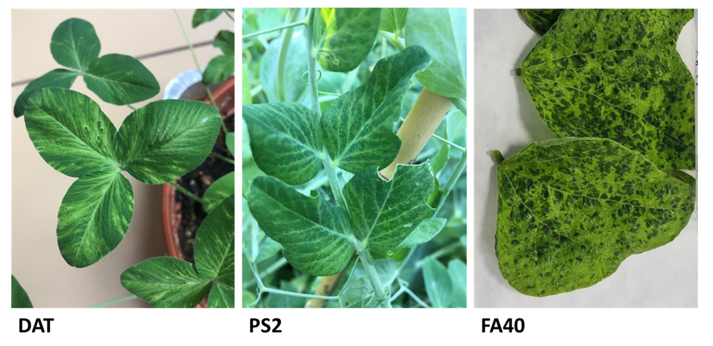
Figure 1. Leaf symptoms observed in Fabaceae plants at the time of sampling for HTS analysis (DAT—red clover (T. pratense), PS2—pea (P. sativum), FA40 common bean (P. vulgaris).
Horticulturae 10 00262 g001
Figure 3. Systemic symptoms developed on N. benthamiana and five commercial pea genotypes after experimental mechanical inoculation with BYMV-PS2 isolate.
Horticulturae 10 00262 g003
Figure 4. Semiquantitative comparison of virus accumulation, measured as the absorbance at 405 nm in DAS-ELISA, in five different pea genotypes 21 days p.i. Infected N. benthamiana and mock-inoculated pea cv. Alderman were used as controls. The graph shows the mean OD values and standard deviation error bars calculated only from BYMV-positive plants, the number of which is indicated. Deviations from the initial number of eight inoculated plants per genotype are caused by plant death, not due to virus infection (Alderman, Hercules, Senator) or uncomplete infection (Gloriosa). Compact letter display method was used to interpret results of post-hoc TukeyHSD test, showing differences between individual plant groups OD values. Groups sharing the same superscript letter are not significantly different (p > 0.05).
Horticulturae 10 00262 g004
Figure 5. Schematic representation of the collection of different samples for the purpose of conducting relative comparison of virus accumulation within a single pea plant by immunoblot analysis (A). Four independent tests of infected cv. Alderman showing relative densitometric analysis of specific bands, performed using Image Studio Lite quantification software v. 5.2. (B).
Horticulturae 10 00262 g005
Table 1. List of Fabaceae samples used for HTS analysis and their characteristics.
SampleNatural HostLocationYear of
Sampling
Leaf SymptomsViruses Identified in the
Sample 1
DATTrifolium pratenseBratislava2020MosaicBYMV, SbDV, BGCV-2
PS2Pisum sativumPezinok2021Mottling, mild leaf distortionsBYMV
FA40Phaseolus vulgarisVrbová nad Váhom2022Severe mosaic, yellowingBYMV, CMV
1 SbDV: soybean dwarf virus (genus Luteovirus), BGCV-2: black grass cryptic virus-2 (unassigned), CMV: cucumber mosaic virus (genus Cucumovirus).
Table 2. Analysis of HTS data from the three Fabaceae samples related to BYMV genome reconstruction.
SampleTotal Number of Reads/Average Length (bp)Reads Mapped against BYMV Reference NC_003492Percentage of the Full-Length
Genome Covered
Coverage DepthThe Closest BLAST Relative
DAT4,449,806/137.1662,10599.9%9764.5×AB373203, pea, Japan
PS24,025,394/103.7414,92599.9%4794.5×AB373203, pea, Japan
FA4016,948,908/117.51,075,14399.9%14,139.7×HG970866, lupine, Australia
Disclaimer/Publisher’s Note: The statements, opinions and data contained in all publications are solely those of the individual author(s) and contributor(s) and not of MDPI and/or the editor(s). MDPI and/or the editor(s) disclaim responsibility for any injury to people or property resulting from any ideas, methods, instructions or products referred to in the content.