Article

Light Induction of Seed Culture Accelerates Lutein Accumulation in Heterotrophic Fermentation of Chlorella protothecoides CS-41

1
Institute of Ocean Research, College of Engineering, Peking University, Beijing 100871, China
2
Shenzhen Key Laboratory of Marine Microbiome Engineering, Institute for Advanced Study, Shenzhen University, Shenzhen 518060, China
3
Institute for Innovative Development of Food Industry, Shenzhen University, Shenzhen 518060, China
4
Institute of New Materials and Advanced Manufacturing, Beijing Academy of Science and Technology, Beijing 100089, China
*
Authors to whom correspondence should be addressed.
Fermentation 2023, 9(8), 768; https://doi.org/10.3390/fermentation9080768
Submission received: 27 July 2023 / Revised: 15 August 2023 / Accepted: 16 August 2023 / Published: 18 August 2023
(This article belongs to the Special Issue Microalgae: Biofactory for Valuable Products)

Abstract

:
Microalgae are recognized as a promising and valuable source of lutein. However, the current two-stage method for lutein production has drawbacks, such as complex operations and a long cultivation time. Additionally, utilizing heterotrophic fermentation to cultivate microalgae for lutein production leads to low lutein content due to the absence of light. In this study, we proposed a novel cultivation method that involves light induction of the seed culture to enhance lutein production during the heterotrophic cultivation phase of Chlorella protothecoides CS-41. To gain comprehensive insights into the underlying mechanisms of this method, we conducted qualitative and quantitative analyses of specific metabolites related to central carbon metabolism. The results revealed that low-light induction of seeds exhibited higher carbon efficiency compared to cells continuously subjected to heterotrophic cultivation, which may explain the observed increase in biomass and lutein content in cultures. Cultures after low-light induction of seed exhibited significantly higher lutein content (2.71 mg/g), yield (66.49 mg/L) and productivity (8.59 mg/L/d) compared to those consistently cultivated under heterotrophic conditions (2.37 mg/g, 37.45 mg/L, 4.68 mg/L/d). This cultivation strategy effectively enhances lutein yields, reduces production costs and holds the potential for broader application in other algal species for pigment production.

1. Introduction

Lutein, a member of the carotenoid family, is a prominent pigment found in microalgae and higher plants. It serves as an accessory pigment that plays essential roles in light harvesting and nonphotochemical quenching within the photosystem of microalgae. Its natural color and potent eye-protective properties have led to its widespread use in feed additives and healthcare products [1]. Lutein has gained attention not only for its antioxidant properties but also for its potential anticancer effects and involvement in infant neural development [2]. Particularly noteworthy is its ability to protect the eyes from oxidative damage caused by blue light. Clinical studies have demonstrated that lutein can help prevent cataracts and reduce the risk of age-related macular degeneration, a common cause of vision loss in older individuals [3]. The market for lutein has been steadily growing, with a valuation of USD 324.6 million in 2022. It is projected to reach USD 491.4 million by 2029 [4]. The increasing awareness of the benefits of lutein and its applications in food and feed additives have contributed to its market expansion.
Microalgae, ancient photosynthetic organisms, are known for their remarkable diversity and abundance. Microalgae play a crucial role as primary producers, accounting for more than 40% of global photosynthetic carbon sequestration. Due to their high productivity, rapid growth cycle, rich nutrient content and minimal need for arable land and freshwater resources, they are regarded as a promising food source to address the challenges posed by a growing population and environmental degradation. They possess the ability to accumulate various nutritional products such as lipids [5], proteins [6] and pigments [7], which have found extensive applications in industries related to food, nutraceuticals, healthcare and cosmetics [8,9].
Microalgae are considered a highly promising source of lutein due to their exceptionally fast growth rate, adaptability to diverse environments and independence from seasonal limitations, in contrast to the marigold flower, which is currently the only commercially available natural source of lutein. To date, many microalgal species are able to accumulate high contents of lutein, such as Chlorella sorokiniana F31 [10], Parachlorella sp. JD-076 [11], Chlorella sorokiniana Kh12 [12], Chlorella sorokiniana FZU60 [13,14], Chlorella sorokiniana MB-1-M12 [15], C. protothecoides CS-41 [16,17], etc.
However, the current commonly used two-stage processes for lutein production are often complex. For instance, they involve initial heterotrophic cultivation followed by light induction [18] or mixotrophic cultivation followed by autotrophic cultivation [13]. While these two-step methods often yield favorable results in laboratory conditions, their application in large-scale production poses challenges. The method of first achieving high biomass through heterotrophic cultivation and then inducing light stimulation requires dilution, leading to increased water usage and subsequent harvesting costs. Additionally, transferring the culture to different containers adds extra steps. On the other hand, heterotrophic cultivation in a fermenter often results in low lutein content due to the lack of light. To address this issue, this study proposes a novel cultivation approach that enhances lutein productivity of C. protothecoides CS-41 during the heterotrophic stage by subjecting the seed culture to light induction. Through this approach, we aim to achieve higher biomass accumulation in fermentation tanks while simultaneously increasing the accumulation of lutein, thereby achieving a greater lutein yield.

2. Materials and Methods

2.1. Strains and Growth Conditions

C. protothecoides CS-41 was obtained from the CSIRO Marine Laboratory (Hobart, Australia). The strains were maintained and cultivated on a plate medium of Modified Basal with 1.0% agar and 10 g/L glucose and then transferred to 20 mL liquid Modified Basal medium in a 100-mL Erlenmeyer flask at 28 °C for 5 days with orbital shaking at 180 rpm in the dark. The derived cells were then inoculated at 10% (v/v) into a 250-mL Erlenmeyer flask containing 100 mL fresh Modified Basal medium under the same conditions as above for another 4 days to prepare seeds. The cultivation conditions used were derived from the previously optimized conditions by Shi et al. [16,17] including the addition of 40 g/L glucose and 3.6 g/L urea into the Modified Basal medium at 28 °C with orbital shaking at 180 rpm in the dark. The Modified Basal medium consisted of (per liter) 1.25 g KH2PO4, 1 g MgSO4·7H2O, 0.5 g EDTA·Na2, 0.1142 g H3BO3, 0.111 g CaCl2·2H2O, 0.0498 g FeSO4·7H2O, 0.0882 g ZnSO4·7H2O, 0.0142 g MnCl2·4H2O, 0.0157 g CuSO4·5H2O; 0.0049 g Co (NO3)2·6H2O and 0.0071 g MoO3. The pH value was adjusted to 6.1 before autoclaving. All experiments were operated in triplicate.

2.1.1. Optimization of Light Intensity

Seeds were inoculated at 10% (v/v) into 200 mL fresh Modified Basal medium in a 500-mL flask at 28 °C with orbital shaking at 180 rpm for 10 days. The cultures were cultivated under three cultivation conditions: low light (LL), 30 μmol/m2/s; high light (HL), 150 μmol/m2/s; and in the dark (DK).

2.1.2. Optimization of Glucose Concentration

Seeds were inoculated at 10% (v/v) into 100 mL fresh Modified Basal medium in a 250-mL flask at 28 °C with orbital shaking at 180 rpm for 72 h in the dark. The cultures were cultivated under four glucose concentration additions: 5 g/L, 10 g/L, 20 g/L, 40 g/L.

2.1.3. Seed Induction under Different Light Conditions

Seeds were inoculated at 10% (v/v) into 200 mL fresh Modified Basal medium in a 500-mL flask at 28 °C with orbital shaking at 180 rpm for 2–3 days under three precultivation conditions: LL (30 μmol/m2/s), HL (150 μmol/m2/s) and DK. This precultivation procedure was repeated again to obtain stable seeds. The derived cells were inoculated into 200 mL fresh Modified Basal medium in a 500-mL flask with the same initial inoculation dosage at 28 °C with orbital shaking at 180 rpm for 5 days in the dark.

2.1.4. Seed Induction before Fed-Batch Cultivation

Seeds were inoculated at 10% (v/v) into 200 mL fresh Modified Basal medium in a 500-mL flask at 28 °C with orbital shaking at 180 rpm for 2–3 days under two precultivation conditions: LL (30 μmol/ m2/s) and DK. This precultivation procedure was repeated again to obtain stable seeds. The derived cells were inoculated into 100 mL fresh Modified Basal medium in a 250-mL flask with the same initial inoculation dosage at 28 °C with orbital shaking at 180 rpm for 12 days in the dark. Glucose and urea were added during the cultivation process to ensure continuous growth of C. protothecoides CS-41.

2.2. Determination of Biomass, Glucose Concentration and Urea Concentration

To determine the biomass concentration of the culture, 1–5 mL algal culture was collected and centrifuged at 5000 rpm for 3 min. The pellet was washed twice with ultrapure water and suction filtrated with the dry preweighed filter paper. The filter paper was then dried to a constant weight and measured using an analytical balance. The biomass concentration was expressed as cells' dry weight per liter (g/L). The specific growth rate (µ) was determined using the following equation:
μ = (ln(Xt) − ln(X0))/t
Here, X₀ represents the initial biomass concentration (g/L) and Xt denotes the biomass concentration (g/L) at the cultivation period of t hours.
The supernatant of the culture derived from centrifugation was measured to confirm the concentration of glucose and urea. The glucose concentration was determined using the DNS method. In brief, 100 μL diluted supernatant and 500 μL DNS reagent were mixed well and heated for 10 min at 100 °C and then cooled for 5 min in an ice bath. An amount of 100 μL reaction mixture was added to 96-well plates. The absorbance reading at 570 nm was recorded with an ELIASA (TECAN SPARK). The standard curve was drawn by hierarchical diluted 5 g/L glucose standard solution at 570 nm absorbance. The urea concentration was determined with urea assay kits (Shanghai Yuanye Science and Technology Co., Ltd., Shanghai, China) according to the manufacturer’s instructions.

2.3. Determination of Lutein, Total Carotenoids and Total Chlorophylls

A total of 5–7 mg lyophilized biomass was weighed and then ground with 3–4 stainless steel beads for 10 min with the Tissue Lyser II (QIAGEN, Hilden, Germany). The debris was extracted with 1–2 mL pure methanol until colorless. The methanol layer was collected using centrifugation at 15,000 rpm for 10 min at 4 °C for further analysis. Pigment profile analysis and lutein content quantification were performed using Waters 2695 HPLC system (Waters, Milford, MA, USA) with a 2998 PDA detector and a CAPCELL PAK C18 reverse phase bar. The absorbance values of the supernatants at 480, 652 and 665 nm were measured with a spectrophotometer for total carotenoids and total chlorophylls quantifications, which were calculated as described in [19].
All procedures were operated under darkness to avoid photo-oxidation. The standard lutein was obtained from Sigma Chemical Co. (St. Louis, MO, USA). All solvents used were high-performance liquid chromatography (HPLC) grade.

2.4. Quantification of Protein, Lipid and Carbohydrate Concentration

Protein, lipid and carbohydrate concentrations were assessed following the methods described in a previous study [20]. Briefly, to extract protein, sodium hydroxide was employed and the BCA Protein Assay Kit (Beyotime, P0010) was used for protein quantification. Lipid quantification was carried out using the sulfo-phosphor-vanillin reaction. The total carbohydrate concentration was determined utilizing the phenol sulfuric acid method.

2.5. Quantification of Maximum Quantum Yield

The maximum quantum yield (Fv/Fm) of photosystem II was assessed using pulse-amplitude-modulated fluorometry (Walz, Effeltrich, Germany). Microalgal cells were adjusted to an approximate optical density of 680 nm and incubated in darkness for 20 min. Subsequently, the cells were subjected to fluorometry analysis. The fluorometry measurements provided the maximum fluorescence level (Fm) and the minimum fluorescence level (F0). The Fv/Fm value was calculated using the following equation:
F v / F m = F m F 0 / F m

2.6. RNA Extraction and Analysis of Differentially Expressed Genes

Cultures of C. protothecoides CS-41 under different light intensities on day 2 were collected and immediately prefrozen with liquid nitrogen. Total RNA extraction was performed using the Trizol method. The cells were disrupted using Tissue Lyser II (QIAGEN, Hilden, Germany) with beads and transferred into 1.5 mL tubes containing precooled Trizol, followed by centrifugation at 13,000 rpm for 5 min at 4 °C and the addition of 200 μL precooled chloroform to the supernatant. The shaken well mixture was centrifuged at 13,000 rpm for 15 min. Afterward, 400 μL of the supernatant was transferred into tubes and mixed with equal volumes of precooled isopropanol for 10 min. After centrifuging the mixture at 13,000 rpm for 10 min, the supernatant was carefully discarded. Next, 1 mL of precooled 75% ethanol was added to the tubes. The tubes were then centrifuged at 12,000 rpm for 5 min to remove the ethanol supernatant. A quick 30-s centrifugation was performed to discard any residual liquid clinging to the tube walls. The tubes were left at room temperature for 3 min. Finally, 20–50 μL of ddH2O was added to elute the RNA from the pellet. To ensure the quality of the RNA samples (OD260/280 = 1.8–2.2, OD260/230 ≥ 2.0, RIN ≥ 6.5 and 28S/18S ≥ 1.0), their concentration and quality were assessed using an Agilent 2100 Bioanalyzer and a Nanodrop Technologies ND-2000 spectrophotometer. For transcriptome sequencing, 2 μg of total RNA was used for each sample.
The RSEM software was utilized to calculate the reads per Kilobase of exon per million reads mapped (FPKM) values. The EdgeR software was employed to calculate the log2 (fold change) and determine the significance of differences using p-values. Following multiple hypothesis testing, the p-values were adjusted using the P-adjust method. Differentially expressed genes were identified based on specific criteria: an absolute log2 (fold change) greater than 1 and a P-adjust value below 0.05. Further enrichment analyses were conducted, including KEGG pathway analysis using KOBAS and Gene Ontology (GO) analysis using Goatools.

2.7. Metabolite Extraction and Analysis

A 10 mL fresh microalgae culture under different precultivation conditions was concentrated using centrifugation at 4 °C for 5 min at 5000 rpm to 0.5 mL. A precise volume of 10 μL of the sample was transferred and combined with 10 μL of isotopic internal standard, 40 μL of 50% methanol–water solution and 140 μL of acetonitrile. The mixture was vortexed for 1 min at 4 °C and then centrifuged at 14,000 rcf for 20 min. Subsequently, 100 μL of the supernatant was transferred to a 1.5-mL centrifuge tube and 25 μL of 200 mM 3NPH.HCl and 25 μL of 120 mM EDC.HCl (containing 6% pyridine) solution was added. The mixture was vortexed for 30 s, briefly centrifuged for 5 s and then subjected to a 60 °C temperature-controlled oscillator for 40 min. After completion of the reaction, the mixture was vortexed for 30 s and centrifuged at 14,000 rcf for 20 min at 4 °C. The supernatant was collected and transferred to a vial for subsequent analysis. The target analytes in the samples were qualitatively and quantitatively analyzed using LC-ESI-MS/MS (UHPLC-Qtrap).
The procedure for preparing the standard solution is as follows: accurately weigh the appropriate amounts of central carbon metabolism standard compounds and prepare a single stock solution using a 50% methanol–water mixture. Combine the individual stock solutions in suitable proportions to create a mixed standard solution, which is then diluted with 50% methanol–water to achieve the desired concentration, resulting in a working standard solution. Additionally, weigh the suitable amounts of isotope standard compounds (succinic acid-D4, levocarnitine-D3, cholic acid-D4, salicylic acid-D4) and prepare individual stock solutions using 50% methanol–water. Dilute the isotope standard solutions with 50% methanol–water to prepare an isotope internal standard mixture (with respective internal standard concentrations of 5 μg/mL, 5 μg/mL, 20 μg/mL, 30 μg/mL).

2.8. Statistical Analysis and Equations

Statistical analysis was performed using IBM SPSS Statistics 27 (SPSS, Chicago, IL, USA). One-way ANOVA with subsequent post hoc multiple-comparison LSD tests were used to detect significant differences.

3. Results

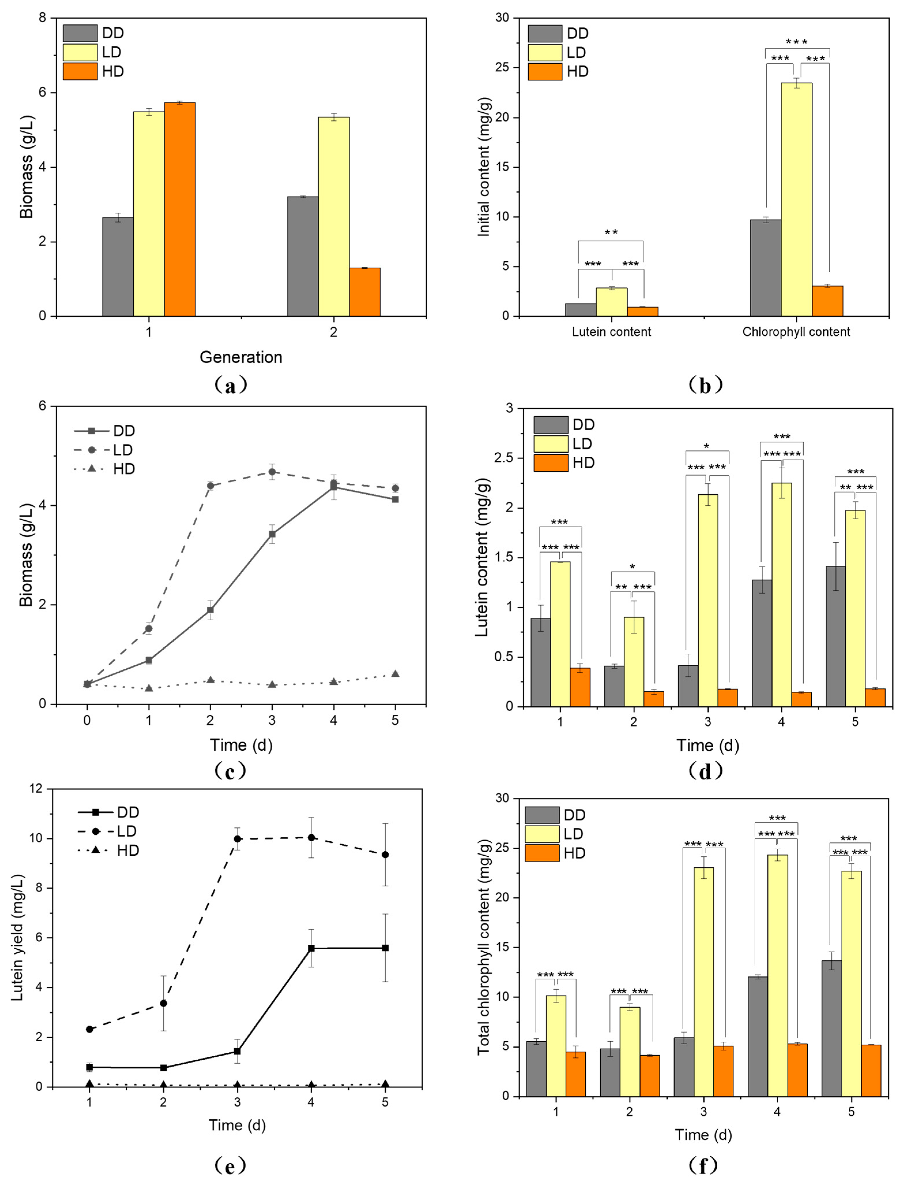
3.1. Low-Light Intensity Contributed to Lutein Biosynthesis

Three light conditions, including LL, HL and DK, were applied to cultivate C. protothecoides CS-41 in order to determine the optimal light conditions for growth and lutein accumulation. As shown in Figure 1a, culture grown under LL exhibited a higher growth rate compared to those under HL or DK, while the final biomass under LL and DK were nearly the same. However, due to severe light damage, the growth rate and final biomass were significantly lower under HL. This is also evident in Figure 1b, where the Fv/Fm value dropped to 0 after day 4. The reduction in maximum photochemical efficiency (Fv/Fm) under varying light intensities is regarded as an indication of photoinhibition in microalgae [1]. The Fv/Fm values under LL during the cultivation process were higher compared to those under DK and HL, indicating that a low-light environment enhanced the photosynthetic capacity of the microalgal cells. C. protothecoides CS-41 accumulated a higher lutein content under LL (4.51 mg/g), which was 1.73 times higher than that under DK (2.61 mg/g) and 4.65 times higher than that under HL (0.97 mg/g) (Figure 1c). Consequently, the final lutein yields under LL were remarkably higher, reaching 75.97 mg/L, which was 1.65 times higher than that under DK (45.99 mg/L) and 16.73 times higher than that under HL (4.54 mg/L). The total chlorophyll content was also measured, as shown in Figure 1e, and the results revealed no significant difference in total chlorophyll content between LL and DK, indicating that C. protothecoides CS-41 retained the ability to synthesize chlorophylls even in the absence of light. However, due to the light damage caused by high light intensity, the total chlorophyll content under HL was considerably lower. The total carbohydrates, protein and lipid contents are presented in Figure 1d. It was observed that cells tended to accumulate more carbohydrates under HL, more proteins under LL and more lipids in the DK. This might be related to cell viability. The contents of different metabolites varied over time under different conditions. Cells grown under HL accumulated more carbohydrates but had lower protein content throughout the cultivation process, indicating reduced activity due to light damage (Figure S1). Furthermore, the fatty acid profile of C. protothecoides CS-41 cultivated under HL also exhibited significant differences compared to that under LL and DK (Figure 1f).

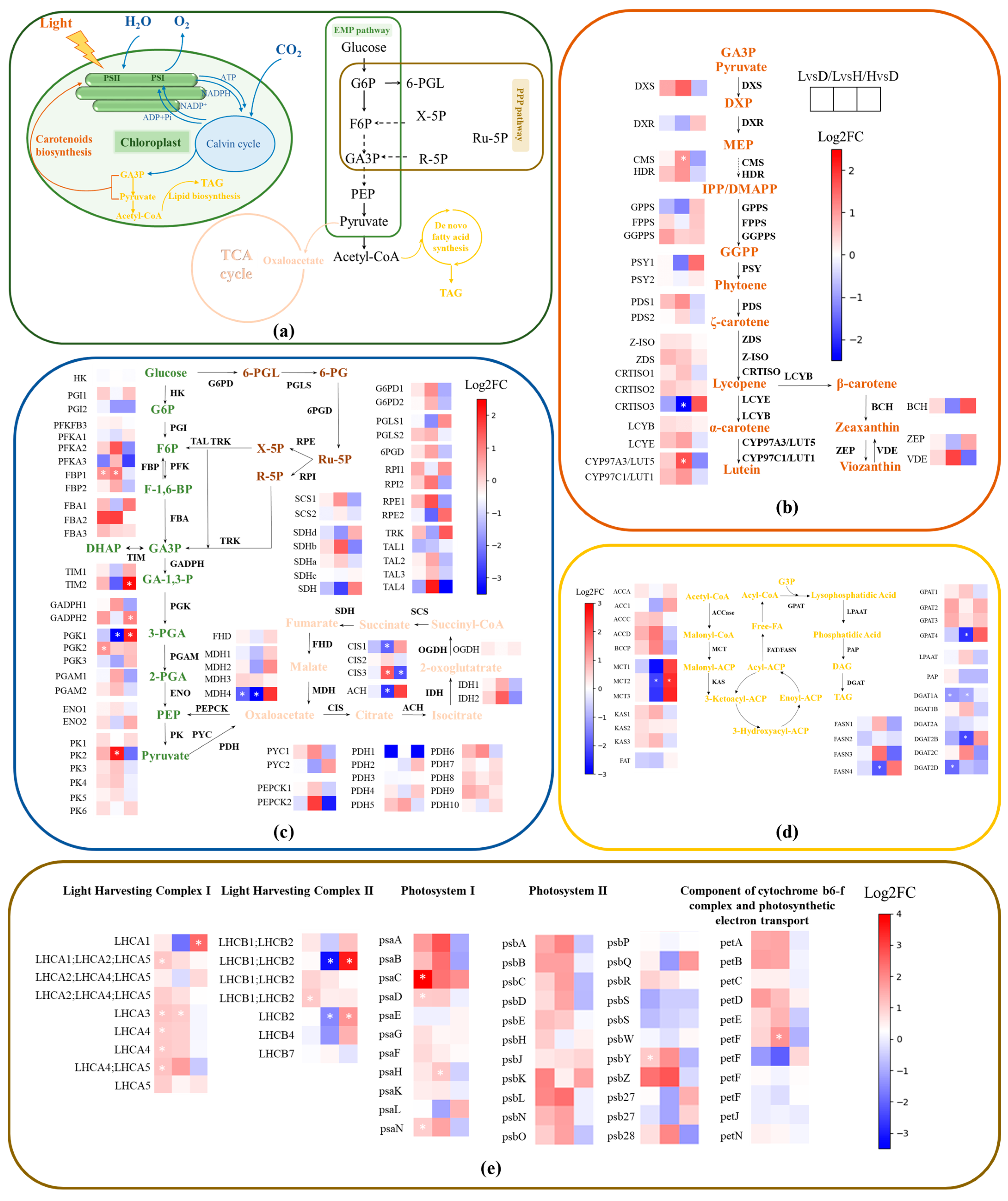
3.2. Transcriptome Data within the Metabolic Network of C. protothecoides CS-41

To better understand the impact of different light intensities on enzyme regulation within the metabolic network of C. protothecoides CS-41, the transcription levels of genes involved in carotenoid synthesis, lipid synthesis, central carbon metabolism and photosynthesis were examined and compared pairwise. Figure 2b illustrates the differential expression of 23 genes involved in the lutein synthesis pathway under different light intensities. Among these genes, the three genes on the right side are key genes in the β-branch, which competes with the α-branch where lutein synthesis occurs. Carotenoid synthesis starts from pyruvate and GA3P and through the regulation of multiple key enzymes, eventually leads to lutein synthesis, as described in previous reviews [1]. When LL is compared to DK, 17 genes were upregulated and 3 genes were downregulated among the 20 genes that induced lutein synthesis, indicating a more active lutein synthesis under low light compared to DK. When comparing low-light to highlight conditions, 15 genes were upregulated and 8 genes were downregulated, with CMS and LUT5 significantly upregulated and CRTISO3 significantly downregulated. LUT5, encoding the β-hydroxylase enzyme directly involved in lutein synthesis, is a crucial enzyme in the process. Meanwhile, the β-branch genes BCH and ZEP were downregulated, suggesting that under low-light conditions, the flux is biased toward the branch leading to lutein synthesis, facilitating lutein accumulation compared to high-light conditions. Although there were no further significant differences observed, it is evident that low light is more favorable for lutein synthesis compared to both dark and high-light conditions.
As components of the light-harvesting complex (LHC), genes related to photosynthesis are closely associated with the accumulation of lutein. After annotating 60 genes associated with photosynthetic systems, our analysis revealed that 50 genes were upregulated, with 10 genes showing significant upregulation under LL compared to DK, as depicted in Figure 2e. This indicates that low light is more favorable for photosynthesis in microalgae. When comparing LL to HL, 40 genes were upregulated, with 3 genes significantly upregulated and 20 genes downregulated, with 2 genes significantly downregulated. This further supports the notion that low light is more conducive to photosynthesis. However, when comparing high-light to dark conditions, only 23 genes were upregulated, with 3 genes significantly upregulated. Despite providing light, the photosynthetic activity of cells under high-light conditions is not stronger than that under dark conditions, which is consistent with the results shown in Figure 1b. This is attributed to the severe photo damage caused by high-light conditions.
The situation is different for lipid synthesis. Figure 2d illustrates the gene expression patterns involved in triacylglycerol (TAG) synthesis. A comparison between LL and HL revealed a significant downregulation of key genes involved in lipid synthesis, including MCT, FASN, GPAT and DGAT. This indicates that high light is more conducive to the synthesis of neutral lipids compared to low-light conditions.
Figure 2c illustrates the transcriptional profiles of genes associated with central carbon metabolism. Among the 43 genes involved in glycolysis and pentose phosphate pathway (PPP), 33 genes were upregulated, with 2 genes showing significant upregulation under LL compared to DK. When comparing LL to HL, 27 genes were upregulated, with FBP and PK exhibiting significant upregulation, while PGK showed significant downregulation. The upregulation of PK, a key enzyme in the synthesis of pyruvate, provides more substrates for carotenoid biosynthesis.

3.3. Seed Induction under Low Light Accelerate Lutein Accumulation in Heterotrophic Cultivation

Four distinct initial glucose concentrations were subjected to cultivating C. protothecoides CS-41, the resulting growth curves were plotted (Figure S2a) and the specific growth rates were calculated using Equation (1) (Figure S2b). Notably, initial glucose concentrations of 5 g/L and 10 g/L demonstrated higher specific growth rates. To facilitate the forthcoming fed-batch fermentation procedures, a 10 g/L glucose concentration was employed for subsequent experimental investigations.
To validate the feasibility of the novel cultivation strategy, we cultured seeds under HL (HL inoculate to DK, HD), LL (LL inoculate to DK, LD) and DK conditions (DK inoculate to DK, DD) prior to heterotrophic cultivation. Figure 3a depicts the biomass over two generations of seed cultivation under the three conditions, showing a similar trend to the previous findings, with higher biomass observed under LD. Figure 3b presents the initial levels of lutein and chlorophyll before heterotrophic cultivation, indicating significantly higher levels of pigments after two generations under LD compared to DD and HD. Figure 3c demonstrates the biomass changes during heterotrophic cultivation following seed cultivation under the three strategies. Although there was no significant difference in final biomass between LD and DD, cells of C. protothecoides CS-41 under LD exhibited a higher growth rate. Conversely, cells under HD failed to recover activity in the dark due to photo damage. Figure 3d displays the lutein content under the three modes, revealing consistently higher lutein levels under LD throughout the cultivation process. On the third day, the lutein content was 5.19 times higher than that of DD and 12.53 times higher than that of HD. Similar trends were observed in chlorophyll content (Figure 3f). The final lutein yield under LD was 9.38 mg/L, which was 67% higher than that of DD and 85 times higher than that of HD (Figure 3e).

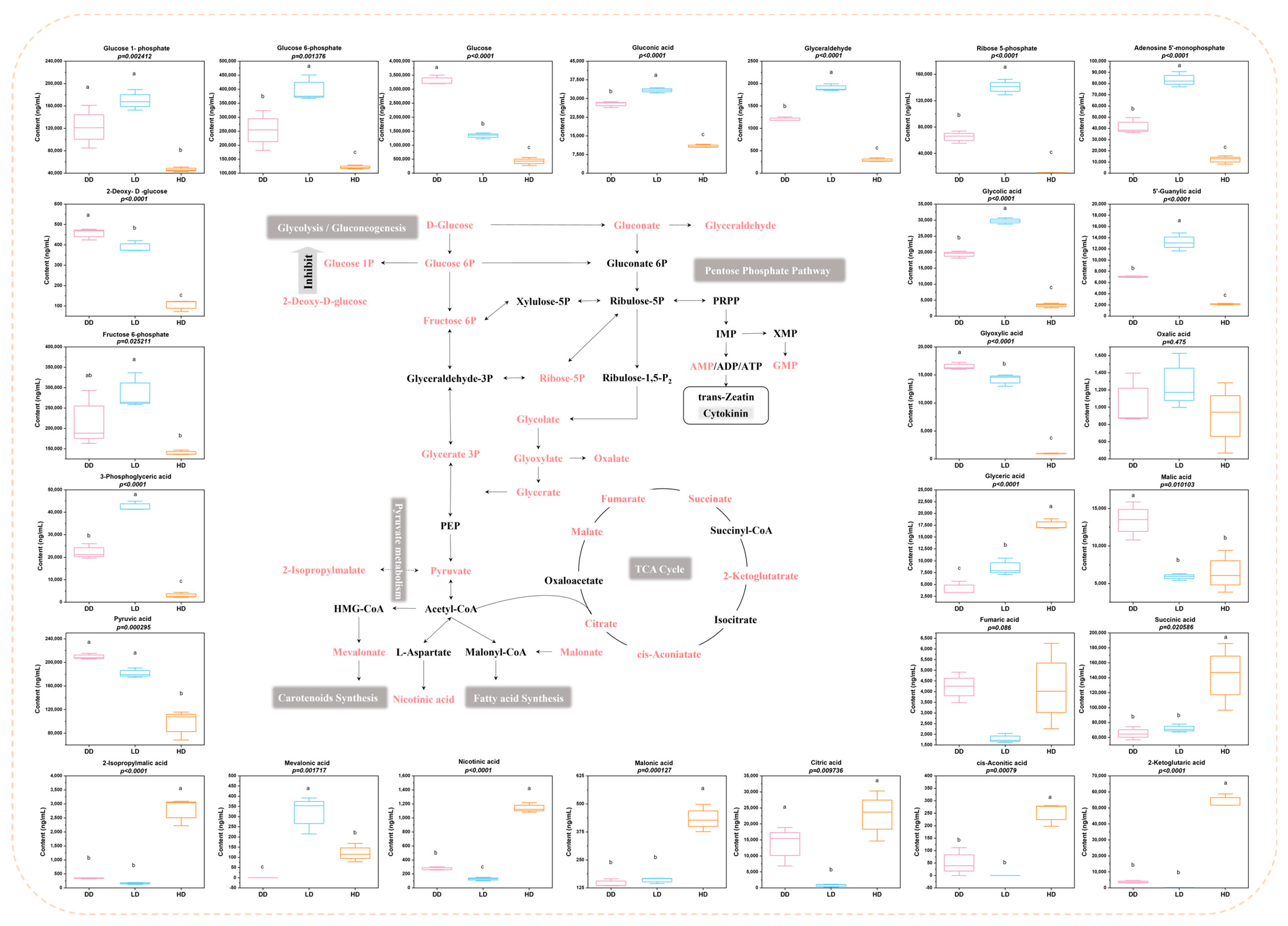
3.4. Metabolites Involved in Central Carbon Metabolism under Different Seed Cultivation Conditions

To investigate the impact of seed cultivation conditions on algal cells under heterotrophic cultivation, we quantified the levels of 27 metabolites related to central carbon metabolism (Figure 4). Figure 4 illustrates the central carbon metabolism network, highlighting the metabolites measured in this study in pink (isocitrate was not included in Figure 4 due to its undetectable levels under all three conditions). Out of the 26 analyzed metabolites, 24 showed significant differences. When comparing only LD and DD conditions, 16 metabolites showed significant differences. Among the six metabolites in the glycolysis/gluconeogenesis and PPP, algal cells under LD had lower glucose levels and higher levels of glucose 6P, glycerate 3P and ribose-5P. Additionally, algal cells under LD exhibited lower levels of 2-Deoxy-D-glucose, a known inhibitor of glycolysis. Among the six metabolites in the TCA cycle, Malate, cis-aconitate and citrate were significantly lower in LD compared to DD, while the other three metabolites did not show significant differences. Furthermore, several metabolites, including gluconate, glyceraldehyde, glycolate and glycerate, exhibited higher levels under LD. Notably, AMP, a precursor for cell growth regulators, showed higher levels under LD. Moreover, mevalonate, an intermediate metabolite in the MVA pathway (carotenoid precursors biosynthetic pathway), also exhibited higher levels under LD.

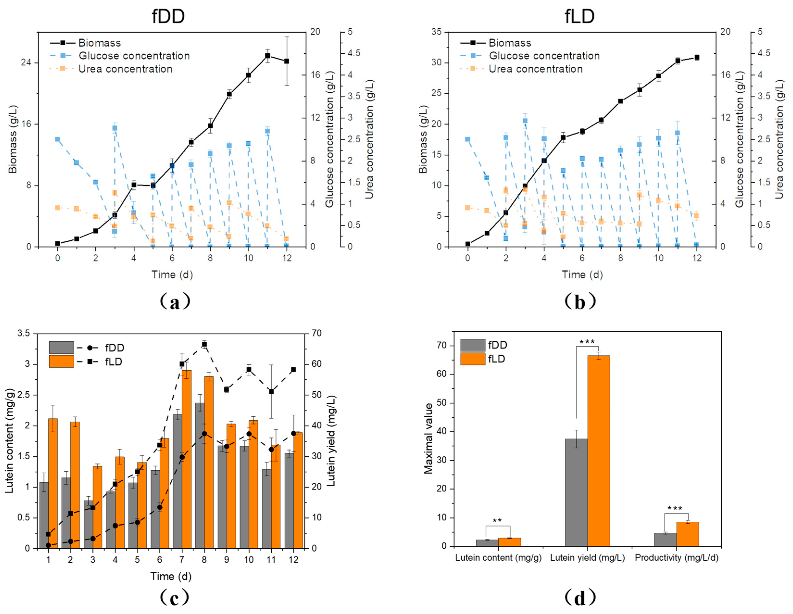
3.5. Fed Batch Cultivation of C. protothecoides CS-41 with Seed Cultivation under Low Light

Finally, fed-batch fermentation under two conditions, low-light seed cultivation (LL inoculate to DK fed-batch culture, fLD) and dark seed cultivation (DK inoculate to DK fed-batch culture, fDD), was conducted. Figure 5a,b depict the growth curves of C. protothecoides CS-41 under fDD and fLD, along with the glucose and urea consumption and supplementation profiles. The final biomass achieved under fDD was 24.90 g/L, while under fLD it reached 30.85 g/L. Figure 5c displays the lutein content and lutein yield under both cultivation strategies, revealing higher lutein content and yield under fLD. Figure 5d compares the maximum lutein content, yield and productivity between the two cultivation strategies, with fLD exhibiting significantly higher lutein content (23% increase), yield (78% increase) and productivity (84% increase) compared to fDD. Cultures after low-light induction of the seeds showed significantly higher pigment values (2.71 mg/g, 66.49 mg/L and 8.59 mg/L/d) compared to those consistently cultivated under heterotrophic conditions (2.37 mg/g, 37.45 mg/L, 4.68 mg/L/d). These results demonstrate that the cultivation strategy of low-light precultivation followed by heterotrophic fermentation effectively enhances the maximum biomass, lutein content and final lutein yield of C. protothecoides CS-41. Further validation in larger-scale fermenters is warranted. This cultivation strategy offers a cost-effective alternative to the two-stage cultivation strategy. The traditional two-stage cultivation involves high-density fermentation followed by dilution and light stimulation, which increases the cost of the subsequent harvesting process. On the other hand, pure heterotrophic fermentation is simple but yields low lutein content. The proposed strategy of low-light precultivation followed by heterotrophic fermentation addresses these challenges effectively and can be widely applied to other algal species for pigment accumulation.

4. Discussion

This study investigated the impact of light intensity on C. protothecoides CS-41 and revealed that low-light conditions are more conducive to its growth and lutein accumulation. This conclusion is consistent with multiple previous studies [1]. Since lutein is part of the light-harvesting complex, cells may tend to accumulate more light-harvesting pigments to enhance photosynthetic efficiency under low-light conditions. By comparing the transcriptomic profiles under HL, LL and DK conditions, we can find that genes related to lutein biosynthesis, photosynthetic system, glycolysis and PPP were mostly upregulated in C. protothecoides CS-41 under LL compared to HL and DK conditions, revealing the regulatory mechanisms favoring cell growth and lutein accumulation under LL. On the other hand, HL is more conducive to lipid synthesis, which is consistent with a previous study [21].
In the preceding cultivation process, we employed elevated concentrations of glucose with the aim of attaining higher biomass and lutein yields. However, in the later stages of cultivation, when the biomass concentration exceeded 10 g/L, it became challenging for external light sources to penetrate the dense culture and reach the majority of cells within the flask. Consequently, the culture transitioned into a state resembling heterotrophic growth, yet the lutein content remained significantly higher compared to the initial culture grown under dark conditions. Based on this observation, we postulate that light stimulation during the early logarithmic growth phase yields greater efficacy. Therefore, a novel cultivation strategy can be proposed, wherein light stimulation is administered to seed culture prior to heterotrophic cultivation, with the expectation of accumulating more lutein while preserving biomass accumulation. The results indicate that LD effectively enhances the lutein content and yield of C. protothecoides CS-41 during heterotrophic cultivation, demonstrating the feasibility of this strategy.
The levels of central carbon metabolism-related metabolites under LD, HD and DD conditions provide us with deeper insights into the underlying reasons for the increase in lutein production in LD. Firstly, the LD group exhibited lower glucose levels accompanied by higher contents of Glucose 6P, Fructose 6P, Glycerate 3P and Ribose-5P, indicating higher carbon availability. Previous studies have shown that elevated carbon availability is beneficial for lipid accumulation in Chlamydomonas reinhardtii [22] and astaxanthin biosynthesis in Chromochloris zofingiensis [23] under stressful conditions. In LD, the higher levels of these intermediate metabolites in the glycolysis pathway were observed, but there was no significant difference in the levels of pyruvate and its metabolite 2-Isopropylmalate compared to DD. This suggests that more pyruvate substrates were utilized for pigment metabolism, increasing the flux of carotenoid biosynthesis pathway, which can explain the increase in lutein content. Additionally, the levels of metabolites in the TCA cycle can be roughly summarized as LD < DD < HD. The TCA cycle provides carbon skeletons for the biosynthesis of various compounds and generates a significant amount of ATP molecules. Since we only measured the levels of each metabolite and do not have information on the flux of metabolites, combined with the final biomass, we can infer that the TCA cycle efficiency is higher in LD, resulting in the accumulation of fewer intermediate products. An active central carbon metabolism is crucial for biomass accumulation [24,25]. These findings suggest that algal cells under the LD strategy demonstrate higher carbon efficiency, which promotes cell growth and provides more substrates for carotenoid accumulation.
Finally, fed-batch cultures after low-light induction of seed exhibited significantly higher lutein content, yield and productivity compared to those consistently cultivated under heterotrophic conditions. Currently, despite the advantage of the high biomass yield of microalgae, their lower lutein content compared to marigold has been shown to result in higher economic costs for lutein production from autotrophic microalgae, requiring a lutein content exceeding 10 mg/g to compete with marigold-derived lutein [26]. Heterotrophic microalgae production of lutein offers advantages such as exceptionally high biomass, minimal land requirement and the potential for industrial-scale production. However, the low lutein content poses a limitation for achieving industrial-scale production. The two-stage cultivation method incurs higher costs and increases the risk of contamination by other microorganisms due to the additional steps. The method proposed in this study can enhance lutein content during heterotrophic cultivation and need to be further validated in fermentation tanks to demonstrate the general significance of this approach in industrial lutein production from microalgae.

Supplementary Materials

The following supporting information can be downloaded at: https://www.mdpi.com/article/10.3390/fermentation9080768/s1, Figure S1: Contents of carbohydrate (a), protein (b) and lipid (c) under dark, low light and high light conditions; Figure S2: The biomass (a) and specific growth rate (b) under different concentrations of glucose.

Author Contributions

Conceptualization, Y.F. and H.S.; Data curation, Y.F.; Funding acquisition, S.Y. and H.S.; Investigation, Y.F.; Methodology, X.L. and B.L.; Project administration, H.S.; Supervision, F.C., K.C. and X.W.; Validation, L.Y. and S.Y.; Writing—original draft, Y.F.; Writing—review and editing, J.H., H.S. and X.W. All authors have read and agreed to the published version of the manuscript.

Funding

This research was funded by Shenzhen Science and Technology R & D Fund (No. 20220809171532001), Guangdong Province Zhujiang Talent Program (No. 2019ZT08H476), Science and Technology Innovation Commission of Shenzhen (No. KQTD20180412181334790) and Scientific Research Program of BJAST (Nos. 23CB107).

Institutional Review Board Statement

Not applicable.

Informed Consent Statement

Not applicable.

Data Availability Statement

Data is contained within the article or Supplementary Material.

Conflicts of Interest

The authors declare no conflict of interest.

References

  1. Fu, Y.; Wang, Y.; Yi, L.; Liu, J.; Yang, S.; Liu, B.; Chen, F.; Sun, H. Lutein production from microalgae: A review. Bioresour. Technol. 2023, 376, 128875. [Google Scholar] [CrossRef] [PubMed]
  2. Hu, J.; Nagarajan, D.; Zhang, Q.; Chang, J.-S.; Lee, D.-J. Heterotrophic cultivation of microalgae for pigment production: A review. Biotechnol. Adv. 2018, 36, 54–67. [Google Scholar] [CrossRef] [PubMed]
  3. Manayi, A.; Abdollahi, M.; Raman, T.; Nabavi, S.F.; Habtemariam, S.; Daglia, M.; Nabavi, S.M. Lutein and cataract: From bench to bedside. Crit. Rev. Biotechnol. 2016, 36, 829–839. [Google Scholar] [CrossRef] [PubMed]
  4. Research, M.M. Lutein Market–Global Industry Analysis and Forecast (2022–2029); Maximize Market Research PVT. LTD.: Chicago, IL, USA, 2022. [Google Scholar]
  5. Zhao, W.; Sun, H.; Ren, Y.; Wu, T.; He, Y.; Chen, F. Chlorella zofingiensis as a promising strain in wastewater treatment. Bioresour. Technol. 2018, 268, 286–291. [Google Scholar] [CrossRef] [PubMed]
  6. Wang, J.; Wang, Y.; Gu, Z.; Mou, H.; Sun, H. Stimulating carbon and nitrogen metabolism of Chlorella pyrenoidosa to treat aquaculture wastewater and produce high-quality protein in plate photobioreactors. Sci. Total Environ. 2023, 878, 163061. [Google Scholar] [CrossRef]
  7. Sun, H.; Wang, Y.; He, Y.; Liu, B.; Mou, H.; Chen, F.; Yang, S. Microalgae-derived pigments for the food industry. Mar. Drugs 2023, 21, 82. [Google Scholar] [CrossRef]
  8. Yang, S.; Fan, Y.; Cao, Y.; Wang, Y.; Mou, H.; Sun, H. Technological readiness of commercial microalgae species for foods. Crit. Rev. Food Sci. 2023, 1–25. [Google Scholar] [CrossRef]
  9. Sun, H.; Yang, S.; Zhao, W.; Kong, Q.; Zhu, C.; Fu, X.; Zhang, F.; Liu, Z.; Zhan, Y.; Mou, H.; et al. Fucoxanthin from marine microalgae: A promising bioactive compound for industrial production and food application. Crit. Rev. Food Sci. 2022, 1–17. [Google Scholar] [CrossRef]
  10. Zhao, X.; Yan, J.; Yang, T.; Xiong, P.; Zheng, X.; Lu, Y.; Jing, K. Exploring engineering reduced graphene oxide-titanium dioxide (RGO-TiO2) nanoparticles treatment to effectively enhance lutein biosynthesis with Chlorella sorokiniana F31 under different light intensity. Bioresour. Technol. 2022, 348, 126816. [Google Scholar] [CrossRef]
  11. Heo, J.; Shin, D.-S.; Cho, K.; Cho, D.-H.; Lee, Y.J.; Kim, H.-S. Indigenous microalga Parachlorella sp. JD-076 as a potential source for lutein production: Optimization of lutein productivity via regulation of light intensity and carbon source. Algal Res. 2018, 33, 1–7. [Google Scholar] [CrossRef]
  12. Vadrale, A.P.; Dong, C.-D.; Haldar, D.; Wu, C.-H.; Chen, C.-W.; Singhania, R.R.; Patel, A.K. Bioprocess development to enhance biomass and lutein production from Chlorella sorokiniana Kh12. Bioresour. Technol. 2023, 370, 128583. [Google Scholar] [CrossRef]
  13. Ma, R.; Zhang, Z.; Ho, S.-H.; Ruan, C.; Li, J.; Xie, Y.; Shi, X.; Liu, L.; Chen, J. Two-stage bioprocess for hyper-production of lutein from microalga Chlorella sorokiniana FZU60: Effects of temperature, light intensity, and operation strategies. Algal Res. 2020, 52, 102119. [Google Scholar] [CrossRef]
  14. Xie, Y.; Zhang, Z.; Ma, R.; Liu, X.; Miao, M.; Ho, S.-H.; Chen, J.; Leong, Y.K.; Chang, J.-S. High-cell-density heterotrophic cultivation of microalga Chlorella sorokiniana FZU60 for achieving ultra-high lutein production efficiency. Bioresour. Technol. 2022, 365, 128130. [Google Scholar] [CrossRef]
  15. Chen, J.-H.; Chen, C.-Y.; Chang, J.-S. Lutein production with wild-type and mutant strains of Chlorella sorokiniana MB-1 under mixotrophic growth. J. Taiwan Inst. Chem. Eng. 2017, 79, 66–73. [Google Scholar] [CrossRef]
  16. Shi, X.-M.; Liu, H.-J.; Zhang, X.-W.; Chen, F. Production of biomass and lutein by Chlorella protothecoides at various glucose concentrations in heterotrophic cultures. Process Biochem. 1999, 34, 341–347. [Google Scholar] [CrossRef]
  17. Shi, X.-M.; Zhang, X.-W.; Chen, F. Heterotrophic production of biomass and lutein by Chlorella protothecoides on various nitrogen sources. Enzym. Microb. Technol. 2000, 27, 312–318. [Google Scholar] [CrossRef] [PubMed]
  18. Flórez-Miranda, L.; Cañizares-Villanueva, R.O.; Melchy-Antonio, O.; Martínez-Jerónimo, F.; Flores-Ortíz, C.M. Two stage heterotrophy/photoinduction culture of Scenedesmus incrassatulus: Potential for lutein production. J. Biotechnol. 2017, 262, 67–74. [Google Scholar] [CrossRef]
  19. Gu, N.; Lin, Q.; Li, G.; Qin, G.; Lin, J.; Huang, L. Effect of salinity change on biomass and biochemical composition of Nannochloropsis oculata. J. World Aquacult. Soc. 2012, 43, 97–106. [Google Scholar] [CrossRef]
  20. Li, Y.; Sun, H.; Wu, T.; Fu, Y.; He, Y.; Mao, X.; Chen, F. Storage carbon metabolism of Isochrysis zhangjiangensis under different light intensities and its application for co-production of fucoxanthin and stearidonic acid. Bioresour. Technol. 2019, 282, 94–102. [Google Scholar] [CrossRef] [PubMed]
  21. Zhu, L.; Li, Z.; Hiltunen, E. Strategies for lipid production improvement in microalgae as a biodiesel feedstock. BioMed Res. Int. 2016, 2016, 8792548. [Google Scholar] [CrossRef]
  22. Sun, H.; Mao, X.; Wu, T.; Ren, Y.; Chen, F.; Liu, B. Novel insight of carotenoid and lipid biosynthesis and their roles in storage carbon metabolism in Chlamydomonas reinhardtii. Bioresour. Technol. 2018, 263, 450–457. [Google Scholar] [CrossRef]
  23. Sun, H.; Ren, Y.; Lao, Y.; Li, X.; Chen, F. A novel fed-batch strategy enhances lipid and astaxanthin productivity without compromising biomass of Chromochloris zofingiensis. Bioresour. Technol. 2020, 308, 123306. [Google Scholar] [CrossRef]
  24. Li, Y.; Sun, H.; Wang, Y.; Yang, S.; Wang, J.; Wu, T.; Lu, X.; Chu, Y.; Chen, F. Integrated metabolic tools reveal carbon alternative in Isochrysis zhangjiangensis for fucoxanthin improvement. Bioresour. Technol. 2022, 347, 126401. [Google Scholar] [CrossRef] [PubMed]
  25. Wang, Y.; Wang, J.; Gu, Z.; Yang, S.; He, Y.; Mou, H.; Sun, H. Altering autotrophic carbon metabolism of Nitzschia closterium to mixotrophic mode for high-value product improvement. Bioresour. Technol. 2023, 371, 128596. [Google Scholar] [CrossRef] [PubMed]
  26. Lin, J.-H.; Lee, D.-J.; Chang, J.-S. Lutein production from biomass: Marigold flowers versus microalgae. Bioresour. Technol. 2015, 184, 421–428. [Google Scholar] [CrossRef] [PubMed]
Figure 1. Effect of LL, HL and DK on biomass (a), Fv/Fm (b), lutein content and lutein yield (c), chlorophyll content (e), carbohydrate, protein and lipid content (d) and fatty acid composition (f) after 10 days of cultivation. Post-hoc comparisons, different numbers of asterisks are used to indicate significant differences. One asterisk indicates a significance level of p < 0.05, two asterisks indicate a significance level of p < 0.01 and three asterisks indicate a significance level of p < 0.001, N = 3.
Fermentation 09 00768 g001
Figure 2. Overview of the metabolic network (a) and the regulation of gene transcription in carotenoids synthesis (b), central carbon metabolism (c), fatty acid and TAG biosynthesis (d) and photosynthesis (e) under different light intensities. Abbreviations: 2-PG, 2-phosphoglycerate; 3-PG, 3-phosphoglycerate; 6PGD, 6-phosphogluconate dehydrogenase; 6-PG, 6-phosphogluconate; 6-PGL, 6-phosphogluconolactone; ACCase, acetyl-CoA carboxylase; Acetyl-CoA, acetyl coenzyme A; ACH, aconitase; ACP, acyl carrier protein; ADP, adenosine diphosphate; ATP, adenosine triphosphate; BCH, β-carotene hydroxylase; CIS, citrate synthase; citrate, citric acid; CMS, CDP-ME synthase; CRTISO, carotenoid isomerase; DAG, diacylglycerol; DGAT, diacylglycerol acyltransferase; DHAP, dihydroxyacetone phosphate; DMAPP, dimethylallyl diphosphate; DXP, deoxy-D-xylulose 5-phosphate; DXR, deoxy-D-xylulose 5-phosphate reductoisomerase; DXS, deoxy-D-xylulose 5-phosphate synthase; ENO, enolase; F-1,6-BP, fructose-1,6-bisphosphate; F6P, fructose-6-phosphate; FAT/FASN, fatty acid synthase; FBA, fructose-1,6-bisphosphatase aldolase; FBP, fructose-1,6-bisphosphatase; FHD, fumarate hydratase; FPPS, farnesyl diphosphate synthase; fumarate, fumaric acid; G6P, glucose-6-phosphate; G6PD, glucose-6-phosphate dehydrogenase; GA-1,3-P, glyceraldehyde-1,3-bisphosphate; GA3P, glyceraldehyde-3-phosphate; GADPH, glyceraldehyde-3-phosphate dehydrogenase; GGPP, geranylgeranyl diphosphate; GGPPs, geranylgeranyl diphosphate synthase; GPAT, glycerol-3-phosphate acyltransferase; GPPS, geranylgeranyl diphosphate synthase; HDR, 4-hydroxy-3-methylbut-2-enyl diphosphate reductase; HK, hexokinase; IDH, isocitrate dehydrogenase; IPP, isopentenyl diphosphate; isocitrate, isocitric acid; KAS, 3-ketoacyl-ACP synthase; LCYB, lycopene β-cyclase; LCYE, lycopene ε-cyclase; LPAAT, lysophosphatidic acid acyltransferase; LUT1, ε-carotene hydroxylase; LUT5, β-carotene hydroxylase; malate, malic acid; MCT, malonyl-CoA Acyl carrier protein transacylase; MDH, malate dehydrogenase; MEP, methylerythritol 4-phosphate; NADP+, nicotinamide adenine dinucleotide phosphate; NADPH, nicotinamide adenine dinucleotide phosphate; OGDH, alpha-ketoglutarate dehydrogenase; oxaloacetate, oxaloacetic acid; PAP, phosphatidate phosphatase; PDH, pyruvate dehydrogenase; PDS, phytoene desaturase; PEP, phosphoenolpyruvate; PEPCK, phosphoenolpyruvate carboxykinase; PFK, phosphofructokinase; PGAM, phosphoglycerate mutase; PGI, phosphoglucose isomerase; PGK, phosphoglycerate kinase; PGLS, phosphogluconolactonase; PK, pyruvate kinase; PSY, phytoene synthase; PYC, pyruvate carboxylase; pyruvate, pyruvic acid; RPE, ribulose-phosphate 3-epimerase; RPI, ribose-5-phosphate isomerase; R-5P, Ribose-5-phosphate; Ru-5P, ribulose-5-phosphate; SCS, succinyl-CoA synthetase; SDH, succinate dehydrogenase; succinate, succinic acid; Succinyl-CoA, succinyl coenzyme A; TAG, triacylglycerol; TAL, transaldolase; TIM, triosephosphate isomerase; TRK, transketolase; VDE, violaxanthin de–epoxidase; X-5P, xylulose-5-phosphate; ZDS, ζ-carotene desaturase; ZEP, zeaxanthin epoxidase; Z-ISO, ζ-carotene isomerase. Asterisk indicates significant difference, N = 3.
Fermentation 09 00768 g002
Figure 3. The biomass (a) and lutein content (b) under precultivation stage and the effect of DD, LD and HD cultivation strategies on biomass (c), lutein content (d), lutein yield (e) and chlorophyll content (f). Post-hoc comparisons, asterisks indicate significant differences. One asterisk indicates a significance level of p < 0.05, two asterisks indicate a significance level of p < 0.01 and three asterisks indicate a significance level of p < 0.001, N = 3.
Fermentation 09 00768 g003
Figure 4. Effect of DD, LD, HD cultivation strategies on content of several metabolites over central carbon metabolism network. Abbreviations: ADP, adenosine diphosphate; AMP, adenosine monophosphate; ATP, adenosine triphosphate; GMP, guanosine monophosphate; HMG-CoA, 3-hydroxy-3-methylglutaryl-coenzyme A; IMP, inosine monophosphate; P, phosphate; PEP, phosphoenolpyruvate; PRPP, phosphoribosyl pyrophosphate; XMP, xanthosine monophosphate; post-hoc comparisons, different superscript letters indicate significant difference (p < 0.05), N = 3.
Fermentation 09 00768 g004
Figure 5. The growth parameters (a,b) lutein content and lutein yield (c) and maximal value of lutein contents, yields and productivity (d) under two precultivation strategies. Post-hoc comparisons, different numbers of asterisks are used to indicate significant differences. Two asterisks indicate a significance level of p < 0.01 and three asterisks indicate a significance level of p < 0.001, N = 3.
Fermentation 09 00768 g005
Disclaimer/Publisher’s Note: The statements, opinions and data contained in all publications are solely those of the individual author(s) and contributor(s) and not of MDPI and/or the editor(s). MDPI and/or the editor(s) disclaim responsibility for any injury to people or property resulting from any ideas, methods, instructions or products referred to in the content.

