Article

Structural Characterization with Laser Scanning Microscopy and an Analysis of Volatile Components Using GC-MS in Vanilla Pods Coated with Edible Microorganisms

1
Department of Medicinal Botanicals and Foods on Health Applications, Da-Yeh University, Changhua 51591, Taiwan
2
Department of Food Processing, National ChiaTung Agricultural Vocational Senior High School, Pingtung 93130, Taiwan
3
Department of Biological Science and Technology, Meiho University, Pingtung 91202, Taiwan
4
Department of Nutritional Science, Fu Jen Catholic University, New Taipei City 24205, Taiwan
*
Author to whom correspondence should be addressed.
Fermentation 2023, 9(8), 724; https://doi.org/10.3390/fermentation9080724
Submission received: 24 June 2023 / Revised: 22 July 2023 / Accepted: 26 July 2023 / Published: 1 August 2023
(This article belongs to the Special Issue Flavor and Aroma in the Fermented Food)

Abstract

:
The aroma of vanilla pods is mainly derived from vanillin. Microbial biotransformation reactions of vanillin precursors yield “natural” vanillin-related aroma metabolites. In this study, we coated vanilla pods with three edible microorganisms and observed the changes in tissues with a laser scanning microscope during early curing. In addition, the conducted volatile components analysis using gas chromatography-mass spectrometry (GC-MS) with ethanol extracts to investigate the differences in the aroma components of coated and uncoated microbial vanilla pods and to identify the correlation between processing and the oily luster of pods. The results demonstrate that the oily luster on the surface of vanilla pods coated with Bacillus subtilis subsp. subtilis is one of the necessary conditions for a high-quality vanilla product. Eight categories of compounds were found in the ethanol extract of vanilla pods. A total of 69 volatile components were analyzed. Different microbial species significantly influenced the volatile components, with 31 compounds not found in the control group. Furthermore, 30 odor and aroma compounds were identified. This study reveals the role of edible microbial coatings in enhancing the natural aroma of vanilla pods and offers possibilities for the development of new and unique vanilla aroma profiles.

Graphical Abstract

1. Introduction

Vanilla pods are known as the queen of spices [1], and it is during the curing process that their unique aroma is developed [2]. The curing process is divided into four stages: killing, sweating, drying, and conditioning, each of which plays an important role in the formation and retention of flavor compounds [3,4,5,6]. Vanilla pods contain numerous compounds in which the involvement of enzymes produced in endophytic fungi in the vanilla plant leads to the presence of vanilla flavor metabolites and affects the flavor of the product [7,8]. Endophytes in vanilla pods could produce β-glucosidase [9]. Glucovanillin (the glucoside of β-D-vanillin) exists in green beans and could be converted into vanillin under the action of β-glucosidase secreted by microorganisms. This vanillin conversion is vital in developing vanilla aroma during curing [3]. The biotransformation of endophytes may contribute to the complexity of vanilla flavor [10]. Studies have found a correlation between valuable microbes and major flavor compounds, and these results could provide evidence for flavor compound change during vanilla pod fermentation [11].
Vanillin is a vital component in the aroma of vanilla pods [12]. The use of fungal biotransformation in the reactions of vanillin precursors yielded “natural” vanillin-related flavor metabolites. Fungi can partially or fully participate in the vanillin biosynthesis pathway in plants [9]. For example, a microbial transformation using Bacillus subtilis B7-S and Enterobacter hormaechei produces natural vanillin from ferulic acid [13,14]. Amine oxidase from Aspergillus niger, Phanerochaete chrysosporium, and Pycnoporus cinnabarinus are used in the industrial synthesis of vanillin [15,16,17]. Ye et al. used three types of yeasts to ferment vanilla pods to promote vanillin production [18].
In the current research field, there have been several studies on the curing process of vanilla pods, including studies on the microbial involvement of vanilla pods, the analysis of aroma components, and changes in tissue structure [19,20,21,22]. However, few studies have focused on the processing of vanilla pods covered with microorganisms and the effect of microbial species on the internal and external histomorphology, aroma production, and aroma enhancement of vanilla pods. Moreover, studies on the role and influence of different microbial species in the early stages of the curing process are limited. Therefore, the present study fills a gap in this research area and provides new insights into the curing process and aroma formation mechanisms of vanilla pods.
The main objective of this study was to reveal the effects of different treatments on the aroma of vanilla pods by observing morphological changes in the tissues of vanilla pods covered with edible microorganisms under laser scanning microscopy and by analyzing the aroma components. In particular, this study focused on the phenomenon of greasy shine on the surface of vanilla pods under microbial covering treatment and the differential effect of microbial species on aroma composition. With these experimental results, we expect to gain insight into the microbial involvement in the curing process of vanilla pods and provide a basis for the creation of new vanilla aromas.

2. Materials and Methods

2.1. Microbial Coating and Curing Process

The three edible microbial species capable of producing β-glucosidase were selected to coat vanilla pods, namely Saccharomyces cerevisiae (BCRC 21404), Bacillus subtilis subsp. subtilis (BCRC 14714), and Aspergillus oryzae (BCRC 30118). The concentration of suspension 5.80 × 10⁷ CFU/mL of both S. cerevisiae and B. subtilis subsp. subtilis was used. The mycelial concentration of the A. oryzae suspension was 32 mg/mL. Green vanilla pods collected from Nantou, Taiwan, exhibit an average length of 18 ± 1 cm and an average weight of 16 ± 1 g. A total of 40 pods were used (including 3 microbial treatments and a control group, in triplicate). During curing process, the killing stage was first, where the green vanilla pods were treated at 65 °C for 3 min. The pods were punctured and then immersed in microbial suspensions in a constant temperature incubator at 25 °C for 15 h. Subsequently, the vanilla pods (S. cerevisiae vanilla pods [SCV]; B. subtilis subsp. subtilis vanilla pods [BCV]; and A. oryzae vanilla pods [ACV]) were taken out and placed in a container to sweat (45 °C, 24 h) and then until dry. Finally, the vanilla pods coated with microorganisms were observed with a laser scanning microscope and analyzed using GC-MS.

2.2. The Surface and Internal Structure of Vanilla Pods Observed Using Laser Scanning Microscopy

The samples used in the laser scanning microscope were on the 7th day of the curing process, and were cut into circular slices with a thickness of about 15 mm. The surface and internal structure of the samples were inspected using a Keyence confocal laser scanning microscope (LSM, VK-X1050, Keyence Corporation, Osaka, Japan).

2.3. Ethanol Extraction and Sample Preparation

In this experiment, the vanilla pod samples were on the 12th day of the curing process. Subsequently, 10 whole vanilla pods (similar in length and weight) were selected and cut into fine pieces. Next, a 4 g sample was randomly taken, and 25 mL of 70% ethanol was added to the sample bottle (three replicates). The solution was then placed in a sample bottle, and the bottle cap was tightly closed. The samples were soaked for 17 days. Subsequently, the solution was filtered with a 0.5 μm filter, and the sample was concentrated with an evaporator (BOCHI R210 V-850 B-491) at 36 °C. Finally, the ethanol extracts were dissolved in 3 mL of methanol, and 1 μL was injected into the GC inlet for GC-MS analysis. The volatile components were subsequently identified based on the related database.

2.4. Gas Chromatography-Mass Spectrometry (GC-MS)

Volatile compounds were analyzed using a GC-2010 gas chromatograph with a QP2010 mass spectrometer (GC-MS) (Shimadzu, Kyoto, Japan). The capillary column for GC-MS analysis was a 60 m × 0.32 mm × 0.25 μm Hp-innonwax column. Column oven temperature: 50 °C, injection temperature: 250 °C, injection mode: split, flow control: 53.5 kPa, total flow: 5.7 mL/min, column flow: 1.34 mL/min, linear velocity: 29.7 cm/s, purge flow: 3.0 mL/min. The column heating conditions were the oven temperature program at 50 °C for 0 min, 6 °C/min, and 23 min at 270 °C. The GC-MS conditions were ion source temp: 250 °C, interface temp: 250 °C, solvent cut time: 6 min, detector gain: 1.00 kV, and threshold: 100. The MS conditions were start time: 6 min, end time: 60 min, ACQ mode: scan, event time: 0.50 s, scan speed: 588, start: 30.00 m/z, and end: 300.00 m/z.
These components were initially identified by matching their spectra with those recorded in FFNSC 1.2.lib and FFNSC 3.lib. The retention index (RI) of volatile compounds was determined using a C8-C20 saturated alkanes standard solution as a reference. Vanillin (ReagentPlus®, 99%) was used as an external standard, and the above drugs were purchased from Sigma-Aldrich Co., St. Louis, MO, USA. Then, the calculated volatile components as a percent composition based on peak area normalized measurements. The formula is as follows:
R e l a t i v e   p e r c e n t a g e % = V o l a t i l e   c o m p o n e n t   p e a k   a r e a T o t a l   p e a k   a r e a s × 100 %

2.5. Odor Description and Flavor Description Determination

The compounds identified by GC-MS in advance to related to the corresponding odor and flavor descriptions by the website TGSC Information System [20]).

2.6. The Quantitative Analysis of Volatile Compounds in Ethanol Extract of Vanilla Pods

Using vanillin as the external standard, the concentration of each volatile compound was calculated as follows:
Cj = Cs × Aj/As
where Cj is the concentration of volatile compounds; Cs is the concentration of external standard; Aj is the chromatographic peak area of the compound; and As is the chromatographic area of the external standard peak.

2.7. The Odor Threshold and Odor Activity Value (OAV) of the Potent Aromatic Compounds of the Ethanol Extract of Vanilla Pods

2.7.1. Odor Thresholds of Compounds

In accordance with the relevant literature or the database: the TGSC Information System [20]; MFood Flavor Innovation (MFFi) [21], and The LRI & Odour Database [22], to identify the odor thresholds values of potential aromatic volatile compounds.

2.7.2. The Odor Activity Value (OAV) of Compounds

The compounds with threshold values were identified as gaseous active substances by citing references or websites to calculate the OAVs of the volatile compounds, which were calculated as the ratio of the concentration of volatile compounds in the sample extract (Cj) to the threshold value (Tj), i.e., OAV = Cj/Tj.

2.8. Statistical Analysis

Principal component analysis (PCA) was applied to the data set with SPSS 26. In this study, principal component analysis (PCA) was performed with different microbial coatings (ACV, BCV, SCV) as variables and relative percentage changes of volatile compounds as observations. PCA was employed to reduce the dimensionality of the multidimensional data and identify the key elements and axes that maximally reflected the differences among samples. These differences were then expressed on a two-dimensional coordinate system through linear combinations, allowing for the observation of variations between individuals or groups.

3. Results and Discussion

3.1. The Surface and Internal Tissue Changes of Vanilla Pods

Figure 1 presents images of the surface and internal structure of the vanilla pods with microorganisms and control obtained from a laser scanning microscope. The surface of ACV pods had koji and many mycelium clusters observed with a laser scanning microscope (Figure 1A). The internal tissue of ACV had less mucus in the trichome area and the surrounding structure of the placenta (Figure 1E). ACV exhibited the fastest color change to brown and had the darkest tissue color among the groups. As shown in Figure 1B,F, the mucus in the seed and intraluminal interstitial cells in the BCV inner placenta began acting and was the first to produce oil and move to the surface, giving the epidermis fluoreodor an oily luster. According to the ISO5561-1, an international standard specification for vanilla [23], vanilla of the highest grade 1A must have a natural oily luster. Odoux et al. found that the fluorescent oleoresin and mucilage of alkenylmethyldihydro-γ-pyranones stored in the trichomes of fresh vanilla pods [20], presumably via B. subtilis subsp. subtilis, acts on the fermentation of vanilla pods and promotes the rapid production of oily luster. The surface tissue of SCV appeared moist, with less placental mucus than that of BCV and a visibly produced mesocarp hole (Figure 1C,G). The control group exhibited few surface changes (Figure 1D). The mesocarp region tissue contained large amounts of water, and the placenta was full of mucus (Figure 1H), which might be caused by the endophyte effect of the vanilla pod itself. We found vanilla pods coated with different microorganisms that had differences in their epidermal state, internal organization, and manner of color change.

3.2. Gas Chromatographic Profiles

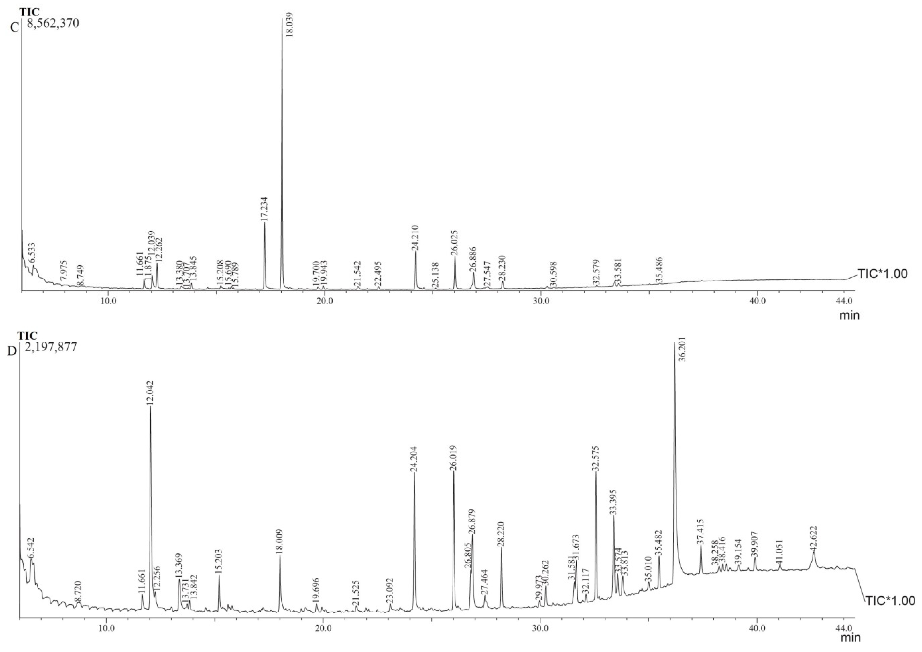
The total ion current (TIC) chromatogram represents the total intensity of the entire mass range. The x-axis and y-axis of the chromatogram represent retention time and TIC, respectively. We analyzed the gas chromatographic profiles of the four different vanilla pod ethanol extracts. The sum of TIC intensity followed by the peak number in each of the four pods were as follows (Figure 2): for ACV, 6,427,299 and 14 peaks (Figure 2A); for BCV, 5,730,438 and 25 peaks (Figure 2B); for SCV, 8,562,370 and 29 peaks (Figure 2C); and for the control group, 2,197,877 and 39 peaks (Figure 2D). The three highest peaks in descending order for each group were 2,3-butadienol, lactic acid, and methyl L-lactate in ACV; vanillin, hydroxyacetone, and guaiacol in BCV; 2,3-butanediol, lactic acid, and mequinol in SCV; and vanillin, 2-propanone, 1-hydroxy-, and mequinol in the control group. Gurnani et al. used n-hexane, methanol, and ethyl acetate to extract Indian vanilla beans using GC-MS and found six, seven, and nine volatile constituents, respectively [24]. In this study, 7, 24, 29, and 38 compounds were detected in the ACV, BCV, SCV, and control groups, respectively, during the early curing stage (the 12th day of the curing process). These numbers are mostly higher than those found in previous studies. It is worth mentioning that, although the number of compounds identified in the control group was higher than that in the microbial treatment group, 31 compounds in the microbial treatment group were not found in the control group, indicating that new combinations of potential aroma components can be produced through microbial treatment.

3.3. Analysis of Volatile Components in Ethanol Extracts

The volatile components of the whole vanilla pod analyzed via ethanol extraction using GC-MS as shown in Table 1, a total of 69 volatile components were identified in ACV, BCV, SCV, and the control groups. The identification of these 69 volatile compounds was based on comparing their spectra with those recorded in FFNSC 1.2.Lib and FFNSC 3.Lib. To determine the retention index (RI) of volatile substances, a standard solution of C8-C20 saturated alkanes was used as a reference. From each peak, the compound with the highest SI value was chosen. This process allowed for the more reliable identification of the volatile compounds. Silva et al. found that the following four components in vanilla pods are the main sources of aroma, including 4-cresol, 2-phenylethanol, guaiacol, and 4-creosol [25], and only 2-phenylethanol was not detected in this study.
In this study of three different processing treatments during the drying stage of early curing, the content of volatile compounds in SCV and the control group had the highest similarity. The use of SCV showed that a total of 50 aroma compounds were analyzed both in the experimental group and the control group, of which 17 were the same compounds, with a similarity of 34%. However, Yeh et al. employed Saccharomycopsis crataegensis in vanilla pods, revealing a similarity of 78.8% between the experimental group and the control group [18]. Compared with the control group, the vanilla pods processed by microorganisms had great differences in volatile compounds. The edible microorganisms in this study helped the vanilla pods to produce new volatile portfolios.
In addition, the aroma index component vanillin was found in BCV and the control group, and its relative percentages were 30.22% and 20.97%, respectively. These two groups had the highest proportion of vanillin as a relative percentage of total compounds. Bacillus isolates in Vanilla planifolia. Andrews beans were involved in the glucovanillin hydrolysis and vanillin formation during the curing process [26]. The microorganism pectinase was used to hydrolyze pectin between the glucovanillin substrate and β-glucosidase [27,28]. Bacillus-assisted curing increased the yield of vanillin, and the content of vanillin added with Bacillus vanillea XY18 and Bacillus subtilis XY20 was higher than that in conventionally cured vanilla beans [26,27,28]. Processing the conditioning step during the curing period needed to continue for above a month was sufficient to obtain the best quality of cured vanilla beans in terms of highest vanillin content [4]. It is worth pointing out that the content of vanillin in the BCV could be analyzed on the 12th day of the process, and the relative percentages were reach 1/3 of the relative content of the total volatile content in this study. It is speculated that the reason may be due to the fact that, during the drying stage of early curing, mucus in the placenta is affected by enzymes secreted by B. subtilis subsp. subtilis, which could decompose the glucovanillin and accelerate the production of vanillin.

3.4. Numbers of Categories of Volatile Compounds in the Ethanol Extracts of Cured Whole Vanilla Pods

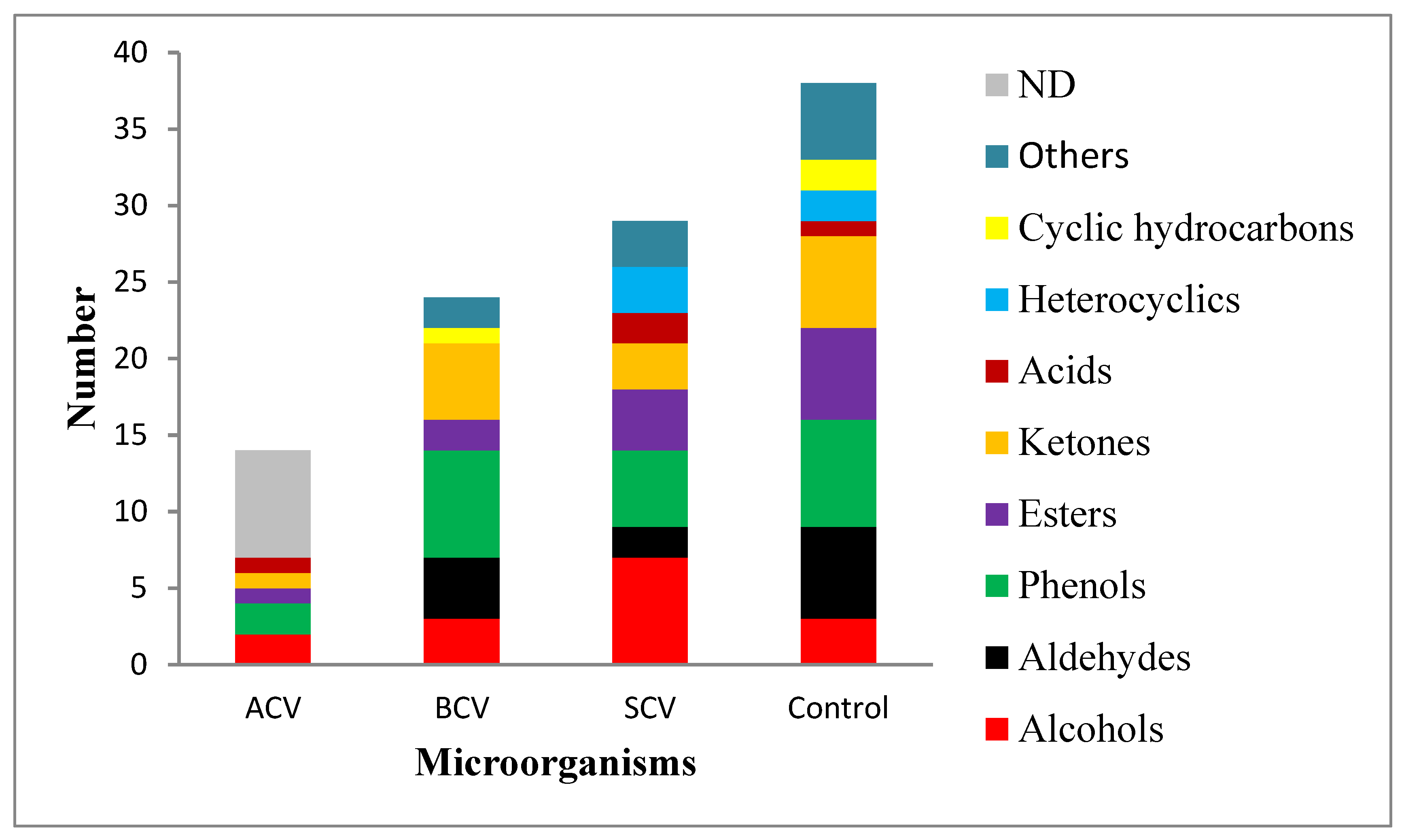
Figure 3 presents the categories of volatile compounds in the ethanol extracts of the cured whole vanilla pods in our study for comparison. The control group contained 38 compounds in 8 categories: 7 phenols, 6 aldehydes, 6 esters, 6 ketones, 3 alcohols, 2 heterocyclics, 2 cyclic hydrocarbons, 1 acid, and 5 others. The control group had the greatest number of compounds. Yah et al. used 35%, 75%, and 95% alcohol to extract vanilla pods and identified 10, 14, and 19 volatile compounds, respectively, which were composed of aldehydes, esters, carboxylic acids, alcohols, ketones, and phenols [29]. Compared with the 70% ethanol extract used in this study, the categories of volatiles were found to be less than this study. There may be two reasons for this. One is that the GC column used by Yah et al. was a non-polar column (DB-1), and here we used the polar (DB-WAX); the second reason is that this experiment has undergone an evaporation concentration step, which increased the concentration of volatiles and was easier to detect. The SCV group had 7 categories: 7 alcohols, 5 phenols, 4 esters, 2 aldehydes, 3 ketones, 3 heterocyclics, 2 acids, and 3 others. The SCV-coated pods had the most categories of compounds of the three modified groups. A total of 6 categories were analyzed in the BCV group, with 7 phenols, 5 ketones, 4 aldehydes, 3 alcohols, 2 esters, 1 cyclic hydrocarbon, and 2 others. The ACV group had the least number of compounds and categories, with only 5 categories, specifically 2 alcohol, 2 phenols, 1 ester, 1 ketone, and 1 acid. Our results showed that the volatile compounds in the ethanol extract of edible microorganisms coated with vanilla pods were mainly phenols and alcohols. Oxidase, peroxidase, and polyphenol oxidase are responsible for the browning of pod tissue and the formation of volatile ketone, aldehyde, and hydrocarbon derivatives that might add to the final vanilla flavor [30]. This study found that the addition of different microorganisms resulted in differences in the composition of volatile components during the drying stage of the early curing of vanilla pods. It may be due to microbial fermentation metabolites in vanilla pods to produce different aromas.

3.5. Odor/Flavor Description and Potential Odor/Flavor Contributors

3.5.1. Odor/Flavor Description

The key components of vanilla responsible for aroma and flavor are volatiles such as aromatic alcohols, aromatic acids, aromatic esters, phenols, aliphatic alcohols, lactones, aromatic, aliphatic hydrocarbons, terpenoids and heterocyclics [4]. The delicate and appreciated aroma of cured vanilla is also due to the participation of minor volatiles. The components jointly impart a delicate, rich, and mellow fragrance with sweet, spicy, woody, and balsamic notes [5]. The odor and flavor descriptions of vanilla pods in this study are in accordance with the related database instead of GC-O and sensory evaluation, owing to both the latter options having reproducibility challenges. The compounds comprised 9 phenols, 6 ketones, 6 alcohols, 6 aldehydes, 2 esters, and 1 acid, totaling 30 odor and flavor description as shown in Table 2. According to Table 2, 6 flavor descriptions of compounds in vanilla pods that were present at the minimum level, ranging from 30 to 75 ppm including (1) furfuryl alcohol (a sweet and caramel-like flavor); (2) 1-hydroxy-2-butanone (notes of toasted grain in its flavor profile); (3) furfural (a sweet, woody, bready, nutty, and caramel-like flavor profile); (4) vanillin methyl ether (a sweet, creamy, and vanilla-like flavor profile); (5) acetyl isovaleryl (a fruity flavor that is creamy with a hint of pineapple); and (6) butyrolactone <gamma-> (a milky flavor profile that is creamy with fruity, peach-like notes).

3.5.2. Potential Odor/Aroma Contributors

A GC-O analysis of the vanilla bean with a pentane/ether extract detected 26 odor-active compounds [31]., less than the number of odors and flavors analyzed by different processing in this study. V. planifolia’s characteristic pod flavor and fragrance is primarily caused by vanillin [32]. GC-Olfactometry (GC-O) was used and indicated that few compounds, including p-cresol, cresol, guaiacol, and 2-phenylethanol, exerted a significant influence on the overall vanilla aroma resulting from the curing of vanilla beans [33]. All the above compounds (except 2-phenylethanol) were identified in this study. In addition, the five volatile compounds selected in Table 2 are proposed as potential odor/aroma contributors according to the relative contents in samples and the related references, including: (1) 2,3-butanediol, (2) lactic acid, (3) hydroxyacetone, (4) acetoin, and (5) mequinol.
(1) 2,3-Butanediol The 2,3-butanediol produced by S. cerevisiae and Paenibacillus polymyxa ZJ-9 is a natural and environmentally friendly polyol that is widely found in fermented foods. [34]. 2,3-Butanediol was present in the ACV, SCV, and control groups and exuded a fruity, creamy, and buttery odor. Only seven compounds were identified in ACV. The relative percentage of 2,3-butanediol was highest in the ACV group at 66.47% and was 46.1% the SCV group.
(2) Lactic acid Lactic acid can be naturally produced by a broad spectrum of microbes, including bacteria, yeast, and filamentous fungi [35]. Lactic acid was found in the ethanol extract of ACV with a relative percentage of 7.69%. The relative percentage of lactic acid in SCV was 11.03%. The fermentation products of vanilla pods using ACV and BCV could produce lactic acid is a new finding in the related study of vanilla pods.
(3) Hydroxyacetone Hydroxyacetone is an aroma compound produced via the liquid fermentation of S. cerevisiae and Zygosaccharomyces rouxii from castor oil through cell permeabilization [36]. It is also one of the flavor compounds in soy sauce fermentation [37]. The relative percentage of hydroxyacetone in the ethanol extract of BCV was 25.25%. The ethanol extract of BCV was an endogenous metabolite with a pungent, sweet, and caramel-like odor.
(4) Acetoin Some bacteria are capable of acetoin biosynthesis from versatile renewable biomass, with the process mainly being used for flavors and fragrances [38]. In this study, BCV had a strong creamy aroma in the drying stage, with a GC-MS analysis showing it to have acetoin compounds. Acetoin has a sweet and creamy odor.
(5) Mequinol Mequinol has a phenolic odor and was found in the SCV and control groups. Mequinol 2%/tretinoin 0.01% solution is a drug commonly prescribed by dermatologists to treat skin conditions such as sunburn and the resulting hyperpigmentation, melasma, or age spots [39]. The relative contents of mequinol in the SCV and control groups were 7.96% and 7.11% in this study, respectively. Mequinol is a metabolite found in vanilla pod extract for the first time, which can be developed into dermatological drugs in the future.

3.6. Odor Activity Value (OAV) for Potential Aromatic Compounds

Coating vanilla pods with various microorganisms results in the production of volatile aromatic compound combinations that are significantly different from those in the control group, leading to variations in aroma. In the identification of the potential contributions of volatile compounds to the aroma of food, odor thresholds and odor activity value (OAV) become crucial [40]. The OAV is commonly used to determine which aromatic compounds are potential components in overall aroma extracts. It assesses the importance of aromatic compounds to the sensory characteristics of food through calculating the ratio between the concentration of a particular compound and its odor threshold in that food. This ratio is referred to as the OAV [41]. The OAV is equal to the ratio of compound concentration to its odor threshold [42,43]. A higher OAV indicates a greater contribution of that component to the overall aromatic odor.
Among the compounds listed in Table 2, which further excluded compounds for which odor thresholds could not be found in the literature, and the odor thresholds and OAVs of the others (15 compounds) as shown in Table 3. Significant variation was observed in the OAV among the three coated microorganisms, ACV, BCV, and SCV, compared to the control. Cresol <para-> (belonging to the phenol odor type) had the highest OAV value in both ACV and BCV treatments, while 3-Pentanol (belonging to the herbal odor type) had the highest OAV value in the SCV treatment. Additionally, 2-Methoxy-4-vinylphenol (belonging to the spicy odor type) had the highest OAV value in the control group, and this compound was not detected in any of the three microorganism treatments (Table 3), indicating variation in the aroma-contributing capabilities of the pods coated with different microorganisms. Previous studies have suggested that 2-Methoxy-4-vinylphenol could serve as a precursor to vanillin [44], which ranked second in terms of OAV in the control group. This could be attributed to the control group samples being early stage fermented vanilla pods, with 2-Methoxy-4-vinylphenol potentially being an intermediate product during the curing process.
In ACV treatment, cresol <para->, acetoin, and 2,3-butanediol exhibited significantly higher OAVs, indicating that ACV contributed to the release of these compounds, enhancing the phenolic, creamy, and buttery aroma in the pods. In contrast, in BCV treatment, the top three OAVs were cresol <para->, vanillin, and butyrolactone, suggesting that BCV had the phenolic, vanilla, and creamy characteristics. In SCV treatment, 3-pentanol, butyrolactone, and 2,3-butanediol present higher OAVs, indicating that SCV imparted herbal characteristics. In the control group, the top three OAVs were 2-methoxy-4-vinylphenol, vanillin, and butyrolactone<gamma->, highlighting the dominant spicy, vanilla, and creamy characteristics.

3.7. Principal Component Analysis (PCA)

In order to gain a better understanding of the impact of different microbial strains on the flavor characteristics of vanilla pods, we conducted principal component analysis (PCA) to observe the differences between samples with and without coating microorganisms (Figure 4). All component data in the PCA were derived from Table 1. The statistical results categorized the samples into four groups, primarily distinguishing the aromas of the ACV, BCV, SCV groups, and the control group of vanilla samples.
Regarding the control and BCV groups, there was not a significant difference in the volatile aroma profiles. However, the aroma profiles of the control group differed significantly from those of the ACV and SCV groups. Based on these statistical findings, it can be inferred that the effects of adding BCV are similar to those generated by the metabolic pathways of endogenous microbes in vanilla pods. The addition of microorganisms during the curing process of vanilla pods leads to the production of β-glucosidase, a compound that influences aroma formation and belongs to the same group as the control. On the other hand, the aroma profiles of vanilla pods processed with A. oryzae and S. cerevisiae differ from those of the control group due to the distinct metabolic pathways of these microorganisms, indicating that they possess different aromatic characteristics. This is consistent with previous studies on the impact of microbes on the flavor characteristics of vanilla, including research indicating that different strains of Bacillus sp. and Aspergillus sp. can generate varying levels of key flavor compounds in vanilla [13,47]. Furthermore, studies on other plants and their associated microorganisms have demonstrated the important role of these microbes’ metabolic activities in shaping the aroma and flavor of plant products [2,48]. In summary, our results indicate that the addition of specific microorganisms during the curing process of vanilla pods can generate distinct aroma characteristics in the product, thus offering the potential to develop new and unique vanilla aroma profiles for various applications.

4. Conclusions

The microbial coating of vanilla pods with different microbial species during the early drying stage resulted in the production of distinct key marker compounds compared to the control group. The primary contributors to the unique aroma were identified as 2,3-butanediol, lactic acid, hydroxyacetone acetoin, and mequinol. Our findings revealed that the vanilla pods exhibited a novel aroma profile characterized by sweetness, creaminess, caramel notes, fruity undertones, woody elements, and distinct vanilla characteristics, along with hints of toasted grains. This aroma profile differed from the traditional fermentation process. Furthermore, the microbial coating technology facilitated early vanillin production and the development of an oily luster. Understanding the volatile compounds involved can aid in enhancing the early aroma development of vanilla pods. This study introduced an innovative microbial coating technique for fermenting vanilla pods, resulting in diverse aroma combinations, thereby contributing to early aroma enhancement and quality improvement. This fermentation technology has important implications for the processing of vanilla pods, especially for those industry applications focused on food flavoring.

Author Contributions

Conceptualization, Y.-S.L. and T.-H.H.; methodology, C.-E.C.; software, H.-C.L.; validation, H.-C.L.; formal analysis, C.-E.C.; investigation, T.-H.H.; resources, Y.-S.L.; data curation, C.-E.C.; writing—original draft preparation, C.-E.C. and T.-H.H.; review and editing, C.-E.C.; writing—review and editing, C.-E.C.; visualization, T.-H.H.; supervision, T.-H.H.; project administration, T.-H.H. All authors have read and agreed to the published version of the manuscript.

Funding

This research was supported by grants from the Ministry of Science and Technology, Executive Yuan, the Republic of China (MOST 108-2221-E-212-008-MY3).

Institutional Review Board Statement

Not applicable.

Informed Consent Statement

Not applicable.

Data Availability Statement

Not applicable.

Acknowledgments

The authors wish to thank Nina Lai, Instrument Department Center, National Cheng Kung University, Tainan, Taiwan for their GC-MS suggestions and assistance.

Conflicts of Interest

The authors declare no conflict of interest.

References

  1. Wilde, A.S.; Frandsen, H.L.; Fromberg, A.; Smedsgaard, J.; Greule, M. Isotopic characterization of vanillin ex glucose by GC-IRMS-New challenge for natural vanilla flavour authentication? Food Control. 2019, 106, 106735. [Google Scholar]
  2. Baqueiro-Peña, I.; Guerrero-Beltrán, J.Á. Vanilla (Vanilla planifolia Andr.), its residues and other industrial by-products for recovering high value flavor molecules: A review. J. Appl. Res. Med. Aromat. Plants 2017, 6, 1–9. [Google Scholar]
  3. Havkin-Frenkel, D.; French, J.C.; Graft, N.M.; Pak, F.; Frenkel, C.; Joel, D. Interrelation of curing and botany in vanilla (Vanilla planifolia) bean. Acta Hortic. 2004, 629, 93–102. [Google Scholar] [CrossRef]
  4. Wongsheree, T.; Wongs-Aree, C.; Srilaong, V.; Jitareerat, P. Vanilla Cultivation and Curing in Thailand; International Society for Horticultural Science (ISHS): Leuven, Belgium, 2013. [Google Scholar]
  5. Sharp, M.D.; Kocaoglu-Vurma, N.A.; Langford, V.; Rodriguez-Saona, L.E.; Harper, W.J. Rapid discrimination and characterization of vanilla bean extracts by attenuated total reflection infrared spectroscopy and selected Ion flow tube mass spectrometry. J. Food Sci. 2012, 77, 284–292. [Google Scholar]
  6. Ramachandra Rao, S.; Ravishankar, G.A. Vanilla flavour: Production by conventional and biotechnological routes. J. Sci. Food Agric. 2000, 80, 289–304. [Google Scholar]
  7. Khoyratty, S.; Kodja, H.; Verpoorte, R. Vanilla flavor production methods: A review. Ind. Crops Prod. 2018, 125, 433–442. [Google Scholar]
  8. Esen, A. ß-Glucosidases Overview. In ß-Glucosidases; American Chemical Society: Washington, DC, USA, 1993; Volume 533, pp. 1–14. [Google Scholar]
  9. Khoyratty, S.; Verpoorte, R.; Kodja, H. Vanillin: Biosynthesis, biotechnology, and bioproduction. In Orchids Phytochemistry, Biology and Horticulture: Fundamentals and Applications; Mérillon, J.M., Kodja, H., Eds.; Springer International Publishing: Cham, Switzerland, 2022; pp. 341–358. [Google Scholar]
  10. Khoyratty, S.; Dupont, J.; Lacoste, S.; Palama, T.L.; Choi, Y.H.; Kim, H.K.; Payet, B.; Grisoni, M.; Fouillaud, M.; Verpoorte, R.; et al. Fungal endophytes of Vanilla planifolia across Réunion Island: Isolation, distribution and biotransformation. BMC Plant Biol. 2015, 15, 142. [Google Scholar]
  11. Che, H.; Yu, J.; Sun, J.; Lu, K.; Xie, W. Bacterial composition changes and volatile compounds during the fermentation of shrimp paste: Dynamic changes of microbial communities and flavor composition. Food Biosci. 2021, 43, 101169. [Google Scholar]
  12. Abd-Aziz, S.; Jenol, M.A.; Ramle, I.K. Biovanillin from oil palm biomass. In Biorefinery of Oil Producing Plants for Value-Added Products; Abd-Aziz, S., Gozan, M., Ibrahim, M.F., Phang, L.Y., Eds.; John Wiley & Sons, Inc.: Hoboken, NJ, USA, 2022; pp. 493–514. [Google Scholar]
  13. Chen, P.; Yan, L.; Wu, Z.; Li, S.; Bai, Z.; Yan, X.; Wang, N.; Liang, N.; Li, H. A microbial transformation using Bacillus subtilis B7-S to produce natural vanillin from ferulic acid. Sci. Rep. 2016, 6, 20400. [Google Scholar]
  14. Mehmood, T.; Ahmed, S.; Waseem, R.; Saeed, S.; Ahmed, W.; Irfan, M.; Ullah, A. Valorization of fruit peels into biovanillin and statistical optimization of process using enterobacter hormaechei through solid-state fermentation. Fermentation 2022, 8, 40–54. [Google Scholar]
  15. Yoshida, A.; Takenaka, Y.; Tamaki, H.; Frébort, I.; Adachi, O.; Kumagai, H. Vanillin formation by mcrobial amine oxidases from vanillylamine. J. Ferment. Bioeng. 1997, 84, 603–605. [Google Scholar]
  16. Rejani, C.T.; Radhakrishnan, S. Microbial conversion of vanillin from ferulic acid extracted from raw coir pith. Nat. Prod. Res. 2020, 36, 901–908. [Google Scholar]
  17. Vandamme, E.J. Bioflavours and fragrances via fungi and their enzymes. Fungal Divers. 2003, 13, 153–166. [Google Scholar]
  18. Yeh, C.H.; Chen, K.Y.; Chou, C.Y.; Liao, H.Y.; Chen, H.C. New insights on volatile components of Vanilla planifolia cultivated in Taiwan. Molecules 2021, 26, 3608. [Google Scholar] [PubMed]
  19. Odoux, E.; Brillouet, J.M. Anatomy, histochemistry and biochemistry of glucovanillin, oleoresin and mucilage accumulation sites in green mature vanilla pod (Vanilla planifolia; Orchidaceae): A comprehensive and critical reexamination. Fruits 2009, 64, 221–241. [Google Scholar]
  20. The Good Scents Company (TGSC). Information System. Available online: http://www.thegoodscentscompany.com/index.html (accessed on 1 December 2022).
  21. MFood Flavor Innovation (MFFi). Available online: https://mffi.sjtu.edu.cn/ (accessed on 16 July 2023).
  22. The LRI & Odour Database—Odour Data. Available online: http://www.odour.org.uk/odour/index.html (accessed on 16 July 2023).
  23. ISO 5565-1:1999; Vanilla [Vanilla fragrans (Salisbury) Ames]—Part 1: Specification. International Organization for Standardization (ISO): Geneva, Switzerland, 1999. Available online: https://www.iso.org/standard/22116.html (accessed on 16 July 2023).
  24. Gurnani, N.; Kapoor, N.; Mehta, D.; Gupta, M.; Mehta, B. Characterization of chemical groups and identification of novel volatile constituents in organic solvent extracts of cured Indian vanilla beans by GC-MS. Middle East J. Sci. Res. 2014, 22, 769–776. [Google Scholar]
  25. Silva, A.P.; Gunata, Z.; Lepoutre, J.P.; Odoux, E. New insight on the genesis and fate of odor-active compounds in vanilla beans (Vanilla planifolia G. Jackson) during traditional curing. Food Res. Int. 2011, 44, 2930–2937. [Google Scholar]
  26. Gu, F.; Chen, Y.; Fang, Y.; Wu, G.; Tan, L. Contribution of Bacillus isolates to the flavor profiles of vanilla beans assessed through aroma analysis and chemometrics. Molecules 2015, 20, 18422–18436. [Google Scholar]
  27. Chen, Y.; Gu, F.; Li, J.; He, S.; Xu, F.; Fang, Y. Involvement of colonizing Bacillus isolates in glucovanillin hydrolysis during the curing of Vanilla planifolia Andrews. Appl. Environ. Microbiol. 2015, 81, 4947–4954. [Google Scholar]
  28. Zhang, Y.; Mo, L.; Chen, F.; Lu, M.; Dong, W.; Wang, Q.; Xu, F.; Gu, F. Optimized production of vanillin from green vanilla pods by enzyme-assisted extraction combined with pre-freezing and thawing. Molecules 2014, 19, 2181–2198. [Google Scholar]
  29. Yeh, C.H.; Chou, C.Y.; Wu, C.S.; Chu, L.P.; Huang, W.J.; Chen, H.C. Effects of different extraction methods on vanilla aroma. Molecules 2022, 27, 4593. [Google Scholar]
  30. Havkin-Frenkel, D.; Belanger, F.C. Handbook of Vanilla Science and Technology; John Wiley & Sons: Hoboken, NJ, USA, 2018; pp. 1–488. [Google Scholar]
  31. Pérez-Silva, A.; Odoux, E.; Brat, P.; Ribeyre, F.; Rodriguez-Jimenes, G.; Robles-Olvera, V.; García-Alvarado, M.; Günata, Z. GC–MS and GC–olfactometry analysis of aroma compounds in a representative organic aroma extract from cured vanilla (Vanilla planifolia G. Jackson) beans. Food Chem. 2006, 99, 728–735. [Google Scholar]
  32. Arya, S.S.; Rookes, J.E.; Cahill, D.M.; Lenka, S.K. Vanillin: A review on the therapeutic prospects of a popular flavouring molecule. Adv. Tradit. Med. 2021, 21, 1–17. [Google Scholar]
  33. Dignum, M.J.W.; van der Heijden, R.; Kerler, J.; Winkel, C.; Verpoorte, R. Identification of glucosides in green beans of Vanilla planifolia Andrews and kinetics of vanilla β-glucosidase. Food Chem. 2004, 85, 199–205. [Google Scholar]
  34. Kim, S.J.; Kim, J.W.; Lee, Y.G.; Park, Y.C.; Seo, J.H. Metabolic engineering of Saccharomyces cerevisiae for 2, 3-butanediol production. Appl. Microbiol. Biotechnol. 2017, 101, 2241–2250. [Google Scholar]
  35. Juturu, V.; Wu, J.C. Microbial production of lactic acid: The latest development. Crit. Rev. Biotechnol. 2016, 36, 967–977. [Google Scholar] [PubMed]
  36. Álvarez, A.; Gutiérrez, A.; Ramírez, C.; Cuenca, F.; Bolívar, G. Aroma compounds produced by liquid fermentation with Saccharomyces cerevisiae and Zygosaccharomyces rouxii from castor oil through cell permeabilization. Biocatal. Agric. Biotechnol. 2022, 39, 102243. [Google Scholar]
  37. Feng, J.; Zhan, X.B.; Zheng, Z.Y.; Wang, D.; Zhang, L.M.; Lin, C.C. New model for flavour quality evaluation of soy sauce. Czech J. Food Sci. 2013, 31, 292–305. [Google Scholar]
  38. Xiao, Z.; Lu, J.R. Strategies for enhancing fermentative production of acetoin: A review. Biotechnol. Adv. 2014, 32, 492–503. [Google Scholar]
  39. Draelos, Z.D. The combination of 2% 4-hydroxyanisole (mequinol) and 0.01% tretinoin effectively improves the appearance of solar lentigines in ethnic groups. J. Cosmet. Dermatol. 2006, 5, 239–244. [Google Scholar] [PubMed]
  40. Cadwallader, K. 18—Measuring cheese flavor. In Improving the Flavour of Cheese; Weimer, B.C., Ed.; Woodhead Publishing: Sawston, UK, 2007; pp. 401–417. [Google Scholar]
  41. Methven, L. 16—Techniques in sensory analysis of flavour. In Flavour Development, Dnalysis and Perception in Food and Beverages; Parker, J.K., Elmore, J.S., Methven, L., Eds.; Woodhead Publishing: Sawston, UK, 2015; pp. 353–368. [Google Scholar]
  42. San, J.F.; Cacho, J.; Ferreira, V.; Escudero, A. Chapter 21—Differences in chemical composition of croma among red wines of different price category. In Flavour Science; Ferreira, V., Lopez, R., Eds.; Academic Press: San Diego, CA, USA, 2014; pp. 117–121. [Google Scholar]
  43. Ferreira, V. 1—Volatile aroma compounds and wine sensory attributes. In Managing Wine Quality; Reynolds, A.G., Ed.; Woodhead Publishing: Sawston, UK, 2010; pp. 3–28. [Google Scholar]
  44. van Schijndel, J.; Molendijk, D.; van Beurden, K.; Canalle, L.A.; Noël, T.; Meuldijk, J. Preparation of bio-based styrene alternatives and their free radical polymerization. Eur. Polym. J. 2020, 125, 109534. [Google Scholar]
  45. Li, R.; Tian, S.; Guo, C.; Li, l.; Liang, M.; Zhang, J. Sensory-guided analysis and recombination of characteristic flavor compoundsin Vanilla planifolia Andrews. Food Sci. 2023, 44, 217–223. (In Chinese) [Google Scholar]
  46. Wang, B.; Xu, H.A.; XU, J.; Qu, M.; Zhang, Y.; Huang, Q.; Xiao, L. Analysis of characteristic volatile aroma components in Inner Mongolia dried Allium chrysanthum. Sci. Technol. Food Ind. 2022, 43, 296–304. (In Chinese) [Google Scholar] [CrossRef]
  47. Maheshwari, R.; Bharadwaj, G.; Bhat, M.K. Thermophilic fungi: Their physiology and enzymes. Microbiol. Mol. Biol. Rev. 2000, 80, 461–488. [Google Scholar]
  48. Gupta, P.; Samant, K.; Sahu, A. Isolation of cellulose-degrading bacteria and determination of their cellulolytic potential. Int. J. Microbiol. 2012, 2012, 578925. [Google Scholar] [CrossRef] [Green Version]
Figure 1. Images of uncoated and coated vanilla pods with different microorganisms with a laser scanning microscope. Surface tissue for (A) ACV; (B) BCV; (C) SCV; and (D) control group. Internal tissue of (E) ACV; (F) BCV; (G) SCV; and (H) control group. (ACV: A. oryzae vanilla pods; BCV: B. subtilis subsp. subtilis vanilla pods; SCV: S. cerevisiae vanilla pods.)
Fermentation 09 00724 g001
Figure 2. Gas chromatographic profiles of (A) ACV; (B) BCV; (C) SCV; and (D) control group. ACV: A. oryzae vanilla pods; BCV: B. subtilis subsp. subtilis vanilla pods; SCV: S. cerevisiae vanilla pods.
Fermentation 09 00724 g002aFermentation 09 00724 g002b
Figure 3. Numbers of categories of volatile compounds in ethanol extracts from whole pods. (ACV: A. oryzae vanilla pods; BCV: B. subtilis subsp. subtilis vanilla pods; SCV: S. cerevisiae vanilla pods; ND: peaks in total ion current (TIC) chromatogram not identified.)
Fermentation 09 00724 g003
Figure 4. Principal component analysis (PCA). ACV: A. oryzae vanilla pods; BCV: B. subtilis subsp. subtilis vanilla pods; SCV: S. cerevisiae vanilla pods.
Fermentation 09 00724 g004
Table 1. Relative percentage (%) of volatile compounds in the ethanol extract of cured vanilla pods.
CompoundsACV aBCV bSCV cControl
1Hexanal 1.82 3.49
21,2-Ethanediamine, N,N′-dimethyl- 0.850.56
32-Butanone, 3-hydroxy- 1.810.74
42-Propanone, 1-hydroxy- 3.2613.56
5Propanoic acid, 2-hydroxy-, methyl ester 4.180.28
6Pentane, 2-bromo- 1.082.6
71-Hydroxy-2-butanone 0.5
8Acetic acid, hydroxy-, ethyl ester 1.040.43
9Pentanoic acid, 3-methyl-2-oxo-, methyl ester 0.671.68
102,3-Butanediol66.47 46.13.25
11Butyrolactone <gamma-> 0.680.530.57
122(5H)-Furanone, 3-methyl- 0.720.31
13Cyclopentane, butyl- 0.33
14Mequinol 7.967.11
15Creosol 4.346.596.54
16Levoglucosenone 1.87
17Phenol 4.464.61
18Benzaldehyde, 4-methoxy- 0.85
19Phenol, 3-methyl- 1.552.99
202-Hydroxy-gamma-butyrolactone 0.33
212-Methoxy-4-vinylphenol 1.12
222,3-Anhydro-d-galactosan 1.33
232,3-Anhydro-d-mannosan 2.32
24Phenol, 2-methoxy-4-(methoxymethyl)- 0.36
25Methyl-(2-hydoxy-3-ethoxy-benzyl)ether 0.295.76
26Vanillin methyl ether 4.24 4.82
271,4:3,6-Dianhydro-.alpha.-d-glucopyranose 0.651.37
282(1H)-Pyridinone 1.46
293-Propylglutaric acid 0.46
30Phenol, 4-(ethoxymethyl)- 0.251.6
31Vanillin 30.22 20.97
322-Propanone, 1-(4-hydroxy-3-methoxyphenyl)- 1.56
334-Hydroxy-2-methoxybenaldehyde 0.33
34Ethyl homovanillate 0.44
353,4-Anhydro-d-galactosan 0.35
36Benzene, 1,1′-[1,2-ethanediylbis(oxy)]bis [2-methoxy- 0.86
37Benzenemethanol, alpha.-(1-methylethyl)-, (R)- 0.28
38Benzaldehyde, 4-hydroxy- 2
39Acetoin2.671.43
40Methyl L-lactate2.86
41Lactic acid 7.69 11.03
42Guaiacol0.4410
43Phenyl alcohol0.654.960.15
44sec-Butylamine 0.5
45Hydroxyacetone 25.25
46Pentene <3-hydroxy-> 1
47Acetyl isovaleryl 1.23
481-Acetoxy-2-propanone 0.29
49Furfuryl alcohol 2.34
50Corylon 0.3
51Cresol <para->0.812.64
52Allyl alcohol 0.61
53Guaiacol <4-vinyl-> 2.08
54Vanillyl butyl ether 1.07
55Ferrolure isomer I 1.28
56Eugenol 1.36
57Guaiacol <4-propyl-> 0.56
58Benzaldehyde <para-hydroxy-> 1.32
59Phytol acetate <(E)-> 0.42
60Pentanal, 3-methyl- 3.06
61dl-Alanyl-l-alanine 0.95
62n-Hexylmethylamine 0.21
633-Pentanol 0.26
64Oxiranemethanol, (R)- 0.5
65Furfural 0.18
662-Furanmethano 0.59
671H-Pyrrole, 2,5-dihydro- 0.48
682-Pyrrolidinone 0.39
69Nicotinyl Alcohol 0.19
a ACV represents A. oryzae vanilla pods. b BCV represents B. subtilis subsp. subtilis vanilla pods. c SCV represents S. cerevisiae vanilla pods.
Table 2. Odor and flavor descriptions of the ethanol extracts of whole vanilla pods after different processing methods.
CompoundsOdor Description aFlavor Description a
Alcohols
2,3-Butanediol Fruity, creamy, buttery
Phenyl alcoholPhenolic
Furfuryl alcoholSweet, caramel, bread, coffeeSweet, caramel-like/at 50.00 ppm
Allyl alcoholPungent, mustard
3-PentanolSweet, herbal, oily, nutty
2-FuranmethanolSweet, caramel, bread, coffeeSweet, caramel-like, bready, coffee
Aldehydes
HexanalFresh, fruityApple, citrus, orange with a fresh, lingering aftertaste
Benzaldehyde,4-methoxy-Sweet, powdery, hawthorn, balsamCreamy, vanilla, spicy with a marshmallow flavor
VanillinSweet, vanilla, creamyVanilla, sweet, creamy, spicy, milky
Benzaldehyde, 4-hydroxy-Sweet, nutty, almond, balsam, woodyCreamy, nutty with vanilla and honey nuances
FurfuralSweet, woody, almond, fragrant, baked, breadSweet, woody, bready, nutty, caramel-like/at 30.00 ppm.
Vanillin methyl etherSweet, woody, vanillaSweet, creamy, vanilla/at 50.00 ppm.
Phenols
CreosolSweet, candy, spice, eugenol, vanillaVanilla, spice, eugenol, woody
PhenolPhenolic
2-Methoxy-4-vinylphenolSweet, spicy, clove, woody, powderySpicy, powdery, clove, woody, balsamic, amber
GuaiacolSpice, vanilla, woodyWoody, phenolic, bacon, savory, smoky, medicinal
Cresol <para->Narcissus, mimosaPhenolic
Guaiacol <4-vinyl->Sweet, spicy, clove, woodySpicy, clove, woody
Guaiacol <4-propyl->Clove, spicy, sweet, allspiceSpicy, clove, allspice, peppery
MequinolPhenolic
Vanillyl butyl etherVanilla, fruity
Esters
Butyrolactone <gamma->Creamy, caramel Milky with fruity peach-like afternotes/at 75.00 ppm
Phytol acetate <(E)->Mild, floral, fruity, orchid, balsamicNutty, waxy, woody, oily, creamy, nut, flesh
Ketones
1-Hydroxy-2-butanoneSweet, coffee, malt, butterscotch Toasted grain notes/at 30.00 ppm.
Acetyl isovalerylSweet, fruity, creamyFruity, creamy with a pineapple nuance/at 50.00 ppm.
AcetoinSweet, buttery, creamy, dairy, milkyCreamy, dairy, sweet, milky, buttery
HydroxyacetonePungent, sweet, caramel-likeSweet, burnt
1-Acetoxy-2-propanoneFruity, buttery, dairy, nutty
CorylonCaramel, maple syrupCaramel-like
Acids
Lactic acid OdorlessSour, acid
a The Good Scents Company (TGSC) information system [20].
Table 3. Odor thresholds and odor activity values (OAVs) of potent aroma components in vanilla ethanol extracts.
CompoundThreshold
(ppm)
OAVOdor Type
ACVBCVSCVControl
2-Methoxy-4-vinylphenol0.003 a---2,740,450spicy
2,3-Butanediol100 b2509-3266238creamy
Furfural 9.562 b--137-bready
Cresol <para->0.0039 b781,2358,101,401--phenolic
3-Pentanol0.12 c--15,400-herbal
Acetoin0.8 c12,60321,371--buttery
Hexanal4.5 c-4828-5682green
Vanillin0.35 d-1,032,425-438,659vanilla
Benzaldehyde, 4-hydroxy-146.67 d---100woody
Guaiacol5.53 d29821,615--smoky
Guaiacol <4-vinyl->1.74 d-3855--spicy
Phenol20.01 d--15801686phenolic
Furfuryl alcohol409.18 d-60--bready
Lactic acid899.35 d32-87-odorless
Butyrolactone <gamma->0.284 e-28,73713,32014,779creamy
ACV: A. oryzae vanilla pods; BCV: B. subtilis subsp. subtilis vanilla pods; SCV: S. cerevisiae vanilla pods. a, b, c, d, and e represent threshold data sources: a The Good Scents Company(TGSC) information system [20]; b MFood Flavor Innovation (MFFi) [21]; c LRI & Odour Database—Odour Data [22]; d Ref. [45]; e Ref. [46]; -: not identified in sample.
Disclaimer/Publisher’s Note: The statements, opinions and data contained in all publications are solely those of the individual author(s) and contributor(s) and not of MDPI and/or the editor(s). MDPI and/or the editor(s) disclaim responsibility for any injury to people or property resulting from any ideas, methods, instructions or products referred to in the content.