Article

The Antioxidant Activity and Protection of Probiotic Bacteria in the In Vitro Gastrointestinal Digestion of a Blueberry Juice and Whey Protein Fermentation System

1
College of Food Science and Engineering, Yangzhou University, Yangzhou 225127, China
2
Jiangsu Key Laboratory of Dairy Biotechnology and Safety Control, Yangzhou University, Yangzhou 225127, China
3
Weiwei Food & Beverage Co., Ltd., Xuzhou 221114, China
4
Shaanxi Key Laboratory of Environmental Engineering, School of Environmental and Municipal Engineering, Xi’an University of Architecture and Technology, Xi’an 710055, China
*
Author to whom correspondence should be addressed.
Fermentation 2023, 9(4), 335; https://doi.org/10.3390/fermentation9040335
Submission received: 21 February 2023 / Revised: 24 March 2023 / Accepted: 25 March 2023 / Published: 28 March 2023
(This article belongs to the Section Fermentation for Food and Beverages)

Abstract

:
Blueberries have received great attention due to the health effects of their bioactive compounds, such as antioxidant, antitumor, and anti-obesity properties. Probiotics also have these health-promoting benefits. However, these biological activities may be affected by the processs of gastrointestinal digestion, which decreases their functionality. This study aimed to use a more convenient method to improve the blueberries’ antioxidant activity and protective effects on probiotic cells by fermentation with whey protein, and to explore the possible mechanisms underlying these effects. This result showed that the total phenolic content, anthocyanin content, reducing power, DPPH radical scavenging capacity, and probiotic cells’ survival in a blueberry juice and whey protein fermentation system were enhanced in a model of in vitro gastrointestinal digestion. The bioactive compounds in blueberry juice interacted with whey protein, as shown through FTIR. The stability of phenolic compounds was enhanced, and the release of functional compounds in the mixture fermentation system was delayed through CLSM. Interactions between bioactive compounds in blueberries, whey protein, and bacterial surface proteins, glycoproteins or polysaccharides during fermentation were studied by SDS-PAGE. Thus, the stability of bioactive activities in the mixed system after fermentation was strengthened by the interaction. The mixed fermentation system has promising potential for improving antioxidant activity and protecting probiotic cells.

1. Introduction

Blueberries, which have a blue color, belong to the family Ericaceae, the subfamily Vacciniaceae, and the genus Vaccinium. They are rich in anthocyanins, other flavonoids, phenolic compounds, and ascorbic acid, which possess a strong capacity for anti-oxidation because of their structure and content. However, due to pH, temperature, metal ions, and enzymatic activity, these functional compounds fail to tolerate the harsh gastrointestinal environment and, thus, cannot maintain their stability and effective release [1]. To enhance the stability of these bioactive compounds, many technologies, such as the preparation of microcapsules and hydrogel particles, have been applied. There was a positive effect on protecting these bioactive compounds, but some drawbacks were shown such as the high cost of the preparation of microcapsules with a freeze-drying process [2]. Therefore, we would like to use a simple, easy and inexpensive method of replacing these processes. It has been widely proven that the interactions of proteins and some active compounds in blueberries could increase their stability and functionality. Anuyahong et al. found that the addition of rice berry extract to yogurt could increase total phenolic content and antioxidant activity before and after in vitro gastrointestinal digestion [3]. Thus, this fermentation method may be a promising way to improve functionality.
Similarly, it is clear that probiotics that contain live microorganisms must keep a sufficient amount (>106 CFU/mL or CFU/g) of the probiotics product in order to deliver health benefits to consumers. Nevertheless, the survival of cells in probiotics is affected by the challenges of pH, temperature, oxygen, and especially human gastrointestinal conditions [4]. Numerous studies have illustrated that the addition to dairy product materials of some bioactive compounds that exert a prebiotic effect is an effective way to reduce the excessive loss of probiotic cells, and the materials further protect bioactive compounds from degradation during storage and in vitro gastrointestinal digestion. Additionally, whey protein could also be used as a carrier of active compounds, such as anthocyanins, making it possible to deliver these functional compounds to the entire intestinal tract and enabling their metabolism by the gut microbiota [1]. Another study showed that the viability of L. casei ATCC 393 when immobilized by sea buckthorn berries still maintained a high level compared to that of free L. casei ATCC 393 during storage for 90 days. Moreover, an in vitro gastrointestinal digestion assay indicated that immobilized L. casei ATCC 393 could be resistant to adverse gastrointestinal environments and that the viable cells remained at a sufficient level at the end of intestinal digestion (>107 CFU/g) [5]. Similar results were also reported showing that the supplementation of fermented milk with anthocyanin extracts from sour cherry skins and a whey protein isolate microcapsule powder promoted the growth of L. casei 431* and maintained viable probiotic cells counts (>1010 CFU/g) during storage for 21 days [1].
Thus, this combination of probiotic cells and functional bioactive compounds in a protein matrix seems to provide numerous benefits during the processing, storage, and consumption of a product. In a previous study, we found that the total phenolic content, antioxidant activity, and viable probiotic cell count increased in a mixed fermentation system after in vitro gastrointestinal digestion as illustrated in Figure 1. This may have been due to the interactions of whey protein and blueberries with bacterial surface proteins or glycoproteins, which protect active compounds in blueberries and form a protective layer around bacteria during the fermentation process as illustrated in Figure 2a.
In order to demonstrate this, ultrafiltration technology was used to separate the bacteria, including their surface molecular substances, from a fermentation broth. We aimed to find the fraction with the highest content of bioactive compounds, antioxidant activity, and protection of probiotic cells during in vitro gastrointestinal digestion by using ultrafiltration technology, as well as to investigate whether interactions could occur between the whey protein or blueberries and surface-layer-proteins (SLPs) or glycoproteins on bacterial surfaces.
The blueberry juice and whey protein fermentation system showed a high release of bioactive compounds, together with antioxidant activity and a protective effect on probiotic cells during in vitro gastrointestinal digestion. Thus, the blueberry juice and whey protein mixture fermentation system may become a functional product and provide a novel idea for protecting probiotic cells.

2. Materials and Methods

2.1. Materials and Lactobacillus Strains

Whey protein (80% protein) was supplied by Fonterra (Auckland, New Zealand). Blueberries (Vaccinium angustifolium) were obtained from farmers in Daxinganling in China during the 2022 harvest period. Lacticaseibacillus casei 54 (L. casei 54) and Lactiplantibacillus plantarum 67 (L. plantarum 67) (Jiangsu Key Laboratory of Dairy Biotechnology and Safety Control, Yangzhou University) were used as the lactobacillus strains for the following preparation of fermentation samples.

2.2. Fermented Beverage Preparation

Blueberries were stored at −20 °C before preparing the fermented blueberry and whey protein beverage. L. plantarum 67 and L. casei 54 were routinely grown in MRS broth at 37 °C for 15–18 h. Blueberry juice was prepared by using a crusher after weighing the blueberries to reach a blueberry juice content of 17% (w/v). Then, 6% (w/v) sugar was added to all beverages, and for the mixed fermented beverages, an equal weight of whey protein was also added. The samples fermented with L. plantarum 67 and L. casei 54 were adjusted to a pH of 6.5 and pasteurized (95 °C, 5 min). After cooling to room temperature, 2% (v/v) inocula of L. plantarum 67 and L. casei 54 were added to the beverages, followed by incubation at 37 °C for 12 h. Then, the fermented beverages were stored under refrigeration until the following tests. The experiment was replicated three times.

2.3. Preparation of Separated Products through Ultrafiltration

The separated products were prepared by using a molecular weight cut-off membrane of 100 kDa under the pressure of 0.25 MPa as shown in Figure 2b. Then, the permeate (MW < 100 kDa) and the concentration (MW > 100 kDa) were obtained. After ultrafiltration, these membranes were rinsed with deionized water and NaOH (0.01 mol/L) for recycling. Finally, the ultrafiltrated fractions were stored at 4 °C, and partial samples were further freeze-dried and, stored at −20 °C for the following analyses. The condition of freeze-drying conditions was that the temperature of the first layer board was kept at −50 °C for 30 min, and then rose to −10 °C within 60 min. The temperature of −10 °C was kept for 24 h, and finally rose to 10 °C. The vacuum degree of the whole process was 0.4 Pa and the temperature of the cold trap was 60 °C.
Figure 2. Schematic diagram showing whey protein and blueberry interaction with bacterial cells’ surface proteins or glycoproteins (a), and ultrafiltration process (b).
Fermentation 09 00335 g002

2.4. Enumeration of the LAB

The viable cell count of probiotic cells before and after in vitro gastrointestinal digestion was assessed by using the plate count method [6]. The samples were diluted with sterile normal saline (0.9%, w/v). Serial dilution (101–106) was performed, and 100 µL of each diluted sample was transferred into 900 µL of sterile normal saline. Then, 5 µL of diluted samples was blended, removed, and placed on the previously prepared MRS agar plates with a pipette. Every dilution time (T) of these samples was performed in triplicate. Finally, the viable cell counts of the samples were obtained after incubation at 37 °C for 36 h, and they were calculated by using Equation (1) [6]:
CFU mL−1 = 10T × 200 × Ncolony

2.5. Determination of Total Phenolic Content (TPC)

The Folin–Ciocalteu method was used to determine the TPC of the samples. Briefly, 150 µL of Folin phenol solution was mixed with an equal volume of the sample. Then, 1.5 mL of Na2CO3 (7.5%, w/v) was immediately added to react for 5 min at room temperature. Subsequently, 3.2 mL of deionized water was added and kept away from light for 5 min. Finally, a measurement of the absorbance at 750 nm was carried out. The TPC was calculated with a standard curve according to gallic acid with different concentrations (0–100 mg L−1) [7]. The standard curve equation of gallic acid was as follows:
y = 0.0024x + 0.049 (R = 0.9998)
The result of TPC was expressed as milligrams of gallic acid equivalents (GAE) per 100 g of sample weight.

2.6. Determination of the Anthocyanin Concentration (TAC)

The TAC was estimated with the pH-differential method. Firstly, 1 mL of sample was mixed with 9 mL of KCl buffer (0.025 M, pH 1.0) and CH3COONa buffer (0.4 M, pH 4.5) in two centrifuge tubes. Then, the absorbance was measured at wavelengths of 520 nm and 700 nm, respectively, after reacting for 15–30 min. The experiments were conducted in triplicate [8]. The calculation formula of TAC content was as follows:
ΔA = (A520 − A700)pH1.0 − (A520 − A700)pH4.5
TAC = ( Δ A × MW × DF × 100 ) / ( ε × 1 )
where ΔA is the absorbance difference, DF is the dilution factor, MW is the molecular weight of cyanidin-3-glucoside (449.20 g mol−1), ε is the molar absorptivity of cyanidin-3-O-glucoside (26,900 L mol−1 cm−1), and l is a path length of 1 cm.

2.7. Determination of Reducing Power

The reducing power of the samples was measured based on the method used in [9] with some slight modifications. Firstly, sodium phosphate buffer (0.2 M) was prepared, and the pH of the buffer was adjusted to 6.6. Then, 2.5 mL of the buffer was mixed with 0.5 mL of sample in a centrifuge tube; 2.5 mL of K3Fe(CN)6 (1%, w/v) was added to the above centrifuge tube with a pipette and allowed to further react for 20 min at 50 °C. After 20 min, an equal volume of trichloroacetic acid (10%, v/v) was also added with a pipette. If the reaction solution was turbid, it was centrifuged at 750 g for 10 min, and 2.5 mL of the supernatant was removed and placed in a new centrifuge tube. Finally, an equal volume of distilled water and 0.5 mL of FeCl3 (0.1%, w/v) were added to the above centrifuge tube. The reducing power of the samples was assessed by measuring the absorbance at a wavelength of 700 nm. The calculation formula for the reducing power was as follows:
Reducing power = A1 − A0
where A1 is the absorbance of the mixture with samples, A0 is the absorbance of the mixture without samples.

2.8. Determination of DPPH Radical-Scavenging Activity

The effects of the samples on DPPH radicals were determined according to the method previously described by [10] with some modifications. A total of 1.0 mL of the sample was added to a fourfold volume of DPPH radical solution (0.1 mM), mixed evenly, and allowed to react for 30 min in the dark. Finally, the scavenging activity was calculated using the absorbance at a wavelength of 517 nm, which was measured with a UNICO UV-2100 spectrophotometer. The calculation formula of DPPH radical-scavenging activity was as follows:
DPPH   radical-scavening   activity = 1     A   sample     A   control A   blank
where A sample is the absorbance of the sample at 517 nm, A control is the absorbance of the control at 517 nm and A blank is the absorbance of the blank at 517 nm.

2.9. In Vitro Gastrointestinal Digestion

For in vitro gastrointestinal digestion, 10 mM of Tris-HCl buffer, the simulated gastric juice (SGJ), and the simulated duodenal juice (SDJ) were prepared in advance, and the pH of the buffer was adjusted to 7.5. Porcine pepsin (4%, w/v, EC 232-629-3) was dissolved in 0.1 M HCl to prepare SGJ. Pancreatin (0.2%, w/v, EC 232-468-9) was dissolved in 0.9 M NaHCO3 to prepare SDJ. Then, 500 µL of the above buffer was mixed with 5 mL of the samples in centrifuge tubes. Subsequently, 11 mL of SGJ was added to each of the centrifuge tubes, and the pH of the digestion mixtures was adjusted to 2.0. Then, they were shaken at 100 rpm for 37 °C. Equal parts of gastric chyme collected after 2 h were mixed with an equal volume of SDJ, and the final pH of the intestinal mixtures was adjusted to 7.0. Finally, the mixtures were shaken at 100 rpm and 37 °C for 2 h. At the end of intestinal digestion, the intestinal chyme was collected and measured.

2.10. Fourier-Transform Infrared Spectroscopy (FT-IR)

The structural changes in the freeze-dried samples were characterized using an FTIR spectrometer (Cary 610/670 Microscopic Infrared Spectrometer, Varian-American) equipped with a diamond accessory with a spectral region of 3800–500 cm−1, using 36 scans, and a resolution of 8 cm−1. At the same time, air was used as the background.

2.11. Confocal-Laser-Scanning Microscopy (CLSM)

The release of anthocyanin, morphological changes and the distribution of probiotic organisms in the samples were studied by using CLSM [11]. Briefly, 0.1% acridine orange solution (Sigma-Aldrich) (λexcitation 480 nm) was used to stain the probiotic organisms and protein in the samples taking note of the autofluorescence characteristics of the anthocyanin (λexcitation 543 nm) in blueberries, and the distributions of anthocyanin, protein, and probiotic organisms were observed. Finally, the stained samples were mounted on slides, sealed, and observed with a Zeiss confocal laser scanning system (LSM 880NOL) equipped with an Ar-laser (458, 488, 514 nm), DPSS laser (diode–pumped solid state: −405 nm), HeNe-laser (543, 594 nm), Rack laser (633 nm), and femtosecond two-photon laser (680–1080 nm) in dual-channel mode.

2.12. SDS-PAGE

Bacterial cells were grown in 500 mL of MRS to a stationary phase (15 h), collected through centrifugation (5000× g at 4 °C for 10 min), and washed twice with phosphate-buffered saline (PBS, pH 7.2). Cells were agitated for 10 min at 4 °C following the addition of 5 M LiCl (Thermo Fisher Scientific, Shanghai, China) in a proportion of 4 mL of solution per mL of the bacterial suspension. Then, the mixture was incubated at 200 rpm and 37 °C for 60 min in a shaking incubator. Supernatants, that contained SLPs, were harvested via centrifugation at 12,000× g for 30 min (4 °C), transferred to a 6000–8000 kDa regenerated cellulose membrane (SpectraPor membrane tube, Spectrum Medical Industries, Santa Monica, CA, USA), and dialyzed against distilled water at 4 °C for 24 h. The water was changed every 2 h for the first 8 h. Then, the surface–layer proteins were lyophilized (Labconco Corp., Kansas City, MO, USA) and stored at −20 °C [12].
SDS-PAGE was performed according to the method described by Wang Wenqiong [13]. The stacking gel was 5% (pH 6.8) and separating gel was 12% (pH 8.8). The samples (L. plantarum 67 and L. casei 54 S-layer proteins, whey protein fermentation with combined bacteria, blueberry fermentation with combined bacteria, whey protein and blueberry mixture fermentation with combined bacteria) were separated by ultrafiltration. The unfermented whey protein and unfermented whey protein and blueberry mixture were mixed with a sample buffer containing 2% SDS and 5% β-ME (ratio 1:8, m/v). The mixtures were then heated to 90 °C for 5 min before loading. The samples were processed at 120 V and 80 mA in a Mini-PROTEAN II Electrophoresis Cell for 1.5 h. Subsequently, the gel was stained with 0.1% Coomasie Brilliant Blue R-250 in a mixed solution of 40% methanol and 10% acetic acid, and it was destained in a solution containing 40% methanol and 10% acetic acid. The gel was then photographed with a CANON IXUS 430 digital camera. The protein fractions were identified by using Sigma’s Pretrained Protein Maker.

2.13. Statistical Analysis

In the above study, the data were statistically analyzed by SPSS 11.5 software (SPSS, Inc., Chicago, IL, USA). One-way ANOVAs were applied to examine the effects of different treatments.

3. Results and Discussion

3.1. The Viable Probiotic Cell Count, Bioactive Compounds (TPC and TAC) and Antioxidant Activity (Reducing Power and DPPH) of the Fermented Samples after In Vitro Gastrointestinal Digestion

In order to maintain a sufficient number of probiotic microorganisms during in vitro gastrointestinal digestion, symbiotic mixtures that are composed of probiotics and prebiotics are widely used and added to dairy products [5]. Natural fruits and some of their bioactive compounds have been utilized as carriers for probiotics and prebiotics [14].
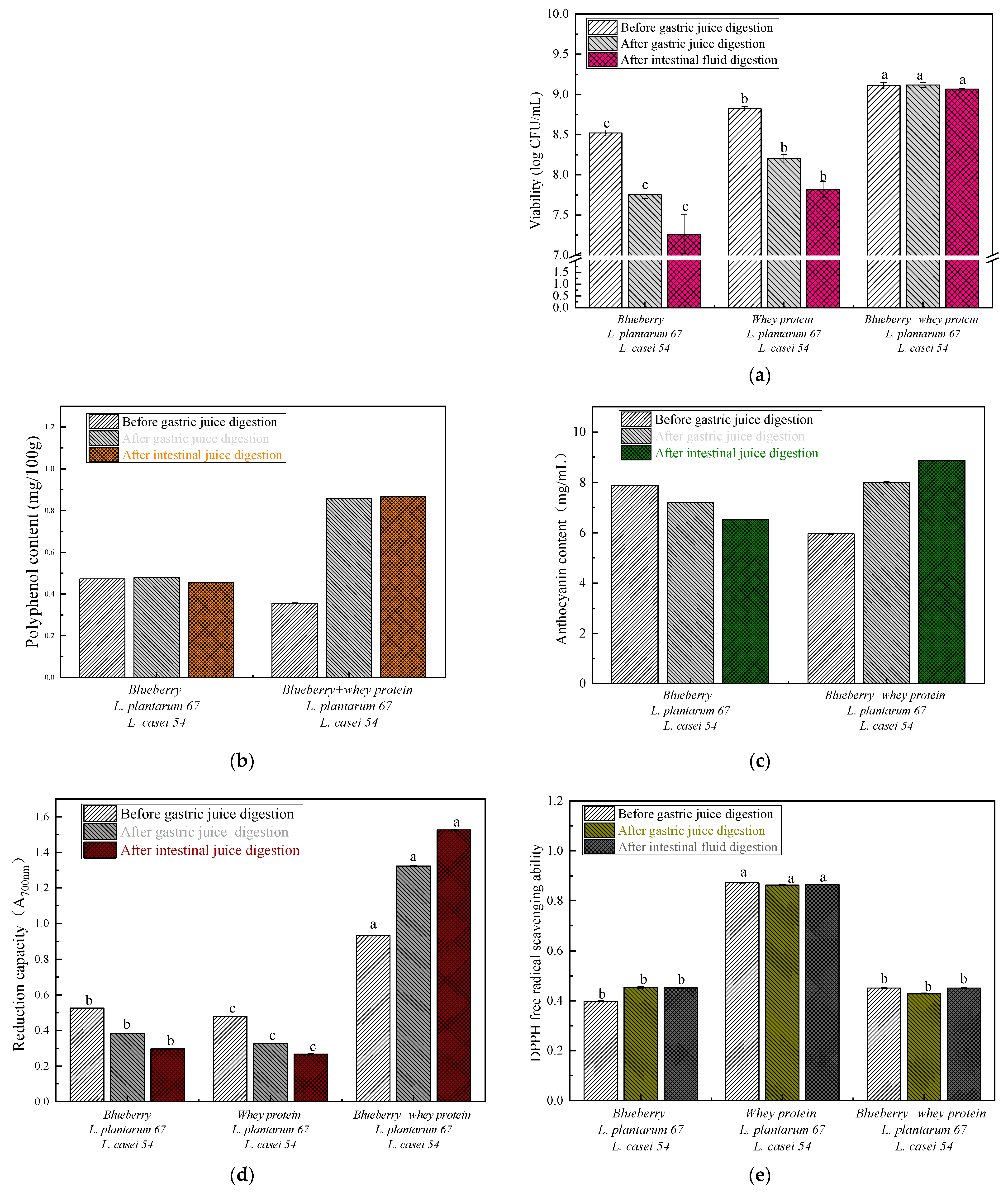
As shown in Figure 3a, the viable probiotic counts of the separate blueberry juice and whey protein systems presented a gradually decreasing trend during the gastrointestinal digestion process. Direct exposure to the low pH and proteolytic enzymes in the stomach, as well as the pH difference and pancreatic fluids in the intestine may disrupt the stability of these bacterial cells’ membranes. In the end, this could lead to an internal imbalance and cell death, which may cause a loss of viable cells [15]. Moreover, direct contact with high contents of polyphenols (>0.1 g/L or 0.2 g/L) could also inhibit the growth of bacteria [16]. However, the viable probiotic count in the mixed blueberry and whey protein system fermented with L. casei 54 and L. plantarum 67 was significantly higher than those of the separate systems (p < 0.05), and the viable probiotic cell level was maintained at up to 109 CFU/mL after intestinal fluid digestion, which was similar to that before gastric digestion (p < 0.05). This indicated that the mixed blueberry and whey protein system may have formed a physical barrier that could protect probiotic cells from the severe gastrointestinal environment. This is in accordance with the findings that probiotic cells were protected from exposure to antibacterial components in the matrix, which led directly to lower rates of cell injury and death [16].
The total phenolic content of the blueberry system fermented by L. plantarum 67 and L. casei 54 was higher than the corresponding value for the mixed blueberry and whey protein system before gastric juice digestion, as shown in Figure 3b, which suggested that phenolic compounds had high affinity with whey protein, and that the phenolic–protein interactions may have masked the phenolic content in the protein, resulting in a reduction in the measured phenolic content [17]. However, the total phenolic content of the mixed blueberry and whey protein system fermented by L. plantarum 67 and L. casei 54 was sharply increased from gastric to intestinal juice digestion in comparison with that before gastric juice digestion. This result was consistent with an increasing trend for phenolic content during in vitro gastrointestinal digestion [18]. This may be related to the protective effect of whey protein, which is considered as a natural encapsulation material that protects hydrophilic compounds from phenolic compounds and further delays the release of phenolic compounds during in vitro gastrointestinal digestion [11].
As displayed in Figure 3c, the TAC of the blueberry system fermented by L. plantarum 67 and L. casei 54 presented a decreasing trend during in vitro gastrointestinal digestion. Compared to the separate system, the TAC of the mixed blueberry and whey protein system fermented with L. plantarum 67 and L. casei 54 steadily increased throughout the digestive process. This may be attributed to the protective effect of whey protein on anthocyanins [11]. Whey protein has a high resistance to pepsin in an acidic stomach environment; thus, as packaging material for bioactive compounds, it prolongs the release of anthocyanin in gastric juices and transfers these compounds from the encapsulated microparticles into the intestinal juices [11]. This indicates that whey protein can protect anthocyanin to achieve a complete release at the end of digestion with intestinal fluids.
Phenolic compounds contain various units of phenol and have strong antioxidant activity due to their capacity for releasing hydrogen ions, which can combine with free radicals and terminate radical polymerization [9]. The antioxidant activity in the fermented samples that underwent in vitro gastrointestinal digestion was determined according to the ferric iron reduction capacity and DPPH–radical–scavenging ability. As presented in Figure 3d, the reduction capacity of the mixed blueberry and whey protein system fermented with L. plantarum 67 and L. casei 54 was approximately two times higher than that of the separate systems. Thus, the mixed blueberry and whey protein system fermented with L. plantarum 67 and L. casei 54 was superior to the single systems in terms of the ferric iron reduction capacity, and the addition of whey protein was able to increase the reduction capacity of the blueberry system. Previous studies suggested that antioxidant capacity is mainly related to the presence of polyphenols and anthocyanins [3]. Additionally, whey protein isolates could improve the stability of anthocyanidins and further enhance their antioxidant capacity [19]. The reduction capacity of the separate systems after gastric and intestinal digestion gradually decreased. However, the total phenolic contents of blueberry juice during in vitro gastrointestinal digestion were almost equivalent. This was caused by the diversity of polyphenols in the samples [18]. The reduction capacity of the mixed blueberry and whey protein system fermented by L. plantarum 67 and L. casei 54 significantly increased after gastric and intestinal digestion (p < 0.05). The result was similar to that of an increase in the release of phenolic compounds and the reduction capacity using whey protein microcapsules [20]. This may be because whey protein was digested into different peptide fractions, and further co-precipitated with polyphenols, or reduced ferric ions to ferrous ions relying on their redox potential [18].
The DPPH-scavenging activity was also used to detect the antioxidant activity in the fermented samples that underwent in vitro gastrointestinal digestion. The results demonstrated that the DPPH-radical-scavenging activity of the whey protein system fermented with L. plantarum 67 and L. casei 54 in Figure 3e significantly increased (p < 0.05), which may have been related to the release of antioxidant peptides when whey protein was hydrolyzed by lactic acid bacteria [21]. It is especially important to mention that poor DPPH-radical-scavenging activity of the mixed blueberry and whey protein fermented with L. plantarum 67 and L. casei 54 was observed, although there was a high content of total phenolics, as shown in Figure 3b. This could potentially be ascribed to phenolic-protein interactions that were compound-specific [22]. Different phenolic compounds have different radical scavenging activities and their interactions could influence the antioxidant activity of phenolic compounds. A similar observation was reported, in which DPPH-radical-scavenging activity did not merely depend on the content of total phenolics and anthocyanins, rather on complex phenolic-protein interactions [22].

3.2. FTIR Spectrum Analysis

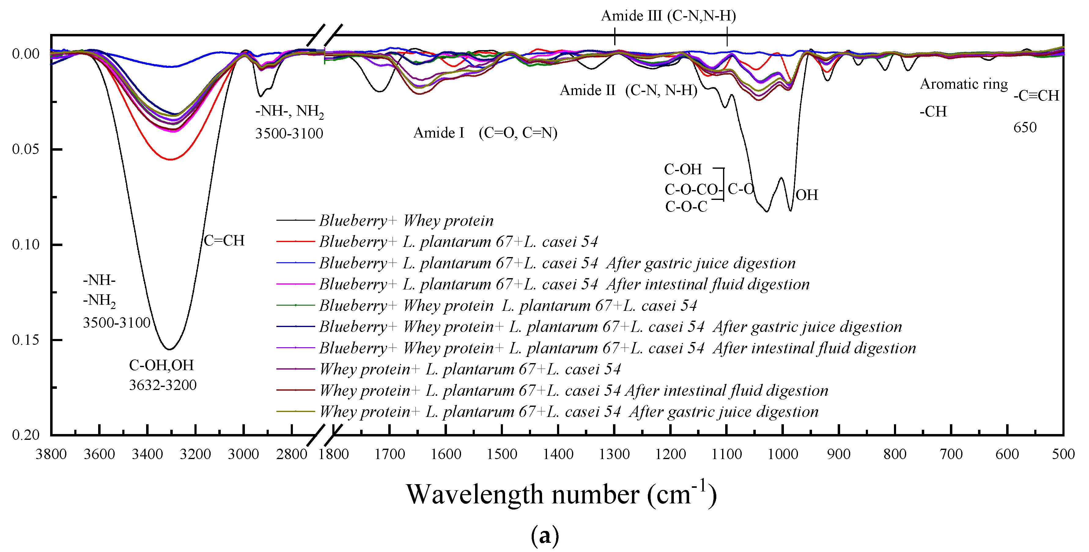
Fourier transform infrared spectra were used to examine changes in the secondary structures of proteins after the mutual binding between proteins and bioactive compounds, such as the anthocyanins from blueberries. It is well known that these changes are mostly shown by the positions of characteristic peaks, including those of amide A at 3300–3400 cm−1, amide I (mainly C=O stretch, 1600–1700 cm−1), amide II (C-N stretching coupled with N-H bending modes, 1500–1600 cm−1) and amide III (C-N stretching and N-H deformation, 1200–1300 cm−1) [23]. Figure 4a displays the FTIR spectra of the pure components and the mixed blueberry and whey protein system fermented with L. plantarum 67 and L. casei 54 after in vitro gastrointestinal at 3800–500 cm−1. The secondary structure of whey protein in the mixed system fermented with L. plantarum 67 and L. casei 54 changed as illustrated by the slight shift in the amide I band and amide II band [24]. The shift in the amide A band further confirmed the interactions between whey protein and anthocyanins from blueberries, as evidenced by the C-N and N-H groups [23]. The shifting of the above amide bands showed that the anthocyanins from blueberries mainly combined with the C-O, C-N, and N-H groups in whey protein [11]. Meanwhile, it was reported that interactions between anthocyanins and proteins occurred via the formation of hydrogen bonds, where the interactions of phenolic hydroxyl groups and certain groups of the proteins, such as –OH and –NH2 groups, play a vital role [25]. As shown in the spectra of all samples, due to the stretching of the O-H groups, narrower absorption peaks were shown in the wavelength range from 3000 to 3600 cm−1. In addition, broader absorption peaks appeared in the wavelength range from 1600 to 1700 cm−1, which resulted from vibration when water molecules were absorbed. It can be observed in Figure 4a, the FTIR spectrum of the mixed blueberry and whey protein system depicted strong absorption peaks with broader bands at about 3100–3600 cm−1, which was attributable to the stretching and vibration in the absorption of O-H groups. A peak occurred in the wavelength range from 2800 to 3000 cm−1, which was due to –CH stretching. Moreover, a special peak in the wavelength range from 950 to 1100 cm−1 was generated by the stretching and vibration of C-O-C groups [26]. The peak at 750–850 cm−1 was due to -CH stretching from the aromatic ring in whey protein [27].
At 1600–1700 cm−1, amide I had distinct polypeptide absorption bands. In addition, the bands of C-O stretching in the mixed blueberry and whey protein system fermented with L. plantarum 67 and L. casei 54 became stronger and wider than those of the blueberry or whey protein system fermented with L. plantarum 67 and L. casei 54. This may have been because the active groups of amino acids had interacted with anthocyanins from blueberries [28].
The mixed blueberry and whey protein system fermented with L. plantarum 67 and L. casei 54 after in vitro gastrointestinal digestion had stronger and wider absorption bands. From Figure 4c, the positions of the amide A and amide II peaks slightly shifted from 1535.061 cm−1 to 1544.706 cm−1 after gastric juices digestion, and from 1544.706 cm−1 to 1550.490 cm−1 after intestinal fluids digestion. For the whey protein system fermented with L. plantarum 67 and L. casei 54, the positions of the amide A and amide II peaks had similar changes. Furthermore, the bands of the peaks for amide I and amide II in the mixed blueberry and whey protein system fermented with L. plantarum 67 and L. casei 54 changed less than those of the whey protein system fermented with L. plantarum 67 and L. casei 54 after gastric juices digestion. The result indicated that the stability of the whey protein structure improved, which may have been because the presence of anthocyanin from blueberries inhibited the digestive enzymes and indirectly made the proteins insensitive to pepsin [11]. The stability of protein structure was advantageous in providing a relatively complete structure to protect the bioactive compounds during the gastric juices digestion, enabling their transfer to the intestinal tract and subsequent release.

3.3. CLSM Images of the In Vitro Release of Anthocyanins and the Distribution of Probiotics

To determine the in vitro release of anthocyanins and the distribution of probiotics, the samples were observed through confocal laser scanning microscopy, as shown in Figure 5. The samples without any bacteria were analyzed as control groups, which have no fluorescent green bacillus-shaped structures. However, there were green bacillus-shaped structures in the samples fermented with L. plantarum 67 and L. casei 54. It is worth mentioning that greater amounts of microorganisms were observed in the mixed blueberry and whey protein system fermented with L. plantarum 67 and L. casei 54, and this result was reflected by the data on viable cell counts in Figure 3a. This illustrates the promoting effect of anthocyanin and whey protein microcapsules which were used as immobilization carriers, in terms of the growth of probiotic cells. During subsequent in vitro gastrointestinal digestion, the probiotics released from the mixed matrix were gradually distributed in the solution as free probiotic cells. The in vitro release of anthocyanins is also shown in Figure 5. As shown in the CLSM images of the mixed blueberry and whey protein system fermented with L. plantarum 67 and L. casei 54, the anthocyanins from blueberries and whey protein formed large curds. In addition, the anthocyanins (in red) and the green probiotics with a bacillus shape were evenly distributed within the whey protein curds. After gastric juices digestion, the anthocyanins scattered around separated from the surfaces of the whey protein curds, which may be attributed to their degradation by gastric acid [11]. At the end of the intestinal fluid digestion, the red color of anthocyanins was clearly observed, indicated that they could be were transported to the intestinal tract.

3.4. The Effect of Ultrafiltration Time on the Survival of Probiotics

In order to demonstrate how whey protein and the anthocyanins or phenolics of blueberries interacted with the surface proteins of bacteria during the fermentation process, ultrafiltration technology was used to separate the bacteria and free molecules in the samples. Firstly, the effect of the operation time on the probiotic viability at a certain transmembrane pressure was investigated before in vitro gastrointestinal digestion in order to choose a suitable ultrafiltration operation time. It was found that the probiotics were retained in the concentration fractions (>100 kDa). As shown in Figure 6a, the probiotic viability in the mixed blueberry and whey protein system fermented with L. casei 54 and L. plantarum 67 was higher than that in the separate systems (p < 0.05) at the beginning of the operation. However, the probiotic viability started to decrease (p < 0.05) at 15 min and was lower than the initial viable probiotic cell count by the operation time of 25 min. To reduce the effect of the ultrafiltration operation on probiotic cells, the end time of the ultrafiltration study was set to 15 min. The products that were separated through ultrafiltration were obtained to study their protective effects on probiotic cells during in vitro gastrointestinal digestion. As illustrated in Figure 6b, the survival rate of probiotic cells in the blueberry juice system rapidly decreased during the digestive process, and a significant decrease (p < 0.05) in the survival rate (about 8.26%) was observed at the end of intestinal fluid digestion. This result was consistent with the above change in probiotic viability in the blueberry juice system and indicated that the effect of blueberry juice on maintaining the minimum probiotic cell amounts was limited after in vitro digestion. In the whey protein system, the survival rate of probiotics was improved, and the result suggested that whey protein could protect probiotic cells and transfer them into intestinal fluid conditions due to their partial degradation in an acidic environment [29]. The survival rate of probiotics in the mixed blueberry and whey protein system was higher than those in the separate systems after gastric juice digestion (p < 0.05). At the end of intestinal digestion, the survival rate of probiotic cells in the mixed blueberry and whey protein system was maintained at 84.49%. This demonstrated that the mixed blueberry and whey protein system could offer protection to probiotic cells during in vitro gastrointestinal digestion. This result was in accordance with the findings that probiotic cells were immobilized and protected by sea buckthorn berries with high viable cell counts [5].

3.5. The Total Phenolics Content and Antioxidant Activity (Reducing Power) in Products Separated through Ultrafiltration after In Vitro Gastrointestinal Digestion

To distinguish which membrane fractions could remain and improve the bioavailability of bioactive compounds, the total phenolic content in the product separated through ultrafiltration was evaluated after in vitro gastrointestinal digestion. As shown in Figure 6c, there was a higher content of total phenolics in the concentration fractions (>100 kDa), while there was a lower content of total phenolics in the permeate fractions (<100 kDa). This suggested that ultrafiltration had the effect of intercepting phenolic compounds. In these concentration fractions (>100 kDa), the total phenolic content of the concentration of 100 kDa in the blueberry system was higher than that of the mixed blueberry and whey protein system. This result was consistent with that of the total phenolic content of the fermented samples before gastric juices digestion. The addition of whey protein lowered the content of total phenolics in the blueberry system. At the same time, a slight reduction in the total phenolic content also occurred after fermentation, although many studies have illustrated that the lactic acid bacteria fermentation can increase the content of total phenolics in blueberries and, thus, improve their functionality [30]. This reduction may be due to the oxidation caused by residual oxygen in the fermentation system during a short fermentation period [30]. The total phenolic contents in the blueberry system and the mixed blueberry and whey protein system fermented with L. plantarum 67 and L. casei 54 were slightly decreased upon exposure to proteases in SGJ and SDJ [31].
The total reduction capacity during in vitro gastrointestinal digestion was used to determine the antioxidant activity in the products separated through ultrafiltration. As shown in Figure 6d, the total reduction capacity of the concentration fractions (>100 kDa) was superior to that of the permeate fractions (<100 kDa), which indicated that there was more capacity in the concentration fractions (>100 kDa) to block free radical reactions [32]. The phenolic compounds in the products separated through ultrafiltration were donors that offered hydrogen ions [33]. The trends of the changes in the total reduction capacity in the mixed blueberry and whey protein system fermented with L. plantarum 67 and L. casei 54 were similar to the changes in the total phenolics during in vitro gastrointestinal digestion. In particular, there was an increase in the total reduction capacity at the end of intestinal digestion. This result could be explained by the hypothesis that enzymatic activity is improved during gastric digestion, and that the enzymes further stimulate the release of phenolic compounds from phenolic–protein complexes [3].

3.6. SDS-PAGE

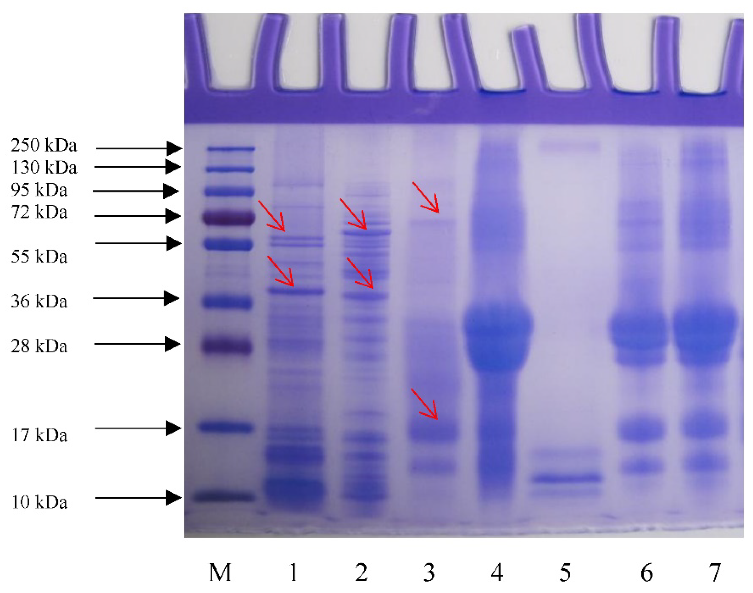
SDS-PAGE was used to identify surface proteins in samples extracted with LiCl solution. As shown in Figure 7, the surface protein fraction of L. plantarum 67 showed eleven protein bands. The bands with higher protein concentrations than those of other bands are indicated by the arrows in the ranges of 36–55 kDa and 55–72 kDa. The main surface protein fractions of L. casei 54 were present in the bands ranging from 36 kDa to 55 kDa and from 72 to 95 kDa. Intense bands were observed at 36–55 kDa and 55–72 kDa, which showed that the concentration of the surface proteins was higher in L. casei 54. However, other protein concentrations were relatively lower. This was consistent with the report that the molecular weight of surface proteins of lactic acid bacteria was 25–72 kDa [34]. However, there were several polypeptide bands at 10–17 kDa in the samples of blueberry juice fermented with the combined bacteria. There were no significant differences between the unfermented whey protein and the unfermented mixture. Six protein bands and two polypeptide bands were observed in the sample of whey protein fermented with the combined bacteria. In addition, one protein band at around 72 kDa and two polypeptides bands at around 17 kDa were observed in the concentration fraction (>100 kDa) of the mixture of whey protein and blueberry fermented with the combined bacteria separated through ultrafiltration. In comparison with the SLPs of L. plantarum 67 and L. casei 54, these bands disappeared, and the new protein bands appeared in the all of the fermented samples. The result may have indicated that lactic acid bacteria made contact with the components of the fermented samples and further interacted with them, which was supported by the images from CLSM [35]. Compared with the protein bands of the unfermented whey protein, the intensity of the bands at around 28 kDa increased. Thus, interactions between the SLPs of the strain and whey protein may have occurred. The addition of blueberry juice could have resulted in the increase in intensity of the bands at 10–17 kDa in the mixture that was fermented with the combined bacteria. The interactions between proteins and active compounds in blueberries were confirmed [25].The interactions of whey protein or blueberry and SLPs may have formed conjugates of whey protein–blueberry and some SLPs. This was supported by the relatively higher viable cell counts of the mixture fermented with L. plantarum 67 and L. casei 54 during gastric juice digestion, which suggested that the conjugates could protect probiotic cells from adverse gastrointestinal environments by acting as a protective layer during in vitro digestion. This result was in agreement with the findings of that the surface proteins of L. acidophilus L-92 can be combined with the proteins of host cells in stress environments [36]. A similar study reported that the surface proteins of L. kefiri HBA20 could interact with the mannans of S. cerevisiae, and this interaction further induced a combination of different probiotics [37].

4. Conclusions

An in vitro gastrointestinal digestion assay indicated that in a mixed blueberry and whey protein system fermented with L. plantarum 67 and L. casei 54, whey protein could effectively slow down the release and degradation of anthocyanins especially in the stomach. It appears that whey protein, which has natural physicochemical properties, may be a protective material due to its interactions with bioactive components such as anthocyanins in the blueberries; in addition, it can influence the targeted release of bioactive compounds and maintain the survival of probiotics. The above results indicate that this blueberry juice and whey protein fermentation system may improve bioactivity and functionality and offer protection for probiotic cells during in vitro gastrointestinal digestion. The results of SDS-PAGE further showed that blueberry–whey protein–bacterial surface protein conjugates may have been formed. However, the binding patterns of the conjugates are not yet clear. In future studies, we aim to explore the types and structures of the combined active compounds in blueberries, whey protein, and surface proteins or glycoproteins of bacterial cells, to explore the binding types of these molecules, the conformation changes of the combined proteins, and possible molecular binding sites, as well as to put this interaction to work in food processing and other fields.

Author Contributions

Q.Y.: methodology, FTIR measurement analysis, writing—original draft; W.W.: investigation, formal analysis, funding support; X.L.: SDS-PAGE; W.S.: resourcing of original materials; R.G.: revision, funding support; C.T.: methodology. All authors have read and agreed to the published version of the manuscript.

Funding

This research was funded by the project of the National Science Foundation for Young Scientists of China (Grant No. 31901715), the Jiangsu postdoctoral Foundation (2021K317C) and the Project funded by China Postdoctoral Science Foundation (2022M712433).

Institutional Review Board Statement

Not applicable.

Informed Consent Statement

Not applicable.

Data Availability Statement

Not applicable.

Conflicts of Interest

The authors declare no conflict of interest.

References

  1. Oancea, A.-M.; Hasan, M.; Vasile, A.M.; Barbu, V.; Enachi, E.; Bahrim, G.; Râpeanu, G.; Silvi, S.; Stănciuc, N. Functional evaluation of microencapsulated anthocyanins from sour cherries skins extract in whey proteins isolate. LWT-Food Sci. Technol. 2018, 95, 129–134. [Google Scholar] [CrossRef]
  2. Yang, W.; Guo, Y.; Liu, M.; Chen, X.; Xiao, X.; Wang, S.; Gong, P.; Ma, Y.; Chen, F. Structure and function of blueberry anthocyanins: A review of recent advances. J. Funct. Foods 2022, 88, 104864. [Google Scholar] [CrossRef]
  3. Anuyahong, T.; Chusak, C.; Adisakwattana, S. Incorporation of anthocyanin-rich riceberry rice in yogurts: Effect on physicochemical properties, antioxidant activity and in vitro gastrointestinal digestion. LWT-Food Sci. Technol. 2020, 129, 109571. [Google Scholar] [CrossRef]
  4. Afzaal, M.; Saeed, F.; Saeed, M.; Azam, M.; Hussain, S.; Mohamed, A.A.; Alamri, M.S.; Anjum, F.M. Survival and stability of free and encapsulated probiotic bacteria under simulated gastrointestinal and thermal conditions. LWT-Food Sci. Technol. 2020, 23, 1899–1912. [Google Scholar] [CrossRef]
  5. Terpou, A.; Papadaki, A.; Bosnea, L.; Kanellaki, M.; Kopsahelis, N. Novel frozen yogurt production fortified with sea buckthorn berries and probiotics. LWT-Food Sci. Technol. 2019, 105, 242–249. [Google Scholar] [CrossRef]
  6. Zhang, C.; Yang, L.; Ding, Z.; Yin, B.; Chen, D.; Guan, C.; Gu, R. New selective media for isolation and enumeration of Lactobacillus rhamnosus and Streptococcus thermophilus. J. Food Meas. Charact. 2019, 13, 1431–1439. [Google Scholar] [CrossRef]
  7. Shen, X.; Sun, X.; Xie, Q.; Liu, H.; Zhao, Y.; Pan, Y.; Hwang, C.-A.; Wu, V.C.H. Antimicrobial effect of blueberry (Vaccinium corymbosum L.) extracts against the growth of Listeria monocytogenes and Salmonella Enteritidis. Food Control. 2014, 35, 159–165. [Google Scholar] [CrossRef]
  8. Lin, Y.; Wang, Y.; Li, B.; Tan, H.; Li, D.; Li, L.; Liu, X.; Han, J.; Meng, X. Comparative transcriptome analysis of genes involved in anthocyanin synthesis in blueberry. Plant Physiol. Bioch. 2018, 127, 561–572. [Google Scholar] [CrossRef]
  9. Lu, C.; Li, C.; Chen, B.; Shen, Y. Composition and antioxidant, antibacterial, and anti-HepG2 cell activities of polyphenols from seed coat of Amygdalus pedunculata Pall. Food Chem. 2018, 265, 111–119. [Google Scholar] [CrossRef]
  10. Zhou, L.; Xie, M.; Yang, F.; Liu, J. Antioxidant activity of high purity blueberry anthocyanins and the effects on human intestinal microbiota. LWT-Food Sci. Technol. 2020, 117, 108621. [Google Scholar] [CrossRef]
  11. Liao, M.; Ma, L.; Miao, S.; Hu, X.; Liao, X.; Chen, F.; Ji, J. The in-vitro digestion behaviors of milk proteins acting as wall materials in spray-dried microparticles: Effects on the release of loaded blueberry anthocyanins. Food Hydrocoll. 2021, 115, 106620. [Google Scholar] [CrossRef]
  12. Meng, J.; Wang, Y.-Y.; Hao, Y.-P. Protective function of surface layer protein from Lactobacillus casei fb05 against intestinal pathogens in vitro. Biochem. Biophys. Res. Commun. 2021, 546, 15–20. [Google Scholar] [CrossRef] [PubMed]
  13. Wang, W.-Q.; Bao, Y.-H.; Chen, Y. Characteristics and antioxidant activity of water-soluble Maillard reaction products from interactions in a whey protein isolate and sugars system. Food Chem. 2013, 139, 355–361. [Google Scholar] [CrossRef] [PubMed]
  14. Bambace, M.F.; Alvarez, M.V.; Moreira, M.R. Ready-to-eat blueberries as fruit-based alternative to deliver probiotic microorganisms and prebiotic compounds. LWT-Food Sci. Technol. 2021, 142, 111009. [Google Scholar] [CrossRef]
  15. Balthazar, C.F.; Santillo, A.; Guimaraes, J.T.; Capozzi, V.; Russo, P.; Caroprese, M.; Marino, R.; Esmerino, E.A.; Raices, R.S.L.; Silva, M.C.; et al. Novel milk-juice beverage with fermented sheep milk and strawberry (Fragaria x ananassa): Nutritional and functional characterization. J. Dairy Sci. 2019, 102, 10724–10736. [Google Scholar] [CrossRef] [Green Version]
  16. Zhang, C.; Quek, S.Y.; Fu, N.; Su, Y.; Kilmartin, P.A.; Chen, X.D. Storage stability and in vitro digestion of microencapsulated powder containing fermented noni juice and probiotics. Food Biosci. 2020, 37, 100740. [Google Scholar] [CrossRef]
  17. Ozdal, T.; Capanoglu, E.; Altay, F. A review on protein–phenolic interactions and associated changes. Food Res. Int. 2013, 51, 954–970. [Google Scholar] [CrossRef]
  18. Flores, F.P.; Singh, R.K.; Kerr, W.L.; Phillips, D.R.; Kong, F. In vitro release properties of encapsulated blueberry (Vaccinium ashei) extracts. Food Chem. 2015, 168, 225–232. [Google Scholar] [CrossRef]
  19. Zang, Z.; Chou, S.; Tian, J.; Lang, Y.; Shen, Y.; Ran, X.; Gao, N.; Li, B. Effect of whey protein isolate on the stability and antioxidant capacity of blueberry anthocyanins: A mechanistic and in vitro simulation study. Food Chem. 2021, 336, 127700. [Google Scholar] [CrossRef]
  20. Flores, F.P.; Singh, R.K.; Kerr, W.L.; Pegg, R.B.; Kong, F. Total phenolics content and antioxidant capacities of microencapsulated blueberry anthocyanins during in vitro digestion. Food Chem. 2014, 153, 272–278. [Google Scholar] [CrossRef]
  21. Hu, S.-M.; Liu, B.-H.; Sun, X.-Y.; Li, X.-D.; Wang, C.-C. The process and optimization of antioxidant peptide preparation by whey protein fermentation. China Dairy 2018, 4, 71–81. [Google Scholar] [CrossRef]
  22. Tang, P.-L.; Hao, E.-W.; Deng, J.-G.; Hou, X.-T.; Zhang, Z.-H.; Xie, J.-L. Boost anti-oxidant activity of yogurt with extract and hydrolysate of cinnamon residues. Chin. Herb. Med. 2019, 11, 417–422. [Google Scholar] [CrossRef]
  23. Zhang, Y.; Chen, S.; Qi, B.; Sui, X.; Jiang, L. Complexation of thermally-denatured soybean protein isolate with anthocyanins and its effect on the protein structure and in vitro digestibility. Food Res. Int. 2018, 106, 619–625. [Google Scholar] [CrossRef] [PubMed]
  24. Jiang, L.; Liu, Y.; Li, L.; Qi, B.; Ju, M.; Xu, Y.; Zhang, Y.; Sui, X. Covalent conjugates of anthocyanins to soy protein: Unravelling their structure features and in vitro gastrointestinal digestion fate. Food Res. Int. 2019, 120, 603–609. [Google Scholar] [CrossRef]
  25. Quan, T.H.; Benjakul, S.; Sae-leaw, T.; Balange, A.K.; Maqsood, S. Protein–polyphenol conjugates: Antioxidant property, functionalities and their applications. Trends Food Sci. Technol. 2019, 91, 507–517. [Google Scholar] [CrossRef]
  26. Lan, W.; Zhang, R.; Ji, T.; E, S.D.; Saeed, A.; Qin, W.; Dai, J.; He, L.; Liu, Y. Improving nisin production by encapsulated Lactococcus lactis with starch/carboxymethyl cellulose edible films. Carbohydr. Polym. 2020, 251, 117062. [Google Scholar] [CrossRef]
  27. Gutiérrez, T.J.; Ponce, A.G.; Alvarez, V.A. Nano-clays from natural and modified montmorillonite with and without added blueberry extract for active and intelligent food nanopackaging materials. Mater. Chem. Phys. 2017, 194, 283–292. [Google Scholar] [CrossRef] [Green Version]
  28. Khalifa, I.; Nie, R.; Ge, Z.; Li, K.; Li, C. Understanding the shielding effects of whey protein on mulberry anthocyanins: Insights from multispectral and molecular modelling investigations. Int. J. Biol. Macromol. 2018, 119, 116–124. [Google Scholar] [CrossRef]
  29. Gbassi, G.K.; Vandamme, T.; Ennahar, S.; Marchioni, E. Microencapsulation of Lactobacillus plantarum spp. in an alginate matrix coated with whey proteins. Int. J. Biol. Macromol. 2009, 129, 103–105. [Google Scholar] [CrossRef]
  30. Gao, B.; Wang, J.; Wang, Y.; Xu, Z.; Li, B.; Meng, X.; Sun, X.; Zhu, J. Influence of fermentation by lactic acid bacteria and in vitro digestion on the biotransformations of blueberry juice phenolics. Food Control. 2022, 133, 1431–1439. [Google Scholar] [CrossRef]
  31. Correa-Betanzo, J.; Allen-Vercoe, E.; McDonald, J.; Schroeter, K.; Corredig, M.; Paliyath, G. Stability and biological activity of wild blueberry (Vaccinium angustifolium) polyphenols during simulated in vitro gastrointestinal digestion. Food Chem. 2014, 165, 522–531. [Google Scholar] [CrossRef] [PubMed]
  32. Xu, Z.; Li, X.; Feng, S.; Liu, J.; Zhou, L.; Yuan, M.; Ding, C. Characteristics and bioactivities of different molecular weight polysaccharides from camellia seed cake. Int. J. Biol. Macromol. 2016, 91, 1025–1032. [Google Scholar] [CrossRef]
  33. Zhang, L.; Zhang, M.; Mujumdar, A.S. New technology to overcome defects in production of fermented plant products—A review. Trends Food Sci. Technol. 2021, 116, 829–841. [Google Scholar] [CrossRef]
  34. Xiao, Z.; Bin, H.; Jingyan, L.; Huibo, L.; Rongrong, L. Isolation, structure and properties of surface protein of lactic acid bacteria. Food Ferment. Ind. 2011, 37, 19–24. [Google Scholar] [CrossRef]
  35. Burgain, J.; Scher, J.; Francius, G.; Borges, F.; Corgneau, M.; Revol-Junelles, A.M.; Cailliez-Grimal, C.; Gaiani, C. Lactic acid bacteria in dairy food: Surface characterization and interactions with food matrix components. Adv. Colloid Interface Sci. 2014, 213, 21–35. [Google Scholar] [CrossRef] [PubMed]
  36. Wakai, T.; Kano, C.; Karsens, H.; Kok, J.; Yamamoto, N. Functional role of surface layer proteins of Lactobacillus acidophilus L-92 in stress tolerance and binding to host cell proteins. Biosci. Microbiota Food Health 2021, 40, 33–42. [Google Scholar] [CrossRef]
  37. Fu, M.; Mao, K.; Gao, J.; Wang, X.; Sadiq, F.A.; Li, J.; Sang, Y. Characteristics of surface layer protein from Lactobacillus kefiri HBA20 and the role in mediating interactions with Saccharomyces cerevisiae Y8. Int. J. Biol. Macromol. 2022, 201, 254–261. [Google Scholar] [CrossRef]
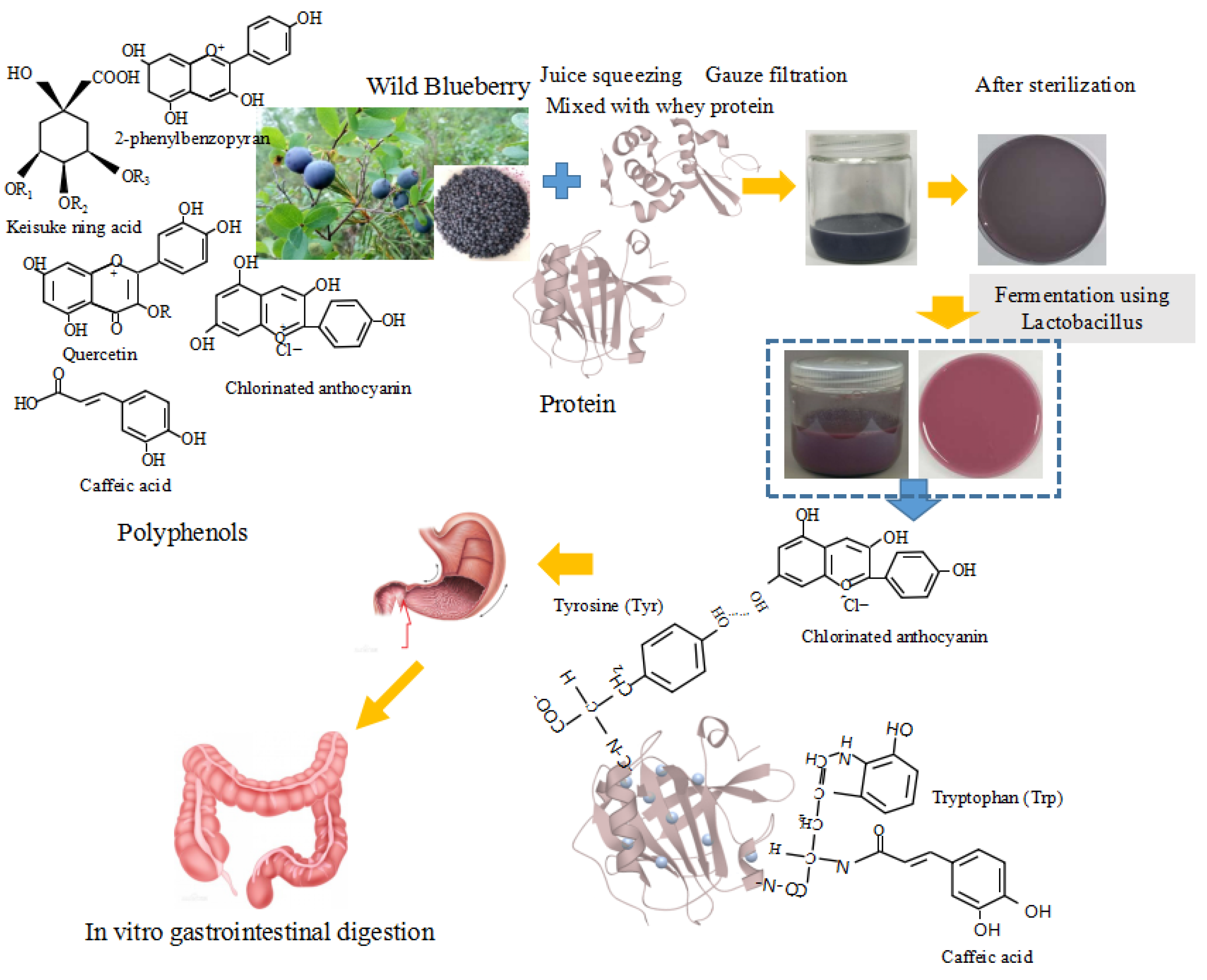
Figure 1. Schematic diagram showing preparation and analysis of blueberry juice and whey protein fermentation system with L. plantarum 67 and L. casei 54.
Fermentation 09 00335 g001
Figure 3. The total colony count (a), total phenol content (b), total anthocyanin content (c), reduction capacity (d) and DPPH-radical-scavenging activity (e) of the fermentation system with L. plantarum 67 and L. casei 54 before and after in vitro gastrointestinal digestion. Different letters within the same column indicate significant differences (p < 0.05).
Fermentation 09 00335 g003
Figure 4. FTIR spectrum analysis at 3800–500 cm−1 (a), spectrum analysis at 3800–2800 cm−1 (b), and spectrum analysis at 1800–700 cm−1 (c), of blueberry juice and whey protein fermentation system in vitro gastrointestinal digestion.
Fermentation 09 00335 g004aFermentation 09 00335 g004b
Figure 5. CLSM pictures of the dynamic release of anthocyanins and distribution of the probiotics during in vitro gastrointestinal digestion. Proteins: the voluminous structures with green color; anthocyanins: red color; probiotics: fluorescent bacillus and chain-shaped structures with green color. The magnification of whey protein was 20× and the magnification of other samples was 100×.
Fermentation 09 00335 g005
Figure 6. The effect of operation time on the probiotic viability (a), the survival of probiotics (b), the total phenol content (c) and the total reduction capacity (d) of products separated by ultrafiltration before and after in vitro gastrointestinal digestion. Different letters within the same column indicate significant differences (p < 0.05).
Fermentation 09 00335 g006
Figure 7. SDS-PAGE analysis of surface protein of samples extracted with LiCl solution. Samples: M: protein marker, 1: L. casei 54, 2: L. plantarum 67, 3: whey protein and blueberry concentration mixture fermentation with combined bacteria separated by ultrafiltration, 4: whey protein concentration mixture fermentation with combined bacteria separated by ultrafiltration, 5: blueberry concentration mixture fermentation with combined bacteria separated by ultrafiltration, 6: unfermented whey protein, 7: unfermented whey protein and blueberry mixture.
Fermentation 09 00335 g007
Disclaimer/Publisher’s Note: The statements, opinions and data contained in all publications are solely those of the individual author(s) and contributor(s) and not of MDPI and/or the editor(s). MDPI and/or the editor(s) disclaim responsibility for any injury to people or property resulting from any ideas, methods, instructions or products referred to in the content.