Article

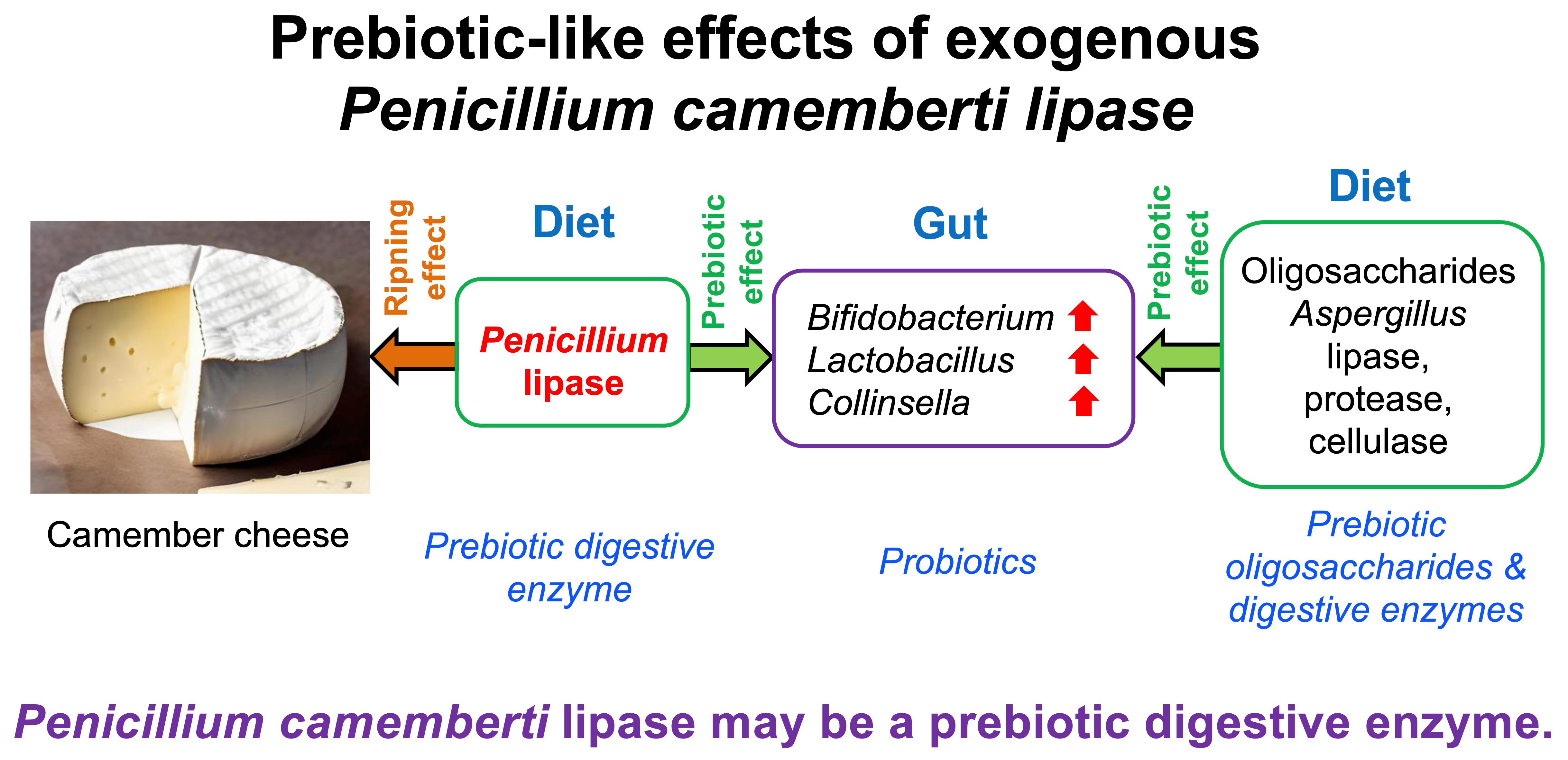
Exogenous Penicillium camemberti Lipase Preparation Exerts Prebiotic-like Effects by Increasing Cecal Bifidobacterium and Lactobacillus Abundance in Rats

1
School of Life Sciences, Anhui University, Hefei 230601, China
2
Graduate School of Agricultural and Life Sciences, The University of Tokyo, Tokyo 113-8657, Japan
3
Graduate School of Integrated Sciences for Life, Hiroshima University, Higashi-Hiroshima 739-8528, Japan
4
The Faculty of Health Sciences, Hiroshima Shudo University, Hiroshima 731-3195, Japan
5
Institute for Advanced Biosciences, Keio University, Tsuruoka 997-0052, Japan
6
Transborder Medical Research Center, Faculty of Medicine, University of Tsukuba, Tsukuba 305-8575, Japan
7
Gut Environmental Design Group, Kanagawa Institute of Industrial Science and Technology, Kawasaki 210-0821, Japan
8
Laboratory for Regenerative Microbiology, Juntendo University Graduate School of Medicine, Tokyo 113-8421, Japan
9
Amano Enzyme Inc., Nagoya 460-8630, Japan
*
Authors to whom correspondence should be addressed.
Fermentation 2023, 9(3), 227; https://doi.org/10.3390/fermentation9030227
Submission received: 13 February 2023 / Revised: 24 February 2023 / Accepted: 24 February 2023 / Published: 27 February 2023
(This article belongs to the Special Issue Nutrition and Health of Fermented Foods, 2nd Edition)

Abstract

:
Penicillium camemberti is used in cheese production; however, its health benefits remain to be elucidated. We previously found that supplemental Aspergillus-derived lipase preparation exerts a strong bifidogenic effect in rats fed a high-fat diet. This study investigated the effects of the feeding of a diet containing a 0.2% or 0.4% Penicillium camemberti-derived lipase preparation (PCL) for two weeks on the cecal microbiota in rats. According to 16S rRNA gene sequencing analysis, both PCL supplements significantly (p < 0.05) affected the cecal microbial community. At the genus level, supplemental 0.4% PCL significantly increased the relative abundance of beneficial bacteria such as Bifidobacterium, Lactobacillus, and Collinsella (127-fold, 6-fold, and 193-fold increase, respectively). The abundance of these bacteria in the 0.2% PCL group was between that of the control and 0.4% PCL groups. Notably, the effects of supplemental 0.4% PCL on modulating the abundance of these bacteria matched the effects observed in studies on typical prebiotic oligosaccharides. PICRUSt analysis revealed that PCL supplements significantly modulated the relative abundance of bacterial genes associated with 27 metabolic pathways, some of which were similar to those reported for prebiotic oligosaccharides. This study provides the first evidence indicating that supplemental PCL exerts prebiotic-like effects by modulating the abundance of the gut microbiota.

Graphical Abstract

1. Introduction

Several epidemiological studies have found that cheese consumption reduces the risk of chronic diseases, including cardiovascular disease, type 2 diabetes, colorectal cancer, cognitive decline, and depression [1,2,3,4,5,6]. However, the active compounds in cheese and the mechanisms underlying their beneficial effects remain to be elucidated. Penicillium camemberti is used in the production of Camembert and Brie cheeses, on which fungus colonies form a white crust [7]. Several Penicillium fungi-derived proteases and lipases are responsible for the ripening process through the enhanced release of substances associated with flavor and aroma, including amino acids, peptides, free fatty acids, and volatile fatty acids. Recently, Ano et al. demonstrated that consumption of a Camembert cheese extract prevented the development of an Alzheimer’s pathology by using transgenic model mice [8]. Oleamide and dehydroergosterol generated during Penicillium fermentation exert beneficial impacts through anti-inflammatory effects [9]. However, information on the roles of Penicillium fungi and their related products in health and disease is limited.
In a previous study, we found that the consumption of an Aspergillus niger-derived lipase preparation (ANL) caused a powerful bifidogenic effect in rats fed a high-fat (HF) diet [10]. Furthermore, the consumption of Aspergillus-derived protease and cellulase caused similar bifidogenic effects in rats fed a HF diet [11,12,13]. Additionally, the consumption of acid protease purified from Aspergillus oryzae had a similar effect on gut Bifidobacteium [14]. Among the digestive enzymes studied in our laboratory, the bifidogenic effects of ANL were the most remarkable. These findings raise the question of whether lipases derived from other fungi, such as Penicillium spp., have a similar effect. In a preliminary study using qPCR analysis, we found that rats fed a P. camemberti-derived lipase preparation (PCL) exhibited a significant increase in the relative abundance of cecal Bifidobacterium and Lactobacillus. Therefore, in the present study, we performed 16S rRNA gene sequencing analysis to determine the effects of dietary supplemental PCL on the entire profiles of the gut microbiota in rats fed a HF diet. This study provides the first evidence indicating that exogenous PCL exerts prebiotic-like effects. To investigate the functional effects of dietary supplemental PCL on gut health and disease, the relative abundance of bacterial genes associated with various metabolic pathways was also investigated using PICRUSt analysis.

2. Materials and Methods

2.1. Animals and Diets

Sixteen male Sprague Dawley rats (four weeks old) were purchased from Charles River Japan. The rats were individually housed in cages in a controlled-temperature environment (23 ± 2 °C), with a 12 h light–dark cycle and relative humidity of 50–60%. After being acclimatized for 7 days, the rats were randomly divided into the following three groups based on their diets: a HF diet (control; Ctrl, five rats) or a HF diet mixed with 0.2% (w/w) PCL (P. camemberti-derived lipase preparation, commercial name: Lipase G “Amano” 50, Amano Enzyme Inc. Nagoya, Japan, six rats), or 0.4% (w/w) PCL (six rats). The lipase activity of PCL is 55,000 U/g at pH 6.0 and the protease activity in the PCL was not detectable. The HF diet used in this study contained 30% beef tallow, 20% casein, 0.3% L-cystine, 1% vitamin mixture (AIN-93), 3.5% mineral mixture (AIN-93G), 5% cellulose, 20% sucrose, and 20.2% α-corn starch [11]. During the two-week experimental period, equal amounts of the experimental diets were given daily in food cups (9, 10, 12, 14, and 15 g on days 1, 2–4, 5–7, 8–12, and 13–14, respectively) to prevent differences in food intake. All of the given diets were consumed each day. The rats had ad libitum access to drinking water. The study protocols were approved by the Ethics Committee of Hiroshima University (protocol identification no. C15-12).

2.2. Sample Collection

At the end of the two-week treatment period, the rats were anesthetized (13:00–15:00 h) by inhaling isoflurane in a desiccator to minimize suffering, and then euthanized by decapitation. The cecum was immediately excised, and its contents were removed entirely, weighed, and stored at −80 °C until the subsequent analysis of the cecal microbiota and organic acids.

2.3. Bacterial DNA Extraction and 16S rRNA Gene-Based Microbiome Analysis

Total bacterial DNA in the cecal contents was extracted with a QIAamp Stool Mini Kit (Qiagen, Hilden, Germany) according to the protocol. Subsequently, the extracted bacterial DNA was quantified via NanoDrop spectrometry. The V1–V2 region of the 16S rRNA gene was amplified with universal primers described in our previous study [10], followed by incorporating Illumina adapters and barcode sequences for further sequencing. Subsequently, the library size was confirmed using the Agilent 2100 Bioanalyzer (Agilent Technologies, Santa Clara, CA, USA), and all libraries were pooled in a single Illumina MiSeq run (MiSeq Reagent Kit V3; Illumina, San Diego, CA, USA) according to the manufacturer’s instructions.

2.4. Bioinformatics Analysis

Sequencing data were analyzed using QIIME2 (v. 2021.11) [15]. Following the pipeline, paired-end sequences were imported into QIIME2, and trimmed and combined using DADA2. The sequences were grouped into an amplicon sequence variant table based on 100% sequence similarity. Subsequently, alpha diversity and beta diversity were analyzed with QIIME2, and plotted using the R software (v.4.0.2). For taxonomic classification, SILVA (v138) was utilized as a 16S rRNA gene database. The microbial functionality profiles were predicted with PICRUSt2 (Phylogenetic Investigation of Communities by Reconstruction of Unobserved States) (v.2.1.4) to generate the Kyoto Encyclopedia of Genes and Genomes (KEGG) pathway data [16]. The predicted metagenomic data were aligned to the KEGG database, and the difference between groups was compared with STAMP (Welch’s t-test, two-sided).

2.5. Analysis of Cecal Organic Acids

The concentrations of organic acids in cecal contents were measured by performing ion-exclusion high-performance liquid chromatography, as previously described [17].

2.6. Data Analysis

Data are expressed as mean ± standard error or boxplots with minimum, maximum, and median. Statistical analysis was performed by Dunnett’s test, and a value of p < 0.05 was considered statistically significant. Data separation in the principal coordinate analysis (PCoA) ordination of beta diversity was tested using the ANOSIM statistical test in vegan-R, and p-values were generated based on 999 permutations. Some bacterial taxa data were subjected to linear discriminant analysis effect size (LEfSe), which uses the two-tailed nonparametric Kruskal–Wallis test to evaluate the significance of differences between taxa. For the relationship between organic acids and microbiota composition, Pearson’s correlation coefficient (r) was calculated, and the resulting correlation matrix was visualized by using the R software (ver. 4.0.2).

3. Results

3.1. Food Intake, Body Weight, and Cecal Content Weight

Total food intake for two weeks and final body weight were unaffected by dietary treatment (data not shown). The weight of cecal contents in the 0.4% PCL group (4.54 ± 0.25 g) was significantly greater than that in the Ctrl group (1.39 ± 0.19 g) (p < 0.05), but that (2.12 ± 0.26 g) in the 0.2% PCL group was not different from that in the Ctrl group (p > 0.05).

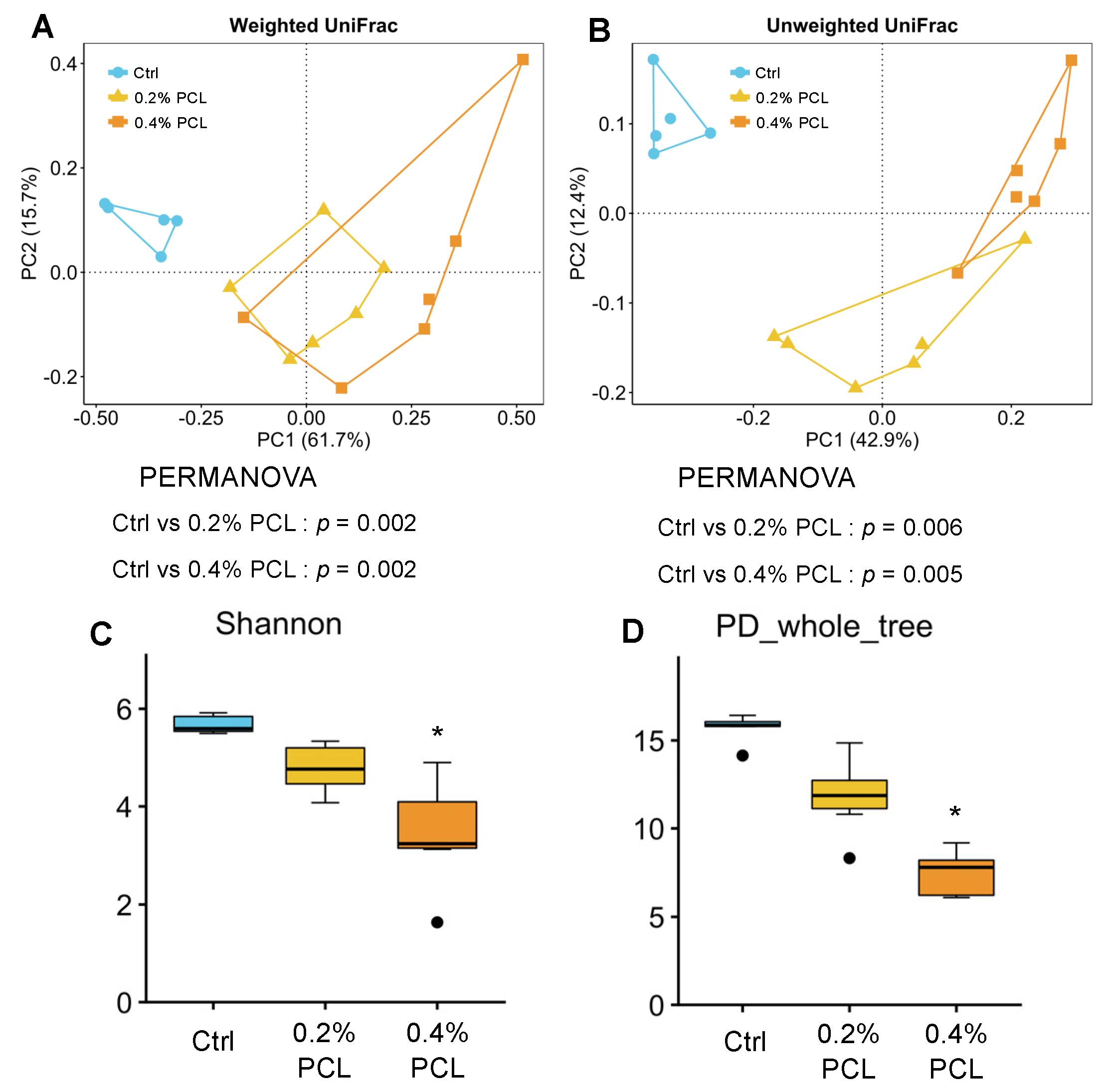
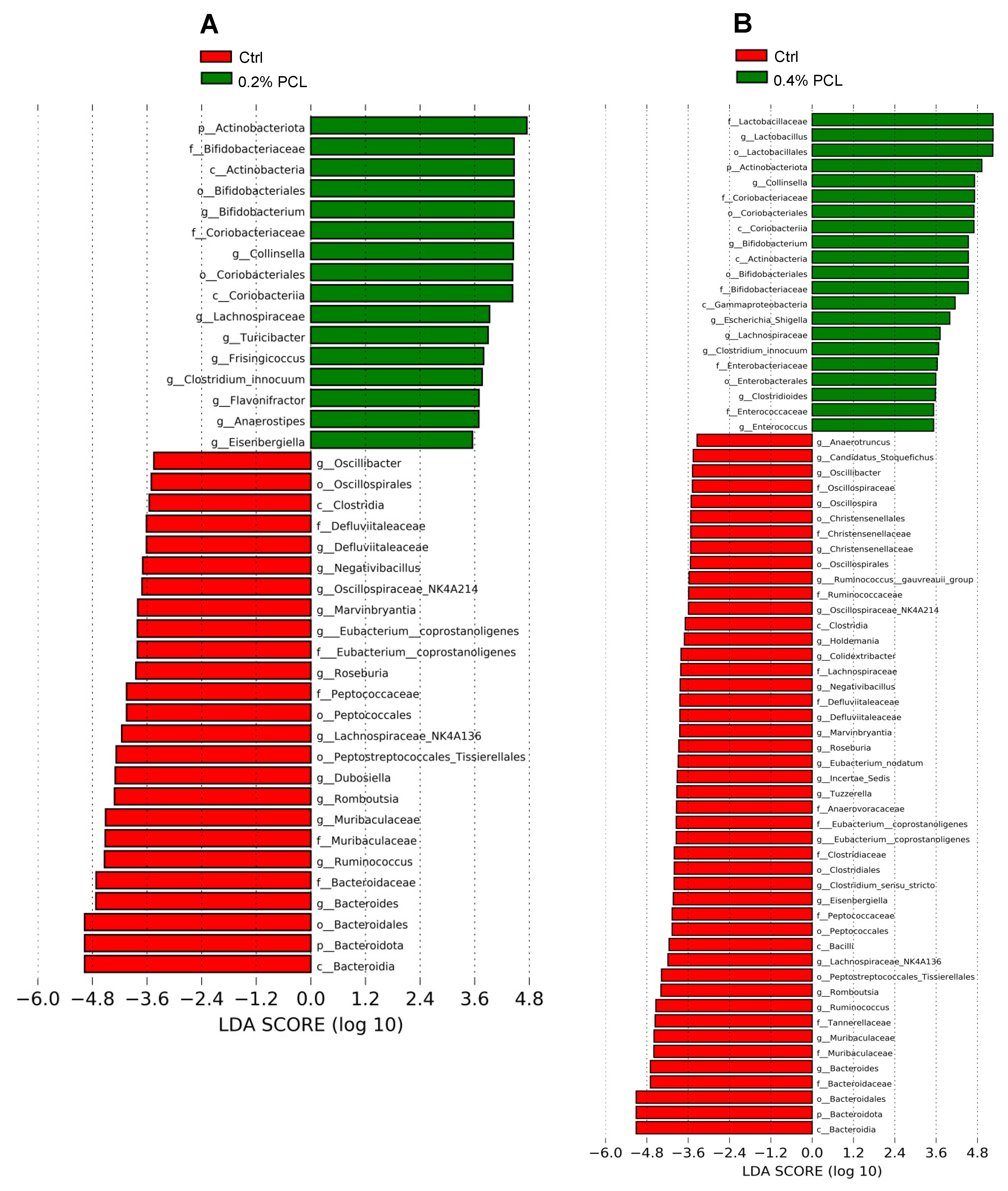
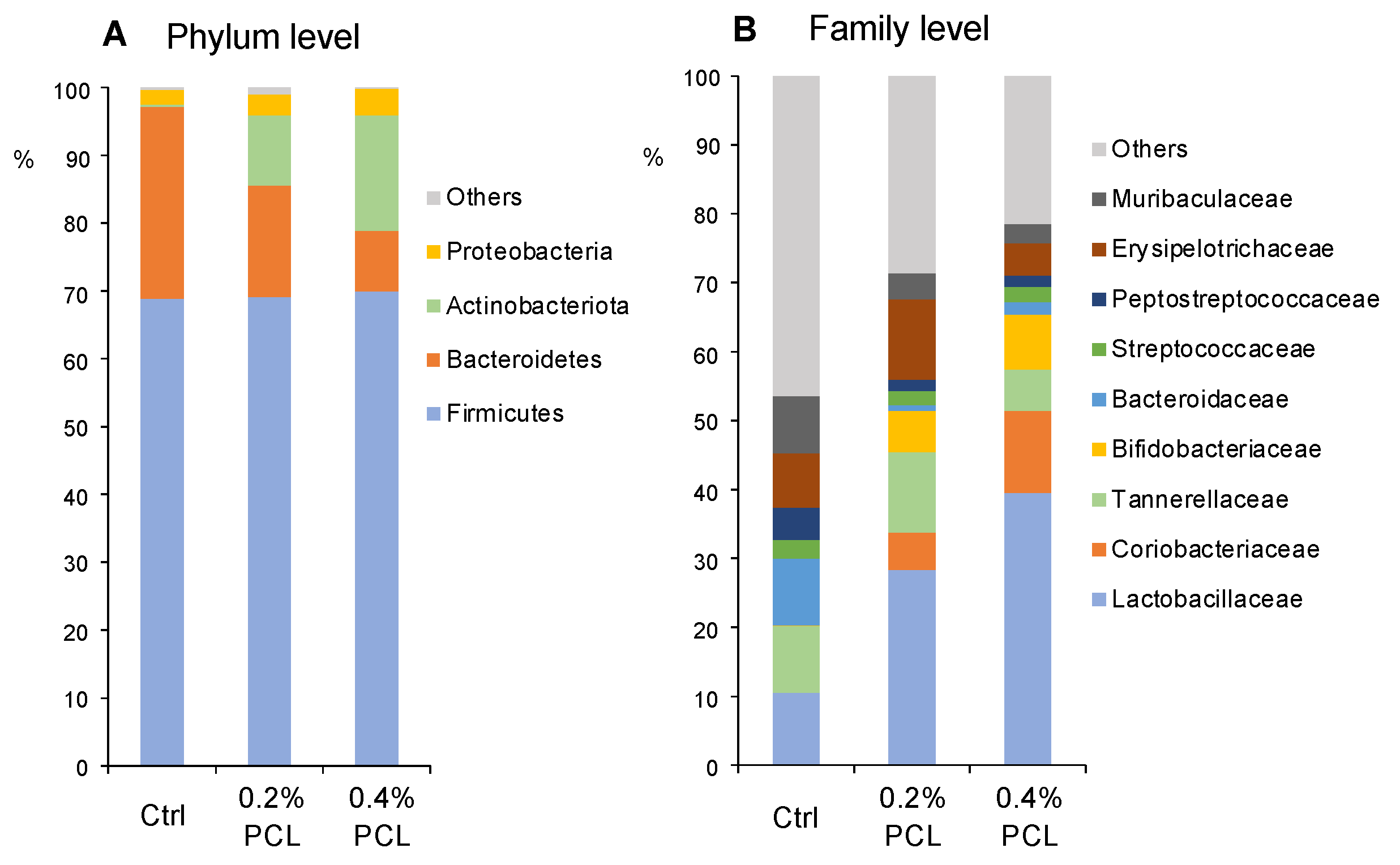
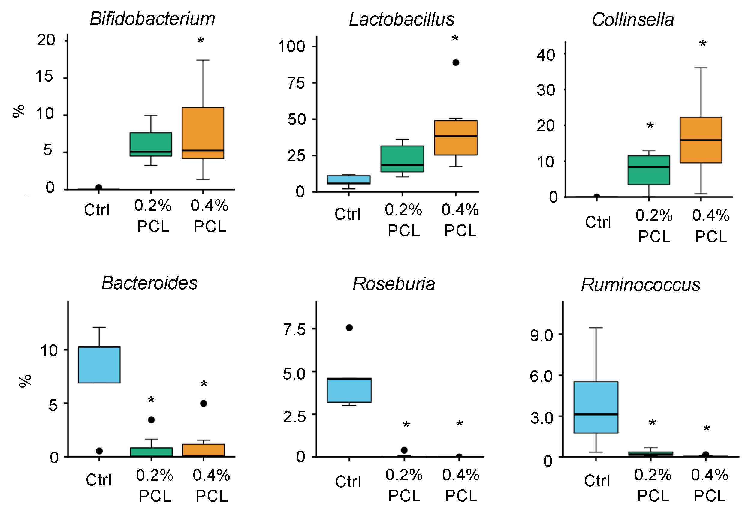
3.2. Cecal Microbiota

For microbiota analysis by 16S rRNA gene sequencing, a total of 790,645 high-quality reads were passed using the QIIME filter. Unweighted and weighted UniFrac PCoA, and PERMANOVA analyses were conducted to compare the microbial structures. The results indicated that the microbial composition differed between the Ctrl and 0.2% PCL groups and between the Ctrl and 0.4% PCL groups (both unweighted and weighted) (Figure 1A,B, p < 0.05). The different alpha diversity indices indicated a lower bacterial diversity in the 0.4% PCL group than in the Ctrl group (Figure 1C,D, p < 0.05).
LEfSe analysis indicated that the bacterial taxa differed between the Ctrl and 0.2% PCL groups (Figure 2A) and between the Ctrl and 0.4% PCL groups (Figure 2B). In both PCL groups, this analysis identified some bacterial taxa, such as Collinsella, Bifidobacterium, Actinobacteriota, Bifidobacteriaceae, Lachnospiraceae, Chlostridium innocuum, Coriobacteriaceae, Coriobacteriia, Lachnospiraceae_NK4A136, Defluvitaleaceae, Oscillibacter, Oscilliopira, Oscillospiraceae_NK4A214, Romboutsia, Roseburia, Peptostreptococcales_Tissierellales, Maruvinbryantia, Ruminococcus, Romboutsia, Muribaculaceae, Bacteroidaceae, Bacteroides, Bacteroidales, Bacteroidota, and Bacteroidia, that varied between the Ctrl and 0.2% PCL groups and between the Ctrl and 0.4% PCL groups. Lactobacillus differed between the Ctrl and 0.4% PCL groups.
Among the four most abundant phyla (Figure 3A), supplemental 0.4% PCL significantly decreased the relative abundance of Bacteroidetes but enriched the relative abundance of Actinobacteria (p < 0.05, Figure 3A). The top nine bacterial taxa are displayed at the family level to address the domain taxa of the microbial groups (Figure 3B). Supplemental 0.4% PCL significantly increased the relative abundance of Bifidobacteriaceae, Lactobacillaceae, and Coribacteriaceae (p < 0.05). In contrast, it reduced the relative abundance of Bacteroidaceae (p < 0.05).
The results of the relative abundance of the six most abundant genera significantly affected by PCL are shown in Figure 4. Compared with the Ctrl group, supplemental 0.4% PCL significantly increased the relative abundance of Bifidobacterium, Lactobacillus, and Collinsella (127-fold, 6-fold, and 193-fold increase, respectively, p < 0.05). Supplemental 0.2% PCL significantly increased the relative abundance of Collinsella (85-fold increase, p < 0.05), but did not affect that of Bifidobacterium and Lactobacillus (p > 0.05). Meanwhile, the relative abundance of Bacteroides was significantly lower in the 0.2% and 0.4% PCL groups than in the Ctrl group (p < 0.05). In the PCL groups, Roseburia and Ruminococcus were mostly not detected, whereas these genera were detectable in all rats of the Ctrl group.

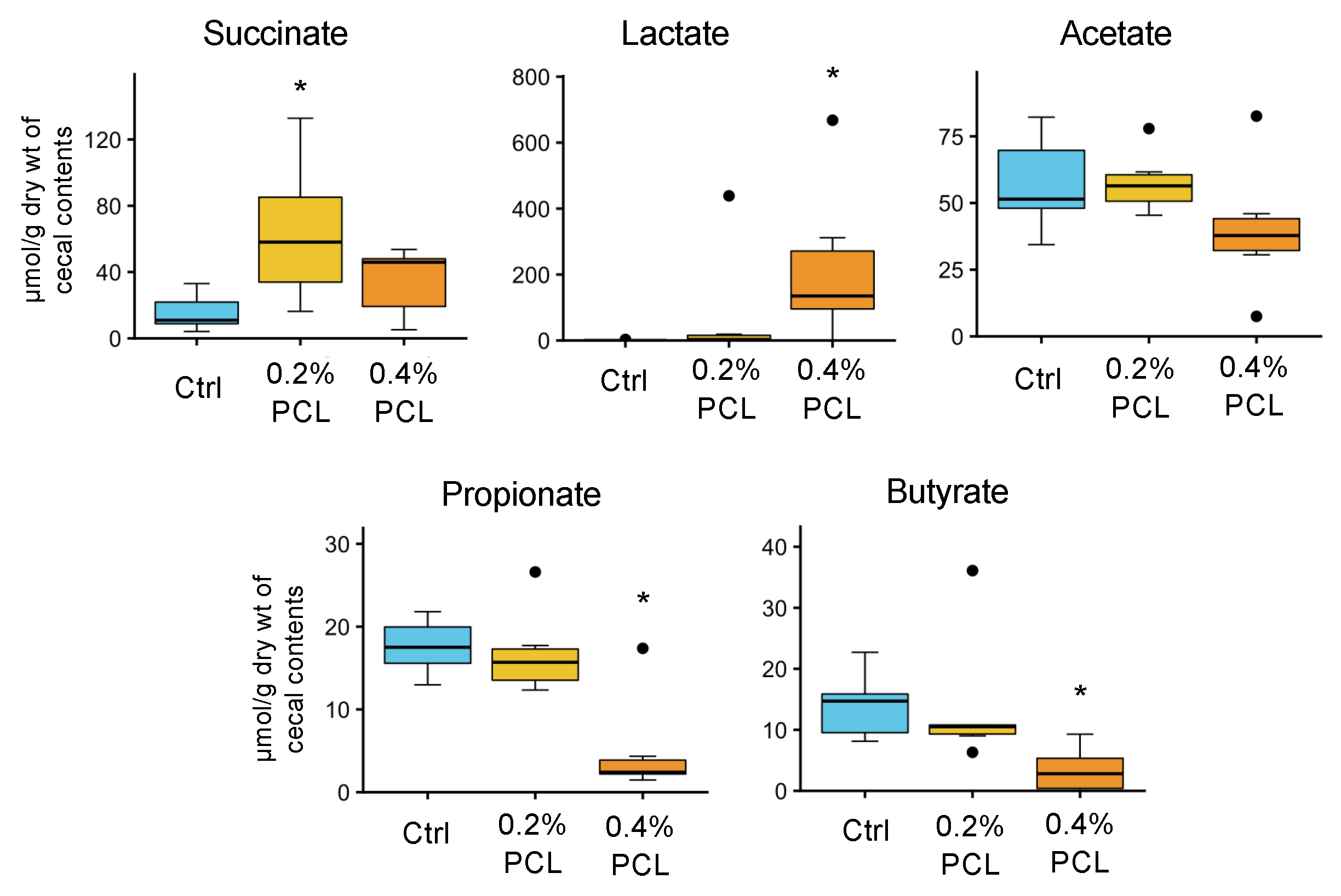
3.3. Cecal Organic Acids

Figure 5 indicates the concentrations of cecal organic acids. Treatment with 0.4% PCL significantly increased the concentrations of lactate (429-fold) but decreased those of propionate (3.4-fold) and n-butyrate (5.5-fold) (p < 0.05). Acetate levels were unaffected. Dietary 0.4% PCL significantly increased the levels of total organic acids (2.7-fold, p < 0.05), but 0.2% PCL did not (p > 0.05). Supplemental Figure S1 further indicates the relationship between the levels of organic acids and various bacterial taxa. Among several bacteria, the relative abundance of Lactobacillus had a strong positive association with the lactate levels, and a strong negative association with propionate.

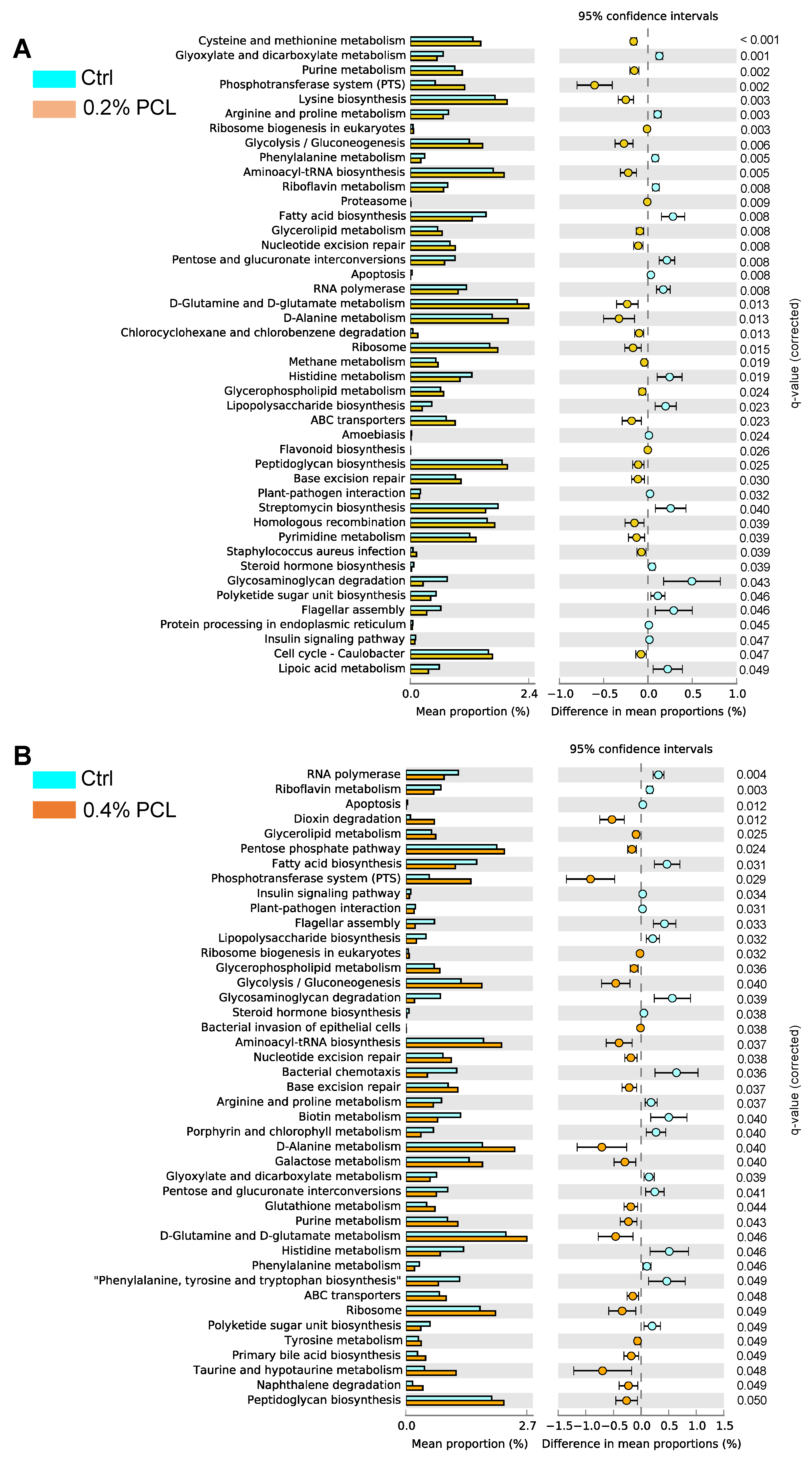
3.4. PICRUSt Analysis

To gain a better understanding of the changed profiles of the gut microbiota by PCL supplementation, PCRUSt analysis was performed to predict the bacterial genes’ functional profiles in the PCL treatment groups according to the composition of the gut microbiota (Figure 6A,B). The results indicated that the relative bacterial gene abundances of 16 metabolic pathways involved in fatty acid biosynthesis, the phosphotransferase (PTS) system, ribosome biogenesis in eukaryotes, glycerophospholipid metabolism, glycerolipid metabolism, glycolysis/gluconeogenesis, aminoacyl-tRNA biosynthesis, nucleotide excision repair, base excision repair, D-alanine metabolism, D-glutamine and D-glutamate metabolism, purine metabolism, ABC transporters, ribosomes, and peptidoylglycan biosynthesis were higher in the 0.2% and 0.4% PCL groups (p < 0.05) than in the Crtl group, while the relative gene abundances of 11 metabolic pathways involved in RNA polymerase, riboflavin metabolism, flagellar assembly, lipopolysaccharide (LPS) biosynthesis, glycosaminoglycan degradation, arginine and proline metabolism, glyoxylate and dicarboxylate metabolism, pentose and glucuronate interconversion, histidine metabolism, phenylalanine metabolism, and polyketide sugar unit biosynthesis were underrepresented in both PCL groups compared to the Crtl group (p < 0.05).

4. Discussion

4.1. Bifidobacterium, Lactobacillus, and Collinsella

This study is the first to present evidence of the health benefits of P. camemberti: rats fed a 0.4% PCL-containing diet exhibited a higher abundance of Bifidobacterium, Lactobacillus, and Collinsella. Similar findings were reported in studies on diets containing preparations of lipase, protease, and cellulase derived from Aspergillus [10,11,13] and those containing prebiotic oligosaccharides [18]. Bifidobacterium and Lactobacillus species are well-known beneficial bacteria. Recent studies suggest that Collinsella species might be beneficial to health [13]. Thus, the present study indicates that beneficial effects on probiotics are not limited to Aspergillus-derived lipase preparations but are also induced by preparations from other fungi, such as Penicillium spp. lipases; in particular, those of microbiological origin (e.g., Aspergillus spp. and Penicillium spp.) are receiving attention in relation to several industrial processes associated with food, drink, pharmaceuticals, medicine, biodiesel, leather, textiles, and detergents [19]. Our findings on the prebiotic-like effects of Aspergillus and Penicillium lipases provide insights that could lead to the novel application of such lipases.
Although PCL exerted a bifidogenic effect that was similar to an Aspergillus nigar lipase preparation (ANL) [10], unexpectedly, the bifidogenic effect of PCL appeared to be far weaker than that of ANL. In fact, a 0.1% ANL supplement induced a 120-fold increase in the relative abundance of cecal Bifidobacterium, whereas a 0.4% PCL supplement induced a 127-fold increase. However, the in vitro lipase activities of ANL and PCL were 60,000 and 55,000 U/g, respectively, which were not significantly different. A. niger-derived lipase is known as an acid lipase [20]. In a preliminary study, Kuroda et al. incubated PCL and ANL at pH 2.0 and 37 °C for 30 min, respectively, which caused the complete inactivation of PCL activity but did not affect ANL activity (unpublished data). These findings indicate the acid-resistant property of ANL, which appears to be lacking in PCL. This characteristic may protect ANL from inactivation by the acidic environment of the stomach lumen, allowing the active lipase to enter the colon lumen. The active lipase in the lumen might at least partly favor the growth of probiotics by enhancing the utilization of triglycerides. Similarly, in our previous study, consumption of a purified acid protease derived from A. oryzae exerted a marked bifidogenic effect, whereas consumption of a purified alkaline protease derived from A. oryzae caused no such effect [14]. This difference might be at least partly attributable to the acid-resistant property of the acid protease. Overall, the greater acid-resistant digestive enzymes in the large intestine might have stronger prebiotic impacts on the gut microbiota through increasing the utilization of energy substrates. To explore this possibility, it is necessary to compare the effects of graded doses of several lipases with different acid-resistant properties on the gut microbiota in the same experiment.

4.2. Other Affected Genera

We found a lower abundance of Bacteroides in the PCL-supplemented groups. Preclinical trials have indicated that Bacteroides are a source of novel beneficial candidates that attenuate inflammation via the regulation of lymphocytes and cytokine expression, as well as controlling metabolism and preventing cancer [21]. However, unlike traditional probiotics such as Bifidobacterium and Lactobacillus, the health-promoting characteristics of Bacteroides are strictly strain-dependent. Accordingly, further study is necessary to determine the specific effects of PCL on several species of cecal Bacteroides.
Interestingly, our study showed that PCL supplementation significantly reduced the abundance of Ruminococcus spp. Similarly, several studies found that mice, rats, and humans that received prebiotic oligosaccharides, such as fructooligosaccharides and galactooligosaccharides, exhibited a lower abundance of Ruminococcus spp. [22,23,24]. Although Ruminococus spp. can generally produce short-chain fatty acids (beneficial organic acids), recent microbiome studies have linked Ruminococcus gnavus to inflammatory bowel disease, namely Crohn’s disease and ulcerative colitis, which are characterized by chronic mucosal inflammation of the gastrointestinal tract, suggesting that R. gnavus may induce aberrant inflammatory responses in the intestinal mucosa [25]. Notably, R. gnavus produces a proinflammatory glucorhamnan polysaccharide that potently induces the toll-like receptor 4-dependent secretion of TNF-α by dendritic cells [26]. In addition, R. gnavus degrades mucin for use as a carbon source, causing the breakdown of the mucus layer and its barrier function [27]. These metabolic properties may increase the exposure of the intestinal immune system to the glucorhamnan polysaccharide, enhancing the host’s inflammatory tone. R. gnavus may also contribute to systemic hyperinflammation characteristics in COVID-19 patients [28]. Accordingly, further study is necessary to investigate whether supplemental PCL reduces the abundance of Ruminococcus spp. linked to chronic inflammation.
Curiously, PCL supplements lowered the abundance of Roseburia spp. Recent studies provided evidence suggesting that Roseburia intestinalis and Roseburia hominis might be probiotics through anti-inflammatory effects, barrier-protective effects, and the production of short-chain fatty acids [29,30]. Accordingly, the lower abundance of Roseburia spp. in rats fed supplemental PCL might have induced adverse effects on host health and disease. In terms of the practical application of PCL, the decreased abundance of Roseburia spp. following PCL supplementation is concerning. Cai et al. found that high doses of Lactobacillus plantarum FRT10 decreased the abundance of Roseburia spp. in mice fed a high-fat diet [31]. Therefore, the increased abundance of Lactobacillus by PCL supplementation may have decreased the abundance of Roseburia. Further study is warranted to determine the implications of reduced Roseburia spp.

4.3. Bacterial Diversity

In the present study, dietary 0.4% PCL supplementation significantly lowered the bacterial diversity compared with that in the Ctrl. Microbial diversity is considered beneficial for community stability and host health [32]. However, this may not always be the case, and assumptions regarding the beneficial effects of increased diversity could be oversimplified considering the complicated interactive mechanisms underlying health and disease [33]. Similarly, lower bacterial diversity was observed in rats fed diets containing lipase, protease, and cellulase preparations derived from Aspergillus. However, the implications of the lower bacterial diversity caused by supplemental prebiotic digestive enzymes are unknown.

4.4. Organic Acids

In the current study, 0.4% PCL supplementation significantly increased cecal lactate levels, which were strongly associated with the relative abundance of Lactobacillus (a major lactate producer). In contrast, 0.4% PCL supplementation decreased the levels of propionate and butyrate. These changes are similar to those caused by Aspergillus lipase and protease in our previous studies [10,13]. Propionate is microbially produced from lactate in the human colon [34]. Accordingly, the metabolic conversion of lactate into propionate might be reduced by dietary PCL supplementation. Recent studies have found that lactate plays an anti-inflammatory role in the gut, and propionate and butyrate both exert anti-inflammatory effects [35,36,37]. However, the PCL-induced effects of increased lactate levels and decreased propionate and butyrate levels on gut health and disease are unknown and warrant further study.

4.5. PICRUSt Analysis

A previous study involving PICRUSt analysis found that the consumption of chitooligosaccharides by neonatal rats modulated the abundance of bacterial genes involved in metabolism, including D-alanine metabolism, the phosphotransferase system, nucleotide excision repair, aminoacyl-tRNA biosynthesis, and the glycosaminoglycan degradation of the gut microbiota [38]. Similar results were observed following dietary supplementation with 0.2% and 0.4% PCL in the present study; thus, KEGG analysis revealed some similarity between the effects of PCL and prebiotic oligosaccharides on the gene abundance in the colon microbiota. Furthermore, interestingly, we found that dietary PCL supplementation increased the abundance of genes involved in the metabolism of D-amino acids, such as D-alanine, D-glutamine, and D-glutamate. Bacteria in the colonic lumen and fermented foods synthesize diverse D-amino acids, which are enantioselectively recognized by some receptors and enzymes, including D-amino acid oxidase (DAO), in mammals [39]. At the host–microbe interface in neutrophiles and the intestinal mucosa, DAO catalyzes the oxidation of bacterial D-amino acids, such as D-alanine, and generates H2O2, which is linked to antimicrobial activity [40]. Interestingly, the abundance of genes associated with the biosynthesis of lipopolysaccharide (LPS), which stimulates inflammation, was decreased in the PCL-supplemented groups. Further study is warranted to explore the implications of PCL-mediated changes in the abundance of genes associated with D-amino acid metabolism and LPS synthesis.

4.6. Limitations of this Study

One limitation of this study was the use of crude and unpurified PCL, despite the PCL exhibiting high lipase activity. Therefore, factors related to the lipase preparation rather than the lipase itself may have been responsible for modulating the abundance of the gut microbiota. Further research is necessary to determine the effects of purified Penicillium-derived lipase on the intestinal microbiota, as well as the relationship between lipase activity and gut microbiota modulation. Our results on the modulation of genes involved in D-amino acids and LPS by PICRUSt analysis, mentioned above, are insightful. However, they indicate the relative gene abundance of metabolic pathways rather than gene expression. Hence, further analysis is necessary to investigate the gene expression and the levels of metabolites such as D-amino acids and LPS before evaluating the implications of the alteration of gene abundance.

5. Conclusions

This study is the first to show that supplemental 0.4% PCL increases the relative abundance of beneficial bacteria, such as Bifidobacterium, Lactobacillus, and Collinsella, in the gut. These changes are consistent with those reported in studies on prebiotic oligosaccharides. Dietary PCL supplementation modulated the relative abundance of bacterial genes involved in several metabolic pathways, some of which were similar to those affected following the consumption of prebiotic oligosaccharides. Thus, these results imply that the exogenous Penicillium lipase preparation used here exerts prebiotic-like effects and could be studied further to determine the health benefits of Penicillium-fermented foods in the colon. Our findings on the prebiotic-like effects of Penicillium lipase provide insights for the novel application of the lipase as a health ingredient in the food, nutraceutical, drink, and animal feed industries, in addition to the established benefits for lipid digestion. Nevertheless, further study is required to determine the mechanisms underlying the prebiotic effects of exogenous Penicillium lipase on colon Bifidobacterium and Lactobacillus. Additionally, since our study used crude lipase and wild-type rats, the effects of highly purified fungal lipase on the intestinal microbiota of wild-type and disease model animals remain to be elucidated.

Supplementary Materials

The following supporting information can be downloaded at: https://www.mdpi.com/article/10.3390/fermentation9030227/s1, Figure S1: Correlation matrix (correlation coefficient, r) between cecal levels of organic acids and microbiota composition. The deep color bars indicate correlation strength. * p < 0.05, ** p < 0.01.

Author Contributions

Conceptualization, investigation, validation, writing—original draft: Y.Y., T.K. and N.K.; formal analysis, software, methodology, visualization: Y.Y., H.J., C.A., T.K., A.K., M.K. and K.N.; literature search: N.K. and A.K.; project administration, funding acquisition, supervision, writing—review and editing: T.K., N.K., H.K., S.F. and S.Y. All authors have read and agreed to the published version of the manuscript.

Funding

This study was supported, in part, by grants from the HIRAKU Consortium, Hiroshima University (to T.K.), Amano Enzyme Inc. (to N.K.), JSPS KAKENHI (22H03541 to S.F.), AMED-CREST (JP22gm1010009 to S.F.), JST ERATO (JPMJER1902 to S.F.), and the Food Science Institute Foundation (to S.F.).

Institutional Review Board Statement

The study was conducted according to the guidelines of the Declaration of Helsinki and approved by the Ethics Committee of Hiroshima University (ethical approval no. C15-12), approved on 16 June 2015.

Informed Consent Statement

Not applicable.

Data Availability Statement

The data of the 16S rRNA gene sequences of gut microbiota presented in this study are available from the NCBI database (http://www.ncbi.nlm.nih.gov/bioproject/918447 (accessed on 25 January 2023) with BioProject record PRJNA918447. Further inquiries can be directed to the corresponding authors.

Conflicts of Interest

This study received funding from Amano Enzyme Inc. The funders had no role in the design of the study; in the collection, analyses, or interpretation of data; in the writing of the manuscript, or in the decision to publish the results. All authors declare no other competing interests in the current study.

References

  1. Hu, M.-J.; Tan, J.-S.; Gao, X.-J.; Yang, J.-G.; Yang, Y.-J. Effect of Cheese Intake on Cardiovascular Diseases and Cardiovascular Biomarkers. Nutrients 2022, 14, 2936. [Google Scholar] [CrossRef] [PubMed]
  2. Alvarez-Bueno, C.; Cavero-Redondo, I.; Martinez-Vizcaino, V.; Sotos-Prieto, M.; Ruiz, J.R.; Gil, A. Effects of Milk and Dairy Product Consumption on Type 2 Diabetes: Overview of Systematic Reviews and Meta-Analyses. Adv. Nutr. 2019, 10 (Suppl. 2), S154–S163. [Google Scholar] [CrossRef] [PubMed]
  3. Papadimitriou, N.; Bouras, E.; Brandt, P.A.V.D.; Muller, D.C.; Papadopoulou, A.; Heath, A.K.; Critselis, E.; Gunter, M.J.; Vineis, P.; Ferrari, P.; et al. A Prospective Diet-Wide Association Study for Risk of Colorectal Cancer in EPIC. Clin. Gastroenterol. Hepatol. 2022, 20, 864–873.e13. [Google Scholar] [CrossRef] [PubMed]
  4. Gil, Á.; Ortega, R.M. Introduction and Executive Summary of the Supplement, Role of Milk and Dairy Products in Health and Prevention of Noncommunicable Chronic Diseases: A Series of Systematic Reviews. Adv. Nutr. 2019, 10, S67–S73. [Google Scholar] [CrossRef] [PubMed] [Green Version]
  5. Park, K.M.; Fulgoni, V.L. The association between dairy product consumption and cognitive function in the National Health and Nutrition Examination Survey. Br. J. Nutr. 2013, 109, 1135–1142. [Google Scholar] [CrossRef] [PubMed] [Green Version]
  6. Luo, Y.; Li, Z.; Gu, L.; Zhang, K. Fermented dairy foods consumption and depressive symptoms: A meta-analysis of cohort studies. PLoS ONE 2023, 18, e0281346. [Google Scholar] [CrossRef]
  7. Bruna, J.M.; Hierro, E.M.; de la Hoz, L.; Mottram, D.S.; Fernández, M.; Ordóñez, J.A. Changes in selected biochemical and sensory parameters as affected by the superficial inoculation of Penicillium camemberti on dry fermented sausages. Int. J. Food Microbiol. 2003, 85, 111–125. [Google Scholar] [CrossRef]
  8. Ano, Y.; Ozawa, M.; Kutsukake, T.; Sugiyama, S.; Uchida, K.; Yoshida, A.; Nakayama, H. Preventive Effects of a Fermented Dairy Product against Alzheimer’s Disease and Identification of a Novel Oleamide with Enhanced Microglial Phagocytosis and Anti-Inflammatory Activity. PLoS ONE 2015, 10, e0118512. [Google Scholar] [CrossRef] [Green Version]
  9. Ano, Y.; Nakayama, H. Preventive Effects of Dairy Products on Dementia and the Underlying Mechanisms. Int. J. Mol. Sci. 2018, 19, 1927. [Google Scholar] [CrossRef] [Green Version]
  10. Yang, Y.; Kumrungsee, T.; Kato, N.; Fukuda, S.; Kuroda, M.; Yamaguchi, S. Supplemental Aspergillus Lipase and Protease Preparations Display Powerful Bifidogenic Effects and Modulate the Gut Microbiota Community of Rats. Fermentation 2021, 7, 294. [Google Scholar] [CrossRef]
  11. Yang, Y.; Sitanggang, N.V.; Kato, N.; Inoue, J.; Murakami, T.; Watanabe, T.; Iguchi, T.; Okazaki, Y. Beneficial effects of protease preparations derived from Aspergillus on the colonic luminal environment in rats consuming a high-fat diet. Biomed. Rep. 2015, 3, 715–720. [Google Scholar] [CrossRef] [Green Version]
  12. Yang, Y.; Kumrungsee, T.; Kuroda, M.; Yamaguchi, S.; Kato, N. Feeding Aspergillus protease preparation combined with adequate protein diet to rats increases levels of cecum gut-protective amino acids, partially linked to Bifidobacterium and Lactobacillus. Biosci. Biotech. Biochem. 2019, 83, 1901–1911. [Google Scholar] [CrossRef]
  13. Yang, Y.; Kumrungsee, T.; Kato, N.; Fukuda, S.; Kuroda, M.; Yamaguchi, S. Aspergillus-Derived Cellulase Preparation Exhibits Prebiotic-like Effects on Gut Microbiota in Rats. Fermentation 2022, 8, 71. [Google Scholar] [CrossRef]
  14. Yang, Y.; Iwamoto, A.; Kumrungsee, T.; Okazaki, Y.; Kuroda, M.; Yamaguchi, S.; Kato, N. Consumption of an acid protease derived from Aspergillus oryzae causes bifidogenic effect in rats. Nutr. Res. 2017, 44, 60–66. [Google Scholar] [CrossRef] [PubMed]
  15. Bolyen, E.; Rideout, J.R.; Dillon, M.R.; Bokulich, N.A.; Abnet, C.C.; Al-Ghalith, G.A.; Alexander, H.; Alm, E.J.; Arumugam, M.; Asnicar, F.; et al. Reproducible, Interactive, Scalable and Extensible Microbiome Data Science using QIIME 2. Nat. Biotechnol. 2019, 37, 852–857. [Google Scholar] [CrossRef]
  16. Douglas, G.M.; Maffei, V.J.; Zaneveld, J.; Yurgel, S.N.; Brown, J.R.; Taylor, C.M.; Huttenhower, C.; Langille, M.G.I. PIC-RUSt2: An improved and customizable approach for metagenome inference. BioRxiv 2020, 672295. [Google Scholar] [CrossRef] [Green Version]
  17. Ramli, N.S.; Jia, H.; Sekine, A.; Lyu, W.; Furukawa, K.; Saito, K.; Hasebe, Y.; Kato, H. Eggshell membrane powder lowers plasma triglyceride and liver total cholesterol by modulating gut microbiota and accelerating lipid metabolism in high-fat diet-fed mice. Food Sci. Nutr. 2020, 8, 2512–2523. [Google Scholar] [CrossRef] [Green Version]
  18. Dewulf, E.M.; Cani, P.; Claus, S.; Fuentes, S.; Puylaert, P.G.B.; Neyrinck, A.; Bindels, L.B.; De Vos, W.M.; Gibson, G.R.; Thissen, J.-P.; et al. Insight into the prebiotic concept: Lessons from an exploratory, double blind intervention study with inulin-type fructans in obese women. Gut 2013, 62, 1112. [Google Scholar] [CrossRef]
  19. Chandra, P.; Enespa; Singh, R.; Arora, P.K. Microbial lipases and their industrial applications: A comprehensive review. Microb. Cell Fact. 2020, 19, 169. [Google Scholar] [CrossRef]
  20. Torossian, K.; Bell, A.W. Purification and characterization of an acid-resistant triacylglycerol lipase from Aspergillus niger. Biotechnol. Appl. Biochem. 1991, 13, 205–211. [Google Scholar]
  21. Tan, H.; Zhai, Q.; Chen, W. Investigations of Bacteroides spp. towards next-generation probiotics. Food Res. Int. 2019, 116, 637–644. [Google Scholar] [CrossRef] [PubMed]
  22. Burokas, A.; Arboleya, S.; Moloney, R.D.; Peterson, V.L.; Murphy, K.; Clarke, G.; Stanton, C.; Dinan, T.G.; Cryan, J.F. Targeting the Microbiota-Gut-Brain Axis: Prebiotics Have Anxiolytic and Antidepressant-like Effects and Reverse the Impact of Chronic Stress in Mice. Biol. Psychiatry 2017, 82, 472–487. [Google Scholar] [CrossRef]
  23. Yang, X.-D.; Wang, L.-K.; Wu, H.-Y.; Jiao, L. Effects of prebiotic galacto-oligosaccharide on postoperative cognitive dysfunction and neuroinflammation through targeting of the gut-brain axis. BMC Anesthesiol. 2018, 18, 177. [Google Scholar] [CrossRef] [Green Version]
  24. Liu, F.; Li, P.; Chen, M.; Luo, Y.; Prabhakar, M.; Zheng, H.; He, Y.; Qi, Q.; Long, H.; Zhang, Y.; et al. Fructooligosaccharide (FOS) and Galactooligosaccharide (GOS) Increase Bifidobacterium but Reduce Butyrate Producing Bacteria with Adverse Glycemic Metabolism in healthy young population. Sci. Rep. 2017, 7, 11789. [Google Scholar] [CrossRef] [PubMed] [Green Version]
  25. Rajilić-Stojanović, M.; Jonkers, D.M.; Salonen, A.; Hanevik, K.; Raes, J.; Jalanka, J.; de Vos, W.M.; Manichanh, C.; Golic, N.; Enck, P.; et al. Intestinal Microbiota and Diet in IBS: Causes, Consequences, or Epiphenomena? Am. J. Gastroenterol. 2015, 110, 278–287. [Google Scholar] [CrossRef] [PubMed] [Green Version]
  26. Henke, M.T.; Kenny, D.J.; Cassilly, C.D.; Vlamakis, H.; Xavier, R.J.; Clardy, J. Ruminococcus gnavus, a member of the human gut microbiome associated with Crohn’s disease, produces an inflammatory polysaccharide. Proc. Natl. Acad. Sci. USA 2019, 116, 12672–12677. [Google Scholar] [CrossRef] [PubMed] [Green Version]
  27. Bell, A.; Brunt, J.; Crost, E.; Vaux, L.; Nepravishta, R.; Owen, C.D.; Latousakis, D.; Xiao, A.; Li, W.; Chen, X.; et al. Elucidation of a sialic acid metabolism pathway in mucus-foraging Ruminococcus gnavus unravels mechanisms of bacterial adaptation to the gut. Nat. Microbiol. 2019, 4, 2393–2404. [Google Scholar] [CrossRef]
  28. Kageyama, Y.; Akiyama, T.; Nakamura, T. Intestinal Dysbiosis and Probiotics in COVID-19. J. Clin. Trials 2020, 10, 421. [Google Scholar]
  29. Nie, K.; Ma, K.; Luo, W.; Shen, Z.; Yang, Z.; Xiao, M.; Tong, T.; Yang, Y.; Wang, X. Roseburia intestinalis: A Beneficial Gut Organism from the Discoveries in Genus and Species. Front. Cell. Infect. Microbiol. 2021, 11, 757718. [Google Scholar] [CrossRef]
  30. Song, L.; Sun, Q.; Zheng, H.; Zhang, Y.; Wang, Y.; Liu, S.; Duan, L. Roseburia hominis Alleviates Neuroinflammation via Short-Chain Fatty Acids through Histone Deacetylase Inhibition. Mol. Nutr. Food Res. 2022, 66, 2200164. [Google Scholar] [CrossRef]
  31. Cai, H.; Wen, Z.; Li, X.; Meng, K.; Yang, P. Lactobacillus plantarum FRT10 alleviated high-fat diet–induced obesity in mice through regulating the PPARα signal pathway and gut microbiota. Appl. Microbiol. Biotechnol. 2020, 104, 5959–5972. [Google Scholar] [CrossRef] [PubMed]
  32. Sjöberg, F.; Barkman, C.; Nookaew, I.; Östman, S.; Adlerberth, I.; Saalman, R.; Wold, A.E. Low-complexity microbiota in the duodenum of children with newly diagnosed ulcerative colitis. PLoS ONE 2017, 12, e0186178. [Google Scholar] [CrossRef] [PubMed] [Green Version]
  33. Shade, A. Diversity is the question, not the answer. ISME J. 2017, 11, 1–6. [Google Scholar] [CrossRef] [PubMed] [Green Version]
  34. Flint, H.J.; Duncan, S.H.; Scott, K.P.; Louis, P. Links between diet, gut microbiota composition and gut metabolism. Proc. Nutr. Soc. 2015, 74, 13–22. [Google Scholar] [CrossRef] [Green Version]
  35. Manosalva, C.; Quiroga, J.; Hidalgo, A.I.; Alarcón, P.; Anseoleaga, N.; Hidalgo, M.A.; Burgos, R.A. Role of Lactate in Inflammatory Processes: Friend or Foe. Front. Immunol. 2022, 12, 808799. [Google Scholar] [CrossRef]
  36. Li, X.; Yang, Y.; Zhang, B.; Lin, X.; Fu, X.; An, Y.; Zou, Y.; Wang, J.X.; Wang, Z.; Yu, T. Lactate metabolism in human health and disease. Signal Transduct. Target. Ther. 2022, 7, 305. [Google Scholar] [CrossRef]
  37. Park, B.O.; Kang, J.S.; Paudel, S.; Park, S.G.; Park, B.C.; Han, S.B.; Kwak, Y.S.; Kim, J.H.; Kim, S. Novel GPR43 Agonists Exert an Anti-Inflammatory Effect in a Colitis Model. Biomol. Ther. 2022, 30, 48–54. [Google Scholar] [CrossRef]
  38. Wei, X.; Yu, L.; Zhang, C.; Ni, Y.; Zhao, J.; Zhang, H.; Chen, W.; Zhai, Q.; Tian, F. Prebiotic activity of chitooligosaccharides and their ability to alleviate necrotizing enterocolitis in newborn rats. Carbohydr. Polym. 2023, 299, 120156. [Google Scholar] [CrossRef]
  39. Matsumoto, M.; Kunisawa, A.; Hattori, T.; Kawana, S.; Kitada, Y.; Tamada, H.; Kawano, S.; Hayakawa, Y.; Iida, J.; Fukusaki, E. Free D-amino acids produced by commensal bacteria in the colonic lumen. Sci. Rep. 2018, 8, 17915. [Google Scholar] [CrossRef] [Green Version]
  40. Sasabe, J.; Suzuki, M. Emerging Role of D-Amino Acid Metabolism in the Innate Defense. Front. Microbiol. 2018, 9, 933. [Google Scholar] [CrossRef] [Green Version]
Figure 1. Effects of supplemental PCL on cecal microbiome profiles and alpha diversity. PCoA of unweighted (A) and weighted UniFrac (B) and PERMANOVA analysis were performed to compare the gut microbiome profiles of the experimental groups. The alpha diversity of the gut microbiota within samples was measured by (C) Shannon index and (D) PD whole tree. Data are presented as a boxplot with median and min–max whiskers. The dots (•) in the boxplots are outliers. * p < 0.05 means significantly different when compared to the Ctrl group (Dunnett’s test).
Fermentation 09 00227 g001
Figure 2. Different bacterial taxa between the Ctrl and 0.2% PCL groups (A) or between the Ctrl and 0.4% PCL groups (B) (LEfSe analysis). The two-tailed nonparametric Kruskal–Wallis test was used to evaluate the significance of differences in taxa at p < 0.05.
Fermentation 09 00227 g002
Figure 3. Effects of supplemental PCL on cecal microbiota composition (average, % of total bacteria) at the phylum (A) and family (B) levels.
Fermentation 09 00227 g003
Figure 4. Effects of supplemental PCL on cecal microbiota composition at the genus level (% of total bacteria). * p < 0.05 means significantly different when compared to the Ctrl group (Dunnett’s test). Data are presented as a boxplot with median and min–max whiskers. The dots (•) in the boxplots are outliers.
Fermentation 09 00227 g004
Figure 5. Effects of supplemental PCL on cecal levels of organic acids. * p < 0.05 means significantly different when compared to the Ctrl group (Dunnett’s test). Data are presented as a boxplot with median and min–max whiskers. The dots (•) in the boxplots are outliers.
Fermentation 09 00227 g005
Figure 6. Prediction of bacterial genes’ functions to make inferences from KEGG annotated databases using PICRUSt analysis. The gene abundances of the metabolic pathways significantly affected by PCL are shown in (A) (0.2% PCL effects) and (B) (0.4% PCL effects). The mean proportion of each pathway is displayed in bar graphs on left.
Fermentation 09 00227 g006
Disclaimer/Publisher’s Note: The statements, opinions and data contained in all publications are solely those of the individual author(s) and contributor(s) and not of MDPI and/or the editor(s). MDPI and/or the editor(s) disclaim responsibility for any injury to people or property resulting from any ideas, methods, instructions or products referred to in the content.

