Article

Bacteriocinogenic Enterococcus casseliflavus Isolated from Fresh Guava Fruit (Psidium guajava): Characterization of Bacteriocin ST192Gu and Some Aspects of Its Mode of Action on Listeria spp. and Enterococcus spp.

by
Svetoslav Dimitrov Todorov
1,2,3,*,
Wilhelm Heinrich Holzapfel
1 and
John Robert Tagg
4
1
ProBacLab, Department of Advanced Convergence, Handong Global University, Gyeongbuk, Pohang 37554, Republic of Korea
2
ProBacLab, Laboratório de Microbiologia de Alimentos, Departamento de Alimentos e Nutrição Experimental, Faculdade de Ciências Farmacêuticas, Universidade de São Paulo, São Paulo 05508-000, Brazil
3
Food Research Center (FoRC), Departamento de Alimentos e Nutrição Experimental, Faculdade de Ciências Farmacêuticas, Universidade de São Paulo, São Paulo 05508-000, Brazil
4
BLIS Technologies Ltd., 81 Glasgow Street, South Dunedin 9012, New Zealand
*
Author to whom correspondence should be addressed.
Fermentation 2023, 9(3), 226; https://doi.org/10.3390/fermentation9030226
Submission received: 14 February 2023 / Revised: 23 February 2023 / Accepted: 25 February 2023 / Published: 27 February 2023
(This article belongs to the Section Microbial Metabolism, Physiology & Genetics)

Abstract

:
Strain ST182Gu, isolated from fresh guava fruit, was identified as Enterococcus casseliflavus on the basis of biochemical tests, sugar fermentation reactions (API20Strip), PCR with genus-specific primers, and 16S rRNA sequencing. This appears to be the first documentation of the presence of this species in guava. E. casseliflavus ST182Gu was shown to produce a 4.8 kDa class IIa bacteriocin, active against various lactic acid bacteria including Enterococcus spp. and Streptococcus spp., and Staphylococcus aureus, and different serotypes of Listeria spp. The activity of the peptide was reduced by treatment with 0.1 mg/mL proteolytic enzymes, but not by α-amylase, catalase, lipase, and 1% (w/v) sodium dodecyl sulphate (SDS), Tween-20, Tween-80, urea, NaCl, and EDTA. No change in activity was recorded after adjustment to pH values of between 2.0 and 12.0 for 2 h, and after treatment at 100 °C for 120 min or 121°C for 20 min, compared with non-treated antimicrobial peptide. The mode of action against representative susceptible bacteria was shown to be bactericidal and associated with cell lysis and enzyme- and DNA-leakage. These susceptible bacteria, Listeria ivanovii subsp. ivanovii ATCC 19119, Listeria monocytogenes ATCC 15313, and Enterococcus faecalis ATCC 19443 differed however in their sensitivity to bacteriocin ST182Gu (6,553,600 AU/mL, 102,400 AU/mL, and 51,200 AU/mL, respectively). No significant differences were detected in cell growth and bacteriocin production when strain ST182Gu was grown in MRS broth at 26 °C, 30 °C, and 37 °C for 24 h. Bacteriocin ST182Gu recovery from the surface of the producer cells showed different activity, dependent of the applied test organisms (3200, 800 and 400 AU/mL, evaluated versus L. ivanovii subsp. ivanovii ATCC 19119, L. monocytogenes ATCC 15313 and E. faecalis ATCC 19443, respectively), however, with proportional values with the activity recorded in cell free supernatant versus same test microorganisms. When bacteriocin ST182Gu was combined with sublethal doses of ciprofloxacin, synergistic inhibition of L. ivanovii subsp. ivanovii ATCC 19119 was demonstrated. This increase in ciprofloxacin sensitivity may be due to the dissipation of the proton gradient in the cell membrane of the target organism associated with exposure to bacteriocin ST182Gu. Apart from reducing the MIC of classical therapeutic antibiotics, bacteriocins such as ST182Gu may also play an important role in the treatment of multidrug resistant strains.

1. Introduction

The history of human civilization has been markedly influenced by periodic exposures to regional and global epidemics. Millions of lives have been prematurely lost due to the ravages of infectious diseases and absence of adequate medical therapy. Even after the onset of the antibiotic era, countless humans and other animals have perished due to the unavailability of effective antimicrobials for treatment of infections by some pathogenic microbes. In many cases, these pathogens have developed strategies to resist the activity of existing antimicrobials and these multiple manifestations of antibiotic resistance have become one of the major challenges facing modern medicine. The urgent need for novel antimicrobials now provides a pressing challenge for scientists, not only for treatment purposes in human and veterinary medicine, but also for potential pathogen and food spoilage control in the food industry [1].
Antimicrobial peptides (bacteriocins) produced by different microorganisms, including lactic acid bacteria (LAB), are considered to be important potential alternatives in the development of new approaches for the improvement of food safety and in the combating of microbial infections both in humans and in veterinary practice [2]. Now well known within the scientific community, knowledge of LAB bacteriocins began with the discovery of nisin, produced by Lactococcus lactis [3]. Still one of the best studied bacteriocins, nisin was at first predominantly recognized as a safe and potent antimicrobial suitable for application in food biopreservation processes [4]. However, in the last decade, it has also been promoted as a potential adjunct or alternative for the treatment of infections [5,6].
The number of research projects that are focused upon the isolation and characterization of bacteriocins have continued to escalate and according to www.scopus.com (accessed on 1 February 2023), more than 13.686 research papers have been published concerning different aspects of the production, characterization, and application of bacteriocins.
Tropical fruits are an underexplored potential source of bacteriocin producers. Antimicrobial peptides produced by plant-adapted indigenous microbes undoubtedly contribute to the biological protection of fruits against extrinsic pathogenic microbes. This research group has previously reported that Pediococcus pentosaceus ST44AM, isolated from marula fruit, is a producer of a pediocin PA-1-like bacteriocin [7] and Lactiplantibacillus plantarum ST16Pa, isolated from papaya, expresses plantaricin [8].
Bacteriocins have generally been found to exhibit low cytotoxicity for eukaryotic cells, and for example, nisin has a similar cytotoxicity to NaCl [9]. On the other hand, some other bacteriocins have been shown to be toxic for eukaryotic cells [9] and of special interest are those having cytotoxic activity against cancer cells [10].
Stringent safety evaluation of bacteriocin producers is considered to be a milestone criterion in the research pathway supporting the evaluation, characterization, and practical application of bacteriocin producers either as biopreservatives or as putative probiotics. Although bacteriocin production is not considered to be a mandatory characteristic of a candidate probiotic strain, it is generally regarded as potentially important since some bacteriocins may help suppress the growth of important pathogens and thereby contribute to the beneficial profile of the probiotic.
The principal objective of the present study was to evaluate the bacteriocin-like characteristics of an antimicrobial peptide produced by E. casseliflavus ST182Gu isolated from fresh guava fruits. Also explored was the potential for application of this peptide as a means of enhancing the antibiotic potency of ciprofloxacin.

2. Materials and Methods

2.1. Isolation, Differentiation, and Identification of Bacteriocin Producer/s

2.1.1. Isolation of the Bacteriocin Producer/s and Evaluation of Antimicrobial Activity

Guava fruits (Psidium guajava), freshly obtained from a local farmers market (Sao Paulo, SP, Brazil), were rinsed with sterile distilled water and macerated in a stomacher with sterile saline in proportion 1:9 (fruit mass/grams: volume of saline/mL). Ten-fold serial dilutions of the fruit puree were prepared and plated (100 μL) on MRS (Difco, Franklin Lakes, NJ, USA), supplemented with 2% (w/v) agar (Difco) and then overlayered with 5 mL of 2% (w/v) agar. Incubation was at 37 °C for 24–48 h in aerobic conditions and the colony count was expressed as CFU/g, according to standard microbiological practice. Plates showing individual bacterial colonies were selected and addition layer of 10 mL Brain Heart Infusion (BHI) (Difco), supplemented with 1% (w/v) agar (temperate to around 45 °C) and Listeria monocytogenes ATCC 15313, Listeria ivanovii subsp. ivanovii ATCC 19119, or Enterococcus faecalis ATCC 19443 at 105 CFU/mL final concentration were added on the top of previous growth media with already grown colonies, according to the recommendations of dos Santos et al. [11]. These plates were incubated for an additional 18–24 h and then assessed for inhibition zones around the previously identified bacterial colonies. Bacterial colonies surrounded by inhibition zones were subcultured and then purified by consecutive streaking on MRS with 2% (w/v) agar and followed by growth in MRS broth for 18–24 h at 37 °C. Putative bacteriocin activity was detected as follows, according to the recommendations of dos Santos et al. [11]. Cell free supernatant (CFS) was obtained by centrifugation (6000× g, 15 min, 20 °C) and separated into two equal volumes. The pH of one of these aliquots was adjusted with 1M NaOH to 5.5–6.5 and the second was not pH-adjusted. Both aliquots were heated for 10 min at 80 °C to inactivate any putatively produced proteolytic enzymes or H2O2. Samples (10 μL) were then spotted on BHI supplemented with 1% (w/v) agar and seeded with either L. monocytogenes ATCC 15313, L. ivanovii subsp. ivanovii ATCC 19119, or E. faecalis ATCC 19443 at 105 CFU/mL final concentration, according to the recommendations of dos Santos et al. [11]. Following incubation for 18–24 h at 37 °C, the cultures were evaluated for the presence of inhibitory zones and having zones larger than 3 mm diameter were putatively considered to be producing antimicrobial metabolites. For the quantification of expressed bacteriocins, the recommendations of dos Santos et al. [11] were followed. Serial two-fold dilutions of CFS (pH neutralized to 5.5–6.5 and treated for 10 min at 80 °C) were prepared using 100 mM sodium phosphate buffer (pH 6.5) and 10 μL of each dilution was spotted on the surface of BHI supplemented with 1% agar and L. monocytogenes ATCC 15313, L. ivanovii subsp. ivanovii ATCC 19119, or E. faecalis ATCC 19443 incorporated into the agar at 105 CFU/mL final concentration, incubated, and evaluated as described before. The putative bacteriocin titer was expressed in AU/mL, taking into consideration the critical dilution needed for observation of clear inhibition zone and the volume of the deposited bacteriocin containing CFS, using the following formula:
AU / mL = D n × 1000 p
where D is the type of serial dilution, n represents the highest dilution where at least 3 mm of inhibition was recorded, p is the volume of spotted BLIS containing material in μL, and 1000 is the conversion factor between μL and mL [11].
All bacterial isolates of interest and other cultures applied either as test organisms in the evaluation of the spectrum of activity or as control strains in subsequent experiments were stored at −80 °C using 30% (v/v) glycerol as a cryogenic protector. All strains requiring recovery from storage were first grown for at least 2 cycles in an appropriate growth medium and temperature prior to their use in experiments.

2.1.2. Differentiation and Identification of Bacteriocin Producer/s

Isolates identified as potential producers of bacteriocin/s were grown on MRS agar and in MRS broth for 18–24 h at 37 °C in order to evaluate their colony morphology (on plates) and Gram-stained appearance (from liquid cultures). In addition, catalase and oxidase reactions were recorded. The isolates selected for future studies were Gram-positive, chain-forming, cocci-exhibiting negative reactions for both the catalase and oxidase tests. Primary identification of the selected bacterial isolates was based initially on recommendations from Bergey’s Manual of Systematic Bacteriology of Archaea and Bacteria [12], including growth at different temperatures and in different concentrations of NaCl. This was followed by evaluation of their sugar fermentation profiles using API50CHL and API20Strip (BioMerieux, Marcy-l’Étoile, France), according to the manufacturer’s recommendations.
Further differentiation of the selected isolates was performed according to de Moraes et al. [13] by RAPD-PCR using primers OPL03 (5′-CAT AGA GCG G-3′), OPL06 (5′-GAG GGA AGA G-3′), OPL11 (5′-ACG ATG AGC C-3′), and OPL17 (5′-AGC CTG AGC C-3′). DNA extracts were obtained from the cultures grown in MRS (for 24 h at 37 °C) using ZR Fungal/Bacterial DNA Kits (Zymo Research, Irvine, CA, USA), according to the manufacturer’s instructions. The obtained DNA was evaluated on NanoDrop (Thermo Fisher, Waltham, MA, USA). PCR reactions were performed using a Veriti 96-well thermal cycler (Thermo Fisher) and the obtained amplicons were visualized on 1.5% (w/v) agarose gel in the presence of ethidium bromide.
Strains of interest were subjected to genus identification using genus-specific PCR according to Ke et al. [14] using primers EntF (5′-TAC TGA CAA ACC ATT CAT GAT G-3′) and EntR (5′-AAC TTC GTC ACC AAC GCG AAC-3′) followed by PCR targeting the 16S rRNA gene, performed on a Veriti 96-well thermal cycler (Thermo Fisher) with primers F8 (5′-CAC GGA TCC AGA CTT TGA TYM TGG CTC AG-3′) and R1512 (5′-GTG AAG CTT ACG GYT AGC TTG TTA CGA CTT-3′) [11] and appropriate amplicons sequenced by a contracted service. The obtained sequences were analyzed on https://blast.ncbi.nlm.nih.gov/Blast.cgi (accessed on 24 May 2022) and compared with the existing database. Additional biochemical and physiological tests for differentiation of Enterococcus spp. were performed, according to recommendations of de Vos et al. [12].

2.2. Safety Evaluation of the Selected Isolate

The recommendations of Fugaban et al. [15] were adopted for the preliminary safety assessments. Characteristics assessed included hemolysin, DNase, gelatinase and lipase activities, sensitivity to selected clinically important antibiotics, and the production of biogenic amines. All tests were performed at least twice at both 30 °C and 37 °C.

2.3. Confirmation of Protein Nature and Stability to pH, Temperature, and to Chemicals of Potential Practical Relevance

The initial presumptive assessment of the protein nature of the (bacteriocin-like) antimicrobial agents produced by inhibitory isolates followed the recommendations of Fugaban et al. [15]. CFS from 24h, 37 °C cultures of each evaluated strain was incubated for 1 h at 37 °C in the presence of 0.1 mg/ mL(final concentration) of either proteinase K (Roche, Indianapolis, IN, USA), pronase (Sigma-Aldrich, St. Luis, MO, USA), α-amylase (Sigma-Aldrich), lipase (Sigma-Aldrich), or catalase (Roche Indianapolis, IN, USA). The added enzymes were inactivated by heating for 5 min at 95–97 °C and if required, the pH was adjusted to 6.0–6.5 prior to assaying the residual inhibitory activity of the enzyme-treated preparations. Inhibitory activity was assessed as described above against L. monocytogenes ATCC 15313, L. ivanovii subsp. ivanovii ATCC 19119, and E. faecalis ATCC 19443.
Additional studies were undertaken to assess the influence of selected chemicals and of exposure to various changes in temperature and pH. For the purpose of these experiments, 2 mL of the CFS obtained as described before was exposed to either 25, 30, 37, 45, or 100 °C for 60 and 120 min, and at 121 °C for 15 min, according to recommendations of Fugaban et al. [15]. The residual bacteriocin activity was determined as described above against the principal test indicator strains.
The influence of pH on putative bacteriocin stability was evaluated according to the recommendations of Fugaban et al. [15]. CFS (10) mL was adjusted to pH 3.0, 4.0, 6.0, 7.0, 8.0, or 9.0 with sterile 1 M HCl or 1 M NaOH and incubated for 2 h at 37 °C. Before evaluation for the residual bacteriocin activity against the principal test indicator strains, the pH was adjusted to 5.5–6.5.
Some chemicals commonly used either in the food industry or in bacteriocin research were tested for their influence on the bioactivity of the inhibitory activities produced by the current LAB isolates. These chemicals were skimmed milk (Difco), NaCl, SDS, Tween 20, Tween 80, and EDTA (all from Sigma-Aldrich), according to Fugaban et al. [15]. The chemicals were added as 10% (w/v) to the CFS of the selected LAB isolates and incubated for 1 h at 37 °C. When required, the pH was adjusted to 5.5–6.5. Residual bacteriocin activity was determined as previously described.
All experiments were minimally performed in duplicate and on two independent occasions. Appropriate controls, including non-treated CFS, were included in all experiments.

2.4. Spectrum of Inhibitory Activity

CFS from the chosen LAB was prepared as previously described with the pH being adjusted to 5.5–6.5 followed by heating for 10 min at 80 °C. Samples (10 μL) of these preparations were then spotted on BHI or MRS, supplemented with 1% (w/v) agar plates and incorporated test microorganisms (final concentration of 105 CFU/mL) listed in Table 1. After incubation for 24 h at 37 °C, plates were evaluated for inhibition zones, where zones with diameters larger than 3 mm were considered positive.

2.5. Adsorption of Produced Bacteriocin to the Producer Cells

Assessment of levels of bacteriocin activity present on the surface of the producer cells followed the recommendations of Yang et al. [16]. Cultures (10 mL) of the LAB test strains were grown in MRS for 24 h at 37 °C and the pH was adjusted to pH 6.0 with 1 M NaOH. The bacterial cells were recovered by centrifugation (5000× g, 15 min, 4 °C) and washed with sterile 0.1 M sodium phosphate buffer (pH 6.5) followed by resuspension in 10 mL 100 mM NaCl (pH 2.0). After stirring these suspensions for 1 h at 4 °C, the CFS was recovered by centrifugation (5000× g, 15 min, 4 °C) and the pH was adjusted to 5.5–6.5 with sterile 1 M NaOH. Bacteriocin activity in these extracts was assessed as previously described.

2.6. Bacterial Growth, Changes in pH, and Production of Bacteriocin

An overnight culture of the tested LAB was inoculated (2%, v/v) into 200 mL MRS broth (Difco) and incubated at 26, 30, and 37 °C, respectively, without agitation, for 48 h. Samples were collected every hour and the growth of the culture (OD600 nm) and pH changes (a consequence of the production of organic acids) were monitored. Bacteriocin activity (expressed as AU/mL) was evaluated in samples taken every 3 h as described above.

2.7. Mode of Action

2.7.1. Partial Purification

CFS from tested LAB grown in MRS for 24 h at 37 °C (500 mL pH adjusted to 5.5–6.5 and heated for 10 min at 80 °C) was treated with 60% saturation of ammonium sulfate for 4 h at 4 °C. The precipitated proteins were harvested by centrifugation (20,000× g, 60 min, at 4 °C) followed by resuspension in 1/10 of the initial volume of 25 mM sodium phosphate buffer (pH 6.5). This preparation was further fractionated by chromatography on a SepPak C18 hydrophobic column (Waters, Millipore, MA, USA) using a step gradient of 20, 40, 60, and 80% (v/v) isopropanol in 25 mM sodium phosphate buffer (pH 6.5) [17]. Eluted fractions were tested for inhibitory activity (as previously described) against L. monocytogenes ATCC 15313, L. ivanovii subsp. ivanovii ATCC 19119, and E. faecalis ATCC 19443. Active fractions were stored at −20 °C for further analysis.

2.7.2. Effect of Bacteriocin-Containing CFS on the Exponential Growth of Test Microorganisms

The evaluation of the effect of the bacteriocin-containing preparations on the indicator bacteria (L. monocytogenes ATCC 15313, L. ivanovii subsp. ivanovii ATCC 19119, and E. faecalis ATCC 19443) was performed, following the recommendations of Favaro et al. [18]. BHI (200 mL) inoculated (1%, v/v) with a culture of indicator bacteria was incubated for 4 h at 37 °C, allowing the culture to enter an early exponential growth phase. CFS containing the studied bacteriocin, obtained as described before with pH adjusted to 5.5–6.5 and treated for 10 min at 80 °C, was filter-sterilized (0.22 μm Millipore sterile filters, Millipore, Burlington, MA, USA) and added to the applied test microorganisms as 10% (v/v) at 3 h after initiation of the experiment. The OD at 600 nm of the cultures was monitored every hour for 24 h. Moreover, the indicator culture viability (CFU/mL) was determined for the experimental and control cultures after 8, 10, and 12 h of incubation. The experiments were performed in duplicate on two independent occasions.

2.7.3. Determination of Cell Lysis by Measuring the Extracellular Levels of β-Galactosidase and DNA-Leakage

Cultures of the target bacteriocin indicator bacteria (L. monocytogenes ATCC 15313, L. ivanovii subsp. ivanovii ATCC 19119, and E. faecalis ATCC 19443) were grown in 100 mL BHI for 12 h at 37 °C. The bacterial cells were harvested by centrifugation (5000× g, 15 min, 20 °C) and washed twice with 50 mL 0.03 M sodium phosphate buffer (pH 6.5). After re-suspension of the cells in 10 mL of the same buffer, the cell suspension was treated with 1/10 volume (1 mL cell suspension and 0.1 mL partially purified bacteriocin, 60% iso-propanol fraction after SepPak C18 chromatography) of the partially purified bacteriocin (previously sterilized by filtration through a 0.20 μM membrane filter, Millipore). Incubation was then for 5 min at 25 °C, followed by supplementation with 0.2 mL 0.1 M ONPG (O-nitrophenyl-β-D-galactopyranoside, Sigma)/0.03 M sodium phosphate buffer (pH 6.8) and additional incubation for 10 min at 25 °C. The reaction was stopped by adding 2.0 mL 0.1 M sodium carbonate. The CFS was obtained by centrifugation (8000× g, 15 min, 25 °C) and absorbance at 420 nm of the supernatant was recorded [7].
In a similar experiment, cell suspensions of the applied test microorganisms (L. monocytogenes ATCC 15313, L. ivanovii subsp. ivanovii ATCC 19119, and E. faecalis ATCC 19443) were first prepared as described above. Cell suspensions were then treated with 1/10 volume of semi-purified bacteriocin and incubated for 10 min at 25 °C. The CFS was obtained by centrifugation (8000× g, 15 min, 25 °C) and the absorbance at 230 nm of the supernatant was recorded to detect the presence of proteins and nucleic acids in the CFS. As controls, semi-purified bacteriocin and cells of the evaluated test microorganisms that had been mechanically disrupted using 0.1 mm diameter glass beads were used [7]. The experiment was performed in duplicate on two independent assays.

2.7.4. Determination of Cell Lysis of Target Microorganisms in the Presence of Bacteriocin ST192Gu

Cell suspensions of the evaluated test bacteria (L. monocytogenes ATCC 15313, L. ivanovii subsp. ivanovii ATCC 19119, and E. faecalis ATCC 19443) were prepared from cultures grown in 10 mL BHI for 18h at 37 °C followed by harvesting of the cells by centrifugation (6000× g, 4 °C, 10 min), washing twice with sodium phosphate buffer (20 mM, pH 6.5) and resuspension in 5 mL of the same buffer. Aliquots (100 μL) of the prepared cell suspensions were individually distributed in sterile flat-bottom 96-well microtiter plates (Nunc, Thermo Fisher Scientific) and supplemented with 50 μL of semi-purified bacteriocin (60% iso-propanol fraction) at different serial 1/2 dilutions. Incubation was then for 24 h at 37 °C and the absorbance at 655 nm was measured on a microplate reader (Thermo Fisher). Cell lysis of the evaluated test microorganisms was calculated as [100 − (At/Ao × 100)], where Ao was the absorbance measured at time 0 and At the absorbance at 3, 6, 9, or 24 h of incubation [7].

2.7.5. Determination of the Reduction of Viability of the Target Microorganisms in the Presence of Bacteriocin ST192Gu

Cell suspensions of the applied test microorganisms (L. monocytogenes ATCC 15313, L. ivanovii subsp. ivanovii ATCC 19119, and E. faecalis ATCC 19443) were prepared as described above and supplemented with equal volumes of CFS from the bacteriocin producing LAB (prepared as described above, with pH adjusted to 5.5–6.5 and treated for 10 min at 80 °C) filter-sterilized (0.20 μm, Minisart®, Sartorius). Incubation was then for 30 min at 37 °C and the CFU/mL (representing the viable cell numbers) was determined both before and after incubation by plating onto BHI agar and incubating for 48 h at 37 °C. Controls (non-treated cell suspensions of the applied test microorganisms) were also included. The experiment was performed in duplicate on two independent occasions.

2.7.6. Cell Growth and Bacteriocin Production in Mixed Cultures

A combination of overnight cultures of E. casseliflavus ST192Gu and L. ivanovii subsp. ivanovii ATCC 19119 were inoculated into MRS broth at 2.0% (v/v) and 0.1% (v/v), respectively, with the objective of modelling the levels of starter culture and contaminant in fermented food products. The mixed culture was incubated at 37 °C for 32 h. Samples were periodically taken to evaluate changes in pH and cell numbers (CFU/mL) of L. ivanovii subsp. ivanovii ATCC 19119 on RapidMono selective for Listeria spp. media (BioRad, Hercules, CA, USA) and also the total cell count on MRS supplemented with 2% (w/v) agar. Bacteriocin activity was monitored by using the previously described method. The experiment was repeated on three independent occasions.

2.7.7. Combined Application of Ciprofloxacin and Bacteriocin ST192Gu on Growth of L. ivanovii subsp. ivanovii ATCC 19119

Initially, the minimal inhibition concentration (MIC) of ciprofloxacin (Sigma) against L. ivanovii subsp. ivanovii ATCC 19119 was determined in BHI at 37 °C at ciprofloxacin concentrations from 0.03 μg/mL to 1280 μg/mL. The growth of L. ivanovii subsp. ivanovii ATCC 19119 was also spectrophotometrically examined in BHI supplemented with a combination of ciprofloxacin and the tested bacteriocin using sterile flat-bottom microtiter plates (Nunc). Each well received 180 µL of BHI and 5 µL of different concentrations of ciprofloxacin (above and below the MIC, final concentrations between 320 μg/mL and 5 μg/mL), 5 µL semi-purified bacteriocin (final concentrations 1312.22 AU/mL, 656.11 AU/mL, 328.06 AU/mL, 164.03 AU/mL, 82.02 AU/mL, 41.01 AU/mL, 20.51 AU/mL, 10.26 AU/mL, 5.13 AU/mL, and 0.3 AU/mL), and 10 µL of an 18 h culture of L. ivanovii subsp. ivanovii ATCC 19119. Bacterial growth was monitored by changes in OD at 655 nm (Thermo Fisher microplate reader) over 30 h. As controls, the growth of L. ivanovii subsp. ivanovii ATCC 19119 in BHI with no bacteriocin or ciprofloxacin was applied. The experiment was performed on two independent occasions.

2.8. Determination of Approximate Molecular Weight of the Bacteriocin ST192Gu by SDS-PAGE

Semi-purified bacteriocin was fractionated by tricine-SDS–PAGE, as suggested by Todorov et al. (2010). A low molecular weight marker preparation (2.5 to 46 kDa, Amersham International, UK) was used. For the visualization of the results, the gels were fixed and one half overlaid with E. faecalis ATCC 19443 (106 CFU/mL), embedded in BHI agar (Difco), as described by Todorov et al. [19].

3. Results and Discussion

3.1. Isolation, Differentiation, and Identification of Bacteriocinogenic Enterococcus casseliflavus

Eleven guava fruit samples were collected from different farmer markets in Sao Paulo and evaluated for their content of bacteriocin-producing LAB. The screening process yielded 17 colonies as potential producers of antimicrobial peptides. The bacterial counts on MRS agar for the investigated fruits ranged between 1.57 × 10⁴ CFU/g and 2.81 × 10⁵ CFU/g. These counts appear to be relatively low, taking into consideration that guava is a fruit with available sugars [20]. Two of these isolates were Gram-negative bacteria and 15 were assessed as Gram-positive, catalase negative, with either coccoid morphology (9 isolates) or rod shaped (6 isolates). Of the 15 Gram-positive isolates, only four (3 coccoid and one rod-shaped, identified as Lb. plantarum) identified as Enterococcus spp. based on applied genus-specific PCR (Figure 1A) assessed as potential bacteriocin producers after implementing the protocols recommended by dos Santos et al. [11]. The other 11 isolates evaluated as putative bacteriocin producers were not further considered either because their antimicrobial activity was eliminated by pH adjustment to 5.5–6.5 or because their antimicrobial activity was not reduced after treatment with proteolytic enzymes.
LAB are known for the production of different antimicrobial metabolites that can affect the growth of Listeria spp. and/or Enterococcus spp., including a variety of organic acids such as lactic and phenyl-lactic in addition to other metabolic products including diacetyl, low weight organic molecules, reuterin, and various other antimicrobial peptides [21].
In the present study, the four isolates identified as bacteriocin producers were pre-identified as LAB based on their colony morphology on MRS agar, catalase reaction, and Gram-stained appearance. The rod-shaped isolate was identified by API50CHL as Lb. plantarum. The coccoid morphology isolates were initially assessed based on API50CHL as Lactococcus lactis. However, API20Strip evaluation of the coccoid isolates indicated these to be Enterococcus spp. Although presumptive identification on the basis of API50CHL and API20Strip assessment has been considered appropriate in the past, with the development of biomolecular tools, biochemical testing is now of supplementary value in microbial identification. Nevertheless, the API test results do provide some valuable practical information relating to the metabolic characteristics of the studied strains of relevance for the conducting of fermentation processes and providing data complementary to that obtained using biomolecular tools. After performing RAPD-PCR (Figure 1B), it was noted that three of the isolates (all sourced from the same fruit sample and all having coccoid morphology) should be considered as likely to be replicas of the same strain since they all generated the same profile. In the process of screening for strains of biotechnology interest, replicas of the same strain can, of course, be isolated from a single source. Thus, appropriate differentiation of the primary cultures is an essential step in the selection process. Previously, for example, Fugaban et al. [22] pointed out that in the preliminary screening of 22 isolates, 16 generated identical repPCR profiles and were identified as Pediococcus pentosaceus, while the other six isolates appeared to be representatives of a single strain of Pediococcus acidilactici. In conclusion, of the 22 primary selected isolates, only two different strains appeared to be present. In the present study, 16S rRNA partial gene analysis indicated that two isolates were Lb. plantarum (isolates with rod morphology) and three isolates were E. casseliflavus (with coccoid morphology) shown by RAPD-PCR to be representatives of the same strain. Based on its expressed antimicrobial activity, E. casseliflavus ST192Gu was selected for further studies. In addition, physiological and biochemical tests recommended by de Vos et al. [12] were taken into consideration for the appropriate microbial identification.

3.2. Safety Evaluation of Enterococcus casseliflavus ST182Gu

It is not surprising to detect Enterococcus spp. associated with fruits and vegetables. Enterococci are considered to be naturally occurring colonizers of plants in various ecological environments [23]. Enterococci can play a specific role in the natural fermentation processes of plants. On the other hand, on the basis of safety evaluations, these plant-associated enterococci can be considered as either beneficial or as spoilage-initiating and/or potential pathogens for humans and other animals [24]. Some enterococci have been classified as serious health hazard-associated strains for reasons such as their hemolytic activity, the presence of antibiotic resistance genes, or their production of biogenic amines [25]. Thus, one of the primary experiments in the present study was to begin to evaluate the safety of strain ST192Gu.
E. casseliflavus ST192Gu was characterized as γ-hemolytic and having a similar growth profile to that of Lb. plantarum ATCC 14917, which was used as a positive control strain. In other tests, strain ST192Gu was negative for DNase, gelatinase, and lipase and it did not produce biogenic amines at either 30 °C or 37 °C. Most of the tested antibiotics inhibited the growth of E. casseliflavus ST192Gu (Table 2).
Safety assessment is a priority criterion for any strain selected for potential application as a starter, adjunct, and/or probiotic culture in the food or health-promoting industries. The GRAS status of some species is a good recommendation for safety applications; however, strain-specific safety always needs to be evaluated to help establish that any newly recommended strains will not compromise the health of consumers. Known for their dual properties (beneficial and/or pathogenic), enterococci are one of the iconic examples where strain-specific safety needs to be evaluated since some strains are well known for their beneficial properties as starter cultures and effective probiotics, but others are carriers of multiple antibiotic resistance genes and are often identified as pathogens associated with different clinical diseases [23].
Antibiotic resistance in beneficial strains and especially in probiotics is a very delicate issue. On one hand, it is recommended [26] that these strains be applied as probiotics and should be completely free of antibiotic resistance encoding genetic determinants, with the aim being to not facilitate the spread of antibiotic resistance. On the other hand, some health-promoting strains may benefit from being administered in combination with antibiotics. Moderate levels of antibiotic resistance in beneficial microorganisms may facilitate their colonization and health-promoting activities when these strains are co-administered with these antibiotics. In depth analysis of the molecular basis of the antibiotic resistance in the putative beneficial bacterium is required in order to reduce the risks of intra-species antibiotic resistance gene transmission, while nevertheless also implementing schemes for the combined applications of beneficial strains and antibiotics. Suvorov [27] has discussed similar issues regarding the safety of enterococci, their antibiotic resistance, and realistic scenarios for the distribution of the resistance genes in the human gastrointestinal tract.

3.3. Establishing the Protein Nature and pH, Temperature, and Chemical Stability of Bacteriocin ST192Gu

In order to be classified as a bacteriocin, an antimicrobial agent must be established as a protein by biochemical nature [28]. Establishing sensitivity to proteolytic enzymes is one simple test that can assist with the preliminary characterization of an inhibitory agent as a bacteriocin. The antimicrobial agent produced by E. casseliflavus ST192Gu was inactivated upon treatment with either Proteinase K or pronase. On the other hand, its activity was not diminished by α-amylase, lipase, or catalase. These results are supportive of the inhibitory agent having an essential protein composition and without associated essential carbohydrate or lipid components. A few examples exist of bacteriocins having associated carbohydrate or lipid components possibly involved in stabilization or structural configuration of the bacteriocin [29,30]. These bacteriocins are considered to be unusual and normally are classified as class IV complex bacteriocins consisting of a protein with a lipid and/or carbohydrate attached [31].
The antimicrobial activity of bacteriocin ST192Gu was not reduced after exposure to 25, 30, 37, 45, and 100 °C for 2 h, or to 121 °C for 15 min. Furthermore, the bacteriocin appeared stable to pH adjustment to 3.0, 4.0, 6.0, 7.0, 8.0, or 9.0, or in the presence of 1% (w/v) skim milk, NaCl, SDS, Tween 20, Tween 80, or EDTA (data not shown) A slight reduction (ca. 20%) of the activity (based on the recorded diameter of the generated inhibition zones) was detected against L. monocytogenes ATCC 15313 or E. faecalis ATCC 19443, but not against L. ivanovii subsp. ivanovii ATCC 19119 after exposure of the bacteriocin for 2 h at 100 °C or 15 min at 121 °C and also following treatment with 1% (w/v) EDTA.
Bacteriocins are defined as polypeptides or small proteins, most being less than 10 kDa and with hydrophobic and cationic properties [32]. Most are stable through an extensive range of pH and at temperatures below 100 °C. As a consequence of them having these molecular characteristics, bacteriocins have increasingly found applications in different food processing and fermentation processes, and since they can be subjected to heat sterilization processes, many have found applications as pharmaceutical product supplements. Several bacteriocins produced by different LAB [33,34,35,36] have been previously shown to have stability characteristics similar to that reported for bacteriocin ST192Gu. On the other hand, many other bacteriocins are inactivated when exposed to extreme levels of pH or when autoclaved or heated at 100 °C [33,34,35,36]. Since the molecular stability of a bacteriocin is clearly dependent on the intricacies of the amino-acid structure of the peptide, each newly isolated bacteriocin needs to be specifically evaluated to assess both its specific application spectrum and its physicochemical stability.
Bacteriocin stability in the presence of specific food additives or chemicals applied in the production or purification of bacteriocin preparations are clearly relevant issues when characterizing a bacteriocin in order to help predict their potential for practical applications. It is important to establish whether a bacteriocin’s activity may be compromised by any other specific chemicals of relevance in biopreservation or in the treatment/prevention of infections. For example, some bacteriocins are sensitive to SDS or to Tween 80, an agent presents in commercial MRS preparations [37].

3.4. Spectrum of Inhibitory Activity of Bacteriocin ST192Gu

Bacteriocin ST192Gu exhibited strong inhibitory activity against most of the tested Listeria spp., Staphylococcus spp., and Enterococcus spp. strains and also some of the tested LAB (Table 1). The characteristic of having a relatively narrow antibacterial spectrum is considered to be a positive feature of bacteriocins from the perspective of supporting their potential application as therapeutics [6] since compared with antibiotics (known for their broad spectrum of activity), they may be able to inhibit infection-causing bacteria without markedly disrupting the overall microbiome composition. Bacteriocins are also considered valuable potential adjuncts in food fermentation processes since by appropriate selection of bacteriocin additives, spoilage bacteria and foodborne pathogens can be inhibited/killed without interfering with the growth of starter or adjunct beneficial cultures [38]. Moreover, suggestions that bacteriocins can be applied as replacement or parallel antimicrobial agents in human and veterinary medicine are based upon the same arguments—the capacity of bacteriocins to function as strong antimicrobials interfering with the replication of pathogens, but not affecting the proliferation of indigenous bacteria present in the vicinity of the infection. Umu et al. [39] evaluated the role of some bacteriocins when applied to control L. monocytogenes growth in the intestinal tracts of experimental animals and concluded that bacteriocins were able to kill targeted pathogens without interfering with the overall microbial balance within the animal’s intestinal microbiota.
Evaluation of the spectrum of inhibitory activity is considered a key step in the characterization of newly discovered bacteriocins since establishing the key potential targets for an antimicrobial as well as the specificity of its antibacterial action against potential disease-causing or spoilage-associated microorganisms will often help motivate further investigations of a producer strain and its bacteriocin product(s).

3.5. Adsorption of Bacteriocin ST192Gu to the Producer Cells

Bacteriocins are typically secreted into the producer cell environment using ABC or Sec-dependent transporters [40]. Their underlying biological role appears to be the killing of other relatively-closely related bacteria, thereby providing them with enhanced access to growth resources and improving their survival prospects in that specific environment [38]. Additional biological roles for bacteriocins include their mediation of intracellular communication between bacteria and also potentially including eukaryote cells [28].
In the process of secretion of bacteriocin molecules, some may be retained for some time on the cell surface of the producer bacteria. These cell-surface interactions are fostered by the hydrophobic and cationic nature of most bacteriocins [40]. However, because of the specific immunity of producer cells to their own bacteriocins, this cell surface adhesion does not result in the expression of antibacterial activity [41]. Moreover, Yang et al. [16] have suggested that this specific adsorption of bacteriocin molecules to their own producer cells can be exploited as a way of increasing bacteriocin recovery from these bacteria and can also potentially be adapted to assist with bacteriocin purification processes.
In order to achieve realistic applications, the level of adsorption of a bacteriocin to its producer cells needs to be relatively high. Unfortunately, in most instances, the levels of cell-bound bacteriocins are quite low [15]. In our case, the bacteriocin recovered from the surface of E. casseliflavus ST192Gu was of titer 400 AU/mL for E. faecalis ATCC, 800 AU/mL for L. monocytogenes ATCC 15313, and 3200 AU/mL for L. ivanovii subsp. ivanovii ATCC 19119. However, even if reasonably high bacteriocin activity was recorded, L. ivanovii subsp. ivanovii ATCC 19119 was applied as test microorganisms. We need to compare these results with activity recorded in the CFS since these proportions will give us knowledge of the relevance of recorded results that may be associated with the levels of sensitivity of applied test microorganisms as well.

3.6. Dynamics of Bacterial Growth, Changes in pH, and Production of Bacteriocin

Information from the bacterial growth performance, in combination with acidification and bacteriocin production kinetics, are relevant not only for the design of fermentation processes, but also when assessing the general nature of expressed antimicrobials. The growth of E. casseliflavus ST192Gu at different incubation temperatures in MRS did not appear to significantly affect growth kinetics, changes in pH, and bacteriocin expression (Figure 2B). Enterococcus spp. are generally well known for their ability to grow well over a relatively large range of temperatures [33,34,35,36]. The growth characteristics of E. casseliflavus ST192Gu in MRS broth followed the pattern previously recorded for other representatives of the genus Enterococcus including other bacteriocinogenic enterococci [34,35,36]. Moreover, the pH profile changes followed similar trends when E. casseliflavus ST192Gu was grown at different growth temperatures (Figure 2A). Moreover, bacteriocin levels in the cultures also followed similar profiles when E. casseliflavus ST192Gu was grown at 26, 30, or 37 °C (Figure 2C–E). Only slight differences were detected in the bacteriocin activity, expressed as AU/mL. It is important to note that the growth temperature of the culture did not appear to be a key factor for the expression of the bacteriocin-associated genes. In some previous studies, it has been shown that bacteriocin production can be decreased by raising the incubation temperature and that this effect occurs independently of the bacterial cell yield [34,35,36]. Consistency of bacteriocin production at different growth temperatures possibly increases the possibilities for more diverse applications of this bacteriocinogenic strain in fermentation processes. From a biotechnological perspective, high bacteriocin production at lower fermentation temperatures may potentially help lower production costs.

3.7. Mode of Action

3.7.1. Partial Purification of Bacteriocin ST192Gu

Preliminary assays (data not shown) indicated that 60% saturation with ammonium sulphate optimized the precipitation of bacteriocin ST192Gu from culture supernatants. Further purification of the active bacteriocin was achieved with the use of SepPak C18 hydrophobic chromatography using a step gradient of 20, 40, 60, and 80% (v/v) isopropanol in 25 mM sodium phosphate buffer (pH 6.5). The bacteriocin activity was associated with the 60% isopropanol fraction.
The process of purification of proteins, including antimicrobial peptides, typically utilizes a combination of analytical and preparative methods frequently designated as “the art of biochemistry”. By nature, bacteriocins are characterized as hydrophobic cationic peptides and purification steps designed to accommodate and to exploit these characteristics are typically recommended and applied with varying levels of success [42]. A combination of precipitation with ammonium sulphate and chromatography on SepPak C18 hydrophobic columns has been an approach successfully used previously for a variety of bacteriocins [17,43,44,45,46].

3.7.2. Effect of Bacteriocin-Containing CFS on Exponentially Growing Test Bacteria

Treatment of exponentially growing cultures of L. monocytogenes ATCC 15313, L. ivanovii subsp. ivanovii ATCC 19119, or E. faecalis ATCC 19443 with CFS from E. casseliflavus ST192Gu (with corrected pH, treated for 10 min at 80 °C, and filter-sterilized via 0.22 μm filter), in each case resulted in complete growth inhibition (Figure 3). Bacterial growth in these experiments was assessed by monitoring the OD of the treated cultures compared with that of the controls grown in the absence of bacteriocin. Criticisms of these experiments include the observation that an absence of OD increase does not necessarily infer a bactericidal effect. Indeed, in some cases, the effect on the targeted bacteria may be bacteriostatic and after an extended period of time, the test organisms recover and continue to replicate. In order to assess the likelihood of this scenario, samples were withdrawn at specified time points and evaluated for viable cells by plating on BHI supplemented with 2% agar. No viable cells of L. monocytogenes ATCC 15313, L. ivanovii subsp. ivanovii ATCC 19119, or E. faecalis ATCC 19443 were detected. The mode of action for bacteriocins produced by different LAB can be classified as bactericidal or bacteriostatic, can be associated with different processes of interaction with target cells, and some authors even suggested that some mechanisms of resistance can be involved [47]. Moreover, some evidence for dose-dependent effects can be associated with effective interactions between target microorganisms and bacteriocins, further influencing the recovery of the culture after a specific time of interaction and presence of the antimicrobial peptides. Evaluation of the effect of bacteriocins on actively growing test organisms was applied in the evaluation of different antimicrobial peptides involving different food spoilage, human, and other animal-related pathogens [33,34,35,36].

3.7.3. Determination of Cell Lysis by Measuring the Extracellular Levels of β-Galactosidase and DNA

Detection of increased extracellular levels of DNA, RNA, proteins, or β-galactosidase can be considered as evidence for the disruption of test microorganisms as a result of the bactericidal mode of action of bacteriocins. A similar rationale has been applied when evaluating the effect of various previously reported bacteriocins such as buchnericin LB, plantaricin 423, pediocin AcH, and bacteriocin HV219 [37,48,49,50]. In the current study, treatment of cells of L. monocytogens ATCC 15313, L. ivanovii subsp. ivanovii ATCC 19119, and E. faecalis ATCC 19443 with the semi-purified bacteriocin produced by E. casseliflavus ST192Gu resulted in increased levels of DNA, RNA, proteins, and β-galactosidase in the CFS of treated test organisms (data not shown). The detection of intracellular material in the CFS of test microorganisms following their exposure to a bacteriocin can be considered an indication of non-reversible action associated with the killing of the target organisms. This is clear evidence for a bactericidal mode of action by an antimicrobial agent.

3.7.4. Determination of Cell Lysis of Target Microorganisms in the Presence of Bacteriocin

Interaction between cell suspensions of L. monocytogenes ATCC 15313, L. ivanovii subsp. ivanovii ATCC 19119, or E. faecalis ATCC 19443 (prepared as described before) with different concentrations of bacteriocin ST192Gu (from 25,600 AU/mL to 400 AU/mL) resulted in inhibition associated with cell lysis even after 3 h of contact (Figure 4). In the experiment, when cells of L. monocytogens ATCC 15313 were treated with bacteriocin ST192Gu, cell lysis ranged from 90.03% to 98.74% (recorded after 24 h). Similarly, L. ivanovii subsp. ivanovii ATCC 19119 treated with bacteriocin ST192Gu experienced from 91.69% to 99.32% cell lysis (recorded after 24 h) and for E. faecalis ATCC 19443, from 91.02% to 97.06% (recorded after 24 h), clearly associated with the applied concentrations (AU/mL) of the studied bacteriocin (Figure 4). These results provide further support of the potential implementation of bacteriocin ST192Gu in the control of Listeria spp. or Enterococcus spp. associated contamination in the food industry or for infection treatment in human or veterinary medical practice. However, appropriate further research evaluation is now required to support the present observations, especially including confirmation of the safety aspects of such applications. Moreover, it is important to underline that in the currently performed experimental design, high initial cell numbers or approximate 108 CFU/mL of test organisms (L. monocytogenes ATCC 15313, L. ivanovii subsp. ivanovii ATCC 19119, or E. faecalis ATCC 19443) were applied. In a realistic scenario associated with food processing facilities, much lower cell numbers will be encountered and, in such circumstances, bacteriocin ST192Gu may indeed be proved to be an effective biocide for control of bacterial proliferation.

3.7.5. Determination of the Reduction of Viable Cells of Target Microorganisms in the Presence of the Bacteriocin

In the experiment, with stationary phase cell suspensions of L. monocytogens ATCC 15313, L. ivanovii subsp. ivanovii ATCC 19119, or E. faecalis ATCC 19443 (108–109 CFU/mL) in sterile saline, exposure to bacteriocin ST192Gu resulted in the complete killing of the target bacteria. Even after 30 min of contact between the test microorganisms and bacteriocin ST192Gu, no viable cells were detected (data not shown). Control cell suspensions exhibited no significant changes in cell numbers. Similar experimental protocols have previously been applied in the evaluation of the bactericidal activity of a variety of other bacteriocins [22,25,34,35,36].

3.7.6. Cell Growth and Bacteriocin Production in Mixed Culture

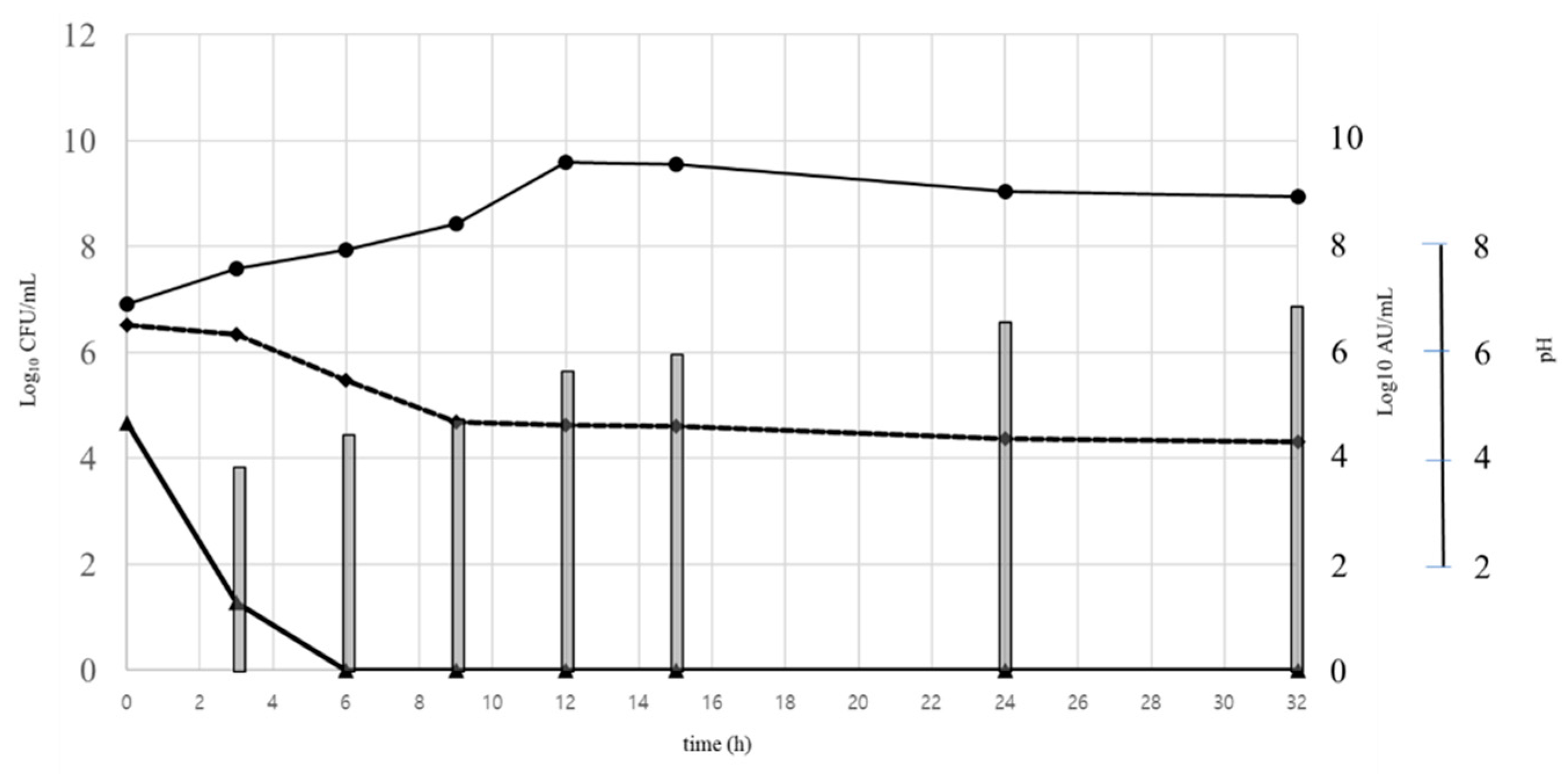
When grown in co-culture with L. ivanovii subsp. ivanovii ATCC 19119, E. casseliflavus ST192Gu was grown well and able to produce the bacteriocins in similar levels as compared with fermentation in monoculture. High cell numbers on MRS plates (E. casseliflavus ST192Gu and L. ivanovii subsp. ivanovii ATCC19119) were recorded representing the total cells recorded in the experimental set-up (Figure 5). However, when media selective for Listeria spp. was applied for the enumeration, a decrease of Listeria ivanovii subsp. ivanovii ATCC 19119 was indicated from 4.7 × 10⁴ CFU/mL to 1.9 × 10 CFU/mL in the first 3 h and to undetectable levels after 6 h and further during the monitored experimental time period (Figure 5). This indicated that the recorded viable cells on MRS plates were representing the population of E. casseliflavus ST192Gu. Inhibition of L. ivanovii subsp. ivanovii ATCC19119 most probably needs to be associated with production of the bacteriocin ST192Gu since production of lactic acid and the thus associated decrease in culture pH was not significant in the first hours of the fermentation process, where decline of listerial cells was already observed, and only after 12 h of fermentation, more relevant reduction of pH was observed (Figure 5). Moreover, as previously indicated, Listeria spp. are generally acid tolerant [51]. Levels of bacteriocin activity produced by E. casseliflavus ST192Gu in fact could be higher since some of the active peptides remain bound to the cell surface of the target listerial strain or even can be affected by the production of some proteases by L. ivanovii subsp. ivanovii ATCC 19119 that can be increased after cell lysis of dead listerial cells. The latter phenomenon has previously been reported by Yildirim et al. [48].

3.7.7. Combined Application of Ciprofloxacin and Bacteriocin on Growth of L. ivanovii subsp. ivanovii ATCC 19119

In the following experiment, the combined application of ciprofloxacin and bacteriocin ST192Gu was evaluated with the intention of detecting potential antagonistic synergetic interactions. As the first step of this evaluation, the MIC (minimal inhibition concentration) for the applied antibiotic (ciprofloxacin) was determined to be 80 μg/mL versus L. ivanovii subsp. ivanovii ATCC 19119. However, when ciprofloxacin was applied in combination with bacteriocin ST192Gu, synergism was observed, and inhibition was detected in concentrations of ciprofloxacin below the MIC (Figure 6). Based on observed inhibitory levels of L. ivanovii subsp. ivanovii ATCC 19119 in performed experiments, it can be suggested that lower levels of ciprofloxacin (below MIC e.g., 40 μg/mL or even less) can be effective in inhibition of Listeria spp. and/or other relevant foodborne and clinical pathogens when administered in combination with the bacteriocin ST192Gu. As results of applied combination of ciprofloxacin and bacteriocin ST192Gu, growth inhibition of L. ivanovii subsp. ivanovii ATCC 19119 was detected during the first 12h and this corresponds with the general recommendations for application of antimicrobials in human and veterinary medical practices. A similar research approach was already reported by Todorov and Dicks [7] regarding the synergetic effects of the combined application between ciprofloxacin and bacteriocin produced by P. pentosaceus ST44AM, supporting further investigation of this approach to more effectively control these infections in humans and other animals and potentially reducing the application of antibiotics. In a similar study, Minahk et al. [52] have explored potential applications of sub-lethal concentrations of enterocin CRL35 to increase the activity of erythromycin, chloramphenicol, and tetracycline. Achieving membrane depolarization of L. monocytogenes has been considered to be an essential initial step in fostering the inhibitory activity of different antimicrobials. However, often this is not sufficient to achieve cell death when antibiotics are applied as the sole therapeutic agent and the supplementary presence of a bacteriocin can potentially enhance the killing effect of antibiotics by augmenting this primary step of the antimicrobial mode of action [7,52].

3.8. Determination of Approximate Molecular Weight of Bacteriocin ST192Gu by SDS-PAGE

Enterocins are a very diverse group of antimicrobial peptides, most having linear structures, but some also reported to be cyclic peptides. Molecular weights are typically from 2–3 kDa, as for enterocin MC13 [53], and are reported to be thermosensitive [54]. Valledor et al. [25] reported two bacteriocins produced by different E. faecium strains having molecular weights of around 4–6 kDa. In the present study, the bacteriocin produced by E. casseliflavus ST192Gu was evaluated to be 4.8 kD, based on Tricine-SDS-PAGE. Similar results are reported for the molecular weights of other enterocins. Yamamoto et al. [55] found enterocin RJ-11 to be 5 kDa and Tulini et al. [56] reported E. faecium 130 to produce 3.5 and 6.5 kDa bacteriocins. Other reports include the 4.8 kDa enterocin A [57], the 5.6 kDa enterocin B [58], and the 4.5 kDa enterocin P [42]. For more precise identification and characterization of the expressed antimicrobial peptide, further purification, amino acid sequencing, and spectrometry is required. Nevertheless, the findings of the present study indicate that the antimicrobial activity expressed by E. casseliflavus ST192Gu is a small peptide, probably belonging to the class II bacteriocin family.

4. Conclusions

This appears to be the first report of the detection of E. casseliflavus associated with guava. The isolation of strains with antimicrobial properties and further characterization of the bioactive agents responsible for their antagonistic activity is opening new perspectives in the selection of potent antimicrobials with applications in food safety and medical (human and veterinary) practices. The urgent need for new antimicrobials is a scientific fact, especially for agents that are effective against multidrug-resistant pathogens. Bacteriocins clearly have immense potential as alternatives or as adjuncts for combating emerging pathogens. In this context, bacteriocin ST192Gu, isolated and partially purified from E. casseliflavus strain ST192Gu, as shown in the present study to act synergistically with ciprofloxacin to effect growth inhibition of L. ivanovii subsp. ivanovii ATCC19119, can be considered to be a candidate having good potential for further investigation.

Author Contributions

Conceptualization, S.D.T.; methodology, S.D.T.; formal analysis, S.D.T.; resources, S.D.T. and W.H.H.; writing—original draft preparation, S.D.T., W.H.H. and J.R.T.; writing—review and editing, W.H.H. and J.R.T.; funding acquisition, S.D.T. and W.H.H. All authors have read and agreed to the published version of the manuscript.

Funding

This research was funded by FAPESP (Grant 2013/07914-8), and Visiting Professor at University of Sao Paulo, Sao Paulo, SP, Brazil (2016.1.920.93) for providing scholarships for visiting professors. The APC was covered by MDPI.

Institutional Review Board Statement

Not applicable.

Informed Consent Statement

Not applicable.

Data Availability Statement

All data generated by the current project is available upon request.

Acknowledgments

To Mia Miau for her unconditional support.

Conflicts of Interest

The research was conducted in the absence of any commercial or financial relationships that could be construed as a potential conflict of interest.

References

  1. Telhig, S.; Ben Said, L.; Zirah, S.; Fliss, I.; Rebuffat, S. Bacteriocins to thwart bacterial resistance in Gram-negative bacteria. Front. Microbiol. 2020, 11, 586433. [Google Scholar] [CrossRef]
  2. Yang, S.C.; Lin, C.H.; Sung, C.T.; Fang, J.Y. Antibacterial activities of bacteriocins: Application in foods and pharmaceuticals. Front. Microbiol. 2014, 5, 241, Erratum in: Front. Microbiol. 2014, 5, 683. [Google Scholar] [CrossRef] [PubMed] [Green Version]
  3. Favaro, L.; Penna, A.L.B.; Todorov, S.D. Bacteriocinogenic LAB from cheeses—Application in biopreservation? Trends Food Sci. Technol. 2015, 41, 37–48. [Google Scholar] [CrossRef]
  4. Haider, T.; Pandey, V.; Behera, C.; Kumar, P.; Gupta, P.N.; Soni, V. Nisin and nisin-loaded nanoparticles: A cytotoxicity investigation. Drug. Dev. Ind. Pharm. 2022, 48, 310–321. [Google Scholar] [CrossRef] [PubMed]
  5. Shin, J.M.; Gwak, J.W.; Kamarajan, P.; Fenno, J.C.; Rickard, A.H.; Kapila, Y.L. Biomedical applications of nisin. J. Appl. Microbiol. 2016, 120, 1449–1465. [Google Scholar] [CrossRef] [PubMed] [Green Version]
  6. Field, D.; Blake, T.; Mathur, H.; O’Connor, P.M.; Cotter, P.D.; Paul Ross, R.; Hill, C. Bioengineering nisin to overcome the nisin resistance protein. Mol. Microbiol. 2019, 111, 717–731. [Google Scholar] [CrossRef]
  7. Todorov, S.D.; Dicks, L.M. Bacteriocin production by Pediococcus pentosaceus isolated from marula (Scerocarya birrea). Int. J. Food Microbiol. 2009, 132, 117–126. [Google Scholar] [CrossRef]
  8. Todorov, S.D.; Prévost, H.; Lebois, M.; Dousset, X.; LeBlanc, J.G.; Franco, B.D.G.M. Bacteriocinogenic Lactobacillus plantarum ST16Pa isolated from papaya (Carica papaya)—from isolation to application: Characterization of a bacteriocin. Food Res. Int. 2011, 44, 1351–1363. [Google Scholar] [CrossRef]
  9. Soltani, S.; Zirah, S.; Rebuffat, S.; Couture, F.; Boutin, Y.; Biron, E.; Subirade, M.; Fliss, I. Gastrointestinal stability and cytotoxicity of bacteriocins from Gram-positive and Gram-negative bacteria: A comparative in vitro study. Front. Microbiol. 2022, 12, 780355. [Google Scholar] [CrossRef]
  10. Kaur, S.; Kaur, S. Bacteriocins as potential anticancer agents. Front. Pharmacol. 2015, 6, 272. [Google Scholar] [CrossRef] [Green Version]
  11. dos Santos, K.M.O.; de Matos, C.R.; Salles, H.O.; Franco, B.D.G.D.M.; Arellano, K.; Holzapfel, W.H.; Todorov, S.D. Exploring beneficial/virulence properties of two dairy-related strains of Streptococcus infantarius subsp. infantarius. Prob. Antimicrob. Prot. 2020, 12, 1524–1541. [Google Scholar] [CrossRef]
  12. De Vos, P.; Garrity, G.M.; Jones, D.; Kreig, N.R.; Ludwig, W.; Rainey, F.A.; Schleifel, K.-H.; Whitman, W.B. Bergey’s manual of systematic bacteriology. In The Firmicutes, 2nd ed.; Springer: Dordrecht, The Netherlands, 2009; Volume 3. [Google Scholar]
  13. de Moraes, G.M.D.; de Abreu, L.R.; do Egito, A.S.; Salles, H.O.; da Silva, L.M.F.; Nero, L.A.; Todorov, S.D.; dos Santos, K.M.O. Functional properties of Lactobacillus mucosae strains isolated from Brazilian goat milk. Prob. Antimicrob. Prot. 2016, 9, 235–245. [Google Scholar] [CrossRef]
  14. Ke, D.; Picard, F.J.; Martineau, F.; Ménard, C.; Roy, P.H.; Onellette, M.; Bergeron, M.G. Development of a PCR assay for rapid detection of Enterococci. J. Clin. Microbiol. 1999, 37, 3497–3503. [Google Scholar] [CrossRef] [Green Version]
  15. Fugaban, J.I.I.; Vazquez Bucheli, J.E.; Holzapfel, W.H.; Todorov, S.D. Characterization of partially purified bacteriocins produced by Enterococcus faecium strains isolated from soybean paste active against Listeria spp. and Vancomycin-resistant enterococci. Microorganisms 2021, 9, 1085. [Google Scholar] [CrossRef]
  16. Yang, R.; Johnson, M.C.; Ray, B.I.B.E.K. Novel method to extract large amounts of bacteriocins from lactic acid bacteria. Appl. Environ. Microbiol. 1992, 58, 3355–3359. [Google Scholar] [CrossRef] [Green Version]
  17. Metivier, A.; Pilet, M.-F.; Dousset, X.; Sorokine, O.; Anglade, P.; Zagorec, M.; Piard, J.-C.; Marlon, D.; Cenatiempo, Y.; Fremaux, C. Divercin V41, a new bacteriocin with two disulphide bonds produced by Carnobacterium divergens V41: Primary structure and genomic organization. Microbiology 1998, 144, 2837–2844. [Google Scholar] [CrossRef] [PubMed] [Green Version]
  18. Favaro, F.; Basaglia, M.; Casella, S.; Hue, I.; Dousset, X.; Franco, B.D.G.M.; Todorov, S.D. Bacteriocinogenic potential and safety evaluation of non starter Enterococcus faecium strains isolated from home made white brine cheese. Food Microbiol. 2014, 38, 228–239. [Google Scholar] [CrossRef] [PubMed]
  19. Todorov, S.D.; Wachsman, M.; Tomé, E.; Dousset, X.; Destro, M.T.; Dicks, L.M.T.; Franco, B.D.G.M.; Vaz-Velho, M.; Drider, D. Characterisation of an antiviral pediocin-like bacteriocin produced by Enterococcus faecium. Food Microbiol. 2010, 27, 869–879. [Google Scholar] [CrossRef] [PubMed]
  20. Hussain, S.Z.; Naseer, B.; Qadri, T.; Fatima, T.; Bhat, T.A. Guava (Psidium Guajava)- Morphology, Taxonomy, Composition and Health Benefits. In Fruits Grown in Highland Regions of the Himalayas; Syed, Z.H., Bazila, N., Tahiya, Q., Tabasum, F., Tashooq, A.B., Eds.; Springer: Cham, Switzerland, 2021. [Google Scholar] [CrossRef]
  21. Fugaban, J.I.I.; Holzapfel, W.H.; Todorov, S.D. The overview of natural by-products of beneficial lactic acid bacteria as promising antimicrobial agents. Appl. Food Biotechnol. 2002, 9, 127–143. [Google Scholar]
  22. Fugaban, J.I.I.; Bucheli, J.E.V.; Park, Y.J.; Suh, D.H.; Jung, E.S.; Franco, B.D.G.M.; Ivanova, I.V.; Holzapfel, W.H.; Todorov, S.D. Antimicrobial properties of Pediococcus acidilactici and Pediococcus pentosaceus isolated from silage. J. Appl. Microbiol. 2022, 132, 311–330. [Google Scholar] [CrossRef]
  23. Micallef, S.A.; Goldstein, R.E.; George, A.; Ewing, L.; Tall, B.D.; Boyer, M.S.; Joseph, S.W.; Sapkota, A.R. Diversity, distribution and antibiotic resistance of Enterococcus spp. recovered from tomatoes, leaves, water and soil on U.S. Mid-Atlantic farms. Food Microbiol. 2013, 36, 465–474. [Google Scholar] [CrossRef] [PubMed]
  24. Vasilakopoulou, A.; Vourli, S.; Siafakas, N.; Kavatha, D.; Tziolos, N.; Pournaras, S. Enterococcus casseliflavus bacteraemia in a patient with chronic renal disease. Infect. Dis. Rep. 2020, 12, 70–73. [Google Scholar] [CrossRef] [PubMed]
  25. Valledor, S.J.D.; Dioso, C.M.; Bucheli, J.E.V.; Park, Y.J.; Suh, D.H.; Jung, E.S.; Kim, B.; Holzapfel, W.H.; Todorov, S.D. Characterization and safety evaluation of two beneficial, enterocin-producing Enterococcus faecium strains isolated from kimchi, a Korean fermented cabbage. Food Microbiol. 2022, 102, 103886. [Google Scholar] [CrossRef] [PubMed]
  26. Floch, M.H.; Madsen, K.K.; Jenkins, D.J.; Guandalini, S.; Katz, J.A.; Onderdonk, A.; Walker, W.A.; Fedorak, R.N.; Camilleri, M. Recommendations for probiotic use. J. Clin. Gastroenterol. 2006, 40, 275–278. [Google Scholar] [CrossRef] [PubMed]
  27. Suvorov, A. What is wrong with enterococcal probiotics? Probiotics Antimicrob. Proteins 2020, 12, 1–4. [Google Scholar] [CrossRef] [PubMed]
  28. Chikindas, M.L.; Weeks, R.; Drider, D.; Chistyakov, V.A.; Dicks, L.M. Functions and emerging applications of bacteriocins. Curr. Opin. Biotechnol. 2018, 49, 23–28. [Google Scholar] [CrossRef] [PubMed]
  29. Lewus, C.B.; Kaiser, A.; Montville, T.J. Inhibition of food-borne bacteria pathogens by bacteriocins from lactic acid bacteria isolated from meat. Appl. Environ. Microbiol. 1991, 57, 1683–1688. [Google Scholar] [CrossRef] [Green Version]
  30. Todorov, S.D. Diversity of bacteriocinogenic lactic acid bacteria isolated from boza, a cereal-based fermented beverage from Bulgaria. Food Control 2010, 21, 1011–1021. [Google Scholar] [CrossRef]
  31. Ouwehand, A.C.; Vesterlund, S. Antimicrobial components from lactic acid bacteria. In Lactic Acid Bacteria Microbiological and Functional Aspects, 3rd ed.; Revised and Expanded; Taylor & Francis Group: London, UK, 2004; pp. 375–395. [Google Scholar]
  32. Heng, N.C.K.; Wescombe, P.A.; Burton, J.P.; Jack, R.W.; Tagg, J.R. The diversity of bacteriocins in gram-positive bacteria. In Bacteriocins; Riley, M.A., Chavan, M.A., Eds.; Springer: Berlin/Heidelberg, Germany, 2007; pp. 45–92. [Google Scholar] [CrossRef]
  33. Wu, Y.; Pang, X.; Wu, Y.; Liu, X.; Zhang, X. Enterocins: Classification, synthesis, antibacterial mechanisms and food applications. Molecules 2022, 27, 2258. [Google Scholar] [CrossRef]
  34. Cintas, L.M.; Casaus, P.; Holo, H.; Hernandez, P.E.; Nes, I.F.; Håvarstein, L.S. Enterocins L50A and L50B, two novel bacteriocins from Enterococcus faecium L50, are related to staphylococcal hemolysins. J. Bacteriol. 1998, 180, 1988–1994. [Google Scholar] [CrossRef] [Green Version]
  35. De Kwaadsteniet, M.; Todorov, S.D.; Knoetze, H.; Dicks, L.M.T. Characterization of a 3 944 Dalton bacteriocin, produced by Enterococcus mundtii ST15, with activity against Gram-positive and Gram-negative bacteria. Int. J. Food Microbiol. 2005, 105, 433–444. [Google Scholar] [CrossRef]
  36. Todorov, S.D.; Wachsman, M.B.; Knoetze, H.; Meincken, M.; Dicks, L.M.T. An antibacterial and antiviral peptide produced by Enterococcus mundtii ST4V isolated from soybeans. Int. J. Antimicrob. Agents 2005, 25, 508–513. [Google Scholar] [CrossRef] [PubMed]
  37. Todorov, S.D.; Danova, S.T.; Van Reenen, C.A.; Meincken, M.; Dinkova, G.; Ivanova, I.V.; Dicks, L.M.T. Characterization of bacteriocin HV219, produced by Lactococcus lactis subsp. lactis HV219 isolated from human vaginal secretions. J. Basic Microbiol. 2006, 46, 226–238. [Google Scholar] [PubMed]
  38. Todorov, S.D.; Franco, B.D.G.M.; Tagg, J.R. Bacteriocins of Gram-positive bacteria having activity spectra extending beyond closely related species. Benef. Microbs. 2019, 10, 315–328. [Google Scholar] [CrossRef] [PubMed]
  39. Umu, Ö.C.O.; Rudi, K.; Diep, D.B. Modulation of the gut microbiota by prebiotic fibres and bacteriocins. Microb. Ecol. Health. Dis. 2017, 28, 1348886. [Google Scholar] [CrossRef] [PubMed] [Green Version]
  40. Parente, E.; Ricciardi, A. Production, recovery and purification of bacteriocins from lactic acid bacteria. Appl. Microbiol. Biotechnol. 1999, 52, 628–638. [Google Scholar] [CrossRef]
  41. Yamashita, H.; Tomita, H.; Inoue, T.; Ike, Y. Genetic organization and mode of action of a novel bacteriocin, bacteriocin 51: Determinant of VanA-type vancomycin-resistant Enterococcus faecium. Antimicrob. Agents Chemother. 2011, 55, 4352–4360. [Google Scholar] [CrossRef] [PubMed] [Green Version]
  42. Ennahar, S.; Sashihara, T.; Sonomoto, K.; Ishizaki, A. Class IIa bacteriocins: Biosynthesis, structure and activity. FEMS Microbiol. Rev. 2000, 24, 85–106. [Google Scholar] [CrossRef]
  43. Todorov, S.; Onno, B.; Sorokine, O.; Chobert, J.; Ivanova, I.; Dousset, X. Detection and characterization of a novel antibacterial substance produced by Lactobacillus plantarum ST31 isolated from sourdough. Int. J. Food Microbiol. 1999, 48, 167–177. [Google Scholar] [CrossRef]
  44. Bhugaloo-Vial, P.; Grajek, W.; Dousset, X.; Boyaval, P. Continuous bacteriocin production with high cell density bioreactors. Enzym. Microb. Technol. 1997, 21, 450–457. [Google Scholar] [CrossRef]
  45. Song, D.; Zhu, M.; Gu, Q. Purification and characterization of plantaricin ZJ5, a new bacteriocin produced by Lactobacillus plantarum ZJ5. PLoS ONE 2014, 9, e105549. [Google Scholar] [CrossRef] [PubMed]
  46. Surovtsev, V.; Borzenkov, V.; Levchuk, V. Purification of bacteriocins by chromatographic methods. Appl. Biochem. Microbiol. 2015, 51, 881–886. [Google Scholar] [CrossRef]
  47. Bastos, M. do C.; Coelho, M.L.; Santos, O.C. Resistance to bacteriocins produced by Gram-positive bacteria. Microbiol. 2015, 161 Pt 4, 683–700. [Google Scholar] [CrossRef] [PubMed] [Green Version]
  48. Todorov, S.D.; Dicks, L.M.T. Screening for bacteriocin producer lactic acid bacteria from boza, a traditional cereal beverage from Bulgaria. Characterization of produced bacteriocins. Process Biochem. 2006, 41, 11–19. [Google Scholar] [CrossRef]
  49. Yıldırım, Z.; Avşar, Y.K.; Yıldırım, M. Factors affecting the adsorption of buchnericin LB, a bacteriocin produced by Lactocobacillus buchneri. Microbiol. Res. 2002, 157, 103–107. [Google Scholar] [CrossRef] [PubMed]
  50. Bhunia, A.K.; Johnson, M.C.; Ray, B.; Kalchayanand, N. Mode of action of pediocin AcH from Pediococcus acidilactici H on sensitive bacterial strains. J. Appl. Bacteriol. 1991, 70, 25–33. [Google Scholar] [CrossRef]
  51. Gandhi, M.; Chikindas, M.L. Listeria: A foodborne pathogen that knows how to survive. Int. J. Food Microbiol. 2007, 113, 1–15. [Google Scholar] [CrossRef]
  52. Minahk, C.J.; Dupuy, F.; Morero, R.D. Enhancement of antibiotic activity by sub-lethal concentrations of enterocin CRL35. J. Antimicrob. Chemother. 2004, 53, 240–246. [Google Scholar] [CrossRef] [Green Version]
  53. Satish, K.R.; Kanmani, P.; Yuvaraj, N.; Paari, K.A.; Pattukumar, V.; Arul, V. Purification and characterization of enterocin MC13 produced by a potential aquaculture probiont Enterococcus faecium MC13 isolated from the gut of Mugil cephalus. Can. J. Microbiol. 2011, 57, 993–1001. [Google Scholar] [CrossRef]
  54. Krämer, J.; Henning, B. Mode of Action of Two Streptococcus faecium Bacteriocins. Antimicrob. Agents Chemoth. 1975, 7, 117–120. [Google Scholar] [CrossRef] [Green Version]
  55. Yamamoto, Y.; Togawa, Y.; Shimosaka, M.; Okazaki, M. Purification and characterization of a novel bacteriocin produced by Enterococcus faecalis strain RJ-11. Appl. Environ. Microbiol. 2003, 69, 5746–5753. [Google Scholar] [CrossRef] [PubMed]
  56. Tulini, F.L.; Gomes, B.C.; de Martins, E.C.P. Partial purification and characterization of a bacteriocin produced by Enterococcus faecium 130 isolated from mozzarella cheese. Food Sci. Technol. 2011, 31, 155–159. [Google Scholar] [CrossRef] [Green Version]
  57. Aymerich, T.; Holo, H.; Havarstein, L.S.; Hugas, M.; Garriga, M.; Nes, I. Biochemical and genetic characterization of enterocin A from Enterococcus faecium, a new antilisterial bacteriocin in the pediocin family of bacteriocins. Appl. Environ. Microbiol. 1996, 62, 1676–1682. [Google Scholar] [CrossRef] [PubMed] [Green Version]
  58. du Toit, M.; Franz, C.M.; Dicks, L.M.; Holzapfel, W.H. Preliminary characterization of bacteriocins produced by Enterococcus faecium and Enterococcus faecalis isolated from pig faeces. J. Appl. Microbiol. 2000, 88, 482–494. [Google Scholar] [CrossRef] [PubMed]
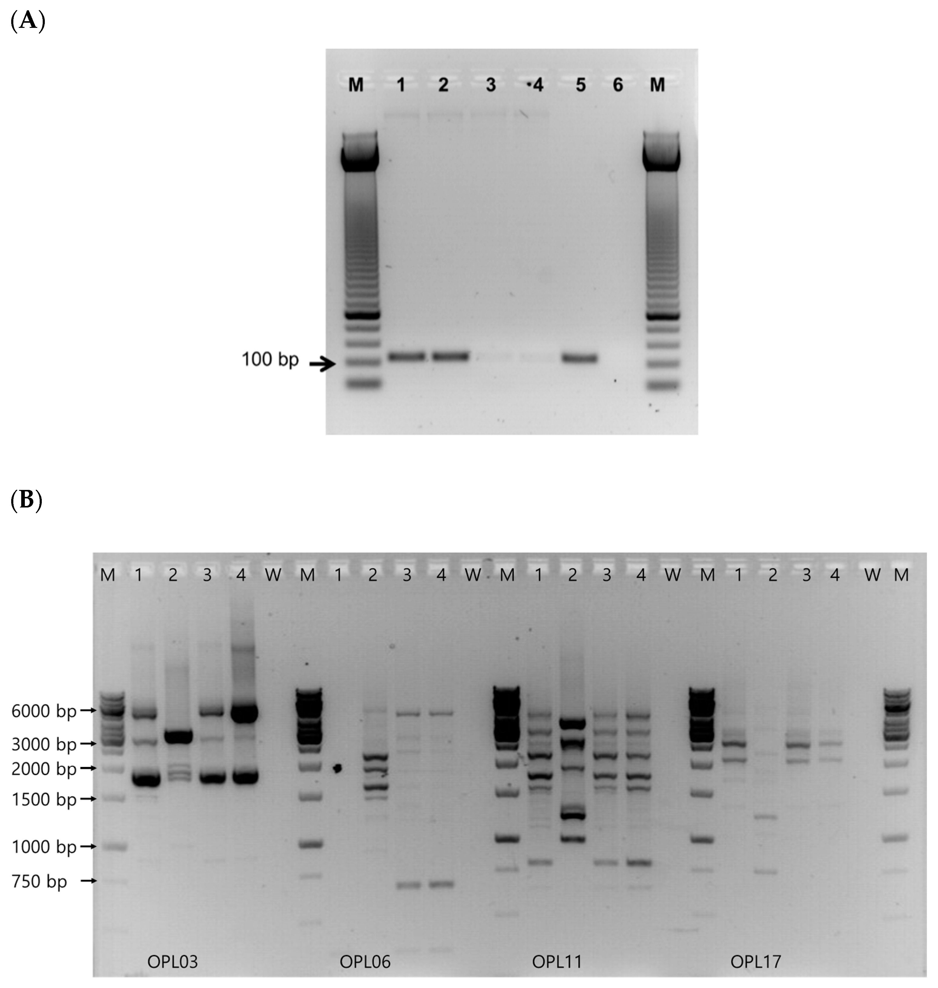
Figure 1. (A) Agarose gels showing DNA fragments obtained after PCR with genus-specific primers. Lanes 1 and 2: isolate ST192Gu, lanes 3 and 4: Lb. plantarum ATCC 14917, lane 5: E. faecium ATCC 19443, lane 6: no DNA loaded, lane M: O’GeneRulertm 50-bp DNA Ladder (Fermentas). (B) RAPD-PCR with primers OPL03 (5′-CAT AGA GCG G-3′), OPL6 (5′-GAG GGA AGA G-3′), OPL11 (5′-ACG ATG AGC C-3′), and OPL17 (5′-AGC CTG AGC C-3′), according to de Moraes et al. [13] on the profiles of 4 bacterial isolates with antimicrobial activity obtained in the screening process for new bacteriocinogenic strains isolated from guava. M: 1 kb ladder (Thermo Fisher), W: no DNA added to the PCR reaction, 1–4: isolates evaluated in present study.
Fermentation 09 00226 g001
Figure 2. Growth kinetics of Enterococcus casseliflavus ST192Gu (▲ 26 °C, ● 30 °C, ◆ 37 °C): Acidification (A), dynamic of bacterial growth (B), and bacteriocin production recorded as log10 AU/mL against (C) L. ivanovii subsp. ivanovii ATCC 19119, (D) L. monocytogenes ATCC 15313, and (E) E. faecalis ATCC 19443 (white histograms at 26 °C, grey histograms at 30 °C, and black histograms at 37 °C). Results are the means of two replicates and each with two repeats with standard deviation, if any, always less than 2%.
Fermentation 09 00226 g002
Figure 3. Cell lysis of Listeria ivanovii subsp. ivanovii ATCC 19119 (▲), Listeria monocytogenes ATCC 15313 (◆), and Enterococcus faecalis ATCC 19443 (●) (solid lines) in presence of bacteriocin produced by Enterococcus casseliflavus ST192Gu. Bacteriocins were added 3 h after initial incubation of test microorganisms. Growth of test organisms without addition of studied bacteriocins served as controls (dotted lines). Results are the means of two replicates and each with two repeats with standard deviation, if any, always less than 2%.
Fermentation 09 00226 g003
Figure 4. Cell lysis (%) of L. ivanovii subsp. ivanovii ATCC 19119, Listeria monocytogenes ATCC 15313, and Enterococcus faecalis ATCC 19443 evaluated after 3, 6, 9, and 24 h incubation time in the presence of semi-purified bacteriocin produced E. casseliflavus ST192Gu applied from 400 AU/mL to 25,600 AU/mL illustrating the effect of different concentrations of the studied bacteriocin on killing of applied test organisms and the role of contact time between the studied antimicrobials and evaluated test microorganism. Results are the average of three independent experiments and SD are less than 4%.
Fermentation 09 00226 g004
Figure 5. Combined growth of Enterococcus casseliflavus ST192Gu and Listeria ivanovii subsp. ivanovii ATCC 19119. Monitoring of bacterial growth on MRS agar (●); on RapidMono selective for Listeria spp. (▲), changes in pH (◆), and bacteriocin production (histogram) activity recorded against L. ivanovii subsp. ivanovii ATCC 19119.
Fermentation 09 00226 g005
Figure 6. Combined effect of ciprofloxacin (from 0 to 320 μg/mL) and semi-purified bacteriocin produced by Enterococcus casseliflavus ST192Gu (from 1.31 × 10³ to 3.0 × 10⁻¹ AU/mL) on growth of L. ivanovii subsp. ivanovii ATCC 19119 as presented for 4 h (A), 8 h (B), 12 h (C), and 30 h (D) of incubation at 37 °C.
Fermentation 09 00226 g006
Table 1. Antagonism of bacteriocin produced by Enterococcus casseliflavus ST192Gu against various target strains cultured at 37 °C. # numbers of test organisms sensitive to the evaluated bacteriocins from the total numbers of strains tested.
Target Strains Growth MediumInhibition #
Acinetobacter baumanniiBHI0/2
Bacteroides fragilisBHI0/1
Escherichia coliBHI0/4
Enterobacter cloacaeBHI0/1
E. faecalisMRS13/14
E. faeciumMRS9/9
Klebsiella pneumoniaeBHI0/2
Lactobacillus acidophilusMRS2/2
Lb. curvatusMRS0/4
Lb. delbrueckiiMRS0/6
Lb. fermentumMRS0/4
Lb. paracaseiMRS1/6
Lb. plantarumMRS0/10
Lb. rhamnosusMRS0/5
Lb. salivariusMRS0/2
Lb. sakeiMRS0/3
Lactococcus lactis subsp. lactisMRS1/1
Leuconostoc lactisBHI2/2
Listeria innocuaBHI6/6
L. ivanovii subsp. ivanovii BHI1/1
L. monocytogenesBHI27/27
Pediococcus acidilacticiMRS0/2
Pediococcus pentosaceusMRS1/6
Staphylococcus aureusBHI2/10
Staph. uberisBHI1/1
Streptococcus agalactiaeBHI0/3
Str. caprinusBHI2/2
Str. gallolyticus subsp. macedonicusMRS0/1
Str. infantarius subsp. infantariusMRS2/2
Str. pneumoniaeBHI0/5
Bacillus subtilisBHI0/5
Bacillus sp.BHI0/8
Table 2. Antibiotic susceptibility/resistance for Enterococcus casseliflavus ST192Gu based on antibiotic disk diffusion method. The results representing the average of the two repetitions performed, respectively, at 30 and 37 °C.
Antibiotics Inhibition (mm)
amoxacilin/clavulonic acid (30 μg per disk)30
ampicillin/sulbactam (20 μg per disk)32
bacitracin (10 units per disk)09
cefepime (30 μg per disk)15
cefotaxim (30 μg per disk)30
cefriaxon (30 μg per disk)34
ceftiofur (30 μg per disk)35
cefuroxim (30 μg per disk)31
chloramphenicol (30 μg per disk)32
ciprofloxacin (5 μg per disk)13
clindamycin (2 μg per disk)21
enrofloxacin (5 μg per disk)11
erytromycin (15 μg per disk)23
florfenicol (30 μg per disk) 12
gentamicin (10 μg per disk) 12
imipenem (10 μg per disk)21
levofloxacine (5 μg per disk) 12
neomycin N (10 μg per disk)10
penicillin (10 units per disk)23
tetracycline (30 μg per disk) 24
vancomycin (30 μg per disk)22
Disclaimer/Publisher’s Note: The statements, opinions and data contained in all publications are solely those of the individual author(s) and contributor(s) and not of MDPI and/or the editor(s). MDPI and/or the editor(s) disclaim responsibility for any injury to people or property resulting from any ideas, methods, instructions or products referred to in the content.