Article

Selection of Yeast and Lactic Acid Bacteria Strains, Isolated from Spontaneous Raw Milk Fermentation, for the Production of a Potential Probiotic Fermented Milk

1
Department of Agriculture, Food, Environment and Forestry (DAGRI), University of Florence, 50145 Florence, Italy
2
FoodMicroTeam s.r.l., 50125 Florence, Italy
*
Author to whom correspondence should be addressed.
Fermentation 2022, 8(8), 407; https://doi.org/10.3390/fermentation8080407
Submission received: 29 July 2022 / Revised: 17 August 2022 / Accepted: 18 August 2022 / Published: 19 August 2022
(This article belongs to the Special Issue High Quality Functional Food: Potential of Probiotics)

Abstract

:
Probiotic milk is a class of fermented milk that possesses health-promoting effects, not only due to the lactic acid bacteria (LAB) presence but potentially also to yeast activity. Hence, the aim of this work was to isolate and select yeasts from spontaneous milk fermentations to be used as inoculum, together with LAB, for manufacturing a potentially probiotic acidic low-alcohol fermented milk. Six yeast species were detected from the spontaneous milk fermentation. A screening of 13 yeast strains and 14 previously isolated LAB strains, based on the resistance to bile salts and to acidic conditions, was carried out. The best performing strains were successively tested for in vitro gastrointestinal tolerance. A strain of Kluyveromyces marxianus and a strain of Lactococcus lactis were selected for the manufacturing of two different fermented milk. The values of the main technological and microbiological parameters (pH, organic acids, ethanol, and microbial concentrations) of the experimental milk were in the range of those reported for this category of products. The evaluation of microorganism survival in fermented milk samples subjected to simulated gastrointestinal conditions highlighted a high resistance of both strains. In conclusion, the selected microbial starter culture enabled the setting up of potential probiotic fermented milk.

1. Introduction

According to the Food and Agriculture Organization (FAO), fermented milk is defined as a “product obtained by the fermentation of milk by the action of suitable microorganisms, which shall be viable, active and abundant in the product to the date of minimum durability and resulting in reduction of pH” [1]. Fermented milk is usually classified into two major groups based on the type of fermentation carried out by the occurring microbiota: lactic fermentation, where lactic acid bacteria (LAB) are the dominant species, and fungal–lactic fermentation, where, in addition to LAB, there are yeasts carrying out the alcoholic fermentation (e.g., kefir, Koumiss, Viili, etc.). Products by lactic fermentation can be further classified based on the LAB features into mesophilic fermented milk (e.g., buttermilk), thermophilic (e.g., yogurt, acidophilus milk), and probiotics products [2].
In regards to the technology of production, spontaneous fermentation is the oldest method of raw milk processing, as well as the use of backslopping method, where a part of the previous batch of a fermented product is used to inoculate the new batch. However, nowadays, especially for large-scale products, the use of selected starter cultures rather than traditional methods is widespread [3]. Among the criteria for starter selection, the microorganism probiotic potential is of particular interest for the manufacturing of fermented milk.
Probiotics are defined as microorganisms that, in adequate amounts, confer a health benefit on the host [4]; they should satisfy several safety, functional, and technological criteria in order to develop a probiotic food product. Probiotics should be viable during processing operation and storage, survive the gastrointestinal transit, and possess human- health-promoting effect, mainly through the maintenance of normal intestinal microflora, and are hence mitigation of the inflammatory bowel disease (IBD) and irritable bowel syndrome (IBS) symptoms, and protection against diarrhea [5,6,7]. The most commonly used probiotics include lactobacilli, and Bifidobacterium spp., followed by several strains of Lactococcus spp., Enterococcus spp., and Streptococcus spp. [8]. Lactobacilli are also widely employed due to their resistance to low pH, adaptation to milk and other food substrates, and their GRAS (generally regarded as safe) status.
Compared to bacteria, less attention has been given by researchers to probiotic yeasts, and a lower number of commercial preparations are available in the market. One of the advantages of yeasts is their natural resistance to antibacterial antibiotics, whereas the possible transfer of antibiotic resistance between lactobacilli and pathogenic bacteria constitutes a threat. Saccharomyces cerevisiae var. boulardii is the most common probiotic yeast distributed in the market presently. Several clinical trials have assessed its probiotic properties and ensured its use in the prevention and treatment of various forms of ailments, especially gastrointestinal diseases [9,10]. Shruthi et al. [11] have recently reviewed the potential probiotic properties of non-Saccharomyces species belonging to the genera Debaryomyces spp., Pichia spp., Yarrowia spp., Meyerozyma spp., Candida spp., and Kluyveromyces spp.
Among the yeast species, Kluyveromyces marxianus is one of the most emerging and promising non-Saccharomyces yeast for biotechnological applications thanks to its ability to utilize a broad range of sugars, including lactose, its thermotolerance, lytic enzymes secretion, and high growth rate [12,13]. This yeast species has been isolated from several matrices, often from dairy products such as kefir and cheese [14,15,16]. Moreover, the European Food Safety Authority (EFSA) has granted the QPS (qualified presumption of safety) status to only a few yeast species, which might be used as “food additive”, including K. marxianus [17].
In light of this, the work was initially targeted to isolate yeasts from spontaneous cow raw milk fermentations and to assess some potential probiotic features (resistance to bile salts, acidic condition, and in vitro digestion survival). The best performing strain was then used together with a lactic acid bacteria strain (selected based on the same probiotic features) for the manufacturing of a potentially probiotic acidic low-alcohol fermented milk.

2. Materials and Methods

2.1. Raw Milk Spontaneous Fermentation

Cow raw milk was provided by Centro di Ricerche Agro-Ambientali Enrico Avanzi, University of Pisa. In order to allow spontaneous fermentation and yeast population development, milk was divided into two sterile flasks and incubated at 30 and 40 °C for 48 h. In the beginning, after 24 and 48 h, in the spontaneously fermented milk, yeast populations were quantified, and the pH was measured by a pHmeter (Metrohm Italiana Srl, Varese, Italy).

2.2. Microorganism Enumeration

For microorganism enumeration, 1 mL of sample was homogenized with 9 mL of sterile saline solution (0.9% NaCl w/v), and serial dilutions were performed. The diluted suspensions of yeasts were plated on MYPG, a medium containing (in g/L): malt extract 5, yeast extract 3, meat extract 5, and glucose 10. For lactic acid bacteria enumeration MRS agar (Oxoid, Basingstoke, Hampshire, UK) and M17 agar (Oxoid, Basingstoke, Hampshire, UK) were used for lactobacilli and lactococci, respectively. Yeast colonies were counted after 24–48 h of incubation at 30 °C in aerobic conditions. LAB colonies were counted after 24–48 h of incubation at 30 °C under microaerophilic conditions in jars containing AnaeroGen enzymatic kit (Oxoid, Basingstoke, Hampshire, UK).

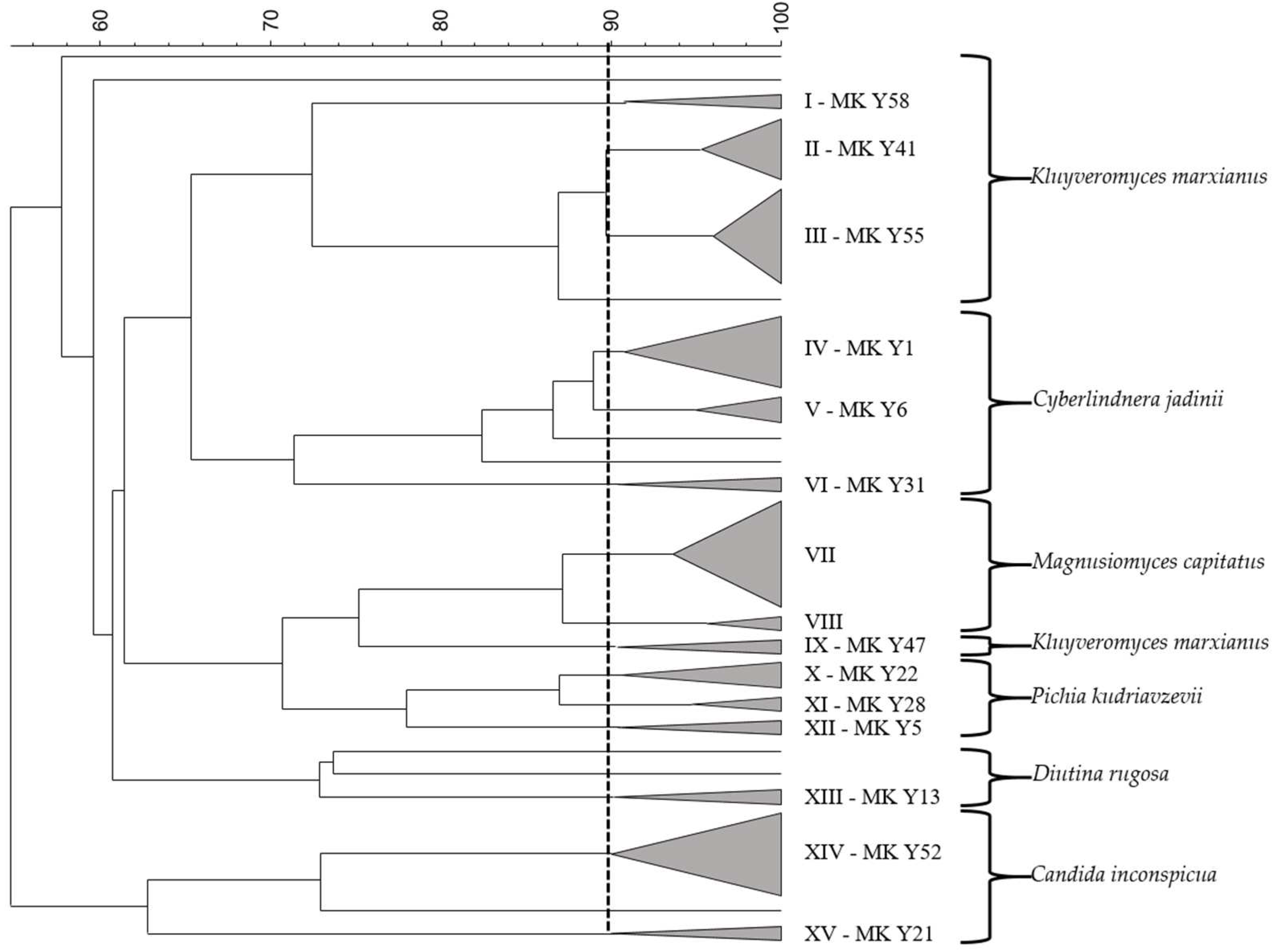
2.3. Identification and Typing of Yeasts

A total of 70 colonies from the various sampling points were picked up and subjected to molecular analysis. In order to identify and characterize the yeasts, a protocol according to Mari et al. [18] was followed. Initially, the isolates were subjected to Randomly amplified polymorphic DNA (RAPD) analysis by using the primer M13 (50-GAGGGTGGCGGTTCT-30) [19] and the PCR protocol according to Reguant and Bordons [20]. All PCR reactions included both negative (DNA-free) and positive controls and were processed in an Applied Biosystems® 2720 Thermal Cycler (Life Technologies, Monza, Italy). Pattern evaluation was made using Dice algorithm, and cluster analysis was carried out by the Unweighted Pair Group Method using Arithmetic Averages (UPGMA). Analysis was performed by using Gel Compar II 6.6 Software (Applied Maths NV, St-Martens-Latem, Belgium). At least two yeast isolates were chosen as representative of different RAPD patterns and were assayed by PCR–RFLP (Restriction Fragment Length Polymorphis) analysis of the rDNA-ITS (Internal transcribed spacer) region as described by Granchi et al. [21], using CfoI, HaeIII and HinfI (Fermentas Inc, Burlington, ON, Canada) as restriction endonucleases. In order to confirm the identification obtained by PCR–RFLP, the ITS rDNA region products were purified using Nucleo Spin Extract II (Macherey-Nagel GmbH & Co. KG, Düren, Germany) and sent to BMR Genomics (Padua, Italy) for sequencing. The sequence obtained in FASTA format was compared with those deposited in GenBank DNA database (http://www.ncbi.nlm.nih.gov/, accessed on 17 August 2022) using the basic BLAST search tools.

2.4. Microorganisms and Culture Condition

Yeast strains were aerobically cultured for 24 h at 30 °C in YEPD, a medium containing (in g/L): malt extract 5, yeast extract 3, meat extract 5, and glucose 10. LAB used in this experiment was isolated from spontaneous raw cow milk fermentation by Galli et al. [22]. The 14 strains were routinely propagated for 24–48 h in MRS medium at 30 °C before their utilization. LAB belonged to 6 species, Lc. lactis (MK L1, MK L37, MK L81, MK L82, MK L84, MK L86) Lactiplantibacillus rhamnosus (MK L20, MK L65), Lacticaseibacillus paracasei (MK L49, MK L53, MK L75), Lacticaseibacillus casei (MK L63), Enterococcus faecium (MK L64), and Limosilactobacillus fermentum (MK L56).

2.5. Yeast and Lactic Acid Bacteria Probiotic Features Assessment

LAB and yeasts were studied for their potentially probiotic characteristics. Before the bile salt and acidic tolerance assays, yeast strains were aerobically cultured for 24 h at 30 °C in YEPD; LAB strains were cultured for 24 h at 30 °C in MRS medium.

2.5.1. Bile Salt Tolerance

Cultures grown as described above were screened for their ability to withstand bile salts (BS). The yeast strains were inoculated 1% (v/v) on YEPD medium with or without BS at 0.3 and 1.0%. Growth was monitored by measuring absorbance at 600 nm using a spectrophotometer (V730, Jasco, Tokyo, Japan) in the beginning and after 3, 6, and 24 h. LAB were inoculated 1% (v/v) in MRS medium with the same two percentages of BS, 0.3 and 1.0%. As for yeasts, culture absorbance was periodically monitored.

2.5.2. Tolerance to Acidic Condition

Resistance to low pH was estimated according to Diosma et al. [14] with some modifications. Yeasts were grown in YEPD broth for 24 h at 30 °C, centrifuged, and resuspended at a concentration of 106 CFU/mL in sterile saline solution acidified to pH 3 with HCl. Samples were taken immediately (T0) and after 4 h (T4) at 37 °C, and serial dilutions were plated on MYPG agar in order to determine the number of viable cells. The same method was used for the lactic acid bacteria with the following differences, the initial inoculum was ca 108 CFU/mL, and MRS agar was used as medium.

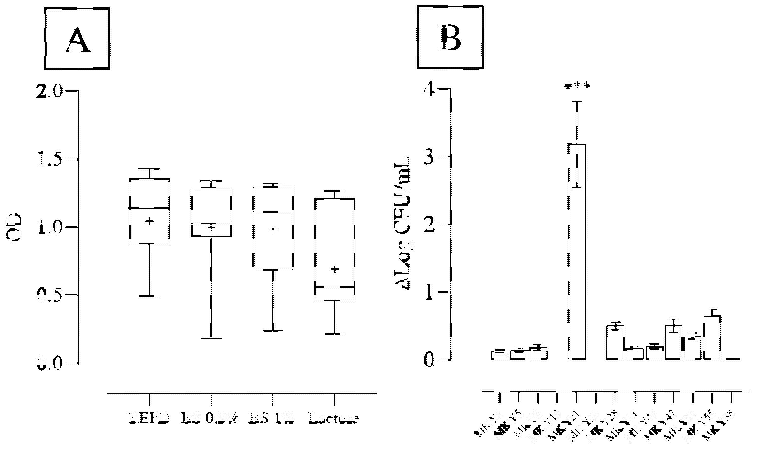
2.6. Lactose Utilization

Yeast growth was also tested on YEPD medium with lactose (YEPD-lactose) as unique carbon source. Cell cultures were inoculated 1% v/v, and the growth was monitored by measuring absorbance at 600 nm after 6, 24, and 30 h at 30 °C.

2.7. Resistance to Simulated Human Gastrointestinal (GI) Conditions

Simulated gastric juice and intestinal fluid were prepared according to Fernández et al. [23]. After washing in sterile saline solution and centrifugation, each cell suspension, grown for 24 h, was separately added to the simulated gastric juice (pH 3.0): LAB at 108 CFU/mL and yeasts at 106 CFU/mL. The microbial suspensions were then incubated at 37 °C under stirring conditions (100 rpm), and after 3 h, the enumeration of LAB and yeasts were carried out by serial dilutions. After 3 h in gastric juice, cultures were centrifuged, washed with sterile physiological solution, and resuspended in the simulated intestinal fluid for 4 h at 37 °C under stirring conditions. Intestinal transit tolerance was studied by determining the viable counts of LAB and yeasts by serial dilution and plate counts.

2.8. Manufacture of Fermented Milk and Microbial Viability during Storage

For the manufacture of fermented milk, raw cow milk was pre-treated, as reported by Galli et al. [22], before being inoculated by the microbial combination of LAB and yeast. LAB cultures, grown overnight in MRS broth, were centrifuged, washed with sterile saline solution, and resuspended in milk at a concentration of 107 CFU/mL. Yeasts, grown overnight in YEPD broth, were centrifuged, washed with sterile saline solution, and resuspended in milk at a concentration of 105 CFU/mL. Fermented milk was manufactured by using two methods: fermented milk obtained by the direct inoculum of the mixed starter culture (FM-S) and fermented milk obtained by the use of a previous fermented batch as inoculum (10% v/v) (FM-B). The inoculated milk was fermented for 24 h at 30 °C, cooled to 4–6 °C, and stored at 4 °C for 28 days. Fermented milk was analyzed in the end of the fermentation and during the storage to monitor acidification (pH values and organic acids concentration), LAB viability (on MRS agar medium), and yeast viability (on MYPG agar medium). To confirm the presence and dominance of the inoculated strains, RAPD-PCR was carried out.

2.9. Resistance to Simulated Gastrointestinal Conditions of Microorganisms in Fermented Milk

The resistance to simulated gastrointestinal conditions of microorganisms (LAB and yeasts) was also tested in the fermented milk. The assay was carried out according to Fernández et al. [23] with some modifications. Fermented milk was added to the gastric juice 10 % v/v (NaOH 1N in phosphate buffer, pH 2.4–2.7, and 3 g/L of pepsin). Samples were placed at 37 °C under stirring conditions. After 2 h (T2), microbiological analyses were carried out to determine LAB and yeast concentrations. The intestinal situation was simulated by modifying the pH to 5.4–5.7 and adding bile salts (10 g/L) and pancreatin (1 g/L). After 2 h (T4) at 37 °C, the viability of the microorganisms was assessed by serial dilution and plate counts. pH was modified to 7.2–7.4 and samples placed at 37 °C for 2 h (T6). In the end, microorganism enumeration was performed.

2.10. Organic Acid Determination by High-Pressure Liquid Chromatography (HPLC)

For the organic acid determination, fermented milk was centrifuged at 11,300× g for 10 min; supernatant was filtered by Amicon® Ultra-4 Centrifugal Filters (3.000 Da NMWL) (Merck Millipore, Burlington, MA, USA) before the injection. Organic acids were determined by HPLC analysis (Varian Inc, Palo Alto, CA, USA). Separation was obtained with a Rezex ROA organic acid H+ column (300 × 7.8 mm; Phenomenex, Castel Maggiore, Bologna, Italy), connected to a refractive index detector (Knauer K-2301, Knauer GmbH, Berlin, Germany) and UV detector (λ = 210). Elution was performed at 65 °C with 0.013 N H2SO4 eluent at flow rate of 0.6 mL min−1. Data were collected and analyzed by using the Galaxie software (Varian Inc, Palo Alto, CA, USA). Quantitative analysis was carried out by standard curves designed for each compound.

2.11. Statistical Analysis

The level of statistical significance was determined using one-way ANOVA (for multiple groups) followed by Tukey’s Test or Student’s t-test (for comparisons between two groups) (GraphPad Prism 8.0.1 software package). A p value of < 0.05 was considered to be significant.

3. Results and Discussion

3.1. Raw Cow Milk Spontaneous Fermentation and Yeast Identification

Raw cow milk was left to spontaneously ferment at 30 and 40 °C in order to allow the indigenous microbiota to develop. Microbiological analyses were carried out on raw milk after 24 and 48 h of fermentation to isolate yeast strains to be tested for potentially probiotic properties and technological features. The yeast population in raw milk was (1.26 ± 0.54) × 102 CFU/mL, in accordance with the range found by Lavoie et al. [24] of 1 to 3 log CFU/mL. After 24 h at 30 °C, yeasts developed up to (9.60 ± 0.85) × 104 CFU/mL, and further to (2.08 ± 0.46) × 105 CFU/mL after 48 h. Yeast growth was higher at 40 °C indeed after 24 h, yeast cell densities were (3.20 ± 0.28) × 105 CFU/mL and raised to (1.41 ± 0.43) × 106 CFU/mL after 48 h. pH decrease was also observed, likely due to the bacterial contamination of milk; indeed, after 48 h, the pH attained 4.12 at 30 °C and 3.56 at 40 °C. A total of 70 colonies of presumptive yeasts from the different sampling points were picked up and purified. Isolates were identified by combining RAPD-PCR, analysis, and sequencing of the rDNA-ITS region, following the protocol by Mari et al. [18]. Initially, isolates were characterized by RAPD-PCR with M13 primer. Cluster analysis of the obtained patterns generated the dendrogram reported in Figure 1. A similarity level of 90% was chosen on the basis of the reproducibility between different RAPD–PCR patterns for the same isolate. A total of 62 out 70 isolates were grouped into 15 clusters.
From each cluster, two isolates were chosen and identified using ITS-RLFP gene analysis and sequencing (Supplementary Table S1). The strains belonged to six species, the majority (7) to K. marxianus, followed by Cyberlindnera jadinii (5), Pichia kudriavzevii (3), Diutina rugosa (3), Candida inconspicua (2), and Magnusiomyces capitatus (2). K. marxianus MK Y50, MK Y40, MK Y48, C. jadinii MK Y2, and MK Y15, D. rugosa MK Y32 and MK Y49, and C. inconspicua MK Y23 isolates were not included in any clusters and showed a detection frequency below 2%; hence, they were not considered for the subsequent screening. In this regard, also M. capitatus strains were excluded. Indeed, although commonly found in normal human microflora, this species is known as an agent of human opportunistic infection by colonizing skin and mucosa of the respiratory and digestive tracts, moreover is a biosafety level two (BSL-2) microorganism [25]. Results of the identified species are in agreement with those reported by other authors. Delavenne et al. [26] indicated K. marxianus and Candida spp. (C. catenulata and C. inconspicua) as commonly encountered genera in cow and goat milk; Merchán et al. [27] found K. marxianus as dominant species in Spanish soft cheese, and they also detected P. kudrivazevii and C. jadinii; K. marxianus was found as predominant species also in traditional Brazilian cheese [28] and natural whey starter (NWS) for the PDO Parmigiano Reggiano cheese production [29]. The frequent detection of Kluyveromyces spp. in the dairy source is overall due to its ability to use lactose as a carbon source [13]. The majority of the K. marxianus isolates were found at a fermentation temperature of 30 °C, closer to its optimal growth temperature. D. rugosa and M. capitatatus species were found only at 40 °C, while the other species were detected at both temperatures. Among the isolated species, the qualified presumption of safety (QPS) status by the European Food Safety Authority [30] panel has been attributed only to C. jadinii and K. marxianus species. The number of studies on the isolation, characterization, and identification of non-Saccharomyces yeasts in dairy products has significantly increased together with the assessment of their potential probiotic features. In this experiment, the following 13 strains, developed from spontaneous milk fermentation, were tested for several probiotic features: K. marxianus MK 41, MK Y47, MK Y55, and MK Y58; C. jadinii MK Y1, MK Y6, and MK Y31; P. kudriavzevii MK Y5, MK Y22, MK Y28; D. rugosa MK Y13; C. inconspicua MK Y21, MK Y52.

3.2. Yeast Screening for Probiotic Features

Tolerance to low pH and bile salts is considered a prerequisite for probiotic strains to exert their beneficial effect on the human gut since they have to maintain viability after ingestion. In addition, due to their use in fermented milk, the capacity to metabolize lactose is of technological relevance. Thus, the 13 yeast strains were tested for their tolerance to two concentrations of bile salts (0.3 and 1%), to an acidic environment of pH 3 at 37 °C, that is the corporal temperature, and for their ability to ferment lactose. Results are reported in Figure 2.
Data obtained from these preliminary tests were elaborated according to percentile distribution. It is possible to observe that, in general, the addition of bile salts did not strongly affect yeast growth. The mean values were not statistically different, 1.0 ± 0.32 in YEPD, 1.00 ± 0.35 in YEPD with 0.3% BS, and 0.99 ± 0.35 in YEPD with 1% BS. The K. marxianus strains showed values above the 75th percentile for all the tested properties, whereas D. rugosa and C. inconspicua strains exhibited the worse performances. Indeed, for all the tested parameters, the recorded values of these two species were below the median value. The lowest value of growth in YEPD, 0.49, was shown by C. inconspicua MK Y52, and the highest, 1.43, by K. marxianus MK Y55. This strain also showed the highest value for the growth with lactose and with BS at 1%. In regards to lactose utilization, the increase in absorbance after 24 h was observed by the K. marxianus strains; on the contrary, the other species showed a low or no increase due to weak lactose utilization. A general high resistance of yeasts isolated from different food matrices and beverages to bile salts (0.3%), with the exception of C. inonspicua, was also observed by Gil-Rodríguez et al. [31]. In regard to D. rugosa, Wang et al. [32] tested the probiotic properties of D. rugosa ATCC 10,571 type strain and of a strain isolated from chicken feces; the latter showed better results in terms of bile salts resistance. Results reported by Merchán et al. [27] on strains belonging to Kluveromyces spp., Pichia spp., Candida spp., and Debaryomyces spp. highlighted a high growth of the Pichia and Kluveromyces genera in the presence of bile salts, even if dependent on the strain. Helmy et al. [33] individuated 2% of bile salts as a critical threshold for yeast growth; below this percentage, yeast showed high tolerance. Bile salt is toxic to the cell as it disorganizes the cell membrane structure; the bile resistance of microorganisms is related to bile salt hydrolase activity, which contributes to the reduction of the toxic effect of conjugated bile salts [34]. Together with bile salt tolerance, the resistance to acidic conditions is another requisite for a probiotic microorganism because yeasts have to survive the low pH that characterizes the human stomach particularly. The capacity to survive at a pH 3 by yeasts was assessed and reported in Figure 2B. Most of the strains were able to maintain their viability after being subjected to acidic conditions; indeed, the majority of the strains’ decrease in cell concentration was below 1 log unit. Only C. inconspicua MK Y21 strongly reduced its concentration of ca 3 log unit, additionally pointing out the scarce performance of these strains. In order to resist acidic conditions and general stress, the main strategies are the adjustment of yeast cell walls and the activation of the cell wall integrity [35]. When considering all the tested parameters, the best performing strains belonged all to K. marxianus species and were MK Y41, MK Y47, MK Y55, and MK Y58; thus, they were chosen for testing their resistance to simulated gastrointestinal conditions.

3.3. Resistance of Yeasts to Simulated Gastrointestinal (GI) Conditions

The K. marxianus strains were subjected to in vitro simulated gastrointestinal conditions; the results are reported in Figure 3.
The in vitro assay indicated a high resistance of all the strains to the gastrointestinal conditions. None of the strains showed a remarkable reduction, reflecting the observed resistance to acidic conditions, even though the stomach pH with high gastric juice secretion is usually around two, and bile salts displayed by these strains in the preliminary test. No differences were detected among the strains; however, we decided to choose the K. marxianus MK Y55 for fermented milk preparation because it was the most frequently detected strain in the spontaneous fermentation, representing 13% of the total isolates. Several strains of K. marxianus have been studied for their probiotic properties. Maccaferri et al. [36] highlighted the beneficial effect of a K. marxianus strain that was able to affect colonic microbiota, increasing the bifidobacterial concentration in the colonic model system, and was highly adhesive to human enterocyte-like Caco-2 cells. The capacity to adhere to Caco-2 by a K. marxianus strain isolated from kefir was also observed by Cho et al. [37], together with a high growth with bile salts (0.3%) and survival in acidic conditions above 50%. Xie et al. [38] investigated the hypocholesterolemic of a K. marxianus strain and found out that it was able to potentially reduce serum cholesterol and triglyceride levels in rats.

3.4. Lactic Acid Bacteria Screening for Probiotic Features

Lactic acid bacteria tested in this experiment were isolated by raw milk or spontaneously fermented cow milk, as reported in our previous work [22], and belonged to six species. Growth and acidification of the 14 lactic acid bacteria strains were monitored, testing the addition of bile salts (BS) at 0.3 and 1%, together with the resistance to an acidic environment for the yeasts. Data obtained from these assays were elaborated according to percentile distribution, reported in Figure 4.
A certain variability among the strains and the species was observed, and, as expected, the addition of bile salts negatively affected the bacterial growth and acidification; particularly, this negative effect was shown to a greater extent with the 1% of bile salts addition. The mean values of growth measured by the absorbance decreased from 1.09 ± 0.62 in MRS medium to 0.57 ± 0.29 in MRS with 0.3% BS and further to 0.31 ± 0.25 in MRS with 1% BS. Only L. fermentum MK L56 exhibited the OD values with both bile salts concentrations above the 75th percentile. Apart from Lc. lactis MK L1, the other lactococci strains growth was strongly inhibited by the presence of bile salts; none of the values recorded for these strains were above the mean value in the tested conditions; differently from the findings of Kondotiene et al. [39], who found tolerance of more than 80% also with 1% of bile salts of 33 Lc. lactis strains isolated from raw and fermented milk. This trend was also reflected in the acidification trial, where these strains led to a slight decrease in the pH media. Conversely, L. fermentum MK L56 showed the highest acidification in all the tested conditions, reaching a pH of 4.48 in 0.3% BS and 4.85 in 1% BS. The mean values for the pH increased from 4.23 ± 0.40 in the MRS medium to 4.80 ± 0.40 in MRS with 0.3% BS and further to 5.44 ± 0.41 in MRS with 1% BS. pH values below the median were observed in the media with 0.3% BS, inoculated by Lc. lactis MK L81, MK L82, MK L84, MK L86, E. faecium MK L64, L. rhamnosus MK L65, and L. paracasei MK L53. In regards to pH values in 1% BS medium, Lc. lactis MK L81, MK L82, MK L86, L. rhamnosus MK L65, and MK L20, L. paracasei MK L75 and MK L53, and L. casei MK L63, showed values below the median. As reported by Hyacinta et al. [40], the bile tolerance of LAB might be related to the activity of bile salts hydrolases (BSH) enzyme, which catalyzes the deconjugation of bile salts, helping to tolerate their antimicrobial effect in the small intestine and to reduce cholesterol level. Hernández-Gómez et al. [41] assessed the BSH activity of five probiotic LAB and pointed out that BSH content and substrate specificity depend strongly on the species and the strain.
Strains that showed at least three values out of the six tested parameters above the median were selected for further evaluation of the survival to acidic conditions (Figure 4B). Accordingly, the following eight strains were selected: Lc. lactis MK L1, L. rhamnosus MK L20, MK L65 L. paracasei MK L49, MK L75, L. fermentum MK L56, L. casei MK L63, E. faecium MK L64. In addition, Lc. lactis MK L84 and MK L37 were considered since both the strains have proven to be high GABA-producing strains, able to improve the nutritional value of fermented milk [22]. The data concerning the survival in acidic conditions (pH 3) of the selected strains are reported in Figure 4B, which shows the differences between the initial (ca 8 log CFU/mL) and final cell densities. The lowest decrease in cell concentration was detected for L. rhamnosus MK L20 and E. faecium MK L64, which were reduced to ca 2 log CFU/mL. This reduction was not statistically different from L. fermentum MK L56, L. casei MK L63, L. rhamnosus MK L65, and L. paracasei MK L75. The highest reduction, hence the lowest resistance to acidic conditions, was exhibited by Lc. lactis MK L84 and MK L37, in agreement with the previous results. Indeed, their concentration decreased by 4 and 5 log CFU/mL, respectively. The results are in agreement with those found by Faye et al. [42], who observed a lower tolerance to bile salts and acidic conditions by Lactococcus spp. compared to lactobacilli strains.

3.5. Resistance of Lactic Acid Bacteria to Simulated Gastrointestinal Conditions

The LAB strains were also subjected to in vitro simulated gastrointestinal conditions, as reported in Figure 5.
Among the 11 tested strains, only 4 were able to survive the transition to the simulated gastric and enteric phase. The survival after 3 h of gastric juice, which was slightly reduced from the initial concentration, was not different among the strains. Probably also because the optimum activity of the gastric enzyme pepsin, which may also impact probiotic viability, is closer to pH 2.0 rather than 3. The results are partially in accordance with the preliminary test, where the acidic condition led to a LAB decrease. The successive step, simulating the enteric digestion, caused a greater reduction in the LAB population; indeed, 6 out of 10 strains were not detectable (<102 CFU/mL). The four remaining strain concentrations were not significantly different from each other and above 5 log CFU/mL. As for the yeast selection, we decided to choose Lc. lactis MK L1 due to its high detection frequency compared to the other strains in the spontaneous cow milk fermentation [22].

3.6. Fermented Milk Production

The selected microbial starter was composed of K. marxianus MK Y55 and Lc. lactis MK L1. The fermented milk was produced by using two production processes: fermented milk, coded FM-S, was obtained by directly inoculating the selected mixed culture to start the fermentation; the other fermented milk, coded FM-B, was produced by simulating a traditional process; that is, pre-fermented milk obtained by the selected mixed culture was used as inoculum 10% (v/v) to restart the fermentation of the final product. Table 1 reports the microbial and chemical characterization of the two fermented milk after 24 h of fermentation.
As shown in Table 1, the cell concentration of LAB and yeasts did not differ between the samples; LAB concentration was ca 100 fold higher compared to yeasts in both FM. The CODEX standard for fermented milk by FAO [1] indicated the minimum concentration of 107 CFU/g and 104 CFU/g for bacteria and yeasts, respectively; hence, both the FM are in conformity with the recommendations. In regard to acidification, the initial pHs of both the FM were similar, ca 6.5, whereas, after 24 h, the FM-S pH was lower than that of the FM-B. The lower pH of FM-B was reflected by the organic acid content, which was higher in FM-S. In FM-S and FM-B, ethanol did not exceed 0.8%. The pH values of the FM were in the range of those found by other authors in similar low-alcohol fermented milk beverages. Among this type of product, kefir is increasing in popularity and consumption due to its sensory properties, health-promoting features, and its status as a natural probiotic [43]. It generally contains (per 100 g) 3.0–3.4 g of protein, 1.5 g of fat, and 2.0–3.5 g of lactose (after the fermentation stage). In regards to acidity, lactic acid content may range between 0.6 and 1.0 mL per 100 mL of the final product [44], although this type of product does not have a uniform composition. Garofalo et al. [43], assessing the composition of kefir, found the content of lactic ranging from 1.48 g/L and 5.79 g/L in backslopped and traditional kefir, respectively. Koumiss, a traditional low-alcohol fermented milk drink of nomadic cattle-breeders in Central Asia, generally contains about 2% alcohol, 0.5–1.5% lactic acid, 2–4% milk sugar with a pH ranging from 3.6 (light Koumiss)–5 (strong Koumiss) [45]. Gul et al. [46] studied the composition of kefir made with cow and buffalo milk that was characterized by a pH between 4.26 and 4.64; a pH value between 4.0 and 4.7, depending on the fermentation time, was also found in kefir by Savastano et al. [47]. Garofalo et al. [43] found a pH ranging comprised between 3.7 and 4.30 in traditional and backslopped kefir, respectively. Chaves-Lopez et al. [48] detected a pH ranging between 3.9 and 4.5 and the alcohol content between 1% and 2% in Kumis fermented cow milk widely consumed in rural and urban areas in the Colombian southwest. Ethanol and CO2 are the main compounds produced by yeasts during the production of acidic low-alcohol fermented milk. They have an important role because they provide a slightly effervescent and mildly alcoholic taste to fermented milk [49]. Ethanol content varies depending on the yeast species and on the production process, Gul et al. [46] reported an initial concentration of 31.8 mg/L that increased during the storage up to 42.2 mg/L in kefir. Kök-Taş et al. [50] reported a wide range of ethanol concentrations from 76.5 to 5.147 mg/L during the storage; however, they did not identify the yeast species in kefir.
Fermented milk was also analyzed during the storage in refrigerated conditions at 4 °C for 28 days to monitor microbial viability and pH trend (Supplementary Table S2). The concentration and microbial viability related to the end of storage are decisive parameters for determining the probiotic potential of the produced fermented milk. Indeed, their viability should remain, for the entire product commercialization time, above a threshold level, defined as the minimum intake necessary for promoting their health properties. The bacterial concentration of both the FM increased during the storage, particularly the FM-S reached (1.66 ± 0.44) × 1010 CFU/mL, while FM-B had a slightly significantly lower concentration of (8.74 ± 0.12) × 109 CFU/mL. The FM-S sample displayed a significantly lower pH compared to FM-B, 3.61 and 4.09, respectively. However, taking into account the initial pH, the decrease in this parameter was similar between the two samples, 0.6 and 0.5, respectively. Leite et al. [51] observed a similar pH reduction after 28 days in refrigerated conditions in kefir, equal to 0.54, with a final pH of 4.31 and a similar LAB cell densities, approximately 10 log units. The yeast concentrations increased to a less extent. The viability of K. marxianus had to be maintained while avoiding its growth, which could affect product quality and stability (ethanol and CO2 production etc.).

3.7. Microbial Survival during Simulated Gastrointestinal Conditions in Fermented Milk

The concentrations of LAB and yeasts in fermented milk during each phase of the simulated gastrointestinal conditions are reported in Figure 6.
Results obtained showed that the bacterial concentration of the FM-S product remained above 9 log CFU/mL during the simulated digestion; however, it was reduced by 57% with a final cell density of (1.26 ± 0.17) × 109 CFU/mL. In the FM-B product, the bacterial concentration underwent a higher reduction soon after the first 2 h, from (8.74 ± 0.87) × 109 CFU/mL to (6.47 ± 0.17) × 108 CFU/mL and further to (2.12 ± 0.02) × 108 CFU/mL after the enteric phase. As observed in the previous results, yeasts demonstrated better resistance to the digestive tract conditions. Indeed, K. marxianus, in both the assays, maintained its viability until the end of the simulated digestion. Lc. lactis MK L1 showed higher resistance to simulate gastrointestinal conditions in milk rather than as pure bacterial culture. The dairy matrix has previously been reported to improve probiotic survival, especially of lactobacilli, during digestion when compared with non-dairy products. This could be due to reducing probiotic physical contact with the harsh condition of the gastrointestinal tract and providing a buffering capacity via the milk and milk-fat contents. This indicates the effect of carrier food substrates on probiotic efficacy [52]. These results are consistent with findings reported by Faye et al. [42]. Indeed, they observed a significant improvement in survival, particularly by Lactococcus spp. Ziarno et al. [53] observed that bifidobacteria included in fermented milk presented better survival rates under simulated gastrointestinal conditions as compared to pure bacterial culture. Ranadheera et al. [54] evaluated the viabilities of three different species of probiotics, Lactobacillus acidophilus, Bifidobacterium animalis subsp. lactis, and Propionibacterium jensenii, using three carrier foods: goat’s milk ice cream, plain and fruit yogurts. They found a significant influence on the in vitro gastrointestinal tolerance of all the considered probiotics when exposed to both highly acidic conditions (pH 2.0) and 0.3% bile, even if with some differences depending on the strain. All three probiotics demonstrated significantly better acid and bile tolerance when they were incorporated into ice cream compared to yogurts. Food components such as fat or proteins could exert a protective effect on bacterial cells, even if the performance of each probiotic strain should have been tested. Even though there are some differences based on the type of production process, the fermented milk contained concentrations above 106 CFU/mL of both species, which is the minimum dose for the food product to be labeled as probiotic at the time of consumption. Although a definitive demonstration of the optimal probiotic cells number to be ingested to obtain an effect on the host has not been clearly shown, a daily intake of 109 CFU/daily dose on labels of probiotic microorganisms is considered essential to achieve probiotic action in the human organism [8,55,56].

4. Conclusions

The spontaneous milk fermentation enabled the development of several yeast strains belonging to six species. The assessment of some prerequisites for evaluating the microorganism probiotic potential indicated a strain of K. marxianus and a strain of Lc. lactis as the best performing microorganisms. Their combination allowed the production of fermented milk characterized by technological features in the range of those reported for this category of products until the end of the storage. The simulation of the gastrointestinal digestion of the milk showed a high survival of microbial starter cultures, which remained in the products at concentrations indispensable for probiotic definition. This study provided a first step for the setting up of a novel potential probiotic product; however, further research is necessary for the evaluation of the effective health-promoting effect of the resulted fermented milk.

Supplementary Materials

The following supporting information can be downloaded at: https://www.mdpi.com/article/10.3390/fermentation8080407/s1, Table S1: Cluster with representative strains and source of isolation of the yeasts from the spontaneous fermentation at 30 °C and 40 °C after 24 h and 48 h, and from raw milk. NC: Single strain not included in any cluster; Table S2: Microbial concentration and pH values after 28 days of storage. Different letters in the same raw correspond to significant differences (p < 0.05).

Author Contributions

Conceptualization, V.G. and M.V.; formal analysis, V.G., M.V. and E.M.; investigation, V.G. and M.V.; writing—original draft preparation, V.G.; writing—review and editing, M.V., S.G. and L.G.; supervision, L.G.; funding acquisition, L.G. All authors have read and agreed to the published version of the manuscript.

Funding

This research was funded by The Italian Ministry of Agricultural, Food and Forestry Policies (MIPAAF) for the financial support whitin the project “Alimentazione di precisione con sanse da olio extra vergine di oliva: modulazione del metabolismo delle bovine da latte per la messa a punto di nuovi prodotti lattiero-caseari nutraceutici-EVOLAT“ (grant number Decreto Ministeriale di concessione del contributo n. 16807. del 11/04/2019).

Institutional Review Board Statement

Not applicable.

Informed Consent Statement

Not applicable.

Data Availability Statement

Not applicable.

Conflicts of Interest

The authors declare no conflict of interest.

References

  1. CXS 243–2003; Codex Standard for Fermented Milks. WHO: Geneva, Switzerland, 2003.
  2. Tamang, J.P.; Watanabe, K.; Holzapfel, W.H. Review: Diversity of microorganisms in global fermented foods and beverages. Front. Microbiol. 2016, 7, 377. [Google Scholar] [CrossRef] [PubMed]
  3. O’Brien, K.V.; Aryana, K.J.; Prinyawiwatkul, W.; Ordonez, K.C.; Boeneke, C.A. The effects of frozen storage on the survival of probiotic microorganisms found in traditionally and commercially manufactured kefir. J. Dairy Sci. 2016, 99, 7043–7048. [Google Scholar] [CrossRef] [PubMed]
  4. FAO/WHO. Food and Agriculture Organization and World Health Organization Expert Consultation. In Evaluation of Health and Nutritional Properties of Powder Milk and Live Lactic Acid Bacteria; Food and Agriculture Organization of the United Nations and World Health Organization: Córdoba, Argentina, 2001; Available online: http://ftp.fao.org/es/esn/food/probio_report_en.pdf (accessed on 13 May 2022).
  5. Islam, S.U. Clinical uses of probiotics. Medicine 2016, 95, e2658. [Google Scholar] [CrossRef] [PubMed]
  6. Martoni, C.J.; Srivastava, S.; Leyer, G.J. Lactobacillus acidophilus DDS-1 and Bifidobacterium lactis UABla-12 improve abdominal pain severity and symptomology in irritable bowel syndrome: Randomized controlled trial. Nutrients 2020, 12, 363. [Google Scholar] [CrossRef]
  7. Pesce, M.; Suella, L.; Del Re, A.; Lu, J.; Palenca, I.; Corpetti, C.; Rurgo, S.; Sanseverino, W.; Sarnelli, G.; Esposito, G. Next-Generation Probiotics for Inflammatory Bowel Disease. Int. J. Mol. Sci. 2022, 23, 5466. [Google Scholar] [CrossRef]
  8. Tripathi, M.K.; Giri, S.K. Probiotic functional foods: Survival of probiotics during processing and storage. J. Funct. Foods 2014, 9, 225–241. [Google Scholar] [CrossRef]
  9. Abid, R.; Waseem, H.; Ali, J.; Ghazanfar, S.; Muhammad Ali, G.; Elasbali, A.M.; Alharethi, S.H. Probiotic Yeast Saccharomyces: Back to Nature to Improve Human Health. J. Fungi. 2022, 8, 444. [Google Scholar] [CrossRef]
  10. Pais, P.; Almeida, V.; Yılmaz, M.; Teixeira, M.C. Saccharomyces boulardii: What makes it tick as successful probiotic? J. Fungi. 2020, 6, 78. [Google Scholar] [CrossRef]
  11. Shruthi, B.; Deepa, N.; Somashekaraiah, R.; Adithi, G.; Divyashree, S.; Sreenivasa, M.Y. Exploring biotechnological and functional characteristics of probiotic yeasts: A review. Biotechnol. Rep. 2022, 34, e00716. [Google Scholar] [CrossRef]
  12. Karim, A.; Gerliani, N.; Aïder, M. Kluyveromyces marxianus: An emerging yeast cell factory for applications in food and biotechnology. Int. J. Food Microbiol. 2020, 333, 108818. [Google Scholar] [CrossRef]
  13. Lane, M.M.; Morrissey, J.P. Kluyveromyces marxianus: A yeast emerging from its sister’s shadow. Fungal Biol. Rev. 2010, 24, 17–26. [Google Scholar] [CrossRef]
  14. Diosma, G.; Romanin, D.E.; Rey-Burusco, M.F.; Londero, A.; Garrote, G.L. Yeasts from kefir grains: Isolation, identification, and probiotic characterization. World J. Microbiol. Biotechnol. 2014, 30, 43–53. [Google Scholar] [CrossRef] [PubMed]
  15. Gethins, L.; Rea, M.C.; Stanton, C.; Ross, R.P.; Kilcawley, K.; O’Sullivan, M.; Crotty, S.; Morrissey, J.P. Acquisition of the yeast Kluyveromyces marxianus from unpasteurised milk by a kefir grain enhances kefir quality. FEMS Microbiol. Lett. 2016, 363, fnw165. [Google Scholar] [CrossRef] [PubMed]
  16. Tofalo, R.; Fasoli, G.; Schirone, M.; Perpetuini, G.; Pepe, A.; Corsetti, A.; Suzzi, G. The predominance, biodiversity and biotechnological properties of Kluyveromyces marxianus in the production of Pecorino di Farindola cheese. Int. J. Food Microbial. 2014, 187, 41–49. [Google Scholar] [CrossRef] [PubMed]
  17. Panel, E.B.; Herman, L. Scientific Opinion on the update of the list of QPS-recommended biological agents intentionally added to food or feed as notified to EFSA (2017–2019). EFSA J. 2020, 18, e05966. [Google Scholar]
  18. Mari, E.; Guerrini, S.; Granchi, L.; Vincenzini, M. Enumeration and rapid identification of yeasts during extraction processes of extra virgin olive oil in Tuscany. World J. Microbiol. Biotechnol. 2016, 32, 1–10. [Google Scholar] [CrossRef]
  19. Huey, B.I.N.G.; Hall, J.E.F.F. Hypervariable DNA fingerprinting in Escherichia coli: Minisatellite probe from bacteriophage M13. J. Bacteriol. 1989, 171, 2528–2532. [Google Scholar] [CrossRef]
  20. Reguant, C.; Bordons, A. Typification of Oenococcus oeni strains by multiplex RAPD-PCR and study of population dynamics during malolactic fermentation. J. Appl. Microbiol. 2003, 95, 344–353. [Google Scholar] [CrossRef]
  21. Granchi, L.; Bosco, M.; Messini, A.; Vincenzini, M. Rapid detection and quantification of yeast species during spontaneous wine fermentation by PCR–RFLP analysis of the rDNA ITS region. J. Appl. Microbiol. 1999, 87, 949–956. [Google Scholar] [CrossRef]
  22. Galli, V.; Venturi, M.; Mari, E.; Guerrini, S.; Granchi, L. Gamma-aminobutyric acid (GABA) production in fermented milk by lactic acid bacteria isolated from spontaneous raw milk fermentation. Int. Dairy J. 2022, 127, 105284. [Google Scholar] [CrossRef]
  23. Fernández, M.F.; Boris, S.; Barbes, C. Probiotic properties of human lactobacilli strains to be used in the gastrointestinal tract. J. Appl. Microbiol. 2003, 94, 449–455. [Google Scholar] [CrossRef] [PubMed]
  24. Lavoie, K.; Touchette, M.; St-Gelais, D.; Labrie, S. Characterization of the fungal microflora in raw milk and specialty cheeses of the province of Quebec. Dairy Sci. Technol. 2012, 92, 455–468. [Google Scholar] [CrossRef] [PubMed]
  25. Brejová, B.; Lichancová, H.; Brázdovič, F.; Hegedűsová, E.; Forgáčová Jakúbková, M.; Hodorová, V.; Džugasová, V.; Baláž, A.; Zeiselová, L.; Cillingová, A.; et al. Genome sequence of the opportunistic human pathogen Magnusiomyces capitatus. Curr. Genet. 2019, 65, 539–560. [Google Scholar] [CrossRef]
  26. Delavenne, E.; Mounier, J.; Asmani, K.; Jany, J.L.; Barbier, G.; Le Blay, G. Fungal diversity in cow, goat and ewe milk. Int. J. Food Microbiol. 2011, 151, 247–251. [Google Scholar] [CrossRef] [PubMed]
  27. Merchán, A.V.; Benito, M.J.; Galván, A.I.; de Herrera, S.R.M.S. Identification and selection of yeast with functional properties for future application in soft paste cheese. LW-Food Sci. Technol. 2020, 124, 109173. [Google Scholar] [CrossRef]
  28. Cardoso, V.M.; Borelli, B.M.; Lara, C.A.; Soares, M.A.; Pataro, C.; Bodevan, E.C.; Rosa, C.A. The influence of seasons and ripening time on yeast communities of a traditional Brazilian cheese. Food Res. Int. 2015, 69, 331–340. [Google Scholar] [CrossRef]
  29. Coloretti, F.; Chiavari, C.; Luise, D.; Tofalo, R.; Fasoli, G.; Suzzi, G.; Grazia, L. Detection and identification of yeasts in natural whey starter for Parmigiano Reggiano cheese-making. Int. Dairy J. 2017, 66, 13–17. [Google Scholar] [CrossRef]
  30. EFSA Panel on Biological Hazards (BIOHAZ); Koutsoumanis, K.; Allende, A.; Alvarez-Ordóñez, A.; Bolton, D.; Bover-Cid, S.; Herman, L. Update of the list of QPS-recommended biological agents intentionally added to food or feed as notified to EFSA 15: Suitability of taxonomic units notified to EFSA until September 2021. EFSA J. 2022, 20, e07045. [Google Scholar]
  31. Gil-Rodríguez, A.M.; Carrascosa, A.V.; Requena, T. Yeasts in foods and beverages: In vitro characterisation of probiotic traits. LWT-Food Sci. Technol. 2015, 64, 1156–1162. [Google Scholar] [CrossRef]
  32. Wang, J.; Zhang, H.; Du, H.; Wang, F.; Li, H.; Zhao, X. Identification and characterization of Diutina rugosa SD-17 for potential use as a probiotic. LWT-Food Sci. Technol. 2019, 109, 283–288. [Google Scholar] [CrossRef]
  33. Helmy, E.A.; Soliman, S.A.; Abdel-Ghany, T.M.; Ganash, M. Evaluation of potentially probiotic attributes of certain dairy yeast isolated from buffalo sweetened Karish cheese. Heliyon 2019, 5, e01649. [Google Scholar] [CrossRef] [PubMed]
  34. Liu, H.; Xie, Y.H.; Xiong, L.X.; Dong, R.T.; Pan, C.L.; Teng, G.X.; Zhang, H.X. Effect and mechanism of cholesterol-lowering by Kluyveromyces from tibetan kefir. Adv. Mat. Res. 2012, 343, 1290–1298. [Google Scholar] [CrossRef]
  35. Lucena, R.M.; Dolz-Edo, L.; Brul, S.; de Morais, M.A., Jr.; Smits, G. Extreme low cytosolic pH is a signal for cell survival in acid stressed yeast. Genes 2020, 11, 656. [Google Scholar] [CrossRef] [PubMed]
  36. Maccaferri, S.; Klinder, A.; Brigidi, P.; Cavina, P.; Costabile, A. Potential probiotic Kluyveromyces marxianus B0399 modulates the immune response in Caco-2 cells and peripheral blood mononuclear cells and impacts the human gut microbiota in an in vitro colonic model system. Appl. Environ. Microbiol. 2012, 78, 956–964. [Google Scholar] [CrossRef] [PubMed]
  37. Cho, Y.J.; Kim, D.H.; Jeong, D.; Seo, K.H.; Jeong, H.S.; Lee, H.G.; Kim, H. Characterization of yeasts isolated from kefir as a probiotic and its synergic interaction with the wine byproduct grape seed flour/extract. LWT-Food Sci. Technol. 2018, 90, 535–539. [Google Scholar] [CrossRef]
  38. Xie, Y.; Zhang, H.; Liu, H.; Xiong, L.; Gao, X.; Jia, H.; Lian, Z.; Tong, N.; Han, T. Hypocholesterolemic effects of Kluyveromyces marxianus M3 isolated from Tibetan mushrooms on diet-induced hypercholesterolemia in rat. Braz. J. Microbiol. 2015, 46, 389–395. [Google Scholar] [CrossRef]
  39. Kondrotiene, K.; Lauciene, L.; Andruleviciute, V.; Kasetiene, N.; Serniene, L.; Sekmokiene, D.; Malakauskas, M. Safety assessment and preliminary in vitro evaluation of probiotic potential of Lactococcus lactis strains naturally present in raw and fermented milk. Curr. Microbiol. 2020, 77, 3013–3023. [Google Scholar] [CrossRef]
  40. Hyacinta, M.; Hana, K.S.; Andrea, B.; Barbora, C. Bile tolerance and its effect on antibiotic susceptibility of probiotic Lactobacillus candidates. Folia Microbiol. 2015, 60, 253–257. [Google Scholar] [CrossRef]
  41. Hernández-Gómez, J.G.; López-Bonilla, A.; Trejo-Tapia, G.; Ávila-Reyes, S.V.; Jiménez-Aparicio, A.R.; Hernández-Sánchez, H. In vitro bile salt hydrolase (BSH) Activity screening of different probiotic microorganisms. Foods 2021, 10, 674. [Google Scholar] [CrossRef]
  42. Faye, T.; Tamburello, A.; Vegarud, G.E.; Skeie, S. Survival of lactic acid bacteria from fermented milks in an in vitro digestion model exploiting sequential incubation in human gastric and duodenum juice. J. Dairy Sci. 2012, 95, 558–566. [Google Scholar] [CrossRef]
  43. Garofalo, C.; Ferrocino, I.; Reale, A.; Sabbatini, R.; Milanović, V.; Alkić-Subašić, M.; Boscaino, F.; Aquilanti, L.; Pasquini, M.; Trombetta, M.F.; et al. Study of kefir drinks produced by backslopping method using kefir grains from Bosnia and Herzegovina: Microbial dynamics and volatilome profile. Food Res. Int. 2020, 137, 109369. [Google Scholar] [CrossRef] [PubMed]
  44. Baschali, A.; Tsakalidou, E.; Kyriacou, A.; Karavasiloglou, N.; Matalas, A.L. Traditional low-alcoholic and non-alcoholic fermented beverages consumed in European countries: A neglected food group. Nutr. Res. Rev. 2017, 30, 1–24. [Google Scholar] [CrossRef] [PubMed]
  45. Danova, S.; Petrov, K.; Pavlov, P.; Petrova, P. Isolation and characterization of Lactobacillus strains involved in koumiss fermentation. Int. J. Dairy Technol. 2005, 58, 100–105. [Google Scholar] [CrossRef]
  46. Gul, O.; Mortas, M.; Atalar, I.; Dervisoglu, M.; Kahyaoglu, T. Manufacture and characterization of kefir made from cow and buffalo milk, using kefir grain and starter culture. J. Dairy Sci. 2015, 98, 1517–1525. [Google Scholar] [CrossRef]
  47. Savastano, M.L.; Pati, S.; Bevilacqua, A.; Corbo, M.R.; Rizzuti, A.; Pischetsrieder, M.; Losito, I. Influence of the production technology on kefir characteristics: Evaluation of microbiological aspects and profiling of phosphopeptides by LC-ESI-QTOF-MS/MS. Food Res. Int. 2020, 129, 108853. [Google Scholar] [CrossRef]
  48. Chaves-López, C.; Serio, A.; Grande-Tovar, C.D.; Cuervo-Mulet, R.; Delgado-Ospina, J.; Paparella, A. Traditional fermented foods and beverages from a microbiological and nutritional perspective: The Colombian heritage. Compr. Rev. Food Sci. 2014, 13, 1031–1048. [Google Scholar] [CrossRef]
  49. Beshkova, D.M.; Simova, E.D.; Simov, Z.I.; Frengova, G.I.; Spasov, Z.N. Pure cultures for making kefir. Food Microbiol. 2002, 19, 537–544. [Google Scholar] [CrossRef]
  50. Kök-Taş, T.; Seydim, A.C.; Özer, B.; Guzel-Seydim, Z.B. Effects of different fermentation parameters on quality characteristics of kefir. J. Dairy Sci. 2013, 96, 780–789. [Google Scholar] [CrossRef]
  51. Leite, A.M.O.; Leite, D.C.A.; Del Aguila, E.M.; Alvares, T.S.; Peixoto, R.S.; Miguel, M.A.L.; Silva, J.T.; Paschoalin, V.M.F. Microbiological and chemical characteristics of Brazilian kefir during fermentation and storage processes. J. Dairy Sci. 2013, 96, 4149–4159. [Google Scholar] [CrossRef]
  52. Ryan, J.; Hutchings, S.C.; Fang, Z.; Bandara, N.; Gamlath, S.; Ajlouni, S.; Ranadheera, C.S. Microbial, physico-chemical and sensory characteristics of mango juice-enriched probiotic dairy drinks. Int. J. Dairy Technol. 2020, 73, 182–190. [Google Scholar] [CrossRef]
  53. Ziarno, M.; Zaręba, D. Effects of milk components and food additives on survival of three bifidobacteria strains in fermented milk under simulated gastrointestinal tract conditions. Microb. Ecol. Health Dis. 2015, 26, 27812. [Google Scholar] [CrossRef] [PubMed]
  54. Ranadheera, C.S.; Evans, C.A.; Adams, M.C.; Baines, S.K. In vitro analysis of gastrointestinal tolerance and intestinal cell adhesion of probiotics in goat’s milk ice cream and yogurt. Food Res. Int. 2012, 49, 619–625. [Google Scholar] [CrossRef]
  55. Toscano, M.; De Grandi, R.; Pastorelli, L.; Vecchi, M.; Drago, L. A consumer’s guide for probiotics: 10 golden rules for a correct use. Dig. Liver Dis. 2017, 49, 1177–1184. [Google Scholar] [CrossRef] [PubMed]
  56. Taverniti, V.; Koirala, R.; Dalla Via, A.; Gargari, G.; Leonardis, E.; Arioli, S.; Guglielmetti, S. Effect of cell concentration on the persistence in the human intestine of four probiotic strains administered through a multispecies formulation. Nutrients 2019, 11, 285. [Google Scholar] [CrossRef] [PubMed]
Figure 1. UPGMA dendrogram derived from comparison of the RAPD–PCR patterns obtained with primer M13 for the tested yeast isolates. The vertical red dotted line indicates the 90% of similarity level.
Fermentation 08 00407 g001
Figure 2. (A) Box plot representing microbial growth determined by measuring the absorbance in YEPD medium, YEPD medium with 0.3% of bile salts (BS 0.3%), YEPD medium with 1% of bile salts (BS 1%), and in YEPD medium with lactose as carbohydrate (lactose), of the 13 yeast strains isolated from spontaneous raw milk fermentation. The center line of each box represents the median, the mean is indicated by “+”, the top and bottom of the box represent the 75th and 25th percentile, respectively. (B) Differences in yeast cell counts (mean ± standard deviation, n = 3) between the initial concentration and after 4 h in acidic conditions (*** p < 0.001).
Fermentation 08 00407 g002
Figure 3. Survival (mean ± standard deviation, n = 3) of K. marxianus strains during exposition to simulated gastrointestinal conditions. White bars: initial concentration; light grey bars: yeast concentration after 3 h (gastric phase); and dark grey bars: yeast concentration after 4 h (enteric phase).
Fermentation 08 00407 g003
Figure 4. (A) Box plot representing growth measured by absorbance in MRS medium, MRS medium with 0.3% of bile salts (BS 0.3%), MRS medium with 1% of bile salts (BS 1%), and final pH in these three media, by the 14 lactic acid bacteria strains. The center line of each box represents the median, the mean is indicated by “+”, the top and bottom of the box represent the 75th and 25th percentile, respectively. (B) Differences in lactic acid bacteria cell counts (mean ± standard deviation, n = 3) between the initial concentration and after 4 h in acidic conditions. Different letters indicate significant differences among samples after 4 h (p < 0.05).
Fermentation 08 00407 g004
Figure 5. Survival (mean ± standard deviation, n = 3) of lactic acid bacteria strains during exposition to simulated gastrointestinal conditions for 3 h (T3, end of the gastric phase) and 4 h (T7, end of the enteric phase).
Fermentation 08 00407 g005
Figure 6. Concentrations (mean ± standard deviation, n = 2) of lactic acid bacteria (●) and yeasts. (▲) in the FM-S sample and of lactic acid bacteria (⭘) and yeasts (△) in FM-B during exposition to simulated gastrointestinal conditions for 2 h (T2, gastric phase), 4 h (T4, enteric phase 1), and 6 h (T6 enteric phase 2).
Fermentation 08 00407 g006
Table 1. Microbiological (total lactic acid bacteria and yeasts) and chemical (pH, lactose, ethanol and organic acids) features of fermented milk after 24 h of fermentation. FM-S: fermented milk obtained by the direct inoculum of the starter; FM-B: fermented milk obtained by previous fermented batch of fermented milk as inoculum. ΔpH: difference between the final and initial value of pH. Results are expressed as mean ± standard deviation (n = 3). Different letters in the same raw correspond to significant differences (p < 0.05).
FM-SFM-B
Lactic acid bacteria (CFU/mL)(2.44 ± 0.69) × 109 a(1.42 ± 0.44) × 109 a
Yeasts (CFU/mL)(2.14 ± 0.55) × 107 a(2.76 ± 0.34) × 107 a
Final pH4.21 ± 0.17 b4.59 ± 0.01 a
ΔpH−2.22 ± 0.11 b−1.63 ± 0.04 a
Lactose (g/L)31.75 ± 0.6431.85 ± 0.64
Lactic acid (g/L)7.00 ± 0.49 b5.00 ± 0.26 a
Acetic acid (g/L)0.58 ± 0.05 b0.38 ± 0.10 a
Ethanol (v/v %)0.61 ± 0.06 a0.80 ± 0.07 b
Publisher’s Note: MDPI stays neutral with regard to jurisdictional claims in published maps and institutional affiliations.

